Toxoplasmosis, Congenital
Toxoplasmosis, Ocular
Toxoplasmosis, Cerebral
Toxoplasma
Chorioretinitis
Sulfadiazine
Pregnancy Complications, Parasitic
Immunoglobulin M
Lung Diseases, Parasitic
Antigens, Protozoan
Immunoglobulin G
Eye
Ocular Hypertension
Seroepidemiologic Studies
AIDS-Related Opportunistic Infections
Pregnancy
Enzyme-Linked Immunosorbent Assay
Atovaquone
Protozoan Vaccines
Antibody Affinity
Pyrimethamine
Uveitis
Uveitis, Posterior
Neonatal Screening
Brazil
Aqueous Humor
Pregnancy Complications, Infectious
Infectious Disease Transmission, Vertical
Albinism, Ocular
Dominance, Ocular
Sensitivity and Specificity
Tonometry, Ocular
Immunocompromised Host
Immunoglobulin A
Polymerase Chain Reaction
Vitreous Body
Ocular Motility Disorders
Fatal Outcome
Cat Diseases
Cornea
Ocular Physiological Phenomena
Naphthoquinones
Encephalitis
Food Parasitology
Immunocompetence
Eye Injuries
Prenatal Diagnosis
Agglutination Tests
Suriname
Retinitis
Cats
Tuberculosis, Ocular
Oocysts
HLA-B15 Antigen
Visual Acuity
Kyrgyzstan
Fundus Oculi
Amniotic Fluid
Ophthalmic Solutions
Retinal Diseases
Fluorescent Antibody Technique, Indirect
Vision Disorders
Opportunistic Infections
Trichechus manatus
Uveitis, Anterior
Eye Infections, Parasitic
Conjunctival Diseases
Eye Infections
Hemagglutination Tests
Immunoassay
Dry Eye Syndromes
Conjunctivitis
Ocular Hypotension
Apicomplexa
Host-Parasite Interactions
Central Nervous System Protozoal Infections
Clinical Laboratory Techniques
Disease Models, Animal
Animals, Domestic
Fluorescein Angiography
Iris
Fetal Diseases
The relationship between ocular toxoplasmosis and levels of specific toxoplasma antibodies. (1/160)
The relationship between ocular toxoplasmosis and levels of toxoplasma specific antibodies was examined in 195 patients. Using clinical information collected by questionnaires, patients were divided into: 97 with ocular toxoplasmosis (group 1) and 98 with ocular lesions not due to toxoplasma (group 2). The geometric mean of dye test titres (+/-S.D. natural log titre) in group 1 was 53.2 (+/-0.95) compared with 24.6 (+/-1.11) in group 2 (P < 0.001). Young females tended to have more active lesions compared with young males (P < 0.05). There was an age-dependent difference in dye test titres between the groups (P < 0.001). Group 1 showed a decline in titre with age compared with an increase in group 2. Ocular toxoplasmosis was diagnosed most frequently among 21-30 year olds. More group 1 patients had dye test titres > or = 65 iu/ml than group 2 (P < 0.05). Dye test titres > or = 65 iu/ml support a diagnosis of ocular toxoplasmosis whereas lower titres suggest other causes for eye lesions. (+info)Immunoglobulin G avidity in diagnosis of toxoplasmic lymphadenopathy and ocular toxoplasmosis. (2/160)
Traditional serological techniques have some limitations in evaluating the duration of Toxoplasma gondii infection in pregnant women, patients with lymphadenopathy, and older children suspected of having congenital toxoplasmosis. In these three groups of patients, two variants of T. gondii immunoglobulin G (IgG) avidity tests were used: an EIA Kit (Labsystems) and a noncommercial enzyme-linked immunosorbent assay specially elaborated in the laboratory. The avidity of specific IgG in sera from 23 patients with a known recently acquired infection (mainly pregnant women) was low (less than 30%), whereas that in sera from 19 patients with toxoplasmic lymphadenopathy of 3 weeks to 6 months in duration (mean, 8.3 weeks) covered a large range (between 0.2 and 57.8%; mean, 25. 7%); high avidity results were observed for 10 of 19 patients (52. 6%). The large range of IgG avidity in patients with toxoplasmic lymphadenopathy suggests various durations of infection in these patients, with a tendency for a chronic phase of toxoplasmosis. According to the avidity marker, five patients with lymphadenopathy for less than 3 months did not have a recent Toxoplasma infection. In 6 of 19 patients with lymphadenopathy (31.6%), low IgG avidity values persisted until 5 months after the first serological examination. In all four patients with a documented chronic course of Toxoplasma infection (6 months to 8 years after the first positive serology), high IgG avidity values were observed. Among sera from 10 children and young immunocompetent adults suspected of having ocular reactivation of congenital toxoplasmosis, all had high IgG avidity values (over 40%), suggesting congenitally acquired ocular infection rather than noncongenital infection. In conclusion, the avidity of IgG is a valuable marker of recent toxoplasmosis in pregnant women, suggests the duration of invasion in patients with lymphadenopathy, and may be helpful for differentiation between reactivation of congenital infection and recently acquired ocular toxoplasmosis in immunocompetent patients. A low IgG avidity does not always identify a recent case of toxoplasmosis, but a high IgG avidity can exclude primary infections of less than 5 months' duration. (+info)Value of PCR for detection of Toxoplasma gondii in aqueous humor and blood samples from immunocompetent patients with ocular toxoplasmosis. (3/160)
Toxoplasma gondii infection is an important cause of chorioretinitis in the United States and Europe. Most cases of Toxoplasma chorioretinitis result from congenital infection. Patients are often asymptomatic during life, with a peak incidence of symptomatic illness in the second and third decades of life. Diagnosis is mainly supported by ophthalmological examination and a good response to installed therapy. However, establishment of a diagnosis by ophthalmological examination alone can be difficult in some cases. To determine the diagnostic value of PCR for the detection of T. gondii, 56 blood and 56 aqueous humor samples from 56 immunocompetent patients were examined. Fifteen patients with a diagnosis of ocular toxoplasmosis had increased serum anti-T. gondii immunoglobulin G levels but were negative for anti-T. gondii immunoglobulin M (group 1), and 41 patients were used as controls (group 2). Samples were taken before antiparasitic therapy was initiated, and only one blood sample and one aqueous humor sample were obtained for each patient. Single nested PCRs and Southern blot hybridization were performed with DNA extracted from these samples. The results obtained showed sensitivity and specificity values of 53. 3 and 83%, respectively. Interestingly, among all patients with ocular toxoplasmosis, a positive PCR result with the aqueous humor sample was accompanied by a positive PCR result with the blood sample. This result suggests that ocular toxoplasmosis should not be considered a local event, as PCR testing of blood samples from patients with ocular toxoplasmosis yielded the same result as PCR testing of aqueous humor samples. PCR testing may be useful for discriminating between ocular toxoplasmosis and other ocular diseases, and also can avoid the problems associated with ocular puncture. (+info)Detection of specific immunoglobulin E during maternal, fetal, and congenital toxoplasmosis. (4/160)
Toxoplasma immunoglobulin E (IgE) antibodies in 664 serum samples were evaluated by using an immunocapture method with a suspension of tachyzoites prepared in the laboratory in order to evaluate its usefulness in the diagnosis of acute Toxoplasma gondii infection during pregnancy, congenital infection, and progressive toxoplasmosis. IgE antibodies were never detected in sera from seronegative women, from patients with chronic toxoplasma infection, or from infants without congenital toxoplasmosis. In contrast, they were detected in 86.6% of patients with toxoplasmic seroconversion, and compared with IgA and IgM, the short kinetics of IgE was useful to date the infection precisely. For the diagnosis of congenital toxoplasmosis, specific IgE detected was less frequently than IgM or IgA (25 versus 67.3%), but its detection during follow-up of children may be interesting, reflecting an immunological rebound. Finally, IgE was detected early and persisted longer in progressive toxoplasmosis with cervical adenopathies, so it was also a good marker of the evolution of toxoplasma infection. (+info)Incidence of symptomatic toxoplasma eye disease: aetiology and public health implications. (5/160)
Ocular disease is the commonest disabling consequence of toxoplasma infection. Incidence and lifetime risk of ocular symptoms were determined by ascertaining affected patients in a population-based, active reporting study involving ophthalmologists serving a population of 7.4 million. Eighty-seven symptomatic episodes were attributed to toxoplasma infection. Bilateral visual acuity of 6/12 or less was found in seven episodes (8%) and was likely to have been transient in most cases. Black people born in West Africa had a 100-fold higher incidence of symptoms than white people born in Britain. Only two patients reported symptoms before 10 years of age. The estimated lifetime risk of symptoms in British born individuals (52% of all episodes) was 18/100000 (95% confidence interval: 10.8-25.2). The low risk and mild symptoms in an unscreened British population indicate limited potential benefits of prenatal or postnatal screening. The late age at presentation suggests a mixed aetiology of postnatally acquired and congenital infection for which primary prevention may be appropriate, particularly among West Africans. (+info)Toxoplasma gondii infection induces gene expression and secretion of interleukin 1 (IL-1), IL-6, granulocyte-macrophage colony-stimulating factor, and intercellular adhesion molecule 1 by human retinal pigment epithelial cells. (6/160)
We have used human retinal pigment epithelial (HRPE) cultures to investigate the primary cellular responses of retinal resident cells to intracellular Toxoplasma gondii replication. At 4 days postinoculation, when all of the cells were infected, the secretion of interleukin 1beta (IL-1beta), IL-6, granulocyte-macrophage colony-stimulating factor (GM-CSF), and intercellular adhesion molecule 1 (ICAM-1) was augmented by 23-, 10-, 8-, and 5-fold, respectively, over the control. Northern and reverse transcriptase PCR analyses showed significant upregulation of steady-state levels of mRNA for IL-1beta, IL-6, GM-CSF, and ICAM-1. The secretion of these molecules by HRPE cells may play a critical immunoregulatory role in the pathophysiological processes associated with T. gondii-induced retinochoroiditis. (+info)Frequency of specific anti-Toxoplasma gondii IgM, IgA and IgE in colombian patients with acute and chronic ocular toxoplasmosis. (7/160)
We studied the frequency of specific anti-Toxoplasma IgM, IgA and IgE antibodies in serum of 28 immunocompetent Colombian patients, selected by ophthalmologists and with lesions that were compatible with ocular toxoplasmosis. Patients were classified in three groups: (i) group 1 consisted of ten patients with a first episode; (ii) group 2, with seven patients with a recurrence and (iii) group 3, consisted of eleven patients with chronic chorioretinal lesion without uveitis. We found that 10/28 (35%) of Colombian patients with ocular toxoplasmosis possessed at least one serological marker for Toxoplasma infection different from IgG. In group 1 (first episode), we found simultaneous presence of specific IgM plus IgA plus IgE in 1/10 (10%). In group 2 (recurrences) in 1/7 (14%) we found IgM and IgA test positives and in 1/7 (14%) we found IgM and IgE tests positives. In group 3 (toxoplasmic chorioretinal scar) the IgA serological test was positive in 2/11 (18%). These results show that serum IgM or IgA or IgE can be present during recurrences. (+info)Early aqueous humor analysis in patients with human ocular toxoplasmosis. (8/160)
To evaluate the diagnostic sensitivity of a panel of laboratory tests for ocular toxoplasmosis performed at the time of presentation, paired samples of aqueous humor and serum were collected from 49 consecutive episodes of ocular toxoplasmosis with a clinical course of less than 3 weeks. Total immunoglobulin G (IgG) and Toxoplasma gondii-specific IgG, IgM, and IgA were quantified by enzyme-linked immunosorbent assay. The avidity of T. gondii-specific IgG was determined, and DNA extracted from aqueous humor was amplified for detection of a glycoprotein B gene sequence of T. gondii. The diagnosis was confirmed for 73% (36 of 49) of the patients; this rate rose to 79.5% if data from a later analysis of aqueous humor derived from five of the negative patients were included. The analysis of serum (detection of T. gondii-specific IgM and analysis of consecutive serum samples) alone did not contribute to the diagnosis. Calculation of local antibody production lacked diagnostic sensitivity when it was determined less than 3 weeks after the manifestation of clinical symptoms (28 of 49 patients [57%]), but this rose to 70% after an analysis of a second aqueous humor sample. The antibody avidity index attained diagnostic significance in only 8 of 43 instances (19%), and T. gondii DNA was amplified from no more than 6 of 39 (16%) aqueous humor samples. However, T. gondii-specific IgA was found within the aqueous humors of 11 of 43 patients (26%); measurement of the T. gondii-specific IgA level thus contributed substantially to the diagnostic sensitivity of the laboratory tests. (+info)Toxoplasmosis is a disease caused by the parasitic protozoan Toxoplasma gondii. It can infect humans, birds, and most warm-blooded animals, including marine mammals. In humans, it is usually contracted through eating undercooked, contaminated meat or ingesting oocysts (a form of the parasite) from cat feces, often through contact with litter boxes or gardening in soil that has been contaminated with cat feces.
The infection can also be passed to the fetus if a woman becomes infected during or just before pregnancy. Most healthy individuals who become infected with Toxoplasma gondii experience few symptoms and are not aware they have the disease. However, for those with weakened immune systems, such as people with HIV/AIDS, organ transplant recipients, and pregnant women, toxoplasmosis can cause severe complications, including damage to the brain, eyes, and other organs.
Symptoms of toxoplasmosis in individuals with weakened immune systems may include swollen lymph nodes, fever, fatigue, muscle aches, and headache. In pregnant women, infection can lead to miscarriage, stillbirth, or severe developmental problems in the baby. Treatment typically involves antiparasitic medications such as pyrimethamine and sulfadiazine.
Congenital toxoplasmosis is a medical condition that results from the transmission of the Toxoplasma gondii parasite from an infected pregnant woman to her developing fetus through the placenta. The severity of the infection can vary widely, depending on the stage of pregnancy at which the mother becomes infected.
Infection during early pregnancy is associated with a higher risk of severe symptoms in the newborn, including:
* Intracranial calcifications
* Hydrocephalus (fluid buildup in the brain)
* Microcephaly (abnormally small head)
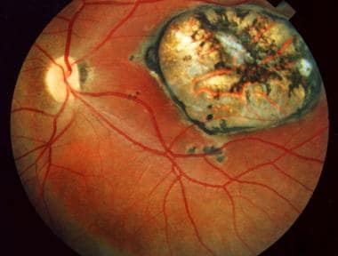
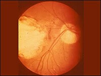
* Chorioretinitis (inflammation of the eye's retina and choroid layer)
* Seizures
* Developmental delays
* Hearing loss
Infection later in pregnancy may result in less severe symptoms or be asymptomatic at birth, but can still lead to developmental delays, learning disabilities, and vision problems as the child grows.
Diagnosis of congenital toxoplasmosis typically involves a combination of tests, such as blood tests to detect antibodies against Toxoplasma gondii, imaging studies (e.g., ultrasound, CT, or MRI) to assess any structural abnormalities in the brain and other organs, and ophthalmologic examinations to evaluate potential eye damage.
Treatment for congenital toxoplasmosis usually involves a combination of antiparasitic medications (such as spiramycin, pyrimethamine, and sulfadiazine) and corticosteroids to reduce inflammation. Early treatment can help minimize the severity of symptoms and improve outcomes for affected children.
Ocular toxoplasmosis is an inflammatory eye disease caused by the parasitic infection of Toxoplasma gondii in the eye's retina. It can lead to lesions and scarring in the retina, resulting in vision loss or impairment. The severity of ocular toxoplasmosis depends on the location and extent of the infection in the eye. In some cases, it may cause only mild symptoms, while in others, it can result in severe damage to the eye. Ocular toxoplasmosis is usually treated with medications that target the Toxoplasma gondii parasite, such as pyrimethamine and sulfadiazine, often combined with corticosteroids to reduce inflammation.
Cerebral toxoplasmosis is a type of toxoplasmosis, which is an infection caused by the Toxoplasma gondii parasite. In cerebral toxoplasmosis, the infection primarily affects the brain, leading to inflammation and the formation of lesions or abscesses in the brain tissue.
This condition is most commonly observed in individuals with weakened immune systems, such as those living with HIV/AIDS, receiving immunosuppressive therapy after organ transplantation, or having other conditions that compromise their immune function. The infection can cause a range of neurological symptoms, including headaches, seizures, confusion, memory loss, poor coordination, and in severe cases, coma or even death. Early diagnosis and treatment with appropriate antiparasitic medications are crucial to manage the infection and prevent complications.
"Toxoplasma" is a genus of protozoan parasites, and the most well-known species is "Toxoplasma gondii." This particular species is capable of infecting virtually all warm-blooded animals, including humans. It's known for its complex life cycle that involves felines (cats) as the definitive host.
Infection in humans, called toxoplasmosis, often occurs through ingestion of contaminated food or water, or through contact with cat feces that contain T. gondii oocysts. While many people infected with Toxoplasma show no symptoms, it can cause serious health problems in immunocompromised individuals and developing fetuses if a woman becomes infected during pregnancy.
It's important to note that while I strive to provide accurate information, this definition should not be used for self-diagnosis or treatment. Always consult with a healthcare professional for medical advice.
Toxoplasmosis is a zoonotic disease, meaning it can be transmitted from animals to humans. It is caused by the intracellular protozoan parasite Toxoplasma gondii. This parasite can infect a wide range of warm-blooded animals, including birds and mammals, as intermediate hosts. However, cats are the primary definitive host for this parasite because the sexual stage of the parasite's life cycle occurs in their intestines, leading to the shedding of oocysts (environmentally resistant stages) in their feces.
Animals can become infected with Toxoplasma gondii through several routes:
1. Ingestion of sporulated oocysts from contaminated soil, water, or food.
2. Consumption of tissue cysts present in the tissues of infected animals during predation.
3. Vertical transmission (transplacental) from an infected mother to her offspring.
Clinical signs and symptoms of toxoplasmosis in animals can vary depending on their age, immune status, and the parasite's virulence. In many cases, animals may not show any apparent signs of infection, but some may develop:
1. Generalized illness with fever, lethargy, and loss of appetite.
2. Lymphadenopathy (swollen lymph nodes).
3. Neurological symptoms such as tremors, ataxia (lack of coordination), or seizures if the central nervous system is affected.
4. Eye lesions, including inflammation and scarring of the retina, which can lead to vision loss in severe cases.
5. Reproductive issues, such as abortion, stillbirths, or birth defects in offspring when pregnant females are infected.
It is important to note that while toxoplasmosis can cause significant health problems in animals, particularly in immunocompromised individuals and developing fetuses, it is often asymptomatic or mild in healthy adult animals. Nonetheless, the zoonotic potential of Toxoplasma gondii highlights the importance of practicing good hygiene and taking necessary precautions when handling infected animals or their waste to minimize the risk of transmission to humans.
Chorioretinitis is a medical term that refers to the inflammation of the choroid and the retina, which are both important structures in the eye. The choroid is a layer of blood vessels that supplies oxygen and nutrients to the retina, while the retina is a light-sensitive tissue that converts light into electrical signals that are sent to the brain and interpreted as visual images.
Chorioretinitis can be caused by various infectious and non-infectious conditions, such as bacterial, viral, fungal, or parasitic infections, autoimmune diseases, or cancer. The symptoms of chorioretinitis may include decreased vision, floaters, blurry vision, sensitivity to light, and eye pain. Treatment for chorioretinitis depends on the underlying cause and may include antibiotics, antiviral medications, corticosteroids, or other immunosuppressive therapies. It is important to seek medical attention promptly if you experience any symptoms of chorioretinitis, as timely diagnosis and treatment can help prevent permanent vision loss.
Antibodies, protozoan, refer to the immune system's response to an infection caused by a protozoan organism. Protozoa are single-celled microorganisms that can cause various diseases in humans, such as malaria, giardiasis, and toxoplasmosis.
When the body is infected with a protozoan, the immune system responds by producing specific proteins called antibodies. Antibodies are produced by a type of white blood cell called a B-cell, and they recognize and bind to specific antigens on the surface of the protozoan organism.
There are five main types of antibodies: IgA, IgD, IgE, IgG, and IgM. Each type of antibody has a different role in the immune response. For example, IgG is the most common type of antibody and provides long-term immunity to previously encountered pathogens. IgM is the first antibody produced in response to an infection and is important for activating the complement system, which helps to destroy the protozoan organism.
Overall, the production of antibodies against protozoan organisms is a critical part of the immune response and helps to protect the body from further infection.
Sulfadiazine is an antibacterial drug, specifically a sulfonamide. It is chemically described as 4-amino-N-(2-pyrimidinyl)benzenesulfonamide. Sulfadiazine works by inhibiting the bacterial synthesis of dihydrofolic acid, which is essential for bacterial growth and reproduction.
It is used to treat a wide range of infections caused by susceptible bacteria, including urinary tract infections, respiratory infections, and certain types of meningitis. Sulfadiazine is often combined with other antibiotics, such as trimethoprim, to increase its effectiveness against certain bacteria.
Like all sulfonamides, sulfadiazine can cause side effects, including skin rashes, allergic reactions, and stomach upset. It should be used with caution in people who are allergic to sulfa drugs or have kidney or liver disease. Additionally, it is important to note that the use of sulfonamides during pregnancy, especially during the third trimester, should be avoided due to the risk of kernicterus in the newborn.
Parasitic pregnancy complications refer to a rare condition where a parasitic twin takes over the development of the dominant twin's reproductive system and becomes pregnant. This condition is also known as fetus in fetu or vanishing twin syndrome with a parasitic twin. The parasitic twin may have some organs developed, but it is not fully formed and relies on the dominant twin for survival. The pregnancy can pose risks to the dominant twin, such as abnormal growth patterns, organ damage, and complications during childbirth. This condition is usually detected during prenatal ultrasound examinations.
Antiprotozoal agents are a type of medication used to treat protozoal infections, which are infections caused by microscopic single-celled organisms called protozoa. These agents work by either killing the protozoa or inhibiting their growth and reproduction. They can be administered through various routes, including oral, topical, and intravenous, depending on the type of infection and the severity of the illness.
Examples of antiprotozoal agents include:
* Metronidazole, tinidazole, and nitazoxanide for treating infections caused by Giardia lamblia and Entamoeba histolytica.
* Atovaquone, clindamycin, and pyrimethamine-sulfadoxine for treating malaria caused by Plasmodium falciparum or other Plasmodium species.
* Pentamidine and suramin for treating African trypanosomiasis (sleeping sickness) caused by Trypanosoma brucei gambiense or T. b. rhodesiense.
* Nitroimidazoles, such as benznidazole and nifurtimox, for treating Chagas disease caused by Trypanosoma cruzi.
* Sodium stibogluconate and paromomycin for treating leishmaniasis caused by Leishmania species.
Antiprotozoal agents can have side effects, ranging from mild to severe, depending on the drug and the individual patient's response. It is essential to follow the prescribing physician's instructions carefully when taking these medications and report any adverse reactions promptly.
Immunoglobulin M (IgM) is a type of antibody that is primarily found in the blood and lymph fluid. It is the first antibody to be produced in response to an initial exposure to an antigen, making it an important part of the body's primary immune response. IgM antibodies are large molecules that are composed of five basic units, giving them a pentameric structure. They are primarily found on the surface of B cells as membrane-bound immunoglobulins (mlgM), where they function as receptors for antigens. Once an mlgM receptor binds to an antigen, it triggers the activation and differentiation of the B cell into a plasma cell that produces and secretes large amounts of soluble IgM antibodies.
IgM antibodies are particularly effective at agglutination (clumping) and complement activation, which makes them important in the early stages of an immune response to help clear pathogens from the bloodstream. However, they are not as stable or long-lived as other types of antibodies, such as IgG, and their levels tend to decline after the initial immune response has occurred.
In summary, Immunoglobulin M (IgM) is a type of antibody that plays a crucial role in the primary immune response to antigens by agglutination and complement activation. It is primarily found in the blood and lymph fluid, and it is produced by B cells after they are activated by an antigen.
Parasitic lung diseases refer to conditions caused by infection of the lungs by parasites. These are small organisms that live on or in a host organism and derive their sustenance at the expense of the host. Parasitic lung diseases can be caused by various types of parasites, including helminths (worms) and protozoa.
Examples of parasitic lung diseases include:
1. Pulmonary echinococcosis (hydatid disease): This is a rare infection caused by the larval stage of the tapeworm Echinococcus granulosus. The larvae form cysts in various organs, including the lungs.
2. Paragonimiasis: This is a food-borne lung fluke infection caused by Paragonimus westermani and other species. Humans become infected by eating raw or undercooked crustaceans (such as crabs or crayfish) that contain the larval stage of the parasite.
3. Toxocariasis: This is a soil-transmitted helminth infection caused by the roundworm Toxocara canis or T. cati, which are found in the intestines of dogs and cats. Humans become infected through accidental ingestion of contaminated soil, undercooked meat, or through contact with an infected animal's feces. Although the primary site of infection is the small intestine, larval migration can lead to lung involvement in some cases.
4. Amebic lung disease: This is a rare complication of amebiasis, which is caused by the protozoan Entamoeba histolytica. The parasite usually infects the large intestine, but it can spread to other organs, including the lungs, through the bloodstream.
5. Cryptosporidiosis: This is a waterborne protozoan infection caused by Cryptosporidium parvum or C. hominis. Although the primary site of infection is the small intestine, immunocompromised individuals can develop disseminated disease, including pulmonary involvement.
Symptoms of parasitic lung diseases vary depending on the specific organism and the severity of infection but may include cough, chest pain, shortness of breath, fever, and sputum production. Diagnosis typically involves a combination of clinical evaluation, imaging studies, and laboratory tests, such as stool or blood examinations for parasites or their antigens. Treatment depends on the specific organism but may include antiparasitic medications, supportive care, and management of complications.
Antigens are substances (usually proteins) found on the surface of cells, or viruses, that can be recognized by the immune system and stimulate an immune response. In the context of protozoa, antigens refer to the specific proteins or other molecules found on the surface of these single-celled organisms that can trigger an immune response in a host organism.
Protozoa are a group of microscopic eukaryotic organisms that include a diverse range of species, some of which can cause diseases in humans and animals. When a protozoan infects a host, the host's immune system recognizes the protozoan antigens as foreign and mounts an immune response to eliminate the infection. This response involves the activation of various types of immune cells, such as T-cells and B-cells, which recognize and target the protozoan antigens.
Understanding the nature of protozoan antigens is important for developing vaccines and other immunotherapies to prevent or treat protozoan infections. For example, researchers have identified specific antigens on the surface of the malaria parasite that are recognized by the human immune system and have used this information to develop vaccine candidates. However, many protozoan infections remain difficult to prevent or treat, and further research is needed to identify new targets for vaccines and therapies.
Immunoglobulin G (IgG) is a type of antibody, which is a protective protein produced by the immune system in response to foreign substances like bacteria or viruses. IgG is the most abundant type of antibody in human blood, making up about 75-80% of all antibodies. It is found in all body fluids and plays a crucial role in fighting infections caused by bacteria, viruses, and toxins.
IgG has several important functions:
1. Neutralization: IgG can bind to the surface of bacteria or viruses, preventing them from attaching to and infecting human cells.
2. Opsonization: IgG coats the surface of pathogens, making them more recognizable and easier for immune cells like neutrophils and macrophages to phagocytose (engulf and destroy) them.
3. Complement activation: IgG can activate the complement system, a group of proteins that work together to help eliminate pathogens from the body. Activation of the complement system leads to the formation of the membrane attack complex, which creates holes in the cell membranes of bacteria, leading to their lysis (destruction).
4. Antibody-dependent cellular cytotoxicity (ADCC): IgG can bind to immune cells like natural killer (NK) cells and trigger them to release substances that cause target cells (such as virus-infected or cancerous cells) to undergo apoptosis (programmed cell death).
5. Immune complex formation: IgG can form immune complexes with antigens, which can then be removed from the body through various mechanisms, such as phagocytosis by immune cells or excretion in urine.
IgG is a critical component of adaptive immunity and provides long-lasting protection against reinfection with many pathogens. It has four subclasses (IgG1, IgG2, IgG3, and IgG4) that differ in their structure, function, and distribution in the body.
Choroiditis is an inflammatory condition that affects the choroid, a layer of blood vessels in the eye located between the retina (the light-sensitive tissue at the back of the eye) and the sclera (the white outer coat of the eye). The choroid provides oxygen and nutrients to the outer layers of the retina.
Choroiditis is characterized by spots or patches of inflammation in the choroid, which can lead to damage and scarring of the tissue. This can result in vision loss if it affects the macula (the central part of the retina responsible for sharp, detailed vision). Symptoms of choroiditis may include blurred vision, floaters, sensitivity to light, and decreased color perception.
There are several types of choroiditis, including:
1. Multifocal choroiditis: This type is characterized by multiple, small areas of inflammation in the choroid, often accompanied by scarring. It can affect both eyes and may cause vision loss if it involves the macula.
2. Serpiginous choroiditis: This is a chronic, relapsing form of choroiditis that affects the outer layers of the retina and the choroid. It typically causes well-defined, wavy or serpentine-shaped lesions in the posterior pole (the back part) of the eye.
3. Birdshot chorioretinopathy: This is a rare form of choroiditis that primarily affects the peripheral retina and choroid. It is characterized by multiple, cream-colored or yellowish spots throughout the fundus (the interior surface of the eye).
4. Sympathetic ophthalmia: This is a rare condition that occurs when one eye is injured, leading to inflammation in both eyes. The choroid and other structures in the uninjured eye become inflamed due to an autoimmune response.
5. Vogt-Koyanagi-Harada (VKH) disease: This is a multisystemic autoimmune disorder that affects the eyes, skin, hair, and inner ear. In the eye, it causes choroiditis, retinal inflammation, and sometimes optic nerve swelling.
Treatment for choroiditis depends on the underlying cause and may include corticosteroids, immunosuppressive medications, or biologic agents to control inflammation. In some cases, laser therapy or surgery might be necessary to address complications such as retinal detachment or cataracts.
The eye is the organ of sight, primarily responsible for detecting and focusing on visual stimuli. It is a complex structure composed of various parts that work together to enable vision. Here are some of the main components of the eye:
1. Cornea: The clear front part of the eye that refracts light entering the eye and protects the eye from harmful particles and microorganisms.
2. Iris: The colored part of the eye that controls the amount of light reaching the retina by adjusting the size of the pupil.
3. Pupil: The opening in the center of the iris that allows light to enter the eye.
4. Lens: A biconvex structure located behind the iris that further refracts light and focuses it onto the retina.
5. Retina: A layer of light-sensitive cells (rods and cones) at the back of the eye that convert light into electrical signals, which are then transmitted to the brain via the optic nerve.
6. Optic Nerve: The nerve that carries visual information from the retina to the brain.
7. Vitreous: A clear, gel-like substance that fills the space between the lens and the retina, providing structural support to the eye.
8. Conjunctiva: A thin, transparent membrane that covers the front of the eye and the inner surface of the eyelids.
9. Extraocular Muscles: Six muscles that control the movement of the eye, allowing for proper alignment and focus.
The eye is a remarkable organ that allows us to perceive and interact with our surroundings. Various medical specialties, such as ophthalmology and optometry, are dedicated to the diagnosis, treatment, and management of various eye conditions and diseases.
Ocular hypertension is a medical condition characterized by elevated pressure within the eye (intraocular pressure or IOP), which is higher than normal but not necessarily high enough to cause any visible damage to the optic nerve or visual field loss. It serves as a significant risk factor for developing glaucoma, a sight-threatening disease.
The normal range of intraocular pressure is typically between 10-21 mmHg (millimeters of mercury). Ocular hypertension is often defined as an IOP consistently above 21 mmHg, although some studies suggest that even pressures between 22-30 mmHg may not cause damage in all individuals. Regular monitoring and follow-up with an ophthalmologist are essential for people diagnosed with ocular hypertension to ensure early detection and management of any potential glaucomatous changes. Treatment options include medications, laser therapy, or surgery to lower the IOP and reduce the risk of glaucoma onset.
Seroepidemiologic studies are a type of epidemiological study that measures the presence and levels of antibodies in a population's blood serum to investigate the prevalence, distribution, and transmission of infectious diseases. These studies help to identify patterns of infection and immunity within a population, which can inform public health policies and interventions.
Seroepidemiologic studies typically involve collecting blood samples from a representative sample of individuals in a population and testing them for the presence of antibodies against specific pathogens. The results are then analyzed to estimate the prevalence of infection and immunity within the population, as well as any factors associated with increased or decreased risk of infection.
These studies can provide valuable insights into the spread of infectious diseases, including emerging and re-emerging infections, and help to monitor the effectiveness of vaccination programs. Additionally, seroepidemiologic studies can also be used to investigate the transmission dynamics of infectious agents, such as identifying sources of infection or tracking the spread of antibiotic resistance.
There doesn't seem to be a specific medical definition for "DNA, protozoan" as it is simply a reference to the DNA found in protozoa. Protozoa are single-celled eukaryotic organisms that can be found in various environments such as soil, water, and the digestive tracts of animals.
Protozoan DNA refers to the genetic material present in these organisms. It is composed of nucleic acids, including deoxyribonucleic acid (DNA) and ribonucleic acid (RNA), which contain the instructions for the development, growth, and reproduction of the protozoan.
The DNA in protozoa, like in other organisms, is made up of two strands of nucleotides that coil together to form a double helix. The four nucleotide bases that make up protozoan DNA are adenine (A), thymine (T), guanine (G), and cytosine (C). These bases pair with each other to form the rungs of the DNA ladder, with A always pairing with T and G always pairing with C.
The genetic information stored in protozoan DNA is encoded in the sequence of these nucleotide bases. This information is used to synthesize proteins, which are essential for the structure and function of the organism's cells. Protozoan DNA also contains other types of genetic material, such as regulatory sequences that control gene expression and repetitive elements with no known function.
Understanding the DNA of protozoa is important for studying their biology, evolution, and pathogenicity. It can help researchers develop new treatments for protozoan diseases and gain insights into the fundamental principles of genetics and cellular function.
AIDS-related opportunistic infections (AROIs) are infections that occur more frequently or are more severe in people with weakened immune systems, such as those with advanced HIV infection or AIDS. These infections take advantage of a weakened immune system and can affect various organs and systems in the body.
Common examples of AROIs include:
1. Pneumocystis pneumonia (PCP), caused by the fungus Pneumocystis jirovecii
2. Mycobacterium avium complex (MAC) infection, caused by a type of bacteria called mycobacteria
3. Candidiasis, a fungal infection that can affect various parts of the body, including the mouth, esophagus, and genitals
4. Toxoplasmosis, caused by the parasite Toxoplasma gondii
5. Cryptococcosis, a fungal infection that affects the lungs and central nervous system
6. Cytomegalovirus (CMV) infection, caused by a type of herpes virus
7. Tuberculosis (TB), caused by the bacterium Mycobacterium tuberculosis
8. Cryptosporidiosis, a parasitic infection that affects the intestines
9. Progressive multifocal leukoencephalopathy (PML), a viral infection that affects the brain
Preventing and treating AROIs is an important part of managing HIV/AIDS, as they can cause significant illness and even death in people with weakened immune systems. Antiretroviral therapy (ART) is used to treat HIV infection and prevent the progression of HIV to AIDS, which can help reduce the risk of opportunistic infections. In addition, medications to prevent specific opportunistic infections may be prescribed for people with advanced HIV or AIDS.
Pregnancy is a physiological state or condition where a fertilized egg (zygote) successfully implants and grows in the uterus of a woman, leading to the development of an embryo and finally a fetus. This process typically spans approximately 40 weeks, divided into three trimesters, and culminates in childbirth. Throughout this period, numerous hormonal and physical changes occur to support the growing offspring, including uterine enlargement, breast development, and various maternal adaptations to ensure the fetus's optimal growth and well-being.
An Enzyme-Linked Immunosorbent Assay (ELISA) is a type of analytical biochemistry assay used to detect and quantify the presence of a substance, typically a protein or peptide, in a liquid sample. It takes its name from the enzyme-linked antibodies used in the assay.
In an ELISA, the sample is added to a well containing a surface that has been treated to capture the target substance. If the target substance is present in the sample, it will bind to the surface. Next, an enzyme-linked antibody specific to the target substance is added. This antibody will bind to the captured target substance if it is present. After washing away any unbound material, a substrate for the enzyme is added. If the enzyme is present due to its linkage to the antibody, it will catalyze a reaction that produces a detectable signal, such as a color change or fluorescence. The intensity of this signal is proportional to the amount of target substance present in the sample, allowing for quantification.
ELISAs are widely used in research and clinical settings to detect and measure various substances, including hormones, viruses, and bacteria. They offer high sensitivity, specificity, and reproducibility, making them a reliable choice for many applications.
A newborn infant is a baby who is within the first 28 days of life. This period is also referred to as the neonatal period. Newborns require specialized care and attention due to their immature bodily systems and increased vulnerability to various health issues. They are closely monitored for signs of well-being, growth, and development during this critical time.
Serologic tests are laboratory tests that detect the presence or absence of antibodies or antigens in a patient's serum (the clear liquid that separates from clotted blood). These tests are commonly used to diagnose infectious diseases, as well as autoimmune disorders and other medical conditions.
In serologic testing for infectious diseases, a sample of the patient's blood is collected and allowed to clot. The serum is then separated from the clot and tested for the presence of antibodies that the body has produced in response to an infection. The test may be used to identify the specific type of infection or to determine whether the infection is active or has resolved.
Serologic tests can also be used to diagnose autoimmune disorders, such as rheumatoid arthritis and lupus, by detecting the presence of antibodies that are directed against the body's own tissues. These tests can help doctors confirm a diagnosis and monitor the progression of the disease.
It is important to note that serologic tests are not always 100% accurate and may produce false positive or false negative results. Therefore, they should be interpreted in conjunction with other clinical findings and laboratory test results.
Atovaquone is an antiprotozoal medication used for the treatment and prevention of certain parasitic infections. It works by inhibiting the mitochondria of the parasites, disrupting their energy production and ultimately leading to their death. Atovaquone is available as a oral suspension or coated tablets and is often prescribed for conditions such as Pneumocystis pneumonia (PCP), Toxoplasma gondii encephalitis, and babesiosis. It is also used for the prevention of PCP in people with weakened immune systems due to HIV/AIDS or other causes.
The medical definition of Atovaquone can be stated as:
"Atovaquone is an antiprotozoal medication (synthetic hydroxynaphthoquinone) that exhibits activity against a variety of protozoa, including Plasmodium falciparum (the parasite responsible for malaria), Pneumocystis jirovecii (the causative agent of PCP), Toxoplasma gondii, and Babesia microti. It is used primarily for the treatment and prevention of PCP in individuals with compromised immune systems, as well as for the treatment of babesiosis and toxoplasmosis."
There is no medical definition for "Protozoan Vaccines" as such because there are currently no licensed vaccines available for human protozoan diseases. Protozoa are single-celled microorganisms that can cause various diseases in humans, such as malaria, toxoplasmosis, and leishmaniasis.
Researchers have been working on developing vaccines against some of these diseases, but none have yet been approved for use in humans. Therefore, it is not possible to provide a medical definition for "Protozoan Vaccines" as a recognized category of vaccines.
Antibody affinity refers to the strength and specificity of the interaction between an antibody and its corresponding antigen at a molecular level. It is a measure of how strongly and selectively an antibody binds to its target antigen. A higher affinity indicates a more stable and specific binding, while a lower affinity suggests weaker and less specific interactions. Affinity is typically measured in terms of the dissociation constant (Kd), which describes the concentration of antigen needed to achieve half-maximal binding to an antibody. Generally, a smaller Kd value corresponds to a higher affinity, indicating a tighter and more selective bond. This parameter is crucial in the development of diagnostic and therapeutic applications, such as immunoassays and targeted therapies, where high-affinity antibodies are preferred for improved sensitivity and specificity.
Pyrimethamine is an antiparasitic medication that is primarily used to treat and prevent protozoan infections, such as toxoplasmosis and malaria. It works by inhibiting the dihydrofolate reductase enzyme, which is essential for the parasite's survival. By doing so, it interferes with the synthesis of folate, a vital component for the growth and reproduction of the parasite.
Pyrimethamine is often used in combination with other medications, such as sulfonamides or sulfones, to increase its effectiveness and prevent the development of drug-resistant strains. Common side effects of pyrimethamine include nausea, vomiting, loss of appetite, and headache. It is important to note that pyrimethamine should only be used under the supervision of a healthcare professional due to its potential for serious side effects and interactions with other medications.
Uveitis is the inflammation of the uvea, the middle layer of the eye between the retina and the white of the eye (sclera). The uvea consists of the iris, ciliary body, and choroid. Uveitis can cause redness, pain, and vision loss. It can be caused by various systemic diseases, infections, or trauma. Depending on the part of the uvea that's affected, uveitis can be classified as anterior (iritis), intermediate (cyclitis), posterior (choroiditis), or pan-uveitis (affecting all layers). Treatment typically includes corticosteroids and other immunosuppressive drugs to control inflammation.
Posterior uveitis is a type of uveitis that specifically affects the back portion of the uvea, which includes the choroid (a layer of blood vessels that provides nutrients to the outer layers of the retina), the retina (the light-sensitive tissue at the back of the eye), and the optic nerve (which carries visual information from the eye to the brain).
Posterior uveitis can cause symptoms such as blurred vision, floaters, sensitivity to light, and decreased vision. It may also lead to complications such as retinal scarring, cataracts, glaucoma, and retinal detachment if left untreated. The condition can be caused by a variety of factors, including infections, autoimmune diseases, and trauma. Treatment typically involves the use of corticosteroids or other immunosuppressive medications to reduce inflammation and prevent complications.
Neonatal screening is a medical procedure in which specific tests are performed on newborn babies within the first few days of life to detect certain congenital or inherited disorders that are not otherwise clinically apparent at birth. These conditions, if left untreated, can lead to serious health problems, developmental delays, or even death.
The primary goal of neonatal screening is to identify affected infants early so that appropriate treatment and management can be initiated as soon as possible, thereby improving their overall prognosis and quality of life. Commonly screened conditions include phenylketonuria (PKU), congenital hypothyroidism, galactosemia, maple syrup urine disease, sickle cell disease, cystic fibrosis, and hearing loss, among others.
Neonatal screening typically involves collecting a small blood sample from the infant's heel (heel stick) or through a dried blood spot card, which is then analyzed using various biochemical, enzymatic, or genetic tests. In some cases, additional tests such as hearing screenings and pulse oximetry for critical congenital heart disease may also be performed.
It's important to note that neonatal screening is not a diagnostic tool but rather an initial step in identifying infants who may be at risk of certain conditions. Positive screening results should always be confirmed with additional diagnostic tests before any treatment decisions are made.
I'm sorry for any confusion, but "Protozoan Proteins" is not a specific medical or scientific term. Protozoa are single-celled eukaryotic organisms, and proteins are large biological molecules consisting of one or more chains of amino acid residues. Therefore, "Protozoan Proteins" generally refers to the various types of proteins found in protozoa.
However, if you're looking for information about proteins specific to certain protozoan parasites with medical relevance (such as Plasmodium falciparum, which causes malaria), I would be happy to help! Please provide more context or specify the particular protozoan of interest.
An acute disease is a medical condition that has a rapid onset, develops quickly, and tends to be short in duration. Acute diseases can range from minor illnesses such as a common cold or flu, to more severe conditions such as pneumonia, meningitis, or a heart attack. These types of diseases often have clear symptoms that are easy to identify, and they may require immediate medical attention or treatment.
Acute diseases are typically caused by an external agent or factor, such as a bacterial or viral infection, a toxin, or an injury. They can also be the result of a sudden worsening of an existing chronic condition. In general, acute diseases are distinct from chronic diseases, which are long-term medical conditions that develop slowly over time and may require ongoing management and treatment.
Examples of acute diseases include:
* Acute bronchitis: a sudden inflammation of the airways in the lungs, often caused by a viral infection.
* Appendicitis: an inflammation of the appendix that can cause severe pain and requires surgical removal.
* Gastroenteritis: an inflammation of the stomach and intestines, often caused by a viral or bacterial infection.
* Migraine headaches: intense headaches that can last for hours or days, and are often accompanied by nausea, vomiting, and sensitivity to light and sound.
* Myocardial infarction (heart attack): a sudden blockage of blood flow to the heart muscle, often caused by a buildup of plaque in the coronary arteries.
* Pneumonia: an infection of the lungs that can cause coughing, chest pain, and difficulty breathing.
* Sinusitis: an inflammation of the sinuses, often caused by a viral or bacterial infection.
It's important to note that while some acute diseases may resolve on their own with rest and supportive care, others may require medical intervention or treatment to prevent complications and promote recovery. If you are experiencing symptoms of an acute disease, it is always best to seek medical attention to ensure proper diagnosis and treatment.
Parasitology is a branch of biology that deals with the study of parasites, their life cycles, the relationship between parasites and their hosts, the transmission of parasitic diseases, and the development of methods for their control and elimination. It involves understanding various types of parasites including protozoa, helminths, and arthropods that can infect humans, animals, and plants. Parasitologists also study the evolution, genetics, biochemistry, and ecology of parasites to develop effective strategies for their diagnosis, treatment, and prevention.
I apologize, but "Brazil" is not a medical term or condition. It is the largest country in both South America and Latin America by land area and population. If you have any questions related to medical terminology or health concerns, please provide more information and I will do my best to help.
Aqueous humor is a clear, watery fluid that fills the anterior and posterior chambers of the eye. It is produced by the ciliary processes in the posterior chamber and circulates through the pupil into the anterior chamber, where it provides nutrients to the cornea and lens, maintains intraocular pressure, and helps to shape the eye. The aqueous humor then drains out of the eye through the trabecular meshwork and into the canal of Schlemm, eventually reaching the venous system.
Lymphadenitis is a medical term that refers to the inflammation of one or more lymph nodes, which are small, bean-shaped glands that are part of the body's immune system. Lymph nodes contain white blood cells called lymphocytes, which help fight infection and disease.
Lymphadenitis can occur as a result of an infection in the area near the affected lymph node or as a result of a systemic infection that has spread through the bloodstream. The inflammation causes the lymph node to become swollen, tender, and sometimes painful to the touch.
The symptoms of lymphadenitis may include fever, fatigue, and redness or warmth in the area around the affected lymph node. In some cases, the overlying skin may also appear red and inflamed. Lymphadenitis can occur in any part of the body where there are lymph nodes, including the neck, armpits, groin, and abdomen.
The underlying cause of lymphadenitis must be diagnosed and treated promptly to prevent complications such as the spread of infection or the formation of an abscess. Treatment may include antibiotics, pain relievers, and warm compresses to help reduce swelling and discomfort.
Coccidiostats are a type of medication used to prevent and treat coccidiosis, which is an infection caused by protozoan parasites of the genus Coccidia. These medications work by inhibiting the growth and reproduction of the parasites in the gastrointestinal tract of animals, particularly poultry and livestock.
Coccidiostats are commonly added to animal feed to prevent infection and reduce the spread of coccidiosis within a flock or herd. They can also be used to treat active infections, often in combination with other medications. Common examples of coccidiostats include sulfaquinoxaline, monensin, and lasalocid.
It's important to note that the use of coccidiostats in food-producing animals is regulated by government agencies such as the US Food and Drug Administration (FDA) and the European Medicines Agency (EMA) to ensure their safe use and to minimize the risk of residues in animal products.
Infectious pregnancy complications refer to infections that occur during pregnancy and can affect the mother, fetus, or both. These infections can lead to serious consequences such as preterm labor, low birth weight, birth defects, stillbirth, or even death. Some common infectious agents that can cause pregnancy complications include:
1. Bacteria: Examples include group B streptococcus, Escherichia coli, and Listeria monocytogenes, which can cause sepsis, meningitis, or pneumonia in the mother and lead to preterm labor or stillbirth.
2. Viruses: Examples include cytomegalovirus, rubella, varicella-zoster, and HIV, which can cause congenital anomalies, developmental delays, or transmission of the virus to the fetus.
3. Parasites: Examples include Toxoplasma gondii, which can cause severe neurological damage in the fetus if transmitted during pregnancy.
4. Fungi: Examples include Candida albicans, which can cause fungal infections in the mother and lead to preterm labor or stillbirth.
Preventive measures such as vaccination, good hygiene practices, and avoiding high-risk behaviors can help reduce the risk of infectious pregnancy complications. Prompt diagnosis and treatment of infections during pregnancy are also crucial to prevent adverse outcomes.
Vertical transmission of infectious diseases refers to the spread of an infection from an infected mother to her offspring during pregnancy, childbirth, or breastfeeding. This mode of transmission can occur through several pathways:
1. Transplacental transmission: The infection crosses the placenta and reaches the fetus while it is still in the womb. Examples include HIV, syphilis, and toxoplasmosis.
2. Intrauterine infection: The mother's infection causes direct damage to the developing fetus or its surrounding tissues, leading to complications such as congenital defects. Examples include rubella and cytomegalovirus (CMV).
3. Perinatal transmission: This occurs during childbirth when the infant comes into contact with the mother's infected genital tract or bodily fluids. Examples include group B streptococcus, herpes simplex virus (HSV), and hepatitis B.
4. Postnatal transmission: This occurs after birth, often through breastfeeding, when the infant ingests infected milk or comes into contact with the mother's contaminated bodily fluids. Examples include HIV and HTLV-I (human T-lymphotropic virus type I).
Vertical transmission is a significant concern in public health, as it can lead to severe complications, congenital disabilities, or even death in newborns. Preventive measures, such as prenatal screening, vaccination, and antimicrobial treatment, are crucial for reducing the risk of vertical transmission and ensuring better outcomes for both mothers and their offspring.
Ocular albinism is a type of albinism that primarily affects the eyes. It is a genetic disorder characterized by the reduction or absence of melanin, the pigment responsible for coloring the skin, hair, and eyes. In ocular albinism, melanin production is deficient in the eyes, leading to various eye abnormalities.
The main features of ocular albinism include:
1. Nystagmus: Rapid, involuntary back-and-forth movement of the eyes.
2. Iris transillumination: The iris appears translucent due to the lack of pigment, allowing light to pass through easily. This can be observed using a light source shone into the eye.
3. Foveal hypoplasia: Underdevelopment or absence of the fovea, a small pit in the retina responsible for sharp, central vision.
4. Photophobia: Increased sensitivity to light due to the lack of pigment in the eyes.
5. Strabismus: Misalignment of the eyes, which can result in double vision or lazy eye.
6. Reduced visual acuity: Decreased ability to see clearly, even with corrective lenses.
Ocular albinism is typically inherited as an X-linked recessive trait, meaning it primarily affects males, while females can be carriers of the condition. However, there are also autosomal recessive forms of ocular albinism that can affect both males and females equally. Treatment for ocular albinism usually involves managing symptoms with corrective lenses, low-vision aids, and vision therapy to improve visual skills.
Ocular dominance refers to the preference of one eye over the other in terms of visual perception and processing. In other words, it is the tendency for an individual to rely more heavily on the input from one particular eye when interpreting visual information. This can have implications in various visual tasks such as depth perception, aiming, and targeting.
Ocular dominance can be determined through a variety of tests, including the Miles test, the Porta test, or simply by observing which eye a person uses to align a visual target. It is important to note that ocular dominance does not necessarily indicate any sort of visual impairment or deficit; rather, it is a normal variation in the way that visual information is processed by the brain.
Sensitivity and specificity are statistical measures used to describe the performance of a diagnostic test or screening tool in identifying true positive and true negative results.
* Sensitivity refers to the proportion of people who have a particular condition (true positives) who are correctly identified by the test. It is also known as the "true positive rate" or "recall." A highly sensitive test will identify most or all of the people with the condition, but may also produce more false positives.
* Specificity refers to the proportion of people who do not have a particular condition (true negatives) who are correctly identified by the test. It is also known as the "true negative rate." A highly specific test will identify most or all of the people without the condition, but may also produce more false negatives.
In medical testing, both sensitivity and specificity are important considerations when evaluating a diagnostic test. High sensitivity is desirable for screening tests that aim to identify as many cases of a condition as possible, while high specificity is desirable for confirmatory tests that aim to rule out the condition in people who do not have it.
It's worth noting that sensitivity and specificity are often influenced by factors such as the prevalence of the condition in the population being tested, the threshold used to define a positive result, and the reliability and validity of the test itself. Therefore, it's important to consider these factors when interpreting the results of a diagnostic test.
Parasitic skin diseases are conditions caused by parasites living on or in the skin. These parasites can be insects, mites, or fungi that feed off of the host for their own survival. They can cause a variety of symptoms including itching, rashes, blisters, and lesions on the skin. Examples of parasitic skin diseases include scabies, lice infestations, and ringworm. Treatment typically involves the use of topical or oral medications to kill the parasites and alleviate symptoms.
Ocular tonometry is a medical test used to measure the pressure inside the eye, also known as intraocular pressure (IOP). This test is an essential part of diagnosing and monitoring glaucoma, a group of eye conditions that can cause vision loss and blindness due to damage to the optic nerve from high IOP.
The most common method of ocular tonometry involves using a tonometer device that gently touches the front surface of the eye (cornea) with a small probe or prism. The device measures the amount of force required to flatten the cornea slightly, which correlates with the pressure inside the eye. Other methods of ocular tonometry include applanation tonometry, which uses a small amount of fluorescein dye and a blue light to measure the IOP, and rebound tonometry, which uses a lightweight probe that briefly touches the cornea and then bounces back to determine the IOP.
Regular ocular tonometry is important for detecting glaucoma early and preventing vision loss. It is typically performed during routine eye exams and may be recommended more frequently for individuals at higher risk of developing glaucoma, such as those with a family history of the condition or certain medical conditions like diabetes.
Ocular refraction is a medical term that refers to the bending of light as it passes through the optical media of the eye, including the cornea and lens. This process allows the eye to focus light onto the retina, creating a clear image. The refractive power of the eye is determined by the curvature and transparency of these structures.
In a normal eye, light rays are bent or refracted in such a way that they converge at a single point on the retina, producing a sharp and focused image. However, if the curvature of the cornea or lens is too steep or too flat, the light rays may not converge properly, resulting in a refractive error such as myopia (nearsightedness), hyperopia (farsightedness), or astigmatism.
Ocular refraction can be measured using a variety of techniques, including retinoscopy, automated refraction, and subjective refraction. These measurements are used to determine the appropriate prescription for corrective lenses such as eyeglasses or contact lenses. In some cases, ocular refractive errors may be corrected surgically through procedures such as LASIK or PRK.
An immunocompromised host refers to an individual who has a weakened or impaired immune system, making them more susceptible to infections and decreased ability to fight off pathogens. This condition can be congenital (present at birth) or acquired (developed during one's lifetime).
Acquired immunocompromised states may result from various factors such as medical treatments (e.g., chemotherapy, radiation therapy, immunosuppressive drugs), infections (e.g., HIV/AIDS), chronic diseases (e.g., diabetes, malnutrition, liver disease), or aging.
Immunocompromised hosts are at a higher risk for developing severe and life-threatening infections due to their reduced immune response. Therefore, they require special consideration when it comes to prevention, diagnosis, and treatment of infectious diseases.
Eye neoplasms, also known as ocular tumors or eye cancer, refer to abnormal growths of tissue in the eye. These growths can be benign (non-cancerous) or malignant (cancerous). Eye neoplasms can develop in various parts of the eye, including the eyelid, conjunctiva, cornea, iris, ciliary body, choroid, retina, and optic nerve.
Benign eye neoplasms are typically slow-growing and do not spread to other parts of the body. They may cause symptoms such as vision changes, eye pain, or a noticeable mass in the eye. Treatment options for benign eye neoplasms include monitoring, surgical removal, or radiation therapy.
Malignant eye neoplasms, on the other hand, can grow and spread rapidly to other parts of the body. They may cause symptoms such as vision changes, eye pain, floaters, or flashes of light. Treatment options for malignant eye neoplasms depend on the type and stage of cancer but may include surgery, radiation therapy, chemotherapy, or a combination of these treatments.
It is important to note that early detection and treatment of eye neoplasms can improve outcomes and prevent complications. Regular eye exams with an ophthalmologist are recommended for early detection and prevention of eye diseases, including eye neoplasms.
Immunoglobulin A (IgA) is a type of antibody that plays a crucial role in the immune function of the human body. It is primarily found in external secretions, such as saliva, tears, breast milk, and sweat, as well as in mucous membranes lining the respiratory and gastrointestinal tracts. IgA exists in two forms: a monomeric form found in serum and a polymeric form found in secretions.
The primary function of IgA is to provide immune protection at mucosal surfaces, which are exposed to various environmental antigens, such as bacteria, viruses, parasites, and allergens. By doing so, it helps prevent the entry and colonization of pathogens into the body, reducing the risk of infections and inflammation.
IgA functions by binding to antigens present on the surface of pathogens or allergens, forming immune complexes that can neutralize their activity. These complexes are then transported across the epithelial cells lining mucosal surfaces and released into the lumen, where they prevent the adherence and invasion of pathogens.
In summary, Immunoglobulin A (IgA) is a vital antibody that provides immune defense at mucosal surfaces by neutralizing and preventing the entry of harmful antigens into the body.
Polymerase Chain Reaction (PCR) is a laboratory technique used to amplify specific regions of DNA. It enables the production of thousands to millions of copies of a particular DNA sequence in a rapid and efficient manner, making it an essential tool in various fields such as molecular biology, medical diagnostics, forensic science, and research.
The PCR process involves repeated cycles of heating and cooling to separate the DNA strands, allow primers (short sequences of single-stranded DNA) to attach to the target regions, and extend these primers using an enzyme called Taq polymerase, resulting in the exponential amplification of the desired DNA segment.
In a medical context, PCR is often used for detecting and quantifying specific pathogens (viruses, bacteria, fungi, or parasites) in clinical samples, identifying genetic mutations or polymorphisms associated with diseases, monitoring disease progression, and evaluating treatment effectiveness.
The vitreous body, also known simply as the vitreous, is the clear, gel-like substance that fills the space between the lens and the retina in the eye. It is composed mainly of water, but also contains collagen fibers, hyaluronic acid, and other proteins. The vitreous helps to maintain the shape of the eye and provides a transparent medium for light to pass through to reach the retina. With age, the vitreous can become more liquefied and may eventually separate from the retina, leading to symptoms such as floaters or flashes of light.
Ocular motility disorders refer to a group of conditions that affect the movement of the eyes. These disorders can result from nerve damage, muscle dysfunction, or brain injuries. They can cause abnormal eye alignment, limited range of motion, and difficulty coordinating eye movements. Common symptoms include double vision, blurry vision, strabismus (crossed eyes), nystagmus (involuntary eye movement), and difficulty tracking moving objects. Ocular motility disorders can be congenital or acquired and may require medical intervention to correct or manage the condition.
A fatal outcome is a term used in medical context to describe a situation where a disease, injury, or illness results in the death of an individual. It is the most severe and unfortunate possible outcome of any medical condition, and is often used as a measure of the severity and prognosis of various diseases and injuries. In clinical trials and research, fatal outcome may be used as an endpoint to evaluate the effectiveness and safety of different treatments or interventions.
There are many diseases that can affect cats, and the specific medical definitions for these conditions can be quite detailed and complex. However, here are some common categories of feline diseases and examples of each:
1. Infectious diseases: These are caused by viruses, bacteria, fungi, or parasites. Examples include:
* Feline panleukopenia virus (FPV), also known as feline parvovirus, which can cause severe gastrointestinal symptoms and death in kittens.
* Feline calicivirus (FCV), which can cause upper respiratory symptoms such as sneezing and nasal discharge.
* Feline leukemia virus (FeLV), which can suppress the immune system and lead to a variety of secondary infections and diseases.
* Bacterial infections, such as those caused by Pasteurella multocida or Bartonella henselae, which can cause abscesses or other symptoms.
2. Neoplastic diseases: These are cancerous conditions that can affect various organs and tissues in cats. Examples include:
* Lymphoma, which is a common type of cancer in cats that can affect the lymph nodes, spleen, liver, and other organs.
* Fibrosarcoma, which is a type of soft tissue cancer that can arise from fibrous connective tissue.
* Squamous cell carcinoma, which is a type of skin cancer that can be caused by exposure to sunlight or tobacco smoke.
3. Degenerative diseases: These are conditions that result from the normal wear and tear of aging or other factors. Examples include:
* Osteoarthritis, which is a degenerative joint disease that can cause pain and stiffness in older cats.
* Dental disease, which is a common condition in cats that can lead to tooth loss, gum inflammation, and other problems.
* Heart disease, such as hypertrophic cardiomyopathy (HCM), which is a thickening of the heart muscle that can lead to congestive heart failure.
4. Hereditary diseases: These are conditions that are inherited from a cat's parents and are present at birth or develop early in life. Examples include:
* Polycystic kidney disease (PKD), which is a genetic disorder that causes cysts to form in the kidneys and can lead to kidney failure.
* Hypertrophic cardiomyopathy (HCM), which can be inherited as an autosomal dominant trait in some cats.
* Progressive retinal atrophy (PRA), which is a group of genetic disorders that cause degeneration of the retina and can lead to blindness.
The cornea is the clear, dome-shaped surface at the front of the eye. It plays a crucial role in focusing vision. The cornea protects the eye from harmful particles and microorganisms, and it also serves as a barrier against UV light. Its transparency allows light to pass through and get focused onto the retina. The cornea does not contain blood vessels, so it relies on tears and the fluid inside the eye (aqueous humor) for nutrition and oxygen. Any damage or disease that affects its clarity and shape can significantly impact vision and potentially lead to blindness if left untreated.
"Ocular physiological phenomena" is not a standardized medical term with a specific definition. However, I can provide some examples of ocular physiological phenomena, which refer to various normal functions and processes that occur in the eye. Here are a few examples:
1. Accommodation: The ability of the eye to change optical power to maintain a clear image or focus on an object as its distance varies. This is primarily achieved by changing the curvature of the lens through the action of the ciliary muscles.
2. Pupillary reflex: The automatic adjustment of the pupil's size in response to changes in light intensity. In bright light, the pupil constricts (miosis), while in dim light, it dilates (mydriasis). This reflex helps regulate the amount of light that enters the eye.
3. Tear production: The continuous secretion of tears by the lacrimal glands to keep the eyes moist and protected from dust, microorganisms, and other foreign particles.
4. Extraocular muscle function: The coordinated movement of the six extraocular muscles that control eyeball rotation and enable various gaze directions.
5. Color vision: The ability to perceive and distinguish different colors based on the sensitivity of photoreceptor cells (cones) in the retina to specific wavelengths of light.
6. Dark adaptation: The process by which the eyes adjust to low-light conditions, improving visual sensitivity primarily through changes in the rod photoreceptors' sensitivity and pupil dilation.
7. Light adaptation: The ability of the eye to adjust to different levels of illumination, mainly through alterations in pupil size and photoreceptor cell response.
These are just a few examples of ocular physiological phenomena. There are many more processes and functions that occur within the eye, contributing to our visual perception and overall eye health.
Naphthoquinones are a type of organic compound that consists of a naphthalene ring (two benzene rings fused together) with two ketone functional groups (=O) at the 1 and 2 positions. They exist in several forms, including natural and synthetic compounds. Some well-known naphthoquinones include vitamin K1 (phylloquinone) and K2 (menaquinone), which are important for blood clotting and bone metabolism. Other naphthoquinones have been studied for their potential medicinal properties, including anticancer, antibacterial, and anti-inflammatory activities. However, some naphthoquinones can also be toxic or harmful to living organisms, so they must be used with caution.
Encephalitis is defined as inflammation of the brain parenchyma, which is often caused by viral infections but can also be due to bacterial, fungal, or parasitic infections, autoimmune disorders, or exposure to toxins. The infection or inflammation can cause various symptoms such as headache, fever, confusion, seizures, and altered consciousness, ranging from mild symptoms to severe cases that can lead to brain damage, long-term disabilities, or even death.
The diagnosis of encephalitis typically involves a combination of clinical evaluation, imaging studies (such as MRI or CT scans), and laboratory tests (such as cerebrospinal fluid analysis). Treatment may include antiviral medications, corticosteroids, immunoglobulins, and supportive care to manage symptoms and prevent complications.
Food parasitology is not a commonly used term in medical or scientific communities. However, it generally refers to the study of parasites that are transmitted through food, including parasitic protozoa, helminths (worms), and arthropods (e.g., tapeworms, roundworms, Giardia, Cryptosporidium, etc.). Food parasitology involves understanding the life cycles, epidemiology, diagnosis, treatment, and prevention of these foodborne parasites. It is an important field within medical and veterinary parasitology, as well as food safety and public health.
Clindamycin is a antibiotic medication used to treat a variety of bacterial infections. It is a type of antibiotic known as a lincosamide, which works by binding to the bacterial ribosome and inhibiting protein synthesis. This leads to the death of the bacteria and helps to clear the infection.
Clindamycin is effective against a wide range of gram-positive and some anaerobic bacteria, making it a useful antibiotic for treating many different types of infections, including skin and soft tissue infections, bone and joint infections, respiratory infections, and dental infections. It is also sometimes used to treat certain types of bacterial vaginal infections.
Like all antibiotics, clindamycin should be used only under the direction of a healthcare provider, as misuse can lead to antibiotic resistance. Additionally, clindamycin can cause side effects such as diarrhea, nausea, and vomiting, and it may increase the risk of developing a serious intestinal infection called Clostridioides difficile-associated diarrhea (CDAD). It is important to follow your healthcare provider's instructions carefully when taking this medication.
Immunocompetence is the condition of having a properly functioning immune system that can effectively respond to the presence of foreign substances, such as pathogens (like bacteria, viruses, and parasites) and other potentially harmful agents. It involves the ability of the immune system to recognize, attack, and eliminate these foreign substances while also maintaining tolerance to self-tissues and promoting tissue repair.
Immunocompetence is essential for overall health and wellbeing, as it helps protect the body from infections and diseases. Factors that can affect immunocompetence include age, genetics, stress, nutrition, sleep, and certain medical conditions or treatments (like chemotherapy or immunosuppressive drugs) that can weaken the immune system.
Eye injuries refer to any damage or trauma caused to the eye or its surrounding structures. These injuries can vary in severity and may include:
1. Corneal abrasions: A scratch or scrape on the clear surface of the eye (cornea).
2. Chemical burns: Occurs when chemicals come into contact with the eye, causing damage to the cornea and other structures.
3. Eyelid lacerations: Cuts or tears to the eyelid.
4. Subconjunctival hemorrhage: Bleeding under the conjunctiva, the clear membrane that covers the white part of the eye.
5. Hyphema: Accumulation of blood in the anterior chamber of the eye, which is the space between the cornea and iris.
6. Orbital fractures: Breaks in the bones surrounding the eye.
7. Retinal detachment: Separation of the retina from its underlying tissue, which can lead to vision loss if not treated promptly.
8. Traumatic uveitis: Inflammation of the uvea, the middle layer of the eye, caused by trauma.
9. Optic nerve damage: Damage to the optic nerve, which transmits visual information from the eye to the brain.
Eye injuries can result from a variety of causes, including accidents, sports-related injuries, violence, and chemical exposure. It is important to seek medical attention promptly for any suspected eye injury to prevent further damage and potential vision loss.
Prenatal diagnosis is the medical testing of fetuses, embryos, or pregnant women to detect the presence or absence of certain genetic disorders or birth defects. These tests can be performed through various methods such as chorionic villus sampling (CVS), amniocentesis, or ultrasound. The goal of prenatal diagnosis is to provide early information about the health of the fetus so that parents and healthcare providers can make informed decisions about pregnancy management and newborn care. It allows for early intervention, treatment, or planning for the child's needs after birth.
Agglutination tests are laboratory diagnostic procedures used to detect the presence of antibodies or antigens in a sample, such as blood or serum. These tests work by observing the clumping (agglutination) of particles, like red blood cells or bacteriophages, coated with specific antigens or antibodies when mixed with a patient's sample.
In an agglutination test, the sample is typically combined with a reagent containing known antigens or antibodies on the surface of particles, such as latex beads, red blood cells, or bacteriophages. If the sample contains the corresponding antibodies or antigens, they will bind to the particles, forming visible clumps or agglutinates. The presence and strength of agglutination are then assessed visually or with automated equipment to determine the presence and quantity of the target antigen or antibody in the sample.
Agglutination tests are widely used in medical diagnostics for various applications, including:
1. Bacterial and viral infections: To identify specific bacterial or viral antigens in a patient's sample, such as group A Streptococcus, Legionella pneumophila, or HIV.
2. Blood typing: To determine the ABO blood group and Rh type of a donor or recipient before a blood transfusion or organ transplantation.
3. Autoimmune diseases: To detect autoantibodies in patients with suspected autoimmune disorders, such as rheumatoid arthritis, systemic lupus erythematosus, or Hashimoto's thyroiditis.
4. Allergies: To identify specific IgE antibodies in a patient's sample to determine allergic reactions to various substances, such as pollen, food, or venom.
5. Drug monitoring: To detect and quantify the presence of drug-induced antibodies, such as those developed in response to penicillin or hydralazine therapy.
Agglutination tests are simple, rapid, and cost-effective diagnostic tools that provide valuable information for clinical decision-making and patient management. However, they may have limitations, including potential cross-reactivity with other antigens, false-positive results due to rheumatoid factors or heterophile antibodies, and false-negative results due to the prozone effect or insufficient sensitivity. Therefore, it is essential to interpret agglutination test results in conjunction with clinical findings and other laboratory data.
I'm sorry for any confusion, but "Suriname" is not a medical term. It is a country located in South America, known for its diverse culture and rich biodiversity. If you have any questions about medical terminology or Surinamese health issues, I would be happy to try to help answer those!
Intraocular pressure (IOP) is the fluid pressure within the eye, specifically within the anterior chamber, which is the space between the cornea and the iris. It is measured in millimeters of mercury (mmHg). The aqueous humor, a clear fluid that fills the anterior chamber, is constantly produced and drained, maintaining a balance that determines the IOP. Normal IOP ranges from 10-21 mmHg, with average values around 15-16 mmHg. Elevated IOP is a key risk factor for glaucoma, a group of eye conditions that can lead to optic nerve damage and vision loss if not treated promptly and effectively. Regular monitoring of IOP is essential in diagnosing and managing glaucoma and other ocular health issues.
Retinitis is a medical term that refers to the inflammation of the retina, which is the light-sensitive tissue located at the back of the eye. The retina is responsible for converting light into electrical signals that are then sent to the brain and interpreted as visual images. Retinitis can be caused by various factors, including infections, autoimmune diseases, or genetic conditions.
The inflammation associated with retinitis can affect any part of the retina, but it typically involves the retinal pigment epithelium (RPE) and the photoreceptor cells (rods and cones). Depending on the severity and location of the inflammation, retinitis can cause a range of visual symptoms, such as blurry vision, floaters, loss of peripheral vision, or night blindness.
Retinitis is often distinguished from another condition called retinopathy, which refers to damage to the retina caused by diabetes or other systemic diseases. While both conditions can affect the retina and cause visual symptoms, retinitis is characterized by inflammation, while retinopathy is characterized by damage due to circulatory problems.
It's important to note that retinitis is a serious condition that requires prompt medical attention. If left untreated, it can lead to permanent vision loss or blindness. Treatment options for retinitis depend on the underlying cause and may include antibiotics, corticosteroids, or other immunosuppressive medications.
"Cat" is a common name that refers to various species of small carnivorous mammals that belong to the family Felidae. The domestic cat, also known as Felis catus or Felis silvestris catus, is a popular pet and companion animal. It is a subspecies of the wildcat, which is found in Europe, Africa, and Asia.
Domestic cats are often kept as pets because of their companionship, playful behavior, and ability to hunt vermin. They are also valued for their ability to provide emotional support and therapy to people. Cats are obligate carnivores, which means that they require a diet that consists mainly of meat to meet their nutritional needs.
Cats are known for their agility, sharp senses, and predatory instincts. They have retractable claws, which they use for hunting and self-defense. Cats also have a keen sense of smell, hearing, and vision, which allow them to detect prey and navigate their environment.
In medical terms, cats can be hosts to various parasites and diseases that can affect humans and other animals. Some common feline diseases include rabies, feline leukemia virus (FeLV), feline immunodeficiency virus (FIV), and toxoplasmosis. It is important for cat owners to keep their pets healthy and up-to-date on vaccinations and preventative treatments to protect both the cats and their human companions.
Ocular tuberculosis (OTB) is a form of extrapulmonary tuberculosis (TB), which results from the spread of Mycobacterium tuberculosis complex bacteria outside the lungs. In ocular tuberculosis, these bacteria primarily affect the eye and its surrounding structures.
The most common form of OTB is tubercular uveitis, which involves inflammation of the uveal tract (iris, ciliary body, and choroid). Other forms of OTB include:
* Tubercular conjunctivitis: Inflammation of the conjunctiva, the mucous membrane that covers the front part of the eye and lines the inside of the eyelids.
* Tubercular keratitis: Inflammation of the cornea, the transparent outer layer at the front of the eye.
* Tubercular scleritis: Inflammation of the sclera, the white protective coating of the eye.
* Tubercular episcleritis: Inflammation of the episclera, a thin layer of tissue between the conjunctiva and sclera.
* Tubercular dacryoadenitis: Inflammation of the lacrimal gland, which produces tears.
* Tubercular optic neuritis: Inflammation of the optic nerve, which transmits visual information from the eye to the brain.
Diagnosis of OTB can be challenging due to its varied clinical presentations and the need for laboratory confirmation. A definitive diagnosis typically requires the isolation of Mycobacterium tuberculosis from ocular tissues or fluids, which may involve invasive procedures. In some cases, a presumptive diagnosis might be made based on clinical findings, epidemiological data, and response to anti-tuberculous therapy.
Treatment for OTB usually involves a standard anti-tuberculosis regimen consisting of multiple drugs (isoniazid, rifampin, ethambutol, and pyrazinamide) for at least six months. Corticosteroids or other immunosuppressive agents might be used concomitantly to manage inflammation and prevent tissue damage. Close monitoring is essential to ensure treatment adherence, assess response to therapy, and detect potential side effects.
An oocyst is a thick-walled, environmentally resistant spore-like structure produced by some protozoan parasites, such as Cryptosporidium and Cyclospora, during their life cycle. These oocysts can survive for long periods in the environment and can infect a host when ingested, leading to infection and disease. The term "oocyst" is specific to certain groups of protozoan parasites and should not be confused with other types of spores produced by fungi or bacteria.
The HLA-B15 antigen is a human leukocyte antigen (HLA) type B serotype that is encoded by the HLA-B gene located on chromosome 6 in humans. The HLA system plays an essential role in the immune system, presenting foreign peptides to T-cells and triggering an immune response.
The HLA-B15 antigen is a type of class I major histocompatibility complex (MHC) protein, which presents endogenous peptides to CD8+ cytotoxic T lymphocytes (CTLs). The HLA-B15 serotype includes several subtypes, such as HLA-B*1501, HLA-B*1502, and others.
The HLA-B15 antigen is associated with certain diseases, including an increased risk of developing Behçet's disease, a rare autoimmune disorder that causes inflammation in various parts of the body. Additionally, the HLA-B*1502 subtype has been found to be strongly associated with carbamazepine-induced Stevens-Johnson syndrome and toxic epidermal necrolysis (SJS/TEN) in individuals of Asian descent.
An aborted fetus refers to a developing human organism that is expelled or removed from the uterus before it is viable, typically as a result of an induced abortion. An abortion is a medical procedure that intentionally ends a pregnancy and can be performed through various methods, depending on the stage of the pregnancy.
It's important to note that the term "abortion" is often used in different contexts and may carry different connotations depending on one's perspective. In medical terminology, an abortion refers specifically to the intentional ending of a pregnancy before viability. However, in other contexts, the term may be used more broadly to refer to any spontaneous or induced loss of a pregnancy, including miscarriages and stillbirths.
The definition of "viable" can vary, but it generally refers to the point at which a fetus can survive outside the uterus with medical assistance, typically around 24 weeks of gestation. Fetal viability is a complex issue that depends on many factors, including the availability and accessibility of medical technology and resources.
In summary, an aborted fetus is a developing human organism that is intentionally expelled or removed from the uterus before it is viable, typically as a result of a medical procedure called an abortion.
Visual acuity is a measure of the sharpness or clarity of vision. It is usually tested by reading an eye chart from a specific distance, such as 20 feet (6 meters). The standard eye chart used for this purpose is called the Snellen chart, which contains rows of letters that decrease in size as you read down the chart.
Visual acuity is typically expressed as a fraction, with the numerator representing the testing distance and the denominator indicating the smallest line of type that can be read clearly. For example, if a person can read the line on the eye chart that corresponds to a visual acuity of 20/20, it means they have normal vision at 20 feet. If their visual acuity is 20/40, it means they must be as close as 20 feet to see what someone with normal vision can see at 40 feet.
It's important to note that visual acuity is just one aspect of overall vision and does not necessarily reflect other important factors such as peripheral vision, depth perception, color vision, or contrast sensitivity.
In medical terms, "tears" are a clear, salty liquid that is produced by the tear glands (lacrimal glands) in our eyes. They serve to keep the eyes moist, protect against dust and other foreign particles, and help to provide clear vision by maintaining a smooth surface on the front of the eye. Tears consist of water, oil, and mucus, which help to prevent evaporation and ensure that the tears spread evenly across the surface of the eye. Emotional or reflexive responses, such as crying or yawning, can also stimulate the production of tears.
I'm sorry for any confusion, but "Kyrgyzstan" is not a medical term. It is the name of a country located in Central Asia. If you have any questions about medical terms or concepts, I would be happy to help clarify those for you.
"Fundus Oculi" is a medical term that refers to the back part of the interior of the eye, including the optic disc, macula, fovea, retinal vasculature, and peripheral retina. It is the area where light is focused and then transmitted to the brain via the optic nerve, forming visual images. Examinations of the fundus oculi are crucial for detecting various eye conditions such as diabetic retinopathy, macular degeneration, glaucoma, and other retinal diseases. The examination is typically performed using an ophthalmoscope or a specialized camera called a retinal camera.
Amniotic fluid is a clear, slightly yellowish liquid that surrounds and protects the developing baby in the uterus. It is enclosed within the amniotic sac, which is a thin-walled sac that forms around the embryo during early pregnancy. The fluid is composed of fetal urine, lung secretions, and fluids that cross over from the mother's bloodstream through the placenta.
Amniotic fluid plays several important roles in pregnancy:
1. It provides a shock-absorbing cushion for the developing baby, protecting it from injury caused by movement or external forces.
2. It helps to maintain a constant temperature around the fetus, keeping it warm and comfortable.
3. It allows the developing baby to move freely within the uterus, promoting normal growth and development of the muscles and bones.
4. It provides a source of nutrients and hydration for the fetus, helping to support its growth and development.
5. It helps to prevent infection by providing a barrier between the fetus and the outside world.
Throughout pregnancy, the volume of amniotic fluid increases as the fetus grows. The amount of fluid typically peaks around 34-36 weeks of gestation, after which it begins to gradually decrease. Abnormalities in the volume of amniotic fluid can indicate problems with the developing baby or the pregnancy itself, and may require medical intervention.
Ophthalmic solutions are sterile, single-use or multi-dose preparations in a liquid form that are intended for topical administration to the eye. These solutions can contain various types of medications, such as antibiotics, anti-inflammatory agents, antihistamines, or lubricants, which are used to treat or prevent ocular diseases and conditions.
The pH and osmolarity of ophthalmic solutions are carefully controlled to match the physiological environment of the eye and minimize any potential discomfort or irritation. The solutions may be packaged in various forms, including drops, sprays, or irrigations, depending on the intended use and administration route.
It is important to follow the instructions for use provided by a healthcare professional when administering ophthalmic solutions, as improper use can lead to eye injury or reduced effectiveness of the medication.
Retinal diseases refer to a group of conditions that affect the retina, which is the light-sensitive tissue located at the back of the eye. The retina is responsible for converting light into electrical signals that are sent to the brain and interpreted as visual images. Retinal diseases can cause vision loss or even blindness, depending on their severity and location in the retina.
Some common retinal diseases include:
1. Age-related macular degeneration (AMD): A progressive disease that affects the central part of the retina called the macula, causing blurred or distorted vision.
2. Diabetic retinopathy: A complication of diabetes that can damage the blood vessels in the retina, leading to vision loss.
3. Retinal detachment: A serious condition where the retina becomes separated from its underlying tissue, requiring immediate medical attention.
4. Macular edema: Swelling or thickening of the macula due to fluid accumulation, which can cause blurred vision.
5. Retinitis pigmentosa: A group of inherited eye disorders that affect the retina's ability to respond to light, causing progressive vision loss.
6. Macular hole: A small break in the macula that can cause distorted or blurry vision.
7. Retinal vein occlusion: Blockage of the retinal veins that can lead to bleeding, swelling, and potential vision loss.
Treatment for retinal diseases varies depending on the specific condition and its severity. Some treatments include medication, laser therapy, surgery, or a combination of these options. Regular eye exams are essential for early detection and treatment of retinal diseases.
The Fluorescent Antibody Technique (FAT), Indirect is a type of immunofluorescence assay used to detect the presence of specific antigens in a sample. In this method, the sample is first incubated with a primary antibody that binds to the target antigen. After washing to remove unbound primary antibodies, a secondary fluorescently labeled antibody is added, which recognizes and binds to the primary antibody. This indirect labeling approach allows for amplification of the signal, making it more sensitive than direct methods. The sample is then examined under a fluorescence microscope to visualize the location and amount of antigen based on the emitted light from the fluorescent secondary antibody. It's commonly used in diagnostic laboratories for detection of various bacteria, viruses, and other antigens in clinical specimens.
Vision disorders refer to a wide range of conditions that affect the visual system and result in various symptoms, such as blurry vision, double vision, distorted vision, impaired depth perception, and difficulty with visual tracking or focusing. These disorders can be categorized into several types, including:
1. Refractive errors: These occur when the shape of the eye prevents light from focusing directly on the retina, resulting in blurry vision. Examples include myopia (nearsightedness), hyperopia (farsightedness), astigmatism, and presbyopia (age-related loss of near vision).
2. Strabismus: Also known as crossed eyes or walleye, strabismus is a misalignment of the eyes where they point in different directions, which can lead to double vision or loss of depth perception.
3. Amblyopia: Often called lazy eye, amblyopia is a condition where one eye has reduced vision due to lack of proper visual development during childhood. It may be caused by strabismus, refractive errors, or other factors that interfere with normal visual development.
4. Accommodative disorders: These involve problems with the focusing ability of the eyes, such as convergence insufficiency (difficulty focusing on close objects) and accommodative dysfunction (inability to maintain clear vision at different distances).
5. Binocular vision disorders: These affect how the eyes work together as a team, leading to issues like poor depth perception, eye strain, and headaches. Examples include convergence insufficiency, divergence excess, and suppression.
6. Ocular motility disorders: These involve problems with eye movement, such as nystagmus (involuntary eye movements), strabismus, or restricted extraocular muscle function.
7. Visual processing disorders: These affect the brain's ability to interpret and make sense of visual information, even when the eyes themselves are healthy. Symptoms may include difficulty with reading, recognizing shapes and objects, and understanding spatial relationships.
8. Low vision: This term refers to significant visual impairment that cannot be fully corrected with glasses, contact lenses, medication, or surgery. It includes conditions like macular degeneration, diabetic retinopathy, glaucoma, and cataracts.
9. Blindness: Complete loss of sight in both eyes, which can be caused by various factors such as injury, disease, or genetic conditions.
Opportunistic infections (OIs) are infections that occur more frequently or are more severe in individuals with weakened immune systems, often due to a underlying condition such as HIV/AIDS, cancer, or organ transplantation. These infections are caused by microorganisms that do not normally cause disease in people with healthy immune function, but can take advantage of an opportunity to infect and cause damage when the body's defense mechanisms are compromised. Examples of opportunistic infections include Pneumocystis pneumonia, tuberculosis, candidiasis (thrush), and cytomegalovirus infection. Preventive measures, such as antimicrobial medications and vaccinations, play a crucial role in reducing the risk of opportunistic infections in individuals with weakened immune systems.
'Trichechus manatus' is the scientific name for the West Indian Manatee, also known as the American Manatee. It is a large, aquatic, herbivorous mammal that lives in warm, shallow waters of the Caribbean Sea, Gulf of Mexico, and Atlantic Ocean along the North American coast. They are known for their slow movement, wrinkled skin, and paddle-like flippers. West Indian Manatees are an endangered species due to habitat loss, boat strikes, and other human activities.
Anterior uveitis is a medical term that refers to the inflammation of the front portion of the uvea, which is the middle layer of the eye. The uvea includes the iris (the colored part of the eye), the ciliary body (a structure behind the iris that helps focus light onto the retina), and the choroid (a layer of blood vessels that supplies oxygen and nutrients to the retina).
Anterior uveitis is characterized by inflammation of the iris and/or the ciliary body, leading to symptoms such as redness, pain, sensitivity to light, blurred vision, and a small pupil. The condition can be caused by various factors, including infections, autoimmune diseases, trauma, or unknown causes (idiopathic).
Treatment of anterior uveitis typically involves the use of topical corticosteroids to reduce inflammation and cycloplegics to relieve pain and prevent spasms of the ciliary muscle. In some cases, oral medications may be necessary to control the inflammation. Prompt treatment is important to prevent complications such as glaucoma, cataracts, or permanent vision loss.
Parasitic eye infections are conditions characterized by the invasion and infestation of the eye or its surrounding structures by parasites. These can be protozoans, helminths, or ectoparasites. Examples of such infections include Acanthamoeba keratitis, which is caused by a free-living amoeba found in water and soil; Toxoplasmosis, which is caused by the protozoan Toxoplasma gondii; Loiasis, which is caused by the parasitic filarial worm Loa loa; and Demodicosis, which is caused by the mite Demodex folliculorum. Symptoms can vary depending on the type of parasite but often include redness, pain, discharge, and vision changes. Treatment typically involves antiparasitic medications and sometimes surgery to remove the parasites or damaged tissue. Prevention measures include good hygiene practices and avoiding contact with contaminated water or soil.
Conjunctival diseases refer to a group of medical conditions that affect the conjunctiva, which is the thin, clear mucous membrane that covers the inner surface of the eyelids and the white part of the eye (known as the sclera). The conjunctiva helps to keep the eye moist and protected from irritants.
Conjunctival diseases can cause a range of symptoms, including redness, itching, burning, discharge, grittiness, and pain. Some common conjunctival diseases include:
1. Conjunctivitis (pink eye): This is an inflammation or infection of the conjunctiva that can be caused by viruses, bacteria, or allergies. Symptoms may include redness, itching, discharge, and watery eyes.
2. Pinguecula: This is a yellowish, raised bump that forms on the conjunctiva, usually near the corner of the eye. It is caused by an overgrowth of connective tissue and may be related to sun exposure or dry eye.
3. Pterygium: This is a fleshy growth that extends from the conjunctiva onto the cornea (the clear front part of the eye). It can cause redness, irritation, and vision problems if it grows large enough to cover the pupil.
4. Allergic conjunctivitis: This is an inflammation of the conjunctiva caused by an allergic reaction to substances such as pollen, dust mites, or pet dander. Symptoms may include redness, itching, watery eyes, and swelling.
5. Chemical conjunctivitis: This is an irritation or inflammation of the conjunctiva caused by exposure to chemicals such as chlorine, smoke, or fumes. Symptoms may include redness, burning, and tearing.
6. Giant papillary conjunctivitis (GPC): This is a type of allergic reaction that occurs in response to the presence of a foreign body in the eye, such as a contact lens. Symptoms may include itching, mucus discharge, and a gritty feeling in the eye.
Treatment for conjunctival diseases depends on the underlying cause. In some cases, over-the-counter medications or home remedies may be sufficient to relieve symptoms. However, more severe cases may require prescription medication or medical intervention. It is important to consult with a healthcare provider if you experience persistent or worsening symptoms of conjunctival disease.
Eye infections, also known as ocular infections, are conditions characterized by the invasion and multiplication of pathogenic microorganisms in any part of the eye or its surrounding structures. These infections can affect various parts of the eye, including the conjunctiva (conjunctivitis), cornea (keratitis), eyelid (blepharitis), or the internal structures of the eye (endophthalmitis, uveitis). The symptoms may include redness, pain, discharge, itching, blurred vision, and sensitivity to light. The cause can be bacterial, viral, fungal, or parasitic, and the treatment typically involves antibiotics, antivirals, or antifungals, depending on the underlying cause.
Hemagglutination tests are laboratory procedures used to detect the presence of antibodies or antigens in a sample, typically in blood serum. These tests rely on the ability of certain substances, such as viruses or bacteria, to agglutinate (clump together) red blood cells.
In a hemagglutination test, a small amount of the patient's serum is mixed with a known quantity of red blood cells that have been treated with a specific antigen. If the patient has antibodies against that antigen in their serum, they will bind to the antigens on the red blood cells and cause them to agglutinate. This clumping can be observed visually, indicating a positive test result.
Hemagglutination tests are commonly used to diagnose infectious diseases caused by viruses or bacteria that have hemagglutinating properties, such as influenza, parainfluenza, and HIV. They can also be used in blood typing and cross-matching before transfusions.
An immunoassay is a biochemical test that measures the presence or concentration of a specific protein, antibody, or antigen in a sample using the principles of antibody-antigen reactions. It is commonly used in clinical laboratories to diagnose and monitor various medical conditions such as infections, hormonal disorders, allergies, and cancer.
Immunoassays typically involve the use of labeled reagents, such as enzymes, radioisotopes, or fluorescent dyes, that bind specifically to the target molecule. The amount of label detected is proportional to the concentration of the target molecule in the sample, allowing for quantitative analysis.
There are several types of immunoassays, including enzyme-linked immunosorbent assay (ELISA), radioimmunoassay (RIA), fluorescence immunoassay (FIA), and chemiluminescent immunoassay (CLIA). Each type has its own advantages and limitations, depending on the sensitivity, specificity, and throughput required for a particular application.
Dry eye syndrome, also known as keratoconjunctivitis sicca, is a condition characterized by insufficient lubrication and moisture of the eyes. This occurs when the tears produced by the eyes are not sufficient in quantity or quality to keep the eyes moist and comfortable. The medical definition of dry eye syndromes includes the following symptoms:
1. A gritty or sandy sensation in the eyes
2. Burning or stinging sensations
3. Redness and irritation
4. Blurred vision that improves with blinking
5. Light sensitivity
6. A feeling of something foreign in the eye
7. Stringy mucus in or around the eyes
8. Difficulty wearing contact lenses
9. Watery eyes, which may seem contradictory but can be a response to dryness
10. Eye fatigue and discomfort after prolonged screen time or reading
The causes of dry eye syndromes can include aging, hormonal changes, certain medical conditions (such as diabetes, rheumatoid arthritis, lupus, Sjogren's syndrome), medications (antihistamines, decongestants, antidepressants, birth control pills), environmental factors (dry air, wind, smoke, dust), and prolonged screen time or reading.
Treatment for dry eye syndromes depends on the severity of the condition and its underlying causes. It may include artificial tears, lifestyle changes, prescription medications, and in some cases, surgical procedures to improve tear production or drainage.
Conjunctivitis is an inflammation or infection of the conjunctiva, a thin, clear membrane that covers the inner surface of the eyelids and the outer surface of the eye. The condition can cause redness, itching, burning, tearing, discomfort, and a gritty feeling in the eyes. It can also result in a discharge that can be clear, yellow, or greenish.
Conjunctivitis can have various causes, including bacterial or viral infections, allergies, irritants (such as smoke, chlorine, or contact lens solutions), and underlying medical conditions (like dry eye or autoimmune disorders). Treatment depends on the cause of the condition but may include antibiotics, antihistamines, anti-inflammatory medications, or warm compresses.
It is essential to maintain good hygiene practices, like washing hands frequently and avoiding touching or rubbing the eyes, to prevent spreading conjunctivitis to others. If you suspect you have conjunctivitis, it's recommended that you consult an eye care professional for a proper diagnosis and treatment plan.
Ocular hypotension is a medical term that refers to a condition where the pressure inside the eye (intraocular pressure or IOP) is lower than normal. The normal range for IOP is typically between 10-21 mmHg (millimeters of mercury). Ocular hypotension can occur due to various reasons, including certain medications, medical conditions, or surgical procedures that affect the eye's ability to produce or drain aqueous humor, the clear fluid inside the eye.
While mild ocular hypotension may not cause any symptoms, more significant cases can lead to complications such as decreased vision, optic nerve damage, and visual field loss. If left untreated, it could potentially result in a condition called glaucoma. It is essential to consult an eye care professional if you suspect ocular hypotension or experience any changes in your vision.
Parasite load, in medical terms, refers to the total number or quantity of parasites (such as worms, protozoa, or other infectious agents) present in a host organism's body. It is often used to describe the severity of a parasitic infection and can be an important factor in determining the prognosis and treatment plan for the infected individual.
Parasite load can vary widely depending on the type of parasite, the route of infection, the immune status of the host, and other factors. In some cases, even a small number of parasites may cause significant harm if they are highly virulent or located in critical areas of the body. In other cases, large numbers of parasites may be necessary to produce noticeable symptoms.
Measuring parasite load can be challenging, as it often requires specialized laboratory techniques and equipment. However, accurate assessment of parasite load is important for both research and clinical purposes, as it can help researchers develop more effective treatments and allow healthcare providers to monitor the progression of an infection and evaluate the effectiveness of treatment.
Apicomplexa is a phylum of single-celled, parasitic organisms that includes several medically important genera, such as Plasmodium (which causes malaria), Toxoplasma (which causes toxoplasmosis), and Cryptosporidium (which causes cryptosporidiosis). These organisms are characterized by the presence of a unique apical complex, which is a group of specialized structures at one end of the cell that are used during invasion and infection of host cells. They have a complex life cycle involving multiple stages, including sexual and asexual reproduction, often in different hosts. Many Apicomplexa are intracellular parasites, meaning they live and multiply inside the cells of their hosts.
Host-parasite interactions refer to the relationship between a parasitic organism (the parasite) and its host, which can be an animal, plant, or human body. The parasite lives on or inside the host and derives nutrients from it, often causing harm in the process. This interaction can range from relatively benign to severe, depending on various factors such as the species of the parasite, the immune response of the host, and the duration of infection.
The host-parasite relationship is often categorized based on the degree of harm caused to the host. Parasites that cause little to no harm are called commensals, while those that cause significant damage or disease are called parasitic pathogens. Some parasites can even manipulate their hosts' behavior and physiology to enhance their own survival and reproduction, leading to complex interactions between the two organisms.
Understanding host-parasite interactions is crucial for developing effective strategies to prevent and treat parasitic infections, as well as for understanding the ecological relationships between different species in natural ecosystems.
Corneal diseases are a group of disorders that affect the cornea, which is the clear, dome-shaped surface at the front of the eye. The cornea plays an important role in focusing vision, and any damage or disease can cause significant visual impairment or loss. Some common types of corneal diseases include:
1. Keratoconus: A progressive disorder in which the cornea thins and bulges outward into a cone shape, causing distorted vision.
2. Fuchs' dystrophy: A genetic disorder that affects the inner layer of the cornea called the endothelium, leading to swelling, cloudiness, and decreased vision.
3. Dry eye syndrome: A condition in which the eyes do not produce enough tears or the tears evaporate too quickly, causing discomfort, redness, and blurred vision.
4. Corneal ulcers: Open sores on the cornea that can be caused by infection, trauma, or other factors.
5. Herpes simplex keratitis: A viral infection of the cornea that can cause recurrent episodes of inflammation, scarring, and vision loss.
6. Corneal dystrophies: Inherited disorders that affect the structure and clarity of the cornea, leading to visual impairment or blindness.
7. Bullous keratopathy: A condition in which the endothelium fails to pump fluid out of the cornea, causing it to swell and form blisters.
8. Corneal trauma: Injury to the cornea caused by foreign objects, chemicals, or other factors that can lead to scarring, infection, and vision loss.
Treatment for corneal diseases varies depending on the specific condition and severity of the disease. Options may include eyedrops, medications, laser surgery, corneal transplantation, or other treatments.
I. Definition:
An abortion in a veterinary context refers to the intentional or unintentional termination of pregnancy in a non-human animal before the fetus is capable of surviving outside of the uterus. This can occur spontaneously (known as a miscarriage) or be induced through medical intervention (induced abortion).
II. Common Causes:
Spontaneous abortions may result from genetic defects, hormonal imbalances, infections, exposure to toxins, trauma, or other maternal health issues. Induced abortions are typically performed for population control, humane reasons (such as preventing the birth of a severely deformed or non-viable fetus), or when the pregnancy poses a risk to the mother's health.
III. Methods:
Veterinarians may use various methods to induce abortion depending on the species, stage of gestation, and reason for the procedure. These can include administering drugs that stimulate uterine contractions (such as prostaglandins), physically removing the fetus through surgery (dilation and curettage or hysterectomy), or using techniques specific to certain animal species (e.g., intrauterine infusion of hypertonic saline in equids).
IV. Ethical Considerations:
The ethics surrounding veterinary abortions are complex and multifaceted, often involving considerations related to animal welfare, conservation, population management, and human-animal relationships. Veterinarians must weigh these factors carefully when deciding whether to perform an abortion and which method to use. In some cases, legal regulations may also influence the decision-making process.
V. Conclusion:
Abortion in veterinary medicine is a medical intervention that can be used to address various clinical scenarios, ranging from unintentional pregnancy loss to deliberate termination of pregnancy for humane or population control reasons. Ethical considerations play a significant role in the decision-making process surrounding veterinary abortions, and veterinarians must carefully evaluate each situation on a case-by-case basis.
Central nervous system (CNS) protozoal infections refer to diseases caused by protozoa that invade and infect the brain and spinal cord. These infections can lead to serious neurological symptoms and complications.
There are several types of protozoa that can cause CNS infections, including:
1. Toxoplasma gondii: This parasite is commonly found in cats and can be transmitted to humans through contact with infected cat feces or consumption of undercooked meat. In people with weakened immune systems, T. gondii can cause severe CNS symptoms such as seizures, confusion, and coma.
2. Naegleria fowleri: Also known as the "brain-eating amoeba," N. fowleri is a free-living protozoan found in warm freshwater environments. When people swim or dive in infected water, the amoeba can enter the body through the nose and travel to the brain, causing primary amoebic meningoencephalitis (PAM), a rare but often fatal CNS infection.
3. Acanthamoeba: Like N. fowleri, Acanthamoeba is a free-living protozoan found in freshwater and soil. It can cause a range of CNS infections, including granulomatous amoebic encephalitis (GAE), which typically affects people with weakened immune systems.
4. Trypanosoma brucei: This parasite is transmitted through the bite of infected tsetse flies and causes African sleeping sickness, a CNS infection that can lead to coma and death if left untreated.
5. Plasmodium falciparum: While not strictly a protozoan, P. falciparum is a parasite that causes malaria, a mosquito-borne disease that can cause severe CNS symptoms such as seizures, coma, and cerebral malaria.
Treatment for CNS protozoal infections depends on the specific type of infection and may include antiprotozoal medications, antibiotics, or supportive care to manage symptoms. Prevention measures include avoiding contact with infected animals or insects, practicing good hygiene, and using appropriate protective measures such as insect repellent or bed nets in areas where these infections are common.
Eye diseases are a range of conditions that affect the eye or visual system, causing damage to vision and, in some cases, leading to blindness. These diseases can be categorized into various types, including:
1. Refractive errors: These include myopia (nearsightedness), hyperopia (farsightedness), astigmatism, and presbyopia, which affect the way light is focused on the retina and can usually be corrected with glasses or contact lenses.
2. Cataracts: A clouding of the lens inside the eye that leads to blurry vision, glare, and decreased contrast sensitivity. Cataract surgery is the most common treatment for this condition.
3. Glaucoma: A group of diseases characterized by increased pressure in the eye, leading to damage to the optic nerve and potential blindness if left untreated. Treatment includes medications, laser therapy, or surgery.
4. Age-related macular degeneration (AMD): A progressive condition that affects the central part of the retina called the macula, causing blurry vision and, in advanced stages, loss of central vision. Treatment may include anti-VEGF injections, laser therapy, or nutritional supplements.
5. Diabetic retinopathy: A complication of diabetes that affects the blood vessels in the retina, leading to bleeding, leakage, and potential blindness if left untreated. Treatment includes laser therapy, anti-VEGF injections, or surgery.
6. Retinal detachment: A separation of the retina from its underlying tissue, which can lead to vision loss if not treated promptly with surgery.
7. Amblyopia (lazy eye): A condition where one eye does not develop normal vision, often due to a misalignment or refractive error in childhood. Treatment includes correcting the underlying problem and encouraging the use of the weaker eye through patching or other methods.
8. Strabismus (crossed eyes): A misalignment of the eyes that can lead to amblyopia if not treated promptly with surgery, glasses, or other methods.
9. Corneal diseases: Conditions that affect the transparent outer layer of the eye, such as keratoconus, Fuchs' dystrophy, and infectious keratitis, which can lead to vision loss if not treated promptly.
10. Uveitis: Inflammation of the middle layer of the eye, which can cause vision loss if not treated promptly with anti-inflammatory medications or surgery.
Clinical laboratory techniques are methods and procedures used in medical laboratories to perform various tests and examinations on patient samples. These techniques help in the diagnosis, treatment, and prevention of diseases by analyzing body fluids, tissues, and other specimens. Some common clinical laboratory techniques include:
1. Clinical chemistry: It involves the analysis of bodily fluids such as blood, urine, and cerebrospinal fluid to measure the levels of chemicals, hormones, enzymes, and other substances in the body. These measurements can help diagnose various medical conditions, monitor treatment progress, and assess overall health.
2. Hematology: This technique focuses on the study of blood and its components, including red and white blood cells, platelets, and clotting factors. Hematological tests are used to diagnose anemia, infections, bleeding disorders, and other hematologic conditions.
3. Microbiology: It deals with the identification and culture of microorganisms such as bacteria, viruses, fungi, and parasites. Microbiological techniques are essential for detecting infectious diseases, determining appropriate antibiotic therapy, and monitoring the effectiveness of treatment.
4. Immunology: This technique involves studying the immune system and its response to various antigens, such as bacteria, viruses, and allergens. Immunological tests are used to diagnose autoimmune disorders, immunodeficiencies, and allergies.
5. Histopathology: It is the microscopic examination of tissue samples to identify any abnormalities or diseases. Histopathological techniques are crucial for diagnosing cancer, inflammatory conditions, and other tissue-related disorders.
6. Molecular biology: This technique deals with the study of DNA, RNA, and proteins at the molecular level. Molecular biology tests can be used to detect genetic mutations, identify infectious agents, and monitor disease progression.
7. Cytogenetics: It involves analyzing chromosomes and genes in cells to diagnose genetic disorders, cancer, and other diseases. Cytogenetic techniques include karyotyping, fluorescence in situ hybridization (FISH), and comparative genomic hybridization (CGH).
8. Flow cytometry: This technique measures physical and chemical characteristics of cells or particles as they flow through a laser beam. Flow cytometry is used to analyze cell populations, identify specific cell types, and detect abnormalities in cells.
9. Diagnostic radiology: It uses imaging technologies such as X-rays, computed tomography (CT), magnetic resonance imaging (MRI), and ultrasound to diagnose various medical conditions.
10. Clinical chemistry: This technique involves analyzing body fluids, such as blood and urine, to measure the concentration of various chemicals and substances. Clinical chemistry tests are used to diagnose metabolic disorders, electrolyte imbalances, and other health conditions.
Animal disease models are specialized animals, typically rodents such as mice or rats, that have been genetically engineered or exposed to certain conditions to develop symptoms and physiological changes similar to those seen in human diseases. These models are used in medical research to study the pathophysiology of diseases, identify potential therapeutic targets, test drug efficacy and safety, and understand disease mechanisms.
The genetic modifications can include knockout or knock-in mutations, transgenic expression of specific genes, or RNA interference techniques. The animals may also be exposed to environmental factors such as chemicals, radiation, or infectious agents to induce the disease state.
Examples of animal disease models include:
1. Mouse models of cancer: Genetically engineered mice that develop various types of tumors, allowing researchers to study cancer initiation, progression, and metastasis.
2. Alzheimer's disease models: Transgenic mice expressing mutant human genes associated with Alzheimer's disease, which exhibit amyloid plaque formation and cognitive decline.
3. Diabetes models: Obese and diabetic mouse strains like the NOD (non-obese diabetic) or db/db mice, used to study the development of type 1 and type 2 diabetes, respectively.
4. Cardiovascular disease models: Atherosclerosis-prone mice, such as ApoE-deficient or LDLR-deficient mice, that develop plaque buildup in their arteries when fed a high-fat diet.
5. Inflammatory bowel disease models: Mice with genetic mutations affecting intestinal barrier function and immune response, such as IL-10 knockout or SAMP1/YitFc mice, which develop colitis.
Animal disease models are essential tools in preclinical research, but it is important to recognize their limitations. Differences between species can affect the translatability of results from animal studies to human patients. Therefore, researchers must carefully consider the choice of model and interpret findings cautiously when applying them to human diseases.
Domestic animals, also known as domestic animals or pets, are species that have been tamed and kept by humans for various purposes. These purposes can include companionship, work, protection, or food production. Some common examples of domestic animals include dogs, cats, cows, sheep, goats, pigs, horses, and chickens.
Domestic animals are distinguished from wild animals in that they are dependent on humans for their survival and are able to live in close proximity to people. They have often been selectively bred over generations to possess certain traits or characteristics that make them more suitable for their intended uses. For example, dogs may be bred for their size, strength, agility, or temperament, while cats may be bred for their coat patterns or behaviors.
It is important to note that the term "domestic animal" does not necessarily mean that an animal is tame or safe to handle. Some domestic animals, such as certain breeds of dogs, can be aggressive or dangerous if not properly trained and managed. It is always important to approach and handle any animal, domestic or wild, with caution and respect.
Fluorescein angiography is a medical diagnostic procedure used in ophthalmology to examine the blood flow in the retina and choroid, which are the inner layers of the eye. This test involves injecting a fluorescent dye, Fluorescein, into a patient's arm vein. As the dye reaches the blood vessels in the eye, a specialized camera takes rapid sequences of photographs to capture the dye's circulation through the retina and choroid.
The images produced by fluorescein angiography can help doctors identify any damage to the blood vessels, leakage, or abnormal growth of new blood vessels. This information is crucial in diagnosing and managing various eye conditions such as age-related macular degeneration, diabetic retinopathy, retinal vein occlusions, and inflammatory eye diseases.
It's important to note that while fluorescein angiography is a valuable diagnostic tool, it does carry some risks, including temporary side effects like nausea, vomiting, or allergic reactions to the dye. In rare cases, severe adverse reactions can occur, so patients should discuss these potential risks with their healthcare provider before undergoing the procedure.
In medical terms, the iris refers to the colored portion of the eye that surrounds the pupil. It is a circular structure composed of thin, contractile muscle fibers (radial and circumferential) arranged in a regular pattern. These muscles are controlled by the autonomic nervous system and can adjust the size of the pupil in response to changes in light intensity or emotional arousal. By constricting or dilating the iris, the amount of light entering the eye can be regulated, which helps maintain optimal visual acuity under various lighting conditions.
The color of the iris is determined by the concentration and distribution of melanin pigments within the iris stroma. The iris also contains blood vessels, nerves, and connective tissue that support its structure and function. Anatomically, the iris is continuous with the ciliary body and the choroid, forming part of the uveal tract in the eye.
Fetal diseases are medical conditions or abnormalities that affect a fetus during pregnancy. These diseases can be caused by genetic factors, environmental influences, or a combination of both. They can range from mild to severe and may impact various organ systems in the developing fetus. Examples of fetal diseases include congenital heart defects, neural tube defects, chromosomal abnormalities such as Down syndrome, and infectious diseases such as toxoplasmosis or rubella. Fetal diseases can be diagnosed through prenatal testing, including ultrasound, amniocentesis, and chorionic villus sampling. Treatment options may include medication, surgery, or delivery of the fetus, depending on the nature and severity of the disease.
Eye abnormalities refer to any structural or functional anomalies that affect the eye or its surrounding tissues. These abnormalities can be present at birth (congenital) or acquired later in life due to various factors such as injury, disease, or aging. Some examples of eye abnormalities include:
1. Strabismus: Also known as crossed eyes, strabismus is a condition where the eyes are misaligned and point in different directions.
2. Nystagmus: This is an involuntary movement of the eyes that can be horizontal, vertical, or rotatory.
3. Cataracts: A cataract is a clouding of the lens inside the eye that can cause vision loss.
4. Glaucoma: This is a group of eye conditions that damage the optic nerve and can lead to vision loss.
5. Retinal disorders: These include conditions such as retinal detachment, macular degeneration, and diabetic retinopathy.
6. Corneal abnormalities: These include conditions such as keratoconus, corneal ulcers, and Fuchs' dystrophy.
7. Orbital abnormalities: These include conditions such as orbital tumors, thyroid eye disease, and Graves' ophthalmopathy.
8. Ptosis: This is a condition where the upper eyelid droops over the eye.
9. Color blindness: A condition where a person has difficulty distinguishing between certain colors.
10. Microphthalmia: A condition where one or both eyes are abnormally small.
These are just a few examples of eye abnormalities, and there are many others that can affect the eye and its functioning. If you suspect that you have an eye abnormality, it is important to consult with an ophthalmologist for proper diagnosis and treatment.
Protozoan infections are diseases caused by microscopic, single-celled organisms known as protozoa. These parasites can enter the human body through contaminated food, water, or contact with an infected person or animal. Once inside the body, they can multiply and cause a range of symptoms depending on the type of protozoan and where it infects in the body. Some common protozoan infections include malaria, giardiasis, amoebiasis, and toxoplasmosis. Symptoms can vary widely but may include diarrhea, abdominal pain, fever, fatigue, and skin rashes. Treatment typically involves the use of antiprotozoal medications to kill the parasites and alleviate symptoms.
 Clindamycin
Toxoplasmic chorioretinitis
Toxoplasmosis
Birth defect
Chorioretinitis
List of diseases (O)
List of systemic diseases with ocular manifestations
List of MeSH codes (C11)
List of MeSH codes (C03)
Panuveitis
Herbert E. Kaufman
Congenital blindness
Toxocara cati
List of MeSH codes (C01)
Parinaud's syndrome
List of diseases (C)
Slit lamp
Epilepsy
Uveitis
Retinitis pigmentosa
Floater
Carnivore protoparvovirus 1
Amniocentesis
Zika fever
Rubella
List of skin conditions
Barred owl
 Ocular Toxoplasmosis: Overview, Pathogenesis, Congenital Versus Acquired Ocular Toxoplasmosis
Serology in ocular toxoplasmosis. | British Journal of Ophthalmology
Figure A1 - Genetic Divergence of Toxoplasma gondii Strains Associated with Ocular Toxoplasmosis, Brazil - Volume 12, Number 6...
 The role of iron in blindness caused by ocular toxoplasmosis - TheSynapse
 Ocular Toxoplasmosis
 Ocular sarcoidosis masked by positive IgM for toxoplasmosis
 CDC - Toxoplasmosis - Resources for Health Professionals
 Existencias: Type I ROP16 regulates retinal inflammatory responses during ocular toxoplasmosis
 Pathophysiological aspects of ocular toxoplasmosis: host-parasite interactions.
- Universidad del Rosario
Pathophysiological aspects of ocular toxoplasmosis: host-parasite interactions.
- Universidad del Rosario
Lupine Publishers: Atypical Presentation of Ocular Toxoplasmosis with Exudative Retinal Detachment (TOOAJ)-Lupine Publishers
Clindamycin - Wikipedia
 Ocular Changes During Pregnancy - American Academy of Ophthalmology
Toxoplasmosis: Background, Etiology and Pathophysiology, Epidemiology
 Risk factors for ocular toxoplasmosis among uveitis patients in Kinshasa, DR Congo. | BMJ Open Ophthalmol;8(1)2023 01. |...
CDC - Parasites
 PAR-13-357: Pre-application: Opportunities for Collaborative Research at the NIH Clinical Center (X02)
 Ocular (Eye) Pain and Squinting in Dogs - Ocular (Eye) Pain and Squinting in Dogs
Uveitis Classification: Classification Systems, Classification Parameters, Patient Demographics
 Frontiers | Testing New Peptides From Toxoplasma gondii SAG1, GRA6, and GRA7 for Serotyping: Better Definition Using GRA6 in...
Frontiers | Newly detected, virulent Toxoplasma gondii COUG strain causing fatal steatitis and toxoplasmosis in southern sea...
 How to Identify and Treat Toxoplasmosis Infection
Inflammation | British Journal of Ophthalmology
 prof. dr. Hans Van Vlierberghe
 Toxoplasmosis in Pregnancy | GLOWM
 Lucy Young, MD, PhD - Ophthalmology
 Bibliography of the Toxoplasma Serology Laboratory | Sutter Health
 Do the placental barrier, parasite genotype and Toll-like receptor polymorphisms contribute to the course of primary infection...
 Publication : USDA ARS
 Toxoplasmosis - Infectious Diseases - MSD Manual Professional Edition
Toxoplasma13
- Ocular toxoplasmosis is a type of inner eye infection that is caused by toxoplasma gondii, a single-celled parasite that can be transmitted through food contamination or exposure to infected cat litter. (retinagroupflorida.com)
- Diagnosis of toxoplasmosis is usually made by detection of Toxoplasma -specific IgG, IgM, IgA, or IgE antibodies. (cdc.gov)
- Ocular toxoplasmosis (OT), mostly retinochorioditis, is a major feature of infection with the protozoan parasite Toxoplasma gondii. (ccb.org.co)
- Influence of Two Major Toxoplasma Gondii Virulence Factors (ROP16 and ROP18) on the Immune Response of Peripheral Blood Mononuclear Cells to Human Toxoplasmosis Infection por: Hernández-de-los-Ríos, Alejandro, et al. (ccb.org.co)
- Host and Toxoplasma gondii genetic and non-genetic factors influencing the development of ocular toxoplasmosis: A systematic review por: Fernández C., et al. (ccb.org.co)
- Toxoplasma gondii variant influences clinical profile in human congenital and ocular toxoplasmosis. (frontiersin.org)
- Toxoplasmosis is a parasitic infection caused by Toxoplasma gondii . (glowm.com)
- Toxoplasmosis Outbreak Associated With Toxoplasma gondii-Contaminated Venison-High Attack Rate, Unusual Clinical Presentation, and Atypical Genotype. (sutterhealth.org)
- Toxoplasma gondii is estimated to chronically infect one third of the world's human population, causing ocular toxoplasmosis in immunocompetent individuals and often-fatal encephalitis in the immunocompromised, as well as birth defects and mortality following vertical transmission to developing fetuses. (usda.gov)
- Toxoplasmosis is infection with Toxoplasma gondii . (msdmanuals.com)
- A single-celled parasite called Toxoplasma gondii causes a disease known as toxoplasmosis. (retinaoc.com)
- While infection with Toxoplasma is extremely common, the most important health statistic is the rate of the disease caused by the infection, which is called toxoplasmosis. (tunefm.net)
- La présente étude menée dans la partie orientale de l'Iraq a examiné le test ELISA d'avidité des anticorps IgG pour dépister les infections à Toxoplasma gondii récentes chez les femmes enceintes, puis a comparé les méthodes immunologiques et la méthode PCR en tant que dosages moléculaires pour la pose du diagnostic de l'infection à T. gondii . (who.int)
Infection18
- Ocular infection gives rise to a spectrum of disease. (medscape.com)
- Early studies proposed that most cases of ocular toxoplasmosis were secondary to congenital infection and that they tended to occur during the chronic phase of infection. (medscape.com)
- Because reports showed that up to 75% of patients with congenital toxoplasmosis had chorioretinal scars at birth, most cases of intraocular toxoplasmosis were believed to be secondary to reactivation of a congenital infection. (medscape.com)
- however, serologic studies suggest that ocular toxoplasmosis is more commonly associated with acquired infection than was previously believed. (medscape.com)
- Later studies demonstrated the importance of acquired infection in the pathogenesis of ocular toxoplasmosis. (medscape.com)
- Ocular infection is characterized by retinochoroiditis and is a frequent cause of visual impairment and blindness. (urosario.edu.co)
- Congenital toxoplasmosis usually is a subclinical infection. (medscape.com)
- Among immunodeficient individuals, toxoplasmosis most often occurs in those with defects of T-cell-mediated immunity, such as those with hematologic malignancies, bone marrow and solid organ transplants, or acquired immunodeficiency syndrome ( AIDS ).In most immunocompetent individuals, primary or chronic (latent) T gondii infection is asymptomatic. (medscape.com)
- Toxoplasmosis is the most common parasitic infection worldwide, because it affects all homoeothermic species, including humans ( Montoya and Liesenfeld, 2004 ). (frontiersin.org)
- Keep reading to learn more about toxoplasmosis, how it's diagnosed and treated, and the steps you can take to avoid getting this parasitic infection. (healthline.com)
- A cat with toxoplasmosis can shed millions of eggs in its feces for 1 to 3 weeks after infection. (healthline.com)
- When acquired during pregnancy, toxoplasmosis often goes unrecognized in the mother, but it can produce a severe congenital infection with ocular and neurologic damage to the infant. (glowm.com)
- Education about the primary prevention of toxoplasmosis has decreased the rate of primary infection, but it is still an important pathogen. (glowm.com)
- Thus, fetal infection has not been reported in women with chronic toxoplasmosis that occurred before pregnancy. (glowm.com)
- T. gondii type II strains, identified predominantly in the populations of some European countries and the United States, were reported to generate congenital toxoplasmosis, including lethal infection, severe neuro-ocular involvement, isolated chorioretinitis, and/or latent toxoplasmosis [ 7 ]. (springer.com)
- T. gondii infection causes toxoplasmosis, which is the leading cause of death associated with foodborne diseases in the United States. (mdpi.com)
- An episode of ocular infection is ultimately self-limiting in immunocompetent patients. (retinaoc.com)
- Current medicines for toxoplasmosis are limited by adverse events and do not eradicate infection from your host, due to their inactivity against bradyzoite cysts. (iassist2012.org)
Uveitis7
- [ 3 ] Ocular toxoplasmosis is a leading cause of posterior uveitis. (medscape.com)
- Ocular toxoplasmosis is also one of the most common infectious causes of posterior uveitis in the U.S. Posterior uveitis is characterized by inflammation of the choroid tissue between the retina and the sclera and can have a significant impact on the retina and optic nerve's ability to function properly. (retinagroupflorida.com)
- Topical treatment for anterior uveitis and oral antibiotics for toxoplasmosis were started, without improvement. (unifesp.br)
- Risk factors for ocular toxoplasmosis among uveitis patients in Kinshasa, DR Congo. (bvsalud.org)
- To identify the risk factors associated with ocular toxoplasmosis (OT) in a cohort of Congolese patients with uveitis . (bvsalud.org)
- She has a particular interest in treating patients with diabetic retinopathy, AIDS-related retinal infections, ocular toxoplasmosis, macular degeneration, retinal detachment, and uveitis. (massgeneral.org)
- Toxoplasmosis is the most common cause of posterior uveitis, that is, inflammation in the back of the eye. (retinaoc.com)
Gondii5
- The name of the parasite that causes toxoplasmosis is T. gondii . (healthline.com)
- Aside from congenital toxoplasmosis, which is passed from a pregnant parent to their baby, most people become infected when they ingest the T. gondii parasite. (healthline.com)
- Hence, it was suggested that T. gondii strains virulent for mice might not lead to a similar course of toxoplasmosis in humans. (springer.com)
- Fetal toxoplasmosis, particularly in detection of circulating DNA is the anti-T. gondii antibodies early pregnancy can cause miscarriage, only clue to its reactivation [15]. (who.int)
- The present findings on human toxoplasmosis in Gabon suggest that at an older age, reactivation of old infections seems more frequent than new infections, as indicated by the presence of T. gondii using PCR among elevated IgG subjects without IgM. (bvsalud.org)
Retinochoroiditis1
- The most common finding in congenital toxoplasmosis is the ophthalmologic manifestation retinochoroiditis, which has a predilection for the posterior pole. (medscape.com)
Retina4
- Rarely, it may gain access through the choroidal circulation as evidenced by the cases of punctate outer retinal toxoplasmosis where the RPE and the outer retina are selectively affected. (medscape.com)
- The ocular fundus is the back of the eye opposite the pupil and includes the retina, the membrane (the choroid) between the retina and the white of the eye, and the optic disk. (merckvetmanual.com)
- When toxoplasmosis affects the retina and choroid, it causes a white spot in the retina, like the one in the accompanying photograph. (retinaoc.com)
- To diagnose ocular toxoplasmosis, a retina examination is necessary, ideally with the pupils dilated. (tunefm.net)
Diagnosis6
- No relation was observed between the level of the dye test titres and the diagnosis of ocular toxoplasmosis (groups I and II). (bmj.com)
- Our study shows that the definite diagnosis of ocular toxoplasmosis or its exclusion by serological means only is not yet feasible. (bmj.com)
- Orbit tomography showed increased lacrimal glands bilaterally, and chest X-ray radiographic findings were consistent with pulmonary sarcoidosis, which supported the presumed ocular sarcoidosis diagnosis. (unifesp.br)
- Ocular sarcoidosis is an important differential diagnosis requiring careful anamnesis and ophthalmological examinations. (unifesp.br)
- The diagnosis was misinterpreted first as sarcoidosis based on HRCT chest picture, later on diagnosed as ocular toxoplasmosis on correlating clinical presentation, fundus fluorescein angiography (FFA), optical coherence tomography (OCT) findings and serology. (blogspot.com)
- Is Real-Time PCR Targeting Rep 529 Suitable for Diagnosis of Toxoplasmosis in Patients Infected with Non-Type II Strains in North America? (sutterhealth.org)
Retinal detachment1
- A 54 year old lady presented with atypical ocular findings of exudative retinal detachment due to toxoplasmosis. (blogspot.com)
Patients with ocular2
- In patients with ocular hypertension, this decrease may be even greater. (aao.org)
- In a study of patients with ocular toxoplasmosis seen at a large ophthalmology clinic, we measured reduced vision to below driving level in more than 50% of eyes, and 25% of eyes were irreversibly blind. (tunefm.net)
Ultrasound2
- The second editon of this popular ultrasound book expands the readers understanding of the clinical applications of ocular ultrasound through a case study approach. (konstadaras.gr)
- The book is appealing to practitioners involved in ocular ultrasound, including ophthalmic technicians, ophthalmologists, optometrists, radiologists and emergency room physicians who, on occasion, are involved in the practice of ophthalmic ultrasound. (konstadaras.gr)
Pathophysiology1
- Gaining insight in the underlying mechanisms that are involved in the pathophysiology of ocular toxoplasmosis may lead to its potential control in the future. (urosario.edu.co)
Self-limiting in immuno1
- Toxoplasmosis is minor and self-limiting in immunocompetent individuals who may be asymptomatic or present with fever, lymphadenopathy, muscle aches and headache. (loinc.org)
Severe6
- In severe cases, ocular toxoplasmosis can cause permanent vision loss. (retinagroupflorida.com)
- [ 3 ] However, certain individuals are at high risk for severe or life-threatening toxoplasmosis . (medscape.com)
- From February 2020 to March 2022, four southern sea otters ( Enhydra lutris nereis ) stranded in California with severe protozoal steatitis and systemic toxoplasmosis. (frontiersin.org)
- High parasite burdens were observed in all tissues except the central nervous system, and toxoplasmosis with severe protozoal steatitis was the primary cause of death for all cases. (frontiersin.org)
- In humans, type I and atypical strains usually associate with severe toxoplasmosis. (springer.com)
- The two more severe forms of the disease are visceral toxocariasis and ocular toxocariasis. (cdc.gov)
Parasite5
- Currently recommended treatment drugs for toxoplasmosis target the tachyzoite stage of the parasite and do not eradicate encysted parasites in the tissues. (cdc.gov)
- Pathophysiological aspects of ocular toxoplasmosis: host-parasite interactions. (urosario.edu.co)
- This review aims to review the current advances in the understanding of the host-parasite relationship in ocular toxoplasmosis and the factors that could change its treatment and outcome, as it is a potentially blinding disease. (urosario.edu.co)
- Of those with the parasite, the people most at risk of serious illness are people with weakened immune systems and babies born to parents with toxoplasmosis. (healthline.com)
- We conclude that studies on TLR contributions in the maternal-fetal transmission of particular parasite strains and congenital toxoplasmosis are warranted. (springer.com)
Serology2
- Serology in ocular toxoplasmosis. (bmj.com)
- IgG serology for toxoplasmosis. (unifesp.br)
Ophthalmologic1
- Treatment for ocular diseases should be based on a complete ophthalmologic evaluation. (cdc.gov)
Systemic1
- Acute macular retinitis associated with primary acquired toxoplasmosis, requiring immediate systemic therapy. (medscape.com)
Infections1
- mon parasitic infections of humans toxoplasmosis in amniotic fluid [13]. (who.int)
Toxocariasis2
Genotype2
- In Europe and the United States, type II genotype is responsible for most cases of congenital toxoplasmosis. (medscape.com)
- The COUG genotype was first isolated from mountain lions in British Columbia, Canada during investigation of a waterborne outbreak of toxoplasmosis in humans. (frontiersin.org)
Inflammatory1
- This review summarizes the applications for SD-OCT imaging in various ocular inflammatory diseases. (reviewofophthalmology.com)
Examination2
- Eye disease is diagnosed primarily by ocular examination. (cdc.gov)
- Some dogs are in such an extreme amount of pain that ocular examination may require sedation of the animal. (petplace.com)
Manifestations2
- Ocular manifestations of pregnancy can be grouped into three categories: physiologic changes, pregnancy-specific eye disease, and modifications of preexisting eye disease. (aao.org)
- What are the Ocular Manifestations of Toxoplasmosis? (retinaoc.com)
Asymptomatic1
- Combined clinical and in vitro studies showed that outbreaks of toxoplasmosis presented with asymptomatic to symptomatic courses related to the genotypes involved. (springer.com)
Diseases1
- Diseases of the ocular fundus may occur on their own or as a part of generalized diseases. (merckvetmanual.com)
Human toxoplasmosis2
- Characterization of ROP18 alleles in human toxoplasmosis por: Sánchez, Víctor, et al. (ccb.org.co)
- Th1 and Th2 immune response to P30 and ROP18 peptides in human toxoplasmosis por: Torres-Morales, Elizabeth, et al. (ccb.org.co)
Fetus2
- Mothers with toxoplasmosis must be completely informed of the disease's potential consequences to the fetus. (medscape.com)
- Pregnant people with toxoplasmosis can also pass it on to the fetus. (healthline.com)
Pregnancy4
- Although the range of possible pregnancy-associated ocular changes is broad, many of these conditions resolve during the postpartum period. (aao.org)
- The ocular adnexa may be affected by chloasma, a hormonally mediated increase in pigmentation around the eyes and cheeks, which is common during pregnancy. (aao.org)
- This leaves about 62% of women at risk to acquire toxoplasmosis during pregnancy. (glowm.com)
- Serums samples were taken from 130 pregnant women at risk of toxoplasmosis and a control group of 25 women with normal pregnancy. (who.int)
Disorders1
- Subretinal fluid, a common clinical pathological entity, is seen in a variety of ocular disorders. (blogspot.com)
Humans1
- Overall, it's very rare for humans to get toxoplasmosis from cats. (healthline.com)
Inflammation2
- The decision to treat ocular disease is dependent on numerous parameters including acuteness of the lesion, degree if inflammation, visual acuity, and lesion size, location, and persistence. (cdc.gov)
- Though inflammation is associated with the development of subretinal fluid, toxoplasmosis is an unusual causative agent. (blogspot.com)
Acute1
- However, due to the inherent difficulty in diagnosing acute toxoplasmosis, physicians are advised to seek confirmatory testing through the reference laboratory at Palo Alto Medical Foundation / Jack S. Remington Laboratory for Specialty Diagnostics. (cdc.gov)
Optic1
- Other ocular abnormalities seen in preeclampsia and eclampsia include white-centered retinal hemorrhages, papillophlebitis, Elschnig spots, macular edema, retinal pigment epithelial (RPE) lesions, retinal artery and vein occlusion, optic neuritis, optic atrophy, and ischemic optic neuropathy. (aao.org)
Lesions3
- In contrast, ocular lesions in patients who acquired toxoplasmosis after birth were not found to be common. (medscape.com)
- Brazilian studies showed that only 1% of young children with toxoplasmosis had ocular lesions, whereas 21% of persons older than 13 years had ocular lesions. (medscape.com)
- [ 10 ] Moreover, in a Canadian epidemic of toxoplasmosis, up to 21% of persons who were affected developed ocular lesions. (medscape.com)
Newborns2
- Individuals at risk for toxoplasmosis include fetuses, newborns, and immunologically impaired patients. (medscape.com)
- About 75 percent of newborns born with toxoplasmosis don't have any noticeable symptoms at birth. (healthline.com)
Symptoms2
- Sometimes babies with congenital toxoplasmosis aren't noticeably ill at birth but develop signs and symptoms as they get older. (healthline.com)
- Cats don't usually show symptoms of toxoplasmosis. (healthline.com)
Ophthalmologists1
- Ophthalmologists and optometrists are quite familiar with managing ocular toxoplasmosis. (tunefm.net)
Cases3
- In some cases, congenital toxoplasmosis can be detected alongside irregularities in a baby's brain and eyes. (healthline.com)
- Most cases of toxoplasmosis choroiretinitis are recurrent. (retinaoc.com)
- In up to one third of cases, ocular sequelae have been reported. (medscape.com)
Disease3
- During the active stages of the disease no typical change of the titres occurred in several longitudinally studied patients with toxoplasmosis. (bmj.com)
- Ocular disease may also occur. (loinc.org)
- We wanted to investigate the prevalence of ocular toxoplasmosis in Australia, but we knew it would be challenging to get funding for a major survey of this neglected disease. (tunefm.net)
Blindness1
- Researchers from Nagoya University Graduate School of Medicine have identified the role of iron in ocular toxoplasmosis (OT), a form of toxoplasmosis that causes blindness. (thesynapse.net)
Brazil1
- Influence of killer immunoglobulin-like receptors genes on the recurrence rate of ocular toxoplasmosis in Brazil. (cdc.gov)
Pregnant1
- Approach to ocular toxoplasmosis including pregnant women. (sutterhealth.org)
Treatment3
- All these factors have possible implications in the treatment and prognosis of ocular toxoplasmosis. (urosario.edu.co)
- Placebo-controlled prospective trials have not been conducted to evaluate the effectiveness of the treatment of toxoplasmosis in utero . (glowm.com)
- More effective therapies that have fewer side effects are needed for treatment of toxoplasmosis. (iassist2012.org)
Scar1
- Macular scar secondary to congenital toxoplasmosis. (medscape.com)
Differential1
- Type II strains, predominantly identified in European countries and the United States, correlate with a differential course of toxoplasmosis. (springer.com)