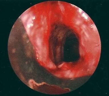
Tracheal Stenosis
Tracheostomy
Laryngostenosis
Intubation, Intratracheal
Tracheal Neoplasms
Trachea
Papio cynocephalus
Tracheoesophageal Fistula
Silicones
Carotid Stenosis
Aortic Valve Stenosis
Tracheal Diseases
Stents
Laser Therapy
Polychondritis, Relapsing
Collagen Diseases
Human papillomavirus 11
Variation in subglottic size in children. (1/197)
The incidence of variation in the subglottic size was investigated in 3304 infants and children. A mild degree of congenital subglottic stenosis was found in 0.91% and a moderate degree of stenosis in 0.06% of the patients. A mild degree of congenital subglottic enlargement was noted in 0.7% and moderate enlargement in 0.06% of the patients. (+info)Localised upper airway obstruction in a patient with acquired immunodeficiency syndrome. (2/197)
We describe a case of rapidly progressive upper airway obstruction due to tracheal Pseudomonas abscesses in a patient with acquired immunodeficiency syndrome. The case highlights the aggressive nature of Pseudomonas infections and the difficulty of eradicating this organism in patients infected with the human immunodeficiency virus. (+info)Tracheobronchial malacia and stenosis in children in intensive care: bronchograms help to predict oucome. (3/197)
BACKGROUND: Severe tracheobronchial malacia and stenosis are important causes of morbidity and mortality in children in intensive care, but little is known about how best to diagnose these conditions or determine their prognosis. METHODS: The records of all 62 children in whom one or both of these conditions had been diagnosed by contrast cinetracheobronchography in our intensive care unit in the period 1986-95 were studied. RESULTS: Seventy four per cent of the 62 children had congenital heart disease; none was a preterm baby with airways disease associated with prolonged ventilation. Fifteen of the children had airway stenosis without malacia; three died because of the stenosis and two died from other causes. Twenty eight of the 47 children with malacia died; only eight children survived without developmental or respiratory handicap. All children needing ventilation for malacia for longer than 14 consecutive days died if their bronchogram showed moderate or severe malacia of either main bronchus (15 cases), or malacia of any severity of both bronchi (three additional cases); all children needing ventilation for malacia for longer than 21 consecutive days died if their bronchogram showed malacia of any severity of the trachea or a main bronchus (three additional cases). These findings were strongly associated with a fatal outcome (p<0.00005); they were present in 21 children (all of whom died) and absent in 26 (of whom seven died, six from non-respiratory causes). They had a positive predictive value for death of 100%, but the lower limit of the 95% confidence interval was 83.9% so up to 16% of patients meeting the criteria might survive. CONCLUSION: In this series the findings on contrast cinetracheobronchography combined with the duration of ventilation provided a useful guide to the prognosis of children with tracheobronchomalacia. The information provided by bronchoscopy was less useful. (+info)Multidisciplinary approach to management of postintubation tracheal stenoses. (4/197)
The optimal management of postintubation tracheal stenosis is not well defined. A therapeutic algorithm was designed by thoracic surgeons, ear, nose and throat (ENT) surgeons, anaesthetists and pulmonologists. Rigid bronchoscopy with neodymium-yttrium aluminium garnet (Nd-YAG) laser resection or stent implantation (removable stent) was proposed as first-line treatment, depending on the type of stenosis (web-like versus complex stenosis). In patients with web-like stenoses, sleeve resection was proposed when laser treatment (up to three sessions) failed. In patients with complex stenoses, operability was assessed 6 months after stent implantation. If the patient was judged operable, the stent was removed and the patient underwent surgery if the stenosis recurred. This algorithm was validated prospectively in a series of 32 consecutive patients. Three patients died from severe coexistent illness shortly after the first bronchoscopy. Of the 15 patients with web-like stenosis, laser resection was curative in 10 (66%). Among the 17 patients with complex stenoses, three remained symptom-free after stent removal. Bronchoscopy alone was thus curative in more than one-third of the patients. Six patients underwent surgery, two after failure of laser resection and four after failure of temporary stenting. Surgery was always performed with the patient in good operative condition. Palliative stenting was the definitive treatment in nine cases. Tracheostomy was the definitive solution in two cases. This approach, including an initial conservative treatment, depending on the type of the stenosis, appears to be applicable to almost all patients and allows secondary surgery to be performed with the patient in good condition. (+info)Fetal tracheal occlusion in the rat model of nitrofen-induced congenital diaphragmatic hernia. (5/197)
Prenatal tracheal occlusion (TO) consistently accelerates lung growth in the sheep model of congenital diaphragmatic hernia (CDH). However, significant variability in lung growth has been observed in early clinical trials of TO. We hypothesized that lung hypoplasia created at relatively late stages of lung development may not be equivalent to human CDH-induced lung hypoplasia, which begins early in gestation. To test this hypothesis, we performed TO in the rat model of nitrofen-induced CDH. Left-sided CDH was induced by administering 100 mg of nitrofen to timed pregnant rats on day 9 of gestation. On day 19 of gestation, four to five fetuses per dam underwent surgical ligation of the trachea. At death (day 21.5), lungs from non-CDH (non-CDH group), left-CDH (CDH group), and trachea-occluded left-CDH fetuses (CDH-TO group) were harvested and compared by weight, DNA and protein content, and stereological morphometry. Wet and dry lung weight-to-body weight ratio, total lung DNA and protein contents, the volume of lung parenchyma, and the total saccular surface area of the CDH-TO group were significantly increased relative to the CDH group and were either greater than or comparable to the non-CDH controls. We conclude that TO accelerates lung growth and increases lung parenchyma in an early-onset model of CDH-induced lung hypoplasia. (+info)Respiratory distress due to tracheal compression by the dilated innominate artery. (6/197)
The case reported is of an 88 yr old female with hypertension and respiratory distress. A chest radiograph revealed a widening of the upper mediastinum. Computed tomographic scanning revealed tracheal compression by the innominate artery, which was elongated and curved. After intubation, she was treated with antihypertensive drugs. This resulted in the remarkable recovery of the patient from respiratory distress. To the authors' knowledge, this is the first reported case of respiratory distress owing to tracheal compression by elongation and curvature of the innominate artery. (+info)A new bronchoscopic method to measure airway size. (7/197)
Bronchoscopic evaluation of stenosis is limited due to radial distortion of bronchoscopic images and the unknown distance between the endoscope and the stenotic area. The purpose of this study was the development and validation of a method for measuring cross-sectional areas in large airways. Distance measurements were performed using a laser probe inserted into the working channel of a bronchoscope. The laser probe was positioned to the locus of interest in the airway, a ring of light (helium/neon) projected on to the luminal wall and the images acquired using an electronic bronchoscope. The images taken were distortion-corrected by means of a computer program. The method was validated by simulating airways using tubes of known diameter. Additionally, distortion-corrected bronchoscopic images were compared with distortion-free videoscopic image analysis of tracheal slices taken from pigs. In the case of the plastic tubes, Pearson's correlation coefficient (r) as well as the intraclass correlation coefficient (ICC) were slightly higher (r=0.99, p<0.01, ICC=0.97) than the correlation of cross-sectional areas between bronchoscopic and videoscopic images of tracheal slices (r=0.88, p<0.01, ICC=0.87). This concept allows accurate and reproducible determination of cross-sectional areas in large airways. (+info)Severe tracheobronchial stenosis in a patient with Crohn's disease. (8/197)
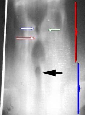
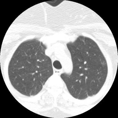
Tracheobronchial involvement in Crohn's disease is rare, usually associated with symptoms of tracheobronchitis, and typically responds well to steroids. The authors report a case of a 29-yr old patient with Crohn's disease, who presented with dyspnoea, fever, and a productive cough. Computed tomography of the chest revealed extensive nodular tracheobronchial stenosis, that was accompanied by severe mucosal inflammation at bronchoscopy. High-dose oral steroids diminished the mucosal inflammation, but had limited efficacy on the underlying tracheobronchial stenosis. It is speculated that this relative ineffectiveness of steroids may be due to the persistence of the untreated inflammatory process. (+info)Tracheal stenosis is a medical condition characterized by the abnormal narrowing of the trachea (windpipe), which can lead to difficulty breathing. This narrowing can be caused by various factors such as inflammation, scarring, or the growth of abnormal tissue in the airway. Symptoms may include wheezing, coughing, shortness of breath, and chest discomfort, particularly during physical activity. Treatment options for tracheal stenosis depend on the severity and underlying cause of the condition and may include medications, bronchodilators, corticosteroids, or surgical interventions such as laser surgery, stent placement, or tracheal reconstruction.
A tracheostomy is a surgically created opening through the neck into the trachea (windpipe). It is performed to provide an airway in cases where the upper airway is obstructed or access to the lower airway is required, such as in prolonged intubation, severe trauma, or chronic lung diseases. The procedure involves making an incision in the front of the neck and creating a direct opening into the trachea, through which a tracheostomy tube is inserted to maintain the patency of the airway. This allows for direct ventilation of the lungs, suctioning of secretions, and prevention of complications associated with upper airway obstruction.
Laryngostenosis is a medical term that refers to a condition where the larynx (or voice box) becomes narrowed. This can occur due to various reasons such as scarring, swelling, or growths in the laryngeal area. The narrowing can cause difficulty with breathing, swallowing, and speaking. In severe cases, it may require medical intervention, such as surgery, to correct the problem.
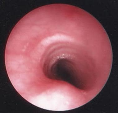
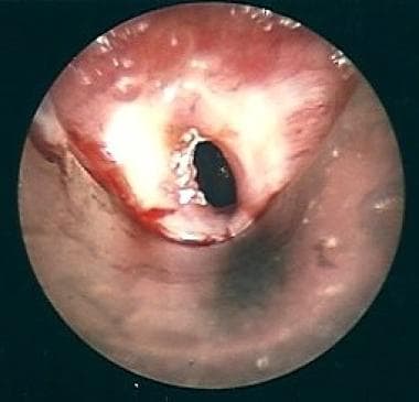
Bronchoscopy is a medical procedure that involves the examination of the inside of the airways and lungs with a flexible or rigid tube called a bronchoscope. This procedure allows healthcare professionals to directly visualize the airways, take tissue samples for biopsy, and remove foreign objects or secretions. Bronchoscopy can be used to diagnose and manage various respiratory conditions such as lung infections, inflammation, cancer, and bleeding. It is usually performed under local or general anesthesia to minimize discomfort and risks associated with the procedure.
Intubation, intratracheal is a medical procedure in which a flexible plastic or rubber tube called an endotracheal tube (ETT) is inserted through the mouth or nose, passing through the vocal cords and into the trachea (windpipe). This procedure is performed to establish and maintain a patent airway, allowing for the delivery of oxygen and the removal of carbon dioxide during mechanical ventilation in various clinical scenarios, such as:
1. Respiratory failure or arrest
2. Procedural sedation
3. Surgery under general anesthesia
4. Neuromuscular disorders
5. Ingestion of toxic substances
6. Head and neck trauma
7. Critical illness or injury affecting the airway
The process of intubation is typically performed by trained medical professionals, such as anesthesiologists, emergency medicine physicians, or critical care specialists, using direct laryngoscopy or video laryngoscopy to visualize the vocal cords and guide the ETT into the correct position. Once placed, the ETT is secured to prevent dislodgement, and the patient's respiratory status is continuously monitored to ensure proper ventilation and oxygenation.
Tracheal neoplasms refer to abnormal growths or tumors in the trachea, which is the windpipe that carries air from the nose and throat to the lungs. These growths can be benign (non-cancerous) or malignant (cancerous). Malignant tracheal neoplasms are relatively rare and can be primary (originating in the trachea) or secondary (spreading from another part of the body, such as lung cancer). Primary tracheal cancers can be squamous cell carcinoma, adenoid cystic carcinoma, mucoepidermoid carcinoma, or sarcomas. Symptoms may include cough, difficulty breathing, wheezing, or chest pain. Treatment options depend on the type, size, and location of the neoplasm and can include surgery, radiation therapy, chemotherapy, or a combination of these approaches.
Inhalation burns, also known as respiratory or pulmonary burns, refer to damage to the airways and lungs caused by inhaling hot gases, smoke, steam, or toxic fumes. This type of injury can occur during a fire or other thermal incidents and can result in significant morbidity and mortality.
Inhalation burns are classified into three categories based on the location and severity of the injury:
1. Upper airway burns: These involve the nose, throat, and voice box (larynx) and are usually caused by inhaling hot gases or steam. Symptoms may include singed nasal hairs, soot in the nose or mouth, coughing, wheezing, and difficulty speaking or swallowing.
2. Lower airway burns: These involve the trachea, bronchi, and bronchioles and are usually caused by inhaling smoke or toxic fumes. Symptoms may include coughing, chest pain, shortness of breath, and wheezing.
3. Systemic burns: These occur when toxic substances are absorbed into the bloodstream and can affect multiple organs. Symptoms may include nausea, vomiting, confusion, and organ failure.
Inhalation burns can lead to complications such as pneumonia, respiratory failure, and acute respiratory distress syndrome (ARDS). Treatment typically involves providing oxygen therapy, removing secretions from the airways, and administering bronchodilators and corticosteroids to reduce inflammation. Severe cases may require intubation and mechanical ventilation.
Prevention of inhalation burns includes avoiding smoke-filled areas during a fire, staying close to the ground where the air is cooler and cleaner, and using appropriate respiratory protection devices when exposed to toxic fumes or gases.
The trachea, also known as the windpipe, is a tube-like structure in the respiratory system that connects the larynx (voice box) to the bronchi (the two branches leading to each lung). It is composed of several incomplete rings of cartilage and smooth muscle, which provide support and flexibility. The trachea plays a crucial role in directing incoming air to the lungs during inspiration and outgoing air to the larynx during expiration.
A tracheotomy is a surgical procedure that involves creating an opening in the neck and through the front (anterior) wall of the trachea (windpipe). This is performed to provide a new airway for the patient, bypassing any obstruction or damage in the upper airways. A tube is then inserted into this opening to maintain it and allow breathing.
This procedure is often conducted in emergency situations when there is an upper airway obstruction that cannot be easily removed or in critically ill patients who require long-term ventilation support. Complications can include infection, bleeding, damage to surrounding structures, and difficulties with speaking, swallowing, or coughing.
Airway obstruction is a medical condition that occurs when the normal flow of air into and out of the lungs is partially or completely blocked. This blockage can be caused by a variety of factors, including swelling of the tissues in the airway, the presence of foreign objects or substances, or abnormal growths such as tumors.
When the airway becomes obstructed, it can make it difficult for a person to breathe normally. They may experience symptoms such as shortness of breath, wheezing, coughing, and chest tightness. In severe cases, airway obstruction can lead to respiratory failure and other life-threatening complications.
There are several types of airway obstruction, including:
1. Upper airway obstruction: This occurs when the blockage is located in the upper part of the airway, such as the nose, throat, or voice box.
2. Lower airway obstruction: This occurs when the blockage is located in the lower part of the airway, such as the trachea or bronchi.
3. Partial airway obstruction: This occurs when the airway is partially blocked, allowing some air to flow in and out of the lungs.
4. Complete airway obstruction: This occurs when the airway is completely blocked, preventing any air from flowing into or out of the lungs.
Treatment for airway obstruction depends on the underlying cause of the condition. In some cases, removing the obstruction may be as simple as clearing the airway of foreign objects or mucus. In other cases, more invasive treatments such as surgery may be necessary.
Respiratory system abnormalities refer to any conditions or structures that do not function properly or are outside the normal range in the respiratory system. The respiratory system is responsible for taking in oxygen and expelling carbon dioxide through the process of breathing. It includes the nose, throat (pharynx), voice box (larynx), windpipe (trachea), bronchi, bronchioles, alveoli, and muscles and nerves that support breathing.
Respiratory system abnormalities can be congenital or acquired. Congenital abnormalities are present at birth and may include conditions such as cystic fibrosis, pulmonary hypoplasia, and congenital diaphragmatic hernia. Acquired abnormalities can develop at any time throughout a person's life due to various factors such as infections, injuries, environmental exposures, or aging. Examples of acquired respiratory system abnormalities include chronic obstructive pulmonary disease (COPD), asthma, pneumonia, lung cancer, and sleep apnea.
Respiratory system abnormalities can cause a range of symptoms, including coughing, wheezing, shortness of breath, chest pain, and fatigue. Treatment for respiratory system abnormalities depends on the specific condition and severity and may include medications, breathing treatments, surgery, or lifestyle changes.
"Papio cynocephalus" is a scientific name for a species of old world monkey, commonly known as the yellow baboon. It's not typically used in a medical context, but I can provide some general biological information about it if that would be helpful.
Yellow baboons are native to the savannas and woodlands of eastern and southern Africa. They have a distinct appearance with a dog-like face (hence the species name "cynocephalus," which means "dog-headed" in Greek) and a long, close-set coat that is yellowish-brown or olive green in color. Adult males can weigh between 33 to 82 pounds (15 to 37 kg), while females are smaller and typically weigh between 14 to 33 pounds (6 to 15 kg).
Yellow baboons live in large social groups called troops, which can consist of up to 200 individuals. They have a complex hierarchical social structure based on age, sex, and dominance. Their diet is omnivorous, consisting of fruits, seeds, nuts, insects, and small vertebrates.
In terms of medical relevance, yellow baboons are sometimes used as animal models in biomedical research due to their close genetic relationship with humans (they share about 96% of their DNA sequence with us). However, it's important to note that using non-human primates in research is a controversial topic and subject to ethical considerations.
A tracheoesophageal fistula (TEF) is an abnormal connection between the trachea (windpipe) and the esophagus (tube that carries food from the mouth to the stomach). This congenital anomaly is usually present at birth and can vary in size and location. It can cause complications such as respiratory distress, feeding difficulties, and recurrent lung infections. TEF is often treated surgically to separate the trachea and esophagus and restore their normal functions.
Silicones are not a medical term, but they are commonly used in the medical field, particularly in medical devices and healthcare products. Silicones are synthetic polymers made up of repeating units of siloxane, which is a chain of alternating silicon and oxygen atoms. They can exist in various forms such as oils, gels, rubbers, and resins.
In the medical context, silicones are often used for their unique properties, including:
1. Biocompatibility - Silicones have a low risk of causing an adverse reaction when they come into contact with living tissue.
2. Inertness - They do not react chemically with other substances, making them suitable for use in medical devices that need to remain stable over time.
3. Temperature resistance - Silicones can maintain their flexibility and elasticity even under extreme temperature conditions.
4. Gas permeability - Some silicone materials allow gases like oxygen and water vapor to pass through, which is useful in applications where maintaining a moist environment is essential.
5. Durability - Silicones have excellent resistance to aging, weathering, and environmental factors, ensuring long-lasting performance.
Examples of medical applications for silicones include:
1. Breast implants
2. Contact lenses
3. Catheters
4. Artificial joints and tendons
5. Bandages and wound dressings
6. Drug delivery systems
7. Medical adhesives
8. Infant care products (nipples, pacifiers)
Dilation, also known as dilatation, refers to the process of expanding or enlarging a body passage or cavity. In medical terms, it typically refers to the widening of a bodily opening or hollow organ, allowing for increased flow or access. This can occur naturally, such as during childbirth when the cervix dilates to allow for the passage of a baby, or it can be induced through medical procedures or interventions.
For example, dilation of the pupils is a natural response to darkness or certain medications, while dilation of blood vessels is a common side effect of some drugs and can also occur in response to changes in temperature or emotional state. Dilation of the stomach or intestines may be necessary for medical procedures such as endoscopies or surgeries.
It's important to note that dilation can also refer to the abnormal enlargement of a body part, such as dilated cardiomyopathy, which refers to an enlarged and weakened heart muscle.
Pulmonary surgical procedures refer to the operations that are performed on the lungs and the surrounding structures, typically to treat or diagnose various respiratory conditions. These procedures can range from minimally invasive techniques to more complex surgeries, depending on the nature and severity of the condition. Here are some examples of pulmonary surgical procedures:
1. Thoracotomy: This is an open surgical procedure where a surgeon makes a large incision in the chest wall to access the lungs. It's typically used to remove lung tumors, repair damaged lung tissue, or perform a lobectomy (removal of a lobe of the lung).
2. Video-assisted thoracoscopic surgery (VATS): This is a minimally invasive procedure where a surgeon makes several small incisions in the chest wall and uses a camera and special instruments to perform the operation. VATS can be used for lung biopsies, lobectomies, and other procedures.
3. Lung biopsy: This is a procedure where a small piece of lung tissue is removed and examined under a microscope to diagnose various conditions such as infections, interstitial lung diseases, or cancer. A biopsy can be performed through a thoracotomy, VATS, or bronchoscopy (a procedure that involves inserting a thin tube with a camera into the airways).
4. Bullectomy: This is a procedure where a surgeon removes large air-filled sacs in the lungs called bullae, which can cause shortness of breath and other symptoms.
5. Lung transplant: This is a complex surgical procedure where a diseased lung is removed and replaced with a healthy one from a donor. It's typically performed on patients with end-stage lung disease such as cystic fibrosis or chronic obstructive pulmonary disease (COPD).
6. Pleurodesis: This is a procedure where the space between the lungs and chest wall is irritated to prevent fluid from accumulating in that space, which can cause shortness of breath and other symptoms. It's typically performed on patients with recurrent pleural effusions (fluid buildup in the pleural space).
These are just a few examples of the many procedures that can be performed to treat various lung conditions.
Carotid stenosis is a medical condition that refers to the narrowing or constriction of the lumen (inner space) of the carotid artery. The carotid arteries are major blood vessels that supply oxygenated blood to the head and neck. Carotid stenosis usually results from the buildup of plaque, made up of fat, cholesterol, calcium, and other substances, on the inner walls of the artery. This process is called atherosclerosis.
As the plaque accumulates, it causes the artery to narrow, reducing blood flow to the brain. Severe carotid stenosis can increase the risk of stroke, as a clot or debris from the plaque can break off and travel to the brain, blocking a smaller blood vessel and causing tissue damage or death.
Carotid stenosis is typically diagnosed through imaging tests such as ultrasound, CT angiography, or MRI angiography. Treatment options may include lifestyle modifications (such as quitting smoking, controlling blood pressure, and managing cholesterol levels), medications to reduce the risk of clots, or surgical procedures like endarterectomy or stenting to remove or bypass the blockage.
Aortic valve stenosis is a cardiac condition characterized by the narrowing or stiffening of the aortic valve, which separates the left ventricle (the heart's main pumping chamber) from the aorta (the large artery that carries oxygen-rich blood to the rest of the body). This narrowing or stiffening prevents the aortic valve from opening fully, resulting in reduced blood flow from the left ventricle to the aorta and the rest of the body.
The narrowing can be caused by several factors, including congenital heart defects, calcification (hardening) of the aortic valve due to aging, or scarring of the valve due to rheumatic fever or other inflammatory conditions. As a result, the left ventricle must work harder to pump blood through the narrowed valve, which can lead to thickening and enlargement of the left ventricular muscle (left ventricular hypertrophy).
Symptoms of aortic valve stenosis may include chest pain or tightness, shortness of breath, fatigue, dizziness or fainting, and heart palpitations. Severe aortic valve stenosis can lead to serious complications such as heart failure, arrhythmias, or even sudden cardiac death. Treatment options may include medications to manage symptoms, lifestyle changes, or surgical intervention such as aortic valve replacement.
Tracheal diseases refer to a group of medical conditions that affect the trachea, also known as the windpipe. The trachea is a tube-like structure made up of rings of cartilage and smooth muscle, which extends from the larynx (voice box) to the bronchi (airways leading to the lungs). Its primary function is to allow the passage of air to and from the lungs.
Tracheal diseases can be categorized into several types, including:
1. Tracheitis: Inflammation of the trachea, often caused by viral or bacterial infections.
2. Tracheal stenosis: Narrowing of the trachea due to scarring, inflammation, or compression from nearby structures such as tumors or goiters.
3. Tracheomalacia: Weakening and collapse of the tracheal walls, often seen in newborns and young children but can also occur in adults due to factors like chronic cough, aging, or connective tissue disorders.
4. Tracheoesophageal fistula: An abnormal connection between the trachea and the esophagus, which can lead to respiratory complications and difficulty swallowing.
5. Tracheal tumors: Benign or malignant growths that develop within the trachea, obstructing airflow and potentially leading to more severe respiratory issues.
6. Tracheobronchial injury: Damage to the trachea and bronchi, often caused by trauma such as blunt force or penetrating injuries.
7. Congenital tracheal abnormalities: Structural defects present at birth, including complete tracheal rings, which can cause narrowing or collapse of the airway.
Symptoms of tracheal diseases may include cough, wheezing, shortness of breath, chest pain, and difficulty swallowing. Treatment options depend on the specific condition and its severity but may involve medications, surgery, or other interventions to alleviate symptoms and improve respiratory function.
A stent is a small mesh tube that's used to treat narrow or weak arteries. Arteries are blood vessels that carry blood away from your heart to other parts of your body. A stent is placed in an artery as part of a procedure called angioplasty. Angioplasty restores blood flow through narrowed or blocked arteries by inflating a tiny balloon inside the blocked artery to widen it.
The stent is then inserted into the widened artery to keep it open. The stent is usually made of metal, but some are coated with medication that is slowly and continuously released to help prevent the formation of scar tissue in the artery. This can reduce the chance of the artery narrowing again.
Stents are also used in other parts of the body, such as the neck (carotid artery) and kidneys (renal artery), to help maintain blood flow and prevent blockages. They can also be used in the urinary system to treat conditions like ureteropelvic junction obstruction or narrowing of the urethra.
Coronary stenosis is a medical condition that refers to the narrowing of the coronary arteries, which supply oxygen-rich blood to the heart muscle. This narrowing is typically caused by the buildup of plaque, made up of fat, cholesterol, and other substances, on the inner walls of the arteries. Over time, as the plaque hardens and calcifies, it can cause the artery to become narrowed or blocked, reducing blood flow to the heart muscle.
Coronary stenosis can lead to various symptoms and complications, including chest pain (angina), shortness of breath, irregular heart rhythms (arrhythmias), and heart attacks. Treatment options for coronary stenosis may include lifestyle changes, medications, medical procedures such as angioplasty or bypass surgery, or a combination of these approaches. Regular check-ups and diagnostic tests, such as stress testing or coronary angiography, can help detect and monitor coronary stenosis over time.
Spinal stenosis is a narrowing of the spinal canal or the neural foramina (the openings through which nerves exit the spinal column), typically in the lower back (lumbar) or neck (cervical) regions. This can put pressure on the spinal cord and/or nerve roots, causing pain, numbness, tingling, or weakness in the affected areas, often in the legs, arms, or hands. It's most commonly caused by age-related wear and tear, but can also be due to degenerative changes, herniated discs, tumors, or spinal injuries.
Laser therapy, also known as phototherapy or laser photobiomodulation, is a medical treatment that uses low-intensity lasers or light-emitting diodes (LEDs) to stimulate healing, reduce pain, and decrease inflammation. It works by promoting the increase of cellular metabolism, blood flow, and tissue regeneration through the process of photobiomodulation.
The therapy can be used on patients suffering from a variety of acute and chronic conditions, including musculoskeletal injuries, arthritis, neuropathic pain, and wound healing complications. The wavelength and intensity of the laser light are precisely controlled to ensure a safe and effective treatment.
During the procedure, the laser or LED device is placed directly on the skin over the area of injury or discomfort. The non-ionizing light penetrates the tissue without causing heat or damage, interacting with chromophores in the cells to initiate a series of photochemical reactions. This results in increased ATP production, modulation of reactive oxygen species, and activation of transcription factors that lead to improved cellular function and reduced pain.
In summary, laser therapy is a non-invasive, drug-free treatment option for various medical conditions, providing patients with an alternative or complementary approach to traditional therapies.
Relapsing polychondritis is a rare autoimmune disease characterized by inflammation and damage to the cartilaginous structures in the body. The condition can affect multiple organs and tissues, including the ears, nose, trachea, bronchi, joints, and cardiovascular system. It is called "relapsing" because it tends to involve recurring episodes of inflammation and damage, followed by periods of remission.
The hallmark symptom of relapsing polychondritis is pain and swelling in the ears, nose, or airways. Other symptoms may include:
* Redness, tenderness, and warmth in affected areas
* Hearing loss or tinnitus (ringing in the ears)
* Nasal congestion, runny nose, or nosebleeds
* Hoarseness or difficulty speaking
* Wheezing, shortness of breath, or coughing
* Joint pain, stiffness, or swelling
* Skin rashes or sores
* Eye inflammation or dryness
* Heart murmurs or other cardiovascular symptoms
The exact cause of relapsing polychondritis is not known, but it is thought to involve an abnormal immune response in which the body's own antibodies attack and damage cartilage and other tissues. The diagnosis of relapsing polychondritis is typically based on a combination of clinical symptoms, laboratory tests, and imaging studies.
There is no cure for relapsing polychondritis, but treatment can help manage the symptoms and prevent complications. Treatment may include corticosteroids, immunosuppressive drugs, and other medications to reduce inflammation and suppress the immune system. In severe cases, surgery may be necessary to repair or replace damaged tissues.
Collagen diseases, also known as collagen disorders or connective tissue diseases, refer to a group of medical conditions that affect the body's connective tissues. These tissues provide support and structure for various organs and systems in the body, including the skin, joints, muscles, and blood vessels.
Collagen is a major component of connective tissues, and it plays a crucial role in maintaining their strength and elasticity. In collagen diseases, the body's immune system mistakenly attacks healthy collagen, leading to inflammation, pain, and damage to the affected tissues.
There are several types of collagen diseases, including:
1. Systemic Lupus Erythematosus (SLE): This is a chronic autoimmune disease that can affect various organs and systems in the body, including the skin, joints, kidneys, heart, and lungs.
2. Rheumatoid Arthritis (RA): This is a chronic inflammatory disease that primarily affects the joints, causing pain, swelling, and stiffness.
3. Scleroderma: This is a rare autoimmune disorder that causes thickening and hardening of the skin and connective tissues, leading to restricted movement and organ damage.
4. Dermatomyositis: This is an inflammatory muscle disease that can also affect the skin, causing rashes and weakness.
5. Mixed Connective Tissue Disease (MCTD): This is a rare autoimmune disorder that combines symptoms of several collagen diseases, including SLE, RA, scleroderma, and dermatomyositis.
The exact cause of collagen diseases is not fully understood, but they are believed to be related to genetic, environmental, and hormonal factors. Treatment typically involves a combination of medications, lifestyle changes, and physical therapy to manage symptoms and prevent complications.
Human papillomavirus type 11 (HPV-11) is a specific type of human papillomavirus that is known to cause benign, or noncancerous, growths called papillomas or warts on the skin and mucous membranes. HPV-11 is one of several types of HPV that are classified as low-risk because they are rarely associated with cancer.
HPV-11 is primarily transmitted through sexual contact and can infect the genital area, leading to the development of genital warts. In some cases, HPV-11 infection may also cause respiratory papillomatosis, a rare condition in which benign growths develop in the airways, including the throat and lungs.
HPV-11 is preventable through vaccination with the human papillomavirus vaccine, which protects against several low-risk and high-risk types of HPV. It is important to note that while HPV-11 is not associated with cancer, other high-risk types of HPV can cause cervical, anal, and oral cancers, so vaccination is still recommended for individuals who are sexually active or plan to become sexually active.
A papilloma is a benign (noncancerous) tumor that grows on a stalk, often appearing as a small cauliflower-like growth. It can develop in various parts of the body, but when it occurs in the mucous membranes lining the respiratory, digestive, or genitourinary tracts, they are called squamous papillomas. The most common type is the skin papilloma, which includes warts. They are usually caused by human papillomavirus (HPV) infection and can be removed through various medical procedures if they become problematic or unsightly.