Tricuspid Atresia
Tricuspid Valve
Tricuspid Valve Stenosis
Tricuspid Valve Insufficiency
Biliary Atresia
Fontan Procedure
Heart Septal Defects, Ventricular
Follicular Atresia
Intestinal Atresia
Heart Defects, Congenital
Esophageal Atresia
Pulmonary Atresia
Transposition of Great Vessels
Heart Ventricles
Abnormalities, Multiple
Pulmonary Artery
Choanal Atresia
Echocardiography
Ebstein Anomaly
Cardiac Valve Annuloplasty
Diagnosis, management, and pathophysiology of post-Fontan hypoxaemia secondary to Glenn shunt related pulmonary arteriovenous malformation. (1/44)
An 8 year old child with tricuspid atresia had developed right sided pulmonary arteriovenous malformations following a previous classic Glenn procedure. These became clinically manifest immediately after Fontan conversion because of severe systemic desaturation. The pathophysiology and postoperative medical management of this case is described and related to current understanding of the aetiology of acquired pulmonary arteriovenous malformations following cavopulmonary shunt. (+info)Impact of early ventricular unloading on exercise performance in preadolescents with single ventricle Fontan physiology. (2/44)
OBJECTIVES: We sought to determine if early ventricular volume unloading improves aerobic capacity in patients with single ventricle Fontan physiology. BACKGROUND: Surgical strategies for patients with single ventricle include intermediate staging or early Fontan completion to reduce the adverse affects of prolonged ventricular volume load. The impact of this strategy on exercise performance has not been evaluated. METHODS: Retrospectively, we reviewed the exercise stress test results of all preadolescents with single ventricle Fontan physiology. "Volume unloading" was considered to have occurred at the time of bidirectional cavopulmonary anastomosis or at the time of Fontan surgery in those patients who did not undergo intermediate staging. Potential predictors of aerobic capacity were analyzed using multivariate regression. RESULTS: The patients (n = 46) achieved a mean percentage predicted of maximal oxygen consumption (VO2max) of 76.1% +/- 21.1%. The mean age at the time of volume unloading was 2.7 +/- 2.4 years, and the mean age at testing was 8.7 +/- 2 years. Intermediate staging was performed in 16 of 46 patients (35%). In multivariate analysis, younger age at volume unloading was associated with increased aerobic capacity (p = 0.003). Other variables were not predictive. The subgroup of patients who underwent volume unloading before two years of age achieved a mean percentage predicted VO2max of 88.6% +/- 24.1%. CONCLUSIONS: Preadolescents with single ventricle who undergo volume unloading surgery at an early age demonstrate superior aerobic capacity compared with those whose surgery is delayed until a later age. (+info)Ebstein's anomaly with imperforate tricuspid valve. Prenatal diagnosis. (3/44)
Ebstein's anomaly is an uncommon congenital heart defect, with a prevalence of 0.3-0.5%. Its association with an imperforate tricuspid valve is an even more rare situation (less than 10% of cases). Prenatal diagnosis of this association by means of fetal echocardiography has not been reported. We describe here this association diagnosed before birth and confirmed after birth. The diagnostic potential and importance of fetal echocardiography during prenatal evaluation of cardiac malformations allows for adequate perinatal planning and management, with an obvious impact on morbidity and mortality. (+info)Double-outlet left ventricle. Echocardiographic diagnosis. (4/44)
This is a case report of a double-outlet left ventricle associated with tricuspid atresia and hypoplasia of the right ventricle, diagnosed during echocardiography with color-flow imaging, in a three-month-old child who presented with fatigue and cyanosis. The child underwent palliative pulmonary arterial banding without an invasive procedure, and showed sustained improvement during follow-up. (+info)The Fontan procedure for tricuspid atresia: early and late results of a 25-year experience with 216 patients. (5/44)
OBJECTIVES: We assessed the operative and late mortality and the present clinical status of 216 patients with tricuspid atresia who had a nonfenestrated Fontan procedure performed at the Mayo Clinic in the 25-year period 1973 to 1998. BACKGROUND: The Fontan operation eliminates the systemic hypoxemia and ventricular volume overload characteristic of prior forms of palliation. However, it originally did so at the cost of systemic venous and right atrial hypertension, and the long-term effects of this "price" were unknown when the procedure was initially proposed. METHODS: We reviewed the clinical records of the 216 patients retrospectively. These were arbitrarily grouped into early (1973 through 1980), middle (1981 through 1987) and late (1988 through 1997) surgical eras. Patient outcome was also analyzed according to age at surgery. Operative and late mortality rates were determined and present clinical status was ascertained in 167 of 171 surviving patients. RESULTS: Overall survival was 79%. Operative mortality steadily declined and was 2% (one of 58 patients) during the most recent decade. Late survival also continues to improve. Age at operation had no effect on operative mortality, and late mortality was significantly increased only in patients who were operated on at age 18 years or older. Eighty-nine percent of surviving patients are currently in New York Heart Association class I or II. CONCLUSIONS: The initial 25-year experience with the nonfenestrated Fontan procedure for tricuspid atresia has been gratifying, with most survivors now leading lives of good quality into adulthood. These results justify continued application of this procedure for children born with tricuspid atresia. (+info)Late clinical outcomes of the fontan operation in patients with tricuspid atresia. (6/44)
OBJECTIVE: Evaluation of the long-term clinical results of the Fontan operation in patients with tricuspid atresia. METHODS: A retrospective analysis was made at the Instituto de Cardiologia do Rio Grande do Sul (Institute of Cardiology of Rio Grande do Sul), from August 1980 through January 2000, of 25 patients with a long-term follow-up, out of a series of 36 patients who underwent the Fontan operation or one of its variants due to tricuspid atresia. Their mean age at surgery was 5.4+/-3.1 years, and their mean weight was 15.8+/-6.1 kg, the majority of them (63.9%) being males. Four patients underwent the classical Fontan operation, 12 the Kreutzer variant, 6 the Bjork variant, 9 total cavopulmonary shunt with a fenestrated tube, and 5 total cavopulmonary shunt with a nonfenestrated tube. RESULTS: The patients were followed-up on an outpatient basis, with a mean long-term survival time of 5.5+/-4.2 years (50 days to 17.8 years) and a late mortality rate of 8%. Arterial saturation increased from 77.2+/-18.8% in the preoperative period to 91+/-6.7% upon the last outpatient visit (p>0.05). At the final check, most (67%) patients were asymptomatic and 87% could tolerate exercise. Ten (40%) patients experienced some kind of complication during the long-term follow-up, such as cardiac arrhythmia, cyanosis, protein-losing enteropathy, neurological events, right heart failure, intolerance to exercise and reoperation. CONCLUSION: The results indicate that, once the immediate postoperative period is over, during which the adaptations to the new circulatory physiology occur, the evolution of patients with tricuspid atresia who underwent the Fontan operation is satisfactory, in spite of a low, yet significant, morbidity. (+info)Tetralogy of fallot and other congenital heart defects in Hey2 mutant mice. (7/44)
Congenital malformations of the heart and circulatory system are the most common type of human birth defect. Recent studies have implicated the Notch signaling pathway in human cardiac development by demonstrating abnormalities of the JAG1 gene as the basis for Alagille syndrome and some cases of isolated tetralogy of Fallot or pulmonic stenosis. How the Notch pathway acts in cardiac development remains unknown, but the Hey family of basic helix-loop-helix (bHLH) transcription factors are candidates for mediating Notch signaling in the developing cardiovascular system. Here, we use gene targeting to determine the developmental functions of mouse Hey2, a Hey family member that is expressed during the embryonic development of the heart, arteries, and other organs. Homozygotes for the Hey2 mutant allele display a spectrum of cardiac malformations including ventricular septal defects, tetralogy of Fallot, and tricuspid atresia, defects that resemble those associated with mutations of human JAG1. These results establish Hey2 as an important regulator of cardiac morphogenesis and suggest a role for Hey2 in mediating or modulating Notch signaling in the developing heart. (+info)Developmental changes in ventricular diastolic function correlate with changes in ventricular myoarchitecture in normal mouse embryos. (8/44)
Both genetic and epigenetic factors, such as abnormal hemodynamics, affect cardiac morphogenesis and the pathogenesis of congenital heart disease. Diastolic function is an important determinant of cardiac function, and tools for evaluating diastolic function in the embryo would be very valuable for assessment of cardiac performance. Using histological measurements of ventricular myoarchitecture, Doppler assessment of ventricular inflow velocities, and direct measurement of ventricular pressure, we investigated developmental changes of ventricular diastolic function in the mouse embryos from embryonic days 9.5 to 19.5. Regression analysis showed that peak velocity of A wave (an index of passive compliance) correlated with the area of trabecular myocardium in right ventricle (RV) (r2=0.92, P<0.0001) and left ventricle (LV) (r2=0.93, P<0.0001). Peak velocity of E wave (an index of active relaxation) exponentially correlated with the area of compact myocardium in RV (r2=0.98, P<0.0001) and LV (r2=0.97, P<0.0001). We used these techniques to analyze FOG-2 null embryos. FOG-2 null embryos had thin compact myocardium, higher EDP and E/A ratio, smaller -dP/dt, and diminished sucking pressure than wild-type littermates, indicating that decreased ventricular diastolic function might be the primary cause of embryonic lethality. In conclusion, during embryogenesis the development of compact myocardium tightly regulates the development of ventricular distensibility. Our study in normal mice forms the basis for future studies of embryonic cardiac function in genetically manipulated mice with abnormalities of the cardiovascular system. (+info)Tricuspid atresia is a congenital heart defect where the tricuspid valve, which regulates blood flow between the right atrium and right ventricle, fails to develop properly. As a result, there is no direct pathway for blood to move from the right atrium to the right ventricle and then to the lungs for oxygenation.
In this condition, blood from the body returning to the heart enters the right atrium but cannot flow through the tricuspid valve into the right ventricle. Instead, it flows through an opening in the interatrial septum (atrial septal defect) into the left atrium and then into the left ventricle. The left ventricle pumps this blood to the body and a portion of it goes to the lungs via a patent ductus arteriosus or other collateral vessels.
Tricuspid atresia is often associated with other heart defects, such as transposition of the great arteries, pulmonary stenosis, or total anomalous pulmonary venous return. Symptoms can vary depending on the severity and associated defects but may include cyanosis (bluish discoloration of the skin), shortness of breath, fatigue, and poor growth. Treatment typically involves surgical interventions to create a path for blood to flow to the lungs and establish proper oxygenation.
The tricuspid valve is the heart valve that separates the right atrium and the right ventricle in the human heart. It is called "tricuspid" because it has three leaflets or cusps, which are also referred to as flaps or segments. These cusps are named anterior, posterior, and septal. The tricuspid valve's function is to prevent the backflow of blood from the ventricle into the atrium during systole, ensuring unidirectional flow of blood through the heart.
Tricuspid valve stenosis is a cardiac condition characterized by the narrowing or stiffening of the tricuspid valve, which is located between the right atrium and right ventricle in the heart. This narrowing or stiffening restricts the normal flow of blood from the right atrium into the right ventricle, causing increased pressure in the right atrium and reduced blood flow to the lungs.
The tricuspid valve typically has three leaflets or cusps that open and close to regulate the flow of blood between the right atrium and right ventricle. In tricuspid valve stenosis, these leaflets become thickened, calcified, or fused together, leading to a reduced opening size and impaired function.
The most common causes of tricuspid valve stenosis include rheumatic heart disease, congenital heart defects, carcinoid syndrome, and infective endocarditis. Symptoms may include fatigue, shortness of breath, swelling in the legs and abdomen, and irregular heartbeats. Treatment options depend on the severity of the condition and underlying causes but may involve medications, surgical repair or replacement of the valve, or catheter-based procedures.
Tricuspid valve insufficiency, also known as tricuspid regurgitation, is a cardiac condition in which the tricuspid valve located between the right atrium and right ventricle of the heart does not close properly, allowing blood to flow back into the right atrium during contraction of the right ventricle. This results in a portion of the blood being pumped inefficiently, which can lead to volume overload of the right side of the heart and potentially result in symptoms such as fatigue, weakness, shortness of breath, and fluid retention. The condition can be congenital or acquired, with common causes including dilated cardiomyopathy, infective endocarditis, rheumatic heart disease, and trauma.
Biliary atresia is a rare, progressive liver disease in infants and children, characterized by the inflammation, fibrosis, and obstruction of the bile ducts. This results in the impaired flow of bile from the liver to the intestine, leading to cholestasis (accumulation of bile in the liver), jaundice (yellowing of the skin and eyes), and eventually liver cirrhosis and failure if left untreated.
The exact cause of biliary atresia is not known, but it is believed to be a combination of genetic and environmental factors. It can occur as an isolated condition or in association with other congenital anomalies. The diagnosis of biliary atresia is typically made through imaging studies, such as ultrasound and cholangiography, and confirmed by liver biopsy.
The standard treatment for biliary atresia is a surgical procedure called the Kasai portoenterostomy, which aims to restore bile flow from the liver to the intestine. In this procedure, the damaged bile ducts are removed and replaced with a loop of intestine that is connected directly to the liver. The success of the Kasai procedure depends on several factors, including the age at diagnosis and surgery, the extent of liver damage, and the skill and experience of the surgeon.
Despite successful Kasai surgery, many children with biliary atresia will eventually develop cirrhosis and require liver transplantation. The prognosis for children with biliary atresia has improved significantly over the past few decades due to earlier diagnosis, advances in surgical techniques, and better postoperative care. However, it remains a challenging condition that requires close monitoring and multidisciplinary management by pediatric hepatologists, surgeons, and other healthcare professionals.
Cineangiography is a medical imaging technique used to visualize the blood flow in the heart and cardiovascular system. It involves the injection of a contrast agent into the bloodstream while X-ray images are taken in quick succession, creating a movie-like sequence that shows the movement of the contrast through the blood vessels and chambers of the heart. This technique is often used to diagnose and evaluate various heart conditions, such as coronary artery disease, valvular heart disease, and congenital heart defects.
The procedure typically involves threading a catheter through a blood vessel in the arm or leg and guiding it to the heart. Once in place, the contrast agent is injected, and X-ray images are taken using a specialized X-ray machine called a fluoroscope. The images captured during cineangiography can help doctors identify areas of narrowing or blockage in the coronary arteries, abnormalities in heart valves, and other cardiovascular problems.
Cineangiography is an invasive procedure that carries some risks, such as bleeding, infection, and reactions to the contrast agent. However, it can provide valuable information for diagnosing and treating heart conditions, and may be recommended when other diagnostic tests have been inconclusive.
The Fontan procedure is a type of open-heart surgery used to treat specific types of complex congenital (present at birth) heart defects. It's typically performed on children with single ventricle hearts, where one of the heart's lower chambers (the right or left ventricle) is underdeveloped or missing.
In a normal heart, oxygen-poor (blue) blood returns from the body to the right atrium, then flows through the tricuspid valve into the right ventricle. The right ventricle pumps the blue blood to the lungs, where it picks up oxygen and turns red. Oxygen-rich (red) blood then returns from the lungs to the left atrium, flows through the mitral valve into the left ventricle, and the left ventricle pumps it out to the body through the aorta.
However, in a single ventricle heart, the underdeveloped or missing ventricle cannot effectively pump blood to the lungs and the body simultaneously. The Fontan procedure aims to separate the blue and red blood circulation to improve oxygenation of the body's tissues.
The Fontan procedure involves two stages:
1. In the first stage, usually performed in infancy, a shunt or a band is placed around the pulmonary artery (the blood vessel that carries blood from the heart to the lungs) to control the amount of blood flowing into the lungs. This helps prevent lung congestion due to excessive blood flow.
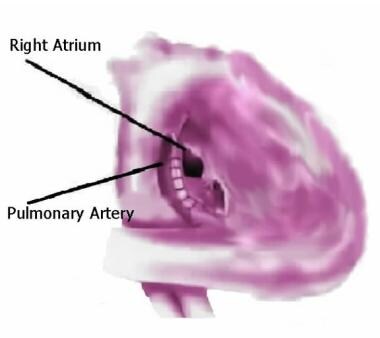
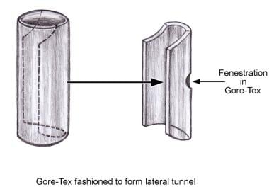
2. The second stage, the Fontan procedure itself, takes place when the child is between 18 months and 4 years old. During this surgery, the surgeon creates a connection between the inferior vena cava (the large vein that returns blue blood from the lower body to the heart) and the pulmonary artery. This allows oxygen-poor blood to flow directly into the lungs without passing through the underdeveloped ventricle.
The Fontan procedure significantly improves the quality of life for many children with single ventricle hearts, although they may still face long-term complications such as heart failure, arrhythmias, and protein-losing enteropathy (a condition where the body loses too much protein in the stool). Regular follow-up care with a pediatric cardiologist is essential to monitor their health and manage any potential issues.
A ventricular septal defect (VSD) is a type of congenital heart defect that involves a hole in the wall separating the two lower chambers of the heart, the ventricles. This defect allows oxygenated blood from the left ventricle to mix with deoxygenated blood in the right ventricle, leading to inefficient oxygenation of the body's tissues. The size and location of the hole can vary, and symptoms may range from none to severe, depending on the size of the defect and the amount of blood that is able to shunt between the ventricles. Small VSDs may close on their own over time, while larger defects usually require medical intervention, such as medication or surgery, to prevent complications like pulmonary hypertension and heart failure.
The pulmonary valve, also known as the pulmonic valve, is a semilunar valve located at the exit of the right ventricle of the heart and the beginning of the pulmonary artery. It has three cusps or leaflets that prevent the backflow of blood from the pulmonary artery into the right ventricle during ventricular diastole, ensuring unidirectional flow of blood towards the lungs for oxygenation.
Follicular atresia is a physiological process that occurs in the ovary, where follicles (fluid-filled sacs containing immature eggs or oocytes) undergo degeneration and disappearance. This process begins after the primordial follicle stage and continues throughout a woman's reproductive years. At birth, a female has approximately 1 to 2 million primordial follicles, but only about 400 of these will mature and release an egg during her lifetime. The rest undergo atresia, which is a natural process that helps regulate the number of available eggs and maintain hormonal balance within the body.
The exact mechanisms that trigger follicular atresia are not fully understood, but it is believed to be influenced by various factors such as hormonal imbalances, oxidative stress, and apoptosis (programmed cell death). In some cases, accelerated or excessive follicular atresia can lead to infertility or early menopause.
Intestinal atresia is a congenital condition characterized by the absence or complete closure of a portion of the intestine, preventing the passage of digested food from the stomach to the remaining part of the intestines. This results in a blockage in the digestive system, which can be life-threatening if not treated promptly after birth. The condition can occur anywhere along the small or large intestine and may affect either a single segment or multiple segments of the intestine.
There are several types of intestinal atresia, including:
1. Jejunal atresia: A closure or absence in the jejunum, a part of the small intestine located between the duodenum and ileum.
2. Ileal atresia: A closure or absence in the ileum, the lower portion of the small intestine that connects to the large intestine (cecum).
3. Colonic atresia: A closure or absence in the colon, a part of the large intestine responsible for storing and eliminating waste.
4. Duodenal atresia: A closure or absence in the duodenum, the uppermost portion of the small intestine that receives chyme (partially digested food) from the stomach.
5. Multiple atresias: When more than one segment of the intestines is affected by atresia.
The exact cause of intestinal atresia remains unclear, but it is believed to be related to disruptions in fetal development during pregnancy. Treatment typically involves surgical correction to reconnect the affected segments of the intestine and restore normal digestive function. The prognosis for infants with intestinal atresia depends on the severity and location of the atresia, as well as any associated conditions or complications.
Congenital heart defects (CHDs) are structural abnormalities in the heart that are present at birth. They can affect any part of the heart's structure, including the walls of the heart, the valves inside the heart, and the major blood vessels that lead to and from the heart.
Congenital heart defects can range from mild to severe and can cause various symptoms depending on the type and severity of the defect. Some common symptoms of CHDs include cyanosis (a bluish tint to the skin, lips, and fingernails), shortness of breath, fatigue, poor feeding, and slow growth in infants and children.
There are many different types of congenital heart defects, including:
1. Septal defects: These are holes in the walls that separate the four chambers of the heart. The two most common septal defects are atrial septal defect (ASD) and ventricular septal defect (VSD).
2. Valve abnormalities: These include narrowed or leaky valves, which can affect blood flow through the heart.
3. Obstruction defects: These occur when blood flow is blocked or restricted due to narrowing or absence of a part of the heart's structure. Examples include pulmonary stenosis and coarctation of the aorta.
4. Cyanotic heart defects: These cause a lack of oxygen in the blood, leading to cyanosis. Examples include tetralogy of Fallot and transposition of the great arteries.
The causes of congenital heart defects are not fully understood, but genetic factors and environmental influences during pregnancy may play a role. Some CHDs can be detected before birth through prenatal testing, while others may not be diagnosed until after birth or later in childhood. Treatment for CHDs may include medication, surgery, or other interventions to improve blood flow and oxygenation of the body's tissues.
Esophageal atresia is a congenital condition in which the esophagus, the tube that connects the throat to the stomach, does not develop properly. In most cases, the upper esophagus ends in a pouch instead of connecting to the lower esophagus and stomach. This condition prevents food and liquids from reaching the stomach, leading to difficulty swallowing and feeding problems in newborn infants. Esophageal atresia often occurs together with a congenital defect called tracheoesophageal fistula, in which there is an abnormal connection between the esophagus and the windpipe (trachea).
The medical definition of 'Esophageal Atresia' is:
A congenital anomaly characterized by the absence of a normal connection between the upper esophagus and the stomach, resulting in the separation of the proximal and distal esophageal segments. The proximal segment usually ends in a blind pouch, while the distal segment may communicate with the trachea through a tracheoesophageal fistula. Esophageal atresia is often associated with other congenital anomalies and can cause serious complications if not diagnosed and treated promptly after birth.
Pulmonary atresia is a congenital heart defect where the pulmonary valve, which controls blood flow from the right ventricle to the lungs, doesn't form properly and instead of being open, there is a membranous obstruction or atresia. This results in an absence of communication between the right ventricle and the pulmonary artery.
The right ventricle is often small and underdeveloped due to this condition, and blood flow to the lungs can be severely limited. In some cases, there may be additional heart defects present, such as a ventricular septal defect (a hole between the two lower chambers of the heart) or patent ductus arteriosus (an abnormal connection between the pulmonary artery and the aorta).
Pulmonary atresia can range from mild to severe, and treatment options depend on the specific anatomy and physiology of each individual case. Treatment may include medications, catheter-based procedures, or open-heart surgery, and in some cases, a heart transplant may be necessary.
Transposition of the Great Vessels is a congenital heart defect in which the two main vessels that carry blood from the heart to the rest of the body are switched in position. Normally, the aorta arises from the left ventricle and carries oxygenated blood to the body, while the pulmonary artery arises from the right ventricle and carries deoxygenated blood to the lungs. In transposition of the great vessels, the aorta arises from the right ventricle and the pulmonary artery arises from the left ventricle. This results in oxygen-poor blood being pumped to the body and oxygen-rich blood being recirculated back to the lungs, which can lead to serious health problems and is often fatal if not corrected through surgery soon after birth.
The heart ventricles are the two lower chambers of the heart that receive blood from the atria and pump it to the lungs or the rest of the body. The right ventricle pumps deoxygenated blood to the lungs, while the left ventricle pumps oxygenated blood to the rest of the body. Both ventricles have thick, muscular walls to generate the pressure necessary to pump blood through the circulatory system.
'Abnormalities, Multiple' is a broad term that refers to the presence of two or more structural or functional anomalies in an individual. These abnormalities can be present at birth (congenital) or can develop later in life (acquired). They can affect various organs and systems of the body and can vary greatly in severity and impact on a person's health and well-being.
Multiple abnormalities can occur due to genetic factors, environmental influences, or a combination of both. Chromosomal abnormalities, gene mutations, exposure to teratogens (substances that cause birth defects), and maternal infections during pregnancy are some of the common causes of multiple congenital abnormalities.
Examples of multiple congenital abnormalities include Down syndrome, Turner syndrome, and VATER/VACTERL association. Acquired multiple abnormalities can result from conditions such as trauma, infection, degenerative diseases, or cancer.
The medical evaluation and management of individuals with multiple abnormalities depend on the specific abnormalities present and their impact on the individual's health and functioning. A multidisciplinary team of healthcare professionals is often involved in the care of these individuals to address their complex needs.
The heart atria are the upper chambers of the heart that receive blood from the veins and deliver it to the lower chambers, or ventricles. There are two atria in the heart: the right atrium receives oxygen-poor blood from the body and pumps it into the right ventricle, which then sends it to the lungs to be oxygenated; and the left atrium receives oxygen-rich blood from the lungs and pumps it into the left ventricle, which then sends it out to the rest of the body. The atria contract before the ventricles during each heartbeat, helping to fill the ventricles with blood and prepare them for contraction.
The pulmonary artery is a large blood vessel that carries deoxygenated blood from the right ventricle of the heart to the lungs for oxygenation. It divides into two main branches, the right and left pulmonary arteries, which further divide into smaller vessels called arterioles, and then into a vast network of capillaries in the lungs where gas exchange occurs. The thin walls of these capillaries allow oxygen to diffuse into the blood and carbon dioxide to diffuse out, making the blood oxygen-rich before it is pumped back to the left side of the heart through the pulmonary veins. This process is crucial for maintaining proper oxygenation of the body's tissues and organs.
A newborn infant is a baby who is within the first 28 days of life. This period is also referred to as the neonatal period. Newborns require specialized care and attention due to their immature bodily systems and increased vulnerability to various health issues. They are closely monitored for signs of well-being, growth, and development during this critical time.
Choanal atresia is a medical condition where the back of the nasal passage (choana) is blocked or narrowed, usually by bone, membrane, or a combination of both. This blockage can be present at birth (congenital) or acquired later in life due to various reasons such as infection, injury, or tumor.
Congenital choanal atresia is more common and occurs during fetal development when the nasal passages fail to open properly. It can affect one or both sides of the nasal passage and can be unilateral (affecting one side) or bilateral (affecting both sides). Bilateral choanal atresia can cause breathing difficulties in newborns, as they are obligate nose breathers and cannot breathe through their mouth yet.
Treatment for choanal atresia typically involves surgical intervention to open up the nasal passage and restore normal breathing. The specific type of surgery may depend on the location and extent of the blockage. In some cases, follow-up surgeries or additional treatments may be necessary to ensure proper functioning of the nasal passage.
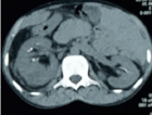
Echocardiography is a medical procedure that uses sound waves to produce detailed images of the heart's structure, function, and motion. It is a non-invasive test that can help diagnose various heart conditions, such as valve problems, heart muscle damage, blood clots, and congenital heart defects.
During an echocardiogram, a transducer (a device that sends and receives sound waves) is placed on the chest or passed through the esophagus to obtain images of the heart. The sound waves produced by the transducer bounce off the heart structures and return to the transducer, which then converts them into electrical signals that are processed to create images of the heart.
There are several types of echocardiograms, including:
* Transthoracic echocardiography (TTE): This is the most common type of echocardiogram and involves placing the transducer on the chest.
* Transesophageal echocardiography (TEE): This type of echocardiogram involves passing a specialized transducer through the esophagus to obtain images of the heart from a closer proximity.
* Stress echocardiography: This type of echocardiogram is performed during exercise or medication-induced stress to assess how the heart functions under stress.
* Doppler echocardiography: This type of echocardiogram uses sound waves to measure blood flow and velocity in the heart and blood vessels.
Echocardiography is a valuable tool for diagnosing and managing various heart conditions, as it provides detailed information about the structure and function of the heart. It is generally safe, non-invasive, and painless, making it a popular choice for doctors and patients alike.
Palliative care is a type of medical care that focuses on relieving the pain, symptoms, and stress of serious illnesses. The goal is to improve quality of life for both the patient and their family. It is provided by a team of doctors, nurses, and other specialists who work together to address the physical, emotional, social, and spiritual needs of the patient. Palliative care can be provided at any stage of an illness, alongside curative treatments, and is not dependent on prognosis.
The World Health Organization (WHO) defines palliative care as: "an approach that improves the quality of life of patients and their families facing the problems associated with life-threatening illness, through the prevention and relief of suffering by means of early identification and impeccable assessment and treatment of pain and other problems, physical, psychological and spiritual."
Ebstein anomaly is a congenital heart defect that affects the tricuspid valve, which is the valve between the right atrium and right ventricle of the heart. In Ebstein anomaly, the tricuspid valve is abnormally formed and positioned, causing it to leak blood back into the right atrium. This can lead to various symptoms such as shortness of breath, fatigue, and cyanosis (bluish discoloration of the skin). Treatment for Ebstein anomaly may include medication, surgery, or a combination of both. It is important to note that the severity of the condition can vary widely among individuals, and some people with Ebstein anomaly may require more intensive treatment than others.
Cardiac valve annuloplasty is a surgical procedure that involves repairing and reinforcing the ring-like structure (annulus) surrounding the heart valves, primarily the mitral or tricuspid valves. This procedure is often performed to correct valve leaks or regurgitation caused by various conditions such as valve disease or dilated cardiomyopathy.
During the annuloplasty procedure, the surgeon typically uses an artificial ring-like device (annuloplasty ring) made of fabric, metal, or a combination of both to reshape and stabilize the damaged annulus. The ring is sewn in place, reducing the size of the valve opening and helping the valve leaflets to coapt properly, thereby preventing valve leaks and improving heart function.
Annuloplasty can be performed as a standalone procedure or in combination with other cardiac surgeries such as valve replacement or repair. The specific technique and approach may vary depending on the individual patient's needs and the surgeon's preference.
A tracheoesophageal fistula (TEF) is an abnormal connection between the trachea (windpipe) and the esophagus (tube that carries food from the mouth to the stomach). This congenital anomaly is usually present at birth and can vary in size and location. It can cause complications such as respiratory distress, feeding difficulties, and recurrent lung infections. TEF is often treated surgically to separate the trachea and esophagus and restore their normal functions.
 Tricuspid atresia
James William Brown
Atresia
Blue baby syndrome
Double inlet left ventricle
Francis Fontan
Fontan procedure
Pulmonary atresia with ventricular septal defect
Bidirectional Glenn procedure
Glenn procedure
Damus-Kaye-Stansel procedure
Protein signalling in heart development
December 1967
Adrian Kantrowitz
Tricuspid valve
Congenital heart defect
Hypoplastic right heart syndrome
Heart valve
Aorticopulmonary septum
Ectopia cordis
List of circulatory system conditions
List of diseases (T)
List of ICD-9 codes 740-759: congenital anomalies
Cyanotic heart defect
Right-to-left shunt
Prostaglandin E1
Pulmonary atresia
Neonatology
List of MeSH codes (C16)
Norwood procedure
Tricuspid atresia - Wikipedia
 Tricuspid Atresia: Background, Pathophysiology, Etiology
 Tricuspid Atresia | Symptoms, Diagnosis & Treatment
 Tricuspid Atresia: Olive's Story | Johns Hopkins Medicine
 Tricuspid Atresia: Causes, Symptoms and Treatment
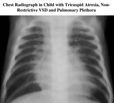
 Tricuspid atresia chest x ray - wikidoc
 Tricuspid Atresia - Pediatrics - Merck Manuals Professional Edition
Information for "Tricuspid atresia other diagnostic studies" - wikidoc
Temporary iatrogenic fetal tricuspid valve atresia in a case of twin to twin transfusion syndrome. | Heart
 CHD | Severe Right Ventricular Dysplasia with Absent Pulmonary Valve Syndrome and Tricuspid Atresia: A Literature Review
Pediatric Tricuspid Atresia Differential Diagnoses
Tricuspid atresia secondary prevention - wikidoc
Pediatric Tricuspid Atresia - Naqlafshk.com
Tricuspid Atresia - Pediatrics - MSD Manual Professional Edition
 Tricuspid atresia and pulmonary stenosis | Hypercholesterolemia Forum
 Pulmonary atresia, intact ventricular septum, and Ebstein anomaly of the tricuspid valve. Anatomic and surgical considerations.
Tricuspid atresia : Causes - Symptoms- Diagnosis -Treatment - Usa-good | Clinic world
 Inpatient Hospitalization Costs Associated with Birth Defects Among Persons Aged 65 Years - United States, 2019 | MMWR
 Congenital heart defect - corrective surgery: MedlinePlus Medical Encyclopedia
 Congenital heart defect - corrective surgery | Lima Memorial Health System
Other Birth Defects of the Heart - Children's Health Issues - MSD Manual Consumer Version
 Types of Heart Valve Disease - UChicago Medicine
 Single Ventricle Heart Conditions And Treatments - Little Hearts Matter
 Words to Know (Heart Glossary) (for Kids) - Aetna Better Health of Virginia (Medicaid)
Tricuspid atresia with absent pulmonary valve and intact ventricular septum: A rare association - Fingerprint - University...
 4Dimensional XStrain echocardiographic assessment by sequential chamber analysis of Double Outlet Left Ventricular with...
 The arterial switch operation in transposition of the great arteries: anatomic indications and contraindications
 Joseph D. Orie, M.D. | UR Medicine
Intact ventricular septum4
- Schneider AW, Blom NA, Bruggemans EF, Hazekamp MG. More than 25 years of experience in managing pulmonary atresia with intact ventricular septum. (medscape.com)
- Pulmonary atresia, intact ventricular septum, and Ebstein anomaly of the tricuspid valve. (unipd.it)
- Pulmonary atresia with intact ventricular septum is a disorder that involves the whole right ventricle. (unipd.it)
- Ebstein anomaly of the tricuspid valve further complicates surgical management and outcome of pulmonary atresia and intact ventricular septum. (unipd.it)
Ventricular septal3
- The signs and symptoms of tricuspid atresia depend on the presence and size of the ventricular septal defect and the relationship of the great arteries. (cincinnatichildrens.org)
- The diagnosis of tricuspid atresia and the associated specific problems such as a ventricular septal defect or transposition of the great arteries can be very accurately diagnosed by echocardiography . (cincinnatichildrens.org)
- In some babies with tricuspid atresia, there's an additional hole between their heart's two lower chambers ( ventricular septal defect ). (clevelandclinic.org)
Cases of tricuspid atresia3
- In most cases of tricuspid atresia, additional defects exist to allow exchange of blood between the loops of systematic circulation and pulmonary circulation, filling in the role of the missing atrioventricular connection. (wikipedia.org)
- Healthcare providers put cases of tricuspid atresia into different categories. (clevelandclinic.org)
- Although the characteristic abnormal superior vector (left axis deviation) of tricuspid atresia is helpful, it is not present in all cases of tricuspid atresia with transposition of the great arteries. (medscape.com)
Great arteries2
- Pulmonary obstruction occurs most often in patients with tricuspid atresia and normally related great arteries. (medscape.com)
- Patients with d-transposed great arteries and tricuspid atresia generally have unobstructed pulmonary blood flow. (medscape.com)
Stenosis13
- [ 4 ] In the absence of pulmonary atresia or pulmonary valve stenosis, the volume of blood to the lungs may be normal with normal oxygenation occurring, resulting in reduced cyanosis. (medscape.com)
- Infrequently, classic tricuspid atresia involves a large VSD and mild pulmonic stenosis, resulting in pulmonary overcirculation. (merckmanuals.com)
- Anatomic variations in underdeveloped right ventricle related to tricuspid atresia and stenosis. (medscape.com)
- Fusion of developing valve leaflet components results in stenosis (partial fusion) or atresia (complete fusion) of the valve. (naqlafshk.com)
- If the valve fusion is incomplete, stenosis of the tricuspid valve develops. (naqlafshk.com)
- The pathologic, clinical, and electrocardiographic features of tricuspid stenosis and atresia are similar. (naqlafshk.com)
- Therefore, the fact that isolated congenital tricuspid stenosis belongs to the group of tricuspid atresia defects and that their embryologic developments are similar is no surprise. (naqlafshk.com)
- Thus, the tricuspid valve stenosis, tricuspid atresia with well-formed but fused valve leaflets, and the muscular type of tricuspid atresia represent a spectrum of morphologic abnormalities. (naqlafshk.com)
- These may include transposition of the great vessels, pulmonary stenosis, or pulmonary atresia. (chkd.org)
- DOLV occurs most commonly in the form of atrial situs solitus with atrioventricular (AV)concordance but is often associated with myriads of cardiac anomalies such as VSD, ASD, PDA,pulmonary stenosis, right ventricular hypoplasia, and tricuspid atresia (TA). (medresearch.in)
- We arepresenting an exceedingly rare case report of DOLV, tricuspid atresia, D-malposition of greatarteries, and mild pulmonary stenosis with the absence of RV hypoplasia, assessed by sequentialchamber analysis, employing 4Dimensional XStrain colour Doppler echocardiography. (medresearch.in)
- Discuss the pathophysiology of pulmonary stenosis/atresia, in contrast to tricuspid atresia. (iame.com)
- Modified technique for dilatation of pulmonary valve stenosis (nearly atresia) in a neonate]. (bvsalud.org)
Forms of tricuspid atresia2
- Considering all forms of tricuspid atresia, no sexual predilection exists. (medscape.com)
- Thus, pulmonary blood flow may be increased or decreased with different forms of tricuspid atresia. (merckmanuals.com)
Baby with tricuspid atresia1
- As a result of a baby with tricuspid atresia may have surgery or alternative procedures shortly after birth, this congenital disorder is taken into account as a vital innate heart defect. (usa-good.com)
Truncus3
- Tricuspid atresia: association with persistent truncus arteriosus. (naqlafshk.com)
- Tricuspid atresia and truncus arteriosus are two medical conditions most parents have never heard of. (umms.org)
- We knew before he was born that he had tricuspid atresia, but since it is so rare to have both tricuspid atresia and truncus arteriosus occur together, we didn't know about it until after he was born," explains Shanade, Tristan's mom. (umms.org)
Regurgitation1
- Hence, dilatation of the LV does not pull the LV papillary muscles apart, whereas dilatation of the RV does pull the RV muscles apart, favoring the development or exacerbation of tricuspid regurgitation. (nih.gov)
Leaflet of the tricuspid valve2
- The septal leaflet of the tricuspid valve mostly develops from the inferior endocardial cushion with a small contribution from the superior cushion. (naqlafshk.com)
- Furthermore, in five cases the anterior leaflet of the tricuspid valve obstructed the right ventricle at the ostium infundibuli level. (unipd.it)
Cyanosis3
- progressive cyanosis poor feeding tachypnea over the first 2 weeks of life holosystolic murmur due to the VSD left axis deviation on electrocardiography and left ventricular hypertrophy (since it must pump blood to both the pulmonary and systemic systems) Normal or mildly enlarged heart Tricuspid atresia is caused by complete absence of the tricuspid valve. (wikipedia.org)
- In this case, the newborn with tricuspid atresia will have a low oxygen level and a dusky, blue color, also called cyanosis. (cincinnatichildrens.org)
- Tricuspid atresia is the most common cause of cyanosis with left ventricular hypertrophy. (naqlafshk.com)
Mitral atresia1
- There are many types of single ventricles including double inlet left ventricle, hypoplastic left heart syndrome, mitral atresia, tricuspid atresia, and common AV valves with only one well-developed ventricle. (stanford.edu)
Univentricular heart2
- Terminology: tricuspid atresia or univentricular heart? (medscape.com)
- Little more than 3 decades ago, the terminology for this defect (eg, tricuspid atresia, univentricular heart, univentricular atrioventricular connection) was intensely debated. (naqlafshk.com)
Defects3
- It is also possible for tricuspid atresia to appear without the life-saving defects. (wikipedia.org)
- Tricuspid atresia is one of the serious heart defects that healthcare providers consider critical congenital heart defects . (clevelandclinic.org)
- Following this report, multiple studies were published demonstrating the effectiveness of this technique in infants with congestive heart failure caused by large VSDs, complex lesions (eg, atrioventricular canal defects), and tricuspid atresia . (medscape.com)
Ventricle16
- [ 1 ] The deformity consists of a complete lack of formation of the tricuspid valve with absence of direct connection between the right atrium and right ventricle. (medscape.com)
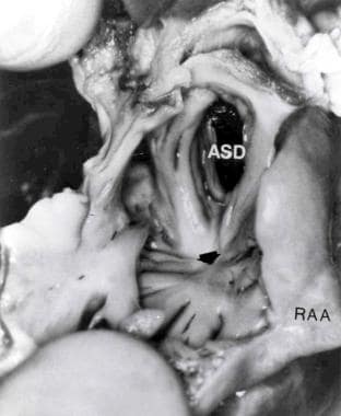
- With the absence of the tricuspid valve and no continuity between the right atrium and right ventricle, venous blood returning to the right atrium can exit only by an intra-atrial communication. (medscape.com)
- The left ventricle comprises most of the ventricular mass in tricuspid atresia. (medscape.com)
- The tricuspid valve is normally between two chambers on the right side of your heart , the right atrium (upper chamber) and right ventricle (lower chamber). (clevelandclinic.org)
- Tricuspid atresia is absence of the tricuspid valve accompanied by a hypoplastic right ventricle. (merckmanuals.com)
- The tricuspid valve is absent, and the right ventricle is hypoplastic. (merckmanuals.com)
- Bharati S, Lev M. The concept of tricuspid atresia complex as distinct from that of the single ventricle complex. (medscape.com)
- An associated Ebstein deformity of the tricuspid valve is found in 10% of the cases, further complicating the anatomy and the function of the right ventricle. (unipd.it)
- The tricuspid valve lies between 2 of the chambers of the heart (the right atrium of the heart and right ventricle). (usa-good.com)
- tricuspid atresia is a congenital disorder of the tricuspid valve, that is, the valve that controls blood results from the right atrium (upper right chamber of the guts) to the proper ventricle (lower right chamber of the heart). (usa-good.com)
- In babies with tricuspid atresia, the right atrioventricular valve that controls blood results the proper atrium to the right ventricle isn't formed, so blood is unable to induce to the right ventricle and dead set the lungs. (usa-good.com)
- In tricuspid atresia, since blood cannot directly flow from the proper atrium to the right ventricle, blood should use alternative routes to bypass the unformed tricuspidata valve. (usa-good.com)
- Because blood doesn't pass through the tricuspid valve, the right ventricle remains small. (chkd.org)
- Infants with tricuspid atresia are born without a tricuspid valve and have an underdeveloped right ventricle. (msdmanuals.com)
- 12. Rocha IEG, Pazin IC, Loops LM: Tricuspid atresia and double outlet left ventricle: A rare association in adulthood. (medresearch.in)
- The tricuspid valve is located between the right atrium and right ventricle and has a valve area of 4-6 cm 2 (see the following image and video). (medscape.com)
Diagnosis1
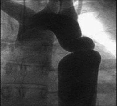
- A chest x-ray may be helpful in the diagnosis of tricuspid atresia. (wikidoc.org)
Occurs6
- It occurs when the heart's tricuspid valve, between the right upper and right lower pumping chamber, doesn't form correctly in the womb. (hopkinsmedicine.org)
- Tricuspid atresia is a congenital (present at birth) heart defect that occurs when the tricuspid valve of the heart doesn't form. (clevelandclinic.org)
- The classic muscular form of tricuspid atresia develops if the embryologic insult occurs early in gestation, and fused valve leaflets occur if the embryologic abnormality occurs slightly later than this in gestation. (naqlafshk.com)
- It occurs when the tricuspid valve doesn't form, or only partly forms. (chkd.org)
- Pulmonary atresia frequently occurs together with other heart disorders and with abnormalities of the coronary arteries. (msdmanuals.com)
- Tricuspid atresia occurs when the tricuspid valve fails to form, leaving two adjoining chambers of the heart without an opening between them. (umms.org)
Symptoms2
- What are the tricuspid atresia symptoms? (clevelandclinic.org)
- In most cases, babies with tricuspid atresia have symptoms within a week of birth. (clevelandclinic.org)
Dysplasia1
- A correlation between the degree of tricuspid valve dysplasia and right ventricular cavity size was observed in all. (unipd.it)
Anatomy3
- Weinberg PM. Anatomy of tricuspid atresia and its relevance to current forms of surgical therapy. (medscape.com)
- Tricuspid atresia: anatomy, imaging, and natural history. (medscape.com)
- The pathologic anatomy of tricuspid atresia is best understood by reviewing variations in valvar morphology. (naqlafshk.com)
Leaflets5
- The tricuspid valve leaflets have several origins. (naqlafshk.com)
- The anterior and posterior tricuspid valve leaflets develop by undermining of a skirt of ventricular muscle tissue. (naqlafshk.com)
- Whether a muscular type of tricuspid atresia develops or whether well-formed but fused tricuspid-valve leaflets develop depends on the stage of development when the embryologic aberration takes place. (naqlafshk.com)
- 5) The mitral valve leaflets are better designed to occlude a circular systemic atrioventricular orifice than are the tricuspid valve leaflets. (nih.gov)
- The right atrioventricular valve complex (the tricuspid valve) is made up of the 3 valve leaflets, the annulus, the supporting chordae tendineae, and the papillary muscles. (medscape.com)
Pulmonary valve1
- In pulmonary atresia, the pulmonic (pulmonary) valve does not form properly, so blood does not flow into the lungs and pick up oxygen. (msdmanuals.com)
Obstruction1
- Depending on the degree of obstruction and associated anomalies, tricuspid atresia may be lethal at birth. (medscape.com)
Anomaly1
- With Ebstein's anomaly , the tricuspid valve is there but doesn't work right. (clevelandclinic.org)
Anomalies2
- Other cardiovascular anomalies occur in 15-20% of patients with tricuspid atresia. (medscape.com)
- Tricuspid atresia accounts for 1 to 3% of congenital heart anomalies. (merckmanuals.com)
Babies4
- An estimated 404 babies are born with tricuspid atresia each year in the United States. (medscape.com)
- Babies with tricuspid atresia may or may not have a heart murmur . (cincinnatichildrens.org)
- About 1 out of 10,000 babies born has tricuspid atresia, regardless of gender. (clevelandclinic.org)
- Babies born with tricuspid abnormal conditions typically even have an associate degree chamber septate defect, that could be a hole between the right and left atria, or a cavum septal defect, which is a hole between the right and left ventricles. (usa-good.com)
Atrial1
- The atrial and ventricular masses, conduction system tissue, and support structure of the fibroelastic cardiac skeleton allow coordinated actions of the tricuspid valve. (medscape.com)
19061
- Although some authors state that Holmes (1824) or Kuhne (1906) first described tricuspid atresia, Rashkind's methodical and thorough historical review indicates that Kreysig (1817) reported the first case in 1817. (naqlafshk.com)
Heart defect7
- Tricuspid atresia is the third most common critical congenital heart defect. (wikipedia.org)
- Olive was born with a heart defect called tricuspid atresia and had only half of a functioning heart. (hopkinsmedicine.org)
- Olive was born with a heart defect called tricuspid atresia . (hopkinsmedicine.org)
- A Type 1 tricuspid atresia heart defect prevents the normal flow of blood through your heart. (clevelandclinic.org)
- People are more likely to get tricuspid atresia or another congenital heart disease if they have Down syndrome or a parent who had a congenital heart defect. (clevelandclinic.org)
- Tricuspid atresia is a congenital (present at birth) heart defect that happens once the right atrioventricular valve of the guts isn't properly formed. (usa-good.com)
- Tricuspid atresia (TA) is a heart defect present at birth (congenital). (chkd.org)
Disease3
- Tricuspid atresia is a form of congenital heart disease whereby there is a complete absence of the tricuspid valve. (wikipedia.org)
- Tricuspid atresia is the third most common form of cyanotic congenital heart disease, with a prevalence of 1.03 per 10,000 live births. (medscape.com)
- Tricuspid atresia is a type of congenital (a condition you are born with) heart disease. (cincinnatichildrens.org)
Posterior2
- therefore, most of the tricuspid annular descent takes place along the margins of the anterior and posterior cusps. (medscape.com)
- The tricuspid subvalvular apparatus consists of anterior, posterior, and septal papillary muscles and their true chordae tendineae. (medscape.com)
Valve leaflet1
- In two of our surgical patients, a protruding anterior tricuspid valve leaflet was identified and excised and both patients survived. (unipd.it)
Absence2
- The intracardiac blood flow in tricuspid atresia further depends on the presence or absence of pulmonary arterial pathology. (medscape.com)
- Tricuspid atresia may be defined as congenital absence or agenesis of the tricuspid valve. (naqlafshk.com)
Anomalous1
- Atresia of the tricuspid valve and total anomalous supracardiac pulmonary venous return. (bvsalud.org)