Triiodothyronine
Triiodothyronine, Reverse
Thyroxine
Thyroid Hormones
Hypothyroidism
Hyperthyroidism
Thyrotropin
Thyroxine-Binding Proteins
Thyroid Gland
Propylthiouracil
Iodide Peroxidase
Receptors, Thyroid Hormone
Thyrotropin-Releasing Hormone
Euthyroid Sick Syndromes
Iodine
Radioimmunoassay
Methimazole
Antithyroid Agents
Thyroid Hormone Receptors alpha
Liver
Serum Globulins
Myxedema
Iodine Isotopes
Thyroid Hormone Receptors beta
Malate Dehydrogenase
Dialysis
Thyroglobulin
Graves Disease
Thyronines
Iodine Radioisotopes
Growth Hormone
Glycerolphosphate Dehydrogenase
Reagent Kits, Diagnostic
Hormones
Pituitary Gland
Hydrocortisone
Rats, Inbred Strains
Thyrotoxicosis
Talc
Adipose Tissue, Brown
Goiter
RNA, Messenger
Body Weight
Thyroxine-Binding Globulin
Basal Metabolism
Pregnancy
Prolactin
Insulin
Amiodarone
Immunoassay
Prealbumin
Reference Values
Rats, Wistar
Ergotamine
Congenital Hypothyroidism
Clinical Chemistry Tests
Pregnenolone Carbonitrile
Serum Albumin, Radio-Iodinated
Selenium
Glucagon
Rats, Sprague-Dawley
Cells, Cultured
Type 1 deiodinase is stimulated by iodothyronines and involved in thyroid hormone metabolism in human somatomammotroph GX cells. (1/147)
BACKGROUND: Local 5'-deiOdination of l-thyroxine (T4) to the active thyroid hormone, 3,3',5-tri-iodothyronine (T3) via two deiodinase isoenzymes (D1 and D2) has an important role for various T3-dependent functions in the anterior pituitary. However, no evidence has been presented yet for thyroid hormone inactivation via the 5-deiodinase (D3) in anterior pituitary models. METHODS: Using the human somatomammotroph cell line, GX, we analysed effects of T3 and its 5'-deiodination product, 3,5-di-iodothyronine (3,5-T2), on deiodinase activities, measuring release of iodide-125 (125I-) from phenolic-ring- or tyrosyl-ring-labelled substrates respectively. RESULTS: T3 and 3,5-T2 rapidly stimulated D1 activity in GX cells in the presence of serum in the culture medium, whereas D2 activity was not detectable under these conditions. However, when the cells were kept under serum-free conditions, specific activity of D2 reached levels similar to those of D1. With tyrosyl-ring labelled 3, 5-[125I]-,3'-T3 as substrate, a significant release of 125I- was observed in GX cell homogenates. This is comparable to the D1 activity of liver membranes, which preferentially catalyses 5'-deiodination, but to some extent also 5-deiodination, at the tyrosyl ring. CONCLUSIONS: D1 activity of human GX cells is increased by T3 and 3,5-T2. Inactivation of T3 in the anterior pituitary might occur by deiodination at the tyrosyl ring via D1, thus terminating the stimulatory thyroid hormone signal in human somatomammotroph cells. (+info)Reverse triiodothyronine, thyroid hormone, and thyrotrophin concentrations in placental cord blood. (2/147)
Reverse triiodothyronine (rT3), triiodothyronine (T3), thyroxine (T4), thyroxine binding globulin (TBG), and thyrotrophin (TSH) were measured in sera from placental cord blood in an unselected series of 272 deliveries. In this series the concentrations of rT3 (mean 3.33 nmol/l, 95% confidence limits 1.6--7.0 nmol/l), were log normally distributed and did not overlap the adult normal range (0.11--0.44 nmol/l). There were no correlations between the cord blood concentrations of rT3, T3, T4, and TSH. The cord serum rT3 concentration was not influenced by maturity, birth-weight, or neonatal risk factors, whereas these factors did affect the concentrations of T3, T4, AND TBG. There is no arteriovenous rT3 concentration difference across the placenta, therefore the cord rT3 reflects the systemic rT3 concentration in the baby at birth. As rT3 in the neonate largely, if not entirely, derives from thyroxine from the fetal thyroid, measurement of the cord rT3 concentration may be a good immediate screening test for neonatal hypothyroidism. (+info)Comparison of mechanisms mediating uptake and efflux of thyroid hormones in the human choriocarcinoma cell line, JAR. (3/147)
We compared the specificities of transport mechanisms for uptake and efflux of thyroid hormones in cells of the human choriocarcinoma cell line, JAR, to determine whether triiodothyronine (T3), thyroxine (T4) and reverse T3 (rT3) are carried by the same transport mechanism. Uptake of 125I-T3, 125I-T4 and 125I-rT3 was saturable and stereospecific, but not specific for T3, T4 and rT3, as unlabelled L-stereoisomers of the thyroid hormones inhibited uptake of each of the radiolabelled hormones. Efflux of 125I-T3 was also saturable and stereospecific and was inhibited by T4 and rT3. Efflux of 125I-T4 or 125I-rT3 was, in contrast, not significantly inhibited by any of the unlabelled thyroid hormones tested. A range of compounds known to interfere with receptor-mediated thyroid hormone uptake in cells inhibited uptake of 125I-T3 and 125I-rT3, but not 125I-T4. We conclude that in JAR cells uptake and efflux of 125I-T3 are mediated by saturable and stereospecific membrane transport processes. In contrast, the uptake, but not the efflux, of 125I-T4 and 125I-rT3 is saturable and stereospecific, indicating that uptake and efflux of T4 and rT3 in JAR cells occur by different mechanisms. These results suggest that in JAR cells thyroid hormones may be transported by at least two types of transporters: a low affinity iodothyronine transporter (Michaelis constant, Km, around 1 microM) which interacts with T3, T4 and rT3, but not amino acids, and an amino acid transporter which takes up T3, but not T4 or rT3. Efflux of T4 and rT3 appears to occur by passive diffusion in these cells. (+info)Effect of nicotine on type 2 deiodinase activity in cultured rat glial cells. (4/147)
Intracellular generation of triiodothyronine (T3) from thyroxine (T4) by type 2 deiodinase (D2) in the mammalian brain, plays a key role in thyroid hormone action. The presence of D2 in rat astrocytes suggests the importance of glial cells in the regulation of intracellular T3 levels in the rat central nervous system (CNS). To analyze further the factors that regulate D2 activity in the CNS, we investigated the effects of nicotine and of mecamylamine, which inhibits the binding of nicotine with nicotinic acetylcholine receptors, on D2 activity in cultured mixed glial cells of the rat brain. We incubated cultured mixed glial cells obtained from neonatal Wistar rats in the presence of 10 mM dithiothreitol, 2 nM [125I] reverse T3 and 1 mM 6-N-propyl-2-thiouracil for 2 h at 37 degrees C, and the released 125I- was counted in a gamma counter. D2 activity of cultured cells was dependent on the temperature and the amount of protein. The basal D2 activity of rat mixed glial cells was 1.9 +/- 0.2 fmol of I- released/mg protein/h (mean +/- SEM). The addition of 10(-11), 2 x 10(-11), 10(-10), and 10(-9) M nicotine significantly increased D2 activity to approximately 2.2-, 2.4, 3.5- and 2.9-fold the basal level, respectively. D2 activity stimulated by 10(-8) M nicotine (2.5-fold) reached a peak after 9 h incubation. The stimulatory effect of nicotine was completely blocked by 10(-6) M mecamylamine. In conclusion, nicotine increases D2 activity probably via nicotinic acetylcholine receptors, and may influence brain function, at least in part, by affecting thyroid hormone metabolism. (+info)Effects of thyroid hormone on action potential and repolarizing currents in rat ventricular myocytes. (5/147)
Thyroid hormones play an important role in cardiac electrophysiology through both genomic and nongenomic mechanisms of action. The effects of triiodothyronine (T(3)) on the electrophysiological properties of ventricular myocytes isolated from euthyroid and hypothyroid rats were studied using whole cell patch clamp techniques. Hypothyroid ventricular myocytes showed significantly prolonged action potential duration (APD(90)) compared with euthyroid myocytes, APD(90) of 151 +/- 5 vs. 51 +/- 8 ms, respectively. Treatment of hypothyroid ventricular myocytes with T(3) (0.1 microM) for 5 min significantly shortened APD by 24% to 115 +/- 10 ms. T(3) similarly shortened APD in euthyroid ventricular myocytes, but only in the presence of 4-aminopyridine (4-AP), an inhibitor of the transient outward current (I(to)), which prolonged the APD by threefold. Transient outward current (I(to)) was not affected by the acute application of T(3) to either euthyroid or hypothyroid myocytes; however, I(to) density was significantly reduced in hypothyroid compared with euthyroid ventricular myocytes. (+info)Myosin V plays an essential role in the thyroid hormone-dependent endocytosis of type II iodothyronine 5'-deiodinase. (6/147)
In astrocytes, thyroxine modulates type II iodothyronine 5'-deiodinase levels by initiating the binding of the endosomes containing the enzyme to microfilaments, followed by actin-based endocytosis. Myosin V is a molecular motor thought to participate in vesicle trafficking in the brain. In this report, we developed an in vitro actin-binding assay to characterize the thyroid hormone-dependent binding of endocytotic vesicles to microfilaments. Thyroxine and reverse triiodothyronine (EC(50) levels approximately 1 nm) were >100-fold more potent than 3,5,3'-triiodothyronine in initiating vesicle binding to actin fibers in vitro. Thyroxine-dependent vesicle binding was calcium-, magnesium-, and ATP-dependent, suggesting the participation of one or more myosin motors, presumably myosin V. Addition of the myosin V globular tail, lacking the actin-binding head, specifically blocked thyroid hormone-dependent vesicle binding, and direct binding of the myosin V tail to enzyme-containing endosomes was thyroxine-dependent. Progressive NH(2)-terminal deletion of the myosin V tail and domain-specific antibody inhibition studies revealed that the thyroxine-dependent vesicle-tethering domain was localized to the last 21 amino acids of the COOH terminus. These data show that myosin V is responsible for thyroid hormone-dependent binding of primary endosomes to the microfilaments and suggest that this motor mediates the actin-based endocytosis of the type II iodothyronine deiodinase. (+info)Variation in values for iodothyronine hormones, thyrotropin, and thyroxine-binding globulin in normal umbilical-cord serum with season and duration of storage. (7/147)
We measured concentrations of thyroxine, triiodothyronine, reverse triiodothyronine, thyroxine-binding globulin, and thyrotropin in pooled samples of cord sera from normal newborns. Sera collected in winter contain significantly (p less than 0.05) higher concentrations of the first tour--14.9, 13.4, 9k9, and 7.5%, respectively--than do sera collected in summer; thyrotropin concentrations are similar in samples collected during winter and summer (p greater than 0.05). With storage, the values for the thyronines and thyrotropin decreased progressively at rates between 0.9 and 5.3% per year; those for thyroxine-binding globulin did not change significantly. (+info)Do adaptive changes in metabolic rate favor weight regain in weight-reduced individuals? An examination of the set-point theory. (8/147)
BACKGROUND: Obese persons generally regain lost weight, suggesting that adaptive metabolic changes favor return to a preset weight. OBJECTIVE: Our objective was to determine whether adaptive changes in resting metabolic rate (RMR) and thyroid hormones occur in weight-reduced persons, predisposing them to long-term weight gain. DESIGN: Twenty-four overweight, postmenopausal women were studied at a clinical research center in four 10-d study phases: the overweight state (phase 1, energy balance; phase 2, 3350 kJ/d) and after reduction to a normal-weight state (phase 3, 3350 kJ/d; phase 4, energy balance). Weight-reduced women were matched with 24 never-overweight control subjects. After each study phase, assessments included RMR (by indirect calorimetry), body composition (by hydrostatic weighing), serum triiodothyronine (T(3)), and reverse T(3) (rT(3)). Body weight was measured 4 y later, without intervention. RESULTS: Body composition-adjusted RMR and T(3):rT(3) fell during acute (phase 2) and chronic (phase 3) energy restriction (P: < 0.01), but returned to baseline in the normal-weight, energy-balanced state (phase 4; mean weight loss: 12.9 +/- 2.0 kg). RMR among weight-reduced women (4771 +/- 414 kJ/d) was not significantly different from that in control subjects (4955 +/- 414 kJ/d; P: = 0.14), and lower RMR did not predict greater 4-y weight regain (r = 0.27, NS). CONCLUSIONS: Energy restriction produces a transient hypothyroid-hypometabolic state that normalizes on return to energy-balanced conditions. Failure to establish energy balance after weight loss gives the misleading impression that weight-reduced persons are energy conservative and predisposed to weight regain. Our findings do not provide evidence in support of adaptive metabolic changes as an explanation for the tendency of weight-reduced persons to regain weight. (+info)Triiodothyronine (T3) is a thyroid hormone, specifically the active form of thyroid hormone, that plays a critical role in the regulation of metabolism, growth, and development in the human body. It is produced by the thyroid gland through the iodination and coupling of the amino acid tyrosine with three atoms of iodine. T3 is more potent than its precursor, thyroxine (T4), which has four iodine atoms, as T3 binds more strongly to thyroid hormone receptors and accelerates metabolic processes at the cellular level.
In circulation, about 80% of T3 is bound to plasma proteins, while the remaining 20% is unbound or free, allowing it to enter cells and exert its biological effects. The primary functions of T3 include increasing the rate of metabolic reactions, promoting protein synthesis, enhancing sensitivity to catecholamines (e.g., adrenaline), and supporting normal brain development during fetal growth and early infancy. Imbalances in T3 levels can lead to various medical conditions, such as hypothyroidism or hyperthyroidism, which may require clinical intervention and management.
Reverse Triiodothyronine (rT3) is a thyroid hormone that is chemically identical to triiodothyronine (T3), but has a reverse configuration at one end of the molecule. It is produced in smaller quantities compared to T3 and its function is not well understood. In some cases, increased levels of rT3 have been associated with decreased thyroid hormone action, such as in non-thyroidal illnesses or during calorie restriction. However, the clinical significance of rT3 levels remains a topic of ongoing research and debate.
Thyroxine (T4) is a type of hormone produced and released by the thyroid gland, a small butterfly-shaped endocrine gland located in the front of your neck. It is one of two major hormones produced by the thyroid gland, with the other being triiodothyronine (T3).
Thyroxine plays a crucial role in regulating various metabolic processes in the body, including growth, development, and energy expenditure. Specifically, T4 helps to control the rate at which your body burns calories for energy, regulates protein, fat, and carbohydrate metabolism, and influences the body's sensitivity to other hormones.
T4 is produced by combining iodine and tyrosine, an amino acid found in many foods. Once produced, T4 circulates in the bloodstream and gets converted into its active form, T3, in various tissues throughout the body. Thyroxine has a longer half-life than T3, which means it remains active in the body for a more extended period.
Abnormal levels of thyroxine can lead to various medical conditions, such as hypothyroidism (underactive thyroid) or hyperthyroidism (overactive thyroid). These conditions can cause a range of symptoms, including weight gain or loss, fatigue, mood changes, and changes in heart rate and blood pressure.
Thyroid hormones are hormones produced and released by the thyroid gland, a small endocrine gland located in the neck that helps regulate metabolism, growth, and development in the human body. The two main thyroid hormones are triiodothyronine (T3) and thyroxine (T4), which contain iodine atoms. These hormones play a crucial role in various bodily functions, including heart rate, body temperature, digestion, and brain development. They help regulate the rate at which your body uses energy, affects how sensitive your body is to other hormones, and plays a vital role in the development and differentiation of all cells of the human body. Thyroid hormone levels are regulated by the hypothalamus and pituitary gland through a feedback mechanism that helps maintain proper balance.
Hypothyroidism is a medical condition where the thyroid gland, which is a small butterfly-shaped gland located in the front of your neck, does not produce enough thyroid hormones. This results in a slowing down of the body's metabolic processes, leading to various symptoms such as fatigue, weight gain, constipation, cold intolerance, dry skin, hair loss, muscle weakness, and depression.
The two main thyroid hormones produced by the thyroid gland are triiodothyronine (T3) and thyroxine (T4). These hormones play crucial roles in regulating various bodily functions, including heart rate, body temperature, and energy levels. In hypothyroidism, the production of these hormones is insufficient, leading to a range of symptoms that can affect multiple organ systems.
Hypothyroidism can be caused by several factors, including autoimmune disorders (such as Hashimoto's thyroiditis), surgical removal of the thyroid gland, radiation therapy for neck cancer, certain medications, and congenital defects. Hypothyroidism is typically diagnosed through blood tests that measure levels of TSH (thyroid-stimulating hormone), T3, and T4. Treatment usually involves taking synthetic thyroid hormones to replace the missing hormones and alleviate symptoms.
Hyperthyroidism is a medical condition characterized by an excessive production and release of thyroid hormones from the thyroid gland, leading to an increased metabolic rate in various body systems. The thyroid gland, located in the front of the neck, produces two main thyroid hormones: triiodothyronine (T3) and thyroxine (T4). These hormones play crucial roles in regulating many bodily functions, including heart rate, digestion, energy levels, and mood.
In hyperthyroidism, the elevated levels of T3 and T4 can cause a wide range of symptoms, such as rapid heartbeat, weight loss, heat intolerance, increased appetite, tremors, anxiety, and sleep disturbances. Some common causes of hyperthyroidism include Graves' disease, toxic adenoma, Plummer's disease (toxic multinodular goiter), and thyroiditis. Proper diagnosis and treatment are essential to manage the symptoms and prevent potential complications associated with this condition.
Thyrotropin, also known as thyroid-stimulating hormone (TSH), is a hormone secreted by the anterior pituitary gland. Its primary function is to regulate the production and release of thyroxine (T4) and triiodothyronine (T3) hormones from the thyroid gland. Thyrotropin binds to receptors on the surface of thyroid follicular cells, stimulating the uptake of iodide and the synthesis and release of T4 and T3. The secretion of thyrotropin is controlled by the hypothalamic-pituitary-thyroid axis: thyrotropin-releasing hormone (TRH) from the hypothalamus stimulates the release of thyrotropin, while T3 and T4 inhibit its release through a negative feedback mechanism.
Thyroxine-binding proteins (TBPs) are specialized transport proteins in the blood that bind and carry thyroid hormones, primarily Thyroxine (T4), but also Triiodothyronine (T3) to a lesser extent. The majority of T4 and T3 in the blood are bound to these proteins, while only a small fraction (0.03% of T4 and 0.3% of T3) remains unbound or free, which is the biologically active form that can enter cells and tissues to exert its physiological effects.
There are three main types of thyroxine-binding proteins:
1. Thyroxine-binding globulin (TBG): This is the major thyroid hormone transport protein, synthesized in the liver and accounting for approximately 70-80% of T4 and T3 binding. TBG has a high affinity but low capacity for thyroid hormones.
2. Transthyretin (TTR), also known as prealbumin: This protein accounts for around 10-20% of T4 and T3 binding. It has a lower affinity but higher capacity for thyroid hormones compared to TBG.
3. Albumin: This is the most abundant protein in the blood and binds approximately 15-20% of T4 and a smaller fraction of T3. Although albumin has a low affinity for thyroid hormones, its high concentration allows it to contribute significantly to their transport.
The binding of thyroid hormones to these proteins helps maintain stable levels in the blood and ensures a steady supply to tissues. Additionally, TBPs protect thyroid hormones from degradation and rapid clearance by the kidneys, thereby extending their half-life in the circulation.
The thyroid gland is a major endocrine gland located in the neck, anterior to the trachea and extends from the lower third of the Adams apple to the suprasternal notch. It has two lateral lobes, connected by an isthmus, and sometimes a pyramidal lobe. This gland plays a crucial role in the metabolism, growth, and development of the human body through the production of thyroid hormones (triiodothyronine/T3 and thyroxine/T4) and calcitonin. The thyroid hormones regulate body temperature, heart rate, and the production of protein, while calcitonin helps in controlling calcium levels in the blood. The function of the thyroid gland is controlled by the hypothalamus and pituitary gland through the thyroid-stimulating hormone (TSH).
Thyroid function tests (TFTs) are a group of blood tests that assess the functioning of the thyroid gland, which is a small butterfly-shaped gland located in the front of the neck. The thyroid gland produces hormones that regulate metabolism, growth, and development in the body.
TFTs typically include the following tests:
1. Thyroid-stimulating hormone (TSH) test: This test measures the level of TSH, a hormone produced by the pituitary gland that regulates the production of thyroid hormones. High levels of TSH may indicate an underactive thyroid gland (hypothyroidism), while low levels may indicate an overactive thyroid gland (hyperthyroidism).
2. Thyroxine (T4) test: This test measures the level of T4, a hormone produced by the thyroid gland. High levels of T4 may indicate hyperthyroidism, while low levels may indicate hypothyroidism.
3. Triiodothyronine (T3) test: This test measures the level of T3, another hormone produced by the thyroid gland. High levels of T3 may indicate hyperthyroidism, while low levels may indicate hypothyroidism.
4. Thyroid peroxidase antibody (TPOAb) test: This test measures the level of TPOAb, an antibody that attacks the thyroid gland and can cause hypothyroidism.
5. Thyroglobulin (Tg) test: This test measures the level of Tg, a protein produced by the thyroid gland. It is used to monitor the treatment of thyroid cancer.
These tests help diagnose and manage various thyroid disorders, including hypothyroidism, hyperthyroidism, thyroiditis, and thyroid cancer.
Propylthiouracil is a medication that is primarily used to treat hyperthyroidism, a condition characterized by an overactive thyroid gland that produces too much thyroid hormone. The medication works by inhibiting the production of thyroid hormones in the body. It belongs to a class of drugs called antithyroid agents or thionamides.
In medical terms, propylthiouracil is defined as an antithyroid medication used to manage hyperthyroidism due to Graves' disease or toxic adenoma. It acts by inhibiting the synthesis of thyroid hormones, triiodothyronine (T3) and thyroxine (T4), in the thyroid gland. Propylthiouracil also reduces the peripheral conversion of T4 to T3. The medication is available as a tablet for oral administration and is typically prescribed at a starting dose of 100-150 mg three times daily, with adjustments made based on the patient's response and thyroid function tests.
It's important to note that propylthiouracil should be used under the close supervision of a healthcare provider due to potential side effects and risks associated with its use. Regular monitoring of thyroid function tests is necessary during treatment, and patients should promptly report any signs or symptoms of adverse reactions to their healthcare provider.
Iodide peroxidase, also known as iodide:hydrogen peroxide oxidoreductase, is an enzyme that belongs to the family of oxidoreductases. Specifically, it is a peroxidase that uses iodide as its physiological reducing substrate. This enzyme catalyzes the oxidation of iodide by hydrogen peroxide to produce iodine, which plays a crucial role in thyroid hormone biosynthesis.
The systematic name for this enzyme is iodide:hydrogen-peroxide oxidoreductase (iodinating). It is most commonly found in the thyroid gland, where it helps to produce and regulate thyroid hormones by facilitating the iodination of tyrosine residues on thyroglobulin, a protein produced by the thyroid gland.
Iodide peroxidase requires a heme cofactor for its enzymatic activity, which is responsible for the oxidation-reduction reactions it catalyzes. The enzyme's ability to iodinate tyrosine residues on thyroglobulin is essential for the production of triiodothyronine (T3) and thyroxine (T4), two critical hormones that regulate metabolism, growth, and development in mammals.
Thyroid hormone receptors (THRs) are nuclear receptor proteins that bind to thyroid hormones, triiodothyronine (T3) and thyroxine (T4), and regulate gene transcription in target cells. These receptors play a crucial role in the development, growth, and metabolism of an organism by mediating the actions of thyroid hormones. THRs are encoded by genes THRA and THRB, which give rise to two major isoforms: TRα1 and TRβ1. Additionally, alternative splicing results in other isoforms with distinct tissue distributions and functions. THRs function as heterodimers with retinoid X receptors (RXRs) and bind to thyroid hormone response elements (TREs) in the regulatory regions of target genes. The binding of T3 or T4 to THRs triggers a conformational change, which leads to recruitment of coactivators or corepressors, ultimately resulting in activation or repression of gene transcription.
Thyroid diseases are a group of conditions that affect the function and structure of the thyroid gland, a small butterfly-shaped endocrine gland located in the base of the neck. The thyroid gland produces hormones that regulate many vital functions in the body, including metabolism, growth, and development.
Thyroid diseases can be classified into two main categories: hypothyroidism and hyperthyroidism. Hypothyroidism occurs when the thyroid gland does not produce enough hormones, leading to symptoms such as fatigue, weight gain, cold intolerance, constipation, and depression. Hyperthyroidism, on the other hand, occurs when the thyroid gland produces too much hormone, resulting in symptoms such as weight loss, heat intolerance, rapid heart rate, tremors, and anxiety.
Other common thyroid diseases include:
1. Goiter: an enlargement of the thyroid gland that can be caused by iodine deficiency or autoimmune disorders.
2. Thyroid nodules: abnormal growths on the thyroid gland that can be benign or malignant.
3. Thyroid cancer: a malignant tumor of the thyroid gland that requires medical treatment.
4. Hashimoto's disease: an autoimmune disorder that causes chronic inflammation of the thyroid gland, leading to hypothyroidism.
5. Graves' disease: an autoimmune disorder that causes hyperthyroidism and can also lead to eye problems and skin changes.
Thyroid diseases are diagnosed through a combination of physical examination, medical history, blood tests, and imaging studies such as ultrasound or CT scan. Treatment options depend on the specific type and severity of the disease and may include medication, surgery, or radioactive iodine therapy.
Thyroidectomy is a surgical procedure where all or part of the thyroid gland is removed. The thyroid gland is a butterfly-shaped endocrine gland located in the neck, responsible for producing hormones that regulate metabolism, growth, and development.
There are different types of thyroidectomy procedures, including:
1. Total thyroidectomy: Removal of the entire thyroid gland.
2. Partial (or subtotal) thyroidectomy: Removal of a portion of the thyroid gland.
3. Hemithyroidectomy: Removal of one lobe of the thyroid gland, often performed to treat benign solitary nodules or differentiated thyroid cancer.
Thyroidectomy may be recommended for various reasons, such as treating thyroid nodules, goiter, hyperthyroidism (overactive thyroid), or thyroid cancer. Potential risks and complications of the procedure include bleeding, infection, damage to nearby structures like the parathyroid glands and recurrent laryngeal nerve, and hypoparathyroidism or hypothyroidism due to removal of or damage to the parathyroid glands or thyroid gland, respectively. Close postoperative monitoring and management are essential to minimize these risks and ensure optimal patient outcomes.
Thyrotropin-Releasing Hormone (TRH) is a tripeptide hormone that is produced and released by the hypothalamus in the brain. Its main function is to regulate the release of thyroid-stimulating hormone (TSH) from the anterior pituitary gland. TRH acts on the pituitary gland to stimulate the synthesis and secretion of TSH, which then stimulates the thyroid gland to produce and release thyroid hormones (triiodothyronine (T3) and thyroxine (T4)) into the bloodstream.
TRH is a tripeptide amino acid sequence with the structure of pGlu-His-Pro-NH2, and it is synthesized as a larger precursor molecule called preprothyrotropin-releasing hormone (preproTRH) in the hypothalamus. PreproTRH undergoes post-translational processing to produce TRH, which is then stored in secretory vesicles and released into the hypophyseal portal system, where it travels to the anterior pituitary gland and binds to TRH receptors on thyrotroph cells.
In addition to its role in regulating TSH release, TRH has been shown to have other physiological functions, including modulation of feeding behavior, body temperature, and neurotransmitter release. Dysregulation of the TRH-TSH axis can lead to various thyroid disorders, such as hypothyroidism or hyperthyroidism.
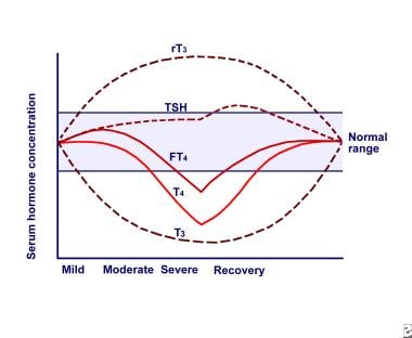
Euthyroid sick syndrome, also known as non-thyroidal illness syndrome (NTIS), is a condition characterized by abnormal thyroid function tests that occur in individuals with underlying non-thyroidal systemic illness. Despite the presence of abnormal test results, these individuals do not have evidence of clinical hypothyroidism or hyperthyroidism.
In euthyroid sick syndrome, the levels of triiodothyronine (T3) and thyroxine (T4) hormones may be decreased, while thyroid-stimulating hormone (TSH) levels remain normal or low. This is thought to occur due to alterations in the peripheral metabolism of thyroid hormones, rather than changes in the function of the thyroid gland itself.
The condition is often seen in individuals with severe illness, such as sepsis, cancer, malnutrition, or following major surgery. It is thought to represent an adaptive response to stress and illness, although the exact mechanisms are not fully understood. In most cases, euthyroid sick syndrome resolves on its own once the underlying illness has been treated.
Iodine is an essential trace element that is necessary for the production of thyroid hormones in the body. These hormones play crucial roles in various bodily functions, including growth and development, metabolism, and brain development during pregnancy and infancy. Iodine can be found in various foods such as seaweed, dairy products, and iodized salt. In a medical context, iodine is also used as an antiseptic to disinfect surfaces, wounds, and skin infections due to its ability to kill bacteria, viruses, and fungi.
Diiodothyronines are hormones that contain two iodine atoms and are produced by the thyroid gland. They are formed when thyroxine (T4), another thyroid hormone, is deiodinated. Diiodothyronines include T2 (3,5-diiodothyronine) and reverse T2 (3,3'-diiodothyronine). These hormones play a role in regulating metabolism and energy production in the body. However, their specific functions and mechanisms of action are not as well understood as those of thyroxine and triiodothyronine (T3), another important thyroid hormone.
Radioimmunoassay (RIA) is a highly sensitive analytical technique used in clinical and research laboratories to measure concentrations of various substances, such as hormones, vitamins, drugs, or tumor markers, in biological samples like blood, urine, or tissues. The method relies on the specific interaction between an antibody and its corresponding antigen, combined with the use of radioisotopes to quantify the amount of bound antigen.
In a typical RIA procedure, a known quantity of a radiolabeled antigen (also called tracer) is added to a sample containing an unknown concentration of the same unlabeled antigen. The mixture is then incubated with a specific antibody that binds to the antigen. During the incubation period, the antibody forms complexes with both the radiolabeled and unlabeled antigens.
After the incubation, the unbound (free) radiolabeled antigen is separated from the antibody-antigen complexes, usually through a precipitation or separation step involving centrifugation, filtration, or chromatography. The amount of radioactivity in the pellet (containing the antibody-antigen complexes) is then measured using a gamma counter or other suitable radiation detection device.
The concentration of the unlabeled antigen in the sample can be determined by comparing the ratio of bound to free radiolabeled antigen in the sample to a standard curve generated from known concentrations of unlabeled antigen and their corresponding bound/free ratios. The higher the concentration of unlabeled antigen in the sample, the lower the amount of radiolabeled antigen that will bind to the antibody, resulting in a lower bound/free ratio.
Radioimmunoassays offer high sensitivity, specificity, and accuracy, making them valuable tools for detecting and quantifying low levels of various substances in biological samples. However, due to concerns about radiation safety and waste disposal, alternative non-isotopic immunoassay techniques like enzyme-linked immunosorbent assays (ELISAs) have become more popular in recent years.
Methimazole is an anti-thyroid medication that is primarily used to treat hyperthyroidism, a condition in which the thyroid gland produces excessive amounts of thyroid hormones. It works by inhibiting the enzyme thyroperoxidase, which is essential for the production of thyroid hormones. By blocking this enzyme, methimazole reduces the amount of thyroid hormones produced by the thyroid gland, helping to restore normal thyroid function.
Methimazole is available in oral tablet form and is typically taken two to three times a day. Common side effects of methimazole include nausea, vomiting, skin rashes, and joint pain. In rare cases, it can cause more serious side effects such as liver damage or agranulocytosis (a severe decrease in white blood cell count).
It is important to note that methimazole should only be used under the close supervision of a healthcare provider, as regular monitoring of thyroid function and potential side effects is necessary. Additionally, it may take several weeks or months of treatment with methimazole before thyroid function returns to normal.
Antithyroid agents are a class of medications that are used to treat hyperthyroidism, a condition in which the thyroid gland produces too much thyroid hormone. These medications work by inhibiting the production of thyroid hormones in the thyroid gland. There are several types of antithyroid agents available, including:
1. Propylthiouracil (PTU): This medication works by blocking the enzyme that is needed to produce thyroid hormones. It also reduces the conversion of thyroxine (T4) to triiodothyronine (T3), another thyroid hormone, in peripheral tissues.
2. Methimazole: This medication works similarly to propylthiouracil by blocking the enzyme that is needed to produce thyroid hormones. However, it does not affect the conversion of T4 to T3 in peripheral tissues.
3. Carbimazole: This medication is converted to methimazole in the body and works similarly to block the production of thyroid hormones.
Antithyroid agents are usually taken orally, and their effects on thyroid hormone production begin within a few hours after ingestion. However, it may take several weeks for patients to notice an improvement in their symptoms. These medications can have side effects, including rash, hives, and joint pain. In rare cases, they can cause liver damage or agranulocytosis, a condition in which the body does not produce enough white blood cells.
It is important to note that antithyroid agents do not cure hyperthyroidism; they only treat the symptoms by reducing thyroid hormone production. Therefore, patients may need to take these medications for several months or even years, depending on their individual circumstances. In some cases, surgery or radioactive iodine therapy may be recommended as alternative treatments for hyperthyroidism.
Thyroid hormone receptors (THRs) are nuclear receptor proteins that bind to thyroid hormones and mediate their effects in the body. There are two main types of THRs, referred to as THRα and THRβ.
THRα is a subtype of thyroid hormone receptor that is primarily expressed in tissues such as the heart, skeletal muscle, and brown adipose tissue. It plays an important role in regulating metabolism, growth, and development in these tissues. THRα has two subtypes, THRα1 and THRα2, which have different functions and are expressed in different tissues.
THRα1 is the predominant form of THRα and is found in many tissues, including the heart, skeletal muscle, and brown adipose tissue. It regulates genes involved in metabolism, growth, and development, and has been shown to play a role in regulating heart rate and contractility.
THRα2, on the other hand, is primarily expressed in the brain and pituitary gland, where it regulates the production of thyroid-stimulating hormone (TSH). THRα2 is unable to bind to thyroid hormones, but can form heterodimers with THRα1 or THRβ1, which allows it to modulate their activity.
Overall, THRα plays an important role in regulating various physiological processes in the body, and dysregulation of THRα function has been implicated in a number of diseases, including heart disease, muscle wasting, and neurological disorders.
The liver is a large, solid organ located in the upper right portion of the abdomen, beneath the diaphragm and above the stomach. It plays a vital role in several bodily functions, including:
1. Metabolism: The liver helps to metabolize carbohydrates, fats, and proteins from the food we eat into energy and nutrients that our bodies can use.
2. Detoxification: The liver detoxifies harmful substances in the body by breaking them down into less toxic forms or excreting them through bile.
3. Synthesis: The liver synthesizes important proteins, such as albumin and clotting factors, that are necessary for proper bodily function.
4. Storage: The liver stores glucose, vitamins, and minerals that can be released when the body needs them.
5. Bile production: The liver produces bile, a digestive juice that helps to break down fats in the small intestine.
6. Immune function: The liver plays a role in the immune system by filtering out bacteria and other harmful substances from the blood.
Overall, the liver is an essential organ that plays a critical role in maintaining overall health and well-being.
Iopanoic acid is a contrast medium, specifically a radiocontrast agent, that is used during imaging examinations such as X-rays and CT scans to help improve the visibility of internal body structures. It works by blocking the absorption of X-rays in the digestive tract, making it possible to visualize the gastrointestinal tract more clearly on imaging studies. Iopanoic acid is typically given orally before the examination.
It's important to note that the use of iopanoic acid and other radiocontrast agents should be carefully weighed against the potential risks, as they can cause allergic reactions, kidney damage, and other complications in some individuals. Therefore, it is usually reserved for situations where the benefits of improved imaging outweigh these potential risks.
Serum globulins are a group of proteins present in the liquid portion of blood, known as serum. They are produced by the immune system in response to foreign substances such as bacteria, viruses, and allergens. Serum globulins include several types of immunoglobulins (antibodies), complement components, and other proteins involved in the immune response.
The serum globulin level is often measured as part of a complete blood count (CBC) or a protein electrophoresis test. An elevated serum globulin level may indicate an ongoing infection, inflammation, or an autoimmune disorder. Conversely, a decreased level may suggest a liver or kidney disease, or a malnutrition condition. It is important to note that the interpretation of serum globulin levels should be done in conjunction with other laboratory and clinical findings.
Myxedema is not a term used in modern medicine to describe a specific medical condition. However, historically, it was used to refer to the severe form of hypothyroidism, a condition characterized by an underactive thyroid gland that doesn't produce enough thyroid hormones. In hypothyroidism, various body functions slow down, which can lead to symptoms such as fatigue, weight gain, cold intolerance, constipation, and dry skin.
Myxedema specifically refers to the physical signs of severe hypothyroidism, including swelling (edema) and thickening of the skin, particularly around the face, hands, and feet, as well as a puffy appearance of the face. The term myxedema coma was used to describe a rare but life-threatening complication of long-standing, untreated hypothyroidism, characterized by altered mental status, hypothermia, and other systemic manifestations.
Nowadays, healthcare professionals use more precise medical terminology to describe these conditions, such as hypothyroidism or myxedematous edema, rather than the outdated term myxedema.
Iodine isotopes are different forms of the chemical element iodine, which have different numbers of neutrons in their nuclei. Iodine has a total of 53 protons in its nucleus, and its stable isotope, iodine-127, has 74 neutrons, giving it a mass number of 127. However, there are also radioactive isotopes of iodine, which have different numbers of neutrons and are therefore unstable.
Radioactive isotopes of iodine emit radiation as they decay towards a stable state. For example, iodine-131 is a commonly used isotope in medical imaging and therapy, with a half-life of about 8 days. It decays by emitting beta particles and gamma rays, making it useful for treating thyroid cancer and other conditions that involve overactive thyroid glands.
Other radioactive iodine isotopes include iodine-123, which has a half-life of about 13 hours and is used in medical imaging, and iodine-125, which has a half-life of about 60 days and is used in brachytherapy (a type of radiation therapy that involves placing radioactive sources directly into or near tumors).
It's important to note that exposure to radioactive iodine isotopes can be harmful, especially if it occurs through inhalation or ingestion. This is because the iodine can accumulate in the thyroid gland and cause damage over time. Therefore, appropriate safety measures must be taken when handling or working with radioactive iodine isotopes.
Thyroid hormone receptors (THRs) are nuclear receptor proteins that bind to thyroid hormones and mediate their effects in target cells. There are two main types of THRs, referred to as THR alpha and THR beta. THR beta is further divided into two subtypes, THR beta1 and THR beta2.
THR beta is a type of nuclear receptor that is primarily expressed in the liver, kidney, and heart, as well as in the central nervous system. It plays an important role in regulating the metabolism of carbohydrates, lipids, and proteins, as well as in the development and function of the heart. THR beta is also involved in the regulation of body weight and energy expenditure.
THR beta1 is the predominant subtype expressed in the liver and is responsible for many of the metabolic effects of thyroid hormones in this organ. THR beta2, on the other hand, is primarily expressed in the heart and plays a role in regulating cardiac function.
Abnormalities in THR beta function can lead to various diseases, including thyroid hormone resistance, a condition in which the body's cells are unable to respond properly to thyroid hormones. This can result in symptoms such as weight gain, fatigue, and cold intolerance.
Malate Dehydrogenase (MDH) is an enzyme that plays a crucial role in the Krebs cycle, also known as the citric acid cycle or tricarboxylic acid (TCA) cycle. It catalyzes the reversible oxidation of malate to oxaloacetate, while simultaneously reducing NAD+ to NADH. This reaction is essential for energy production in the form of ATP and NADH within the cell.
There are two main types of Malate Dehydrogenase:
1. NAD-dependent Malate Dehydrogenase (MDH1): Found primarily in the cytoplasm, this isoform plays a role in the malate-aspartate shuttle, which helps transfer reducing equivalents between the cytoplasm and mitochondria.
2. FAD-dependent Malate Dehydrogenase (MDH2): Located within the mitochondrial matrix, this isoform is involved in the Krebs cycle for energy production.
Abnormal levels of Malate Dehydrogenase enzyme can be indicative of certain medical conditions or diseases, such as myocardial infarction (heart attack), muscle damage, or various types of cancer. Therefore, MDH enzyme activity is often assessed in diagnostic tests to help identify and monitor these health issues.
Dialysis is a medical treatment that is used to remove waste and excess fluid from the blood when the kidneys are no longer able to perform these functions effectively. This life-sustaining procedure uses a specialized machine, called a dialyzer or artificial kidney, to filter the blood outside of the body and return clean, chemically balanced blood back into the body.
There are two main types of dialysis: hemodialysis and peritoneal dialysis.
1. Hemodialysis: In this method, a patient's blood is passed through an external filter (dialyzer) that removes waste products, toxins, and excess fluids. The cleaned blood is then returned to the body with the help of a specialized machine. Hemodialysis typically requires access to a large vein, often created by a surgical procedure called an arteriovenous (AV) fistula or graft. Hemodialysis sessions usually last for about 3-5 hours and are performed three times a week in a clinical setting, such as a dialysis center or hospital.
2. Peritoneal Dialysis: This method uses the lining of the patient's own abdomen (peritoneum) as a natural filter to clean the blood. A sterile dialysate solution is introduced into the peritoneal cavity via a permanently implanted catheter. The solution absorbs waste products and excess fluids from the blood vessels lining the peritoneum through a process called diffusion. After a dwell time, usually several hours, the used dialysate is drained out and replaced with fresh dialysate. This process is known as an exchange and is typically repeated multiple times throughout the day or night, depending on the specific type of peritoneal dialysis (continuous ambulatory peritoneal dialysis or automated peritoneal dialysis).
Both methods have their advantages and disadvantages, and the choice between them depends on various factors, such as a patient's overall health, lifestyle, and personal preferences. Dialysis is a life-saving treatment for people with end-stage kidney disease or severe kidney dysfunction, allowing them to maintain their quality of life and extend their lifespan until a kidney transplant becomes available or their kidney function improves.
Thyroglobulin is a protein produced and used by the thyroid gland in the production of thyroid hormones, primarily thyroxine (T4) and triiodothyronine (T3). It is composed of two subunits, an alpha and a beta or gamma unit, which bind iodine atoms necessary for the synthesis of the thyroid hormones. Thyroglobulin is exclusively produced by the follicular cells of the thyroid gland.
In clinical practice, measuring thyroglobulin levels in the blood can be useful as a tumor marker for monitoring treatment and detecting recurrence of thyroid cancer, particularly in patients with differentiated thyroid cancer (papillary or follicular) who have had their thyroid gland removed. However, it is important to note that thyroglobulin is not specific to thyroid tissue and can be produced by some non-thyroidal cells under certain conditions, which may lead to false positive results in some cases.
Graves' disease is defined as an autoimmune disorder that leads to overactivity of the thyroid gland (hyperthyroidism). It results when the immune system produces antibodies that stimulate the thyroid gland, causing it to produce too much thyroid hormone. This can result in a variety of symptoms such as rapid heartbeat, weight loss, heat intolerance, and bulging eyes (Graves' ophthalmopathy). The exact cause of Graves' disease is unknown, but it is more common in women and people with a family history of the disorder. Treatment may include medications to control hyperthyroidism, radioactive iodine therapy to destroy thyroid tissue, or surgery to remove the thyroid gland.
Thyronines are a type of hormone that is produced and released by the thyroid gland. They are iodinated amino acids, specifically triiodothyronine (T3) and thyroxine (T4), that are essential for regulating the body's metabolic rate, growth, and development. These hormones play a crucial role in maintaining the body's energy balance, brain development, and overall health. They work by binding to specific receptors in cells throughout the body, where they help to regulate gene expression and various cellular processes. Disorders of thyronine production or function can lead to a variety of medical conditions, such as hypothyroidism or hyperthyroidism.
Iodine radioisotopes are radioactive isotopes of the element iodine, which decays and emits radiation in the form of gamma rays. Some commonly used iodine radioisotopes include I-123, I-125, I-131. These radioisotopes have various medical applications such as in diagnostic imaging, therapy for thyroid disorders, and cancer treatment.
For example, I-131 is commonly used to treat hyperthyroidism and differentiated thyroid cancer due to its ability to destroy thyroid tissue. On the other hand, I-123 is often used in nuclear medicine scans of the thyroid gland because it emits gamma rays that can be detected by a gamma camera, allowing for detailed images of the gland's structure and function.
It is important to note that handling and administering radioisotopes require specialized training and safety precautions due to their radiation-emitting properties.
Growth Hormone (GH), also known as somatotropin, is a peptide hormone secreted by the somatotroph cells in the anterior pituitary gland. It plays a crucial role in regulating growth, cell reproduction, and regeneration by stimulating the production of another hormone called insulin-like growth factor 1 (IGF-1) in the liver and other tissues. GH also has important metabolic functions, such as increasing glucose levels, enhancing protein synthesis, and reducing fat storage. Its secretion is regulated by two hypothalamic hormones: growth hormone-releasing hormone (GHRH), which stimulates its release, and somatostatin (SRIF), which inhibits its release. Abnormal levels of GH can lead to various medical conditions, such as dwarfism or gigantism if there are deficiencies or excesses, respectively.
Glycerol-3-phosphate dehydrogenase (GPD) is an enzyme that plays a crucial role in the metabolism of glucose and lipids. It catalyzes the conversion of dihydroxyacetone phosphate (DHAP) to glycerol-3-phosphate (G3P), which is a key intermediate in the synthesis of triglycerides, phospholipids, and other glycerophospholipids.
There are two main forms of GPD: a cytoplasmic form (GPD1) and a mitochondrial form (GPD2). The cytoplasmic form is involved in the production of NADH, which is used in various metabolic processes, while the mitochondrial form is involved in the production of ATP, the main energy currency of the cell.
Deficiencies or mutations in GPD can lead to a variety of metabolic disorders, including glycerol kinase deficiency and congenital muscular dystrophy. Elevated levels of GPD have been observed in certain types of cancer, suggesting that it may play a role in tumor growth and progression.
Reagent kits, diagnostic are prepackaged sets of chemical reagents and other components designed for performing specific diagnostic tests or assays. These kits are often used in clinical laboratories to detect and measure the presence or absence of various biomarkers, such as proteins, antibodies, antigens, nucleic acids, or small molecules, in biological samples like blood, urine, or tissues.
Diagnostic reagent kits typically contain detailed instructions for their use, along with the necessary reagents, controls, and sometimes specialized equipment or supplies. They are designed to simplify the testing process, reduce human error, and increase standardization, ensuring accurate and reliable results. Examples of diagnostic reagent kits include those used for pregnancy tests, infectious disease screening, drug testing, genetic testing, and cancer biomarker detection.
Hormones are defined as chemical messengers that are produced by endocrine glands or specialized cells and are transported through the bloodstream to tissues and organs, where they elicit specific responses. They play crucial roles in regulating various physiological processes such as growth, development, metabolism, reproduction, and mood. Examples of hormones include insulin, estrogen, testosterone, adrenaline, and thyroxine.
The pituitary gland is a small, endocrine gland located at the base of the brain, in the sella turcica of the sphenoid bone. It is often called the "master gland" because it controls other glands and makes the hormones that trigger many body functions. The pituitary gland measures about 0.5 cm in height and 1 cm in width, and it weighs approximately 0.5 grams.
The pituitary gland is divided into two main parts: the anterior lobe (adenohypophysis) and the posterior lobe (neurohypophysis). The anterior lobe is further divided into three zones: the pars distalis, pars intermedia, and pars tuberalis. Each part of the pituitary gland has distinct functions and produces different hormones.
The anterior pituitary gland produces and releases several important hormones, including:
* Growth hormone (GH), which regulates growth and development in children and helps maintain muscle mass and bone strength in adults.
* Thyroid-stimulating hormone (TSH), which controls the production of thyroid hormones by the thyroid gland.
* Adrenocorticotropic hormone (ACTH), which stimulates the adrenal glands to produce cortisol and other steroid hormones.
* Follicle-stimulating hormone (FSH) and luteinizing hormone (LH), which regulate reproductive function in both males and females.
* Prolactin, which stimulates milk production in pregnant and lactating women.
The posterior pituitary gland stores and releases two hormones that are produced by the hypothalamus:
* Antidiuretic hormone (ADH), which helps regulate water balance in the body by controlling urine production.
* Oxytocin, which stimulates uterine contractions during childbirth and milk release during breastfeeding.
Overall, the pituitary gland plays a critical role in maintaining homeostasis and regulating various bodily functions, including growth, development, metabolism, and reproductive function.
Hydrocortisone is a synthetic glucocorticoid, which is a class of steroid hormones. It is identical to the naturally occurring cortisol, a hormone produced by the adrenal gland that helps regulate metabolism and helps your body respond to stress. Hydrocortisone has anti-inflammatory effects and is used to treat various inflammatory conditions such as allergies, skin disorders, and autoimmune diseases. It works by suppressing the immune system's response to reduce swelling, redness, itching, and other symptoms caused by inflammation.
Hydrocortisone is available in different forms, including oral tablets, topical creams, lotions, gels, and ointments, as well as injectable solutions. The specific use and dosage depend on the condition being treated and the individual patient's medical history and current health status.
As with any medication, hydrocortisone can have side effects, especially when used in high doses or for extended periods. Common side effects include increased appetite, weight gain, mood changes, insomnia, and skin thinning. Long-term use of hydrocortisone may also increase the risk of developing osteoporosis, diabetes, cataracts, and other health problems. Therefore, it is essential to follow your healthcare provider's instructions carefully when using this medication.
Iodides are chemical compounds that contain iodine in the form of an iodide ion (I-). Iodide ions are negatively charged ions that consist of one iodine atom and an extra electron. Iodides are commonly found in dietary supplements and medications, and they are often used to treat or prevent iodine deficiency. They can also be used as expectorants to help thin and loosen mucus in the respiratory tract. Examples of iodides include potassium iodide (KI) and sodium iodide (NaI).
"Inbred strains of rats" are genetically identical rodents that have been produced through many generations of brother-sister mating. This results in a high degree of homozygosity, where the genes at any particular locus in the genome are identical in all members of the strain.
Inbred strains of rats are widely used in biomedical research because they provide a consistent and reproducible genetic background for studying various biological phenomena, including the effects of drugs, environmental factors, and genetic mutations on health and disease. Additionally, inbred strains can be used to create genetically modified models of human diseases by introducing specific mutations into their genomes.
Some commonly used inbred strains of rats include the Wistar Kyoto (WKY), Sprague-Dawley (SD), and Fischer 344 (F344) rat strains. Each strain has its own unique genetic characteristics, making them suitable for different types of research.
Thyrotoxicosis is a medical condition that results from an excess of thyroid hormones in the body, leading to an overactive metabolic state. It can be caused by various factors such as Graves' disease, toxic adenoma, Plummer's disease, or excessive intake of thyroid hormone medication. Symptoms may include rapid heart rate, weight loss, heat intolerance, tremors, and increased sweating, among others. Thyrotoxicosis is not a diagnosis itself but a manifestation of various underlying thyroid disorders. Proper diagnosis and management are crucial to prevent complications and improve quality of life.
Talc is a mineral composed of hydrated magnesium silicate with the chemical formula H2Mg3(SiO3)4 or Mg3Si4O10(OH)2. It is widely used in various industries including pharmaceuticals and cosmetics due to its softness, lubricity, and ability to absorb moisture. In medical contexts, talc is often found in powdered products used for personal hygiene or as a drying agent in medical dressings. However, it should be noted that the use of talcum powder in the genital area has been linked to an increased risk of ovarian cancer, although the overall evidence remains controversial.
Adipose tissue, brown, also known as brown adipose tissue (BAT), is a type of fat in mammals that plays a crucial role in non-shivering thermogenesis, which is the process of generating heat and maintaining body temperature through the burning of calories. Unlike white adipose tissue, which primarily stores energy in the form of lipids, brown adipose tissue contains numerous mitochondria rich in iron, giving it a brown appearance. These mitochondria contain a protein called uncoupling protein 1 (UCP1), which allows for the efficient conversion of stored energy into heat rather than ATP production.
Brown adipose tissue is typically found in newborns and hibernating animals, but recent studies have shown that adults also possess functional brown adipose tissue, particularly around the neck, shoulders, and spine. The activation of brown adipose tissue has been suggested as a potential strategy for combating obesity and related metabolic disorders due to its ability to burn calories and increase energy expenditure. However, further research is needed to fully understand the mechanisms underlying brown adipose tissue function and its therapeutic potential in treating these conditions.
Goiter is a medical term that refers to an enlarged thyroid gland. The thyroid gland is a small, butterfly-shaped gland located in the front of your neck below the larynx or voice box. It produces hormones that regulate your body's metabolism, growth, and development.
Goiter can vary in size and may be visible as a swelling at the base of the neck. It can be caused by several factors, including iodine deficiency, autoimmune disorders, thyroid cancer, pregnancy, or the use of certain medications. Depending on the underlying cause and the severity of the goiter, treatment options may include medication, surgery, or radioactive iodine therapy.
Messenger RNA (mRNA) is a type of RNA (ribonucleic acid) that carries genetic information copied from DNA in the form of a series of three-base code "words," each of which specifies a particular amino acid. This information is used by the cell's machinery to construct proteins, a process known as translation. After being transcribed from DNA, mRNA travels out of the nucleus to the ribosomes in the cytoplasm where protein synthesis occurs. Once the protein has been synthesized, the mRNA may be degraded and recycled. Post-transcriptional modifications can also occur to mRNA, such as alternative splicing and addition of a 5' cap and a poly(A) tail, which can affect its stability, localization, and translation efficiency.
Organ size refers to the volume or physical measurement of an organ in the body of an individual. It can be described in terms of length, width, and height or by using specialized techniques such as imaging studies (like CT scans or MRIs) to determine the volume. The size of an organ can vary depending on factors such as age, sex, body size, and overall health status. Changes in organ size may indicate various medical conditions, including growths, inflammation, or atrophy.
Body weight is the measure of the force exerted on a scale or balance by an object's mass, most commonly expressed in units such as pounds (lb) or kilograms (kg). In the context of medical definitions, body weight typically refers to an individual's total weight, which includes their skeletal muscle, fat, organs, and bodily fluids.
Healthcare professionals often use body weight as a basic indicator of overall health status, as it can provide insights into various aspects of a person's health, such as nutritional status, metabolic function, and risk factors for certain diseases. For example, being significantly underweight or overweight can increase the risk of developing conditions like malnutrition, diabetes, heart disease, and certain types of cancer.
It is important to note that body weight alone may not provide a complete picture of an individual's health, as it does not account for factors such as muscle mass, bone density, or body composition. Therefore, healthcare professionals often use additional measures, such as body mass index (BMI), waist circumference, and blood tests, to assess overall health status more comprehensively.
Thyroxine-binding globulin (TBG) is a glycoprotein found in human plasma that has a high affinity for binding thyroid hormones, specifically Thyroxine (T4) and Triiodothyronine (T3). It is produced by the liver and plays a crucial role in maintaining the balance of these hormones in the body. TBG binds to approximately 70-80% of circulating T4 and about 55% of circulating T3, acting as a transport protein that carries these hormones throughout the body. The amount of TBG in the blood can vary due to factors such as genetics, sex hormones, and certain medications, which can affect the levels of free (unbound) thyroid hormones and contribute to various thyroid-related disorders.
Basal metabolism, also known as basal metabolic rate (BMR) or resting metabolic rate (RMR), is the amount of energy expended by an organism at rest, in a neutrally temperate environment, while in the post-absorptive state. It is the minimum amount of energy required to maintain basic bodily functions such as breathing, heartbeat, and maintenance of body temperature.
The BMR is typically measured in units of energy per unit time, such as kilocalories per day (kcal/day) or watts (W). In humans, the BMR is usually around 10-15% of a person's total daily energy expenditure. It can vary depending on factors such as age, sex, body size and composition, and genetics.
The BMR can be measured in a variety of ways, including direct calorimetry, indirect calorimetry, or by using predictive equations based on factors such as age, weight, and height. It is an important concept in the study of energy balance, nutrition, and metabolism.
Pregnancy is a physiological state or condition where a fertilized egg (zygote) successfully implants and grows in the uterus of a woman, leading to the development of an embryo and finally a fetus. This process typically spans approximately 40 weeks, divided into three trimesters, and culminates in childbirth. Throughout this period, numerous hormonal and physical changes occur to support the growing offspring, including uterine enlargement, breast development, and various maternal adaptations to ensure the fetus's optimal growth and well-being.
Prolactin is a hormone produced by the pituitary gland, a small gland located at the base of the brain. Its primary function is to stimulate milk production in women after childbirth, a process known as lactation. However, prolactin also plays other roles in the body, including regulating immune responses, metabolism, and behavior. In men, prolactin helps maintain the sexual glands and contributes to paternal behaviors.
Prolactin levels are usually low in both men and non-pregnant women but increase significantly during pregnancy and after childbirth. Various factors can affect prolactin levels, including stress, sleep, exercise, and certain medications. High prolactin levels can lead to medical conditions such as amenorrhea (absence of menstruation), galactorrhea (spontaneous milk production not related to childbirth), infertility, and reduced sexual desire in both men and women.
Insulin is a hormone produced by the beta cells of the pancreatic islets, primarily in response to elevated levels of glucose in the circulating blood. It plays a crucial role in regulating blood glucose levels and facilitating the uptake and utilization of glucose by peripheral tissues, such as muscle and adipose tissue, for energy production and storage. Insulin also inhibits glucose production in the liver and promotes the storage of excess glucose as glycogen or triglycerides.
Deficiency in insulin secretion or action leads to impaired glucose regulation and can result in conditions such as diabetes mellitus, characterized by chronic hyperglycemia and associated complications. Exogenous insulin is used as a replacement therapy in individuals with diabetes to help manage their blood glucose levels and prevent long-term complications.
Amiodarone is a Class III antiarrhythmic medication used to treat and prevent various types of irregular heart rhythms (arrhythmias). It works by stabilizing the electrical activity of the heart and slowing down the nerve impulses in the heart tissue. Amiodarone is available in oral tablet and injection forms.
The medical definition of 'Amiodarone' is:
A benzofuran derivative with Class III antiarrhythmic properties, used for the treatment of ventricular arrhythmias. It has a relatively slow onset of action and is therefore not useful in acute situations. Additionally, it has negative inotropic effects and may exacerbate heart failure. The most serious adverse effect is pulmonary fibrosis, which occurs in approximately 1-2% of patients. Other important side effects include corneal microdeposits, hepatotoxicity, thyroid dysfunction, and photosensitivity. Amiodarone has a very long half-life (approximately 50 days) due to its extensive tissue distribution. It is metabolized by the liver and excreted in bile and urine.
Sources:
1. UpToDate - Amiodarone use in adults: Indications, dosing, and adverse effects.
2. Micromedex - Amiodarone.
3. Drugs.com - Amiodarone.
In the context of medicine and pharmacology, "kinetics" refers to the study of how a drug moves throughout the body, including its absorption, distribution, metabolism, and excretion (often abbreviated as ADME). This field is called "pharmacokinetics."
1. Absorption: This is the process of a drug moving from its site of administration into the bloodstream. Factors such as the route of administration (e.g., oral, intravenous, etc.), formulation, and individual physiological differences can affect absorption.
2. Distribution: Once a drug is in the bloodstream, it gets distributed throughout the body to various tissues and organs. This process is influenced by factors like blood flow, protein binding, and lipid solubility of the drug.
3. Metabolism: Drugs are often chemically modified in the body, typically in the liver, through processes known as metabolism. These changes can lead to the formation of active or inactive metabolites, which may then be further distributed, excreted, or undergo additional metabolic transformations.
4. Excretion: This is the process by which drugs and their metabolites are eliminated from the body, primarily through the kidneys (urine) and the liver (bile).
Understanding the kinetics of a drug is crucial for determining its optimal dosing regimen, potential interactions with other medications or foods, and any necessary adjustments for special populations like pediatric or geriatric patients, or those with impaired renal or hepatic function.
An immunoassay is a biochemical test that measures the presence or concentration of a specific protein, antibody, or antigen in a sample using the principles of antibody-antigen reactions. It is commonly used in clinical laboratories to diagnose and monitor various medical conditions such as infections, hormonal disorders, allergies, and cancer.
Immunoassays typically involve the use of labeled reagents, such as enzymes, radioisotopes, or fluorescent dyes, that bind specifically to the target molecule. The amount of label detected is proportional to the concentration of the target molecule in the sample, allowing for quantitative analysis.
There are several types of immunoassays, including enzyme-linked immunosorbent assay (ELISA), radioimmunoassay (RIA), fluorescence immunoassay (FIA), and chemiluminescent immunoassay (CLIA). Each type has its own advantages and limitations, depending on the sensitivity, specificity, and throughput required for a particular application.
Prealbumin, also known as transthyretin, is a protein produced primarily in the liver and circulates in the blood. It plays a role in transporting thyroid hormones and vitamin A throughout the body. Prealbumin levels are often used as an indicator of nutritional status and liver function. Low prealbumin levels may suggest malnutrition or inflammation, while increased levels can be seen in certain conditions like hyperthyroidism. It is important to note that prealbumin levels should be interpreted in conjunction with other clinical findings and laboratory tests for a more accurate assessment of a patient's health status.
Reference values, also known as reference ranges or reference intervals, are the set of values that are considered normal or typical for a particular population or group of people. These values are often used in laboratory tests to help interpret test results and determine whether a patient's value falls within the expected range.
The process of establishing reference values typically involves measuring a particular biomarker or parameter in a large, healthy population and then calculating the mean and standard deviation of the measurements. Based on these statistics, a range is established that includes a certain percentage of the population (often 95%) and excludes extreme outliers.
It's important to note that reference values can vary depending on factors such as age, sex, race, and other demographic characteristics. Therefore, it's essential to use reference values that are specific to the relevant population when interpreting laboratory test results. Additionally, reference values may change over time due to advances in measurement technology or changes in the population being studied.
"Wistar rats" are a strain of albino rats that are widely used in laboratory research. They were developed at the Wistar Institute in Philadelphia, USA, and were first introduced in 1906. Wistar rats are outbred, which means that they are genetically diverse and do not have a fixed set of genetic characteristics like inbred strains.
Wistar rats are commonly used as animal models in biomedical research because of their size, ease of handling, and relatively low cost. They are used in a wide range of research areas, including toxicology, pharmacology, nutrition, cancer, cardiovascular disease, and behavioral studies. Wistar rats are also used in safety testing of drugs, medical devices, and other products.
Wistar rats are typically larger than many other rat strains, with males weighing between 500-700 grams and females weighing between 250-350 grams. They have a lifespan of approximately 2-3 years. Wistar rats are also known for their docile and friendly nature, making them easy to handle and work with in the laboratory setting.
In the field of medicine, "time factors" refer to the duration of symptoms or time elapsed since the onset of a medical condition, which can have significant implications for diagnosis and treatment. Understanding time factors is crucial in determining the progression of a disease, evaluating the effectiveness of treatments, and making critical decisions regarding patient care.
For example, in stroke management, "time is brain," meaning that rapid intervention within a specific time frame (usually within 4.5 hours) is essential to administering tissue plasminogen activator (tPA), a clot-busting drug that can minimize brain damage and improve patient outcomes. Similarly, in trauma care, the "golden hour" concept emphasizes the importance of providing definitive care within the first 60 minutes after injury to increase survival rates and reduce morbidity.
Time factors also play a role in monitoring the progression of chronic conditions like diabetes or heart disease, where regular follow-ups and assessments help determine appropriate treatment adjustments and prevent complications. In infectious diseases, time factors are crucial for initiating antibiotic therapy and identifying potential outbreaks to control their spread.
Overall, "time factors" encompass the significance of recognizing and acting promptly in various medical scenarios to optimize patient outcomes and provide effective care.
Ergotamine is a type of ergopeptine alkaloid, derived from the ergot fungus (Claviceps purpurea) that parasitizes certain grains, particularly rye. It is a potent vasoconstrictor and has been used medically to prevent migraines and treat cluster headaches, as well as for other uses such as controlling postpartum hemorrhage and reducing symptoms of orthostatic hypotension.
Ergotamine works by binding to serotonin receptors in the brain and causing vasoconstriction of cranial blood vessels, which can help to relieve migraine headaches. However, it can also cause serious side effects such as nausea, vomiting, muscle pain, numbness or tingling in the extremities, and in rare cases, more severe reactions such as ergotism, a condition characterized by vasoconstriction of peripheral blood vessels leading to gangrene.
Ergotamine is usually taken orally, but can also be administered rectally or by inhalation. It is important to follow the dosage instructions carefully and avoid taking excessive amounts, as this can increase the risk of serious side effects. Ergotamine should not be taken during pregnancy or while breastfeeding, and it may interact with other medications, so it is important to inform your healthcare provider of all medications you are taking before starting ergotamine therapy.
In the context of medical research, "methods" refers to the specific procedures or techniques used in conducting a study or experiment. This includes details on how data was collected, what measurements were taken, and what statistical analyses were performed. The methods section of a medical paper allows other researchers to replicate the study if they choose to do so. It is considered one of the key components of a well-written research article, as it provides transparency and helps establish the validity of the findings.
Congenital hypothyroidism is a medical condition characterized by the partial or complete absence of thyroid hormone production in the baby's body at birth. The thyroid gland, which is located in the front of the neck, produces hormones that are essential for normal growth and development of the brain and body.
Congenital hypothyroidism can occur due to various reasons such as the absence or abnormal development of the thyroid gland, or a defect in the production or regulation of thyroid hormones. In some cases, it may be caused by genetic mutations that affect the development or function of the thyroid gland.
If left untreated, congenital hypothyroidism can lead to mental and physical retardation, growth problems, and other health issues. Therefore, it is important to diagnose and treat this condition as early as possible, usually within the first few weeks of life. Treatment typically involves replacing the missing thyroid hormones with synthetic medications, which are safe and effective when administered under a doctor's supervision.
Clinical chemistry tests are a type of laboratory test that measure the levels of various chemicals or substances in the body. These tests can be used to help diagnose and monitor a wide range of medical conditions, including diabetes, liver disease, heart disease, and kidney disease. Some common clinical chemistry tests include:
1. Blood glucose test: Measures the level of glucose (sugar) in the blood. This test is commonly used to diagnose and monitor diabetes.
2. Electrolyte panel: Measures the levels of important electrolytes such as sodium, potassium, chloride, and bicarbonate in the blood. Imbalances in these electrolytes can indicate a variety of medical conditions.
3. Liver function tests (LFTs): Measure the levels of various enzymes and proteins produced by the liver. Abnormal results can indicate liver damage or disease.
4. Kidney function tests: Measure the levels of various substances such as creatinine and blood urea nitrogen (BUN) in the blood. Elevated levels of these substances can indicate kidney dysfunction or disease.
5. Lipid panel: Measures the levels of different types of cholesterol and triglycerides in the blood. Abnormal results can indicate an increased risk of heart disease.
6. Thyroid function tests: Measure the levels of hormones produced by the thyroid gland. Abnormal results can indicate thyroid dysfunction or disease.
Clinical chemistry tests are usually performed on a sample of blood, urine, or other bodily fluid. The results of these tests can provide important information to help doctors diagnose and manage medical conditions.
Pregnenolone carbonitrile is not a recognized or established medical term or substance. It's possible that there may be some confusion with the term "pregnenolone," which is a steroid hormone produced in the body. Pregnenolone is a precursor to other steroid hormones, including progesterone, cortisol, and the sex hormones (estrogens, androgens).
Carbonitrile is an organic functional group consisting of a cyano group (-CN) attached to a carbon atom. It's not typically associated with pregnenolone or any other steroid hormones in medical or scientific contexts.
Therefore, I cannot provide a medical definition for 'Pregnenolone Carbonitrile,' as it is not a recognized term in the field of medicine or biology.
Food deprivation is not a medical term per se, but it is used in the field of nutrition and psychology. It generally refers to the deliberate withholding of food for a prolonged period, leading to a state of undernutrition or malnutrition. This can occur due to various reasons such as famine, starvation, anorexia nervosa, or as a result of certain medical treatments or conditions. Prolonged food deprivation can have serious consequences on physical health, including weight loss, muscle wasting, organ damage, and decreased immune function, as well as psychological effects such as depression, anxiety, and cognitive impairment.
Radio-iodinated serum albumin refers to human serum albumin that has been chemically bonded with radioactive iodine isotopes, typically I-125 or I-131. This results in a radiolabeled protein that can be used in medical imaging and research to track the distribution and movement of the protein in the body.
In human physiology, serum albumin is the most abundant protein in plasma, synthesized by the liver, and it plays a crucial role in maintaining oncotic pressure and transporting various molecules in the bloodstream. Radio-iodination of serum albumin allows for non-invasive monitoring of its behavior in vivo, which can be useful in evaluating conditions such as protein losing enteropathies, nephrotic syndrome, or liver dysfunction.
It is essential to handle and dispose of radio-iodinated serum albumin with proper radiation safety protocols due to its radioactive nature.
Selenium is a trace element that is essential for the proper functioning of the human body. According to the medical definitions provided by the National Institutes of Health (NIH), selenium is a component of several major metabolic pathways, including thyroid hormone metabolism, antioxidant defense systems, and immune function.
Selenium is found in a variety of foods, including nuts (particularly Brazil nuts), cereals, fish, and meat. It exists in several forms, with selenomethionine being the most common form found in food. Other forms include selenocysteine, which is incorporated into proteins, and selenite and selenate, which are inorganic forms of selenium.
The recommended dietary allowance (RDA) for selenium is 55 micrograms per day for adults. While selenium deficiency is rare, chronic selenium deficiency can lead to conditions such as Keshan disease, a type of cardiomyopathy, and Kaschin-Beck disease, which affects the bones and joints.
It's important to note that while selenium is essential for health, excessive intake can be harmful. High levels of selenium can cause symptoms such as nausea, vomiting, hair loss, and neurological damage. The tolerable upper intake level (UL) for selenium is 400 micrograms per day for adults.
Glucagon is a hormone produced by the alpha cells of the pancreas. Its main function is to regulate glucose levels in the blood by stimulating the liver to convert stored glycogen into glucose, which can then be released into the bloodstream. This process helps to raise blood sugar levels when they are too low, such as during hypoglycemia.
Glucagon is a 29-amino acid polypeptide that is derived from the preproglucagon protein. It works by binding to glucagon receptors on liver cells, which triggers a series of intracellular signaling events that lead to the activation of enzymes involved in glycogen breakdown.
In addition to its role in glucose regulation, glucagon has also been shown to have other physiological effects, such as promoting lipolysis (the breakdown of fat) and inhibiting gastric acid secretion. Glucagon is often used clinically in the treatment of hypoglycemia, as well as in diagnostic tests to assess pancreatic function.
Sprague-Dawley rats are a strain of albino laboratory rats that are widely used in scientific research. They were first developed by researchers H.H. Sprague and R.C. Dawley in the early 20th century, and have since become one of the most commonly used rat strains in biomedical research due to their relatively large size, ease of handling, and consistent genetic background.
Sprague-Dawley rats are outbred, which means that they are genetically diverse and do not suffer from the same limitations as inbred strains, which can have reduced fertility and increased susceptibility to certain diseases. They are also characterized by their docile nature and low levels of aggression, making them easier to handle and study than some other rat strains.
These rats are used in a wide variety of research areas, including toxicology, pharmacology, nutrition, cancer, and behavioral studies. Because they are genetically diverse, Sprague-Dawley rats can be used to model a range of human diseases and conditions, making them an important tool in the development of new drugs and therapies.
"Cells, cultured" is a medical term that refers to cells that have been removed from an organism and grown in controlled laboratory conditions outside of the body. This process is called cell culture and it allows scientists to study cells in a more controlled and accessible environment than they would have inside the body. Cultured cells can be derived from a variety of sources, including tissues, organs, or fluids from humans, animals, or cell lines that have been previously established in the laboratory.
Cell culture involves several steps, including isolation of the cells from the tissue, purification and characterization of the cells, and maintenance of the cells in appropriate growth conditions. The cells are typically grown in specialized media that contain nutrients, growth factors, and other components necessary for their survival and proliferation. Cultured cells can be used for a variety of purposes, including basic research, drug development and testing, and production of biological products such as vaccines and gene therapies.
It is important to note that cultured cells may behave differently than they do in the body, and results obtained from cell culture studies may not always translate directly to human physiology or disease. Therefore, it is essential to validate findings from cell culture experiments using additional models and ultimately in clinical trials involving human subjects.
Dexamethasone is a type of corticosteroid medication, which is a synthetic version of a natural hormone produced by the adrenal glands. It is often used to reduce inflammation and suppress the immune system in a variety of medical conditions, including allergies, asthma, rheumatoid arthritis, and certain skin conditions.
Dexamethasone works by binding to specific receptors in cells, which triggers a range of anti-inflammatory effects. These include reducing the production of chemicals that cause inflammation, suppressing the activity of immune cells, and stabilizing cell membranes.
In addition to its anti-inflammatory effects, dexamethasone can also be used to treat other medical conditions, such as certain types of cancer, brain swelling, and adrenal insufficiency. It is available in a variety of forms, including tablets, liquids, creams, and injectable solutions.
Like all medications, dexamethasone can have side effects, particularly if used for long periods of time or at high doses. These may include mood changes, increased appetite, weight gain, acne, thinning skin, easy bruising, and an increased risk of infections. It is important to follow the instructions of a healthcare provider when taking dexamethasone to minimize the risk of side effects.
Genetic transcription is the process by which the information in a strand of DNA is used to create a complementary RNA molecule. This process is the first step in gene expression, where the genetic code in DNA is converted into a form that can be used to produce proteins or functional RNAs.
During transcription, an enzyme called RNA polymerase binds to the DNA template strand and reads the sequence of nucleotide bases. As it moves along the template, it adds complementary RNA nucleotides to the growing RNA chain, creating a single-stranded RNA molecule that is complementary to the DNA template strand. Once transcription is complete, the RNA molecule may undergo further processing before it can be translated into protein or perform its functional role in the cell.
Transcription can be either "constitutive" or "regulated." Constitutive transcription occurs at a relatively constant rate and produces essential proteins that are required for basic cellular functions. Regulated transcription, on the other hand, is subject to control by various intracellular and extracellular signals, allowing cells to respond to changing environmental conditions or developmental cues.
 Reverse triiodothyronine
Triiodothyronine
Thyroid function tests
Thyroid
Functional hypothalamic amenorrhea
Development of the endocrine system
Endocrine system
C15H12I3NO4
List of MeSH codes (D12.125)
Proopiomelanocortin
List of MeSH codes (D06)
Iodothyronine deiodinase
Deiodinase
List of biomolecules
Thyroid-stimulating hormone
Sum activity of peripheral deiodinases
Iodine in biology
Index of biochemistry articles
Pyruvate kinase
Endocrine gland
Desiccated thyroid extract
Euthyroid sick syndrome
Iodine
Antidepressant
Endocrinology
Effects of stress on memory
Plastic pollution
Health effects of Bisphenol A
Reverse triiodothyronine - Wikipedia
 Reverse Triiodothyronine | BioVendor R&D
 Diagnostic antibodies and antigens for thyroid disease testing: Reverse triiodothyronine (rT3), Thyroid Stimulating Hormone ...
 Euthyroid Sick Syndrome: Practice Essentials, Pathophysiology, Epidemiology
 Search - NeL.edu
 Bumrungrad International Hospital | Bangkok Thailand
 Reverse T3 Test Online | Affordable and Nationwide | Request A Test
 A Natural Program Proven to Increase Muscle and Decrease Fat
 Triostat (Liothyronine) - Published Studies
 Baby’s Pregnancy Calendar
 Gastrointestinal Testing Archives - Wellsource
Screen shot 2017-09-18 at 9.11.43 PM - Wellsource
 Clearing up Confusion about Reverse T3: Part 1. The Deiodinases & Thyroid Hormone Bioavailability - ZRT Laboratory
 Patient Assessment Panels by LifeLabs - Rocky Mountain Analytical
 Development of a pregnancy-specific reference material for thyroid biomarkers, vitamin D, and nutritional trace elements in...
 PRIME PubMed | Decline of T3 and elevation in reverse T3 induced by hyperglucagonemia: changes in thyroid hormone metabolism,...
 Pacerone: Uses, Dosage, Side Effects, Interactions, Warning
Cytomel (Liothyronine Sodium): Uses, Dosage, Side Effects, Interactions, Warning
 English
 Normal Thyroid Stimulating Hormone (TSH) Levels - by Age - Star Health
 Thyroid, porcine: Uses, Interactions, Mechanism of Action | DrugBank Online
 mRNA half-lives of growth hormone gene - Mouse Mus musculus - BNID 115286
 Always tired and falling asleep, what vitamins should I have? - AgingCare.com
 Thyroid Issues? Low Energy Intake Triggers Low T3 / High rT3 Syndrome in Exercising Women |19kcal/kg LBM Avail. Energy Required...
 Plus it
 Medical Abbreviations & Acronyms: R | OpenMD.com
 Pesquisa | Portal Regional da BVS
Paul E Fenster - Scholarly Works - University of Arizona
What The Government And Corporate America Doesn't Want You To know About Iodine And Salt - DPL
 Thyroid and Antithyroid Drugs | Goodman & Gilman's: The Pharmacological Basis of Therapeutics, 14e | AccessHemOnc | McGraw Hill...
Serum triiodothyronine4
- The most prominent alterations are low serum triiodothyronine (T3) and elevated reverse T3 (rT3), leading to the general term "low T3 syndrome. (medscape.com)
- Recently we reported that hyperglucagonemia induced by glucagon infusion causes a decline in serum Triiodothyronine (T3) and a rise in reverse T3 (rT3) in euthyroid healthy volunteers. (unboundmedicine.com)
- Since its description in the 1970s, it has been described that the thyroid profile shows decreased serum triiodothyronine (T3) levels and increased serum reverse triiodothyronine (T3r) levels in patients with acute illness with no history of thyroid disease. (andrologica.org)
- Low serum triiodothyronine (T3) and hypothyroidism in anorexia nervosa. (tpauk.com)
Thyroxine and triiodothyronine2
- A randomized placebo-controlled clinical trial of the effectiveness of thyroxine and triiodothyronine and short-term exposure to bright light in prevention of decrements in cognitive performance and mood during prolonged Antarctic residence. (druglib.com)
- Only the combined treatment with thyroxine and triiodothyronine ensures euthyroidism in all tissues of the thyroidectomized rat. (tpauk.com)
Hormone21
- The most commonly measured thyroid hormones are thyroid stimulating hormone (TSH), total thyroxine (T4), free T4, total triiodothyronine (T3), and free T3. (genemedi.net)
- After its release from the thyroid gland, T4 is converted to T3, which is an active thyroid hormone, or to Reverse triiodothyronine (rT3), which is considered an inactive form. (genemedi.net)
- Hence, the measurement of reverse triiodothyronine, thyroid-stimulating hormone and so on is useful to diagnose the thyroid disorder. (genemedi.net)
- The Reverse T3 (RT3) test measures the inactive form of the hormone T3 or Triiodothyronine . (requestatest.com)
- Even when apparently adequate amounts of thyroxine are produced by the thyroid gland, thyroid function is profoundly affected by anything that disrupts conversion of thyroxine (T4) to the active thyroid hormone, triiodothyronine (T3). (zrtlab.com)
- Thyroid hormone drugs are natural or synthetic preparations containing tetraiodothyronine (T4, levothyroxine ) sodium or triiodothyronine (T3, liothyronine) sodium or both. (rxlist.com)
- Cytomel (liothyronine sodium) Tablets contain liothyronine (L-triiodothyronine or LT3), a synthetic form of a natural thyroid hormone, and is available as the sodium salt. (rxlist.com)
- The thyroid hormone exists in the main forms - thyroxine (T4) and triiodothyronine (T3). (starhealth.in)
- T3 is a thyroid hormone known as triiodothyronine . (nesaz.com)
- Reverse T3 (rT3) is the inactive form of the hormone. (nesaz.com)
- This is the active thyroid hormone, triiodothyronine (T3). (andrologica.org)
- The main hormone is triiodothyronine or T3 in short. (tastycoffeemaker.com)
- Hypothyroidism (underactive thyroid) occurs when the thyroid gland produces too little thyroid hormone, when there is decreased conversion from T4 to T3, when there is an overproduction of reverse T3 (Wilson's syndrome), or when the body is not efficiently using thyroid hormone. (vanitadahia.com)
- Free T3 (Triiodothyronine) is the active form of thyroid hormone. (tiffanydeluisi.com)
- Triiodothyronine (T3) levels are obtained in suspected cases of hyperthyroidism, either because the patient has typical symptoms or when the thyroid-stimulation hormone (TSH) levels are lower than normal. (medscape.com)
- T3 Total: This test measures the total amount of triiodothyronine (T3) hormone in the blood, including both the bound and free forms. (ultalabtests.com)
- Reverse T3 produced in the thyroid comes from the conversion of the storage hormone T4. (ultalabtests.com)
- Selenium is crucial for the proper conversion of the less active thyroid hormone, thyroxine (T4), into the metabolically active triiodothyronine (T3). (macdonaldsrxshop.ca)
- The thyroid secretes thyroxine (T4), & triiodothyronine (T3) that exert effects on peripheral tissues exerting the actions of thyroid hormone. (cdc.gov)
- This study was planned to have 80% the production of triiodothyronine (T3) and power to detect thyroid hormone serum are responsible for thyroid hormone degra- levels at the 5% significance level. (who.int)
- The relationship reverse T3 (RT3) hormone and T4 in tissues between creatinine or BUN and thyroid such as liver, kidneys and muscles [ 8,9 ]. (who.int)
Acute3
- The effects of acute triiodothyronine therapy on myocardial gene expression in brain stem dead cardiac donors. (druglib.com)
- Elevated total triiodothyronine values are found in hyperthyroidism, triiodothyronine thyrotoxicosis, and acute thyroiditis. (medscape.com)
- Reduced total triiodothyronine values are found in clinical or subclinical hypothyroidism, starvation, stress, and acute illness, as well as in the setting of reduced levels of thyroid-binding globulin. (medscape.com)
Hypothyroidism5
- Elevated levels of Reverse T3 are common in people with hypothyroidism . (requestatest.com)
- While a reverse T3 test can be ordered at any time, it is typically used when someone is experiencing symptoms of hypothyroidism, especially if they have abnormal results from other Thyroid tests. (requestatest.com)
- Combined therapy with L-thyroxine and L-triiodothyronine compared to L-thyroxine alone in the treatment of primary hypothyroidism. (druglib.com)
- Gomes-Lima C, Wartofsky L, Burman K. Can Reverse T3 Assay Be Employed to Guide T4 vs. T4/T3 Therapy in Hypothyroidism? (nesaz.com)
- Sjöberg S, Eriksson M, Werner S, Bjellerup P, Nordin C. L-thyroxine treatment in primary hypothyroidism does not increase the content of free triiodothyronine in cerebrospinal fluid: a pilot study. (tpauk.com)
Hyperthyroidism2
- The symptoms are reversed to hyperthyroidism such as weight gain, feeling cold and longer menstruations. (agingcare.com)
- Increased free T3 values are found in triiodothyronine toxicosis, hyperthyroidism, and peripheral resistance syndrome. (medscape.com)
Thyroid hormones3
- Reverse T3 is not regularly checked as often as other thyroid hormones but may be tested when someone is experiencing symptoms of low thyroid production but shows normal results from other thyroid tests. (requestatest.com)
- Thyroid hormones, thyroxine (T4) and triiodothyronine (T3) are responsible for metabolic processes in the body. (agingcare.com)
- The thyroid gland is one of many glands associated with the endocrine system and it is responsible for the production and secretion of the thyroid hormones, triiodothyronine (T 3 ) and thyroxine (T 4 ). (ascls.org)
Assays1
- Summary: This review represents the conclusions on the clinical use of current routine TSH and TH (thyroxine [T4] and triiodothyronine [T3]) assays, taking into account geographic differences in disease prevalence and clinical and laboratory practice among writing members. (bvsalud.org)
Differs2
- 3,3′,5′-Triiodo-L-thyronine also known as reverse triiodothyronine or reverse T3 (rT3), differs from 3,3′,5-Triiodo-L-thyronine (T3) in the positions of the iodine atoms in the molecule. (biovendor.com)
- Reverse triiodothyronine (rT3) differs from triiodothyronine (T3) in the positions of the iodine atoms attached to the aromatic rings. (bumrungrad.com)
Euthyroid1
- In hospitalized or sick patients with low triiodothyronine (T3) values, elevated reverse triiodothyronine (rT3) values are consistent with 'sick euthyroid' syndrome. (bumrungrad.com)
Free7
- This test is also available in our Thyroid Comprehensive Panel with Reverse and Free for customers who require an in-depth thyroid testing package. (requestatest.com)
- Free thyroxin (T4) and reverse T3 levels vary according to the underlying disease. (vanitadahia.com)
- Free Thyroxine (T4) can then be converted to either Free Tri-iodothyronine (T3) or Reverse T3 (RT3). (lakesidenaturalmedicine.com)
- The reference range of free triiodothyronine (FT3) in adults is 260-480 pg/dL, or 4-7.4 pmol/L. It is usually not measured in children. (medscape.com)
- The laboratory diagnosis of thyroid dysfunction relies on measurement of circulating concentrations of thyrotropin (TSH), free thyroxine (T4), and, in some cases, free triiodothyronine. (medscape.com)
- [ 5 ] which showed TSHhad greater sensitivity and specificity than T4 or triiodothyronine for detecting thyroid dysfunction, and a retrospective study by Bauer and Brown, [ 6 ] which found that measurement of free T4 did not contribute to patient management when TSH was within the reference range. (medscape.com)
- [ 7 ] found that the initial measurement of TSH, followed by measurement of free T4 if the TSH level was abnormal, was preferable to the reverse sequence. (medscape.com)
Metabolic3
- Asper SP Jr, Selenkow HA, and Plamondon CA. A comparaison of the metabolic activities of 3,5,3-triiodothyronine and l-thyroxine in myxedema. (tpauk.com)
- Without selenium, the metabolically inactive reverse T3 (rT3) is created instead, putting our bodies into a disadvantageous metabolic state. (macdonaldsrxshop.ca)
- T4 can be deiodinated to lose an atom of iodine of the inner ring to produce reverse-T3, which has no metabolic activity. (cdc.gov)
Tissues1
- Much experimental research into the deiodinase systems in various tissues has necessarily been done in animals other than humans, and when studying the research on reverse T3 (rT3) it is important to remember that humans are not the same as rodents. (zrtlab.com)
Levels3
- Decreased triiodothyronine (T3) levels are most common. (msdmanuals.com)
- They are also increased in association with a daily dosage of 25 µg or more of triiodothyronine or 300 µg of thyroxine (T4) and elevation in the levels of thyroid-binding globulin. (medscape.com)
- Blood samples were taken before and after haemodialysis to measure blood urea nitrogen and serum creatinine, triiodothyronine (T3) and thyroxine (T4) levels. (who.int)
Body's1
- One of the thyroid's primary functions is to produce hormones, such as triiodothyronine (T3) and thyroxine (T4), which help regulate your body's metabolism. (healthheropharmacy.com)
Form2
- Thyroxine is released from its precursor protein thyroglobulin through proteolysis and secreted into the blood where is it then peripherally deiodinated to form triiodothyronine (T3) which exerts a broad spectrum of stimulatory effects on cell metabolism. (itopmeds.com)
- T3 Reverse (RT3): This test measures the level of reverse T3, an inactive form of T3, which is produced when the body is under stress. (ultalabtests.com)
Active2
- Certain conditions may cause the body to convert T4 into Reverse T3 instead of active T3 as it normally does. (requestatest.com)
- Approximately one third of the active T4 is deiodinated to the active T3, one third is deiodinated to the inactive reverse T3 (rT3), and one third is glucuronidated or sulfated. (drugbank.com)
Converts1
- There's also thyroxin or T4 which converts into T3, and the reverse-T3 or RT3 that negates triiodothyronine's effect. (tastycoffeemaker.com)
Total2
- New liquid chromatography tandem mass spectrometry (LC-MS/MS) methods were developed and applied to the measurement of total thyroxine (T4), total tri-iodothyronine (T3), total reverse tri-iodothyronine (rT3), vitamin D metabolites, and vitamin D binding protein (VDBP). (nist.gov)
- P.567 caption to table 1: 'After treatment with 100 nM T3 [triiodothyronine] for 24 h, cells were incubated with 1 μg/mL actinomycin D and 100 nM T3 for 1, 2, 4, or 6 h followed by extraction of total RNA and reverse transcription. (harvard.edu)
Patients4
- Permanent atrial fibrillation in heart failure patients as another condition with increa se d rever se triiodothyronine concentration. (nel.edu)
- Generally, reverse triiodothyronine tests are not necessary since triiodothyronine should not be ordered in hospitalized or sick patients. (bumrungrad.com)
- Hair regrowth with topical triiodothyronine ointment in patients with alopecia areata: a double-blind, randomized pilot clinical trial of efficacy. (druglib.com)
- Carter JN, Eastman CJ, Corcoran JM, and Lazarus L. Inhibition of conversion of thyroxine to triiodothyronine in patients with severe chronic illness. (tpauk.com)
Clinical1
- Halsall DJ, Oddy S. Clinical and laboratory aspects of 3,3',5'-triiodothyronine (reverse T3). (nesaz.com)
ELISA2
- Competitive immunoassay-validated hapten-carrier conjugates [BSA-&OVA-Reverse triiodothyronine (rT3)], Reverse triiodothyronine (rT3) antibodies binding, Immunogen in Competitive immunoassay validation, Elisa, lateral-flow tests,and other immunoassays as control material in rT3 level test of Thyroid diseases (stress ) and related syndrome evaluation. (genemedi.net)
- Human Reverse triiodothyronine (rT3) binding, in Competitive immunoassay validation, Elisa, lateral-flow tests, and other immunoassays in rT3 level test of Thyroid diseases (stress ) and related syndrome evaluation. (genemedi.net)
Disease1
- Walfish PG.Triiodothyronine and thyroxine interrelationships in health and disease.Can Med Ass. (tpauk.com)
Effects1
- Blackburn CM, McConahey WM, Keating FR Jr, Albert A. Calorigenic effects of single intravenous doses of l-triiodothyronine and l-thyroxine in myxedematous persons. (tpauk.com)
Body1
- Production of Reverse T3 is typically triggered when the body is under significant stress. (requestatest.com)
Blood1
- This test measures the amount of triiodothyronine, or T3, in the blood. (ultalabtests.com)
Activity1
- Gross J, Pitt-Rivers R. Physiological activity of 3:5:3′-L-triiodothyronine. (tpauk.com)
Opposite1
- The inactive or Reverse T3 turns the opposite way and locks the energy out of the cell. (lakesidenaturalmedicine.com)
Report1
- Decreased triiodothyronines in depression: A preliminary report. (tpauk.com)
Process1
- This process should be reversed in the final days of the diet program, clenbuterol magnus pharmaceuticals avis. (zangerpartners.com)
Days1
- Turnaround time for the Reverse T3 test is typically 3-5 business days. (requestatest.com)
Natural1
- Luckily, there are natural remedies for treating Reverse T3. (nesaz.com)