Tuberculosis
Drug Resistance, Multiple
Tuberculosis, Multidrug-Resistant
Mycobacterium tuberculosis
Drug Resistance, Multiple, Bacterial
P-Glycoprotein
Multidrug Resistance-Associated Proteins
Isoniazid
Microbial Sensitivity Tests
Drug Resistance
Genes, MDR
Rifampin
Drug Resistance, Neoplasm
Drug Resistance, Bacterial
Tuberculosis, Miliary
Antitubercular Agents
Doxorubicin
Latent Tuberculosis
Tuberculosis, Lymph Node
Sputum
P-Glycoproteins
Tuberculosis, Gastrointestinal
Tuberculosis, Spinal
Acinetobacter baumannii
ATP-Binding Cassette Transporters
Tuberculosis, Bovine
Drug Resistance, Microbial
Tuberculosis, Cutaneous
Rhodamine 123
Tuberculin Test
Drug Resistance, Multiple, Fungal
Menogaril
Daunorubicin
Antibiotics, Antitubercular
Mycobacterium bovis
Extensively Drug-Resistant Tuberculosis
Tuberculosis, Pleural
Tuberculosis, Urogenital
BCG Vaccine
Nogalamycin
DNA Fingerprinting
Tuberculosis, Meningeal
Typhoid Fever
Polymerase Chain Reaction
Tumor Cells, Cultured
India
Vault Ribonucleoprotein Particles
Molecular Sequence Data
Antibiotics, Antineoplastic
Bacterial Typing Techniques
Apiaceae
Drug Screening Assays, Antitumor
Mycobacterium
Molecular Epidemiology
Tuberculosis, Ocular
Ethambutol
KB Cells
Mycobacterium smegmatis
Carubicin
Genotype
Tuberculosis, Hepatic
Tuberculosis, Female Genital
beta-Lactamases
Multilocus Sequence Typing
Carbapenems
Directly Observed Therapy
Mutation
Euphorbia
Sensitivity and Specificity
Integrons
Anti-Infective Agents
Membrane Transport Proteins
Escherichia coli
Biological Transport
Tuberculosis, Endocrine
Tuberculosis, Central Nervous System
Tuberculosis, Laryngeal
K562 Cells
Colchicine
Inhibitory Concentration 50
Pseudomonas aeruginosa
Electrophoresis, Gel, Pulsed-Field
Klebsiella pneumoniae
Base Sequence
Dihydropyridines
Prevalence
Sequence Analysis, DNA
Neoplasm Proteins
Cell Survival
Cluster Analysis
Acinetobacter
Phenotype
Gram-Negative Bacterial Infections
AIDS-Related Opportunistic Infections
Interferon-gamma
Dose-Response Relationship, Drug
Carrier Proteins
Retrospective Studies
Contact Tracing
Streptomycin
Polymorphism, Restriction Fragment Length
Antineoplastic Agents, Phytogenic
Amino Acid Sequence
RNA, Messenger
South Africa
Plasmids
Lung
Ethidium
Tuberculin
Gram-Negative Bacteria
HIV Infections
Tubulin Modulators
Treatment Outcome
Gene Expression Regulation, Bacterial
Apoptosis
Flow Cytometry
Etoposide
Staphylococcus aureus
Risk Factors
Quinolines
Macrophages
Molecular Structure
Reverse Transcriptase Polymerase Chain Reaction
DNA Transposable Elements
Bacteria
Gene Expression
Mycolic Acids
Structure-Activity Relationship
Salmonella
Glutathione
Tuberculosis, Male Genital
Peritonitis, Tuberculous
Streptococcus pneumoniae
Mycobacteriophages
beta-Lactam Resistance
Cell Membrane
Granuloma
Colony Count, Microbial
Antiporters
Hospitals, University
Carcinoma, Small Cell
Mice, Inbred C57BL
Virulence
Culture Media
Emigration and Immigration
Antimalarials
Nontuberculous Mycobacteria
Species Specificity
Methicillin-Resistant Staphylococcus aureus
Interferon-gamma Release Tests
Prospective Studies
Serotyping
Coinfection
Mice, Inbred BALB C
Communicable Disease Control
Minisatellite Repeats
Reagent Kits, Diagnostic
Tuberculous meningitis in South African urban adults. (1/959)
We retrospectively reviewed 56 adults with culture-proven tuberculous meningitis (TBM), investigating clinical signs, cerebrospinal fluid (CSF) findings and outcome. There were 50 patients, aged 18-59 years, 39 with and 11 without human immunodeficiency virus (HIV) infection. Six were aged 60 years or older. Neurological signs of TBM in 18-59-year-olds were unaffected by HIV serostatus while, compared to those > or = 60 years of age, there were more patients with meningism (86.0% vs. 33.3%; p = 0.011) and fewer with seizures (12.0% vs. 50.0%; p = 0.046). The HIV-infected 18-59-year-olds had significantly more extrameningeal tuberculosis compared to the non-HIV-infected (76.9% vs. 9.1%; p = 0.0001) and 23.1% had 'breakthrough' TBM. CSF analysis revealed 12 patients (21.4%) with acellular fluid (more common in those > or = 60 years of age, p = 0.016), of whom three had completely normal CSF. A neutrophil predominance was found in 22 patients (39.3%). Only three patients (5.4%) had a positive CSF smear for acid-fast bacilli. In-hospital mortality occurred in 39 patients (69.1%), was similar in all study groups, and was not related to neurological stage. The diagnosis of TBM can be masked by lack of meningism in the elderly and by atypical CSF findings. (+info)Epidemiology of drug-resistant tuberculosis in Texas. (2/959)
During 1987-1996, over 22,000 tuberculosis cases were reported in Texas, at an average annual incidence rate of 12.5 cases per 100,000 population. Counties with the highest rates were located along the Mexico-Texas border and in northwestern Texas. Nine percent of cases were resistant to at least one of the five first-line antituberculosis drugs used for treatment. Almost 5 percent (4.6%) were resistant to isoniazid, either alone or in combination with other antibiotics; 2.3% were resistant to rifampin; and only 1.3% were resistant to both isoniazid and rifampin. Being a recurrent case, being foreign-born, being 20-39 years of age, and residing in a Mexico-Texas border county were independent risk factors for isoniazid resistance and rifampin resistance. Tuberculosis patients with human immunodeficiency virus (HIV) infection were more likely to have rifampin resistance and less likely to have isoniazid resistance than patients without HIV infection. Factors associated with multi-drug-resistant tuberculosis included a history of previous tuberculosis (relative risk (RR) = 4.91, 95% confidence interval (CI) 3.5-6.8), non-US birth (RR = 2.69, 95% CI 2.1-3.5), age younger than 20 years (RR = 1.97, 95% CI 1.1-3.5), age 20-39 years (RR = 1.82, 95% CI 1.3-2.6), and residence in a Mexico-Texas border county (RR = 2.33, 95% CI 1.8-3.1). (+info)Issues in the treatment of active tuberculosis in human immunodeficiency virus-infected patients. (3/959)
Most HIV-infected patients with tuberculosis can be treated satisfactorily with standard regimens with expectations of good results. Treatment of tuberculosis in these patients has been complicated by the introduction of HAART, which relies on drugs that interfere with the most potent class of antituberculous medications. Rifampin-free regimens or regimens that employ rifabutin may be acceptable strategies for patients who are receiving protease inhibitors, although these regimens have not been rigorously evaluated in patients with AIDS. At present, there is good reason to believe that a 6-month course of a rifabutin-containing regimen or a 9-12-month course of a regimen of streptomycin, isoniazid, and pyrazinamide should be adequate therapy for most patients with drug-susceptible disease. As the treatment of HIV infection with antiretroviral agents evolves, the treatment of tuberculosis in patients with AIDS is likely to evolve as well. This will require careful coordination of antituberculosis and antiretroviral therapies. (+info)Susceptibility of multidrug-resistant strains of Mycobacterium tuberculosis to amoxycillin in combination with clavulanic acid and ethambutol. (4/959)
Thirty clinical isolates of Mycobacterium tuberculosis, 20 of which were multidrug-resistant (MDR), were tested for susceptibility to different combinations of amoxycillin, clavulanic acid and subinhibitory concentrations of ethambutol. beta-Lactamase production was assessed semiquantitatively with the nitrocefin method and susceptibility testing was performed with the BACTEC method. All isolates were beta-lactamase positive and were resistant to 16 mg/L amoxycillin. The MIC of amoxycillin in combination with clavulanic acid was > or =2 mg/L for 27/30 (90%) isolates. Addition of subinhibitory concentrations of ethambutol significantly reduced the MIC of amoxycillin for all tested isolates. Twenty-nine (97%) isolates had an MIC of amoxycillin of < or =0.5 mg/L when subinhibitory concentrations of ethambutol were added; this is well below the concentrations achievable in serum and tissue. (+info)Rapid film-based determination of antibiotic susceptibilities of Mycobacterium tuberculosis strains by using a luciferase reporter phage and the Bronx Box. (5/959)
Detecting antibiotic resistance in Mycobacterium tuberculosis is becoming increasingly important with the global recognition of drug-resistant strains and their adverse impact on clinical outcomes. Current methods of susceptibility testing are either time-consuming or costly; rapid, reliable, simple, and inexpensive methods would be highly desirable, especially in the developing world where most tuberculosis is found. The luciferase reporter phage is a unique reagent well-suited for this purpose: upon infection with viable mycobacteria, it produces quantifiable light which is not observed in mycobacterial cells treated with active antimicrobials. In this report, we describe a modification of our original assay, which allows detection of the emitted light with a Polaroid film box designated the Bronx Box. The technique has been applied to 25 M. tuberculosis reference and clinical strains, and criteria are presented which allow rapid and simple discrimination among strains susceptible or resistant to isoniazid and rifampin, the major antituberculosis agents. (+info)rpoB mutations in multidrug-resistant strains of Mycobacterium tuberculosis isolated in Italy. (6/959)
Mutations of rpoB associated with rifampin resistance were studied in 37 multidrug-resistant (MDR) clinical strains of Mycobacterium tuberculosis isolated in Italy. At least one mutated codon was found in each MDR strain. It was always a single-base substitution leading to an amino acid change. Nine different rpoB alleles, three of which had not been reported before, were found. The relative frequencies of specific mutations in this sample were different from those previously reported from different geographical areas, since 22 strains (59.5%) carried the mutated codon TTG in position 531 (Ser-->Leu) and 11 (29.7%) had GAC in position 526 (His-->Asp). (+info)Multiple drug resistant tuberculosis: aetiology, diagnosis and outcome. (7/959)
Tuberculosis is an increasing problem worldwide both in terms of disease burden and resistance to conventional antibiotic therapy. Studies of outbreaks involving resistant strains have highlighted the need for both improved infection control and the rapid provision of accurate susceptibility data. Each patient should undergo a risk assessment for possible resistance and those in whom risk factors exist should be investigated by means of rapid molecular techniques or other phenotypic methods, so that appropriate management can be instituted with minimal delay. The ultimate outcome will vary according to whether the patient is immunosuppressed, the time taken to make a diagnosis, the severity of disease as well as the degree of resistance. The prognosis can be improved when adequate antibiotic therapy is started as soon as resistance is suspected. Adjuncts to conventional treatment, such as surgery and perhaps immunotherapy may be considered when response to antimicrobial chemotherapy has been suboptimal. (+info)Spread of strain W, a highly drug-resistant strain of Mycobacterium tuberculosis, across the United States. (8/959)
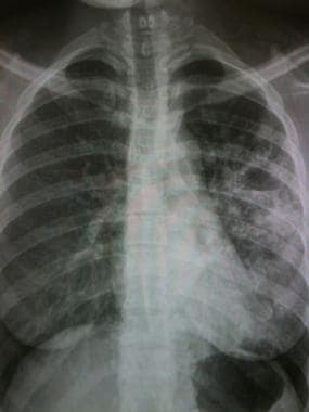
Strain W, a highly drug-resistant strain of Mycobacterium tuberculosis, was responsible for large nosocomial outbreaks in New York in the early 1990s. To describe the spread of strain W outside New York, we reviewed data from epidemiologic investigations, national tuberculosis surveillance, regional DNA fingerprint laboratories, and the Centers for Disease Control and Prevention Mycobacteriology Laboratory to identify potential cases of tuberculosis due to strain W. From January 1992 through February 1997, 23 cases were diagnosed in nine states and Puerto Rico; 8 were exposed to strain W in New York before their diagnosis; 4 of the 23 transmitted disease to 10 others. Eighty-six contacts of the 23 cases are presumed to be infected with strain W; 11 completed alternative preventive therapy. Strain W tuberculosis cases will occur throughout the United States as persons infected in New York move elsewhere. To help track and contain this strain, health departments should notify the Centers for Disease Control and Prevention of cases of tuberculosis resistant to isoniazid, rifampin, streptomycin, and kanamycin. (+info)Tuberculosis (TB) is a chronic infectious disease caused by the bacterium Mycobacterium tuberculosis. It primarily affects the lungs but can also involve other organs and tissues in the body. The infection is usually spread through the air when an infected person coughs, sneezes, or talks.
The symptoms of pulmonary TB include persistent cough, chest pain, coughing up blood, fatigue, fever, night sweats, and weight loss. Diagnosis typically involves a combination of medical history, physical examination, chest X-ray, and microbiological tests such as sputum smear microscopy and culture. In some cases, molecular tests like polymerase chain reaction (PCR) may be used for rapid diagnosis.
Treatment usually consists of a standard six-month course of multiple antibiotics, including isoniazid, rifampin, ethambutol, and pyrazinamide. In some cases, longer treatment durations or different drug regimens might be necessary due to drug resistance or other factors. Preventive measures include vaccination with the Bacillus Calmette-Guérin (BCG) vaccine and early detection and treatment of infected individuals to prevent transmission.
Pulmonary tuberculosis (TB) is an infectious disease caused by the bacterium Mycobacterium tuberculosis. It primarily affects the lungs and can spread to other parts of the body through the bloodstream or lymphatic system. The infection typically enters the body when a person inhales droplets containing the bacteria, which are released into the air when an infected person coughs, sneezes, or talks.
The symptoms of pulmonary TB can vary but often include:
* Persistent cough that lasts for more than three weeks and may produce phlegm or blood-tinged sputum
* Chest pain or discomfort, particularly when breathing deeply or coughing
* Fatigue and weakness
* Unexplained weight loss
* Fever and night sweats
* Loss of appetite
Pulmonary TB can cause serious complications if left untreated, including damage to the lungs, respiratory failure, and spread of the infection to other parts of the body. Treatment typically involves a course of antibiotics that can last several months, and it is essential for patients to complete the full treatment regimen to ensure that the infection is fully eradicated.
Preventive measures include vaccination with the Bacillus Calmette-Guérin (BCG) vaccine, which can provide some protection against severe forms of TB in children, and measures to prevent the spread of the disease, such as covering the mouth and nose when coughing or sneezing, wearing a mask in public places, and avoiding close contact with people who have active TB.
"Multiple drug resistance" (MDR) is a term used in medicine to describe the condition where a patient's infection becomes resistant to multiple antimicrobial drugs. This means that the bacteria, virus, fungus or parasite that is causing the infection has developed the ability to survive and multiply despite being exposed to medications that were originally designed to kill or inhibit its growth.
In particular, MDR occurs when an organism becomes resistant to at least one drug in three or more antimicrobial categories. This can happen due to genetic changes in the microorganism that allow it to survive in the presence of these drugs. The development of MDR is a significant concern for public health because it limits treatment options and can make infections harder, if not impossible, to treat.
MDR can develop through several mechanisms, including mutations in the genes that encode drug targets or enzymes involved in drug metabolism, as well as the acquisition of genetic elements such as plasmids and transposons that carry resistance genes. The overuse and misuse of antimicrobial drugs are major drivers of MDR, as they create selective pressure for the emergence and spread of resistant strains.
MDR infections can occur in various settings, including hospitals, long-term care facilities, and communities. They can affect people of all ages and backgrounds, although certain populations may be at higher risk, such as those with weakened immune systems or chronic medical conditions. Preventing the spread of MDR requires a multifaceted approach that includes surveillance, infection control, antimicrobial stewardship, and research into new therapies and diagnostics.
Multidrug-resistant tuberculosis (MDR-TB) is a form of tuberculosis (TB) infection caused by bacteria that are resistant to at least two of the first-line anti-TB drugs, isoniazid and rifampin. This makes MDR-TB more difficult and expensive to treat, requiring longer treatment durations and the use of second-line medications, which can have more severe side effects.
MDR-TB can occur when there are errors in prescribing or taking anti-TB drugs, or when people with TB do not complete their full course of treatment. It is a significant global health concern, particularly in low- and middle-income countries where TB is more prevalent and resources for diagnosis and treatment may be limited.
MDR-TB can spread from person to person through the air when someone with the infection coughs, speaks, or sneezes. People at higher risk of contracting MDR-TB include those who have been in close contact with someone with MDR-TB, people with weakened immune systems, and healthcare workers who treat TB patients.
Preventing the spread of MDR-TB involves early detection and prompt treatment, as well as infection control measures such as wearing masks, improving ventilation, and separating infected individuals from others. It is also important to ensure that anti-TB drugs are used correctly and that patients complete their full course of treatment to prevent the development of drug-resistant strains.
'Mycobacterium tuberculosis' is a species of slow-growing, aerobic, gram-positive bacteria that demonstrates acid-fastness. It is the primary causative agent of tuberculosis (TB) in humans. This bacterium has a complex cell wall rich in lipids, including mycolic acids, which provides a hydrophobic barrier and makes it resistant to many conventional antibiotics. The ability of M. tuberculosis to survive within host macrophages and resist the immune response contributes to its pathogenicity and the difficulty in treating TB infections.
M. tuberculosis is typically transmitted through inhalation of infectious droplets containing the bacteria, which primarily targets the lungs but can spread to other parts of the body (extrapulmonary TB). The infection may result in a spectrum of clinical manifestations, ranging from latent TB infection (LTBI) to active disease. LTBI represents a dormant state where individuals are infected with M. tuberculosis but do not show symptoms and cannot transmit the bacteria. However, they remain at risk of developing active TB throughout their lifetime, especially if their immune system becomes compromised.
Effective prevention and control strategies for TB rely on early detection, treatment, and public health interventions to limit transmission. The current first-line treatments for drug-susceptible TB include a combination of isoniazid, rifampin, ethambutol, and pyrazinamide for at least six months. Multidrug-resistant (MDR) and extensively drug-resistant (XDR) strains of M. tuberculosis present significant challenges in TB control and require more complex treatment regimens.
Multiple bacterial drug resistance (MDR) is a medical term that refers to the resistance of multiple strains of bacteria to several antibiotics or antimicrobial agents. This means that these bacteria have developed mechanisms that enable them to survive and multiply despite being exposed to drugs that were previously effective in treating infections caused by them.
MDR is a significant public health concern because it limits the treatment options available for bacterial infections, making them more difficult and expensive to treat. In some cases, MDR bacteria may cause severe or life-threatening infections that are resistant to all available antibiotics, leaving doctors with few or no effective therapeutic options.
MDR can arise due to various mechanisms, including the production of enzymes that inactivate antibiotics, changes in bacterial cell membrane permeability that prevent antibiotics from entering the bacteria, and the development of efflux pumps that expel antibiotics out of the bacteria. The misuse or overuse of antibiotics is a significant contributor to the emergence and spread of MDR bacteria.
Preventing and controlling the spread of MDR bacteria requires a multifaceted approach, including the judicious use of antibiotics, infection control measures, surveillance, and research into new antimicrobial agents.
P-glycoprotein (P-gp) is a type of membrane transport protein that plays a crucial role in the efflux (extrusion) of various substrates, including drugs and toxins, out of cells. It is also known as multidrug resistance protein 1 (MDR1).
P-gp is encoded by the ABCB1 gene and is primarily located on the apical membrane of epithelial cells in several tissues, such as the intestine, liver, kidney, and blood-brain barrier. Its main function is to protect these organs from harmful substances by actively pumping them out of the cells and back into the lumen or bloodstream.
In the context of pharmacology, P-gp can contribute to multidrug resistance (MDR) in cancer cells. When overexpressed, P-gp can reduce the intracellular concentration of various anticancer drugs, making them less effective. This has led to extensive research on inhibitors of P-gp as potential adjuvants for cancer therapy.
In summary, P-glycoprotein is a vital efflux transporter that helps maintain homeostasis by removing potentially harmful substances from cells and can impact drug disposition and response in various tissues, including the intestine, liver, kidney, and blood-brain barrier.
Multidrug Resistance-Associated Proteins (MRPs) are a subfamily of ATP-binding cassette (ABC) transporter proteins that play a crucial role in the efflux of various substrates, including drugs and organic anions, out of cells. They are located in the plasma membrane of many cell types, including epithelial cells in the liver, intestine, kidney, and blood-brain barrier.
MRPs are known to transport a wide range of molecules, such as glutathione conjugates, bilirubin, bile acids, and various clinical drugs. One of the most well-known MRPs is MRP1 (ABCC1), which was initially identified in drug-resistant tumor cells. MRP1 can confer resistance to chemotherapeutic agents by actively pumping them out of cancer cells, thereby reducing their intracellular concentration and effectiveness.
The activity of MRPs can have significant implications for the pharmacokinetics and pharmacodynamics of drugs, as they can affect drug absorption, distribution, metabolism, and excretion (ADME). Understanding the function and regulation of MRPs is essential for developing strategies to overcome multidrug resistance in cancer therapy and optimizing drug dosing regimens in various clinical settings.
Isoniazid is an antimicrobial medication used for the prevention and treatment of tuberculosis (TB). It is a first-line medication, often used in combination with other TB drugs, to kill the Mycobacterium tuberculosis bacteria that cause TB. Isoniazid works by inhibiting the synthesis of mycolic acids, which are essential components of the bacterial cell wall. This leads to bacterial death and helps to control the spread of TB.
Isoniazid is available in various forms, including tablets, capsules, and liquid solutions. It can be taken orally or given by injection. The medication is generally well-tolerated, but it can cause side effects such as peripheral neuropathy, hepatitis, and skin rashes. Regular monitoring of liver function tests and supplementation with pyridoxine (vitamin B6) may be necessary to prevent or manage these side effects.
It is important to note that Isoniazid is not effective against drug-resistant strains of TB, and its use should be guided by the results of drug susceptibility testing. Additionally, it is essential to complete the full course of treatment as prescribed to ensure the successful eradication of the bacteria and prevent the development of drug-resistant strains.
Microbial sensitivity tests, also known as antibiotic susceptibility tests (ASTs) or bacterial susceptibility tests, are laboratory procedures used to determine the effectiveness of various antimicrobial agents against specific microorganisms isolated from a patient's infection. These tests help healthcare providers identify which antibiotics will be most effective in treating an infection and which ones should be avoided due to resistance. The results of these tests can guide appropriate antibiotic therapy, minimize the potential for antibiotic resistance, improve clinical outcomes, and reduce unnecessary side effects or toxicity from ineffective antimicrobials.
There are several methods for performing microbial sensitivity tests, including:
1. Disk diffusion method (Kirby-Bauer test): A standardized paper disk containing a predetermined amount of an antibiotic is placed on an agar plate that has been inoculated with the isolated microorganism. After incubation, the zone of inhibition around the disk is measured to determine the susceptibility or resistance of the organism to that particular antibiotic.
2. Broth dilution method: A series of tubes or wells containing decreasing concentrations of an antimicrobial agent are inoculated with a standardized microbial suspension. After incubation, the minimum inhibitory concentration (MIC) is determined by observing the lowest concentration of the antibiotic that prevents visible growth of the organism.
3. Automated systems: These use sophisticated technology to perform both disk diffusion and broth dilution methods automatically, providing rapid and accurate results for a wide range of microorganisms and antimicrobial agents.
The interpretation of microbial sensitivity test results should be done cautiously, considering factors such as the site of infection, pharmacokinetics and pharmacodynamics of the antibiotic, potential toxicity, and local resistance patterns. Regular monitoring of susceptibility patterns and ongoing antimicrobial stewardship programs are essential to ensure optimal use of these tests and to minimize the development of antibiotic resistance.
Drug resistance, also known as antimicrobial resistance, is the ability of a microorganism (such as bacteria, viruses, fungi, or parasites) to withstand the effects of a drug that was originally designed to inhibit or kill it. This occurs when the microorganism undergoes genetic changes that allow it to survive in the presence of the drug. As a result, the drug becomes less effective or even completely ineffective at treating infections caused by these resistant organisms.
Drug resistance can develop through various mechanisms, including mutations in the genes responsible for producing the target protein of the drug, alteration of the drug's target site, modification or destruction of the drug by enzymes produced by the microorganism, and active efflux of the drug from the cell.
The emergence and spread of drug-resistant microorganisms pose significant challenges in medical treatment, as they can lead to increased morbidity, mortality, and healthcare costs. The overuse and misuse of antimicrobial agents, as well as poor infection control practices, contribute to the development and dissemination of drug-resistant strains. To address this issue, it is crucial to promote prudent use of antimicrobials, enhance surveillance and monitoring of resistance patterns, invest in research and development of new antimicrobial agents, and strengthen infection prevention and control measures.
"MDR" is an abbreviation for "Multidrug Resistance." In the context of genetics, MDR genes are those that encode for proteins, typically transmembrane pumps, which can actively transport various drugs out of cells. This results in reduced drug accumulation within cells and decreased effectiveness of these drugs.
MDR genes play a crucial role in conferring resistance to chemotherapy agents in cancer cells, making treatment more challenging. One well-known MDR gene is the ABCB1 (ATP Binding Cassette Subfamily B Member 1) gene, which encodes for the P-glycoprotein efflux pump. Overexpression of such MDR genes can lead to cross-resistance to multiple drugs, further complicating treatment strategies.
Rifampin is an antibiotic medication that belongs to the class of drugs known as rifamycins. It works by inhibiting bacterial DNA-dependent RNA polymerase, thereby preventing bacterial growth and multiplication. Rifampin is used to treat a variety of infections caused by bacteria, including tuberculosis, Haemophilus influenzae, Neisseria meningitidis, and Legionella pneumophila. It is also used to prevent meningococcal disease in people who have been exposed to the bacteria.
Rifampin is available in various forms, including tablets, capsules, and injectable solutions. The medication is usually taken two to four times a day, depending on the type and severity of the infection being treated. Rifampin may be given alone or in combination with other antibiotics.
It is important to note that rifampin can interact with several other medications, including oral contraceptives, anticoagulants, and anti-seizure drugs, among others. Therefore, it is essential to inform your healthcare provider about all the medications you are taking before starting treatment with rifampin.
Rifampin may cause side effects such as nausea, vomiting, diarrhea, dizziness, headache, and changes in the color of urine, tears, sweat, and saliva to a reddish-orange color. These side effects are usually mild and go away on their own. However, if they persist or become bothersome, it is important to consult your healthcare provider.
In summary, rifampin is an antibiotic medication used to treat various bacterial infections and prevent meningococcal disease. It works by inhibiting bacterial DNA-dependent RNA polymerase, preventing bacterial growth and multiplication. Rifampin may interact with several other medications, and it can cause side effects such as nausea, vomiting, diarrhea, dizziness, headache, and changes in the color of body fluids.
Anti-bacterial agents, also known as antibiotics, are a type of medication used to treat infections caused by bacteria. These agents work by either killing the bacteria or inhibiting their growth and reproduction. There are several different classes of anti-bacterial agents, including penicillins, cephalosporins, fluoroquinolones, macrolides, and tetracyclines, among others. Each class of antibiotic has a specific mechanism of action and is used to treat certain types of bacterial infections. It's important to note that anti-bacterial agents are not effective against viral infections, such as the common cold or flu. Misuse and overuse of antibiotics can lead to antibiotic resistance, which is a significant global health concern.
A tuberculosis vaccine, also known as the BCG (Bacillus Calmette-Guérin) vaccine, is a type of immunization used to prevent tuberculosis (TB), a bacterial infection caused by Mycobacterium tuberculosis. The BCG vaccine contains a weakened strain of the bacteria that causes TB in cattle.
The BCG vaccine works by stimulating an immune response in the body, which helps to protect against severe forms of TB, such as TB meningitis and TB in children. However, it is not very effective at preventing pulmonary TB (TB that affects the lungs) in adults.
The BCG vaccine is not routinely recommended for use in the United States due to the low risk of TB infection in the general population. However, it may be given to people who are at high risk of exposure to TB, such as healthcare workers, laboratory personnel, and people traveling to countries with high rates of TB.
It is important to note that the BCG vaccine does not provide complete protection against TB and that other measures, such as testing and treatment for latent TB infection, are also important for controlling the spread of this disease.
Drug resistance in neoplasms (also known as cancer drug resistance) refers to the ability of cancer cells to withstand the effects of chemotherapeutic agents or medications designed to kill or inhibit the growth of cancer cells. This can occur due to various mechanisms, including changes in the cancer cell's genetic makeup, alterations in drug targets, increased activity of drug efflux pumps, and activation of survival pathways.
Drug resistance can be intrinsic (present at the beginning of treatment) or acquired (developed during the course of treatment). It is a significant challenge in cancer therapy as it often leads to reduced treatment effectiveness, disease progression, and poor patient outcomes. Strategies to overcome drug resistance include the use of combination therapies, development of new drugs that target different mechanisms, and personalized medicine approaches that consider individual patient and tumor characteristics.
Bacterial drug resistance is a type of antimicrobial resistance that occurs when bacteria evolve the ability to survive and reproduce in the presence of drugs (such as antibiotics) that would normally kill them or inhibit their growth. This can happen due to various mechanisms, including genetic mutations or the acquisition of resistance genes from other bacteria.
As a result, bacterial infections may become more difficult to treat, requiring higher doses of medication, alternative drugs, or longer treatment courses. In some cases, drug-resistant infections can lead to serious health complications, increased healthcare costs, and higher mortality rates.
Examples of bacterial drug resistance include methicillin-resistant Staphylococcus aureus (MRSA), vancomycin-resistant Enterococci (VRE), and multidrug-resistant tuberculosis (MDR-TB). Preventing the spread of bacterial drug resistance is crucial for maintaining effective treatments for infectious diseases.
Miliary tuberculosis is a disseminated form of tuberculosis (TB), a bacterial infection caused by Mycobacterium tuberculosis. The term "miliary" refers to the tiny millet-like size (2-5 microns in diameter) of the TB foci observed in the lungs or other organs during autopsy or on imaging studies. In military tuberculosis, these small granules are widespread throughout the body, affecting multiple organs such as the lungs, liver, spleen, bones, and brain. It can occur in people with weakened immune systems, including those with HIV/AIDS, or in individuals who have recently been infected with TB bacteria. Symptoms may include fever, night sweats, weight loss, fatigue, and cough. Early diagnosis and treatment are crucial to prevent severe complications and improve outcomes.
Verapamil is a calcium channel blocker medication that is primarily used to treat hypertension (high blood pressure), angina (chest pain), and certain types of cardiac arrhythmias (irregular heart rhyats). It works by relaxing the smooth muscle cells in the walls of blood vessels, which causes them to dilate or widen, reducing the resistance to blood flow and thereby lowering blood pressure. Verapamil also slows down the conduction of electrical signals within the heart, which can help to regulate the heart rate and rhythm.
In addition to its cardiovascular effects, verapamil is sometimes used off-label for the treatment of other conditions such as migraine headaches, Raynaud's phenomenon, and certain types of tremors. It is available in various forms, including immediate-release tablets, extended-release capsules, and intravenous (IV) injection.
It is important to note that verapamil can interact with other medications, so it is essential to inform your healthcare provider about all the drugs you are taking before starting this medication. Additionally, verapamil should be used with caution in people with certain medical conditions, such as heart failure, liver disease, and low blood pressure.
Bacterial proteins are a type of protein that are produced by bacteria as part of their structural or functional components. These proteins can be involved in various cellular processes, such as metabolism, DNA replication, transcription, and translation. They can also play a role in bacterial pathogenesis, helping the bacteria to evade the host's immune system, acquire nutrients, and multiply within the host.
Bacterial proteins can be classified into different categories based on their function, such as:
1. Enzymes: Proteins that catalyze chemical reactions in the bacterial cell.
2. Structural proteins: Proteins that provide structural support and maintain the shape of the bacterial cell.
3. Signaling proteins: Proteins that help bacteria to communicate with each other and coordinate their behavior.
4. Transport proteins: Proteins that facilitate the movement of molecules across the bacterial cell membrane.
5. Toxins: Proteins that are produced by pathogenic bacteria to damage host cells and promote infection.
6. Surface proteins: Proteins that are located on the surface of the bacterial cell and interact with the environment or host cells.
Understanding the structure and function of bacterial proteins is important for developing new antibiotics, vaccines, and other therapeutic strategies to combat bacterial infections.
Antitubercular agents, also known as anti-tuberculosis drugs or simply TB drugs, are a category of medications specifically used for the treatment and prevention of tuberculosis (TB), a bacterial infection caused by Mycobacterium tuberculosis. These drugs target various stages of the bacteria's growth and replication process to eradicate it from the body or prevent its spread.
There are several first-line antitubercular agents, including:
1. Isoniazid (INH): This is a bactericidal drug that inhibits the synthesis of mycolic acids, essential components of the mycobacterial cell wall. It is primarily active against actively growing bacilli.
2. Rifampin (RIF) or Rifampicin: A bactericidal drug that inhibits DNA-dependent RNA polymerase, preventing the transcription of genetic information into mRNA. This results in the interruption of protein synthesis and ultimately leads to the death of the bacteria.
3. Ethambutol (EMB): A bacteriostatic drug that inhibits the arabinosyl transferase enzyme, which is responsible for the synthesis of arabinan, a crucial component of the mycobacterial cell wall. It is primarily active against actively growing bacilli.
4. Pyrazinamide (PZA): A bactericidal drug that inhibits the synthesis of fatty acids and mycolic acids in the mycobacterial cell wall, particularly under acidic conditions. PZA is most effective during the initial phase of treatment when the bacteria are in a dormant or slow-growing state.
These first-line antitubercular agents are often used together in a combination therapy to ensure complete eradication of the bacteria and prevent the development of drug-resistant strains. Treatment duration typically lasts for at least six months, with the initial phase consisting of daily doses of INH, RIF, EMB, and PZA for two months, followed by a continuation phase of INH and RIF for four months.
Second-line antitubercular agents are used when patients have drug-resistant TB or cannot tolerate first-line drugs. These include drugs like aminoglycosides (e.g., streptomycin, amikacin), fluoroquinolones (e.g., ofloxacin, moxifloxacin), and injectable bacteriostatic agents (e.g., capreomycin, ethionamide).
It is essential to closely monitor patients undergoing antitubercular therapy for potential side effects and ensure adherence to the treatment regimen to achieve optimal outcomes and prevent the development of drug-resistant strains.
Doxorubicin is a type of chemotherapy medication known as an anthracycline. It works by interfering with the DNA in cancer cells, which prevents them from growing and multiplying. Doxorubicin is used to treat a wide variety of cancers, including leukemia, lymphoma, breast cancer, lung cancer, ovarian cancer, and many others. It may be given alone or in combination with other chemotherapy drugs.
Doxorubicin is usually administered through a vein (intravenously) and can cause side effects such as nausea, vomiting, hair loss, mouth sores, and increased risk of infection. It can also cause damage to the heart muscle, which can lead to heart failure in some cases. For this reason, doctors may monitor patients' heart function closely while they are receiving doxorubicin treatment.
It is important for patients to discuss the potential risks and benefits of doxorubicin therapy with their healthcare provider before starting treatment.
Latent Tuberculosis (TB) infection is defined as a state of persistent immune response to stimulation by Mycobacterium tuberculosis without evidence of clinically manifest active TB disease. The individuals with latent TB infection do not feel ill and are not infectious. However, they may develop active TB disease later in their lives, typically within the first 2 years after infection. It's estimated that about 5-10% of people with latent TB infection will develop active TB disease during their lifetime. The risk is higher in people who have weakened immune systems due to HIV infection, malnutrition, aging, or use of immunosuppressive medications. Diagnosis of latent TB infection is typically made through a tuberculin skin test or an interferon-gamma release assay (IGRA). Treatment of latent TB infection can reduce the risk of developing active TB disease.
Tuberculosis (TB) of the lymph node, also known as scrofula or tuberculous lymphadenitis, is a specific form of extrapulmonary tuberculosis. It involves the infection and inflammation of the lymph nodes (lymph glands) by the Mycobacterium tuberculosis bacterium. The lymph nodes most commonly affected are the cervical (neck) and supraclavicular (above the collarbone) lymph nodes, but other sites can also be involved.
The infection typically spreads to the lymph nodes through the bloodstream or via nearby infected organs, such as the lungs or intestines. The affected lymph nodes may become enlarged, firm, and tender, forming masses called cold abscesses that can suppurate (form pus) and eventually rupture. In some cases, the lymph nodes may calcify, leaving hard, stone-like deposits.
Diagnosis of tuberculous lymphadenitis often involves a combination of clinical evaluation, imaging studies (such as CT or MRI scans), and microbiological or histopathological examination of tissue samples obtained through fine-needle aspiration biopsy or surgical excision. Treatment typically consists of a standard anti-tuberculosis multi-drug regimen, which may include isoniazid, rifampin, ethambutol, and pyrazinamide for at least six months. Surgical intervention might be necessary in cases with complications or treatment failure.
Sputum is defined as a mixture of saliva and phlegm that is expelled from the respiratory tract during coughing, sneezing or deep breathing. It can be clear, mucoid, or purulent (containing pus) depending on the underlying cause of the respiratory issue. Examination of sputum can help diagnose various respiratory conditions such as infections, inflammation, or other lung diseases.
P-glycoproteins (P-gp), also known as multidrug resistance proteins (MDR), are a type of transmembrane protein that functions as an efflux pump, actively transporting various substrates out of cells. They play a crucial role in the protection of cells against xenobiotics, including drugs, toxins, and carcinogens. P-gp is expressed in many tissues, such as the intestine, liver, kidney, and blood-brain barrier, where it helps limit the absorption and distribution of drugs and other toxic substances.
In the context of medicine and pharmacology, P-glycoproteins are particularly relevant due to their ability to confer multidrug resistance in cancer cells. Overexpression of P-gp in tumor cells can lead to reduced intracellular drug concentrations, making these cells less sensitive to chemotherapeutic agents and contributing to treatment failure. Understanding the function and regulation of P-glycoproteins is essential for developing strategies to overcome multidrug resistance in cancer therapy.
"Prosopis" is a genus of flowering plants in the pea family, Fabaceae. It includes several species of spiny trees and shrubs that are native to arid and semi-arid regions of America, Africa, and Asia. Some common names for Prosopis species include mesquite, algarrobo, and jand. These plants are known for their ability to fix nitrogen in the soil, making them valuable for improving soil fertility in areas where they grow. They also produce seed pods that are a valuable food source for wildlife and humans in some regions. However, Prosopis species can also be invasive in some areas, outcompeting native vegetation and altering ecosystems.
Osteoarticular tuberculosis is a form of extrapulmonary tuberculosis (TB) that involves the bones and joints. It is caused by the bacterium Mycobacterium tuberculosis. The infection can spread to the bones and joints through the bloodstream or from nearby infected organs, such as the lungs.
The most commonly affected sites are the spine (Pott's disease), hip, knee, wrist, and small bones of the hands and feet. Symptoms may include pain, swelling, stiffness, and decreased range of motion in the affected joint or bone. In some cases, the infection can lead to deformity, chronic disability, or even death if left untreated.
Diagnosis typically involves a combination of medical history, physical examination, imaging studies (such as X-rays, CT scans, or MRI), and laboratory tests (such as blood tests, sputum cultures, or biopsy). Treatment usually consists of a long course of antibiotics (usually for at least six months) to kill the bacteria. Surgery may also be necessary in some cases to remove infected tissue or stabilize damaged joints.
Gastrointestinal tuberculosis (GTB) is a type of tuberculosis that affects the gastrointestinal tract, including the stomach, intestines, and associated organs such as the liver and spleen. It is caused by the bacterium Mycobacterium tuberculosis, which typically infects the lungs (pulmonary TB) but can spread to other parts of the body through the bloodstream or lymphatic system.
In GTB, the bacteria invade the tissues of the gastrointestinal tract and cause inflammation, ulceration, and thickening of the intestinal wall. This can lead to a variety of symptoms, including abdominal pain, diarrhea (which may be bloody), weight loss, fever, and fatigue. GTB can also cause complications such as bowel obstruction, perforation, or fistula formation.
Diagnosis of GTB can be challenging, as the symptoms are non-specific and can mimic those of other gastrointestinal disorders. Diagnostic tests may include endoscopy, biopsy, culture, and molecular testing for the presence of M. tuberculosis. Treatment typically involves a prolonged course of multiple antibiotics, such as isoniazid, rifampin, ethambutol, and pyrazinamide, administered under the guidance of a healthcare provider.
It's worth noting that GTB is relatively rare in developed countries with low rates of tuberculosis, but it is more common in areas where TB is endemic or among populations with weakened immune systems, such as those with HIV/AIDS.
Tuberculosis (TB) of the spine, also known as Pott's disease, is a specific form of extrapulmonary tuberculosis that involves the vertebral column. It is caused by the Mycobacterium tuberculosis bacterium, which primarily affects the lungs but can spread through the bloodstream to other parts of the body, including the spine.
In Pott's disease, the infection leads to the destruction of the spongy bone (vertebral body) and the intervertebral disc space, resulting in vertebral collapse, kyphosis (hunchback deformity), and potential neurological complications due to spinal cord compression. Common symptoms include back pain, stiffness, fever, night sweats, and weight loss. Early diagnosis and treatment with a multidrug antibiotic regimen are crucial to prevent long-term disability and further spread of the infection.
'Acinetobacter baumannii' is a gram-negative, aerobic, coccobacillus-shaped bacterium that is commonly found in the environment, including water, soil, and healthcare settings. It is known to cause various types of infections in humans, particularly in hospitalized patients or those with weakened immune systems.
This bacterium can cause a range of infections, such as pneumonia, bloodstream infections, meningitis, and wound infections. 'Acinetobacter baumannii' is often resistant to multiple antibiotics, making it difficult to treat the resulting infections. This has led to its classification as a "superbug" or a multidrug-resistant organism (MDRO).
The medical community continues to research and develop new strategies to prevent and treat infections caused by 'Acinetobacter baumannii' and other antibiotic-resistant bacteria.
Acinetobacter infections are caused by bacteria that can be found in various environments, such as soil, water, and healthcare facilities. These bacteria can cause a range of illnesses, from mild skin infections to serious respiratory and bloodstream infections. They are often resistant to multiple antibiotics, making them difficult to treat.
Acinetobacter baumannii is the species most commonly associated with human infection. It is known for its ability to survive on dry surfaces for extended periods of time, which can contribute to its spread in healthcare settings. Infections caused by Acinetobacter are a particular concern in critically ill patients, such as those in intensive care units, and in individuals with weakened immune systems.
Symptoms of an Acinetobacter infection depend on the site of infection but may include fever, cough, shortness of breath, wound drainage, or skin redness or swelling. Treatment typically involves the use of antibiotics that are still effective against the bacteria, which can be determined through laboratory testing. In some cases, infection control measures, such as contact precautions and environmental cleaning, may also be necessary to prevent the spread of Acinetobacter in healthcare settings.
ATP-binding cassette (ABC) transporters are a family of membrane proteins that utilize the energy from ATP hydrolysis to transport various substrates across extra- and intracellular membranes. These transporters play crucial roles in several biological processes, including detoxification, drug resistance, nutrient uptake, and regulation of cellular cholesterol homeostasis.
The structure of ABC transporters consists of two nucleotide-binding domains (NBDs) that bind and hydrolyze ATP, and two transmembrane domains (TMDs) that form the substrate-translocation pathway. The NBDs are typically located adjacent to each other in the cytoplasm, while the TMDs can be either integral membrane domains or separate structures associated with the membrane.
The human genome encodes 48 distinct ABC transporters, which are classified into seven subfamilies (ABCA-ABCG) based on their sequence similarity and domain organization. Some well-known examples of ABC transporters include P-glycoprotein (ABCB1), multidrug resistance protein 1 (ABCC1), and breast cancer resistance protein (ABCG2).
Dysregulation or mutations in ABC transporters have been implicated in various diseases, such as cystic fibrosis, neurological disorders, and cancer. In cancer, overexpression of certain ABC transporters can contribute to drug resistance by actively effluxing chemotherapeutic agents from cancer cells, making them less susceptible to treatment.
Bovine tuberculosis (BTB) is a chronic infectious disease caused by the bacterium Mycobacterium bovis. It primarily affects cattle but can also spread to other mammals including humans, causing a similar disease known as zoonotic tuberculosis. The infection in animals typically occurs through inhalation of infectious droplets or ingestion of contaminated feed and water.
In cattle, the disease often affects the respiratory system, leading to symptoms such as chronic coughing, weight loss, and difficulty breathing. However, it can also affect other organs, including the intestines, lymph nodes, and mammary glands. Diagnosis of BTB typically involves a combination of clinical signs, laboratory tests, and epidemiological data.
Control measures for BTB include regular testing and culling of infected animals, movement restrictions, and vaccination of susceptible populations. In many countries, BTB is a notifiable disease, meaning that cases must be reported to the authorities. Proper cooking and pasteurization of dairy products can help prevent transmission to humans.
Bacterial DNA refers to the genetic material found in bacteria. It is composed of a double-stranded helix containing four nucleotide bases - adenine (A), thymine (T), guanine (G), and cytosine (C) - that are linked together by phosphodiester bonds. The sequence of these bases in the DNA molecule carries the genetic information necessary for the growth, development, and reproduction of bacteria.
Bacterial DNA is circular in most bacterial species, although some have linear chromosomes. In addition to the main chromosome, many bacteria also contain small circular pieces of DNA called plasmids that can carry additional genes and provide resistance to antibiotics or other environmental stressors.
Unlike eukaryotic cells, which have their DNA enclosed within a nucleus, bacterial DNA is present in the cytoplasm of the cell, where it is in direct contact with the cell's metabolic machinery. This allows for rapid gene expression and regulation in response to changing environmental conditions.
Microbial drug resistance is a significant medical issue that refers to the ability of microorganisms (such as bacteria, viruses, fungi, or parasites) to withstand or survive exposure to drugs or medications designed to kill them or limit their growth. This phenomenon has become a major global health concern, particularly in the context of bacterial infections, where it is also known as antibiotic resistance.
Drug resistance arises due to genetic changes in microorganisms that enable them to modify or bypass the effects of antimicrobial agents. These genetic alterations can be caused by mutations or the acquisition of resistance genes through horizontal gene transfer. The resistant microbes then replicate and multiply, forming populations that are increasingly difficult to eradicate with conventional treatments.
The consequences of drug-resistant infections include increased morbidity, mortality, healthcare costs, and the potential for widespread outbreaks. Factors contributing to the emergence and spread of microbial drug resistance include the overuse or misuse of antimicrobials, poor infection control practices, and inadequate surveillance systems.
To address this challenge, it is crucial to promote prudent antibiotic use, strengthen infection prevention and control measures, develop new antimicrobial agents, and invest in research to better understand the mechanisms underlying drug resistance.
Cutaneous tuberculosis (CTB) is a rare form of tuberculosis that affects the skin. It is caused by the Mycobacterium tuberculosis complex, including M. tuberculosis, M. bovis, and M. africanum. CTB can occur as a primary infection after direct inoculation of the skin with the bacteria, or it can be secondary to a distant focus of infection such as lung or lymph node TB.
The clinical presentation of CTB is varied and can include papules, nodules, pustules, ulcers, plaques, or scaly lesions. The lesions may be painless or painful, and they can be associated with systemic symptoms such as fever, night sweats, and weight loss.
CTB can be diagnosed through a combination of clinical examination, skin biopsy, culture, and PCR testing. Treatment typically involves a prolonged course of multiple antibiotics, often for six to nine months or more. The most commonly used drugs are isoniazid, rifampin, ethambutol, and pyrazinamide. Surgical excision may be necessary in some cases.
Prevention measures include early detection and treatment of pulmonary TB, BCG vaccination, and avoiding contact with people with active TB.
Rhodamine 123 is not a medical term, but a chemical compound. It's a fluorescent dye used in various scientific and research applications, particularly in the field of cell biology. Rhodamine 123 has an affinity for mitochondria, the energy-producing structures in cells, making it useful as a marker to study mitochondrial function and distribution within cells.
In summary, Rhodamine 123 is not a medical definition itself, but it can be used in medical research contexts to investigate cellular processes.
A tuberculin test is a medical procedure used to determine if someone has developed an immune response to the bacterium that causes tuberculosis (TB), Mycobacterium tuberculosis. The test involves injecting a small amount of purified protein derivative (PPD) from the TB bacteria under the skin, usually on the forearm. After 48-72 hours, the area is examined for signs of a reaction, such as swelling, redness, or hardness. A positive result suggests that the person has been infected with TB at some point in the past, although it does not necessarily mean that they have active TB disease. However, individuals who have a positive tuberculin test should be evaluated further to determine if they need treatment for latent TB infection or active TB disease.
Multiple drug resistance in fungi refers to the ability of certain fungal strains or species to resist the effects of multiple antifungal agents. This occurs when these organisms develop mechanisms that prevent the drugs from interfering with their growth and survival. As a result, the drugs become less effective or even completely ineffective at treating fungal infections caused by these resistant strains or species.
Multiple drug resistance in fungi can arise due to various factors, including genetic mutations, overuse or misuse of antifungal agents, and the ability of fungi to exchange genetic material with other fungi. This makes treatment of fungal infections more challenging, as doctors may need to use higher doses of drugs or try alternative therapies that may have more side effects or be less effective.
Multiple drug resistance in fungi is a significant concern in healthcare settings, particularly for patients who are immunocompromised or have underlying medical conditions that make them more susceptible to fungal infections. It is essential to take measures to prevent the development and spread of multiple drug-resistant fungi, such as using antifungal agents appropriately, practicing good infection control practices, and conducting surveillance for resistant strains.
I'm sorry for any confusion, but "Menogaril" does not appear to have a widely recognized or established medical definition. It is possible that you may be referring to a specific chemical compound or experimental drug. However, without more context or information, it is difficult for me to provide a precise definition.
Menogaril is a synthetic compound that has been studied in preclinical trials for its potential anticancer properties. It is an analog of the natural product dictyostatin and has been shown to inhibit the activity of several enzymes involved in DNA replication and repair, including topoisomerase II and poly(ADP-ribose) polymerase (PARP). However, it is important to note that Menogaril is not currently approved for use in clinical medicine, and its safety and efficacy have not been established in human trials.
Daunorubicin is an anthracycline antibiotic used in the treatment of various types of cancer, including leukemia, Hodgkin's lymphoma, and breast cancer. It works by intercalating with DNA and inhibiting topoisomerase II, which results in DNA damage and ultimately cell death.
The drug is administered intravenously and may cause side effects such as nausea, vomiting, hair loss, mouth sores, and damage to the heart muscle (cardiotoxicity) with long-term use. Regular monitoring of cardiac function is recommended during treatment with daunorubicin.
It's important to note that this medication should only be used under the supervision of a qualified healthcare professional, as it can have serious and potentially life-threatening consequences if not used correctly.
Antitubercular antibiotics are a class of medications specifically used to treat tuberculosis (TB) and other mycobacterial infections. Tuberculosis is caused by the bacterium Mycobacterium tuberculosis, which can affect various organs, primarily the lungs.
There are several antitubercular antibiotics available, with different mechanisms of action that target the unique cell wall structure and metabolism of mycobacteria. Some commonly prescribed antitubercular antibiotics include:
1. Isoniazid (INH): This is a first-line medication for treating TB. It inhibits the synthesis of mycolic acids, a crucial component of the mycobacterial cell wall. Isoniazid can be bactericidal or bacteriostatic depending on the concentration and duration of treatment.
2. Rifampin (RIF): Also known as rifampicin, this antibiotic inhibits bacterial DNA-dependent RNA polymerase, preventing the transcription of genetic information into mRNA. It is a potent bactericidal agent against mycobacteria and is often used in combination with other antitubercular drugs.
3. Ethambutol (EMB): This antibiotic inhibits the synthesis of arabinogalactan and mycolic acids, both essential components of the mycobacterial cell wall. Ethambutol is primarily bacteriostatic but can be bactericidal at higher concentrations.
4. Pyrazinamide (PZA): This medication is active against dormant or slow-growing mycobacteria, making it an essential component of TB treatment regimens. Its mechanism of action involves the inhibition of fatty acid synthesis and the disruption of bacterial membrane potential.
5. Streptomycin: An aminoglycoside antibiotic that binds to the 30S ribosomal subunit, inhibiting protein synthesis in mycobacteria. It is primarily used as a second-line treatment for drug-resistant TB.
6. Fluoroquinolones: These are a class of antibiotics that inhibit DNA gyrase and topoisomerase IV, essential enzymes involved in bacterial DNA replication. Examples include ciprofloxacin, moxifloxacin, and levofloxacin, which can be used as second-line treatments for drug-resistant TB.
These antitubercular drugs are often used in combination to prevent the development of drug resistance and improve treatment outcomes. The World Health Organization (WHO) recommends a standardized regimen consisting of isoniazid, rifampicin, ethambutol, and pyrazinamide for the initial two months, followed by isoniazid and rifampicin for an additional four to seven months. However, treatment regimens may vary depending on the patient's clinical presentation, drug susceptibility patterns, and local guidelines.
"Mycobacterium bovis" is a species of slow-growing, aerobic, gram-positive bacteria in the family Mycobacteriaceae. It is the causative agent of tuberculosis in cattle and other animals, and can also cause tuberculosis in humans, particularly in those who come into contact with infected animals or consume unpasteurized dairy products from infected cows. The bacteria are resistant to many common disinfectants and survive for long periods in a dormant state, making them difficult to eradicate from the environment. "Mycobacterium bovis" is closely related to "Mycobacterium tuberculosis," the bacterium that causes tuberculosis in humans, and both species share many genetic and biochemical characteristics.
Extensively Drug-Resistant Tuberculosis (XDR-TB) is a term used to describe a rare, severe form of tuberculosis (TB) that is resistant to the majority of available drugs used to treat TB. This means that the bacteria that cause TB have developed resistance to at least four of the core anti-TB drugs, including isoniazid and rifampin, as well as any fluoroquinolone and at least one of the three injectable second-line drugs (amikacin, capreomycin, or kanamycin).
XDR-TB can be challenging to diagnose and treat due to its resistance to multiple drugs. It is also more likely to cause severe illness, spread from person to person, and result in poor treatment outcomes compared to drug-susceptible TB. XDR-TB is a public health concern, particularly in areas with high rates of TB and limited access to effective treatments.
It's important to note that XDR-TB should not be confused with Multi-Drug Resistant Tuberculosis (MDR-TB), which refers to TB that is resistant to at least isoniazid and rifampin, but not necessarily to the other second-line drugs.
Pyrazinamide is an antituberculosis agent, a type of medication used to treat tuberculosis (TB) caused by Mycobacterium tuberculosis. It is an antimicrobial drug that works by inhibiting the growth of the bacterium. Pyrazinamide is often used in combination with other TB drugs such as isoniazid, rifampin, and ethambutol.
The medical definition of Pyrazinamide is: a synthetic antituberculosis agent, C6H5N3O (a pyridine derivative), used in the treatment of tuberculosis, especially in combination with isoniazid and rifampin. It is converted in the body to its active form, pyrazinoic acid, which inhibits the growth of Mycobacterium tuberculosis by interfering with bacterial cell wall synthesis.
It's important to note that Pyrazinamide should be used under the supervision of a healthcare professional and is usually prescribed for several months to ensure complete eradication of the TB bacteria. As with any medication, it can cause side effects, and individuals should report any unusual symptoms to their healthcare provider.
Pleural Tuberculosis is a form of extrapulmonary tuberculosis (EPTB) that involves the infection and inflammation of the pleura, which are the thin membranes that surround the lungs and line the inside of the chest cavity. This condition is caused by the Mycobacterium tuberculosis bacterium, which can spread through the air when an infected person coughs, sneezes, or talks.
In pleural tuberculosis, the bacteria reach the pleura either through direct extension from a nearby lung infection or via bloodstream dissemination. The infection can cause the pleura to become inflamed and produce excess fluid, leading to pleural effusion. This accumulation of fluid in the pleural space can cause chest pain, coughing, and difficulty breathing.
Diagnosis of pleural tuberculosis typically involves a combination of medical history, physical examination, imaging studies such as chest X-rays or CT scans, and laboratory tests such as acid-fast bacilli (AFB) smear microscopy, culture, and nucleic acid amplification tests (NAATs) to detect the presence of M. tuberculosis in the pleural fluid or tissue samples.
Treatment of pleural tuberculosis typically involves a standard course of anti-tuberculosis therapy (ATT), which includes a combination of multiple antibiotics such as isoniazid, rifampin, ethambutol, and pyrazinamide. The duration of treatment may vary depending on the severity of the infection and the patient's response to therapy. In some cases, surgical intervention may be necessary to drain the pleural effusion or remove the infected pleura.
Tuberculosis (TB) is a bacterial infection caused by Mycobacterium tuberculosis. Urogenital tuberculosis (UTB) is a less common form of TB that affects the urinary and genital systems. It occurs when the bacteria spread through the bloodstream from the initial site of infection, usually the lungs, to the kidneys. The infection can then spread to other parts of the urinary system, including the ureters, bladder, and urethra, as well as the genital organs in both men and women.
UTB symptoms may include:
- Persistent dull pain in the lower back or side
- Frequent urination or urgent need to urinate
- Painful urination (dysuria)
- Blood in the urine (hematuria)
- Incontinence
- Sexual dysfunction in men, such as epididymitis or infertility
- Scrotal mass in men
- Amenorrhea or irregular menstruation in women
Diagnosis of UTB typically involves a combination of medical history, physical examination, imaging tests (such as ultrasound, CT scan, or MRI), urine analysis and culture, and sometimes biopsy. Treatment usually consists of a prolonged course of multiple antibiotics to eliminate the infection. Surgery may be required in some cases to repair damaged organs or remove scar tissue.
BCG (Bacillus Calmette-Guérin) vaccine is a type of immunization used primarily to prevent tuberculosis (TB). It contains a live but weakened strain of Mycobacterium bovis, which is related to the bacterium that causes TB in humans (Mycobacterium tuberculosis).
The BCG vaccine works by stimulating an immune response in the body, enabling it to better resist infection with TB bacteria if exposed in the future. It is often given to infants and children in countries where TB is common, and its use varies depending on the national immunization policies. The protection offered by the BCG vaccine is moderate and may not last for a very long time.
In addition to its use against TB, the BCG vaccine has also been investigated for its potential therapeutic role in treating bladder cancer and some other types of cancer. The mechanism of action in these cases is thought to be related to the vaccine's ability to stimulate an immune response against abnormal cells.
Nogalamycin is not typically considered as a medical term, but it is a type of antibiotic that is used in research and microbiology. Here's the definition from a scientific perspective:
Nogalamycin is an anthracycline antitumor antibiotic produced by Streptomyces nogalater. It is a DNA-intercalating agent, which means it can insert itself between the base pairs of DNA and disrupt the structure and function of the genetic material in bacteria and cancer cells. Nogalamycin has been studied for its potential use as an anticancer drug, but its clinical use has been limited due to toxicity concerns.
DNA fingerprinting, also known as DNA profiling or genetic fingerprinting, is a laboratory technique used to identify and compare the unique genetic makeup of individuals by analyzing specific regions of their DNA. This method is based on the variation in the length of repetitive sequences of DNA called variable number tandem repeats (VNTRs) or short tandem repeats (STRs), which are located at specific locations in the human genome and differ significantly among individuals, except in the case of identical twins.
The process of DNA fingerprinting involves extracting DNA from a sample, amplifying targeted regions using the polymerase chain reaction (PCR), and then separating and visualizing the resulting DNA fragments through electrophoresis. The fragment patterns are then compared to determine the likelihood of a match between two samples.
DNA fingerprinting has numerous applications in forensic science, paternity testing, identity verification, and genealogical research. It is considered an essential tool for providing strong evidence in criminal investigations and resolving disputes related to parentage and inheritance.
Meningeal tuberculosis, also known as Tuberculous meningitis, is a severe form of tuberculosis (TB) that affects the meninges, which are the membranes covering the brain and spinal cord. It is caused by the Mycobacterium tuberculosis bacterium, which can spread through the bloodstream from a primary infection site in the lungs or elsewhere in the body.
In meningeal tuberculosis, the bacteria cause inflammation and thickening of the meninges, leading to increased intracranial pressure, cerebral edema, and vasculitis. These conditions can result in various neurological symptoms such as headache, fever, stiff neck, altered mental status, seizures, and focal neurologic deficits. If left untreated, meningeal tuberculosis can lead to severe complications, including brain damage, hydrocephalus, and even death.
Diagnosis of meningeal tuberculosis typically involves a combination of clinical symptoms, cerebrospinal fluid (CSF) analysis, imaging studies, and sometimes molecular or culture-based tests to detect the presence of Mycobacterium tuberculosis in the CSF. Treatment usually involves a prolonged course of antibiotics specifically designed to target TB, such as isoniazid, rifampin, ethambutol, and pyrazinamide, often administered for six to nine months or longer. In some cases, corticosteroids may also be used to reduce inflammation and prevent complications.
Typhoid fever is an acute illness caused by the bacterium Salmonella enterica serovar Typhi. It is characterized by sustained fever, headache, constipation or diarrhea, rose-colored rash (in some cases), abdominal pain, and weakness. The bacteria are spread through contaminated food, water, or direct contact with an infected person's feces. If left untreated, typhoid fever can lead to severe complications and even be fatal. It is diagnosed through blood, stool, or urine tests and treated with antibiotics. Vaccination is available for prevention.
Polymerase Chain Reaction (PCR) is a laboratory technique used to amplify specific regions of DNA. It enables the production of thousands to millions of copies of a particular DNA sequence in a rapid and efficient manner, making it an essential tool in various fields such as molecular biology, medical diagnostics, forensic science, and research.
The PCR process involves repeated cycles of heating and cooling to separate the DNA strands, allow primers (short sequences of single-stranded DNA) to attach to the target regions, and extend these primers using an enzyme called Taq polymerase, resulting in the exponential amplification of the desired DNA segment.
In a medical context, PCR is often used for detecting and quantifying specific pathogens (viruses, bacteria, fungi, or parasites) in clinical samples, identifying genetic mutations or polymorphisms associated with diseases, monitoring disease progression, and evaluating treatment effectiveness.
'Tumor cells, cultured' refers to the process of removing cancerous cells from a tumor and growing them in controlled laboratory conditions. This is typically done by isolating the tumor cells from a patient's tissue sample, then placing them in a nutrient-rich environment that promotes their growth and multiplication.
The resulting cultured tumor cells can be used for various research purposes, including the study of cancer biology, drug development, and toxicity testing. They provide a valuable tool for researchers to better understand the behavior and characteristics of cancer cells outside of the human body, which can lead to the development of more effective cancer treatments.
It is important to note that cultured tumor cells may not always behave exactly the same way as they do in the human body, so findings from cell culture studies must be validated through further research, such as animal models or clinical trials.
I'm sorry for any confusion, but "India" is not a medical term or concept. It is a country in South Asia, the second-most populous country in the world, known for its rich history, diverse culture, and numerous contributions to various fields including medicine. If you have any questions related to medical topics, I would be happy to help answer them!
Vault ribonucleoprotein particles (vault RNPs or vaults) are large, complex, and evolutionarily conserved cellular structures found in the cytoplasm of eukaryotic cells. They were first identified in 1986 due to their vault-like morphology observed under an electron microscope. Vault RNPs have a hollow barrel-shaped structure with a height of approximately 67 nm and a diameter of about 42 nm, composed primarily of multiple copies of three proteins (major vault protein, MVP; telomerase-associated protein 1, TEP1; and vault poly(ADP-ribose) polymerase, VPARP) and small untranslated RNAs called vault RNAs (vRNAs).
The exact function of vault RNPs remains to be fully elucidated, but they have been implicated in various cellular processes such as:
1. Intracellular transport and trafficking: Vault RNPs may play a role in the transport of molecules across membranes, including drug efflux pumps and the export of specific mRNAs from the nucleus to the cytoplasm.
2. Cell signaling and regulation: Vault RNPs have been suggested to participate in cellular responses to stress, inflammation, and hypoxia by modulating various signaling pathways, including those involving MAPK, PI3K/AKT, and NF-κB.
3. Drug resistance: The overexpression of vault components has been associated with multidrug resistance in cancer cells, possibly due to their involvement in drug efflux pumps or other detoxification mechanisms.
4. Viral defense: Vault RNPs may contribute to the cell's antiviral response by interacting with viral particles and inhibiting their replication.
5. Telomere maintenance: TEP1, a component of vault RNPs, has been found to associate with telomerase, suggesting a potential role in telomere maintenance and chromosome stability.
Despite these proposed functions, further research is needed to fully understand the molecular mechanisms and physiological relevance of vault RNPs in various cellular processes.
Molecular sequence data refers to the specific arrangement of molecules, most commonly nucleotides in DNA or RNA, or amino acids in proteins, that make up a biological macromolecule. This data is generated through laboratory techniques such as sequencing, and provides information about the exact order of the constituent molecules. This data is crucial in various fields of biology, including genetics, evolution, and molecular biology, allowing for comparisons between different organisms, identification of genetic variations, and studies of gene function and regulation.
Antibiotics are a type of medication used to treat infections caused by bacteria. They work by either killing the bacteria or inhibiting their growth.
Antineoplastics, also known as chemotherapeutic agents, are a class of drugs used to treat cancer. These medications target and destroy rapidly dividing cells, such as cancer cells, although they can also affect other quickly dividing cells in the body, such as those in the hair follicles or digestive tract, which can lead to side effects.
Antibiotics and antineoplastics are two different classes of drugs with distinct mechanisms of action and uses. It is important to use them appropriately and under the guidance of a healthcare professional.
Vinblastine is an alkaloid derived from the Madagascar periwinkle plant (Catharanthus roseus) and is primarily used in cancer chemotherapy. It is classified as a vinca alkaloid, along with vincristine, vinorelbine, and others.
Medically, vinblastine is an antimicrotubule agent that binds to tubulin, a protein involved in the formation of microtubules during cell division. By binding to tubulin, vinblastine prevents the assembly of microtubules, which are essential for mitosis (cell division). This leads to the inhibition of cell division and ultimately results in the death of rapidly dividing cells, such as cancer cells.
Vinblastine is used to treat various types of cancers, including Hodgkin's lymphoma, non-Hodgkin's lymphoma, testicular cancer, breast cancer, and others. It is often administered intravenously in a healthcare setting and may be given as part of a combination chemotherapy regimen with other anticancer drugs.
As with any medication, vinblastine can have side effects, including bone marrow suppression (leading to an increased risk of infection, anemia, and bleeding), neurotoxicity (resulting in peripheral neuropathy, constipation, and jaw pain), nausea, vomiting, hair loss, and mouth sores. Regular monitoring by a healthcare professional is necessary during vinblastine treatment to manage side effects and ensure the safe and effective use of this medication.
Bacterial typing techniques are methods used to identify and differentiate bacterial strains or isolates based on their unique characteristics. These techniques are essential in epidemiological studies, infection control, and research to understand the transmission dynamics, virulence, and antibiotic resistance patterns of bacterial pathogens.
There are various bacterial typing techniques available, including:
1. **Bacteriophage Typing:** This method involves using bacteriophages (viruses that infect bacteria) to identify specific bacterial strains based on their susceptibility or resistance to particular phages.
2. **Serotyping:** It is a technique that differentiates bacterial strains based on the antigenic properties of their cell surface components, such as capsules, flagella, and somatic (O) and flagellar (H) antigens.
3. **Biochemical Testing:** This method uses biochemical reactions to identify specific metabolic pathways or enzymes present in bacterial strains, which can be used for differentiation. Commonly used tests include the catalase test, oxidase test, and various sugar fermentation tests.
4. **Molecular Typing Techniques:** These methods use genetic markers to identify and differentiate bacterial strains at the DNA level. Examples of molecular typing techniques include:
* **Pulsed-Field Gel Electrophoresis (PFGE):** This method uses restriction enzymes to digest bacterial DNA, followed by electrophoresis in an agarose gel under pulsed electrical fields. The resulting banding patterns are analyzed and compared to identify related strains.
* **Multilocus Sequence Typing (MLST):** It involves sequencing specific housekeeping genes to generate unique sequence types that can be used for strain identification and phylogenetic analysis.
* **Whole Genome Sequencing (WGS):** This method sequences the entire genome of a bacterial strain, providing the most detailed information on genetic variation and relatedness between strains. WGS data can be analyzed using various bioinformatics tools to identify single nucleotide polymorphisms (SNPs), gene deletions or insertions, and other genetic changes that can be used for strain differentiation.
These molecular typing techniques provide higher resolution than traditional methods, allowing for more accurate identification and comparison of bacterial strains. They are particularly useful in epidemiological investigations to track the spread of pathogens and identify outbreaks.
Apiaceae is a family of flowering plants also known as Umbelliferae. It includes aromatic herbs and vegetables such as carrots, parsley, celery, fennel, and dill. The plants in this family are characterized by their umbrella-shaped clusters of flowers (umbels) and hollow stems. Some members of Apiaceae contain toxic compounds, so caution should be taken when identifying and consuming wild plants from this family.
Drug screening assays for antitumor agents are laboratory tests used to identify and evaluate the effectiveness of potential drugs or compounds that can inhibit the growth of tumor cells or induce their death. These assays are typically performed in vitro (in a test tube or petri dish) using cell cultures of various types of cancer cells.
The assays measure different parameters such as cell viability, proliferation, apoptosis (programmed cell death), and cytotoxicity to determine the ability of the drug to kill or inhibit the growth of tumor cells. The results of these assays can help researchers identify promising antitumor agents that can be further developed for clinical use in cancer treatment.
There are different types of drug screening assays for antitumor agents, including high-throughput screening (HTS) assays, which allow for the rapid and automated testing of a large number of compounds against various cancer cell lines. Other types of assays include phenotypic screening assays, target-based screening assays, and functional screening assays, each with its own advantages and limitations.
Overall, drug screening assays for antitumor agents play a critical role in the development of new cancer therapies by providing valuable information on the activity and safety of potential drugs, helping to identify effective treatments and reduce the time and cost associated with bringing new drugs to market.
"Mycobacterium" is a genus of gram-positive, aerobic, rod-shaped bacteria that are characterized by their complex cell walls containing large amounts of lipids. This genus includes several species that are significant in human and animal health, most notably Mycobacterium tuberculosis, which causes tuberculosis, and Mycobacterium leprae, which causes leprosy. Other species of Mycobacterium can cause various diseases in humans, including skin and soft tissue infections, lung infections, and disseminated disease in immunocompromised individuals. These bacteria are often resistant to common disinfectants and antibiotics, making them difficult to treat.
Molecular epidemiology is a branch of epidemiology that uses laboratory techniques to identify and analyze the genetic material (DNA, RNA) of pathogens or host cells to understand their distribution, transmission, and disease associations in populations. It combines molecular biology methods with epidemiological approaches to investigate the role of genetic factors in disease occurrence and outcomes. This field has contributed significantly to the identification of infectious disease outbreaks, tracking the spread of antibiotic-resistant bacteria, understanding the transmission dynamics of viruses, and identifying susceptible populations for targeted interventions.
Ocular tuberculosis (OTB) is a form of extrapulmonary tuberculosis (TB), which results from the spread of Mycobacterium tuberculosis complex bacteria outside the lungs. In ocular tuberculosis, these bacteria primarily affect the eye and its surrounding structures.
The most common form of OTB is tubercular uveitis, which involves inflammation of the uveal tract (iris, ciliary body, and choroid). Other forms of OTB include:
* Tubercular conjunctivitis: Inflammation of the conjunctiva, the mucous membrane that covers the front part of the eye and lines the inside of the eyelids.
* Tubercular keratitis: Inflammation of the cornea, the transparent outer layer at the front of the eye.
* Tubercular scleritis: Inflammation of the sclera, the white protective coating of the eye.
* Tubercular episcleritis: Inflammation of the episclera, a thin layer of tissue between the conjunctiva and sclera.
* Tubercular dacryoadenitis: Inflammation of the lacrimal gland, which produces tears.
* Tubercular optic neuritis: Inflammation of the optic nerve, which transmits visual information from the eye to the brain.
Diagnosis of OTB can be challenging due to its varied clinical presentations and the need for laboratory confirmation. A definitive diagnosis typically requires the isolation of Mycobacterium tuberculosis from ocular tissues or fluids, which may involve invasive procedures. In some cases, a presumptive diagnosis might be made based on clinical findings, epidemiological data, and response to anti-tuberculous therapy.
Treatment for OTB usually involves a standard anti-tuberculosis regimen consisting of multiple drugs (isoniazid, rifampin, ethambutol, and pyrazinamide) for at least six months. Corticosteroids or other immunosuppressive agents might be used concomitantly to manage inflammation and prevent tissue damage. Close monitoring is essential to ensure treatment adherence, assess response to therapy, and detect potential side effects.
Antineoplastic agents are a class of drugs used to treat malignant neoplasms or cancer. These agents work by inhibiting the growth and proliferation of cancer cells, either by killing them or preventing their division and replication. Antineoplastic agents can be classified based on their mechanism of action, such as alkylating agents, antimetabolites, topoisomerase inhibitors, mitotic inhibitors, and targeted therapy agents.
Alkylating agents work by adding alkyl groups to DNA, which can cause cross-linking of DNA strands and ultimately lead to cell death. Antimetabolites interfere with the metabolic processes necessary for DNA synthesis and replication, while topoisomerase inhibitors prevent the relaxation of supercoiled DNA during replication. Mitotic inhibitors disrupt the normal functioning of the mitotic spindle, which is essential for cell division. Targeted therapy agents are designed to target specific molecular abnormalities in cancer cells, such as mutated oncogenes or dysregulated signaling pathways.
It's important to note that antineoplastic agents can also affect normal cells and tissues, leading to various side effects such as nausea, vomiting, hair loss, and myelosuppression (suppression of bone marrow function). Therefore, the use of these drugs requires careful monitoring and management of their potential adverse effects.
Ethambutol is an antimycobacterial medication used for the treatment of tuberculosis (TB). It works by inhibiting the synthesis of mycobacterial cell walls, which leads to the death of the bacteria. Ethambutol is often used in combination with other TB drugs, such as isoniazid and rifampin, to prevent the development of drug-resistant strains of the bacteria.
The most common side effect of ethambutol is optic neuritis, which can cause visual disturbances such as decreased vision, color blindness, or blurred vision. This side effect is usually reversible if the medication is stopped promptly. Other potential side effects include skin rashes, joint pain, and gastrointestinal symptoms such as nausea and vomiting.
Ethambutol is available in oral tablet and solution forms, and is typically taken once or twice daily. The dosage of ethambutol is based on the patient's weight, and it is important to follow the healthcare provider's instructions carefully to avoid toxicity. Regular monitoring of visual acuity and liver function is recommended during treatment with ethambutol.
Splenic tuberculosis is a form of extrapulmonary tuberculosis (ETB), which refers to a manifestation of the disease outside of the lungs. It is caused by the bacterium Mycobacterium tuberculosis.
In splenic tuberculosis, the infection involves the spleen (an organ located in the upper left part of the abdomen that filters blood and helps fight infection). The infection can occur through the hematogenous spread (dissemination via the bloodstream) from a primary focus elsewhere in the body, such as the lungs.
The disease presents with various symptoms, including fever, fatigue, weight loss, abdominal pain, and splenomegaly (enlargement of the spleen). Diagnosis often requires a combination of clinical evaluation, imaging studies, and microbiological or histopathological confirmation. Treatment typically involves a prolonged course of multidrug antibiotics to eliminate the infection and prevent complications.
Bacterial antigens are substances found on the surface or produced by bacteria that can stimulate an immune response in a host organism. These antigens can be proteins, polysaccharides, teichoic acids, lipopolysaccharides, or other molecules that are recognized as foreign by the host's immune system.
When a bacterial antigen is encountered by the host's immune system, it triggers a series of responses aimed at eliminating the bacteria and preventing infection. The host's immune system recognizes the antigen as foreign through the use of specialized receptors called pattern recognition receptors (PRRs), which are found on various immune cells such as macrophages, dendritic cells, and neutrophils.
Once a bacterial antigen is recognized by the host's immune system, it can stimulate both the innate and adaptive immune responses. The innate immune response involves the activation of inflammatory pathways, the recruitment of immune cells to the site of infection, and the production of antimicrobial peptides.
The adaptive immune response, on the other hand, involves the activation of T cells and B cells, which are specific to the bacterial antigen. These cells can recognize and remember the antigen, allowing for a more rapid and effective response upon subsequent exposures.
Bacterial antigens are important in the development of vaccines, as they can be used to stimulate an immune response without causing disease. By identifying specific bacterial antigens that are associated with virulence or pathogenicity, researchers can develop vaccines that target these antigens and provide protection against infection.
I'm sorry for any confusion, but "KB cells" is not a widely recognized or established term in medical or scientific communities. It's possible that "KB cells" may refer to a specific cell line used in scientific research. KB cells are a type of cell line derived from a human carcinoma (a type of cancer) of the nasopharynx (the upper part of the throat behind the nose). They are often used in studies related to cancer, virology, and other areas of biomedical research.
However, without more context or information, it's difficult to provide a more precise definition of "KB cells." If you have more information about where you encountered this term or what specific context it was used in, I may be able to give a more accurate answer.
"Mycobacterium smegmatis" is a species of fast-growing, non-tuberculous mycobacteria (NTM). It is commonly found in the environment, including soil and water. This bacterium is known for its ability to form resistant colonies called biofilms. While it does not typically cause disease in humans, it can contaminate medical equipment and samples, potentially leading to misdiagnosis or infection. In rare cases, it has been associated with skin and soft tissue infections. It is often used in research as a model organism for studying mycobacterial biology and drug resistance due to its relatively harmless nature and rapid growth rate.
Carubicin is an antineoplastic antibiotic, which means it is used to treat cancer. It is a type of drug called an anthracycline, which works by interfering with the DNA in cancer cells and preventing them from dividing and growing. Carubicin is specifically used to treat soft tissue sarcomas, which are cancers that develop in the connective tissues such as muscles, tendons, and cartilage. It may be given by injection into a vein (intravenously) or muscle (intramuscularly).
It is important to note that Carubicin can have serious side effects, including damage to the heart and bone marrow. Therefore, it should only be used under the close supervision of a healthcare professional who has experience in administering cancer chemotherapy.
Genotype, in genetics, refers to the complete heritable genetic makeup of an individual organism, including all of its genes. It is the set of instructions contained in an organism's DNA for the development and function of that organism. The genotype is the basis for an individual's inherited traits, and it can be contrasted with an individual's phenotype, which refers to the observable physical or biochemical characteristics of an organism that result from the expression of its genes in combination with environmental influences.
It is important to note that an individual's genotype is not necessarily identical to their genetic sequence. Some genes have multiple forms called alleles, and an individual may inherit different alleles for a given gene from each parent. The combination of alleles that an individual inherits for a particular gene is known as their genotype for that gene.
Understanding an individual's genotype can provide important information about their susceptibility to certain diseases, their response to drugs and other treatments, and their risk of passing on inherited genetic disorders to their offspring.
Salmonella typhi is a bacterium that causes typhoid fever, a severe and sometimes fatal infectious disease. It is a human-specific pathogen, which means it only infects humans and is not carried in animals or birds. The bacteria are spread through the fecal-oral route, often through contaminated food or water. Once ingested, Salmonella typhi can invade the intestinal tract, causing symptoms such as high fever, headache, abdominal pain, constipation, and rose-colored spots on the chest. If left untreated, typhoid fever can lead to serious complications, including intestinal perforation, bacteremia, and death.
Hepatic tuberculosis (HTB) is a form of extrapulmonary tuberculosis (TB) that involves the liver. It can occur as a result of the spread of Mycobacterium tuberculosis from a primary site of infection, usually the lungs, through the bloodstream to the liver.
In hepatic tuberculosis, the liver may become enlarged and tender, and patients may experience symptoms such as fever, night sweats, loss of appetite, weight loss, and abdominal discomfort. Liver function tests may show elevated levels of certain enzymes, such as alkaline phosphatase and gamma-glutamyl transferase (GGT).
Diagnosis of hepatic tuberculosis can be challenging, as the symptoms and laboratory findings are nonspecific. Imaging studies such as ultrasound, CT scan, or MRI may show evidence of liver involvement, but a definitive diagnosis usually requires histological examination of liver tissue obtained through biopsy.
Treatment of hepatic tuberculosis involves the use of multiple antituberculous drugs, typically including isoniazid, rifampin, ethambutol, and pyrazinamide. The duration of treatment is usually at least six months, but may be longer in some cases. It is important to monitor liver function tests closely during treatment, as these medications can cause liver damage in some individuals.
Female genital tuberculosis (FGTB) is a specific form of tuberculosis (TB) that affects the female reproductive organs. It is caused by the bacterium Mycobacterium tuberculosis, which primarily affects the lungs (pulmonary TB) but can spread to other parts of the body through the bloodstream or lymphatic system.
In FGTB, the bacteria typically infect the fallopian tubes, uterus, ovaries, and/or the cervix, leading to various gynecological symptoms. The infection can cause scarring, blockage of the fallopian tubes, and damage to the reproductive organs, which may result in infertility, ectopic pregnancy, or chronic pelvic pain.
FGTB is often asymptomatic or has non-specific symptoms, making it difficult to diagnose. Common symptoms include irregular menstrual bleeding, postmenopausal bleeding, vaginal discharge, and pelvic pain. Diagnosis typically involves a combination of clinical examination, imaging studies (such as ultrasound or CT scan), and laboratory tests (such as endometrial biopsy, PCR, or culture).
FGTB is usually treated with a standard anti-tuberculosis drug regimen that includes isoniazid, rifampicin, ethambutol, and pyrazinamide for at least six months. In some cases, surgery may be required to manage complications such as hydrosalpinx or chronic pelvic pain. Preventing the spread of pulmonary TB through early detection and treatment is crucial in preventing FGTB.
Beta-lactamases are enzymes produced by certain bacteria that can break down and inactivate beta-lactam antibiotics, such as penicillins, cephalosporins, and carbapenems. This enzymatic activity makes the bacteria resistant to these antibiotics, limiting their effectiveness in treating infections caused by these organisms.
Beta-lactamases work by hydrolyzing the beta-lactam ring, a structural component of these antibiotics that is essential for their antimicrobial activity. By breaking down this ring, the enzyme renders the antibiotic ineffective against the bacterium, allowing it to continue growing and potentially causing harm.
There are different classes of beta-lactamases (e.g., Ambler Class A, B, C, and D), each with distinct characteristics and mechanisms for breaking down various beta-lactam antibiotics. The emergence and spread of bacteria producing these enzymes have contributed to the growing problem of antibiotic resistance, making it increasingly challenging to treat infections caused by these organisms.
To overcome this issue, researchers have developed beta-lactamase inhibitors, which are drugs that can bind to and inhibit the activity of these enzymes, thus restoring the effectiveness of certain beta-lactam antibiotics. Examples of such combinations include amoxicillin/clavulanate (Augmentin) and piperacillin/tazobactam (Zosyn).
Cross infection, also known as cross-contamination, is the transmission of infectious agents or diseases between patients in a healthcare setting. This can occur through various means such as contaminated equipment, surfaces, hands of healthcare workers, or the air. It is an important concern in medical settings and measures are taken to prevent its occurrence, including proper hand hygiene, use of personal protective equipment (PPE), environmental cleaning and disinfection, and safe injection practices.
A cell line that is derived from tumor cells and has been adapted to grow in culture. These cell lines are often used in research to study the characteristics of cancer cells, including their growth patterns, genetic changes, and responses to various treatments. They can be established from many different types of tumors, such as carcinomas, sarcomas, and leukemias. Once established, these cell lines can be grown and maintained indefinitely in the laboratory, allowing researchers to conduct experiments and studies that would not be feasible using primary tumor cells. It is important to note that tumor cell lines may not always accurately represent the behavior of the original tumor, as they can undergo genetic changes during their time in culture.
Multilocus Sequence Typing (MLST) is a standardized method used in microbiology to characterize and identify bacterial isolates at the subspecies level. It is based on the sequencing of several (usually 7-10) housekeeping genes, which are essential for the survival of the organism and have a low rate of mutation. The sequence type (ST) is determined by the specific alleles present at each locus, creating a unique profile that can be used to compare and cluster isolates into clonal complexes or sequence types. This method provides high-resolution discrimination between closely related strains and has been widely adopted for molecular epidemiology, infection control, and population genetics studies of bacterial pathogens.
Carbapenems are a class of broad-spectrum beta-lactam antibiotics, which are used to treat severe infections caused by bacteria that are resistant to other antibiotics. They have a similar chemical structure to penicillins and cephalosporins but are more resistant to the enzymes produced by bacteria that can inactivate these other antibiotics. Carbapenems are often reserved for use in serious infections caused by multidrug-resistant organisms, and they are typically given intravenously in a hospital setting. Examples of carbapenems include imipenem, meropenem, doripenem, and ertapenem.
Fluoroquinolones are a class of antibiotics that are widely used to treat various types of bacterial infections. They work by interfering with the bacteria's ability to replicate its DNA, which ultimately leads to the death of the bacterial cells. Fluoroquinolones are known for their broad-spectrum activity against both gram-positive and gram-negative bacteria.
Some common fluoroquinolones include ciprofloxacin, levofloxacin, moxifloxacin, and ofloxacin. These antibiotics are often used to treat respiratory infections, urinary tract infections, skin infections, and gastrointestinal infections, among others.
While fluoroquinolones are generally well-tolerated, they can cause serious side effects in some people, including tendonitis, nerve damage, and changes in mood or behavior. As with all antibiotics, it's important to use fluoroquinolones only when necessary and under the guidance of a healthcare provider.
Directly Observed Therapy (DOT) is a treatment strategy in which a healthcare professional directly observes the patient taking each dose of their medication, typically used in the context of tuberculosis (TB) treatment. The goal of DOT is to ensure adherence to the prescribed treatment regimen and improve treatment outcomes by reducing the likelihood of missed doses or irregular medication-taking behaviors that can contribute to drug resistance and disease relapse.
In a DOT setting, the healthcare provider, which could be a nurse, community health worker, or other designated individual, directly observes the patient swallowing the medication. This can occur in various settings, such as a clinic, hospital, or even the patient's home, depending on the program and resources available. The frequency of observations may vary based on the specific treatment plan and clinical context.
DOT has been shown to improve treatment completion rates and reduce the risk of TB transmission and drug resistance. It is an essential component of the World Health Organization's (WHO) recommended strategy for TB control and care.
A mutation is a permanent change in the DNA sequence of an organism's genome. Mutations can occur spontaneously or be caused by environmental factors such as exposure to radiation, chemicals, or viruses. They may have various effects on the organism, ranging from benign to harmful, depending on where they occur and whether they alter the function of essential proteins. In some cases, mutations can increase an individual's susceptibility to certain diseases or disorders, while in others, they may confer a survival advantage. Mutations are the driving force behind evolution, as they introduce new genetic variability into populations, which can then be acted upon by natural selection.
Euphorbia is a genus of plants that belongs to the spurge family (Euphorbiaceae). It contains around 2,000 species of shrubs, trees, and herbs that are found worldwide, particularly in tropical and subtropical regions. Many euphorbias are known for their milky sap, which can be toxic or irritating to the skin and mucous membranes. Some species of euphorbia are cultivated as ornamental plants due to their attractive flowers and foliage, while others have medicinal or industrial uses. However, it's important to note that some euphorbias can be invasive and harmful to local ecosystems, so care should be taken when handling or growing them.
Sensitivity and specificity are statistical measures used to describe the performance of a diagnostic test or screening tool in identifying true positive and true negative results.
* Sensitivity refers to the proportion of people who have a particular condition (true positives) who are correctly identified by the test. It is also known as the "true positive rate" or "recall." A highly sensitive test will identify most or all of the people with the condition, but may also produce more false positives.
* Specificity refers to the proportion of people who do not have a particular condition (true negatives) who are correctly identified by the test. It is also known as the "true negative rate." A highly specific test will identify most or all of the people without the condition, but may also produce more false negatives.
In medical testing, both sensitivity and specificity are important considerations when evaluating a diagnostic test. High sensitivity is desirable for screening tests that aim to identify as many cases of a condition as possible, while high specificity is desirable for confirmatory tests that aim to rule out the condition in people who do not have it.
It's worth noting that sensitivity and specificity are often influenced by factors such as the prevalence of the condition in the population being tested, the threshold used to define a positive result, and the reliability and validity of the test itself. Therefore, it's important to consider these factors when interpreting the results of a diagnostic test.
Membrane glycoproteins are proteins that contain oligosaccharide chains (glycans) covalently attached to their polypeptide backbone. They are integral components of biological membranes, spanning the lipid bilayer and playing crucial roles in various cellular processes.
The glycosylation of these proteins occurs in the endoplasmic reticulum (ER) and Golgi apparatus during protein folding and trafficking. The attached glycans can vary in structure, length, and composition, which contributes to the diversity of membrane glycoproteins.
Membrane glycoproteins can be classified into two main types based on their orientation within the lipid bilayer:
1. Type I (N-linked): These glycoproteins have a single transmembrane domain and an extracellular N-terminus, where the oligosaccharides are predominantly attached via asparagine residues (Asn-X-Ser/Thr sequon).
2. Type II (C-linked): These glycoproteins possess two transmembrane domains and an intracellular C-terminus, with the oligosaccharides linked to tryptophan residues via a mannose moiety.
Membrane glycoproteins are involved in various cellular functions, such as:
* Cell adhesion and recognition
* Receptor-mediated signal transduction
* Enzymatic catalysis
* Transport of molecules across membranes
* Cell-cell communication
* Immunological responses
Some examples of membrane glycoproteins include cell surface receptors (e.g., growth factor receptors, cytokine receptors), adhesion molecules (e.g., integrins, cadherins), and transporters (e.g., ion channels, ABC transporters).
A bacterial gene is a segment of DNA (or RNA in some viruses) that contains the genetic information necessary for the synthesis of a functional bacterial protein or RNA molecule. These genes are responsible for encoding various characteristics and functions of bacteria such as metabolism, reproduction, and resistance to antibiotics. They can be transmitted between bacteria through horizontal gene transfer mechanisms like conjugation, transformation, and transduction. Bacterial genes are often organized into operons, which are clusters of genes that are transcribed together as a single mRNA molecule.
It's important to note that the term "bacterial gene" is used to describe genetic elements found in bacteria, but not all genetic elements in bacteria are considered genes. For example, some DNA sequences may not encode functional products and are therefore not considered genes. Additionally, some bacterial genes may be plasmid-borne or phage-borne, rather than being located on the bacterial chromosome.
Integrons are genetic elements that can capture, integrate and express mobile gene cassettes, which are circular DNA molecules containing one or more antibiotic resistance genes. Integrons consist of an integrase gene (intI), a recombination site (attI), and a promoter region that drives the expression of integrated gene cassettes. They play a significant role in the spread and dissemination of antibiotic resistance among bacterial populations, as they can facilitate the acquisition and exchange of resistance genes between different bacteria. Integrons are commonly found on plasmids and transposons, which are mobile genetic elements that can move between different bacterial species, further contributing to the rapid spread of antibiotic resistance.
Anti-infective agents are a class of medications that are used to treat infections caused by various microorganisms such as bacteria, viruses, fungi, and parasites. These agents work by either killing the microorganism or inhibiting its growth, thereby helping to control the infection and alleviate symptoms.
There are several types of anti-infective agents, including:
1. Antibiotics: These are medications that are used to treat bacterial infections. They work by either killing bacteria (bactericidal) or inhibiting their growth (bacteriostatic).
2. Antivirals: These are medications that are used to treat viral infections. They work by interfering with the replication of the virus, preventing it from spreading and causing further damage.
3. Antifungals: These are medications that are used to treat fungal infections. They work by disrupting the cell membrane of the fungus, killing it or inhibiting its growth.
4. Antiparasitics: These are medications that are used to treat parasitic infections. They work by either killing the parasite or inhibiting its growth and reproduction.
It is important to note that anti-infective agents are not effective against all types of infections, and it is essential to use them appropriately to avoid the development of drug-resistant strains of microorganisms.
Membrane transport proteins are specialized biological molecules, specifically integral membrane proteins, that facilitate the movement of various substances across the lipid bilayer of cell membranes. They are responsible for the selective and regulated transport of ions, sugars, amino acids, nucleotides, and other molecules into and out of cells, as well as within different cellular compartments. These proteins can be categorized into two main types: channels and carriers (or pumps). Channels provide a passive transport mechanism, allowing ions or small molecules to move down their electrochemical gradient, while carriers actively transport substances against their concentration gradient, requiring energy usually in the form of ATP. Membrane transport proteins play a crucial role in maintaining cell homeostasis, signaling processes, and many other physiological functions.
'Escherichia coli' (E. coli) is a type of gram-negative, facultatively anaerobic, rod-shaped bacterium that commonly inhabits the intestinal tract of humans and warm-blooded animals. It is a member of the family Enterobacteriaceae and one of the most well-studied prokaryotic model organisms in molecular biology.
While most E. coli strains are harmless and even beneficial to their hosts, some serotypes can cause various forms of gastrointestinal and extraintestinal illnesses in humans and animals. These pathogenic strains possess virulence factors that enable them to colonize and damage host tissues, leading to diseases such as diarrhea, urinary tract infections, pneumonia, and sepsis.
E. coli is a versatile organism with remarkable genetic diversity, which allows it to adapt to various environmental niches. It can be found in water, soil, food, and various man-made environments, making it an essential indicator of fecal contamination and a common cause of foodborne illnesses. The study of E. coli has contributed significantly to our understanding of fundamental biological processes, including DNA replication, gene regulation, and protein synthesis.
Biological transport refers to the movement of molecules, ions, or solutes across biological membranes or through cells in living organisms. This process is essential for maintaining homeostasis, regulating cellular functions, and enabling communication between cells. There are two main types of biological transport: passive transport and active transport.
Passive transport does not require the input of energy and includes:
1. Diffusion: The random movement of molecules from an area of high concentration to an area of low concentration until equilibrium is reached.
2. Osmosis: The diffusion of solvent molecules (usually water) across a semi-permeable membrane from an area of lower solute concentration to an area of higher solute concentration.
3. Facilitated diffusion: The assisted passage of polar or charged substances through protein channels or carriers in the cell membrane, which increases the rate of diffusion without consuming energy.
Active transport requires the input of energy (in the form of ATP) and includes:
1. Primary active transport: The direct use of ATP to move molecules against their concentration gradient, often driven by specific transport proteins called pumps.
2. Secondary active transport: The coupling of the movement of one substance down its electrochemical gradient with the uphill transport of another substance, mediated by a shared transport protein. This process is also known as co-transport or counter-transport.
Endocrine tuberculosis (TB) is a form of extrapulmonary tuberculosis that involves the endocrine glands, such as the thyroid, pituitary, and adrenal glands. The infection can cause inflammation, granulomatous lesions, and tissue damage in these glands, leading to hormonal imbalances and various clinical manifestations.
Tuberculosis bacilli (Mycobacterium tuberculosis) reach the endocrine glands through hematogenous spread from a primary or secondary focus, usually in the lungs. The most common form of endocrine TB is adrenal TB, which can lead to adrenal insufficiency due to destruction of the adrenal cortex. Thyroid TB is rare and typically presents as a cold abscess or a thyroid mass. Pituitary TB is also uncommon but can cause hypopituitarism and visual impairment due to compression of the optic chiasm.
Diagnosis of endocrine TB often involves imaging studies, such as CT or MRI scans, hormonal assessments, and microbiological or histopathological examination of tissue samples obtained through biopsy. Treatment typically consists of a standard anti-tuberculous chemotherapy regimen, which may need to be adjusted based on the patient's hormonal status and clinical response.
Central Nervous System (CNS) Tuberculosis is a specific form of tuberculosis (TB) that refers to the infection and inflammation caused by Mycobacterium tuberculosis in the brain or spinal cord. The two most common forms of CNS tuberculosis are tuberculous meningitis and tuberculomas.
1. Tuberculous Meningitis (TBM): This is the most frequent form of CNS TB, characterized by the inflammation of the membranes surrounding the brain and spinal cord (meninges). The infection can lead to the formation of caseous lesions (granulomas), which may obstruct cerebrospinal fluid (CSF) flow and result in increased intracranial pressure. Symptoms often include headache, fever, altered mental status, neck stiffness, vomiting, and focal neurological deficits.
2. Tuberculomas: These are localized granulomatous lesions formed by the immune response to M. tuberculosis in the brain parenchyma. They can cause various neurological symptoms depending on their size and location, such as seizures, focal deficits, or increased intracranial pressure.
CNS TB is a severe manifestation of tuberculosis that requires prompt diagnosis and treatment to prevent long-term neurological damage or even death. Diagnosis typically involves imaging studies (CT or MRI scans) and analysis of cerebrospinal fluid obtained through lumbar puncture. Treatment usually consists of a prolonged course of multiple antituberculous drugs, along with corticosteroids to manage inflammation and prevent complications.
Dothiepin is a tricyclic antidepressant (TCA) that was commonly used in the past to treat depression and anxiety disorders. It works by increasing the levels of certain neurotransmitters, such as serotonin and noradrenaline, in the brain. However, due to its side effects and the availability of safer and more effective antidepressants, dothiepin is not commonly prescribed anymore.
Dothiepin has a sedative effect, which can help people with depression who have trouble sleeping. It also has an analgesic effect, which can be helpful in treating chronic pain conditions. However, its use is associated with several side effects, including dry mouth, blurred vision, constipation, dizziness, and weight gain. In addition, dothiepin can cause orthostatic hypotension (a drop in blood pressure upon standing), which can increase the risk of falls and fractures in older adults.
Dothiepin is available in immediate-release and sustained-release formulations, and it is usually taken orally once or twice a day. The dosage depends on the individual's response to treatment and the severity of their symptoms. It is important to follow the doctor's instructions carefully when taking dothiepin and to report any bothersome side effects promptly.
Like other TCAs, dothiepin has a potential for overdose and can be fatal if taken in large quantities. Therefore, it should be stored in a safe place, away from children and pets. People who are taking dothiepin should inform their healthcare provider about any other medications they are taking, as well as any medical conditions they have, to avoid dangerous interactions and complications.
Laryngeal tuberculosis is a specific form of tuberculosis (TB), a bacterial infection caused by Mycobacterium tuberculosis, that affects the larynx or voice box. The bacteria typically infect the lungs, leading to pulmonary TB, and can spread through the bloodstream or airways to other parts of the body, including the larynx.
In laryngeal tuberculosis, the infection causes granulomatous inflammation and ulceration in the laryngeal tissues, particularly affecting the vocal cords, epiglottis, and/or false vocal cords. Symptoms may include hoarseness, cough, difficulty swallowing, painful swallowing, stridor (high-pitched whistling sound during breathing), and occasionally respiratory distress or airway obstruction. Diagnosis typically involves a combination of clinical evaluation, imaging studies (such as X-rays or CT scans), endoscopic examination, and microbiological or histopathological confirmation of the presence of TB in tissue samples or secretions. Treatment usually consists of a standard multidrug antituberculosis chemotherapy regimen to eliminate the infection and prevent complications or further spread of the disease.
K562 cells are a type of human cancer cell that are commonly used in scientific research. They are derived from a patient with chronic myelogenous leukemia (CML), a type of cancer that affects the blood and bone marrow.
K562 cells are often used as a model system to study various biological processes, including cell signaling, gene expression, differentiation, and apoptosis (programmed cell death). They are also commonly used in drug discovery and development, as they can be used to test the effectiveness of potential new therapies against cancer.
K562 cells have several characteristics that make them useful for research purposes. They are easy to grow and maintain in culture, and they can be manipulated genetically to express or knock down specific genes. Additionally, K562 cells are capable of differentiating into various cell types, such as red blood cells and megakaryocytes, which allows researchers to study the mechanisms of cell differentiation.
It's important to note that while K562 cells are a valuable tool for research, they do not fully recapitulate the complexity of human CML or other cancers. Therefore, findings from studies using K562 cells should be validated in more complex model systems or in clinical trials before they can be translated into treatments for patients.
Colchicine is a medication that is primarily used to treat gout, a type of arthritis characterized by sudden and severe attacks of pain, swelling, redness, and tenderness in the joints. It works by reducing inflammation and preventing the formation of uric acid crystals that cause gout symptoms.
Colchicine is also used to treat familial Mediterranean fever (FMF), a genetic disorder that causes recurrent fevers and inflammation in the abdomen, chest, and joints. It can help prevent FMF attacks and reduce their severity.
The medication comes in the form of tablets or capsules that are taken by mouth. Common side effects of colchicine include diarrhea, nausea, vomiting, and abdominal pain. In rare cases, it can cause more serious side effects such as muscle weakness, nerve damage, and bone marrow suppression.
It is important to follow the dosage instructions carefully when taking colchicine, as taking too much of the medication can be toxic. People with certain health conditions, such as liver or kidney disease, may need to take a lower dose or avoid using colchicine altogether.
Inhibitory Concentration 50 (IC50) is a measure used in pharmacology, toxicology, and virology to describe the potency of a drug or chemical compound. It refers to the concentration needed to reduce the biological or biochemical activity of a given substance by half. Specifically, it is most commonly used in reference to the inhibition of an enzyme or receptor.
In the context of infectious diseases, IC50 values are often used to compare the effectiveness of antiviral drugs against a particular virus. A lower IC50 value indicates that less of the drug is needed to achieve the desired effect, suggesting greater potency and potentially fewer side effects. Conversely, a higher IC50 value suggests that more of the drug is required to achieve the same effect, indicating lower potency.
It's important to note that IC50 values can vary depending on the specific assay or experimental conditions used, so they should be interpreted with caution and in conjunction with other measures of drug efficacy.
"Pseudomonas aeruginosa" is a medically important, gram-negative, rod-shaped bacterium that is widely found in the environment, such as in soil, water, and on plants. It's an opportunistic pathogen, meaning it usually doesn't cause infection in healthy individuals but can cause severe and sometimes life-threatening infections in people with weakened immune systems, burns, or chronic lung diseases like cystic fibrosis.
P. aeruginosa is known for its remarkable ability to resist many antibiotics and disinfectants due to its intrinsic resistance mechanisms and the acquisition of additional resistance determinants. It can cause various types of infections, including respiratory tract infections, urinary tract infections, gastrointestinal infections, dermatitis, and severe bloodstream infections known as sepsis.
The bacterium produces a variety of virulence factors that contribute to its pathogenicity, such as exotoxins, proteases, and pigments like pyocyanin and pyoverdine, which aid in iron acquisition and help the organism evade host immune responses. Effective infection control measures, appropriate use of antibiotics, and close monitoring of high-risk patients are crucial for managing P. aeruginosa infections.
Renal tuberculosis (TB) is a type of extrapulmonary tuberculosis that occurs when the Mycobacterium tuberculosis bacterium infects and affects the kidneys. It can also spread to other parts of the urinary system, such as the ureters, bladder, or urethra.
In renal TB, the infection typically begins in the renal cortex, where it causes caseous necrosis (formation of areas of tissue death) and granulomas (small clusters of immune cells). Over time, these lesions can lead to scarring, calcification, and destruction of renal tissues.
Symptoms of renal TB may include fever, fatigue, weight loss, flank pain, hematuria (blood in the urine), and sterile pyuria (pus in the urine without evidence of bacterial infection). Diagnosis typically involves a combination of medical history, physical examination, imaging studies (such as CT scans or intravenous pyelograms), and laboratory tests (such as urinalysis, acid-fast bacilli smears, and culture).
Treatment of renal TB usually involves a prolonged course of antibiotics (typically 6 to 9 months) using multiple drugs, such as isoniazid, rifampin, ethambutol, and pyrazinamide. Surgery may be necessary in some cases to remove damaged or infected tissues, or to relieve obstructions caused by scarring or calcification.
Pulsed-field gel electrophoresis (PFGE) is a type of electrophoresis technique used in molecular biology to separate DNA molecules based on their size and conformation. In this method, the electric field is applied in varying directions, which allows for the separation of large DNA fragments that are difficult to separate using traditional gel electrophoresis methods.
The DNA sample is prepared by embedding it in a semi-solid matrix, such as agarose or polyacrylamide, and then subjected to an electric field that periodically changes direction. This causes the DNA molecules to reorient themselves in response to the changing electric field, which results in the separation of the DNA fragments based on their size and shape.
PFGE is a powerful tool for molecular biology research and has many applications, including the identification and characterization of bacterial pathogens, the analysis of genomic DNA, and the study of gene organization and regulation. It is also used in forensic science to analyze DNA evidence in criminal investigations.
"Klebsiella pneumoniae" is a medical term that refers to a type of bacteria belonging to the family Enterobacteriaceae. It's a gram-negative, encapsulated, non-motile, rod-shaped bacterium that can be found in various environments, including soil, water, and the gastrointestinal tracts of humans and animals.
"Klebsiella pneumoniae" is an opportunistic pathogen that can cause a range of infections, particularly in individuals with weakened immune systems or underlying medical conditions. It's a common cause of healthcare-associated infections, such as pneumonia, urinary tract infections, bloodstream infections, and wound infections.
The bacterium is known for its ability to produce a polysaccharide capsule that makes it resistant to phagocytosis by white blood cells, allowing it to evade the host's immune system. Additionally, "Klebsiella pneumoniae" has developed resistance to many antibiotics, making infections caused by this bacterium difficult to treat and a growing public health concern.
In the field of medicine, "time factors" refer to the duration of symptoms or time elapsed since the onset of a medical condition, which can have significant implications for diagnosis and treatment. Understanding time factors is crucial in determining the progression of a disease, evaluating the effectiveness of treatments, and making critical decisions regarding patient care.
For example, in stroke management, "time is brain," meaning that rapid intervention within a specific time frame (usually within 4.5 hours) is essential to administering tissue plasminogen activator (tPA), a clot-busting drug that can minimize brain damage and improve patient outcomes. Similarly, in trauma care, the "golden hour" concept emphasizes the importance of providing definitive care within the first 60 minutes after injury to increase survival rates and reduce morbidity.
Time factors also play a role in monitoring the progression of chronic conditions like diabetes or heart disease, where regular follow-ups and assessments help determine appropriate treatment adjustments and prevent complications. In infectious diseases, time factors are crucial for initiating antibiotic therapy and identifying potential outbreaks to control their spread.
Overall, "time factors" encompass the significance of recognizing and acting promptly in various medical scenarios to optimize patient outcomes and provide effective care.
A base sequence in the context of molecular biology refers to the specific order of nucleotides in a DNA or RNA molecule. In DNA, these nucleotides are adenine (A), guanine (G), cytosine (C), and thymine (T). In RNA, uracil (U) takes the place of thymine. The base sequence contains genetic information that is transcribed into RNA and ultimately translated into proteins. It is the exact order of these bases that determines the genetic code and thus the function of the DNA or RNA molecule.
Vincristine is an antineoplastic agent, specifically a vinca alkaloid. It is derived from the Madagascar periwinkle plant (Catharanthus roseus). Vincristine binds to tubulin, a protein found in microtubules, and inhibits their polymerization, which results in disruption of mitotic spindles leading to cell cycle arrest and apoptosis (programmed cell death). It is used in the treatment of various types of cancer including leukemias, lymphomas, and solid tumors. Common side effects include peripheral neuropathy, constipation, and alopecia.
Dihydropyridines are a class of compounds that contain a core structure of two fused rings, each containing six carbon atoms, with a hydrogen atom attached to each of the two central carbon atoms. They are commonly used in pharmaceuticals, particularly as calcium channel blockers in the treatment of cardiovascular diseases.
Calcium channel blockers, including dihydropyridines, work by blocking the influx of calcium ions into cardiac and vascular smooth muscle cells. This leads to relaxation of the muscles, resulting in decreased peripheral resistance and reduced blood pressure. Dihydropyridines are known for their potent vasodilatory effects and include medications such as nifedipine, amlodipine, and felodipine.
It is important to note that while dihydropyridines can be effective in treating hypertension and angina, they may also have side effects such as headache, dizziness, and peripheral edema. Additionally, they may interact with other medications, so it is essential to consult a healthcare provider before starting or changing any medication regimen.
Prevalence, in medical terms, refers to the total number of people in a given population who have a particular disease or condition at a specific point in time, or over a specified period. It is typically expressed as a percentage or a ratio of the number of cases to the size of the population. Prevalence differs from incidence, which measures the number of new cases that develop during a certain period.
An azide is a chemical compound that contains the functional group -N=N+=N-, which consists of three nitrogen atoms joined by covalent bonds. In organic chemistry, azides are often used as reagents in various chemical reactions, such as the azide-alkyne cycloaddition (also known as the "click reaction").
In medical terminology, azides may refer to a class of drugs that contain an azido group and are used for their pharmacological effects. For example, sodium nitroprusside is a vasodilator drug that contains an azido group and is used to treat hypertensive emergencies.
However, it's worth noting that azides can also be toxic and potentially explosive under certain conditions, so they must be handled with care in laboratory settings.
DNA Sequence Analysis is the systematic determination of the order of nucleotides in a DNA molecule. It is a critical component of modern molecular biology, genetics, and genetic engineering. The process involves determining the exact order of the four nucleotide bases - adenine (A), guanine (G), cytosine (C), and thymine (T) - in a DNA molecule or fragment. This information is used in various applications such as identifying gene mutations, studying evolutionary relationships, developing molecular markers for breeding, and diagnosing genetic diseases.
The process of DNA Sequence Analysis typically involves several steps, including DNA extraction, PCR amplification (if necessary), purification, sequencing reaction, and electrophoresis. The resulting data is then analyzed using specialized software to determine the exact sequence of nucleotides.
In recent years, high-throughput DNA sequencing technologies have revolutionized the field of genomics, enabling the rapid and cost-effective sequencing of entire genomes. This has led to an explosion of genomic data and new insights into the genetic basis of many diseases and traits.
A neoplasm is a tumor or growth that is formed by an abnormal and excessive proliferation of cells, which can be benign or malignant. Neoplasm proteins are therefore any proteins that are expressed or produced in these neoplastic cells. These proteins can play various roles in the development, progression, and maintenance of neoplasms.
Some neoplasm proteins may contribute to the uncontrolled cell growth and division seen in cancer, such as oncogenic proteins that promote cell cycle progression or inhibit apoptosis (programmed cell death). Others may help the neoplastic cells evade the immune system, allowing them to proliferate undetected. Still others may be involved in angiogenesis, the formation of new blood vessels that supply the tumor with nutrients and oxygen.
Neoplasm proteins can also serve as biomarkers for cancer diagnosis, prognosis, or treatment response. For example, the presence or level of certain neoplasm proteins in biological samples such as blood or tissue may indicate the presence of a specific type of cancer, help predict the likelihood of cancer recurrence, or suggest whether a particular therapy will be effective.
Overall, understanding the roles and behaviors of neoplasm proteins can provide valuable insights into the biology of cancer and inform the development of new diagnostic and therapeutic strategies.
Cell survival refers to the ability of a cell to continue living and functioning normally, despite being exposed to potentially harmful conditions or treatments. This can include exposure to toxins, radiation, chemotherapeutic drugs, or other stressors that can damage cells or interfere with their normal processes.
In scientific research, measures of cell survival are often used to evaluate the effectiveness of various therapies or treatments. For example, researchers may expose cells to a particular drug or treatment and then measure the percentage of cells that survive to assess its potential therapeutic value. Similarly, in toxicology studies, measures of cell survival can help to determine the safety of various chemicals or substances.
It's important to note that cell survival is not the same as cell proliferation, which refers to the ability of cells to divide and multiply. While some treatments may promote cell survival, they may also inhibit cell proliferation, making them useful for treating diseases such as cancer. Conversely, other treatments may be designed to specifically target and kill cancer cells, even if it means sacrificing some healthy cells in the process.
Cluster analysis is a statistical method used to group similar objects or data points together based on their characteristics or features. In medical and healthcare research, cluster analysis can be used to identify patterns or relationships within complex datasets, such as patient records or genetic information. This technique can help researchers to classify patients into distinct subgroups based on their symptoms, diagnoses, or other variables, which can inform more personalized treatment plans or public health interventions.
Cluster analysis involves several steps, including:
1. Data preparation: The researcher must first collect and clean the data, ensuring that it is complete and free from errors. This may involve removing outlier values or missing data points.
2. Distance measurement: Next, the researcher must determine how to measure the distance between each pair of data points. Common methods include Euclidean distance (the straight-line distance between two points) or Manhattan distance (the distance between two points along a grid).
3. Clustering algorithm: The researcher then applies a clustering algorithm, which groups similar data points together based on their distances from one another. Common algorithms include hierarchical clustering (which creates a tree-like structure of clusters) or k-means clustering (which assigns each data point to the nearest centroid).
4. Validation: Finally, the researcher must validate the results of the cluster analysis by evaluating the stability and robustness of the clusters. This may involve re-running the analysis with different distance measures or clustering algorithms, or comparing the results to external criteria.
Cluster analysis is a powerful tool for identifying patterns and relationships within complex datasets, but it requires careful consideration of the data preparation, distance measurement, and validation steps to ensure accurate and meaningful results.
Drug synergism is a pharmacological concept that refers to the interaction between two or more drugs, where the combined effect of the drugs is greater than the sum of their individual effects. This means that when these drugs are administered together, they produce an enhanced therapeutic response compared to when they are given separately.
Drug synergism can occur through various mechanisms, such as:
1. Pharmacodynamic synergism - When two or more drugs interact with the same target site in the body and enhance each other's effects.
2. Pharmacokinetic synergism - When one drug affects the metabolism, absorption, distribution, or excretion of another drug, leading to an increased concentration of the second drug in the body and enhanced therapeutic effect.
3. Physiochemical synergism - When two drugs interact physically, such as when one drug enhances the solubility or permeability of another drug, leading to improved absorption and bioavailability.
It is important to note that while drug synergism can result in enhanced therapeutic effects, it can also increase the risk of adverse reactions and toxicity. Therefore, healthcare providers must carefully consider the potential benefits and risks when prescribing combinations of drugs with known or potential synergistic effects.
Pseudomonas infections are infections caused by the bacterium Pseudomonas aeruginosa or other species of the Pseudomonas genus. These bacteria are gram-negative, opportunistic pathogens that can cause various types of infections, including respiratory, urinary tract, gastrointestinal, dermatological, and bloodstream infections.
Pseudomonas aeruginosa is a common cause of healthcare-associated infections, particularly in patients with weakened immune systems, chronic lung diseases, or those who are hospitalized for extended periods. The bacteria can also infect wounds, burns, and medical devices such as catheters and ventilators.
Pseudomonas infections can be difficult to treat due to the bacteria's resistance to many antibiotics. Treatment typically involves the use of multiple antibiotics that are effective against Pseudomonas aeruginosa. In severe cases, intravenous antibiotics or even hospitalization may be necessary.
Prevention measures include good hand hygiene, contact precautions for patients with known Pseudomonas infections, and proper cleaning and maintenance of medical equipment.
'Acinetobacter' is a genus of gram-negative, aerobic bacteria that are commonly found in the environment, including water, soil, and healthcare settings. They are known for their ability to survive in a wide range of temperatures and pH levels, as well as their resistance to many antibiotics.
Some species of Acinetobacter can cause healthcare-associated infections, particularly in patients who are hospitalized, have weakened immune systems, or have been exposed to medical devices such as ventilators or catheters. These infections can include pneumonia, bloodstream infections, wound infections, and meningitis.
Acinetobacter baumannii is one of the most common species associated with human infection and is often resistant to multiple antibiotics, making it a significant public health concern. Infections caused by Acinetobacter can be difficult to treat and may require the use of last-resort antibiotics.
Preventing the spread of Acinetobacter in healthcare settings is important and includes practices such as hand hygiene, environmental cleaning, and contact precautions for patients with known or suspected infection.
A phenotype is the physical or biochemical expression of an organism's genes, or the observable traits and characteristics resulting from the interaction of its genetic constitution (genotype) with environmental factors. These characteristics can include appearance, development, behavior, and resistance to disease, among others. Phenotypes can vary widely, even among individuals with identical genotypes, due to differences in environmental influences, gene expression, and genetic interactions.
Gram-negative bacterial infections refer to illnesses or diseases caused by Gram-negative bacteria, which are a group of bacteria that do not retain crystal violet dye during the Gram staining procedure used in microbiology. This characteristic is due to the structure of their cell walls, which contain a thin layer of peptidoglycan and an outer membrane composed of lipopolysaccharides (LPS), proteins, and phospholipids.
The LPS component of the outer membrane is responsible for the endotoxic properties of Gram-negative bacteria, which can lead to severe inflammatory responses in the host. Common Gram-negative bacterial pathogens include Escherichia coli (E. coli), Klebsiella pneumoniae, Pseudomonas aeruginosa, Acinetobacter baumannii, and Proteus mirabilis, among others.
Gram-negative bacterial infections can cause a wide range of clinical syndromes, such as pneumonia, urinary tract infections, bloodstream infections, meningitis, and soft tissue infections. The severity of these infections can vary from mild to life-threatening, depending on the patient's immune status, the site of infection, and the virulence of the bacterial strain.
Effective antibiotic therapy is crucial for treating Gram-negative bacterial infections, but the increasing prevalence of multidrug-resistant strains has become a significant global health concern. Therefore, accurate diagnosis and appropriate antimicrobial stewardship are essential to ensure optimal patient outcomes and prevent further spread of resistance.
A cell line is a culture of cells that are grown in a laboratory for use in research. These cells are usually taken from a single cell or group of cells, and they are able to divide and grow continuously in the lab. Cell lines can come from many different sources, including animals, plants, and humans. They are often used in scientific research to study cellular processes, disease mechanisms, and to test new drugs or treatments. Some common types of human cell lines include HeLa cells (which come from a cancer patient named Henrietta Lacks), HEK293 cells (which come from embryonic kidney cells), and HUVEC cells (which come from umbilical vein endothelial cells). It is important to note that cell lines are not the same as primary cells, which are cells that are taken directly from a living organism and have not been grown in the lab.
Bacteriological techniques refer to the various methods and procedures used in the laboratory for the cultivation, identification, and study of bacteria. These techniques are essential in fields such as medicine, biotechnology, and research. Here are some common bacteriological techniques:
1. **Sterilization**: This is a process that eliminates or kills all forms of life, including bacteria, viruses, fungi, and spores. Common sterilization methods include autoclaving (using steam under pressure), dry heat (in an oven), chemical sterilants, and radiation.
2. **Aseptic Technique**: This refers to practices used to prevent contamination of sterile materials or environments with microorganisms. It includes the use of sterile equipment, gloves, and lab coats, as well as techniques such as flaming, alcohol swabbing, and using aseptic transfer devices.
3. **Media Preparation**: This involves the preparation of nutrient-rich substances that support bacterial growth. There are various types of media, including solid (agar), liquid (broth), and semi-solid (e.g., stab agar). The choice of medium depends on the type of bacteria being cultured and the purpose of the investigation.
4. **Inoculation**: This is the process of introducing a bacterial culture into a medium. It can be done using a loop, swab, or needle. The inoculum should be taken from a pure culture to avoid contamination.
5. **Incubation**: After inoculation, the bacteria are allowed to grow under controlled conditions of temperature, humidity, and atmospheric composition. This process is called incubation.
6. **Staining and Microscopy**: Bacteria are too small to be seen with the naked eye. Therefore, they need to be stained and observed under a microscope. Gram staining is a common method used to differentiate between two major groups of bacteria based on their cell wall composition.
7. **Biochemical Tests**: These are tests used to identify specific bacterial species based on their biochemical characteristics, such as their ability to ferment certain sugars, produce particular enzymes, or resist certain antibiotics.
8. **Molecular Techniques**: Advanced techniques like PCR and DNA sequencing can provide more precise identification of bacteria. They can also be used for genetic analysis and epidemiological studies.
Remember, handling microorganisms requires careful attention to biosafety procedures to prevent accidental infection or environmental contamination.
Tuberculosis (TB), when referring to "oral" or "oropharyngeal," is a specific form of this infectious disease caused by the bacterium Mycobacterium tuberculosis. In oral TB, the infection primarily affects the tissues in and around the mouth and throat (oropharynx). The most common sites for oral TB are the tongue, palate, tonsils, and buccal mucosa (the lining of the inner cheeks).
Oral TB can present with various symptoms, including:
1. Painless or painful ulcers in the mouth or throat
2. Swelling of the lymph nodes in the neck
3. Difficulty swallowing
4. Persistent cough and hoarseness
5. Fever and fatigue
6. Unintentional weight loss
It is important to note that oral TB is relatively rare compared to pulmonary tuberculosis (TB affecting the lungs). However, it can still be transmitted through respiratory droplets or direct contact with infected sputum or saliva. Diagnosis typically involves a combination of clinical examination, imaging studies, and laboratory tests such as smear microscopy, culture, or molecular techniques like PCR to detect the presence of M. tuberculosis in samples taken from the affected area. Treatment usually consists of a standard anti-TB drug regimen recommended by the World Health Organization (WHO) for at least six months.
A hospital is a healthcare facility where patients receive medical treatment, diagnosis, and care for various health conditions, injuries, or diseases. It is typically staffed with medical professionals such as doctors, nurses, and other healthcare workers who provide round-the-clock medical services. Hospitals may offer inpatient (overnight) stays or outpatient (same-day) services, depending on the nature of the treatment required. They are equipped with various medical facilities like operating rooms, diagnostic equipment, intensive care units (ICUs), and emergency departments to handle a wide range of medical situations. Hospitals may specialize in specific areas of medicine, such as pediatrics, geriatrics, oncology, or trauma care.
Escherichia coli (E. coli) infections refer to illnesses caused by the bacterium E. coli, which can cause a range of symptoms depending on the specific strain and site of infection. The majority of E. coli strains are harmless and live in the intestines of healthy humans and animals. However, some strains, particularly those that produce Shiga toxins, can cause severe illness.
E. coli infections can occur through various routes, including contaminated food or water, person-to-person contact, or direct contact with animals or their environments. Common symptoms of E. coli infections include diarrhea (often bloody), abdominal cramps, nausea, and vomiting. In severe cases, complications such as hemolytic uremic syndrome (HUS) can occur, which may lead to kidney failure and other long-term health problems.
Preventing E. coli infections involves practicing good hygiene, cooking meats thoroughly, avoiding cross-contamination of food during preparation, washing fruits and vegetables before eating, and avoiding unpasteurized dairy products and juices. Prompt medical attention is necessary if symptoms of an E. coli infection are suspected to prevent potential complications.
AIDS-related opportunistic infections (AROIs) are infections that occur more frequently or are more severe in people with weakened immune systems, such as those with advanced HIV infection or AIDS. These infections take advantage of a weakened immune system and can affect various organs and systems in the body.
Common examples of AROIs include:
1. Pneumocystis pneumonia (PCP), caused by the fungus Pneumocystis jirovecii
2. Mycobacterium avium complex (MAC) infection, caused by a type of bacteria called mycobacteria
3. Candidiasis, a fungal infection that can affect various parts of the body, including the mouth, esophagus, and genitals
4. Toxoplasmosis, caused by the parasite Toxoplasma gondii
5. Cryptococcosis, a fungal infection that affects the lungs and central nervous system
6. Cytomegalovirus (CMV) infection, caused by a type of herpes virus
7. Tuberculosis (TB), caused by the bacterium Mycobacterium tuberculosis
8. Cryptosporidiosis, a parasitic infection that affects the intestines
9. Progressive multifocal leukoencephalopathy (PML), a viral infection that affects the brain
Preventing and treating AROIs is an important part of managing HIV/AIDS, as they can cause significant illness and even death in people with weakened immune systems. Antiretroviral therapy (ART) is used to treat HIV infection and prevent the progression of HIV to AIDS, which can help reduce the risk of opportunistic infections. In addition, medications to prevent specific opportunistic infections may be prescribed for people with advanced HIV or AIDS.
Interferon-gamma (IFN-γ) is a soluble cytokine that is primarily produced by the activation of natural killer (NK) cells and T lymphocytes, especially CD4+ Th1 cells and CD8+ cytotoxic T cells. It plays a crucial role in the regulation of the immune response against viral and intracellular bacterial infections, as well as tumor cells. IFN-γ has several functions, including activating macrophages to enhance their microbicidal activity, increasing the presentation of major histocompatibility complex (MHC) class I and II molecules on antigen-presenting cells, stimulating the proliferation and differentiation of T cells and NK cells, and inducing the production of other cytokines and chemokines. Additionally, IFN-γ has direct antiproliferative effects on certain types of tumor cells and can enhance the cytotoxic activity of immune cells against infected or malignant cells.
A dose-response relationship in the context of drugs refers to the changes in the effects or symptoms that occur as the dose of a drug is increased or decreased. Generally, as the dose of a drug is increased, the severity or intensity of its effects also increases. Conversely, as the dose is decreased, the effects of the drug become less severe or may disappear altogether.
The dose-response relationship is an important concept in pharmacology and toxicology because it helps to establish the safe and effective dosage range for a drug. By understanding how changes in the dose of a drug affect its therapeutic and adverse effects, healthcare providers can optimize treatment plans for their patients while minimizing the risk of harm.
The dose-response relationship is typically depicted as a curve that shows the relationship between the dose of a drug and its effect. The shape of the curve may vary depending on the drug and the specific effect being measured. Some drugs may have a steep dose-response curve, meaning that small changes in the dose can result in large differences in the effect. Other drugs may have a more gradual dose-response curve, where larger changes in the dose are needed to produce significant effects.
In addition to helping establish safe and effective dosages, the dose-response relationship is also used to evaluate the potential therapeutic benefits and risks of new drugs during clinical trials. By systematically testing different doses of a drug in controlled studies, researchers can identify the optimal dosage range for the drug and assess its safety and efficacy.
Carrier proteins, also known as transport proteins, are a type of protein that facilitates the movement of molecules across cell membranes. They are responsible for the selective and active transport of ions, sugars, amino acids, and other molecules from one side of the membrane to the other, against their concentration gradient. This process requires energy, usually in the form of ATP (adenosine triphosphate).
Carrier proteins have a specific binding site for the molecule they transport, and undergo conformational changes upon binding, which allows them to move the molecule across the membrane. Once the molecule has been transported, the carrier protein returns to its original conformation, ready to bind and transport another molecule.
Carrier proteins play a crucial role in maintaining the balance of ions and other molecules inside and outside of cells, and are essential for many physiological processes, including nerve impulse transmission, muscle contraction, and nutrient uptake.
Retrospective studies, also known as retrospective research or looking back studies, are a type of observational study that examines data from the past to draw conclusions about possible causal relationships between risk factors and outcomes. In these studies, researchers analyze existing records, medical charts, or previously collected data to test a hypothesis or answer a specific research question.
Retrospective studies can be useful for generating hypotheses and identifying trends, but they have limitations compared to prospective studies, which follow participants forward in time from exposure to outcome. Retrospective studies are subject to biases such as recall bias, selection bias, and information bias, which can affect the validity of the results. Therefore, retrospective studies should be interpreted with caution and used primarily to generate hypotheses for further testing in prospective studies.
Contact tracing is a key public health strategy used to control the spread of infectious diseases. It involves identifying and monitoring individuals (contacts) who have come into close contact with an infected person (case), to prevent further transmission of the disease. The process typically includes:
1. Case identification: Identifying and confirming cases of infection through diagnostic testing.
2. Contact identification: Finding people who may have been in close contact with the infected case during their infectious period, which is the time when they can transmit the infection to others. Close contacts are usually defined as individuals who have had face-to-face contact with a confirmed case within a certain distance (often 6 feet or closer) and/or shared confined spaces for prolonged periods (usually more than 15 minutes).
3. Contact listing: Recording the identified contacts' information, including their names, addresses, phone numbers, and potentially other demographic data.
4. Risk assessment: Evaluating the level of risk associated with each contact based on factors such as the type of exposure, duration of contact, and the infectiousness of the case.
5. Notification: Informing contacts about their potential exposure to the infection and providing them with necessary health information, education, and guidance. This may include recommendations for self-quarantine, symptom monitoring, testing, and vaccination if available.
6. Follow-up: Monitoring and supporting contacts during their quarantine or isolation period, which typically lasts 14 days from the last exposure to the case. Public health professionals will check in with contacts regularly to assess their symptoms, provide additional guidance, and ensure they are adhering to the recommended infection prevention measures.
7. Data management: Documenting and reporting contact tracing activities for public health surveillance, evaluation, and future planning purposes.
Contact tracing is a critical component of infectious disease control and has been used effectively in managing various outbreaks, including tuberculosis, HIV/AIDS, Ebola, and more recently, COVID-19.
Streptomycin is an antibiotic drug derived from the actinobacterium Streptomyces griseus. It belongs to the class of aminoglycosides and works by binding to the 30S subunit of the bacterial ribosome, thereby inhibiting protein synthesis and leading to bacterial death.
Streptomycin is primarily used to treat a variety of infections caused by gram-negative and gram-positive bacteria, including tuberculosis, brucellosis, plague, tularemia, and certain types of bacterial endocarditis. It is also used as part of combination therapy for the treatment of multidrug-resistant tuberculosis (MDR-TB).
Like other aminoglycosides, streptomycin has a narrow therapeutic index and can cause ototoxicity (hearing loss) and nephrotoxicity (kidney damage) with prolonged use or high doses. Therefore, its use is typically limited to cases where other antibiotics are ineffective or contraindicated.
It's important to note that the use of streptomycin requires careful monitoring of drug levels and kidney function, as well as regular audiometric testing to detect any potential hearing loss.
Klebsiella infections are caused by bacteria called Klebsiella spp., with the most common species being Klebsiella pneumoniae. These gram-negative, encapsulated bacilli are normal inhabitants of the human gastrointestinal tract and upper respiratory tract but can cause various types of infections when they spread to other body sites.
Commonly, Klebsiella infections include:
1. Pneumonia: This is a lung infection that can lead to symptoms like cough, chest pain, difficulty breathing, and fever. It often affects people with weakened immune systems, chronic lung diseases, or those who are hospitalized.
2. Urinary tract infections (UTIs): Klebsiella can cause UTIs, particularly in individuals with compromised urinary tracts, such as catheterized patients or those with structural abnormalities. Symptoms may include pain, burning during urination, frequent urges to urinate, and lower abdominal or back pain.
3. Bloodstream infections (bacteremia/septicemia): When Klebsiella enters the bloodstream, it can cause bacteremia or septicemia, which can lead to sepsis, a life-threatening condition characterized by an overwhelming immune response to infection. Symptoms may include fever, chills, rapid heart rate, and rapid breathing.
4. Wound infections: Klebsiella can infect wounds, particularly in patients with open surgical wounds or traumatic injuries. Infected wounds may display redness, swelling, pain, pus discharge, and warmth.
5. Soft tissue infections: These include infections of the skin and underlying soft tissues, such as cellulitis and abscesses. Symptoms can range from localized redness, swelling, and pain to systemic symptoms like fever and malaise.
Klebsiella infections are increasingly becoming difficult to treat due to their resistance to multiple antibiotics, including carbapenems, which has led to the term "carbapenem-resistant Enterobacteriaceae" (CRE) or "carbapenem-resistant Klebsiella pneumoniae" (CRKP). These infections often require the use of last-resort antibiotics like colistin and tigecycline. Infection prevention measures, such as contact precautions, hand hygiene, and environmental cleaning, are crucial to controlling the spread of Klebsiella in healthcare settings.
Restriction Fragment Length Polymorphism (RFLP) is a term used in molecular biology and genetics. It refers to the presence of variations in DNA sequences among individuals, which can be detected by restriction enzymes. These enzymes cut DNA at specific sites, creating fragments of different lengths.
In RFLP analysis, DNA is isolated from an individual and treated with a specific restriction enzyme that cuts the DNA at particular recognition sites. The resulting fragments are then separated by size using gel electrophoresis, creating a pattern unique to that individual's DNA. If there are variations in the DNA sequence between individuals, the restriction enzyme may cut the DNA at different sites, leading to differences in the length of the fragments and thus, a different pattern on the gel.
These variations can be used for various purposes, such as identifying individuals, diagnosing genetic diseases, or studying evolutionary relationships between species. However, RFLP analysis has largely been replaced by more modern techniques like polymerase chain reaction (PCR)-based methods and DNA sequencing, which offer higher resolution and throughput.
Antineoplastic agents, phytogenic, also known as plant-derived anticancer drugs, are medications that are derived from plants and used to treat cancer. These agents have natural origins and work by interfering with the growth and multiplication of cancer cells, helping to slow or stop the spread of the disease. Some examples of antineoplastic agents, phytogenic include paclitaxel (Taxol), vincristine, vinblastine, and etoposide. These drugs are often used in combination with other treatments such as surgery, radiation therapy, and other medications to provide a comprehensive approach to cancer care.
An amino acid sequence is the specific order of amino acids in a protein or peptide molecule, formed by the linking of the amino group (-NH2) of one amino acid to the carboxyl group (-COOH) of another amino acid through a peptide bond. The sequence is determined by the genetic code and is unique to each type of protein or peptide. It plays a crucial role in determining the three-dimensional structure and function of proteins.
Messenger RNA (mRNA) is a type of RNA (ribonucleic acid) that carries genetic information copied from DNA in the form of a series of three-base code "words," each of which specifies a particular amino acid. This information is used by the cell's machinery to construct proteins, a process known as translation. After being transcribed from DNA, mRNA travels out of the nucleus to the ribosomes in the cytoplasm where protein synthesis occurs. Once the protein has been synthesized, the mRNA may be degraded and recycled. Post-transcriptional modifications can also occur to mRNA, such as alternative splicing and addition of a 5' cap and a poly(A) tail, which can affect its stability, localization, and translation efficiency.
I'm sorry for any confusion, but "South Africa" is not a medical term or concept. It is a country located at the southernmost tip of the African continent. If you have any questions related to medical topics, I would be happy to help answer them!
A plasmid is a small, circular, double-stranded DNA molecule that is separate from the chromosomal DNA of a bacterium or other organism. Plasmids are typically not essential for the survival of the organism, but they can confer beneficial traits such as antibiotic resistance or the ability to degrade certain types of pollutants.
Plasmids are capable of replicating independently of the chromosomal DNA and can be transferred between bacteria through a process called conjugation. They often contain genes that provide resistance to antibiotics, heavy metals, and other environmental stressors. Plasmids have also been engineered for use in molecular biology as cloning vectors, allowing scientists to replicate and manipulate specific DNA sequences.
Plasmids are important tools in genetic engineering and biotechnology because they can be easily manipulated and transferred between organisms. They have been used to produce vaccines, diagnostic tests, and genetically modified organisms (GMOs) for various applications, including agriculture, medicine, and industry.
A lung is a pair of spongy, elastic organs in the chest that work together to enable breathing. They are responsible for taking in oxygen and expelling carbon dioxide through the process of respiration. The left lung has two lobes, while the right lung has three lobes. The lungs are protected by the ribcage and are covered by a double-layered membrane called the pleura. The trachea divides into two bronchi, which further divide into smaller bronchioles, leading to millions of tiny air sacs called alveoli, where the exchange of gases occurs.
Ethidium is a fluorescent, intercalating compound that is often used in molecular biology to stain DNA. When ethidium bromide, a common form of ethidium, binds to DNA, it causes the DNA to fluoresce brightly under ultraviolet light. This property makes it useful for visualizing DNA bands on gels, such as agarose or polyacrylamide gels, during techniques like gel electrophoresis.
It is important to note that ethidium bromide is a mutagen and should be handled with care. It can cause damage to DNA, which can lead to mutations, and it can also be harmful if inhaled or ingested. Therefore, appropriate safety precautions must be taken when working with this compound.
Tuberculin is not a medical condition but a diagnostic tool used in the form of a purified protein derivative (PPD) to detect tuberculosis infection. It is prepared from the culture filtrate of Mycobacterium tuberculosis, the bacterium that causes TB. The PPD tuberculin is injected intradermally, and the resulting skin reaction is measured after 48-72 hours to determine if a person has developed an immune response to the bacteria, indicating a past or present infection with TB. It's important to note that a positive tuberculin test does not necessarily mean that active disease is present, but it does indicate that further evaluation is needed.
Gram-negative bacteria are a type of bacteria that do not retain the crystal violet stain used in the Gram staining method, a standard technique used in microbiology to classify and identify different types of bacteria based on their structural differences. This method was developed by Hans Christian Gram in 1884.
The primary characteristic distinguishing Gram-negative bacteria from Gram-positive bacteria is the composition and structure of their cell walls:
1. Cell wall: Gram-negative bacteria have a thin peptidoglycan layer, making it more susceptible to damage and less rigid compared to Gram-positive bacteria.
2. Outer membrane: They possess an additional outer membrane that contains lipopolysaccharides (LPS), which are endotoxins that can trigger strong immune responses in humans and animals. The outer membrane also contains proteins, known as porins, which form channels for the passage of molecules into and out of the cell.
3. Periplasm: Between the inner and outer membranes lies a compartment called the periplasm, where various enzymes and other molecules are located.
Some examples of Gram-negative bacteria include Escherichia coli (E. coli), Pseudomonas aeruginosa, Klebsiella pneumoniae, Salmonella enterica, Shigella spp., and Neisseria meningitidis. These bacteria are often associated with various infections, such as urinary tract infections, pneumonia, sepsis, and meningitis. Due to their complex cell wall structure, Gram-negative bacteria can be more resistant to certain antibiotics, making them a significant concern in healthcare settings.
HIV (Human Immunodeficiency Virus) infection is a viral illness that progressively attacks and weakens the immune system, making individuals more susceptible to other infections and diseases. The virus primarily infects CD4+ T cells, a type of white blood cell essential for fighting off infections. Over time, as the number of these immune cells declines, the body becomes increasingly vulnerable to opportunistic infections and cancers.
HIV infection has three stages:
1. Acute HIV infection: This is the initial stage that occurs within 2-4 weeks after exposure to the virus. During this period, individuals may experience flu-like symptoms such as fever, fatigue, rash, swollen glands, and muscle aches. The virus replicates rapidly, and the viral load in the body is very high.
2. Chronic HIV infection (Clinical latency): This stage follows the acute infection and can last several years if left untreated. Although individuals may not show any symptoms during this phase, the virus continues to replicate at low levels, and the immune system gradually weakens. The viral load remains relatively stable, but the number of CD4+ T cells declines over time.
3. AIDS (Acquired Immunodeficiency Syndrome): This is the most advanced stage of HIV infection, characterized by a severely damaged immune system and numerous opportunistic infections or cancers. At this stage, the CD4+ T cell count drops below 200 cells/mm3 of blood.
It's important to note that with proper antiretroviral therapy (ART), individuals with HIV infection can effectively manage the virus, maintain a healthy immune system, and significantly reduce the risk of transmission to others. Early diagnosis and treatment are crucial for improving long-term health outcomes and reducing the spread of HIV.
Tubulin modulators are a class of drugs that target and alter the function or structure of tubulin, which is a key component of microtubules in cells. These drugs can either stabilize or destabilize microtubules by interacting with tubulin, leading to various effects on cell division and other processes that rely on microtubule dynamics.
There are two main types of tubulin modulators:
1. Microtubule stabilizers: These drugs promote the assembly and stability of microtubules by binding to tubulin, preventing its disassembly. Examples include taxanes (e.g., paclitaxel) and vinca alkaloids (e.g., vinblastine). They are primarily used as anticancer agents because they interfere with the division of cancer cells.
2. Microtubule destabilizers: These drugs inhibit the formation and stability of microtubules by binding to tubulin, promoting its disassembly. Examples include colchicine, vinca alkaloids (e.g., vinorelbine), and combretastatins. They can also be used as anticancer agents because they disrupt the mitotic spindle during cell division, leading to cancer cell death.
Tubulin modulators have various other effects on cells beyond their impact on microtubules, such as interfering with intracellular transport and signaling pathways. These diverse actions contribute to their therapeutic potential in treating diseases like cancer, but they can also lead to side effects that limit their clinical use.
Treatment outcome is a term used to describe the result or effect of medical treatment on a patient's health status. It can be measured in various ways, such as through symptoms improvement, disease remission, reduced disability, improved quality of life, or survival rates. The treatment outcome helps healthcare providers evaluate the effectiveness of a particular treatment plan and make informed decisions about future care. It is also used in clinical research to compare the efficacy of different treatments and improve patient care.
Gene expression regulation in bacteria refers to the complex cellular processes that control the production of proteins from specific genes. This regulation allows bacteria to adapt to changing environmental conditions and ensure the appropriate amount of protein is produced at the right time.
Bacteria have a variety of mechanisms for regulating gene expression, including:
1. Operon structure: Many bacterial genes are organized into operons, which are clusters of genes that are transcribed together as a single mRNA molecule. The expression of these genes can be coordinately regulated by controlling the transcription of the entire operon.
2. Promoter regulation: Transcription is initiated at promoter regions upstream of the gene or operon. Bacteria have regulatory proteins called sigma factors that bind to the promoter and recruit RNA polymerase, the enzyme responsible for transcribing DNA into RNA. The binding of sigma factors can be influenced by environmental signals, allowing for regulation of transcription.
3. Attenuation: Some operons have regulatory regions called attenuators that control transcription termination. These regions contain hairpin structures that can form in the mRNA and cause transcription to stop prematurely. The formation of these hairpins is influenced by the concentration of specific metabolites, allowing for regulation of gene expression based on the availability of those metabolites.
4. Riboswitches: Some bacterial mRNAs contain regulatory elements called riboswitches that bind small molecules directly. When a small molecule binds to the riboswitch, it changes conformation and affects transcription or translation of the associated gene.
5. CRISPR-Cas systems: Bacteria use CRISPR-Cas systems for adaptive immunity against viruses and plasmids. These systems incorporate short sequences from foreign DNA into their own genome, which can then be used to recognize and cleave similar sequences in invading genetic elements.
Overall, gene expression regulation in bacteria is a complex process that allows them to respond quickly and efficiently to changing environmental conditions. Understanding these regulatory mechanisms can provide insights into bacterial physiology and help inform strategies for controlling bacterial growth and behavior.
Salmonella infections, also known as salmonellosis, are a type of foodborne illness caused by the Salmonella bacterium. These bacteria can be found in the intestinal tracts of humans, animals, and birds, especially poultry. People typically get salmonella infections from consuming contaminated foods or water, or through contact with infected animals or their feces. Common sources of Salmonella include raw or undercooked meat, poultry, eggs, and milk products; contaminated fruits and vegetables; and improperly prepared or stored food.
Symptoms of salmonella infections usually begin within 12 to 72 hours after exposure and can include diarrhea, abdominal cramps, fever, nausea, vomiting, and headache. Most people recover from salmonella infections without treatment within four to seven days, although some cases may be severe or even life-threatening, especially in young children, older adults, pregnant women, and people with weakened immune systems. In rare cases, Salmonella can spread from the intestines to the bloodstream and cause serious complications such as meningitis, endocarditis, and arthritis.
Prevention measures include proper food handling, cooking, and storage practices; washing hands thoroughly after using the bathroom, changing diapers, or touching animals; avoiding cross-contamination of foods during preparation; and using pasteurized dairy products and eggs. If you suspect that you have a Salmonella infection, it is important to seek medical attention promptly to prevent complications and reduce the risk of spreading the infection to others.
Apoptosis is a programmed and controlled cell death process that occurs in multicellular organisms. It is a natural process that helps maintain tissue homeostasis by eliminating damaged, infected, or unwanted cells. During apoptosis, the cell undergoes a series of morphological changes, including cell shrinkage, chromatin condensation, and fragmentation into membrane-bound vesicles called apoptotic bodies. These bodies are then recognized and engulfed by neighboring cells or phagocytic cells, preventing an inflammatory response. Apoptosis is regulated by a complex network of intracellular signaling pathways that involve proteins such as caspases, Bcl-2 family members, and inhibitors of apoptosis (IAPs).
Flow cytometry is a medical and research technique used to measure physical and chemical characteristics of cells or particles, one cell at a time, as they flow in a fluid stream through a beam of light. The properties measured include:
* Cell size (light scatter)
* Cell internal complexity (granularity, also light scatter)
* Presence or absence of specific proteins or other molecules on the cell surface or inside the cell (using fluorescent antibodies or other fluorescent probes)
The technique is widely used in cell counting, cell sorting, protein engineering, biomarker discovery and monitoring disease progression, particularly in hematology, immunology, and cancer research.
Etoposide is a chemotherapy medication used to treat various types of cancer, including lung cancer, testicular cancer, and certain types of leukemia. It works by inhibiting the activity of an enzyme called topoisomerase II, which is involved in DNA replication and transcription. By doing so, etoposide can interfere with the growth and multiplication of cancer cells.
Etoposide is often administered intravenously in a hospital or clinic setting, although it may also be given orally in some cases. The medication can cause a range of side effects, including nausea, vomiting, hair loss, and an increased risk of infection. It can also have more serious side effects, such as bone marrow suppression, which can lead to anemia, bleeding, and a weakened immune system.
Like all chemotherapy drugs, etoposide is not without risks and should only be used under the close supervision of a qualified healthcare provider. It is important for patients to discuss the potential benefits and risks of this medication with their doctor before starting treatment.
Mycobacterium infections are a group of infectious diseases caused by various species of the Mycobacterium genus, including but not limited to M. tuberculosis (which causes tuberculosis), M. avium complex (which causes pulmonary and disseminated disease, particularly in immunocompromised individuals), M. leprae (which causes leprosy), and M. ulcerans (which causes Buruli ulcer). These bacteria are known for their ability to resist destruction by normal immune responses and many disinfectants due to the presence of a waxy mycolic acid layer in their cell walls.
Infection typically occurs through inhalation, ingestion, or direct contact with contaminated materials. The severity and manifestations of the disease can vary widely depending on the specific Mycobacterium species involved, the route of infection, and the host's immune status. Symptoms may include cough, fever, night sweats, weight loss, fatigue, skin lesions, or lymphadenitis. Diagnosis often requires specialized laboratory tests, such as culture or PCR-based methods, to identify the specific Mycobacterium species involved. Treatment typically involves a combination of antibiotics and may require long-term therapy.
Staphylococcus aureus is a type of gram-positive, round (coccal) bacterium that is commonly found on the skin and mucous membranes of warm-blooded animals and humans. It is a facultative anaerobe, which means it can grow in the presence or absence of oxygen.
Staphylococcus aureus is known to cause a wide range of infections, from mild skin infections such as pimples, impetigo, and furuncles (boils) to more severe and potentially life-threatening infections such as pneumonia, endocarditis, osteomyelitis, and sepsis. It can also cause food poisoning and toxic shock syndrome.
The bacterium is often resistant to multiple antibiotics, including methicillin, which has led to the emergence of methicillin-resistant Staphylococcus aureus (MRSA) strains that are difficult to treat. Proper hand hygiene and infection control practices are critical in preventing the spread of Staphylococcus aureus and MRSA.
Medical Definition:
"Risk factors" are any attribute, characteristic or exposure of an individual that increases the likelihood of developing a disease or injury. They can be divided into modifiable and non-modifiable risk factors. Modifiable risk factors are those that can be changed through lifestyle choices or medical treatment, while non-modifiable risk factors are inherent traits such as age, gender, or genetic predisposition. Examples of modifiable risk factors include smoking, alcohol consumption, physical inactivity, and unhealthy diet, while non-modifiable risk factors include age, sex, and family history. It is important to note that having a risk factor does not guarantee that a person will develop the disease, but rather indicates an increased susceptibility.
Phylogeny is the evolutionary history and relationship among biological entities, such as species or genes, based on their shared characteristics. In other words, it refers to the branching pattern of evolution that shows how various organisms have descended from a common ancestor over time. Phylogenetic analysis involves constructing a tree-like diagram called a phylogenetic tree, which depicts the inferred evolutionary relationships among organisms or genes based on molecular sequence data or other types of characters. This information is crucial for understanding the diversity and distribution of life on Earth, as well as for studying the emergence and spread of diseases.
Quinolines are a class of organic compounds that consist of a bicyclic structure made up of a benzene ring fused to a piperidine ring. They have a wide range of applications, but they are perhaps best known for their use in the synthesis of various medications, including antibiotics and antimalarial drugs.
Quinolone antibiotics, such as ciprofloxacin and levofloxacin, work by inhibiting the bacterial enzymes involved in DNA replication and repair. They are commonly used to treat a variety of bacterial infections, including urinary tract infections, pneumonia, and skin infections.
Quinoline-based antimalarial drugs, such as chloroquine and hydroxychloroquine, work by inhibiting the parasite's ability to digest hemoglobin in the red blood cells. They are commonly used to prevent and treat malaria.
It is important to note that quinolines have been associated with serious side effects, including tendinitis and tendon rupture, nerve damage, and abnormal heart rhythms. As with any medication, it is important to use quinolines only under the supervision of a healthcare provider, and to follow their instructions carefully.
Macrophages are a type of white blood cell that are an essential part of the immune system. They are large, specialized cells that engulf and destroy foreign substances, such as bacteria, viruses, parasites, and fungi, as well as damaged or dead cells. Macrophages are found throughout the body, including in the bloodstream, lymph nodes, spleen, liver, lungs, and connective tissues. They play a critical role in inflammation, immune response, and tissue repair and remodeling.
Macrophages originate from monocytes, which are a type of white blood cell produced in the bone marrow. When monocytes enter the tissues, they differentiate into macrophages, which have a larger size and more specialized functions than monocytes. Macrophages can change their shape and move through tissues to reach sites of infection or injury. They also produce cytokines, chemokines, and other signaling molecules that help coordinate the immune response and recruit other immune cells to the site of infection or injury.
Macrophages have a variety of surface receptors that allow them to recognize and respond to different types of foreign substances and signals from other cells. They can engulf and digest foreign particles, bacteria, and viruses through a process called phagocytosis. Macrophages also play a role in presenting antigens to T cells, which are another type of immune cell that helps coordinate the immune response.
Overall, macrophages are crucial for maintaining tissue homeostasis, defending against infection, and promoting wound healing and tissue repair. Dysregulation of macrophage function has been implicated in a variety of diseases, including cancer, autoimmune disorders, and chronic inflammatory conditions.
Molecular structure, in the context of biochemistry and molecular biology, refers to the arrangement and organization of atoms and chemical bonds within a molecule. It describes the three-dimensional layout of the constituent elements, including their spatial relationships, bond lengths, and angles. Understanding molecular structure is crucial for elucidating the functions and reactivities of biological macromolecules such as proteins, nucleic acids, lipids, and carbohydrates. Various experimental techniques, like X-ray crystallography, nuclear magnetic resonance (NMR) spectroscopy, and cryo-electron microscopy (cryo-EM), are employed to determine molecular structures at atomic resolution, providing valuable insights into their biological roles and potential therapeutic targets.
Reverse Transcriptase Polymerase Chain Reaction (RT-PCR) is a laboratory technique used in molecular biology to amplify and detect specific DNA sequences. This technique is particularly useful for the detection and quantification of RNA viruses, as well as for the analysis of gene expression.
The process involves two main steps: reverse transcription and polymerase chain reaction (PCR). In the first step, reverse transcriptase enzyme is used to convert RNA into complementary DNA (cDNA) by reading the template provided by the RNA molecule. This cDNA then serves as a template for the PCR amplification step.
In the second step, the PCR reaction uses two primers that flank the target DNA sequence and a thermostable polymerase enzyme to repeatedly copy the targeted cDNA sequence. The reaction mixture is heated and cooled in cycles, allowing the primers to anneal to the template, and the polymerase to extend the new strand. This results in exponential amplification of the target DNA sequence, making it possible to detect even small amounts of RNA or cDNA.
RT-PCR is a sensitive and specific technique that has many applications in medical research and diagnostics, including the detection of viruses such as HIV, hepatitis C virus, and SARS-CoV-2 (the virus that causes COVID-19). It can also be used to study gene expression, identify genetic mutations, and diagnose genetic disorders.
DNA transposable elements, also known as transposons or jumping genes, are mobile genetic elements that can change their position within a genome. They are composed of DNA sequences that include genes encoding the enzymes required for their own movement (transposase) and regulatory elements. When activated, the transposase recognizes specific sequences at the ends of the element and catalyzes the excision and reintegration of the transposable element into a new location in the genome. This process can lead to genetic variation, as the insertion of a transposable element can disrupt the function of nearby genes or create new combinations of gene regulatory elements. Transposable elements are widespread in both prokaryotic and eukaryotic genomes and are thought to play a significant role in genome evolution.
Bacteria are single-celled microorganisms that are among the earliest known life forms on Earth. They are typically characterized as having a cell wall and no membrane-bound organelles. The majority of bacteria have a prokaryotic organization, meaning they lack a nucleus and other membrane-bound organelles.
Bacteria exist in diverse environments and can be found in every habitat on Earth, including soil, water, and the bodies of plants and animals. Some bacteria are beneficial to their hosts, while others can cause disease. Beneficial bacteria play important roles in processes such as digestion, nitrogen fixation, and biogeochemical cycling.
Bacteria reproduce asexually through binary fission or budding, and some species can also exchange genetic material through conjugation. They have a wide range of metabolic capabilities, with many using organic compounds as their source of energy, while others are capable of photosynthesis or chemosynthesis.
Bacteria are highly adaptable and can evolve rapidly in response to environmental changes. This has led to the development of antibiotic resistance in some species, which poses a significant public health challenge. Understanding the biology and behavior of bacteria is essential for developing strategies to prevent and treat bacterial infections and diseases.
Gene expression is the process by which the information encoded in a gene is used to synthesize a functional gene product, such as a protein or RNA molecule. This process involves several steps: transcription, RNA processing, and translation. During transcription, the genetic information in DNA is copied into a complementary RNA molecule, known as messenger RNA (mRNA). The mRNA then undergoes RNA processing, which includes adding a cap and tail to the mRNA and splicing out non-coding regions called introns. The resulting mature mRNA is then translated into a protein on ribosomes in the cytoplasm through the process of translation.
The regulation of gene expression is a complex and highly controlled process that allows cells to respond to changes in their environment, such as growth factors, hormones, and stress signals. This regulation can occur at various stages of gene expression, including transcriptional activation or repression, RNA processing, mRNA stability, and translation. Dysregulation of gene expression has been implicated in many diseases, including cancer, genetic disorders, and neurological conditions.
Mycolic acids are complex, long-chain fatty acids that are a major component of the cell wall in mycobacteria, including the bacteria responsible for tuberculosis and leprosy. These acids contribute to the impermeability and resistance to chemical agents of the mycobacterial cell wall, making these organisms difficult to eradicate. Mycolic acids are unique to mycobacteria and some related actinomycetes, and their analysis can be useful in the identification and classification of these bacteria.
A Structure-Activity Relationship (SAR) in the context of medicinal chemistry and pharmacology refers to the relationship between the chemical structure of a drug or molecule and its biological activity or effect on a target protein, cell, or organism. SAR studies aim to identify patterns and correlations between structural features of a compound and its ability to interact with a specific biological target, leading to a desired therapeutic response or undesired side effects.
By analyzing the SAR, researchers can optimize the chemical structure of lead compounds to enhance their potency, selectivity, safety, and pharmacokinetic properties, ultimately guiding the design and development of novel drugs with improved efficacy and reduced toxicity.
Salmonella is a genus of rod-shaped, Gram-negative bacteria that are facultative anaerobes and are motile due to peritrichous flagella. They are non-spore forming and often have a single polar flagellum when grown in certain conditions. Salmonella species are important pathogens in humans and other animals, causing foodborne illnesses known as salmonellosis.
Salmonella can be found in the intestinal tracts of humans, birds, reptiles, and mammals. They can contaminate various foods, including meat, poultry, eggs, dairy products, and fresh produce. The bacteria can survive and multiply in a wide range of temperatures and environments, making them challenging to control completely.
Salmonella infection typically leads to gastroenteritis, characterized by symptoms such as diarrhea, abdominal cramps, fever, and vomiting. In some cases, the infection may spread beyond the intestines, leading to more severe complications like bacteremia (bacterial infection of the blood) or focal infections in various organs.
There are two main species of Salmonella: S. enterica and S. bongori. S. enterica is further divided into six subspecies and numerous serovars, with over 2,500 distinct serotypes identified to date. Some well-known Salmonella serovars include S. Typhi (causes typhoid fever), S. Paratyphi A, B, and C (cause paratyphoid fever), and S. Enteritidis and S. Typhimurium (common causes of foodborne salmonellosis).
Glutathione is a tripeptide composed of three amino acids: cysteine, glutamic acid, and glycine. It is a vital antioxidant that plays an essential role in maintaining cellular health and function. Glutathione helps protect cells from oxidative stress by neutralizing free radicals, which are unstable molecules that can damage cells and contribute to aging and diseases such as cancer, heart disease, and dementia. It also supports the immune system, detoxifies harmful substances, and regulates various cellular processes, including DNA synthesis and repair.
Glutathione is found in every cell of the body, with particularly high concentrations in the liver, lungs, and eyes. The body can produce its own glutathione, but levels may decline with age, illness, or exposure to toxins. As such, maintaining optimal glutathione levels through diet, supplementation, or other means is essential for overall health and well-being.
Tuberculosis (TB) of the male genital system, also known as genitourinary tuberculosis (GUTB), is a rare form of extrapulmonary tuberculosis that affects the urinary and genital organs. It is caused by the Mycobacterium tuberculosis bacterium, which typically enters the body through inhalation and spreads to other parts of the body via the bloodstream or lymphatic system.
In males, GUTB can affect the epididymis, testes, prostate gland, seminal vesicles, vas deferens, and urethra. The most common site of infection is the epididymis, followed by the prostate gland. Symptoms may include pain or swelling in the affected area, discharge from the urethra, blood in the urine, fever, fatigue, and weight loss.
Diagnosis of GUTB typically involves a combination of medical history, physical examination, imaging studies (such as ultrasound, CT scan, or MRI), and laboratory tests (such as urinalysis, culture, or biopsy). Treatment usually involves a prolonged course of multiple antibiotics that are effective against TB, such as isoniazid, rifampin, ethambutol, and pyrazinamide. Surgery may be necessary in some cases to drain abscesses or remove infected tissue.
GUTB can lead to serious complications if left untreated, including infertility, chronic pain, and spread of the infection to other parts of the body. Therefore, it is important to seek medical attention promptly if you experience any symptoms suggestive of GUTB.
Tuberculous peritonitis is a specific type of peritonitis (inflammation of the peritoneum, the serous membrane that lines the abdominal cavity and covers the abdominal organs) that is caused by the Mycobacterium tuberculosis bacterium. This form of peritonitis is less common than peritonitis caused by other types of bacteria, but it can occur in people with weakened immune systems or those who have been in close contact with individuals with active TB.
The symptoms of tuberculous peritonitis may include abdominal pain and distension, fever, weight loss, decreased appetite, and ascites (accumulation of fluid in the abdominal cavity). Diagnosis typically involves a combination of clinical evaluation, imaging studies, and laboratory tests, such as cultures or nucleic acid amplification tests (NAATs) to detect the presence of M. tuberculosis in the peritoneal fluid or tissue. Treatment usually involves a prolonged course of multiple antibiotics that are active against M. tuberculosis, along with supportive care to manage any complications or symptoms.
Staphylococcal infections are a type of infection caused by Staphylococcus bacteria, which are commonly found on the skin and nose of healthy people. However, if they enter the body through a cut, scratch, or other wound, they can cause an infection.
There are several types of Staphylococcus bacteria, but the most common one that causes infections is Staphylococcus aureus. These infections can range from minor skin infections such as pimples, boils, and impetigo to serious conditions such as pneumonia, bloodstream infections, and toxic shock syndrome.
Symptoms of staphylococcal infections depend on the type and severity of the infection. Treatment typically involves antibiotics, either topical or oral, depending on the severity and location of the infection. In some cases, hospitalization may be necessary for more severe infections. It is important to note that some strains of Staphylococcus aureus have developed resistance to certain antibiotics, making them more difficult to treat.
Lung neoplasms refer to abnormal growths or tumors in the lung tissue. These tumors can be benign (non-cancerous) or malignant (cancerous). Malignant lung neoplasms are further classified into two main types: small cell lung carcinoma and non-small cell lung carcinoma. Lung neoplasms can cause symptoms such as cough, chest pain, shortness of breath, and weight loss. They are often caused by smoking or exposure to secondhand smoke, but can also occur due to genetic factors, radiation exposure, and other environmental carcinogens. Early detection and treatment of lung neoplasms is crucial for improving outcomes and survival rates.
Streptococcus pneumoniae, also known as the pneumococcus, is a gram-positive, alpha-hemolytic bacterium frequently found in the upper respiratory tract of healthy individuals. It is a leading cause of community-acquired pneumonia and can also cause other infectious diseases such as otitis media (ear infection), sinusitis, meningitis, and bacteremia (bloodstream infection). The bacteria are encapsulated, and there are over 90 serotypes based on variations in the capsular polysaccharide. Some serotypes are more virulent or invasive than others, and the polysaccharide composition is crucial for vaccine development. S. pneumoniae infection can be treated with antibiotics, but the emergence of drug-resistant strains has become a significant global health concern.
Ethionamide is an antimicrobial medication used to treat tuberculosis (TB) caused by drug-resistant strains of the bacterium Mycobacterium tuberculosis. It belongs to a class of drugs called thioamides, which work by inhibiting the bacteria's ability to synthesize its cell wall.
Ethionamide is often used in combination with other TB medications to prevent the development of drug-resistant strains and improve treatment outcomes. Common side effects of ethionamide include gastrointestinal symptoms such as nausea, vomiting, and loss of appetite, as well as neurological symptoms such as dizziness, headache, and peripheral neuropathy.
It is important to note that the use of ethionamide should be under the close supervision of a healthcare professional, as it can cause serious side effects and its effectiveness may be affected by drug interactions or individual patient factors.
Mycobacteriophages are viruses that infect and replicate within mycobacteria, which include species such as Mycobacterium tuberculosis and Mycobacterium smegmatis. These viruses are important tools in the study of mycobacterial biology, genetics, and evolution. They have also been explored for their potential therapeutic use in treating mycobacterial infections, including tuberculosis.
Mycobacteriophages typically have double-stranded DNA genomes that range in size from around 50 to 170 kilobases. They can be classified into different groups or "clusters" based on genetic similarities and differences. Some mycobacteriophages are temperate, meaning they can either replicate lytically (killing the host cell) or establish a persistent relationship with the host by integrating their genome into the host's chromosome as a prophage. Others are strictly lytic and always kill the host cell upon infection.
Understanding the biology of mycobacteriophages can provide insights into the basic mechanisms of virus-host interactions, DNA replication, gene regulation, and other fundamental processes. Additionally, studying the diversity of mycobacteriophages can shed light on evolutionary relationships among different mycobacterial species and strains.
Beta-lactam resistance is a type of antibiotic resistance in which bacteria have developed the ability to inactivate or circumvent the action of beta-lactam antibiotics. Beta-lactams are a class of antibiotics that include penicillins, cephalosporins, carbapenems, and monobactams. They work by binding to and inhibiting the activity of enzymes called penicillin-binding proteins (PBPs), which are essential for bacterial cell wall synthesis.
Bacteria can develop beta-lactam resistance through several mechanisms:
1. Production of beta-lactamases: These are enzymes that bacteria produce to break down and inactivate beta-lactam antibiotics. Some bacteria have acquired genes that encode for beta-lactamases that can hydrolyze and destroy the beta-lactam ring, rendering the antibiotic ineffective.
2. Alteration of PBPs: Bacteria can also develop mutations in their PBPs that make them less susceptible to beta-lactams. These alterations can reduce the affinity of PBPs for beta-lactams or change their conformation, preventing the antibiotic from binding effectively.
3. Efflux pumps: Bacteria can also develop efflux pumps that actively pump beta-lactam antibiotics out of the cell, reducing their intracellular concentration and limiting their effectiveness.
4. Biofilm formation: Some bacteria can form biofilms, which are communities of microorganisms that adhere to surfaces and are encased in a protective matrix. Biofilms can make bacteria more resistant to beta-lactams by preventing the antibiotics from reaching their targets.
Beta-lactam resistance is a significant public health concern because it limits the effectiveness of these important antibiotics. The overuse and misuse of beta-lactams have contributed to the emergence and spread of resistant bacteria, making it essential to use these antibiotics judiciously and develop new strategies to combat bacterial resistance.
A cell membrane, also known as the plasma membrane, is a thin semi-permeable phospholipid bilayer that surrounds all cells in animals, plants, and microorganisms. It functions as a barrier to control the movement of substances in and out of the cell, allowing necessary molecules such as nutrients, oxygen, and signaling molecules to enter while keeping out harmful substances and waste products. The cell membrane is composed mainly of phospholipids, which have hydrophilic (water-loving) heads and hydrophobic (water-fearing) tails. This unique structure allows the membrane to be flexible and fluid, yet selectively permeable. Additionally, various proteins are embedded in the membrane that serve as channels, pumps, receptors, and enzymes, contributing to the cell's overall functionality and communication with its environment.
A granuloma is a small, nodular inflammatory lesion that occurs in various tissues in response to chronic infection, foreign body reaction, or autoimmune conditions. Histologically, it is characterized by the presence of epithelioid macrophages, which are specialized immune cells with enlarged nuclei and abundant cytoplasm, often arranged in a palisading pattern around a central area containing necrotic debris, microorganisms, or foreign material.
Granulomas can be found in various medical conditions such as tuberculosis, sarcoidosis, fungal infections, and certain autoimmune disorders like Crohn's disease. The formation of granulomas is a complex process involving both innate and adaptive immune responses, which aim to contain and eliminate the offending agent while minimizing tissue damage.
A "colony count" is a method used to estimate the number of viable microorganisms, such as bacteria or fungi, in a sample. In this technique, a known volume of the sample is spread onto the surface of a solid nutrient medium in a petri dish and then incubated under conditions that allow the microorganisms to grow and form visible colonies. Each colony that grows on the plate represents an individual cell (or small cluster of cells) from the original sample that was able to divide and grow under the given conditions. By counting the number of colonies that form, researchers can make a rough estimate of the concentration of microorganisms in the original sample.
The term "microbial" simply refers to microscopic organisms, such as bacteria, fungi, or viruses. Therefore, a "colony count, microbial" is a general term that encompasses the use of colony counting techniques to estimate the number of any type of microorganism in a sample.
Colony counts are used in various fields, including medical research, food safety testing, and environmental monitoring, to assess the levels of contamination or the effectiveness of disinfection procedures. However, it is important to note that colony counts may not always provide an accurate measure of the total number of microorganisms present in a sample, as some cells may be injured or unable to grow under the conditions used for counting. Additionally, some microorganisms may form clusters or chains that can appear as single colonies, leading to an overestimation of the true cell count.
Antiporters, also known as exchange transporters, are a type of membrane transport protein that facilitate the exchange of two or more ions or molecules across a biological membrane in opposite directions. They allow for the movement of one type of ion or molecule into a cell while simultaneously moving another type out of the cell. This process is driven by the concentration gradient of one or both of the substances being transported. Antiporters play important roles in various physiological processes, including maintaining electrochemical balance and regulating pH levels within cells.
A "University Hospital" is a type of hospital that is often affiliated with a medical school or university. These hospitals serve as major teaching institutions where medical students, residents, and fellows receive their training and education. They are equipped with advanced medical technology and resources to provide specialized and tertiary care services. University hospitals also conduct research and clinical trials to advance medical knowledge and practices. Additionally, they often treat complex and rare cases and provide a wide range of medical services to the community.
Carcinoma, small cell is a type of lung cancer that typically starts in the bronchi (the airways that lead to the lungs). It is called "small cell" because the cancer cells are small and appear round or oval in shape. This type of lung cancer is also sometimes referred to as "oat cell carcinoma" due to the distinctive appearance of the cells, which can resemble oats when viewed under a microscope.
Small cell carcinoma is a particularly aggressive form of lung cancer that tends to spread quickly to other parts of the body. It is strongly associated with smoking and is less common than non-small cell lung cancer (NSCLC), which accounts for about 85% of all lung cancers.
Like other types of lung cancer, small cell carcinoma may not cause any symptoms in its early stages. However, as the tumor grows and spreads, it can cause a variety of symptoms, including coughing, chest pain, shortness of breath, hoarseness, and weight loss. Treatment for small cell carcinoma typically involves a combination of chemotherapy, radiation therapy, and sometimes surgery.
RNA (Ribonucleic acid) is a single-stranded molecule similar in structure to DNA, involved in the process of protein synthesis in the cell. It acts as a messenger carrying genetic information from DNA to the ribosomes, where proteins are produced.
A neoplasm, on the other hand, is an abnormal growth of cells, which can be benign or malignant. Benign neoplasms are not cancerous and do not invade nearby tissues or spread to other parts of the body. Malignant neoplasms, however, are cancerous and have the potential to invade surrounding tissues and spread to distant sites in the body through a process called metastasis.
Therefore, an 'RNA neoplasm' is not a recognized medical term as RNA is not a type of growth or tumor. However, there are certain types of cancer-causing viruses known as oncoviruses that contain RNA as their genetic material and can cause neoplasms. For example, human T-cell leukemia virus (HTLV-1) and hepatitis C virus (HCV) are RNA viruses that can cause certain types of cancer in humans.
C57BL/6 (C57 Black 6) is an inbred strain of laboratory mouse that is widely used in biomedical research. The term "inbred" refers to a strain of animals where matings have been carried out between siblings or other closely related individuals for many generations, resulting in a population that is highly homozygous at most genetic loci.
The C57BL/6 strain was established in 1920 by crossing a female mouse from the dilute brown (DBA) strain with a male mouse from the black strain. The resulting offspring were then interbred for many generations to create the inbred C57BL/6 strain.
C57BL/6 mice are known for their robust health, longevity, and ease of handling, making them a popular choice for researchers. They have been used in a wide range of biomedical research areas, including studies of cancer, immunology, neuroscience, cardiovascular disease, and metabolism.
One of the most notable features of the C57BL/6 strain is its sensitivity to certain genetic modifications, such as the introduction of mutations that lead to obesity or impaired glucose tolerance. This has made it a valuable tool for studying the genetic basis of complex diseases and traits.
Overall, the C57BL/6 inbred mouse strain is an important model organism in biomedical research, providing a valuable resource for understanding the genetic and molecular mechanisms underlying human health and disease.
Virulence, in the context of medicine and microbiology, refers to the degree or severity of damage or harm that a pathogen (like a bacterium, virus, fungus, or parasite) can cause to its host. It is often associated with the ability of the pathogen to invade and damage host tissues, evade or suppress the host's immune response, replicate within the host, and spread between hosts.
Virulence factors are the specific components or mechanisms that contribute to a pathogen's virulence, such as toxins, enzymes, adhesins, and capsules. These factors enable the pathogen to establish an infection, cause tissue damage, and facilitate its transmission between hosts. The overall virulence of a pathogen can be influenced by various factors, including host susceptibility, environmental conditions, and the specific strain or species of the pathogen.
Culture media is a substance that is used to support the growth of microorganisms or cells in an artificial environment, such as a petri dish or test tube. It typically contains nutrients and other factors that are necessary for the growth and survival of the organisms being cultured. There are many different types of culture media, each with its own specific formulation and intended use. Some common examples include blood agar, which is used to culture bacteria; Sabouraud dextrose agar, which is used to culture fungi; and Eagle's minimum essential medium, which is used to culture animal cells.
Emigration is the process of leaving one's country of origin or habitual residence to settle in another country. It involves giving up the rights and privileges associated with citizenship in the country of origin and acquiring new rights and responsibilities as a citizen or resident of the destination country. Emigrants are people who choose to leave their native land to live elsewhere, often driven by factors such as economic opportunities, political instability, or conflict.
Immigration, on the other hand, is the process of entering and settling in a new country with the intention of becoming a permanent resident or citizen. Immigrants are individuals who come from another country to live in a new place, often seeking better job opportunities, education, or quality of life. They must comply with the immigration laws and regulations of the host country and may be required to undergo medical examinations, background checks, and other screening processes before being granted permission to enter and reside in the country.
In summary, emigration refers to leaving one's home country, while immigration refers to entering and settling in a new country.
Antimalarials are a class of drugs that are used for the prevention, treatment, and elimination of malaria. They work by targeting the malaria parasite at various stages of its life cycle, particularly the erythrocytic stage when it infects red blood cells. Some commonly prescribed antimalarials include chloroquine, hydroxychloroquine, quinine, mefloquine, and artemisinin-based combinations. These drugs can be used alone or in combination with other antimalarial agents to increase their efficacy and prevent the development of drug resistance. Antimalarials are also being investigated for their potential use in treating other diseases, such as autoimmune disorders and cancer.
Nontuberculous mycobacteria (NTM) are a group of environmental mycobacteria that do not cause tuberculosis or leprosy. They can be found in water, soil, and other natural environments. Some people may become infected with NTM, leading to various diseases depending on the site of infection, such as lung disease (most common), skin and soft tissue infections, lymphadenitis, and disseminated disease.
The clinical significance of NTM isolation is not always clear, as colonization without active infection can occur. Diagnosis typically requires a combination of clinical, radiological, microbiological, and sometimes molecular evidence to confirm the presence of active infection. Treatment usually involves multiple antibiotics for an extended period, depending on the species involved and the severity of disease.
Species specificity is a term used in the field of biology, including medicine, to refer to the characteristic of a biological entity (such as a virus, bacterium, or other microorganism) that allows it to interact exclusively or preferentially with a particular species. This means that the biological entity has a strong affinity for, or is only able to infect, a specific host species.
For example, HIV is specifically adapted to infect human cells and does not typically infect other animal species. Similarly, some bacterial toxins are species-specific and can only affect certain types of animals or humans. This concept is important in understanding the transmission dynamics and host range of various pathogens, as well as in developing targeted therapies and vaccines.
Methicillin-Resistant Staphylococcus aureus (MRSA) is a type of bacteria that is resistant to many antibiotics, including methicillin and other related antibiotics such as oxacillin, penicillin, and amoxicillin. This bacterium can cause a range of infections, from skin infections to more severe and potentially life-threatening conditions such as pneumonia, bloodstream infections, and surgical site infections.
MRSA is often associated with healthcare settings, where it can spread through contaminated surfaces, equipment, and direct contact with an infected person or carrier. However, community-associated MRSA (CA-MRSA) has also emerged as a significant public health concern, causing infections outside of healthcare facilities, such as in schools, gyms, and other community settings.
It's important to note that while MRSA is resistant to certain antibiotics, there are still some treatment options available for MRSA infections, including vancomycin, linezolid, daptomycin, and others. However, the emergence of MRSA strains with reduced susceptibility to these antibiotics has become a growing concern, highlighting the importance of infection control measures and the development of new antimicrobial agents.
The Interferon-gamma Release Assay (IGRA) is a type of blood test that measures the immune response to the bacterium Mycobacterium tuberculosis, which causes tuberculosis (TB). Specifically, it detects the release of interferon-gamma (IFN-γ), a signaling molecule produced by T cells when they are stimulated by antigens present in the M. tuberculosis complex.
The IGRA test is used as an aid in diagnosing latent TB infection (LTBI) and active TB disease, particularly in individuals who may have an increased risk of progression to active TB or who cannot provide a reliable sputum sample for conventional acid-fast bacilli (AFB) smear microscopy or culture.
There are two commercially available IGRA tests: the QuantiFERON-TB Gold In-Tube test and the T-SPOT.TB test. Both tests involve incubating a patient's whole blood sample with M. tuberculosis-specific antigens, followed by measurement of IFN-γ release from T cells. The QuantiFERON-TB Gold In-Tube test measures IFN-γ in the plasma using an enzyme-linked immunosorbent assay (ELISA), while the T-SPOT.TB test enumerates antigen-specific T cells using an enzyme-linked immunospot (ELISPOT) assay.
IGRA tests have several advantages over traditional tuberculin skin tests (TSTs), including higher specificity, less cross-reactivity with BCG vaccination or non-tuberculous mycobacteria, and greater ease of administration and interpretation. However, IGRAs may still have limitations in certain populations, such as immunocompromised individuals, and should be interpreted in conjunction with clinical symptoms, radiographic findings, and other diagnostic tests.
Monoclonal antibodies are a type of antibody that are identical because they are produced by a single clone of cells. They are laboratory-produced molecules that act like human antibodies in the immune system. They can be designed to attach to specific proteins found on the surface of cancer cells, making them useful for targeting and treating cancer. Monoclonal antibodies can also be used as a therapy for other diseases, such as autoimmune disorders and inflammatory conditions.
Monoclonal antibodies are produced by fusing a single type of immune cell, called a B cell, with a tumor cell to create a hybrid cell, or hybridoma. This hybrid cell is then able to replicate indefinitely, producing a large number of identical copies of the original antibody. These antibodies can be further modified and engineered to enhance their ability to bind to specific targets, increase their stability, and improve their effectiveness as therapeutic agents.
Monoclonal antibodies have several mechanisms of action in cancer therapy. They can directly kill cancer cells by binding to them and triggering an immune response. They can also block the signals that promote cancer growth and survival. Additionally, monoclonal antibodies can be used to deliver drugs or radiation directly to cancer cells, increasing the effectiveness of these treatments while minimizing their side effects on healthy tissues.
Monoclonal antibodies have become an important tool in modern medicine, with several approved for use in cancer therapy and other diseases. They are continuing to be studied and developed as a promising approach to treating a wide range of medical conditions.
Capreomycin is an antibiotic drug that is primarily used to treat tuberculosis (TB) that is resistant to other first-line medications. It belongs to a class of drugs called cyclic polypeptides, which work by inhibiting bacterial protein synthesis. Capreomycin is administered via intramuscular injection and is typically used in combination with other anti-TB drugs as part of a multidrug regimen.
The medical definition of 'Capreomycin' is:
A cyclic polypeptide antibiotic derived from Streptomyces capreolus, used in the treatment of tuberculosis, particularly drug-resistant strains. It inhibits bacterial protein synthesis by binding to the 50S ribosomal subunit and is administered intramuscularly.
Bacterial infections are caused by the invasion and multiplication of bacteria in or on tissues of the body. These infections can range from mild, like a common cold, to severe, such as pneumonia, meningitis, or sepsis. The symptoms of a bacterial infection depend on the type of bacteria invading the body and the area of the body that is affected.
Bacteria are single-celled microorganisms that can live in many different environments, including in the human body. While some bacteria are beneficial to humans and help with digestion or protect against harmful pathogens, others can cause illness and disease. When bacteria invade the body, they can release toxins and other harmful substances that damage tissues and trigger an immune response.
Bacterial infections can be treated with antibiotics, which work by killing or inhibiting the growth of bacteria. However, it is important to note that misuse or overuse of antibiotics can lead to antibiotic resistance, making treatment more difficult. It is also essential to complete the full course of antibiotics as prescribed, even if symptoms improve, to ensure that all bacteria are eliminated and reduce the risk of recurrence or development of antibiotic resistance.
Prospective studies, also known as longitudinal studies, are a type of cohort study in which data is collected forward in time, following a group of individuals who share a common characteristic or exposure over a period of time. The researchers clearly define the study population and exposure of interest at the beginning of the study and follow up with the participants to determine the outcomes that develop over time. This type of study design allows for the investigation of causal relationships between exposures and outcomes, as well as the identification of risk factors and the estimation of disease incidence rates. Prospective studies are particularly useful in epidemiology and medical research when studying diseases with long latency periods or rare outcomes.
Serotyping is a laboratory technique used to classify microorganisms, such as bacteria and viruses, based on the specific antigens or proteins present on their surface. It involves treating the microorganism with different types of antibodies and observing which ones bind to its surface. Each distinct set of antigens corresponds to a specific serotype, allowing for precise identification and characterization of the microorganism. This technique is particularly useful in epidemiology, vaccine development, and infection control.
Coinfection is a term used in medicine to describe a situation where a person is infected with more than one pathogen (infectious agent) at the same time. This can occur when a person is infected with two or more viruses, bacteria, parasites, or fungi. Coinfections can complicate the diagnosis and treatment of infectious diseases, as the symptoms of each infection can overlap and interact with each other.
Coinfections are common in certain populations, such as people who are immunocompromised, have chronic illnesses, or live in areas with high levels of infectious agents. For example, a person with HIV/AIDS may be more susceptible to coinfections with tuberculosis, hepatitis, or pneumocystis pneumonia. Similarly, a person who has recently undergone an organ transplant may be at risk for coinfections with cytomegalovirus, Epstein-Barr virus, or other opportunistic pathogens.
Coinfections can also occur in people who are otherwise healthy but are exposed to multiple infectious agents at once, such as through travel to areas with high levels of infectious diseases or through close contact with animals that carry infectious agents. For example, a person who travels to a tropical area may be at risk for coinfections with malaria and dengue fever, while a person who works on a farm may be at risk for coinfections with influenza and Q fever.
Effective treatment of coinfections requires accurate diagnosis and appropriate antimicrobial therapy for each pathogen involved. In some cases, treating one infection may help to resolve the other, but in other cases, both infections may need to be treated simultaneously to achieve a cure. Preventing coinfections is an important part of infectious disease control, and can be achieved through measures such as vaccination, use of personal protective equipment, and avoidance of high-risk behaviors.
BALB/c is an inbred strain of laboratory mouse that is widely used in biomedical research. The strain was developed at the Institute of Cancer Research in London by Henry Baldwin and his colleagues in the 1920s, and it has since become one of the most commonly used inbred strains in the world.
BALB/c mice are characterized by their black coat color, which is determined by a recessive allele at the tyrosinase locus. They are also known for their docile and friendly temperament, making them easy to handle and work with in the laboratory.
One of the key features of BALB/c mice that makes them useful for research is their susceptibility to certain types of tumors and immune responses. For example, they are highly susceptible to developing mammary tumors, which can be induced by chemical carcinogens or viral infection. They also have a strong Th2-biased immune response, which makes them useful models for studying allergic diseases and asthma.
BALB/c mice are also commonly used in studies of genetics, neuroscience, behavior, and infectious diseases. Because they are an inbred strain, they have a uniform genetic background, which makes it easier to control for genetic factors in experiments. Additionally, because they have been bred in the laboratory for many generations, they are highly standardized and reproducible, making them ideal subjects for scientific research.
Communicable disease control is a branch of public health that focuses on preventing and controlling the spread of infectious diseases within a population. The goal is to reduce the incidence and prevalence of communicable diseases through various strategies, such as:
1. Surveillance: Monitoring and tracking the occurrence of communicable diseases in a population to identify trends, outbreaks, and high-risk areas.
2. Prevention: Implementing measures to prevent the transmission of infectious agents, such as vaccination programs, education campaigns, and environmental interventions (e.g., water treatment, food safety).
3. Case management: Identifying, diagnosing, and treating cases of communicable diseases to reduce their duration and severity, as well as to prevent further spread.
4. Contact tracing: Identifying and monitoring individuals who have been in close contact with infected persons to detect and prevent secondary cases.
5. Outbreak response: Coordinating a rapid and effective response to disease outbreaks, including the implementation of control measures, communication with affected communities, and evaluation of interventions.
6. Collaboration: Working closely with healthcare providers, laboratories, policymakers, and other stakeholders to ensure a coordinated and comprehensive approach to communicable disease control.
7. Research: Conducting research to better understand the epidemiology, transmission dynamics, and prevention strategies for communicable diseases.
Effective communicable disease control requires a multidisciplinary approach that combines expertise in medicine, epidemiology, microbiology, public health, social sciences, and healthcare management.
Minisatellites, also known as VNTRs (Variable Number Tandem Repeats), are repetitive DNA sequences that consist of a core repeat unit of 10-60 base pairs, arranged in a head-to-tail fashion. They are often found in non-coding regions of the genome and can vary in the number of times the repeat unit is present in an individual's DNA. This variation in repeat number can occur both within and between individuals, making minisatellites useful as genetic markers for identification and forensic applications. They are also associated with certain genetic disorders and play a role in genome instability.
Reagent kits, diagnostic are prepackaged sets of chemical reagents and other components designed for performing specific diagnostic tests or assays. These kits are often used in clinical laboratories to detect and measure the presence or absence of various biomarkers, such as proteins, antibodies, antigens, nucleic acids, or small molecules, in biological samples like blood, urine, or tissues.
Diagnostic reagent kits typically contain detailed instructions for their use, along with the necessary reagents, controls, and sometimes specialized equipment or supplies. They are designed to simplify the testing process, reduce human error, and increase standardization, ensuring accurate and reliable results. Examples of diagnostic reagent kits include those used for pregnancy tests, infectious disease screening, drug testing, genetic testing, and cancer biomarker detection.
Breast neoplasms refer to abnormal growths in the breast tissue that can be benign or malignant. Benign breast neoplasms are non-cancerous tumors or growths, while malignant breast neoplasms are cancerous tumors that can invade surrounding tissues and spread to other parts of the body.
Breast neoplasms can arise from different types of cells in the breast, including milk ducts, milk sacs (lobules), or connective tissue. The most common type of breast cancer is ductal carcinoma, which starts in the milk ducts and can spread to other parts of the breast and nearby structures.
Breast neoplasms are usually detected through screening methods such as mammography, ultrasound, or MRI, or through self-examination or clinical examination. Treatment options for breast neoplasms depend on several factors, including the type and stage of the tumor, the patient's age and overall health, and personal preferences. Treatment may include surgery, radiation therapy, chemotherapy, hormone therapy, or targeted therapy.
Thoracic radiography is a type of diagnostic imaging that involves using X-rays to produce images of the chest, including the lungs, heart, bronchi, great vessels, and the bones of the spine and chest wall. It is a commonly used tool in the diagnosis and management of various respiratory, cardiovascular, and thoracic disorders such as pneumonia, lung cancer, heart failure, and rib fractures.
During the procedure, the patient is positioned between an X-ray machine and a cassette containing a film or digital detector. The X-ray beam is directed at the chest, and the resulting image is captured on the film or detector. The images produced can help identify any abnormalities in the structure or function of the organs within the chest.
Thoracic radiography may be performed as a routine screening test for certain conditions, such as lung cancer, or it may be ordered when a patient presents with symptoms suggestive of a respiratory or cardiovascular disorder. It is a safe and non-invasive procedure that can provide valuable information to help guide clinical decision making and improve patient outcomes.
Cord factors are a group of glycolipids that are found on the surface of mycobacteria, including Mycobacterium tuberculosis, which is the bacterium that causes tuberculosis. These cord factors are called "cord factors" because they help to form characteristic "cords" or cable-like structures when mycobacteria grow in clumps.
Cord factors contribute to the virulence of mycobacteria by inhibiting the ability of certain immune cells, such as macrophages, to destroy the bacteria. They do this by preventing the fusion of lysosomes (which contain enzymes that can break down and kill the bacteria) with phagosomes (the compartments in which the bacteria are contained within the macrophage). This allows the mycobacteria to survive and replicate inside the host cells, leading to the development of tuberculosis.
Cord factors have also been shown to induce the production of pro-inflammatory cytokines, which can contribute to tissue damage and the pathogenesis of tuberculosis. Therefore, cord factors are an important target for the development of new therapies and vaccines against tuberculosis.
DNA primers are short single-stranded DNA molecules that serve as a starting point for DNA synthesis. They are typically used in laboratory techniques such as the polymerase chain reaction (PCR) and DNA sequencing. The primer binds to a complementary sequence on the DNA template through base pairing, providing a free 3'-hydroxyl group for the DNA polymerase enzyme to add nucleotides and synthesize a new strand of DNA. This allows for specific and targeted amplification or analysis of a particular region of interest within a larger DNA molecule.
Bacteremia is the presence of bacteria in the bloodstream. It is a medical condition that occurs when bacteria from another source, such as an infection in another part of the body, enter the bloodstream. Bacteremia can cause symptoms such as fever, chills, and rapid heart rate, and it can lead to serious complications such as sepsis if not treated promptly with antibiotics.
Bacteremia is often a result of an infection elsewhere in the body that allows bacteria to enter the bloodstream. This can happen through various routes, such as during medical procedures, intravenous (IV) drug use, or from infected wounds or devices that come into contact with the bloodstream. In some cases, bacteremia may also occur without any obvious source of infection.
It is important to note that not all bacteria in the bloodstream cause harm, and some people may have bacteria in their blood without showing any symptoms. However, if bacteria in the bloodstream multiply and cause an immune response, it can lead to bacteremia and potentially serious complications.
A bacterial genome is the complete set of genetic material, including both DNA and RNA, found within a single bacterium. It contains all the hereditary information necessary for the bacterium to grow, reproduce, and survive in its environment. The bacterial genome typically includes circular chromosomes, as well as plasmids, which are smaller, circular DNA molecules that can carry additional genes. These genes encode various functional elements such as enzymes, structural proteins, and regulatory sequences that determine the bacterium's characteristics and behavior.
Bacterial genomes vary widely in size, ranging from around 130 kilobases (kb) in Mycoplasma genitalium to over 14 megabases (Mb) in Sorangium cellulosum. The complete sequencing and analysis of bacterial genomes have provided valuable insights into the biology, evolution, and pathogenicity of bacteria, enabling researchers to better understand their roles in various diseases and potential applications in biotechnology.
In epidemiology, the incidence of a disease is defined as the number of new cases of that disease within a specific population over a certain period of time. It is typically expressed as a rate, with the number of new cases in the numerator and the size of the population at risk in the denominator. Incidence provides information about the risk of developing a disease during a given time period and can be used to compare disease rates between different populations or to monitor trends in disease occurrence over time.
Bacterial load refers to the total number or concentration of bacteria present in a given sample, tissue, or body fluid. It is a measure used to quantify the amount of bacterial infection or colonization in a particular area. The bacterial load can be expressed as colony-forming units (CFU) per milliliter (ml), gram (g), or other units of measurement depending on the sample type. High bacterial loads are often associated with more severe infections and increased inflammation.
"Mycobacterium leprae" is a slow-growing, rod-shaped, gram-positive bacterium that is the causative agent of leprosy, a chronic infectious disease that primarily affects the skin, peripheral nerves, and mucosal surfaces of the upper respiratory tract. The bacterium was discovered in 1873 by Gerhard Armauer Hansen, a Norwegian physician, and is named after him as "Hansen's bacillus."
"Mycobacterium leprae" has a unique cell wall that contains high amounts of lipids, which makes it resistant to many common disinfectants and antibiotics. It can survive and multiply within host macrophages, allowing it to evade the immune system and establish a chronic infection.
Leprosy is a treatable disease with multidrug therapy (MDT), which combines several antibiotics such as dapsone, rifampicin, and clofazimine. Early diagnosis and treatment can prevent the progression of the disease and reduce its transmission to others.
Leprosstatic agents are substances or drugs that have a specific effect on the bacterium that causes leprosy, also known as Mycobacterium leprae. These agents are used in the treatment and prevention of leprosy, a chronic infectious disease that primarily affects the skin, peripheral nerves, and mucosal surfaces of the upper respiratory tract.
The most common leprostatic agents are antibiotics, which target the bacterial cells and inhibit their growth or kill them. The two main antibiotics used to treat leprosy are dapsone and rifampicin, which are often given in combination with other drugs such as clofazimine to prevent the development of drug-resistant strains of the bacteria.
Leprosstatic agents are usually administered orally or by injection, and the duration of treatment can vary depending on the severity of the disease and the patient's response to therapy. It is important to note that early detection and treatment of leprosy can help prevent the progression of the disease and reduce the risk of transmission to others.
Molecular typing is a laboratory technique used to identify and characterize specific microorganisms, such as bacteria or viruses, at the molecular level. This method is used to differentiate between strains of the same species based on their genetic or molecular differences. Molecular typing techniques include methods such as pulsed-field gel electrophoresis (PFGE), multiple-locus variable number tandem repeat analysis (MLVA), and whole genome sequencing (WGS). These techniques allow for high-resolution discrimination between strains, enabling epidemiological investigations of outbreaks, tracking the transmission of pathogens, and studying the evolution and population biology of microorganisms.
Combination drug therapy is a treatment approach that involves the use of multiple medications with different mechanisms of action to achieve better therapeutic outcomes. This approach is often used in the management of complex medical conditions such as cancer, HIV/AIDS, and cardiovascular diseases. The goal of combination drug therapy is to improve efficacy, reduce the risk of drug resistance, decrease the likelihood of adverse effects, and enhance the overall quality of life for patients.
In combining drugs, healthcare providers aim to target various pathways involved in the disease process, which may help to:
1. Increase the effectiveness of treatment by attacking the disease from multiple angles.
2. Decrease the dosage of individual medications, reducing the risk and severity of side effects.
3. Slow down or prevent the development of drug resistance, a common problem in chronic diseases like HIV/AIDS and cancer.
4. Improve patient compliance by simplifying dosing schedules and reducing pill burden.
Examples of combination drug therapy include:
1. Antiretroviral therapy (ART) for HIV treatment, which typically involves three or more drugs from different classes to suppress viral replication and prevent the development of drug resistance.
2. Chemotherapy regimens for cancer treatment, where multiple cytotoxic agents are used to target various stages of the cell cycle and reduce the likelihood of tumor cells developing resistance.
3. Cardiovascular disease management, which may involve combining medications such as angiotensin-converting enzyme (ACE) inhibitors, beta-blockers, diuretics, and statins to control blood pressure, heart rate, fluid balance, and cholesterol levels.
4. Treatment of tuberculosis, which often involves a combination of several antibiotics to target different aspects of the bacterial life cycle and prevent the development of drug-resistant strains.
When prescribing combination drug therapy, healthcare providers must carefully consider factors such as potential drug interactions, dosing schedules, adverse effects, and contraindications to ensure safe and effective treatment. Regular monitoring of patients is essential to assess treatment response, manage side effects, and adjust the treatment plan as needed.
Molecular cloning is a laboratory technique used to create multiple copies of a specific DNA sequence. This process involves several steps:
1. Isolation: The first step in molecular cloning is to isolate the DNA sequence of interest from the rest of the genomic DNA. This can be done using various methods such as PCR (polymerase chain reaction), restriction enzymes, or hybridization.
2. Vector construction: Once the DNA sequence of interest has been isolated, it must be inserted into a vector, which is a small circular DNA molecule that can replicate independently in a host cell. Common vectors used in molecular cloning include plasmids and phages.
3. Transformation: The constructed vector is then introduced into a host cell, usually a bacterial or yeast cell, through a process called transformation. This can be done using various methods such as electroporation or chemical transformation.
4. Selection: After transformation, the host cells are grown in selective media that allow only those cells containing the vector to grow. This ensures that the DNA sequence of interest has been successfully cloned into the vector.
5. Amplification: Once the host cells have been selected, they can be grown in large quantities to amplify the number of copies of the cloned DNA sequence.
Molecular cloning is a powerful tool in molecular biology and has numerous applications, including the production of recombinant proteins, gene therapy, functional analysis of genes, and genetic engineering.
Neoplasms are abnormal growths of cells or tissues in the body that serve no physiological function. They can be benign (non-cancerous) or malignant (cancerous). Benign neoplasms are typically slow growing and do not spread to other parts of the body, while malignant neoplasms are aggressive, invasive, and can metastasize to distant sites.
Neoplasms occur when there is a dysregulation in the normal process of cell division and differentiation, leading to uncontrolled growth and accumulation of cells. This can result from genetic mutations or other factors such as viral infections, environmental exposures, or hormonal imbalances.
Neoplasms can develop in any organ or tissue of the body and can cause various symptoms depending on their size, location, and type. Treatment options for neoplasms include surgery, radiation therapy, chemotherapy, immunotherapy, and targeted therapy, among others.
Aminosalicylic acid is an anti-inflammatory medication that is primarily used to treat inflammatory bowel diseases such as ulcerative colitis and Crohn's disease. It works by reducing the production of chemicals in the body that cause inflammation in the intestines.
Aminosalicylic acid is available in various forms, including tablets, capsules, and enema formulations. The medication is typically taken at regular intervals, often several times a day, to maintain its effectiveness in reducing inflammation.
Common side effects of aminosalicylic acid include headache, nausea, vomiting, diarrhea, and abdominal pain. In some cases, the medication may cause more serious side effects such as kidney or liver problems, allergic reactions, or blood disorders. It is important to discuss any potential risks or side effects with a healthcare provider before starting treatment with aminosalicylic acid.
"Cells, cultured" is a medical term that refers to cells that have been removed from an organism and grown in controlled laboratory conditions outside of the body. This process is called cell culture and it allows scientists to study cells in a more controlled and accessible environment than they would have inside the body. Cultured cells can be derived from a variety of sources, including tissues, organs, or fluids from humans, animals, or cell lines that have been previously established in the laboratory.
Cell culture involves several steps, including isolation of the cells from the tissue, purification and characterization of the cells, and maintenance of the cells in appropriate growth conditions. The cells are typically grown in specialized media that contain nutrients, growth factors, and other components necessary for their survival and proliferation. Cultured cells can be used for a variety of purposes, including basic research, drug development and testing, and production of biological products such as vaccines and gene therapies.
It is important to note that cultured cells may behave differently than they do in the body, and results obtained from cell culture studies may not always translate directly to human physiology or disease. Therefore, it is essential to validate findings from cell culture experiments using additional models and ultimately in clinical trials involving human subjects.
I must clarify that "Ethiopia" is not a medical term or condition. Ethiopia is a country located in the Horn of Africa, known for its rich history and cultural heritage. It is the second-most populous nation in Africa, with diverse ethnic groups, languages, and religious practices.
If you have any questions related to medical terminology or health-related topics, please feel free to ask! I'm here to help.
Disease notification is the process by which health care professionals, laboratories, or other relevant individuals or organizations inform public health authorities about cases of specific diseases or conditions that are reportable (also known as notifiable) within a particular jurisdiction. Reportable diseases are those that have been designated by law or regulation as posing a significant risk to public health and for which timely reporting is necessary to enable effective surveillance, control measures, and prevention strategies.
The specific diseases and conditions that must be reported, as well as the procedures for reporting, vary by jurisdiction. Common reportable diseases include infectious diseases such as tuberculosis, measles, and sexually transmitted infections (STIs), as well as non-infectious conditions like cancer and lead poisoning.
The purpose of disease notification is to provide public health officials with accurate and up-to-date information about the occurrence and spread of diseases in a population. This information can help inform policy decisions, guide resource allocation, and support the development and implementation of evidence-based interventions to protect and promote the health of communities.
Molecular diagnostic techniques are a group of laboratory methods used to analyze biological markers in DNA, RNA, and proteins to identify specific health conditions or diseases at the molecular level. These techniques include various methods such as polymerase chain reaction (PCR), DNA sequencing, gene expression analysis, fluorescence in situ hybridization (FISH), and mass spectrometry.
Molecular diagnostic techniques are used to detect genetic mutations, chromosomal abnormalities, viral and bacterial infections, and other molecular changes associated with various diseases, including cancer, genetic disorders, infectious diseases, and neurological disorders. These techniques provide valuable information for disease diagnosis, prognosis, treatment planning, and monitoring of treatment response.
Compared to traditional diagnostic methods, molecular diagnostic techniques offer several advantages, such as higher sensitivity, specificity, and speed. They can detect small amounts of genetic material or proteins, even in early stages of the disease, and provide accurate results with a lower risk of false positives or negatives. Additionally, molecular diagnostic techniques can be automated, standardized, and performed in high-throughput formats, making them suitable for large-scale screening and research applications.
Rhodamines are not a medical term, but rather a class of chemical compounds that are commonly used as dyes and fluorescent tracers in various fields, including biology, chemistry, and material science. They absorb light at one wavelength and emit it at another, longer wavelength, which makes them useful for tracking and visualizing processes in living cells and tissues.
In a medical context, rhodamines may be used as part of diagnostic tests or procedures, such as in fluorescence microscopy or flow cytometry, to label and detect specific cells or molecules of interest. However, they are not typically used as therapeutic agents themselves.
I'm sorry for any confusion, but "Peru" is not a medical term. It is a country located in South America, known for its rich history, diverse culture, and beautiful landscapes. If you have any questions about medical terms or concepts, I would be happy to help answer those!
DNA gyrase is a type II topoisomerase enzyme that plays a crucial role in the negative supercoiling and relaxation of DNA in bacteria. It functions by introducing transient double-stranded breaks into the DNA helix, allowing the strands to pass through one another and thereby reducing positive supercoils or introducing negative supercoils as required for proper DNA function, replication, and transcription.
DNA gyrase is composed of two subunits, GyrA and GyrB, which form a heterotetrameric structure (AB-BA) in the functional enzyme. The enzyme's activity is targeted by several antibiotics, such as fluoroquinolones and novobiocin, making it an essential target for antibacterial drug development.
In summary, DNA gyrase is a bacterial topoisomerase responsible for maintaining the correct supercoiling of DNA during replication and transcription, which can be inhibited by specific antibiotics to combat bacterial infections.
A tuberculoma is a granulomatous lesion in the brain caused by the infection of Mycobacterium tuberculosis. It typically consists of caseating necrosis surrounded by a layer of epithelioid histiocytes, Langhans' giant cells, and lymphocytes. Tuberculomas can be single or multiple and may cause various neurological symptoms depending on their size and location. They are often associated with tuberculous meningitis but can also occur in immunocompromised individuals without obvious systemic infection.
Cell proliferation is the process by which cells increase in number, typically through the process of cell division. In the context of biology and medicine, it refers to the reproduction of cells that makes up living tissue, allowing growth, maintenance, and repair. It involves several stages including the transition from a phase of quiescence (G0 phase) to an active phase (G1 phase), DNA replication in the S phase, and mitosis or M phase, where the cell divides into two daughter cells.
Abnormal or uncontrolled cell proliferation is a characteristic feature of many diseases, including cancer, where deregulated cell cycle control leads to excessive and unregulated growth of cells, forming tumors that can invade surrounding tissues and metastasize to distant sites in the body.
I apologize, but "Brazil" is not a medical term or condition. It is the largest country in both South America and Latin America by land area and population. If you have any questions related to medical terminology or health concerns, please provide more information and I will do my best to help.
"Cardiovascular Tuberculosis" refers to a form of tuberculosis (TB) where the bacteria (Mycobacterium tuberculosis) infects the heart or the blood vessels. This is a less common manifestation of TB, but it can have serious consequences if left untreated.
In cardiovascular TB, the bacteria can cause inflammation and damage to the heart muscle (myocarditis), the sac surrounding the heart (pericarditis), or the coronary arteries that supply blood to the heart muscle. This can lead to symptoms such as chest pain, shortness of breath, coughing, fatigue, and fever. In severe cases, it can cause heart failure or life-threatening arrhythmias.
Cardiovascular TB is usually treated with a combination of antibiotics that are effective against the TB bacteria. The treatment may last for several months to ensure that all the bacteria have been eliminated. In some cases, surgery may be necessary to repair or replace damaged heart valves or vessels. Early diagnosis and treatment can help prevent serious complications and improve outcomes in patients with cardiovascular TB.
'Aza compounds' is a general term used in chemistry to describe organic compounds containing a nitrogen atom (denoted by the symbol 'N' or 'aza') that has replaced a carbon atom in a hydrocarbon structure. The term 'aza' comes from the Greek word for nitrogen, 'azote.'
In medicinal chemistry and pharmacology, aza compounds are of particular interest because the presence of the nitrogen atom can significantly affect the chemical and biological properties of the compound. For example, aza compounds may exhibit enhanced bioavailability, metabolic stability, or receptor binding affinity compared to their non-aza counterparts.
Some common examples of aza compounds in medicine include:
1. Aza-aromatic compounds: These are aromatic compounds that contain one or more nitrogen atoms in the ring structure. Examples include pyridine, quinoline, and isoquinoline derivatives, which have been used as anti-malarial, anti-inflammatory, and anti-cancer agents.
2. Aza-heterocyclic compounds: These are non-aromatic compounds that contain one or more nitrogen atoms in a cyclic structure. Examples include azepine, diazepine, and triazole derivatives, which have been used as anxiolytic, anti-viral, and anti-fungal agents.
3. Aza-peptides: These are peptide compounds that contain one or more nitrogen atoms in the backbone structure. Examples include azapeptides and azabicyclopeptides, which have been used as enzyme inhibitors and neuroprotective agents.
4. Aza-sugars: These are sugar derivatives that contain one or more nitrogen atoms in the ring structure. Examples include azasugars and iminosugars, which have been used as glycosidase inhibitors and anti-viral agents.
Overall, aza compounds represent an important class of medicinal agents with diverse chemical structures and biological activities.
Nontuberculous Mycobacterium (NTM) infections refer to illnesses caused by a group of bacteria called mycobacteria that do not cause tuberculosis or leprosy. These bacteria are commonly found in the environment, such as in water, soil, and dust. They can be spread through inhalation, ingestion, or contact with contaminated materials.
NTM infections can affect various parts of the body, including the lungs, skin, and soft tissues. Lung infections are the most common form of NTM infection and often occur in people with underlying lung conditions such as chronic obstructive pulmonary disease (COPD) or bronchiectasis. Symptoms of NTM lung infection may include cough, fatigue, weight loss, fever, and night sweats.
Skin and soft tissue infections caused by NTM can occur through direct contact with contaminated water or soil, or through medical procedures such as contaminated injections or catheters. Symptoms of NTM skin and soft tissue infections may include redness, swelling, pain, and drainage.
Diagnosis of NTM infections typically involves a combination of clinical symptoms, imaging studies, and laboratory tests to identify the specific type of mycobacteria causing the infection. Treatment may involve multiple antibiotics for an extended period of time, depending on the severity and location of the infection.
'Escherichia coli (E. coli) proteins' refer to the various types of proteins that are produced and expressed by the bacterium Escherichia coli. These proteins play a critical role in the growth, development, and survival of the organism. They are involved in various cellular processes such as metabolism, DNA replication, transcription, translation, repair, and regulation.
E. coli is a gram-negative, facultative anaerobe that is commonly found in the intestines of warm-blooded organisms. It is widely used as a model organism in scientific research due to its well-studied genetics, rapid growth, and ability to be easily manipulated in the laboratory. As a result, many E. coli proteins have been identified, characterized, and studied in great detail.
Some examples of E. coli proteins include enzymes involved in carbohydrate metabolism such as lactase, sucrase, and maltose; proteins involved in DNA replication such as the polymerases, single-stranded binding proteins, and helicases; proteins involved in transcription such as RNA polymerase and sigma factors; proteins involved in translation such as ribosomal proteins, tRNAs, and aminoacyl-tRNA synthetases; and regulatory proteins such as global regulators, two-component systems, and transcription factors.
Understanding the structure, function, and regulation of E. coli proteins is essential for understanding the basic biology of this important organism, as well as for developing new strategies for combating bacterial infections and improving industrial processes involving bacteria.
Molecular models are three-dimensional representations of molecular structures that are used in the field of molecular biology and chemistry to visualize and understand the spatial arrangement of atoms and bonds within a molecule. These models can be physical or computer-generated and allow researchers to study the shape, size, and behavior of molecules, which is crucial for understanding their function and interactions with other molecules.
Physical molecular models are often made up of balls (representing atoms) connected by rods or sticks (representing bonds). These models can be constructed manually using materials such as plastic or wooden balls and rods, or they can be created using 3D printing technology.
Computer-generated molecular models, on the other hand, are created using specialized software that allows researchers to visualize and manipulate molecular structures in three dimensions. These models can be used to simulate molecular interactions, predict molecular behavior, and design new drugs or chemicals with specific properties. Overall, molecular models play a critical role in advancing our understanding of molecular structures and their functions.
Microbial viability is the ability of a microorganism to grow, reproduce and maintain its essential life functions. It can be determined through various methods such as cell growth in culture media, staining techniques that detect metabolic activity, or direct observation of active movement. In contrast, non-viable microorganisms are those that have been killed or inactivated and cannot replicate or cause further harm. The measurement of microbial viability is important in various fields such as medicine, food safety, water quality, and environmental monitoring to assess the effectiveness of disinfection and sterilization procedures, and to determine the presence and concentration of harmful bacteria in different environments.
In the context of medicine and pharmacology, "kinetics" refers to the study of how a drug moves throughout the body, including its absorption, distribution, metabolism, and excretion (often abbreviated as ADME). This field is called "pharmacokinetics."
1. Absorption: This is the process of a drug moving from its site of administration into the bloodstream. Factors such as the route of administration (e.g., oral, intravenous, etc.), formulation, and individual physiological differences can affect absorption.
2. Distribution: Once a drug is in the bloodstream, it gets distributed throughout the body to various tissues and organs. This process is influenced by factors like blood flow, protein binding, and lipid solubility of the drug.
3. Metabolism: Drugs are often chemically modified in the body, typically in the liver, through processes known as metabolism. These changes can lead to the formation of active or inactive metabolites, which may then be further distributed, excreted, or undergo additional metabolic transformations.
4. Excretion: This is the process by which drugs and their metabolites are eliminated from the body, primarily through the kidneys (urine) and the liver (bile).
Understanding the kinetics of a drug is crucial for determining its optimal dosing regimen, potential interactions with other medications or foods, and any necessary adjustments for special populations like pediatric or geriatric patients, or those with impaired renal or hepatic function.
Recombinant proteins are artificially created proteins produced through the use of recombinant DNA technology. This process involves combining DNA molecules from different sources to create a new set of genes that encode for a specific protein. The resulting recombinant protein can then be expressed, purified, and used for various applications in research, medicine, and industry.
Recombinant proteins are widely used in biomedical research to study protein function, structure, and interactions. They are also used in the development of diagnostic tests, vaccines, and therapeutic drugs. For example, recombinant insulin is a common treatment for diabetes, while recombinant human growth hormone is used to treat growth disorders.
The production of recombinant proteins typically involves the use of host cells, such as bacteria, yeast, or mammalian cells, which are engineered to express the desired protein. The host cells are transformed with a plasmid vector containing the gene of interest, along with regulatory elements that control its expression. Once the host cells are cultured and the protein is expressed, it can be purified using various chromatography techniques.
Overall, recombinant proteins have revolutionized many areas of biology and medicine, enabling researchers to study and manipulate proteins in ways that were previously impossible.
Ofloxacin is an antibacterial drug, specifically a fluoroquinolone. It works by inhibiting the bacterial DNA gyrase, which is essential for the bacteria to replicate. This results in the death of the bacteria and helps to stop the infection. Ofloxacin is used to treat a variety of bacterial infections, including respiratory tract infections, urinary tract infections, skin infections, and sexually transmitted diseases. It is available in various forms, such as tablets, capsules, and eye drops. As with any medication, it should be used only under the direction of a healthcare professional, and its use may be associated with certain risks and side effects.
I'm sorry for any confusion, but "Russia" is not a medical term or concept. Russia is the largest country in the world by land area, located primarily in Asia with a smaller portion extending into Europe. It is a nation rich in history and culture, known for its diverse landscapes, from tundra and forests to subtropical beaches.
If you have any medical questions or terms that you would like me to define, please feel free to ask!
Acriflavine is an antiseptic and disinfectant substance that has been used in dermatology and veterinary medicine. Its chemical name is trypaflavine, and it is a mixture of basic dyes with the ability to interact with DNA, RNA, and proteins. Acriflavine has shown antibacterial, antifungal, and antiviral properties, although its use in human medicine has been limited due to its potential toxicity and staining effects on tissues. It is still used in some topical preparations for the treatment of skin conditions such as psoriasis and eczema.
"Mycobacterium avium is a species of gram-positive, aerobic bacteria that belongs to the family Mycobacteriaceae. It is a slow-growing mycobacterium that is widely distributed in the environment, particularly in soil and water. M. avium is an opportunistic pathogen that can cause pulmonary disease, lymphadenitis, and disseminated infection in individuals with compromised immune systems, such as those with HIV/AIDS. It is also known to cause pulmonary disease in elderly people with structural lung damage. The bacteria are resistant to many common disinfectants and can survive in hostile environments for extended periods."
Bacterial antibodies are a type of antibodies produced by the immune system in response to an infection caused by bacteria. These antibodies are proteins that recognize and bind to specific antigens on the surface of the bacterial cells, marking them for destruction by other immune cells. Bacterial antibodies can be classified into several types based on their structure and function, including IgG, IgM, IgA, and IgE. They play a crucial role in the body's defense against bacterial infections and provide immunity to future infections with the same bacteria.
Gene deletion is a type of mutation where a segment of DNA, containing one or more genes, is permanently lost or removed from a chromosome. This can occur due to various genetic mechanisms such as homologous recombination, non-homologous end joining, or other types of genomic rearrangements.
The deletion of a gene can have varying effects on the organism, depending on the function of the deleted gene and its importance for normal physiological processes. If the deleted gene is essential for survival, the deletion may result in embryonic lethality or developmental abnormalities. However, if the gene is non-essential or has redundant functions, the deletion may not have any noticeable effects on the organism's phenotype.
Gene deletions can also be used as a tool in genetic research to study the function of specific genes and their role in various biological processes. For example, researchers may use gene deletion techniques to create genetically modified animal models to investigate the impact of gene deletion on disease progression or development.
An Enzyme-Linked Immunosorbent Assay (ELISA) is a type of analytical biochemistry assay used to detect and quantify the presence of a substance, typically a protein or peptide, in a liquid sample. It takes its name from the enzyme-linked antibodies used in the assay.
In an ELISA, the sample is added to a well containing a surface that has been treated to capture the target substance. If the target substance is present in the sample, it will bind to the surface. Next, an enzyme-linked antibody specific to the target substance is added. This antibody will bind to the captured target substance if it is present. After washing away any unbound material, a substrate for the enzyme is added. If the enzyme is present due to its linkage to the antibody, it will catalyze a reaction that produces a detectable signal, such as a color change or fluorescence. The intensity of this signal is proportional to the amount of target substance present in the sample, allowing for quantification.
ELISAs are widely used in research and clinical settings to detect and measure various substances, including hormones, viruses, and bacteria. They offer high sensitivity, specificity, and reproducibility, making them a reliable choice for many applications.
Diarylquinolines are a class of antimicrobial compounds, which include drugs such as bedaquiline and TBA-354. These agents inhibit mycobacterial ATP synthase and have been used in the treatment of drug-resistant tuberculosis (MDR-TB) and extensively drug-resistant tuberculosis (XDR-TB).
Bedaquiline, for example, is a first-in-class diarylquinoline medication that was approved by the US Food and Drug Administration (FDA) in 2012 for use in combination with other antituberculosis drugs to treat adults with pulmonary MDR-TB.
It's important to note that the use of diarylquinolines should be under the guidance of a healthcare professional, as they can have potential side effects and drug interactions.
"Mycobacterium marinum" is a slow-growing, gram-positive bacterium that belongs to the group of nontuberculous mycobacteria (NTM). It is commonly found in fresh and saltwater environments, including aquariums and swimming pools. This pathogen can cause skin infections, known as swimmer's granuloma or fish tank granuloma, in individuals who have exposure to contaminated water. The infection typically occurs through minor cuts or abrasions on the skin, leading to a localized, chronic, and slowly progressive lesion. In some cases, disseminated infection can occur in people with weakened immune systems.
References:
1. Chan, R. C., & Cohen, S. M. (2017). Nontuberculous mycobacterial skin infections. Clinics in dermatology, 35(4), 416-423.
2. Kohler, P., Bloch, A., & Pfyffer, G. E. (2002). Nontuberculous mycobacteria: an overview. Swiss medical weekly, 132(35-36), 548-557.
3. Sanguinetti, M., & Bloch, S. A. (2019). Mycobacterium marinum skin infection. American journal of clinical dermatology, 20(2), 219-226.
T-lymphocytes, also known as T-cells, are a type of white blood cell that plays a key role in the adaptive immune system's response to infection. They are produced in the bone marrow and mature in the thymus gland. There are several different types of T-cells, including CD4+ helper T-cells, CD8+ cytotoxic T-cells, and regulatory T-cells (Tregs).
CD4+ helper T-cells assist in activating other immune cells, such as B-lymphocytes and macrophages. They also produce cytokines, which are signaling molecules that help coordinate the immune response. CD8+ cytotoxic T-cells directly kill infected cells by releasing toxic substances. Regulatory T-cells help maintain immune tolerance and prevent autoimmune diseases by suppressing the activity of other immune cells.
T-lymphocytes are important in the immune response to viral infections, cancer, and other diseases. Dysfunction or depletion of T-cells can lead to immunodeficiency and increased susceptibility to infections. On the other hand, an overactive T-cell response can contribute to autoimmune diseases and chronic inflammation.
Leprosy, also known as Hansen's disease, is a chronic infectious disease caused by the bacterium Mycobacterium leprae. It primarily affects the skin, peripheral nerves, mucosal surfaces of the upper respiratory tract, and the eyes. The disease mainly spreads through droplets from the nose and mouth of infected people.
Leprosy is characterized by granulomatous inflammation, which leads to the formation of distinctive skin lesions and nerve damage. If left untreated, it can cause progressive and permanent damage to the skin, nerves, limbs, and eyes. However, with early diagnosis and multidrug therapy (MDT), the disease can be cured, and disability can be prevented or limited.
The World Health Organization (WHO) classifies leprosy into two types based on the number of skin lesions and bacteriological index: paucibacillary (one to five lesions) and multibacillary (more than five lesions). This classification helps determine the appropriate treatment regimen.
Although leprosy is curable, it remains a public health concern in many developing countries due to its stigmatizing nature and potential for social exclusion of affected individuals.
Protein binding, in the context of medical and biological sciences, refers to the interaction between a protein and another molecule (known as the ligand) that results in a stable complex. This process is often reversible and can be influenced by various factors such as pH, temperature, and concentration of the involved molecules.
In clinical chemistry, protein binding is particularly important when it comes to drugs, as many of them bind to proteins (especially albumin) in the bloodstream. The degree of protein binding can affect a drug's distribution, metabolism, and excretion, which in turn influence its therapeutic effectiveness and potential side effects.
Protein-bound drugs may be less available for interaction with their target tissues, as only the unbound or "free" fraction of the drug is active. Therefore, understanding protein binding can help optimize dosing regimens and minimize adverse reactions.
Cyclosporins are a group of cyclic undecapeptides that have immunosuppressive properties. The most well-known and widely used cyclosporin is cyclosporine A, which is commonly used in organ transplantation to prevent rejection. It works by inhibiting the activation of T-cells, a type of white blood cell that plays a central role in the immune response. By suppressing the activity of T-cells, cyclosporine A reduces the risk of an immune response against the transplanted organ.
Cyclosporins are also used in the treatment of autoimmune diseases, such as rheumatoid arthritis and psoriasis, where they help to reduce inflammation and prevent damage to tissues. Like all immunosuppressive drugs, cyclosporins can increase the risk of infection and cancer, so they must be used with caution and under close medical supervision.
Interspersed Repeats or Interspersed Repetitive Sequences (IRSs) are repetitive DNA sequences that are dispersed throughout the eukaryotic genome. They include several types of repeats such as SINEs (Short INterspersed Elements), LINEs (Long INterspersed Elements), and LTR retrotransposons (Long Terminal Repeat retrotransposons). These sequences can make up a significant portion of the genome, with varying copy numbers among different species. They are typically non-coding and have been associated with genomic instability, regulation of gene expression, and evolution of genomes.
Avian tuberculosis is a zoonotic disease caused by Mycobacterium avium complex (MAC), specifically Mycobacterium avium and Mycobacterium intracellulare. It primarily affects birds, particularly poultry such as chickens and turkeys, but can also rarely infect mammals including humans.
In humans, avian tuberculosis is usually acquired through the inhalation of contaminated aerosols or ingestion of contaminated food or water. The infection typically involves the lungs (pulmonary TB) and less commonly other organs (extrapulmonary TB).
Symptoms of avian tuberculosis in humans may include cough, fever, night sweats, fatigue, weight loss, and chest pain. Diagnosis is confirmed through microbiological culture, PCR, or histopathological examination of tissue samples. Treatment typically involves a combination of antibiotics such as clarithromycin, rifabutin, and ethambutol for an extended period of time.
It's worth noting that avian tuberculosis is not the same as human tuberculosis, which is caused by Mycobacterium tuberculosis and is much more common in humans.
Ciprofloxacin is a fluoroquinolone antibiotic that is used to treat various types of bacterial infections, including respiratory, urinary, and skin infections. It works by inhibiting the bacterial DNA gyrase, which is an enzyme necessary for bacterial replication and transcription. This leads to bacterial cell death. Ciprofloxacin is available in oral and injectable forms and is usually prescribed to be taken twice a day. Common side effects include nausea, diarrhea, and headache. It may also cause serious adverse reactions such as tendinitis, tendon rupture, peripheral neuropathy, and central nervous system effects. It is important to note that ciprofloxacin should not be used in patients with a history of hypersensitivity to fluoroquinolones and should be used with caution in patients with a history of seizures, brain injury, or other neurological conditions.
Pleural effusion is a medical condition characterized by the abnormal accumulation of fluid in the pleural space, which is the thin, fluid-filled space that surrounds the lungs and lines the inside of the chest wall. This space typically contains a small amount of fluid to allow for smooth movement of the lungs during breathing. However, when an excessive amount of fluid accumulates, it can cause symptoms such as shortness of breath, coughing, and chest pain.
Pleural effusions can be caused by various underlying medical conditions, including pneumonia, heart failure, cancer, pulmonary embolism, and autoimmune disorders. The fluid that accumulates in the pleural space can be transudative or exudative, depending on the cause of the effusion. Transudative effusions are caused by increased pressure in the blood vessels or decreased protein levels in the blood, while exudative effusions are caused by inflammation, infection, or cancer.
Diagnosis of pleural effusion typically involves a physical examination, chest X-ray, and analysis of the fluid in the pleural space. Treatment depends on the underlying cause of the effusion and may include medications, drainage of the fluid, or surgery.
Immunologic tests are a type of diagnostic assay that detect and measure the presence or absence of specific immune responses in a sample, such as blood or tissue. These tests can be used to identify antibodies, antigens, immune complexes, or complement components in a sample, which can provide information about the health status of an individual, including the presence of infection, autoimmune disease, or immunodeficiency.
Immunologic tests use various methods to detect these immune components, such as enzyme-linked immunosorbent assays (ELISAs), Western blots, immunofluorescence assays, and radioimmunoassays. The results of these tests can help healthcare providers diagnose and manage medical conditions, monitor treatment effectiveness, and assess immune function.
It's important to note that the interpretation of immunologic test results should be done by a qualified healthcare professional, as false positives or negatives can occur, and the results must be considered in conjunction with other clinical findings and patient history.
X-ray crystallography is a technique used in structural biology to determine the three-dimensional arrangement of atoms in a crystal lattice. In this method, a beam of X-rays is directed at a crystal and diffracts, or spreads out, into a pattern of spots called reflections. The intensity and angle of each reflection are measured and used to create an electron density map, which reveals the position and type of atoms in the crystal. This information can be used to determine the molecular structure of a compound, including its shape, size, and chemical bonds. X-ray crystallography is a powerful tool for understanding the structure and function of biological macromolecules such as proteins and nucleic acids.
Innate immunity, also known as non-specific immunity or natural immunity, is the inherent defense mechanism that provides immediate protection against potentially harmful pathogens (like bacteria, viruses, fungi, and parasites) without the need for prior exposure. This type of immunity is present from birth and does not adapt to specific threats over time.
Innate immune responses involve various mechanisms such as:
1. Physical barriers: Skin and mucous membranes prevent pathogens from entering the body.
2. Chemical barriers: Enzymes, stomach acid, and lysozyme in tears, saliva, and sweat help to destroy or inhibit the growth of microorganisms.
3. Cellular responses: Phagocytic cells (neutrophils, monocytes, macrophages) recognize and engulf foreign particles and pathogens, while natural killer (NK) cells target and eliminate virus-infected or cancerous cells.
4. Inflammatory response: When an infection occurs, the innate immune system triggers inflammation to increase blood flow, recruit immune cells, and remove damaged tissue.
5. Complement system: A group of proteins that work together to recognize and destroy pathogens directly or enhance phagocytosis by coating them with complement components (opsonization).
Innate immunity plays a crucial role in initiating the adaptive immune response, which is specific to particular pathogens and provides long-term protection through memory cells. Both innate and adaptive immunity work together to maintain overall immune homeostasis and protect the body from infections and diseases.
Mycobacterium avium Complex (MAC) is a group of slow-growing mycobacteria that includes Mycobacterium avium and Mycobacterium intracellulare. These bacteria are commonly found in water, soil, and dust, and can cause pulmonary disease, lymphadenitis, and disseminated infection, particularly in individuals with compromised immune systems, such as those with HIV/AIDS. The infection caused by MAC is often chronic and difficult to eradicate, requiring long-term antibiotic therapy.
Animal disease models are specialized animals, typically rodents such as mice or rats, that have been genetically engineered or exposed to certain conditions to develop symptoms and physiological changes similar to those seen in human diseases. These models are used in medical research to study the pathophysiology of diseases, identify potential therapeutic targets, test drug efficacy and safety, and understand disease mechanisms.
The genetic modifications can include knockout or knock-in mutations, transgenic expression of specific genes, or RNA interference techniques. The animals may also be exposed to environmental factors such as chemicals, radiation, or infectious agents to induce the disease state.
Examples of animal disease models include:
1. Mouse models of cancer: Genetically engineered mice that develop various types of tumors, allowing researchers to study cancer initiation, progression, and metastasis.
2. Alzheimer's disease models: Transgenic mice expressing mutant human genes associated with Alzheimer's disease, which exhibit amyloid plaque formation and cognitive decline.
3. Diabetes models: Obese and diabetic mouse strains like the NOD (non-obese diabetic) or db/db mice, used to study the development of type 1 and type 2 diabetes, respectively.
4. Cardiovascular disease models: Atherosclerosis-prone mice, such as ApoE-deficient or LDLR-deficient mice, that develop plaque buildup in their arteries when fed a high-fat diet.
5. Inflammatory bowel disease models: Mice with genetic mutations affecting intestinal barrier function and immune response, such as IL-10 knockout or SAMP1/YitFc mice, which develop colitis.
Animal disease models are essential tools in preclinical research, but it is important to recognize their limitations. Differences between species can affect the translatability of results from animal studies to human patients. Therefore, researchers must carefully consider the choice of model and interpret findings cautiously when applying them to human diseases.
In the context of medical and biological sciences, a "binding site" refers to a specific location on a protein, molecule, or cell where another molecule can attach or bind. This binding interaction can lead to various functional changes in the original protein or molecule. The other molecule that binds to the binding site is often referred to as a ligand, which can be a small molecule, ion, or even another protein.
The binding between a ligand and its target binding site can be specific and selective, meaning that only certain ligands can bind to particular binding sites with high affinity. This specificity plays a crucial role in various biological processes, such as signal transduction, enzyme catalysis, or drug action.
In the case of drug development, understanding the location and properties of binding sites on target proteins is essential for designing drugs that can selectively bind to these sites and modulate protein function. This knowledge can help create more effective and safer therapeutic options for various diseases.
Thioridazine is an antipsychotic medication that belongs to the class of phenothiazines. It works by blocking dopamine receptors in the brain, which helps to reduce psychotic symptoms such as delusions, hallucinations, and disordered thought processes. Thioridazine is used to treat schizophrenia and other mental disorders associated with anxiety, agitation, or hostility.
It's important to note that thioridazine has been associated with serious side effects, including prolongation of the QT interval on the electrocardiogram (ECG), which can lead to potentially fatal arrhythmias. Therefore, its use is generally reserved for patients who have not responded to other antipsychotic medications or who cannot tolerate them. Thioridazine has been withdrawn from the market in many countries due to these safety concerns.
Clofazimine is an antimycobacterial medication used mainly in the treatment of leprosy (Hansen's disease) and also has some activity against Mycobacterium avium complex (MAC) infections. It is an oral riminophenazine dye that accumulates in macrophages and bacterial cells, where it inhibits mycobacterial DNA-dependent RNA polymerase. Its side effects include skin discoloration, gastrointestinal symptoms, and potential eye toxicity.
Sequence homology, amino acid, refers to the similarity in the order of amino acids in a protein or a portion of a protein between two or more species. This similarity can be used to infer evolutionary relationships and functional similarities between proteins. The higher the degree of sequence homology, the more likely it is that the proteins are related and have similar functions. Sequence homology can be determined through various methods such as pairwise alignment or multiple sequence alignment, which compare the sequences and calculate a score based on the number and type of matching amino acids.
I'm sorry for any confusion, but "Uganda" is not a medical term or concept. It is the name of a country located in East Africa, known officially as the Republic of Uganda. If you have any questions about medical terms or concepts, I would be happy to help with those!
A disease outbreak is defined as the occurrence of cases of a disease in excess of what would normally be expected in a given time and place. It may affect a small and localized group or a large number of people spread over a wide area, even internationally. An outbreak may be caused by a new agent, a change in the agent's virulence or host susceptibility, or an increase in the size or density of the host population.
Outbreaks can have significant public health and economic impacts, and require prompt investigation and control measures to prevent further spread of the disease. The investigation typically involves identifying the source of the outbreak, determining the mode of transmission, and implementing measures to interrupt the chain of infection. This may include vaccination, isolation or quarantine, and education of the public about the risks and prevention strategies.
Examples of disease outbreaks include foodborne illnesses linked to contaminated food or water, respiratory infections spread through coughing and sneezing, and mosquito-borne diseases such as Zika virus and West Nile virus. Outbreaks can also occur in healthcare settings, such as hospitals and nursing homes, where vulnerable populations may be at increased risk of infection.
Acyltransferases are a group of enzymes that catalyze the transfer of an acyl group (a functional group consisting of a carbon atom double-bonded to an oxygen atom and single-bonded to a hydrogen atom) from one molecule to another. This transfer involves the formation of an ester bond between the acyl group donor and the acyl group acceptor.
Acyltransferases play important roles in various biological processes, including the biosynthesis of lipids, fatty acids, and other metabolites. They are also involved in the detoxification of xenobiotics (foreign substances) by catalyzing the addition of an acyl group to these compounds, making them more water-soluble and easier to excrete from the body.
Examples of acyltransferases include serine palmitoyltransferase, which is involved in the biosynthesis of sphingolipids, and cholesteryl ester transfer protein (CETP), which facilitates the transfer of cholesteryl esters between lipoproteins.
Acyltransferases are classified based on the type of acyl group they transfer and the nature of the acyl group donor and acceptor molecules. They can be further categorized into subclasses based on their sequence similarities, three-dimensional structures, and evolutionary relationships.
Disease susceptibility, also known as genetic predisposition or genetic susceptibility, refers to the increased likelihood or risk of developing a particular disease due to inheriting specific genetic variations or mutations. These genetic factors can make an individual more vulnerable to certain diseases compared to those who do not have these genetic changes.
It is important to note that having a genetic predisposition does not guarantee that a person will definitely develop the disease. Other factors, such as environmental exposures, lifestyle choices, and additional genetic variations, can influence whether or not the disease will manifest. In some cases, early detection and intervention may help reduce the risk or delay the onset of the disease in individuals with a known genetic susceptibility.
"Evaluation studies" is a broad term that refers to the systematic assessment or examination of a program, project, policy, intervention, or product. The goal of an evaluation study is to determine its merits, worth, and value by measuring its effects, efficiency, and impact. There are different types of evaluation studies, including formative evaluations (conducted during the development or implementation of a program to provide feedback for improvement), summative evaluations (conducted at the end of a program to determine its overall effectiveness), process evaluations (focusing on how a program is implemented and delivered), outcome evaluations (assessing the short-term and intermediate effects of a program), and impact evaluations (measuring the long-term and broad consequences of a program).
In medical contexts, evaluation studies are often used to assess the safety, efficacy, and cost-effectiveness of new treatments, interventions, or technologies. These studies can help healthcare providers make informed decisions about patient care, guide policymakers in developing evidence-based policies, and promote accountability and transparency in healthcare systems. Examples of evaluation studies in medicine include randomized controlled trials (RCTs) that compare the outcomes of a new treatment to those of a standard or placebo treatment, observational studies that examine the real-world effectiveness and safety of interventions, and economic evaluations that assess the costs and benefits of different healthcare options.
Membrane proteins are a type of protein that are embedded in the lipid bilayer of biological membranes, such as the plasma membrane of cells or the inner membrane of mitochondria. These proteins play crucial roles in various cellular processes, including:
1. Cell-cell recognition and signaling
2. Transport of molecules across the membrane (selective permeability)
3. Enzymatic reactions at the membrane surface
4. Energy transduction and conversion
5. Mechanosensation and signal transduction
Membrane proteins can be classified into two main categories: integral membrane proteins, which are permanently associated with the lipid bilayer, and peripheral membrane proteins, which are temporarily or loosely attached to the membrane surface. Integral membrane proteins can further be divided into three subcategories based on their topology:
1. Transmembrane proteins, which span the entire width of the lipid bilayer with one or more alpha-helices or beta-barrels.
2. Lipid-anchored proteins, which are covalently attached to lipids in the membrane via a glycosylphosphatidylinositol (GPI) anchor or other lipid modifications.
3. Monotopic proteins, which are partially embedded in the membrane and have one or more domains exposed to either side of the bilayer.
Membrane proteins are essential for maintaining cellular homeostasis and are targets for various therapeutic interventions, including drug development and gene therapy. However, their structural complexity and hydrophobicity make them challenging to study using traditional biochemical methods, requiring specialized techniques such as X-ray crystallography, nuclear magnetic resonance (NMR) spectroscopy, and single-particle cryo-electron microscopy (cryo-EM).
Cytokines are a broad and diverse category of small signaling proteins that are secreted by various cells, including immune cells, in response to different stimuli. They play crucial roles in regulating the immune response, inflammation, hematopoiesis, and cellular communication.
Cytokines mediate their effects by binding to specific receptors on the surface of target cells, which triggers intracellular signaling pathways that ultimately result in changes in gene expression, cell behavior, and function. Some key functions of cytokines include:
1. Regulating the activation, differentiation, and proliferation of immune cells such as T cells, B cells, natural killer (NK) cells, and macrophages.
2. Coordinating the inflammatory response by recruiting immune cells to sites of infection or tissue damage and modulating their effector functions.
3. Regulating hematopoiesis, the process of blood cell formation in the bone marrow, by controlling the proliferation, differentiation, and survival of hematopoietic stem and progenitor cells.
4. Modulating the development and function of the nervous system, including neuroinflammation, neuroprotection, and neuroregeneration.
Cytokines can be classified into several categories based on their structure, function, or cellular origin. Some common types of cytokines include interleukins (ILs), interferons (IFNs), tumor necrosis factors (TNFs), chemokines, colony-stimulating factors (CSFs), and transforming growth factors (TGFs). Dysregulation of cytokine production and signaling has been implicated in various pathological conditions, such as autoimmune diseases, chronic inflammation, cancer, and neurodegenerative disorders.
Mustelidae is not a medical term, but a biological term referring to a family of mammals that includes weasels, badgers, otters, ferrets, and wolverines. These animals are characterized by their elongated bodies, short legs, and specialized scent glands used for marking territory and communication. While the study of these animals is not typically within the scope of medical science, understanding the biology and behavior of various species can have implications for public health, conservation efforts, and ecological research.
The spleen is an organ in the upper left side of the abdomen, next to the stomach and behind the ribs. It plays multiple supporting roles in the body:
1. It fights infection by acting as a filter for the blood. Old red blood cells are recycled in the spleen, and platelets and white blood cells are stored there.
2. The spleen also helps to control the amount of blood in the body by removing excess red blood cells and storing platelets.
3. It has an important role in immune function, producing antibodies and removing microorganisms and damaged red blood cells from the bloodstream.
The spleen can be removed without causing any significant problems, as other organs take over its functions. This is known as a splenectomy and may be necessary if the spleen is damaged or diseased.
Substrate specificity in the context of medical biochemistry and enzymology refers to the ability of an enzyme to selectively bind and catalyze a chemical reaction with a particular substrate (or a group of similar substrates) while discriminating against other molecules that are not substrates. This specificity arises from the three-dimensional structure of the enzyme, which has evolved to match the shape, charge distribution, and functional groups of its physiological substrate(s).
Substrate specificity is a fundamental property of enzymes that enables them to carry out highly selective chemical transformations in the complex cellular environment. The active site of an enzyme, where the catalysis takes place, has a unique conformation that complements the shape and charge distribution of its substrate(s). This ensures efficient recognition, binding, and conversion of the substrate into the desired product while minimizing unwanted side reactions with other molecules.
Substrate specificity can be categorized as:
1. Absolute specificity: An enzyme that can only act on a single substrate or a very narrow group of structurally related substrates, showing no activity towards any other molecule.
2. Group specificity: An enzyme that prefers to act on a particular functional group or class of compounds but can still accommodate minor structural variations within the substrate.
3. Broad or promiscuous specificity: An enzyme that can act on a wide range of structurally diverse substrates, albeit with varying catalytic efficiencies.
Understanding substrate specificity is crucial for elucidating enzymatic mechanisms, designing drugs that target specific enzymes or pathways, and developing biotechnological applications that rely on the controlled manipulation of enzyme activities.
A phagosome is a type of membrane-bound organelle that forms around a particle or microorganism following its engulfment by a cell, through the process of phagocytosis. This results in the formation of a vesicle containing the ingested material, which then fuses with another organelle called a lysosome to form a phago-lysosome. The lysosome contains enzymes that digest and break down the contents of the phagosome, allowing the cell to neutralize and dispose of potentially harmful substances or pathogens.
In summary, phagosomes are important organelles involved in the immune response, helping to protect the body against infection and disease.
Kanamycin is an aminoglycoside antibiotic that is derived from the bacterium Streptomyces kanamyceticus. It works by binding to the 30S subunit of the bacterial ribosome, thereby inhibiting protein synthesis and leading to bacterial cell death. Kanamycin is primarily used to treat serious infections caused by Gram-negative bacteria, such as Pseudomonas aeruginosa, Escherichia coli, and Klebsiella pneumoniae. It is also used in veterinary medicine to prevent bacterial infections in animals.
Like other aminoglycosides, kanamycin can cause ototoxicity (hearing loss) and nephrotoxicity (kidney damage) with prolonged use or high doses. Therefore, it is important to monitor patients closely for signs of toxicity and adjust the dose accordingly. Kanamycin is not commonly used as a first-line antibiotic due to its potential side effects and the availability of safer alternatives. However, it remains an important option for treating multidrug-resistant bacterial infections.
Mitoxantrone is a synthetic antineoplastic anthracenedione drug, which means it is used to treat cancer. Its medical definition can be found in various authoritative sources such as the Merck Manual or Stedman's Medical Dictionary. Here's a brief version of the definition from MedlinePlus, a service of the US National Library of Medicine:
"Mitoxantrone is used to treat certain types of cancer (e.g., breast cancer, leukemia, non-Hodgkin's lymphoma). It works by slowing or stopping the growth of cancer cells. Mitoxantrone belongs to a class of drugs known as antitumor antibiotics."
Please note that this is a simplified definition meant for general information purposes and does not include all the details that might be present in a comprehensive medical definition. Always consult a healthcare professional or refer to authoritative resources for accurate, detailed, and up-to-date information.
I am not aware of a specific medical definition for the term "China." Generally, it is used to refer to:
1. The People's Republic of China (PRC), which is a country in East Asia. It is the most populous country in the world and the fourth largest by geographical area. Its capital city is Beijing.
2. In a historical context, "China" was used to refer to various dynasties and empires that existed in East Asia over thousands of years. The term "Middle Kingdom" or "Zhongguo" (中国) has been used by the Chinese people to refer to their country for centuries.
3. In a more general sense, "China" can also be used to describe products or goods that originate from or are associated with the People's Republic of China.
If you have a specific context in which you encountered the term "China" related to medicine, please provide it so I can give a more accurate response.
Fluorescein is not a medical condition, but rather a diagnostic dye that is used in various medical tests and procedures. It is a fluorescent compound that absorbs light at one wavelength and emits light at another wavelength, which makes it useful for imaging and detecting various conditions.
In ophthalmology, fluorescein is commonly used in eye examinations to evaluate the health of the cornea, conjunctiva, and anterior chamber of the eye. A fluorescein dye is applied to the surface of the eye, and then the eye is examined under a blue light. The dye highlights any damage or abnormalities on the surface of the eye, such as scratches, ulcers, or inflammation.
Fluorescein is also used in angiography, a medical imaging technique used to examine blood vessels in the body. A fluorescein dye is injected into a vein, and then a special camera takes pictures of the dye as it flows through the blood vessels. This can help doctors diagnose and monitor conditions such as cancer, diabetes, and macular degeneration.
Overall, fluorescein is a valuable diagnostic tool that helps medical professionals detect and monitor various conditions in the body.
A cell wall is a rigid layer found surrounding the plasma membrane of plant cells, fungi, and many types of bacteria. It provides structural support and protection to the cell, maintains cell shape, and acts as a barrier against external factors such as chemicals and mechanical stress. The composition of the cell wall varies among different species; for example, in plants, it is primarily made up of cellulose, hemicellulose, and pectin, while in bacteria, it is composed of peptidoglycan.
Transfection is a term used in molecular biology that refers to the process of deliberately introducing foreign genetic material (DNA, RNA or artificial gene constructs) into cells. This is typically done using chemical or physical methods, such as lipofection or electroporation. Transfection is widely used in research and medical settings for various purposes, including studying gene function, producing proteins, developing gene therapies, and creating genetically modified organisms. It's important to note that transfection is different from transduction, which is the process of introducing genetic material into cells using viruses as vectors.
Tertiary protein structure refers to the three-dimensional arrangement of all the elements (polypeptide chains) of a single protein molecule. It is the highest level of structural organization and results from interactions between various side chains (R groups) of the amino acids that make up the protein. These interactions, which include hydrogen bonds, ionic bonds, van der Waals forces, and disulfide bridges, give the protein its unique shape and stability, which in turn determines its function. The tertiary structure of a protein can be stabilized by various factors such as temperature, pH, and the presence of certain ions. Any changes in these factors can lead to denaturation, where the protein loses its tertiary structure and thus its function.
Tumor Necrosis Factor-alpha (TNF-α) is a cytokine, a type of small signaling protein involved in immune response and inflammation. It is primarily produced by activated macrophages, although other cell types such as T-cells, natural killer cells, and mast cells can also produce it.
TNF-α plays a crucial role in the body's defense against infection and tissue injury by mediating inflammatory responses, activating immune cells, and inducing apoptosis (programmed cell death) in certain types of cells. It does this by binding to its receptors, TNFR1 and TNFR2, which are found on the surface of many cell types.
In addition to its role in the immune response, TNF-α has been implicated in the pathogenesis of several diseases, including autoimmune disorders such as rheumatoid arthritis, inflammatory bowel disease, and psoriasis, as well as cancer, where it can promote tumor growth and metastasis.
Therapeutic agents that target TNF-α, such as infliximab, adalimumab, and etanercept, have been developed to treat these conditions. However, these drugs can also increase the risk of infections and other side effects, so their use must be carefully monitored.
Microscopy is a technical field in medicine that involves the use of microscopes to observe structures and phenomena that are too small to be seen by the naked eye. It allows for the examination of samples such as tissues, cells, and microorganisms at high magnifications, enabling the detection and analysis of various medical conditions, including infections, diseases, and cellular abnormalities.
There are several types of microscopy used in medicine, including:
1. Light Microscopy: This is the most common type of microscopy, which uses visible light to illuminate and magnify samples. It can be used to examine a wide range of biological specimens, such as tissue sections, blood smears, and bacteria.
2. Electron Microscopy: This type of microscopy uses a beam of electrons instead of light to produce highly detailed images of samples. It is often used in research settings to study the ultrastructure of cells and tissues.
3. Fluorescence Microscopy: This technique involves labeling specific molecules within a sample with fluorescent dyes, allowing for their visualization under a microscope. It can be used to study protein interactions, gene expression, and cell signaling pathways.
4. Confocal Microscopy: This type of microscopy uses a laser beam to scan a sample point by point, producing high-resolution images with reduced background noise. It is often used in medical research to study the structure and function of cells and tissues.
5. Scanning Probe Microscopy: This technique involves scanning a sample with a physical probe, allowing for the measurement of topography, mechanical properties, and other characteristics at the nanoscale. It can be used in medical research to study the structure and function of individual molecules and cells.
Western blotting is a laboratory technique used in molecular biology to detect and quantify specific proteins in a mixture of many different proteins. This technique is commonly used to confirm the expression of a protein of interest, determine its size, and investigate its post-translational modifications. The name "Western" blotting distinguishes this technique from Southern blotting (for DNA) and Northern blotting (for RNA).
The Western blotting procedure involves several steps:
1. Protein extraction: The sample containing the proteins of interest is first extracted, often by breaking open cells or tissues and using a buffer to extract the proteins.
2. Separation of proteins by electrophoresis: The extracted proteins are then separated based on their size by loading them onto a polyacrylamide gel and running an electric current through the gel (a process called sodium dodecyl sulfate-polyacrylamide gel electrophoresis or SDS-PAGE). This separates the proteins according to their molecular weight, with smaller proteins migrating faster than larger ones.
3. Transfer of proteins to a membrane: After separation, the proteins are transferred from the gel onto a nitrocellulose or polyvinylidene fluoride (PVDF) membrane using an electric current in a process called blotting. This creates a replica of the protein pattern on the gel but now immobilized on the membrane for further analysis.
4. Blocking: The membrane is then blocked with a blocking agent, such as non-fat dry milk or bovine serum albumin (BSA), to prevent non-specific binding of antibodies in subsequent steps.
5. Primary antibody incubation: A primary antibody that specifically recognizes the protein of interest is added and allowed to bind to its target protein on the membrane. This step may be performed at room temperature or 4°C overnight, depending on the antibody's properties.
6. Washing: The membrane is washed with a buffer to remove unbound primary antibodies.
7. Secondary antibody incubation: A secondary antibody that recognizes the primary antibody (often coupled to an enzyme or fluorophore) is added and allowed to bind to the primary antibody. This step may involve using a horseradish peroxidase (HRP)-conjugated or alkaline phosphatase (AP)-conjugated secondary antibody, depending on the detection method used later.
8. Washing: The membrane is washed again to remove unbound secondary antibodies.
9. Detection: A detection reagent is added to visualize the protein of interest by detecting the signal generated from the enzyme-conjugated or fluorophore-conjugated secondary antibody. This can be done using chemiluminescent, colorimetric, or fluorescent methods.
10. Analysis: The resulting image is analyzed to determine the presence and quantity of the protein of interest in the sample.
Western blotting is a powerful technique for identifying and quantifying specific proteins within complex mixtures. It can be used to study protein expression, post-translational modifications, protein-protein interactions, and more. However, it requires careful optimization and validation to ensure accurate and reproducible results.
Adenosine Triphosphate (ATP) is a high-energy molecule that stores and transports energy within cells. It is the main source of energy for most cellular processes, including muscle contraction, nerve impulse transmission, and protein synthesis. ATP is composed of a base (adenine), a sugar (ribose), and three phosphate groups. The bonds between these phosphate groups contain a significant amount of energy, which can be released when the bond between the second and third phosphate group is broken, resulting in the formation of adenosine diphosphate (ADP) and inorganic phosphate. This process is known as hydrolysis and can be catalyzed by various enzymes to drive a wide range of cellular functions. ATP can also be regenerated from ADP through various metabolic pathways, such as oxidative phosphorylation or substrate-level phosphorylation, allowing for the continuous supply of energy to cells.
"World Health" is not a term that has a specific medical definition. However, it is often used in the context of global health, which can be defined as:
"The area of study, research and practice that places a priority on improving health and achieving equity in health for all people worldwide. It emphasizes trans-national health issues, determinants, and solutions; involves many disciplines within and beyond the health sciences and engages stakeholders from across sectors and societies." (World Health Organization)
Therefore, "world health" could refer to the overall health status and health challenges faced by populations around the world. It encompasses a broad range of factors that affect the health of individuals and communities, including social, economic, environmental, and political determinants. The World Health Organization (WHO) plays a key role in monitoring and promoting global health, setting international standards and guidelines, and coordinating responses to global health emergencies.
Paleopathology is the study of ancient diseases and injuries as recorded in bones, mummies, and other archaeological remains. It is an interdisciplinary field that combines knowledge from pathology, epidemiology, anthropology, and archaeology to understand the health and disease patterns of past populations. The findings of paleopathology can provide valuable insights into the evolution of diseases, the effectiveness of ancient medical practices, and the impact of environmental and social factors on human health over time. Examples of conditions that may be studied in paleopathology include infectious diseases (such as tuberculosis or leprosy), nutritional deficiencies, trauma, cancer, and genetic disorders.
In genetics, sequence alignment is the process of arranging two or more DNA, RNA, or protein sequences to identify regions of similarity or homology between them. This is often done using computational methods to compare the nucleotide or amino acid sequences and identify matching patterns, which can provide insight into evolutionary relationships, functional domains, or potential genetic disorders. The alignment process typically involves adjusting gaps and mismatches in the sequences to maximize the similarity between them, resulting in an aligned sequence that can be visually represented and analyzed.
CD4-positive T-lymphocytes, also known as CD4+ T cells or helper T cells, are a type of white blood cell that plays a crucial role in the immune response. They express the CD4 receptor on their surface and help coordinate the immune system's response to infectious agents such as viruses and bacteria.
CD4+ T cells recognize and bind to specific antigens presented by antigen-presenting cells, such as dendritic cells or macrophages. Once activated, they can differentiate into various subsets of effector cells, including Th1, Th2, Th17, and Treg cells, each with distinct functions in the immune response.
CD4+ T cells are particularly important in the immune response to HIV (human immunodeficiency virus), which targets and destroys these cells, leading to a weakened immune system and increased susceptibility to opportunistic infections. The number of CD4+ T cells is often used as a marker of disease progression in HIV infection, with lower counts indicating more advanced disease.
I'm sorry for any confusion, but "Vietnam" is not a medical term. It is the name of a country located in Southeast Asia. If you have any questions about medical conditions or terminology, I would be happy to help clarify those for you.
Rifamycins are a class of antibiotics derived from the bacterium Amycolatopsis rifamycinica. They have a unique chemical structure and mechanism of action, which involves inhibiting bacterial DNA-dependent RNA polymerase. This leads to the prevention of bacterial transcription and ultimately results in bacteriostatic or bactericidal activity, depending on the drug concentration and the susceptibility of the bacteria.
Rifamycins are primarily used in the treatment of various types of infections caused by gram-positive and gram-negative bacteria, as well as mycobacteria. Some examples of rifamycin antibiotics include rifampin (also known as rifampicin), rifabutin, and rifapentine. These drugs are often used to treat tuberculosis, meningitis, and other serious infections. It is important to note that resistance to rifamycins can develop rapidly if the drugs are not used appropriately or if they are used to treat infections caused by bacteria that are already resistant to these antibiotics.
"Cattle" is a term used in the agricultural and veterinary fields to refer to domesticated animals of the genus *Bos*, primarily *Bos taurus* (European cattle) and *Bos indicus* (Zebu). These animals are often raised for meat, milk, leather, and labor. They are also known as bovines or cows (for females), bulls (intact males), and steers/bullocks (castrated males). However, in a strict medical definition, "cattle" does not apply to humans or other animals.
Population surveillance in a public health and medical context refers to the ongoing, systematic collection, analysis, interpretation, and dissemination of health-related data for a defined population over time. It aims to monitor the health status, identify emerging health threats or trends, and evaluate the impact of interventions within that population. This information is used to inform public health policy, prioritize healthcare resources, and guide disease prevention and control efforts. Population surveillance can involve various data sources, such as vital records, disease registries, surveys, and electronic health records.
Norfloxacin is a fluoroquinolone antibiotic that is primarily used to treat bacterial infections of the urinary tract, prostate, and skin. It works by inhibiting the bacterial DNA gyrase, which is an essential enzyme involved in DNA replication. This leads to bacterial cell death. Norfloxacin is available as a generic medication and is usually prescribed in oral form, such as tablets or suspension.
Here's the medical definition of Norfloxacin:
Norfloxacin (norfloxacinum) - A synthetic fluoroquinolone antibiotic with a broad spectrum of activity against gram-positive and gram-negative bacteria, including Pseudomonas aeruginosa. It is used to treat urinary tract infections, prostatitis, and skin infections. Norfloxacin inhibits bacterial DNA gyrase, which results in bacterial cell death. The drug is available as a generic medication and is usually prescribed in oral form, such as tablets or suspension. Common side effects include nausea, diarrhea, headache, and dizziness. Norfloxacin may also cause serious adverse reactions, including tendinitis, tendon rupture, peripheral neuropathy, and central nervous system effects. It is contraindicated in patients with a history of hypersensitivity to quinolones or fluoroquinolones.
Nucleic acid amplification techniques (NAATs) are medical laboratory methods used to increase the number of copies of a specific DNA or RNA sequence. These techniques are widely used in molecular biology and diagnostics, including the detection and diagnosis of infectious diseases, genetic disorders, and cancer.
The most commonly used NAAT is the polymerase chain reaction (PCR), which involves repeated cycles of heating and cooling to separate and replicate DNA strands. Other NAATs include loop-mediated isothermal amplification (LAMP), nucleic acid sequence-based amplification (NASBA), and transcription-mediated amplification (TMA).
NAATs offer several advantages over traditional culture methods for detecting pathogens, including faster turnaround times, increased sensitivity and specificity, and the ability to detect viable but non-culturable organisms. However, they also require specialized equipment and trained personnel, and there is a risk of contamination and false positive results if proper precautions are not taken.
Specimen handling is a set of procedures and practices followed in the collection, storage, transportation, and processing of medical samples or specimens (e.g., blood, tissue, urine, etc.) for laboratory analysis. Proper specimen handling ensures accurate test results, patient safety, and data integrity. It includes:
1. Correct labeling of the specimen container with required patient information.
2. Using appropriate containers and materials to collect, store, and transport the specimen.
3. Following proper collection techniques to avoid contamination or damage to the specimen.
4. Adhering to specific storage conditions (temperature, time, etc.) before testing.
5. Ensuring secure and timely transportation of the specimen to the laboratory.
6. Properly documenting all steps in the handling process for traceability and quality assurance.
Bacterial RNA refers to the genetic material present in bacteria that is composed of ribonucleic acid (RNA). Unlike higher organisms, bacteria contain a single circular chromosome made up of DNA, along with smaller circular pieces of DNA called plasmids. These bacterial genetic materials contain the information necessary for the growth and reproduction of the organism.
Bacterial RNA can be divided into three main categories: messenger RNA (mRNA), ribosomal RNA (rRNA), and transfer RNA (tRNA). mRNA carries genetic information copied from DNA, which is then translated into proteins by the rRNA and tRNA molecules. rRNA is a structural component of the ribosome, where protein synthesis occurs, while tRNA acts as an adapter that brings amino acids to the ribosome during protein synthesis.
Bacterial RNA plays a crucial role in various cellular processes, including gene expression, protein synthesis, and regulation of metabolic pathways. Understanding the structure and function of bacterial RNA is essential for developing new antibiotics and other therapeutic strategies to combat bacterial infections.
I'm sorry for any confusion, but "Leukemia P388" is not a widely recognized medical term or a specific type of leukemia. The term "P388" is often used to refer to a particular type of mouse leukemia that is commonly used in laboratory research for testing potential anti-cancer drugs.
Leukemia, in general, is a type of cancer that originates in the bone marrow and results in an overproduction of abnormal white blood cells (leukocytes). These abnormal cells crowd out the healthy cells in the bone marrow, leading to a weakened immune system and various complications.
There are many different types of leukemia, classified based on the type of white blood cell affected (myeloid or lymphocytic) and the speed of progression (acute or chronic). If you're looking for information about a specific type of leukemia, I would be happy to help if you could provide more details.
Enzyme inhibitors are substances that bind to an enzyme and decrease its activity, preventing it from catalyzing a chemical reaction in the body. They can work by several mechanisms, including blocking the active site where the substrate binds, or binding to another site on the enzyme to change its shape and prevent substrate binding. Enzyme inhibitors are often used as drugs to treat various medical conditions, such as high blood pressure, abnormal heart rhythms, and bacterial infections. They can also be found naturally in some foods and plants, and can be used in research to understand enzyme function and regulation.
Promoter regions in genetics refer to specific DNA sequences located near the transcription start site of a gene. They serve as binding sites for RNA polymerase and various transcription factors that regulate the initiation of gene transcription. These regulatory elements help control the rate of transcription and, therefore, the level of gene expression. Promoter regions can be composed of different types of sequences, such as the TATA box and CAAT box, and their organization and composition can vary between different genes and species.
Biological models, also known as physiological models or organismal models, are simplified representations of biological systems, processes, or mechanisms that are used to understand and explain the underlying principles and relationships. These models can be theoretical (conceptual or mathematical) or physical (such as anatomical models, cell cultures, or animal models). They are widely used in biomedical research to study various phenomena, including disease pathophysiology, drug action, and therapeutic interventions.
Examples of biological models include:
1. Mathematical models: These use mathematical equations and formulas to describe complex biological systems or processes, such as population dynamics, metabolic pathways, or gene regulation networks. They can help predict the behavior of these systems under different conditions and test hypotheses about their underlying mechanisms.
2. Cell cultures: These are collections of cells grown in a controlled environment, typically in a laboratory dish or flask. They can be used to study cellular processes, such as signal transduction, gene expression, or metabolism, and to test the effects of drugs or other treatments on these processes.
3. Animal models: These are living organisms, usually vertebrates like mice, rats, or non-human primates, that are used to study various aspects of human biology and disease. They can provide valuable insights into the pathophysiology of diseases, the mechanisms of drug action, and the safety and efficacy of new therapies.
4. Anatomical models: These are physical representations of biological structures or systems, such as plastic models of organs or tissues, that can be used for educational purposes or to plan surgical procedures. They can also serve as a basis for developing more sophisticated models, such as computer simulations or 3D-printed replicas.
Overall, biological models play a crucial role in advancing our understanding of biology and medicine, helping to identify new targets for therapeutic intervention, develop novel drugs and treatments, and improve human health.
Antifungal agents are a type of medication used to treat and prevent fungal infections. These agents work by targeting and disrupting the growth of fungi, which include yeasts, molds, and other types of fungi that can cause illness in humans.
There are several different classes of antifungal agents, including:
1. Azoles: These agents work by inhibiting the synthesis of ergosterol, a key component of fungal cell membranes. Examples of azole antifungals include fluconazole, itraconazole, and voriconazole.
2. Echinocandins: These agents target the fungal cell wall, disrupting its synthesis and leading to fungal cell death. Examples of echinocandins include caspofungin, micafungin, and anidulafungin.
3. Polyenes: These agents bind to ergosterol in the fungal cell membrane, creating pores that lead to fungal cell death. Examples of polyene antifungals include amphotericin B and nystatin.
4. Allylamines: These agents inhibit squalene epoxidase, a key enzyme in ergosterol synthesis. Examples of allylamine antifungals include terbinafine and naftifine.
5. Griseofulvin: This agent disrupts fungal cell division by binding to tubulin, a protein involved in fungal cell mitosis.
Antifungal agents can be administered topically, orally, or intravenously, depending on the severity and location of the infection. It is important to use antifungal agents only as directed by a healthcare professional, as misuse or overuse can lead to resistance and make treatment more difficult.
I must clarify that the term "Guinea Pigs" is not typically used in medical definitions. However, in colloquial or informal language, it may refer to people who are used as the first to try out a new medical treatment or drug. This is known as being a "test subject" or "in a clinical trial."
In the field of scientific research, particularly in studies involving animals, guinea pigs are small rodents that are often used as experimental subjects due to their size, cost-effectiveness, and ease of handling. They are not actually pigs from Guinea, despite their name's origins being unclear. However, they do not exactly fit the description of being used in human medical experiments.
Reproducibility of results in a medical context refers to the ability to obtain consistent and comparable findings when a particular experiment or study is repeated, either by the same researcher or by different researchers, following the same experimental protocol. It is an essential principle in scientific research that helps to ensure the validity and reliability of research findings.
In medical research, reproducibility of results is crucial for establishing the effectiveness and safety of new treatments, interventions, or diagnostic tools. It involves conducting well-designed studies with adequate sample sizes, appropriate statistical analyses, and transparent reporting of methods and findings to allow other researchers to replicate the study and confirm or refute the results.
The lack of reproducibility in medical research has become a significant concern in recent years, as several high-profile studies have failed to produce consistent findings when replicated by other researchers. This has led to increased scrutiny of research practices and a call for greater transparency, rigor, and standardization in the conduct and reporting of medical research.
Bacterial outer membrane proteins (OMPs) are a type of protein found in the outer membrane of gram-negative bacteria. The outer membrane is a unique characteristic of gram-negative bacteria, and it serves as a barrier that helps protect the bacterium from hostile environments. OMPs play a crucial role in maintaining the structural integrity and selective permeability of the outer membrane. They are involved in various functions such as nutrient uptake, transport, adhesion, and virulence factor secretion.
OMPs are typically composed of beta-barrel structures that span the bacterial outer membrane. These proteins can be classified into several groups based on their size, function, and structure. Some of the well-known OMP families include porins, autotransporters, and two-partner secretion systems.
Porins are the most abundant type of OMPs and form water-filled channels that allow the passive diffusion of small molecules, ions, and nutrients across the outer membrane. Autotransporters are a diverse group of OMPs that play a role in bacterial pathogenesis by secreting virulence factors or acting as adhesins. Two-partner secretion systems involve the cooperation between two proteins to transport effector molecules across the outer membrane.
Understanding the structure and function of bacterial OMPs is essential for developing new antibiotics and therapies that target gram-negative bacteria, which are often resistant to conventional treatments.
A tuberculoma is a specific type of granulomatous lesion that occurs in the brain due to infection with the Mycobacterium tuberculosis bacterium. This condition is relatively rare in developed countries but is still common in developing nations where tuberculosis (TB) is prevalent.
Intracranial tuberculomas are formed when M. tuberculosis bacteria spread through the bloodstream from a primary focus, usually in the lungs, and lodge in the brain tissue. The bacteria then multiply within the brain, leading to an inflammatory response characterized by the formation of granulomas. These granulomas consist of central caseous necrosis (cheese-like material) surrounded by a layer of epithelioid histiocytes, lymphocytes, and multinucleated giant cells.
Tuberculomas can vary in size from a few millimeters to several centimeters in diameter. They may be solitary or multiple and are often found near the surface of the brain, particularly in the cerebral cortex or meninges (the protective membranes surrounding the brain). The presence of intracranial tuberculomas can lead to various neurological symptoms, such as headaches, seizures, focal deficits, and cognitive impairment.
Diagnosis of intracranial tuberculomas typically involves a combination of imaging techniques (such as CT or MRI scans) and laboratory tests (such as cerebrospinal fluid analysis and PCR for M. tuberculosis). Treatment usually consists of a prolonged course of anti-tuberculous medications, which can help to reduce the size of the lesions and alleviate symptoms. In some cases, surgical intervention may be necessary to remove or decompress large or symptomatic tuberculomas.
Aminoglycosides are a class of antibiotics that are derived from bacteria and are used to treat various types of infections caused by gram-negative and some gram-positive bacteria. These antibiotics work by binding to the 30S subunit of the bacterial ribosome, which inhibits protein synthesis and ultimately leads to bacterial cell death.
Some examples of aminoglycosides include gentamicin, tobramycin, neomycin, and streptomycin. These antibiotics are often used in combination with other antibiotics to treat severe infections, such as sepsis, pneumonia, and urinary tract infections.
Aminoglycosides can have serious side effects, including kidney damage and hearing loss, so they are typically reserved for use in serious infections that cannot be treated with other antibiotics. They are also used topically to treat skin infections and prevent wound infections after surgery.
It's important to note that aminoglycosides should only be used under the supervision of a healthcare professional, as improper use can lead to antibiotic resistance and further health complications.
Recurrence, in a medical context, refers to the return of symptoms or signs of a disease after a period of improvement or remission. It indicates that the condition has not been fully eradicated and may require further treatment. Recurrence is often used to describe situations where a disease such as cancer comes back after initial treatment, but it can also apply to other medical conditions. The likelihood of recurrence varies depending on the type of disease and individual patient factors.
HIV seronegativity is a term used to describe a person who has tested negative for HIV (Human Immunodeficiency Virus) antibodies in their blood. This means that the individual does not show evidence of current or past infection with HIV, which can cause AIDS (Acquired Immune Deficiency Syndrome). However, it's important to note that there is a window period after initial infection during which a person may test negative for HIV antibodies, even though they are indeed infected. This window period typically lasts between 2-6 weeks but can extend up to 3 months in some cases. Therefore, if someone believes they have been exposed to HIV, they should consider getting tested again after this window period has passed.
An operon is a genetic unit in prokaryotic organisms (like bacteria) consisting of a cluster of genes that are transcribed together as a single mRNA molecule, which then undergoes translation to produce multiple proteins. This genetic organization allows for the coordinated regulation of genes that are involved in the same metabolic pathway or functional process. The unit typically includes promoter and operator regions that control the transcription of the operon, as well as structural genes encoding the proteins. Operons were first discovered in bacteria, but similar genetic organizations have been found in some eukaryotic organisms, such as yeast.
Propionates, in a medical context, most commonly refer to a group of medications that are used as topical creams or gels to treat fungal infections of the skin. Propionic acid and its salts, such as propionate, are the active ingredients in these medications. They work by inhibiting the growth of fungi, which causes the infection. Common examples of propionate-containing medications include creams used to treat athlete's foot, ringworm, and jock itch.
It is important to note that there are many different types of medications and compounds that contain the word "propionate" in their name, as it refers to a specific chemical structure. However, in a medical context, it most commonly refers to antifungal creams or gels.
Treatment failure is a term used in medicine to describe the situation when a prescribed treatment or intervention is not achieving the desired therapeutic goals or objectives. This may occur due to various reasons, such as:
1. Development of drug resistance by the pathogen or disease being treated.
2. Inadequate dosage or frequency of the medication.
3. Poor adherence or compliance to the treatment regimen by the patient.
4. The presence of underlying conditions or comorbidities that may affect the efficacy of the treatment.
5. The severity or progression of the disease despite appropriate treatment.
When treatment failure occurs, healthcare providers may need to reassess the patient's condition and modify the treatment plan accordingly, which may include adjusting the dosage, changing the medication, adding new medications, or considering alternative treatments.
A "knockout" mouse is a genetically engineered mouse in which one or more genes have been deleted or "knocked out" using molecular biology techniques. This allows researchers to study the function of specific genes and their role in various biological processes, as well as potential associations with human diseases. The mice are generated by introducing targeted DNA modifications into embryonic stem cells, which are then used to create a live animal. Knockout mice have been widely used in biomedical research to investigate gene function, disease mechanisms, and potential therapeutic targets.
Genetic variation refers to the differences in DNA sequences among individuals and populations. These variations can result from mutations, genetic recombination, or gene flow between populations. Genetic variation is essential for evolution by providing the raw material upon which natural selection acts. It can occur within a single gene, between different genes, or at larger scales, such as differences in the number of chromosomes or entire sets of chromosomes. The study of genetic variation is crucial in understanding the genetic basis of diseases and traits, as well as the evolutionary history and relationships among species.
A case-control study is an observational research design used to identify risk factors or causes of a disease or health outcome. In this type of study, individuals with the disease or condition (cases) are compared with similar individuals who do not have the disease or condition (controls). The exposure history or other characteristics of interest are then compared between the two groups to determine if there is an association between the exposure and the disease.
Case-control studies are often used when it is not feasible or ethical to conduct a randomized controlled trial, as they can provide valuable insights into potential causes of diseases or health outcomes in a relatively short period of time and at a lower cost than other study designs. However, because case-control studies rely on retrospective data collection, they are subject to biases such as recall bias and selection bias, which can affect the validity of the results. Therefore, it is important to carefully design and conduct case-control studies to minimize these potential sources of bias.
A drug interaction is the effect of combining two or more drugs, or a drug and another substance (such as food or alcohol), which can alter the effectiveness or side effects of one or both of the substances. These interactions can be categorized as follows:
1. Pharmacodynamic interactions: These occur when two or more drugs act on the same target organ or receptor, leading to an additive, synergistic, or antagonistic effect. For example, taking a sedative and an antihistamine together can result in increased drowsiness due to their combined depressant effects on the central nervous system.
2. Pharmacokinetic interactions: These occur when one drug affects the absorption, distribution, metabolism, or excretion of another drug. For example, taking certain antibiotics with grapefruit juice can increase the concentration of the antibiotic in the bloodstream, leading to potential toxicity.
3. Food-drug interactions: Some drugs may interact with specific foods, affecting their absorption, metabolism, or excretion. An example is the interaction between warfarin (a blood thinner) and green leafy vegetables, which can increase the risk of bleeding due to enhanced vitamin K absorption from the vegetables.
4. Drug-herb interactions: Some herbal supplements may interact with medications, leading to altered drug levels or increased side effects. For instance, St. John's Wort can decrease the effectiveness of certain antidepressants and oral contraceptives by inducing their metabolism.
5. Drug-alcohol interactions: Alcohol can interact with various medications, causing additive sedative effects, impaired judgment, or increased risk of liver damage. For example, combining alcohol with benzodiazepines or opioids can lead to dangerous levels of sedation and respiratory depression.
It is essential for healthcare providers and patients to be aware of potential drug interactions to minimize adverse effects and optimize treatment outcomes.
Diagnostic errors refer to inaccurate or delayed diagnoses of a patient's medical condition, which can lead to improper or unnecessary treatment and potentially serious harm to the patient. These errors can occur due to various factors such as lack of clinical knowledge, failure to consider all possible diagnoses, inadequate communication between healthcare providers and patients, and problems with testing or interpretation of test results. Diagnostic errors are a significant cause of preventable harm in medical care and have been identified as a priority area for quality improvement efforts.
Host-pathogen interactions refer to the complex and dynamic relationship between a living organism (the host) and a disease-causing agent (the pathogen). This interaction can involve various molecular, cellular, and physiological processes that occur between the two entities. The outcome of this interaction can determine whether the host will develop an infection or not, as well as the severity and duration of the illness.
During host-pathogen interactions, the pathogen may release virulence factors that allow it to evade the host's immune system, colonize tissues, and obtain nutrients for its survival and replication. The host, in turn, may mount an immune response to recognize and eliminate the pathogen, which can involve various mechanisms such as inflammation, phagocytosis, and the production of antimicrobial agents.
Understanding the intricacies of host-pathogen interactions is crucial for developing effective strategies to prevent and treat infectious diseases. This knowledge can help identify new targets for therapeutic interventions, inform vaccine design, and guide public health policies to control the spread of infectious agents.
Vaccination is a simple, safe, and effective way to protect people against harmful diseases, before they come into contact with them. It uses your body's natural defenses to build protection to specific infections and makes your immune system stronger.
A vaccination usually contains a small, harmless piece of a virus or bacteria (or toxins produced by these germs) that has been made inactive or weakened so it won't cause the disease itself. This piece of the germ is known as an antigen. When the vaccine is introduced into the body, the immune system recognizes the antigen as foreign and produces antibodies to fight it.
If a person then comes into contact with the actual disease-causing germ, their immune system will recognize it and immediately produce antibodies to destroy it. The person is therefore protected against that disease. This is known as active immunity.
Vaccinations are important for both individual and public health. They prevent the spread of contagious diseases and protect vulnerable members of the population, such as young children, the elderly, and people with weakened immune systems who cannot be vaccinated or for whom vaccination is not effective.
Benzylisoquinolines are a type of naturally occurring organic compounds found in various plants. These compounds are derived from the combination of a benzyl group and an isoquinoline ring, hence the name "benzylisoquinolines." They are known to have diverse biological activities, including anti-inflammatory, antispasmodic, and antimicrobial properties. Some well-known examples of benzylisoquinoline alkaloids include papaverine, found in the opium poppy, and berberine, found in various medicinal plants such as goldenseal and barberry. These compounds have been used in traditional medicine for centuries and continue to be studied for their potential therapeutic uses.
Acetamides are organic compounds that contain an acetamide functional group, which is a combination of an acetyl group (-COCH3) and an amide functional group (-CONH2). The general structure of an acetamide is R-CO-NH-CH3, where R represents the rest of the molecule.
Acetamides are found in various medications, including some pain relievers, muscle relaxants, and anticonvulsants. They can also be found in certain industrial chemicals and are used as intermediates in the synthesis of other organic compounds.
It is important to note that exposure to high levels of acetamides can be harmful and may cause symptoms such as headache, dizziness, nausea, and vomiting. Chronic exposure has been linked to more serious health effects, including liver and kidney damage. Therefore, handling and use of acetamides should be done with appropriate safety precautions.
Genetic polymorphism refers to the occurrence of multiple forms (called alleles) of a particular gene within a population. These variations in the DNA sequence do not generally affect the function or survival of the organism, but they can contribute to differences in traits among individuals. Genetic polymorphisms can be caused by single nucleotide changes (SNPs), insertions or deletions of DNA segments, or other types of genetic rearrangements. They are important for understanding genetic diversity and evolution, as well as for identifying genetic factors that may contribute to disease susceptibility in humans.
A genetic complementation test is a laboratory procedure used in molecular genetics to determine whether two mutated genes can complement each other's function, indicating that they are located at different loci and represent separate alleles. This test involves introducing a normal or wild-type copy of one gene into a cell containing a mutant version of the same gene, and then observing whether the presence of the normal gene restores the normal function of the mutated gene. If the introduction of the normal gene results in the restoration of the normal phenotype, it suggests that the two genes are located at different loci and can complement each other's function. However, if the introduction of the normal gene does not restore the normal phenotype, it suggests that the two genes are located at the same locus and represent different alleles of the same gene. This test is commonly used to map genes and identify genetic interactions in a variety of organisms, including bacteria, yeast, and animals.
I'm sorry for any confusion, but "Pyronine" is not a medical term. It is a type of basic dye that is often used in histology (the study of the microscopic structure of tissues) and cytology (the study of individual cells). Pyronin Y, a specific type of pyronine dye, is sometimes used to stain acidic components within cells, such as DNA and RNA. However, it is not a term that is typically used in clinical medicine to describe diseases or conditions.
Amidohydrolases are a class of enzymes that catalyze the hydrolysis of amides and related compounds, resulting in the formation of an acid and an alcohol. This reaction is also known as amide hydrolysis or amide bond cleavage. Amidohydrolases play important roles in various biological processes, including the metabolism of xenobiotics (foreign substances) and endogenous compounds (those naturally produced within an organism).
The term "amidohydrolase" is a broad one that encompasses several specific types of enzymes, such as proteases, esterases, lipases, and nitrilases. These enzymes have different substrate specificities and catalytic mechanisms but share the common ability to hydrolyze amide bonds.
Proteases, for example, are a major group of amidohydrolases that specifically cleave peptide bonds in proteins. They are involved in various physiological processes, such as protein degradation, digestion, and regulation of biological pathways. Esterases and lipases hydrolyze ester bonds in various substrates, including lipids and other organic compounds. Nitrilases convert nitriles into carboxylic acids and ammonia by cleaving the nitrile bond (C≡N) through hydrolysis.
Amidohydrolases are found in various organisms, from bacteria to humans, and have diverse applications in industry, agriculture, and medicine. For instance, they can be used for the production of pharmaceuticals, biofuels, detergents, and other chemicals. Additionally, inhibitors of amidohydrolases can serve as therapeutic agents for treating various diseases, such as cancer, viral infections, and neurodegenerative disorders.
Medical mass screening, also known as population screening, is a public health service that aims to identify and detect asymptomatic individuals in a given population who have or are at risk of a specific disease. The goal is to provide early treatment, reduce morbidity and mortality, and prevent the spread of diseases within the community.
A mass screening program typically involves offering a simple, quick, and non-invasive test to a large number of people in a defined population, regardless of their risk factors or symptoms. Those who test positive are then referred for further diagnostic tests and appropriate medical interventions. Examples of mass screening programs include mammography for breast cancer detection, PSA (prostate-specific antigen) testing for prostate cancer, and fecal occult blood testing for colorectal cancer.
It is important to note that mass screening programs should be evidence-based, cost-effective, and ethically sound, with clear benefits outweighing potential harms. They should also consider factors such as the prevalence of the disease in the population, the accuracy and reliability of the screening test, and the availability and effectiveness of treatment options.
An emigrant is a person who leaves their native country to live permanently in another country. The process of leaving one's country to settle in another is called emigration.
On the other hand, an immigrant is a person who comes to live permanently in a foreign country. The process of coming to live permanently in a new country is called immigration.
So, the main difference between emigrants and immigrants lies in the perspective: emigrants are people leaving their own country, while immigrants are people entering a new country.
Oxazolidinones are a class of synthetic antibiotics that work by inhibiting bacterial protein synthesis. They bind to the 23S ribosomal RNA of the 50S subunit, preventing the formation of the initiation complex and thus inhibiting the start of protein synthesis.
The most well-known drug in this class is linezolid (Zyvox), which is used to treat serious infections caused by Gram-positive bacteria, such as methicillin-resistant Staphylococcus aureus (MRSA) and vancomycin-resistant Enterococci (VRE).
Oxazolidinones are typically reserved for use in patients with serious infections who have failed other antibiotic treatments, due to concerns about the development of resistance and potential side effects such as myelosuppression and peripheral neuropathy.
Genetic transcription is the process by which the information in a strand of DNA is used to create a complementary RNA molecule. This process is the first step in gene expression, where the genetic code in DNA is converted into a form that can be used to produce proteins or functional RNAs.
During transcription, an enzyme called RNA polymerase binds to the DNA template strand and reads the sequence of nucleotide bases. As it moves along the template, it adds complementary RNA nucleotides to the growing RNA chain, creating a single-stranded RNA molecule that is complementary to the DNA template strand. Once transcription is complete, the RNA molecule may undergo further processing before it can be translated into protein or perform its functional role in the cell.
Transcription can be either "constitutive" or "regulated." Constitutive transcription occurs at a relatively constant rate and produces essential proteins that are required for basic cellular functions. Regulated transcription, on the other hand, is subject to control by various intracellular and extracellular signals, allowing cells to respond to changing environmental conditions or developmental cues.
Delayed hypersensitivity, also known as type IV hypersensitivity, is a type of immune response that takes place several hours to days after exposure to an antigen. It is characterized by the activation of T cells (a type of white blood cell) and the release of various chemical mediators, leading to inflammation and tissue damage. This reaction is typically associated with chronic inflammatory diseases, such as contact dermatitis, granulomatous disorders (e.g. tuberculosis), and certain autoimmune diseases.
The reaction process involves the following steps:
1. Sensitization: The first time an individual is exposed to an antigen, T cells are activated and become sensitized to it. This process can take several days.
2. Memory: Some of the activated T cells differentiate into memory T cells, which remain in the body and are ready to respond quickly if the same antigen is encountered again.
3. Effector phase: Upon subsequent exposure to the antigen, the memory T cells become activated and release cytokines, which recruit other immune cells (e.g. macrophages) to the site of inflammation. These cells cause tissue damage through various mechanisms, such as phagocytosis, degranulation, and the release of reactive oxygen species.
4. Chronic inflammation: The ongoing immune response can lead to chronic inflammation, which may result in tissue destruction and fibrosis (scarring).
Examples of conditions associated with delayed hypersensitivity include:
* Contact dermatitis (e.g. poison ivy, nickel allergy)
* Tuberculosis
* Leprosy
* Sarcoidosis
* Rheumatoid arthritis
* Type 1 diabetes mellitus
* Multiple sclerosis
* Inflammatory bowel disease (e.g. Crohn's disease, ulcerative colitis)
Biological transport, active is the process by which cells use energy to move materials across their membranes from an area of lower concentration to an area of higher concentration. This type of transport is facilitated by specialized proteins called transporters or pumps that are located in the cell membrane. These proteins undergo conformational changes to physically carry the molecules through the lipid bilayer of the membrane, often against their concentration gradient.
Active transport requires energy because it works against the natural tendency of molecules to move from an area of higher concentration to an area of lower concentration, a process known as diffusion. Cells obtain this energy in the form of ATP (adenosine triphosphate), which is produced through cellular respiration.
Examples of active transport include the uptake of glucose and amino acids into cells, as well as the secretion of hormones and neurotransmitters. The sodium-potassium pump, which helps maintain resting membrane potential in nerve and muscle cells, is a classic example of an active transporter.
Acridines are a class of heterocyclic aromatic organic compounds that contain a nucleus of three fused benzene rings and a nitrogen atom. They have a wide range of applications, including in the development of chemotherapeutic agents for the treatment of cancer and antibacterial, antifungal, and antiparasitic drugs. Some acridines also exhibit fluorescent properties and are used in research and diagnostic applications.
In medicine, some acridine derivatives have been found to intercalate with DNA, disrupting its structure and function, which can lead to the death of cancer cells. For example, the acridine derivative proflavin has been used as an antiseptic and in the treatment of certain types of cancer. However, many acridines also have toxic side effects, limiting their clinical use.
It is important to note that while acridines have potential therapeutic uses, they should only be used under the supervision of a qualified healthcare professional, as they can cause harm if not used properly.
Anti-HIV agents are a class of medications specifically designed to treat HIV (Human Immunodeficiency Virus) infection. These drugs work by interfering with various stages of the HIV replication cycle, preventing the virus from infecting and killing CD4+ T cells, which are crucial for maintaining a healthy immune system.
There are several classes of anti-HIV agents, including:
1. Nucleoside/Nucleotide Reverse Transcriptase Inhibitors (NRTIs): These drugs act as faulty building blocks that the virus incorporates into its genetic material, causing the replication process to halt. Examples include zidovudine (AZT), lamivudine (3TC), and tenofovir.
2. Non-nucleoside Reverse Transcriptase Inhibitors (NNRTIs): These medications bind directly to the reverse transcriptase enzyme, altering its shape and preventing it from functioning properly. Examples include efavirenz, nevirapine, and rilpivirine.
3. Protease Inhibitors (PIs): These drugs target the protease enzyme, which is responsible for cleaving viral polyproteins into functional components. By inhibiting this enzyme, PIs prevent the formation of mature, infectious virus particles. Examples include atazanavir, darunavir, and lopinavir.
4. Integrase Strand Transfer Inhibitors (INSTIs): These medications block the integrase enzyme, which is responsible for inserting the viral genetic material into the host cell's DNA. By inhibiting this step, INSTIs prevent the virus from establishing a permanent infection within the host cell. Examples include raltegravir, dolutegravir, and bictegravir.
5. Fusion/Entry Inhibitors: These drugs target different steps of the viral entry process, preventing HIV from infecting CD4+ T cells. Examples include enfuvirtide (T-20), maraviroc, and ibalizumab.
6. Post-Attachment Inhibitors: This class of medications prevents the virus from attaching to the host cell's receptors, thereby inhibiting infection. Currently, there is only one approved post-attachment inhibitor, fostemsavir.
Combination therapy using multiple classes of antiretroviral drugs has been shown to effectively suppress viral replication and improve clinical outcomes in people living with HIV. Regular adherence to the prescribed treatment regimen is crucial for maintaining an undetectable viral load and reducing the risk of transmission.
Protein conformation refers to the specific three-dimensional shape that a protein molecule assumes due to the spatial arrangement of its constituent amino acid residues and their associated chemical groups. This complex structure is determined by several factors, including covalent bonds (disulfide bridges), hydrogen bonds, van der Waals forces, and ionic bonds, which help stabilize the protein's unique conformation.
Protein conformations can be broadly classified into two categories: primary, secondary, tertiary, and quaternary structures. The primary structure represents the linear sequence of amino acids in a polypeptide chain. The secondary structure arises from local interactions between adjacent amino acid residues, leading to the formation of recurring motifs such as α-helices and β-sheets. Tertiary structure refers to the overall three-dimensional folding pattern of a single polypeptide chain, while quaternary structure describes the spatial arrangement of multiple folded polypeptide chains (subunits) that interact to form a functional protein complex.
Understanding protein conformation is crucial for elucidating protein function, as the specific three-dimensional shape of a protein directly influences its ability to interact with other molecules, such as ligands, nucleic acids, or other proteins. Any alterations in protein conformation due to genetic mutations, environmental factors, or chemical modifications can lead to loss of function, misfolding, aggregation, and disease states like neurodegenerative disorders and cancer.
Organic cation transport proteins (OCTs) are a group of membrane transporters that facilitate the movement of organic cations across biological membranes. These transporters play an essential role in the absorption, distribution, and elimination of various endogenous and exogenous substances, including drugs and toxins.
There are four main types of OCTs, namely OCT1, OCT2, OCT3, and OCTN1 (also known as novel organic cation transporter 1 or OCT6). These proteins belong to the solute carrier (SLC) family, specifically SLC22A.
OCTs have a broad substrate specificity and can transport various organic cations, such as neurotransmitters (e.g., serotonin, dopamine, histamine), endogenous compounds (e.g., creatinine, choline), and drugs (e.g., metformin, quinidine, morphine). The transport process is typically sodium-independent and can occur in both directions, depending on the concentration gradient of the substrate.
OCTs are widely expressed in various tissues, including the liver, kidney, intestine, brain, heart, and placenta. Their expression patterns and functions vary among different OCT types, contributing to their diverse roles in physiology and pharmacology. Dysfunction of OCTs has been implicated in several diseases, such as drug toxicity, neurodegenerative disorders, and cancer.
In summary, organic cation transport proteins are membrane transporters that facilitate the movement of organic cations across biological membranes, playing crucial roles in the absorption, distribution, and elimination of various substances, including drugs and toxins.
Mononuclear leukocytes are a type of white blood cells (leukocytes) that have a single, large nucleus. They include lymphocytes (B-cells, T-cells, and natural killer cells), monocytes, and dendritic cells. These cells play important roles in the body's immune system, including defending against infection and disease, and participating in immune responses and surveillance. Mononuclear leukocytes can be found in the bloodstream as well as in tissues throughout the body. They are involved in both innate and adaptive immunity, providing specific and nonspecific defense mechanisms to protect the body from harmful pathogens and other threats.
Thioacetazone is an antituberculous drug that is primarily used in the treatment of tuberculosis. It works by inhibiting the synthesis of mycobacterial cell walls, thereby preventing the growth and multiplication of the bacteria that cause tuberculosis. Thioacetazone is often used in combination with other antituberculous drugs such as isoniazid and rifampicin to improve treatment outcomes and prevent drug resistance.
The chemical formula for thioacetazone is C4H6Cl2N2OS, and it is available in the form of tablets or capsules for oral administration. Common side effects of thioacetazone include skin rashes, gastrointestinal disturbances, and abnormal liver function tests. Rare but serious side effects may include severe skin reactions, hepatitis, and blood disorders. Thioacetazone is generally well-tolerated, but it should be used with caution in patients with liver or kidney disease, as well as those taking other medications that may interact with thioacetazone.
Lymphocyte activation is the process by which B-cells and T-cells (types of lymphocytes) become activated to perform effector functions in an immune response. This process involves the recognition of specific antigens presented on the surface of antigen-presenting cells, such as dendritic cells or macrophages.
The activation of B-cells leads to their differentiation into plasma cells that produce antibodies, while the activation of T-cells results in the production of cytotoxic T-cells (CD8+ T-cells) that can directly kill infected cells or helper T-cells (CD4+ T-cells) that assist other immune cells.
Lymphocyte activation involves a series of intracellular signaling events, including the binding of co-stimulatory molecules and the release of cytokines, which ultimately result in the expression of genes involved in cell proliferation, differentiation, and effector functions. The activation process is tightly regulated to prevent excessive or inappropriate immune responses that can lead to autoimmunity or chronic inflammation.
I'm sorry for any confusion, but "Pakistan" is a country in South Asia and not a medical term or condition. If you have any medical questions or terms you would like defined, I would be happy to help!
"Mycobacterium kansasii" is a slow-growing, gram-positive bacterium that belongs to the group of nontuberculous mycobacteria (NTM). It is named after the state of Kansas where it was first isolated. This bacterium can cause pulmonary and extrapulmonary infections in humans, particularly in individuals with compromised immune systems or underlying lung diseases such as chronic obstructive pulmonary disease (COPD) and bronchiectasis.
The symptoms of M. kansasii infection are similar to those of tuberculosis and can include cough, fever, night sweats, fatigue, weight loss, and chest pain. The diagnosis of M. kansasii infection is usually made by culturing the bacterium from clinical specimens such as sputum or bronchoalveolar lavage fluid. Treatment typically involves a combination of antibiotics such as rifampin, ethambutol, and isoniazid for an extended period of time, often up to 12-24 months.
Gene expression profiling is a laboratory technique used to measure the activity (expression) of thousands of genes at once. This technique allows researchers and clinicians to identify which genes are turned on or off in a particular cell, tissue, or organism under specific conditions, such as during health, disease, development, or in response to various treatments.
The process typically involves isolating RNA from the cells or tissues of interest, converting it into complementary DNA (cDNA), and then using microarray or high-throughput sequencing technologies to determine which genes are expressed and at what levels. The resulting data can be used to identify patterns of gene expression that are associated with specific biological states or processes, providing valuable insights into the underlying molecular mechanisms of diseases and potential targets for therapeutic intervention.
In recent years, gene expression profiling has become an essential tool in various fields, including cancer research, drug discovery, and personalized medicine, where it is used to identify biomarkers of disease, predict patient outcomes, and guide treatment decisions.
Hydrogen-ion concentration, also known as pH, is a measure of the acidity or basicity of a solution. It is defined as the negative logarithm (to the base 10) of the hydrogen ion activity in a solution. The standard unit of measurement is the pH unit. A pH of 7 is neutral, less than 7 is acidic, and greater than 7 is basic.
In medical terms, hydrogen-ion concentration is important for maintaining homeostasis within the body. For example, in the stomach, a high hydrogen-ion concentration (low pH) is necessary for the digestion of food. However, in other parts of the body such as blood, a high hydrogen-ion concentration can be harmful and lead to acidosis. Conversely, a low hydrogen-ion concentration (high pH) in the blood can lead to alkalosis. Both acidosis and alkalosis can have serious consequences on various organ systems if not corrected.
Cellular immunity, also known as cell-mediated immunity, is a type of immune response that involves the activation of immune cells, such as T lymphocytes (T cells), to protect the body against infected or damaged cells. This form of immunity is important for fighting off infections caused by viruses and intracellular bacteria, as well as for recognizing and destroying cancer cells.
Cellular immunity involves a complex series of interactions between various immune cells and molecules. When a pathogen infects a cell, the infected cell displays pieces of the pathogen on its surface in a process called antigen presentation. This attracts T cells, which recognize the antigens and become activated. Activated T cells then release cytokines, chemicals that help coordinate the immune response, and can directly attack and kill infected cells or help activate other immune cells to do so.
Cellular immunity is an important component of the adaptive immune system, which is able to learn and remember specific pathogens in order to mount a faster and more effective response upon subsequent exposure. This form of immunity is also critical for the rejection of transplanted organs, as the immune system recognizes the transplanted tissue as foreign and attacks it.
Tetracycline is a broad-spectrum antibiotic, which is used to treat various bacterial infections. It works by preventing the growth and multiplication of bacteria. It is a part of the tetracycline class of antibiotics, which also includes doxycycline, minocycline, and others.
Tetracycline is effective against a wide range of gram-positive and gram-negative bacteria, as well as some atypical organisms such as rickettsia, chlamydia, mycoplasma, and spirochetes. It is commonly used to treat respiratory infections, skin infections, urinary tract infections, sexually transmitted diseases, and other bacterial infections.
Tetracycline is available in various forms, including tablets, capsules, and liquid solutions. It should be taken orally with a full glass of water, and it is recommended to take it on an empty stomach, at least one hour before or two hours after meals. The drug can cause tooth discoloration in children under the age of 8, so it is generally not recommended for use in this population.
Like all antibiotics, tetracycline should be used only to treat bacterial infections and not viral infections, such as the common cold or flu. Overuse or misuse of antibiotics can lead to antibiotic resistance, which makes it harder to treat infections in the future.
An amino acid substitution is a type of mutation in which one amino acid in a protein is replaced by another. This occurs when there is a change in the DNA sequence that codes for a particular amino acid in a protein. The genetic code is redundant, meaning that most amino acids are encoded by more than one codon (a sequence of three nucleotides). As a result, a single base pair change in the DNA sequence may not necessarily lead to an amino acid substitution. However, if a change does occur, it can have a variety of effects on the protein's structure and function, depending on the nature of the substituted amino acids. Some substitutions may be harmless, while others may alter the protein's activity or stability, leading to disease.
Catalase is a type of enzyme that is found in many living organisms, including humans. Its primary function is to catalyze the decomposition of hydrogen peroxide (H2O2) into water (H2O) and oxygen (O2). This reaction helps protect cells from the harmful effects of hydrogen peroxide, which can be toxic at high concentrations.
The chemical reaction catalyzed by catalase can be represented as follows:
H2O2 + Catalase → H2O + O2 + Catalase
Catalase is a powerful antioxidant enzyme that plays an important role in protecting cells from oxidative damage. It is found in high concentrations in tissues that produce or are exposed to hydrogen peroxide, such as the liver, kidneys, and erythrocytes (red blood cells).
Deficiency in catalase activity has been linked to several diseases, including cancer, neurodegenerative disorders, and aging. On the other hand, overexpression of catalase has been shown to have potential therapeutic benefits in various disease models, such as reducing inflammation and oxidative stress.
Tuberculous pericarditis is a specific form of pericarditis (inflammation of the pericardium, the thin sac-like membrane that surrounds the heart) that is caused by the bacterial infection of Mycobacterium tuberculosis. This type of pericarditis is more common in areas where tuberculosis is prevalent and can lead to serious complications if not diagnosed and treated promptly.
In tuberculous pericarditis, the bacteria typically spread from the lungs (the most common site of TB infection) or other infected organs through the bloodstream to the pericardium. The infection causes an inflammatory response, leading to the accumulation of fluid in the pericardial space (pericardial effusion), which can put pressure on the heart and impair its function. In some cases, the inflammation may lead to the formation of scar tissue, causing the pericardium to thicken and constrict, a condition known as constrictive pericarditis.
Symptoms of tuberculous pericarditis can include chest pain, cough, fever, fatigue, weight loss, and difficulty breathing. Diagnosis typically involves a combination of medical history, physical examination, imaging tests (such as echocardiography, CT scan, or MRI), and laboratory tests (including analysis of the pericardial fluid). Treatment usually consists of a long course of antibiotics specific to TB, along with anti-inflammatory medications and close monitoring for potential complications.
Clinical laboratory techniques are methods and procedures used in medical laboratories to perform various tests and examinations on patient samples. These techniques help in the diagnosis, treatment, and prevention of diseases by analyzing body fluids, tissues, and other specimens. Some common clinical laboratory techniques include:
1. Clinical chemistry: It involves the analysis of bodily fluids such as blood, urine, and cerebrospinal fluid to measure the levels of chemicals, hormones, enzymes, and other substances in the body. These measurements can help diagnose various medical conditions, monitor treatment progress, and assess overall health.
2. Hematology: This technique focuses on the study of blood and its components, including red and white blood cells, platelets, and clotting factors. Hematological tests are used to diagnose anemia, infections, bleeding disorders, and other hematologic conditions.
3. Microbiology: It deals with the identification and culture of microorganisms such as bacteria, viruses, fungi, and parasites. Microbiological techniques are essential for detecting infectious diseases, determining appropriate antibiotic therapy, and monitoring the effectiveness of treatment.
4. Immunology: This technique involves studying the immune system and its response to various antigens, such as bacteria, viruses, and allergens. Immunological tests are used to diagnose autoimmune disorders, immunodeficiencies, and allergies.
5. Histopathology: It is the microscopic examination of tissue samples to identify any abnormalities or diseases. Histopathological techniques are crucial for diagnosing cancer, inflammatory conditions, and other tissue-related disorders.
6. Molecular biology: This technique deals with the study of DNA, RNA, and proteins at the molecular level. Molecular biology tests can be used to detect genetic mutations, identify infectious agents, and monitor disease progression.
7. Cytogenetics: It involves analyzing chromosomes and genes in cells to diagnose genetic disorders, cancer, and other diseases. Cytogenetic techniques include karyotyping, fluorescence in situ hybridization (FISH), and comparative genomic hybridization (CGH).
8. Flow cytometry: This technique measures physical and chemical characteristics of cells or particles as they flow through a laser beam. Flow cytometry is used to analyze cell populations, identify specific cell types, and detect abnormalities in cells.
9. Diagnostic radiology: It uses imaging technologies such as X-rays, computed tomography (CT), magnetic resonance imaging (MRI), and ultrasound to diagnose various medical conditions.
10. Clinical chemistry: This technique involves analyzing body fluids, such as blood and urine, to measure the concentration of various chemicals and substances. Clinical chemistry tests are used to diagnose metabolic disorders, electrolyte imbalances, and other health conditions.
HIV seropositivity is a term used to describe a positive result on an HIV antibody test. This means that the individual has developed antibodies against the Human Immunodeficiency Virus (HIV), indicating that they have been infected with the virus. However, it's important to note that this does not necessarily mean that the person has AIDS, as there can be a long period between HIV infection and the development of AIDS.
Erythromycin is a type of antibiotic known as a macrolide, which is used to treat various types of bacterial infections. It works by inhibiting the bacteria's ability to produce proteins, which are necessary for the bacteria to survive and multiply. Erythromycin is often used to treat respiratory tract infections, skin infections, and sexually transmitted diseases. It may also be used to prevent endocarditis (inflammation of the lining of the heart) in people at risk of this condition.
Erythromycin is generally considered safe for most people, but it can cause side effects such as nausea, vomiting, and diarrhea. It may also interact with other medications, so it's important to tell your doctor about all the drugs you are taking before starting erythromycin.
Like all antibiotics, erythromycin should only be used to treat bacterial infections, as it is not effective against viral infections such as the common cold or flu. Overuse of antibiotics can lead to antibiotic resistance, which makes it harder to treat infections in the future.
Cyclosporine is a medication that belongs to a class of drugs called immunosuppressants. It is primarily used to prevent the rejection of transplanted organs, such as kidneys, livers, and hearts. Cyclosporine works by suppressing the activity of the immune system, which helps to reduce the risk of the body attacking the transplanted organ.
In addition to its use in organ transplantation, cyclosporine may also be used to treat certain autoimmune diseases, such as rheumatoid arthritis and psoriasis. It does this by suppressing the overactive immune response that contributes to these conditions.
Cyclosporine is available in capsule, oral solution, and injectable forms. Common side effects of the medication include kidney problems, high blood pressure, tremors, headache, and nausea. Long-term use of cyclosporine can also increase the risk of certain types of cancer and infections.
It is important to note that cyclosporine should only be used under the close supervision of a healthcare provider, as it requires regular monitoring of blood levels and kidney function.
A cohort study is a type of observational study in which a group of individuals who share a common characteristic or exposure are followed up over time to determine the incidence of a specific outcome or outcomes. The cohort, or group, is defined based on the exposure status (e.g., exposed vs. unexposed) and then monitored prospectively to assess for the development of new health events or conditions.
Cohort studies can be either prospective or retrospective in design. In a prospective cohort study, participants are enrolled and followed forward in time from the beginning of the study. In contrast, in a retrospective cohort study, researchers identify a cohort that has already been assembled through medical records, insurance claims, or other sources and then look back in time to assess exposure status and health outcomes.
Cohort studies are useful for establishing causality between an exposure and an outcome because they allow researchers to observe the temporal relationship between the two. They can also provide information on the incidence of a disease or condition in different populations, which can be used to inform public health policy and interventions. However, cohort studies can be expensive and time-consuming to conduct, and they may be subject to bias if participants are not representative of the population or if there is loss to follow-up.
Penicillin resistance is the ability of certain bacteria to withstand the antibacterial effects of penicillin, a type of antibiotic. This occurs when these bacteria have developed mechanisms that prevent penicillin from binding to and inhibiting the function of their cell wall biosynthesis proteins, particularly the enzyme transpeptidase.
One common mechanism of penicillin resistance is the production of beta-lactamases, enzymes that can hydrolyze and inactivate the beta-lactam ring structure present in penicillin and other related antibiotics. Another mechanism involves alterations in the bacterial cell wall that prevent penicillin from binding to its target proteins.
Penicillin resistance is a significant concern in clinical settings, as it can limit treatment options for bacterial infections and may necessitate the use of more potent or toxic antibiotics. It is important to note that misuse or overuse of antibiotics can contribute to the development and spread of antibiotic-resistant bacteria, including those resistant to penicillin.
A granuloma in the respiratory tract refers to a small nodular lesion that forms in the lung tissue due to an ongoing immune response. It is typically composed of macrophages, lymphocytes, and other inflammatory cells that cluster together around a foreign substance or organism that the body cannot eliminate.
Granulomas can form in response to various stimuli, including infectious agents such as mycobacteria (tuberculosis, nontuberculous mycobacteria), fungi, and parasites, as well as non-infectious causes like inhaled particles (e.g., silica, beryllium) or autoimmune diseases (e.g., sarcoidosis).
These lesions can cause damage to the lung tissue over time, leading to symptoms such as cough, shortness of breath, chest pain, and fatigue. Diagnosis often involves imaging studies like chest X-rays or CT scans, followed by biopsy and microscopic examination to confirm the presence of granulomas and identify the underlying cause. Treatment depends on the underlying cause but may include antibiotics, corticosteroids, or other immunosuppressive medications.
Cricetinae is a subfamily of rodents that includes hamsters, gerbils, and relatives. These small mammals are characterized by having short limbs, compact bodies, and cheek pouches for storing food. They are native to various parts of the world, particularly in Europe, Asia, and Africa. Some species are popular pets due to their small size, easy care, and friendly nature. In a medical context, understanding the biology and behavior of Cricetinae species can be important for individuals who keep them as pets or for researchers studying their physiology.
Signal transduction is the process by which a cell converts an extracellular signal, such as a hormone or neurotransmitter, into an intracellular response. This involves a series of molecular events that transmit the signal from the cell surface to the interior of the cell, ultimately resulting in changes in gene expression, protein activity, or metabolism.
The process typically begins with the binding of the extracellular signal to a receptor located on the cell membrane. This binding event activates the receptor, which then triggers a cascade of intracellular signaling molecules, such as second messengers, protein kinases, and ion channels. These molecules amplify and propagate the signal, ultimately leading to the activation or inhibition of specific cellular responses.
Signal transduction pathways are highly regulated and can be modulated by various factors, including other signaling molecules, post-translational modifications, and feedback mechanisms. Dysregulation of these pathways has been implicated in a variety of diseases, including cancer, diabetes, and neurological disorders.
Stenotrophomonas maltophilia is a gram-negative, aerobic, non-fermentative bacillus that is commonly found in moist environments such as soil and water. It has emerged as an important nosocomial pathogen, particularly in patients with compromised immune systems or underlying lung diseases.
S. maltophilia can cause a variety of infections, including pneumonia, bacteremia, urinary tract infections, and wound infections. It is inherently resistant to many antibiotics, making it difficult to treat. The bacteria produce biofilms, which can make them even more resistant to antibiotics and host defenses.
Infection with S. maltophilia is associated with high mortality rates, particularly in critically ill patients. Prompt identification and appropriate antimicrobial therapy are essential for the successful management of infections caused by this organism.
Preclinical drug evaluation refers to a series of laboratory tests and studies conducted to determine the safety and effectiveness of a new drug before it is tested in humans. These studies typically involve experiments on cells and animals to evaluate the pharmacological properties, toxicity, and potential interactions with other substances. The goal of preclinical evaluation is to establish a reasonable level of safety and understanding of how the drug works, which helps inform the design and conduct of subsequent clinical trials in humans. It's important to note that while preclinical studies provide valuable information, they may not always predict how a drug will behave in human subjects.
The liver is a large, solid organ located in the upper right portion of the abdomen, beneath the diaphragm and above the stomach. It plays a vital role in several bodily functions, including:
1. Metabolism: The liver helps to metabolize carbohydrates, fats, and proteins from the food we eat into energy and nutrients that our bodies can use.
2. Detoxification: The liver detoxifies harmful substances in the body by breaking them down into less toxic forms or excreting them through bile.
3. Synthesis: The liver synthesizes important proteins, such as albumin and clotting factors, that are necessary for proper bodily function.
4. Storage: The liver stores glucose, vitamins, and minerals that can be released when the body needs them.
5. Bile production: The liver produces bile, a digestive juice that helps to break down fats in the small intestine.
6. Immune function: The liver plays a role in the immune system by filtering out bacteria and other harmful substances from the blood.
Overall, the liver is an essential organ that plays a critical role in maintaining overall health and well-being.
A newborn infant is a baby who is within the first 28 days of life. This period is also referred to as the neonatal period. Newborns require specialized care and attention due to their immature bodily systems and increased vulnerability to various health issues. They are closely monitored for signs of well-being, growth, and development during this critical time.
A CD4 lymphocyte count is a laboratory test that measures the number of CD4 T-cells (also known as CD4+ T-cells or helper T-cells) in a sample of blood. CD4 cells are a type of white blood cell that plays a crucial role in the body's immune response, particularly in fighting off infections caused by viruses and other pathogens.
CD4 cells express a protein on their surface called the CD4 receptor, which is used by human immunodeficiency virus (HIV) to infect and destroy these cells. As a result, people with HIV infection or AIDS often have low CD4 lymphocyte counts, which can make them more susceptible to opportunistic infections and other complications.
A normal CD4 lymphocyte count ranges from 500 to 1,200 cells per cubic millimeter of blood (cells/mm3) in healthy adults. A lower than normal CD4 count is often used as a marker for the progression of HIV infection and the development of AIDS. CD4 counts are typically monitored over time to assess the effectiveness of antiretroviral therapy (ART) and to guide clinical decision-making regarding the need for additional interventions, such as prophylaxis against opportunistic infections.
I'm sorry for any confusion, but "Gambia" is not a medical term. It is the name of a country located in West Africa, officially known as the Republic of The Gambia. If you have any questions about medical terminology or health-related topics, I would be happy to try and help answer those for you.
The term "developing countries" is a socio-economic classification used to describe nations that are in the process of industrialization and modernization. This term is often used interchangeably with "low and middle-income countries" or "Global South." The World Bank defines developing countries as those with a gross national income (GNI) per capita of less than US $12,695.
In the context of healthcare, developing countries face unique challenges including limited access to quality medical care, lack of resources and infrastructure, high burden of infectious diseases, and a shortage of trained healthcare professionals. These factors contribute to significant disparities in health outcomes between developing and developed nations.
Patient-to-professional transmission of infectious diseases refers to the spread of an infectious agent or disease from a patient to a healthcare professional. This can occur through various routes, including:
1. Contact transmission: This includes direct contact, such as touching or shaking hands with an infected patient, or indirect contact, such as touching a contaminated surface or object.
2. Droplet transmission: This occurs when an infected person coughs, sneezes, talks, or breathes out droplets containing the infectious agent, which can then be inhaled by a nearby healthcare professional.
3. Airborne transmission: This involves the spread of infectious agents through the air over long distances, usually requiring specialized medical procedures or equipment.
Healthcare professionals are at risk of patient-to-professional transmission of infectious diseases due to their close contact with patients and the potential for exposure to various pathogens. It is essential for healthcare professionals to follow standard precautions, including hand hygiene, personal protective equipment (PPE), and respiratory protection, to minimize the risk of transmission. Additionally, proper vaccination and education on infection prevention and control measures can further reduce the risk of patient-to-professional transmission of infectious diseases.
Macrolides are a class of antibiotics derived from natural products obtained from various species of Streptomyces bacteria. They have a large ring structure consisting of 12, 14, or 15 atoms, to which one or more sugar molecules are attached. Macrolides inhibit bacterial protein synthesis by binding to the 50S ribosomal subunit, thereby preventing peptide bond formation. Common examples of macrolides include erythromycin, azithromycin, and clarithromycin. They are primarily used to treat respiratory, skin, and soft tissue infections caused by susceptible gram-positive and gram-negative bacteria.
Diagnostic techniques for the respiratory system are methods used to identify and diagnose various diseases and conditions affecting the lungs and breathing. Here are some commonly used diagnostic techniques:
1. Physical Examination: A healthcare provider will listen to your chest with a stethoscope to check for abnormal breath sounds, such as wheezing or crackles. They may also observe your respiratory rate and effort.
2. Chest X-ray: This imaging test can help identify abnormalities in the lungs, such as tumors, fluid accumulation, or collapsed lung sections.
3. Computed Tomography (CT) Scan: A CT scan uses X-rays to create detailed cross-sectional images of the lungs and surrounding structures. It can help detect nodules, cysts, or other abnormalities that may not be visible on a chest X-ray.
4. Pulmonary Function Tests (PFTs): These tests measure how well your lungs are working by assessing your ability to inhale and exhale air. Common PFTs include spirometry, lung volume measurement, and diffusing capacity testing.
5. Bronchoscopy: A thin, flexible tube with a camera and light is inserted through the nose or mouth into the airways to examine the lungs' interior and obtain tissue samples for biopsy.
6. Bronchoalveolar Lavage (BAL): During a bronchoscopy, fluid is introduced into a specific area of the lung and then suctioned out to collect cells and other materials for analysis.
7. Sleep Studies: These tests monitor your breathing patterns during sleep to diagnose conditions like sleep apnea or other sleep-related breathing disorders.
8. Sputum Analysis: A sample of coughed-up mucus is examined under a microscope to identify any abnormal cells, bacteria, or other organisms that may be causing respiratory issues.
9. Blood Tests: Blood tests can help diagnose various respiratory conditions by measuring oxygen and carbon dioxide levels, identifying specific antibodies or antigens, or detecting genetic markers associated with certain diseases.
10. Positron Emission Tomography (PET) Scan: A PET scan uses a small amount of radioactive material to create detailed images of the body's internal structures and functions, helping identify areas of abnormal cell growth or metabolic activity in the lungs.
Adenosine triphosphatases (ATPases) are a group of enzymes that catalyze the conversion of adenosine triphosphate (ATP) into adenosine diphosphate (ADP) and inorganic phosphate. This reaction releases energy, which is used to drive various cellular processes such as muscle contraction, transport of ions across membranes, and synthesis of proteins and nucleic acids.
ATPases are classified into several types based on their structure, function, and mechanism of action. Some examples include:
1. P-type ATPases: These ATPases form a phosphorylated intermediate during the reaction cycle and are involved in the transport of ions across membranes, such as the sodium-potassium pump and calcium pumps.
2. F-type ATPases: These ATPases are found in mitochondria, chloroplasts, and bacteria, and are responsible for generating a proton gradient across the membrane, which is used to synthesize ATP.
3. V-type ATPases: These ATPases are found in vacuolar membranes and endomembranes, and are involved in acidification of intracellular compartments.
4. A-type ATPases: These ATPases are found in the plasma membrane and are involved in various functions such as cell signaling and ion transport.
Overall, ATPases play a crucial role in maintaining the energy balance of cells and regulating various physiological processes.
Fungal drug resistance is a condition where fungi are no longer susceptible to the antifungal drugs that were previously used to treat infections they caused. This can occur due to genetic changes in the fungi that make them less sensitive to the drug's effects, or due to environmental factors that allow the fungi to survive and multiply despite the presence of the drug.
There are several mechanisms by which fungi can develop drug resistance, including:
1. Mutations in genes that encode drug targets: Fungi can acquire mutations in the genes that encode for the proteins or enzymes that the antifungal drugs target. These mutations can alter the structure or function of these targets, making them less susceptible to the drug's effects.
2. Overexpression of efflux pumps: Fungi can increase the expression of genes that encode for efflux pumps, which are proteins that help fungi expel drugs from their cells. This can reduce the intracellular concentration of the drug and make it less effective.
3. Changes in membrane composition: Fungi can alter the composition of their cell membranes to make them less permeable to antifungal drugs, making it more difficult for the drugs to enter the fungal cells and exert their effects.
4. Biofilm formation: Fungi can form biofilms, which are complex communities of microorganisms that adhere to surfaces and are protected by a matrix of extracellular material. Biofilms can make fungi more resistant to antifungal drugs by limiting drug penetration and creating an environment that promotes the development of resistance.
Fungal drug resistance is a significant clinical problem, particularly in patients with weakened immune systems, such as those with HIV/AIDS or cancer. It can lead to treatment failures, increased morbidity and mortality, and higher healthcare costs. To address this issue, there is a need for new antifungal drugs, as well as strategies to prevent and manage drug resistance.
Serologic tests are laboratory tests that detect the presence or absence of antibodies or antigens in a patient's serum (the clear liquid that separates from clotted blood). These tests are commonly used to diagnose infectious diseases, as well as autoimmune disorders and other medical conditions.
In serologic testing for infectious diseases, a sample of the patient's blood is collected and allowed to clot. The serum is then separated from the clot and tested for the presence of antibodies that the body has produced in response to an infection. The test may be used to identify the specific type of infection or to determine whether the infection is active or has resolved.
Serologic tests can also be used to diagnose autoimmune disorders, such as rheumatoid arthritis and lupus, by detecting the presence of antibodies that are directed against the body's own tissues. These tests can help doctors confirm a diagnosis and monitor the progression of the disease.
It is important to note that serologic tests are not always 100% accurate and may produce false positive or false negative results. Therefore, they should be interpreted in conjunction with other clinical findings and laboratory test results.
Oxidoreductases are a class of enzymes that catalyze oxidation-reduction reactions, which involve the transfer of electrons from one molecule (the reductant) to another (the oxidant). These enzymes play a crucial role in various biological processes, including energy production, metabolism, and detoxification.
The oxidoreductase-catalyzed reaction typically involves the donation of electrons from a reducing agent (donor) to an oxidizing agent (acceptor), often through the transfer of hydrogen atoms or hydride ions. The enzyme itself does not undergo any permanent chemical change during this process, but rather acts as a catalyst to lower the activation energy required for the reaction to occur.
Oxidoreductases are classified and named based on the type of electron donor or acceptor involved in the reaction. For example, oxidoreductases that act on the CH-OH group of donors are called dehydrogenases, while those that act on the aldehyde or ketone groups are called oxidases. Other examples include reductases, peroxidases, and catalases.
Understanding the function and regulation of oxidoreductases is important for understanding various physiological processes and developing therapeutic strategies for diseases associated with impaired redox homeostasis, such as cancer, neurodegenerative disorders, and cardiovascular disease.
Gene amplification is a process in molecular biology where a specific gene or set of genes are copied multiple times, leading to an increased number of copies of that gene within the genome. This can occur naturally in cells as a response to various stimuli, such as stress or exposure to certain chemicals, but it can also be induced artificially through laboratory techniques for research purposes.
In cancer biology, gene amplification is often associated with tumor development and progression, where the amplified genes can contribute to increased cell growth, survival, and drug resistance. For example, the overamplification of the HER2/neu gene in breast cancer has been linked to more aggressive tumors and poorer patient outcomes.
In diagnostic and research settings, gene amplification techniques like polymerase chain reaction (PCR) are commonly used to detect and analyze specific genes or genetic sequences of interest. These methods allow researchers to quickly and efficiently generate many copies of a particular DNA sequence, facilitating downstream analysis and detection of low-abundance targets.
Site-directed mutagenesis is a molecular biology technique used to introduce specific and targeted changes to a specific DNA sequence. This process involves creating a new variant of a gene or a specific region of interest within a DNA molecule by introducing a planned, deliberate change, or mutation, at a predetermined site within the DNA sequence.
The methodology typically involves the use of molecular tools such as PCR (polymerase chain reaction), restriction enzymes, and/or ligases to introduce the desired mutation(s) into a plasmid or other vector containing the target DNA sequence. The resulting modified DNA molecule can then be used to transform host cells, allowing for the production of large quantities of the mutated gene or protein for further study.
Site-directed mutagenesis is a valuable tool in basic research, drug discovery, and biotechnology applications where specific changes to a DNA sequence are required to understand gene function, investigate protein structure/function relationships, or engineer novel biological properties into existing genes or proteins.
Organophosphorus compounds are a class of chemical substances that contain phosphorus bonded to organic compounds. They are used in various applications, including as plasticizers, flame retardants, pesticides (insecticides, herbicides, and nerve gases), and solvents. In medicine, they are also used in the treatment of certain conditions such as glaucoma. However, organophosphorus compounds can be toxic to humans and animals, particularly those that affect the nervous system by inhibiting acetylcholinesterase, an enzyme that breaks down the neurotransmitter acetylcholine. Exposure to these compounds can cause symptoms such as nausea, vomiting, muscle weakness, and in severe cases, respiratory failure and death.
Virulence factors are characteristics or components of a microorganism, such as bacteria, viruses, fungi, or parasites, that contribute to its ability to cause damage or disease in a host organism. These factors can include various structures, enzymes, or toxins that allow the pathogen to evade the host's immune system, attach to and invade host tissues, obtain nutrients from the host, or damage host cells directly.
Examples of virulence factors in bacteria include:
1. Endotoxins: lipopolysaccharides found in the outer membrane of Gram-negative bacteria that can trigger a strong immune response and inflammation.
2. Exotoxins: proteins secreted by some bacteria that have toxic effects on host cells, such as botulinum toxin produced by Clostridium botulinum or diphtheria toxin produced by Corynebacterium diphtheriae.
3. Adhesins: structures that help the bacterium attach to host tissues, such as fimbriae or pili in Escherichia coli.
4. Capsules: thick layers of polysaccharides or proteins that surround some bacteria and protect them from the host's immune system, like those found in Streptococcus pneumoniae or Klebsiella pneumoniae.
5. Invasins: proteins that enable bacteria to invade and enter host cells, such as internalins in Listeria monocytogenes.
6. Enzymes: proteins that help bacteria obtain nutrients from the host by breaking down various molecules, like hemolysins that lyse red blood cells to release iron or hyaluronidases that degrade connective tissue.
Understanding virulence factors is crucial for developing effective strategies to prevent and treat infectious diseases caused by these microorganisms.
Organic anion transporters (OATs) are membrane transport proteins that are responsible for the cellular uptake and excretion of various organic anions, such as drugs, toxins, and endogenous metabolites. They are found in various tissues, including the kidney, liver, and brain, where they play important roles in the elimination and detoxification of xenobiotics and endogenous compounds.
In the kidney, OATs are located in the basolateral membrane of renal tubular epithelial cells and mediate the uptake of organic anions from the blood into the cells. From there, the anions can be further transported into the urine by other transporters located in the apical membrane. In the liver, OATs are expressed in the sinusoidal membrane of hepatocytes and facilitate the uptake of organic anions from the blood into the liver cells for metabolism and excretion.
There are several isoforms of OATs that have been identified, each with distinct substrate specificities and tissue distributions. Mutations in OAT genes can lead to various diseases, including renal tubular acidosis, hypercalciuria, and drug toxicity. Therefore, understanding the function and regulation of OATs is important for developing strategies to improve drug delivery and reduce adverse drug reactions.
"Azoles" is a class of antifungal medications that have a similar chemical structure, specifically a five-membered ring containing nitrogen and two carbon atoms (a "azole ring"). The most common azoles used in medicine include:
1. Imidazoles: These include drugs such as clotrimazole, miconazole, and ketoconazole. They are used to treat a variety of fungal infections, including vaginal yeast infections, thrush, and skin infections.
2. Triazoles: These include drugs such as fluconazole, itraconazole, and voriconazole. They are also used to treat fungal infections, but have a broader spectrum of activity than imidazoles and are often used for more serious or systemic infections.
Azoles work by inhibiting the synthesis of ergosterol, an essential component of fungal cell membranes. This leads to increased permeability of the cell membrane, which ultimately results in fungal cell death.
While azoles are generally well-tolerated, they can cause side effects such as nausea, vomiting, and abdominal pain. In addition, some azoles can interact with other medications and affect liver function, so it's important to inform your healthcare provider of all medications you are taking before starting an azole regimen.
DNA topoisomerases are enzymes that regulate the topological state of DNA during various cellular processes such as replication, transcription, and repair. They do this by introducing temporary breaks in the DNA strands and allowing the strands to rotate around each other, thereby relieving torsional stress and supercoiling. Topoisomerases are classified into two types: type I and type II.
Type II topoisomerases are further divided into two subtypes: type IIA and type IIB. These enzymes function by forming a covalent bond with the DNA strands, cleaving them, and then passing another segment of DNA through the break before resealing the original strands. This process allows for the removal of both positive and negative supercoils from DNA as well as the separation of interlinked circular DNA molecules (catenanes) or knotted DNA structures.
Type II topoisomerases are essential for cell viability, and their dysfunction has been linked to various human diseases, including cancer and neurodegenerative disorders. They have also emerged as important targets for the development of anticancer drugs that inhibit their activity and induce DNA damage leading to cell death. Examples of type II topoisomerase inhibitors include etoposide, doxorubicin, and mitoxantrone.
The Predictive Value of Tests, specifically the Positive Predictive Value (PPV) and Negative Predictive Value (NPV), are measures used in diagnostic tests to determine the probability that a positive or negative test result is correct.
Positive Predictive Value (PPV) is the proportion of patients with a positive test result who actually have the disease. It is calculated as the number of true positives divided by the total number of positive results (true positives + false positives). A higher PPV indicates that a positive test result is more likely to be a true positive, and therefore the disease is more likely to be present.
Negative Predictive Value (NPV) is the proportion of patients with a negative test result who do not have the disease. It is calculated as the number of true negatives divided by the total number of negative results (true negatives + false negatives). A higher NPV indicates that a negative test result is more likely to be a true negative, and therefore the disease is less likely to be present.
The predictive value of tests depends on the prevalence of the disease in the population being tested, as well as the sensitivity and specificity of the test. A test with high sensitivity and specificity will generally have higher predictive values than a test with low sensitivity and specificity. However, even a highly sensitive and specific test can have low predictive values if the prevalence of the disease is low in the population being tested.
I'm sorry for any confusion, but the term "Africa" actually refers to a continent, not a medical condition or concept. Africa is the second-largest continent in the world, consisting of 54 countries and a wide range of diverse ethnic groups, cultures, languages, and landscapes. It is home to a vast array of wildlife, including many species that are not found anywhere else in the world. If you have any questions about Africa's geography, history, or culture, I would be happy to try to help answer them!
Multiplex polymerase chain reaction (Multiplex PCR) is a laboratory technique that allows the simultaneous amplification and detection of multiple specific DNA sequences in a single reaction. This method utilizes multiple sets of primers, each specifically designed to recognize and bind to a unique target sequence within the DNA sample.
The process involves several steps:
1. Denaturation: The DNA sample is heated to separate the double-stranded DNA into single strands.
2. Annealing: Primers specific to the target sequences are added, and the mixture is cooled, allowing the primers to attach to their respective complementary sequences on the DNA strands.
3. Extension/Amplification: Polymerase enzymes extend the primers along the DNA template, synthesizing new strands of DNA that contain the target sequence. This step is repeated multiple times (usually 25-40 cycles) to exponentially amplify the targeted sequences.
In multiplex PCR, several primer sets are used in a single reaction, allowing for the simultaneous amplification of different target sequences. After amplification, various methods can be employed to distinguish and detect the specific products, such as gel electrophoresis, capillary electrophoresis, or microarray analysis.
Multiplex PCR is widely used in diagnostic tests, pathogen detection, genetic testing, and research applications where multiple DNA targets need to be analyzed simultaneously.
Paclitaxel is a chemotherapeutic agent derived from the bark of the Pacific yew tree (Taxus brevifolia). It is an antimicrotubule agent that promotes the assembly and stabilization of microtubules, thereby interfering with the normal dynamic reorganization of the microtubule network that is essential for cell division.
Paclitaxel is used in the treatment of various types of cancer including ovarian, breast, lung, and pancreatic cancers. It works by inhibiting the disassembly of microtubules, which prevents the separation of chromosomes during mitosis, leading to cell cycle arrest and apoptosis (programmed cell death).
Common side effects of paclitaxel include neutropenia (low white blood cell count), anemia (low red blood cell count), alopecia (hair loss), peripheral neuropathy (nerve damage causing numbness or tingling in the hands and feet), myalgias (muscle pain), arthralgias (joint pain), and hypersensitivity reactions.
Galactans are a type of complex carbohydrates known as oligosaccharides that are composed of galactose molecules. They can be found in certain plants, including beans, lentils, and some fruits and vegetables. In the human body, galactans are not digestible and can reach the colon intact, where they may serve as a substrate for fermentation by gut bacteria. This can lead to the production of short-chain fatty acids, which have been shown to have various health benefits. However, in some individuals with irritable bowel syndrome or other functional gastrointestinal disorders, consumption of galactans may cause digestive symptoms such as bloating, gas, and diarrhea.
Skin tests are medical diagnostic procedures that involve the application of a small amount of a substance to the skin, usually through a scratch, prick, or injection, to determine if the body has an allergic reaction to it. The most common type of skin test is the patch test, which involves applying a patch containing a small amount of the suspected allergen to the skin and observing the area for signs of a reaction, such as redness, swelling, or itching, over a period of several days. Another type of skin test is the intradermal test, in which a small amount of the substance is injected just beneath the surface of the skin. Skin tests are used to help diagnose allergies, including those to pollen, mold, pets, and foods, as well as to identify sensitivities to medications, chemicals, and other substances.
Aerosols are defined in the medical field as suspensions of fine solid or liquid particles in a gas. In the context of public health and medicine, aerosols often refer to particles that can remain suspended in air for long periods of time and can be inhaled. They can contain various substances, such as viruses, bacteria, fungi, or chemicals, and can play a role in the transmission of respiratory infections or other health effects.
For example, when an infected person coughs or sneezes, they may produce respiratory droplets that can contain viruses like influenza or SARS-CoV-2 (the virus that causes COVID-19). Some of these droplets can evaporate quickly and leave behind smaller particles called aerosols, which can remain suspended in the air for hours and potentially be inhaled by others. This is one way that respiratory viruses can spread between people in close proximity to each other.
Aerosols can also be generated through medical procedures such as bronchoscopy, suctioning, or nebulizer treatments, which can produce aerosols containing bacteria, viruses, or other particles that may pose an infection risk to healthcare workers or other patients. Therefore, appropriate personal protective equipment (PPE) and airborne precautions are often necessary to reduce the risk of transmission in these settings.
Gastric lavage, also known as stomach pumping, is a medical procedure where the stomach's contents are emptied using a tube that is inserted through the mouth or nose and into the stomach. The tube is then connected to suction, which helps remove the stomach contents. This procedure is often used in emergency situations to treat poisonings or overdoses by removing the toxic substance before it gets absorbed into the bloodstream. It can also be used to empty the stomach before certain surgeries or procedures.
Oxazines are heterocyclic organic compounds that contain a six-membered ring with one nitrogen atom, one oxygen atom, and four carbon atoms. The structure of oxazine is similar to benzene, but with one methine group (=CH−) replaced by a nitrogen atom and another methine group replaced by an oxygen atom.
Oxazines have important applications in the pharmaceutical industry as they are used in the synthesis of various drugs, including anti-inflammatory, antiviral, and anticancer agents. However, oxazines themselves do not have a specific medical definition, as they refer to a class of chemical compounds rather than a medical condition or treatment.
Cyclic peroxides, often referred to as cyclic peroxide compounds, are organic substances that contain a ring structure formed by two oxygen atoms bonded together (a peroxide group) and one or more hydrocarbon chains. These compounds can be found in various chemical and biological systems, including some natural products and synthetic materials.
Cyclic peroxides have potential applications in several areas, such as pharmaceuticals, agrochemicals, and polymer chemistry. However, they are also known to be potentially unstable and may decompose under certain conditions, releasing oxygen gas and generating free radicals that can cause oxidative damage to other molecules. Therefore, handling and storing cyclic peroxides require caution and appropriate safety measures.
It is worth noting that the term "P-Oxides" in the question may be a typo or a shorthand for "peroxides," as "P" does not have any specific meaning in this context.
Glycolipids are a type of lipid (fat) molecule that contain one or more sugar molecules attached to them. They are important components of cell membranes, where they play a role in cell recognition and signaling. Glycolipids are also found on the surface of some viruses and bacteria, where they can be recognized by the immune system as foreign invaders.
There are several different types of glycolipids, including cerebrosides, gangliosides, and globosides. These molecules differ in the number and type of sugar molecules they contain, as well as the structure of their lipid tails. Glycolipids are synthesized in the endoplasmic reticulum and Golgi apparatus of cells, and they are transported to the cell membrane through vesicles.
Abnormalities in glycolipid metabolism or structure have been implicated in a number of diseases, including certain types of cancer, neurological disorders, and autoimmune diseases. For example, mutations in genes involved in the synthesis of glycolipids can lead to conditions such as Tay-Sachs disease and Gaucher's disease, which are characterized by the accumulation of abnormal glycolipids in cells.
I'm sorry for any confusion, but "Hospitals, Chronic Disease" is not a recognized medical term or concept. Hospitals are healthcare facilities that provide medical care for patients with various conditions, including both acute and chronic diseases. A chronic disease is a long-term medical condition that often requires ongoing management and care. Examples of chronic diseases include diabetes, heart disease, cancer, and arthritis.
If you have any questions related to the management or treatment of specific chronic diseases in a hospital setting, I would be happy to try to help clarify those topics for you.
Up-regulation is a term used in molecular biology and medicine to describe an increase in the expression or activity of a gene, protein, or receptor in response to a stimulus. This can occur through various mechanisms such as increased transcription, translation, or reduced degradation of the molecule. Up-regulation can have important functional consequences, for example, enhancing the sensitivity or response of a cell to a hormone, neurotransmitter, or drug. It is a normal physiological process that can also be induced by disease or pharmacological interventions.
Quinolones are a class of antibacterial agents that are widely used in medicine to treat various types of infections caused by susceptible bacteria. These synthetic drugs contain a chemical structure related to quinoline and have broad-spectrum activity against both Gram-positive and Gram-negative bacteria. Quinolones work by inhibiting the bacterial DNA gyrase or topoisomerase IV enzymes, which are essential for bacterial DNA replication, transcription, and repair.
The first quinolone antibiotic was nalidixic acid, discovered in 1962. Since then, several generations of quinolones have been developed, with each generation having improved antibacterial activity and a broader spectrum of action compared to the previous one. The various generations of quinolones include:
1. First-generation quinolones (e.g., nalidixic acid): Primarily used for treating urinary tract infections caused by Gram-negative bacteria.
2. Second-generation quinolones (e.g., ciprofloxacin, ofloxacin, norfloxacin): These drugs have improved activity against both Gram-positive and Gram-negative bacteria and are used to treat a wider range of infections, including respiratory, gastrointestinal, and skin infections.
3. Third-generation quinolones (e.g., levofloxacin, sparfloxacin, grepafloxacin): These drugs have enhanced activity against Gram-positive bacteria, including some anaerobes and atypical organisms like Legionella and Mycoplasma species.
4. Fourth-generation quinolones (e.g., moxifloxacin, gatifloxacin): These drugs have the broadest spectrum of activity, including enhanced activity against Gram-positive bacteria, anaerobes, and some methicillin-resistant Staphylococcus aureus (MRSA) strains.
Quinolones are generally well-tolerated, but like all medications, they can have side effects. Common adverse reactions include gastrointestinal symptoms (nausea, vomiting, diarrhea), headache, and dizziness. Serious side effects, such as tendinitis, tendon rupture, peripheral neuropathy, and QT interval prolongation, are less common but can occur, particularly in older patients or those with underlying medical conditions. The use of quinolones should be avoided or used cautiously in these populations.
Quinolone resistance has become an increasing concern due to the widespread use of these antibiotics. Bacteria can develop resistance through various mechanisms, including chromosomal mutations and the acquisition of plasmid-mediated quinolone resistance genes. The overuse and misuse of quinolones contribute to the emergence and spread of resistant strains, which can limit treatment options for severe infections caused by these bacteria. Therefore, it is essential to use quinolones judiciously and only when clinically indicated, to help preserve their effectiveness and prevent further resistance development.
Phenothiazines are a class of heterocyclic organic compounds that contain a phenothiazine nucleus, which consists of a pair of benzene rings fused to a thiazine ring. They have been widely used in medicine as antipsychotic drugs for the treatment of various mental disorders such as schizophrenia and bipolar disorder.
Phenothiazines work by blocking dopamine receptors in the brain, which helps to reduce the symptoms of psychosis such as hallucinations, delusions, and disordered thinking. They also have sedative and antiemetic (anti-nausea) effects. However, they can cause a range of side effects including extrapyramidal symptoms (involuntary muscle movements), tardive dyskinesia (irreversible movement disorder), and neuroleptic malignant syndrome (a rare but potentially fatal reaction to antipsychotic drugs).
Examples of phenothiazine drugs include chlorpromazine, thioridazine, and promethazine. While they have been largely replaced by newer atypical antipsychotics, phenothiazines are still used in some cases due to their lower cost and effectiveness in treating certain symptoms.
A DNA probe is a single-stranded DNA molecule that contains a specific sequence of nucleotides, and is labeled with a detectable marker such as a radioisotope or a fluorescent dye. It is used in molecular biology to identify and locate a complementary sequence within a sample of DNA. The probe hybridizes (forms a stable double-stranded structure) with its complementary sequence through base pairing, allowing for the detection and analysis of the target DNA. This technique is widely used in various applications such as genetic testing, diagnosis of infectious diseases, and forensic science.
Beta-lactams are a class of antibiotics that include penicillins, cephalosporins, carbapenems, and monobactams. They contain a beta-lactam ring in their chemical structure, which is responsible for their antibacterial activity. The beta-lactam ring inhibits the bacterial enzymes necessary for cell wall synthesis, leading to bacterial death. Beta-lactams are commonly used to treat a wide range of bacterial infections, including respiratory tract infections, skin and soft tissue infections, urinary tract infections, and bone and joint infections. However, some bacteria have developed resistance to beta-lactams through the production of beta-lactamases, enzymes that can break down the beta-lactam ring and render the antibiotic ineffective. To overcome this resistance, beta-lactam antibiotics are often combined with beta-lactamase inhibitors, which protect the beta-lactam ring from degradation.
I believe there may be a slight misunderstanding in your question. "Prisons" are not a medical term, but rather a term used to describe facilities where individuals who have been convicted of crimes serve their sentences of incarceration. They are run by correctional systems and law enforcement agencies, not healthcare providers or medical organizations.
However, I can certainly provide information about the potential impact of prisons on health and healthcare. Prisons can have significant effects on the physical and mental health of inmates due to factors such as overcrowding, violence, limited access to healthcare services, and the transmission of infectious diseases like HIV and hepatitis C. Additionally, many inmates have underlying health issues that may be exacerbated by incarceration, including substance use disorders, mental illness, and chronic medical conditions.
Therefore, it is important for correctional facilities to provide adequate healthcare services to their inmates, not only to meet basic human rights standards but also to promote public health more broadly by reducing the spread of infectious diseases and improving the health outcomes of individuals who will eventually be released back into the community.
Secondary protein structure refers to the local spatial arrangement of amino acid chains in a protein, typically described as regular repeating patterns held together by hydrogen bonds. The two most common types of secondary structures are the alpha-helix (α-helix) and the beta-pleated sheet (β-sheet). In an α-helix, the polypeptide chain twists around itself in a helical shape, with each backbone atom forming a hydrogen bond with the fourth amino acid residue along the chain. This forms a rigid rod-like structure that is resistant to bending or twisting forces. In β-sheets, adjacent segments of the polypeptide chain run parallel or antiparallel to each other and are connected by hydrogen bonds, forming a pleated sheet-like arrangement. These secondary structures provide the foundation for the formation of tertiary and quaternary protein structures, which determine the overall three-dimensional shape and function of the protein.
I'm sorry for any confusion, but "Turkey" is not a medical term. It is a common name for the country located in Eastern Europe and Western Asia, as well as a type of large bird native to North America that is often eaten as a holiday meal. If you have any questions about medical terminology or health-related topics, I'd be happy to try and help answer them!
Acquired Immunodeficiency Syndrome (AIDS) is a chronic, life-threatening condition caused by the Human Immunodeficiency Virus (HIV). AIDS is the most advanced stage of HIV infection, characterized by the significant weakening of the immune system, making the person more susceptible to various opportunistic infections and cancers.
The medical definition of AIDS includes specific criteria based on CD4+ T-cell count or the presence of certain opportunistic infections and diseases. According to the Centers for Disease Control and Prevention (CDC), a person with HIV is diagnosed with AIDS when:
1. The CD4+ T-cell count falls below 200 cells per cubic millimeter of blood (mm3) - a normal range is typically between 500 and 1,600 cells/mm3.
2. They develop one or more opportunistic infections or cancers that are indicative of advanced HIV disease, regardless of their CD4+ T-cell count.
Some examples of these opportunistic infections and cancers include:
* Pneumocystis pneumonia (PCP)
* Candidiasis (thrush) affecting the esophagus, trachea, or lungs
* Cryptococcal meningitis
* Toxoplasmosis of the brain
* Cytomegalovirus disease
* Kaposi's sarcoma
* Non-Hodgkin's lymphoma
* Invasive cervical cancer
It is important to note that with appropriate antiretroviral therapy (ART), people living with HIV can maintain their CD4+ T-cell counts, suppress viral replication, and prevent the progression to AIDS. Early diagnosis and consistent treatment are crucial for managing HIV and improving life expectancy and quality of life.
Cell division is the process by which a single eukaryotic cell (a cell with a true nucleus) divides into two identical daughter cells. This complex process involves several stages, including replication of DNA, separation of chromosomes, and division of the cytoplasm. There are two main types of cell division: mitosis and meiosis.
Mitosis is the type of cell division that results in two genetically identical daughter cells. It is a fundamental process for growth, development, and tissue repair in multicellular organisms. The stages of mitosis include prophase, prometaphase, metaphase, anaphase, and telophase, followed by cytokinesis, which divides the cytoplasm.
Meiosis, on the other hand, is a type of cell division that occurs in the gonads (ovaries and testes) during the production of gametes (sex cells). Meiosis results in four genetically unique daughter cells, each with half the number of chromosomes as the parent cell. This process is essential for sexual reproduction and genetic diversity. The stages of meiosis include meiosis I and meiosis II, which are further divided into prophase, prometaphase, metaphase, anaphase, and telophase.
In summary, cell division is the process by which a single cell divides into two daughter cells, either through mitosis or meiosis. This process is critical for growth, development, tissue repair, and sexual reproduction in multicellular organisms.
I'm sorry for any confusion, but "Tanzania" is not a medical term. It is the name of a country located in East Africa. If you have any questions about medical terms or conditions, I would be happy to help clarify those for you.
Rifabutin is an antibiotic drug that belongs to the class of rifamycins. According to the Medical Subject Headings (MeSH) database of the National Library of Medicine, Rifabutin is defined as: "A semi-synthetic antibiotic produced from Streptomyces mediterranei and related to rifamycin B. It has iron-binding properties and is used, usually in combination with other antibiotics, to treat tuberculosis. Its antibacterial action is due to inhibition of DNA-dependent RNA polymerase activity."
Rifabutin is primarily used to prevent and treat Mycobacterium avium complex (MAC) infections in people with human immunodeficiency virus (HIV) infection or acquired immune deficiency syndrome (AIDS). It may also be used off-label for other bacterial infections, such as tuberculosis, atypical mycobacteria, and Legionella pneumophila.
Rifabutin has a unique chemical structure compared to other rifamycin antibiotics like rifampin and rifapentine. This structural difference results in a longer half-life and better tissue distribution, allowing for once-daily dosing and improved penetration into the central nervous system (CNS).
As with any medication, Rifabutin can have side effects, including gastrointestinal disturbances, rashes, and elevated liver enzymes. Additionally, it is known to interact with several other medications, such as oral contraceptives, anticoagulants, and some anti-seizure drugs, which may require dose adjustments or monitoring for potential interactions.
'Onium compounds' is a general term used in chemistry and biochemistry to describe a class of organic compounds that contain a positively charged functional group. The name 'onium' refers to the positive charge, which is usually located on a nitrogen or phosphorus atom.
The most common onium compounds are ammonium compounds (positive charge on a nitrogen atom) and phosphonium compounds (positive charge on a phosphorus atom). Other examples include sulfonium compounds (positive charge on a sulfur atom) and oxonium compounds (positive charge on an oxygen atom).
In the context of medical research, onium compounds may be studied for their potential use as drugs or diagnostic agents. For example, certain ammonium compounds have been shown to have antimicrobial properties and are used in some disinfectants and sanitizers. Phosphonium compounds have been investigated for their potential use as anti-cancer agents, while sulfonium compounds have been studied for their potential as enzyme inhibitors.
It's worth noting that onium compounds can also be found in nature, including in some biological systems. For example, certain enzymes and signaling molecules contain onium groups that are important for their function.
There is no single, universally accepted medical definition of "homeless persons." However, in the public health and healthcare contexts, homeless individuals are often defined as those who lack a fixed, regular, and adequate nighttime residence. This can include people who are living on the streets, in shelters, vehicles, or other temporary or emergency housing situations. The McKinney-Vento Homeless Assistance Act, a major federal law in the United States that provides funding for homeless services programs, defines homeless individuals as those who lack a fixed, regular, and adequate nighttime residence, and includes people who are living in shelters, transitional housing, or doubled up with family or friends due to loss of housing, economic hardship, or similar reasons.
Nitroimidazoles are a class of antibiotic drugs that contain a nitro group (-NO2) attached to an imidazole ring. These medications have both antiprotozoal and antibacterial properties, making them effective against a range of anaerobic organisms, including bacteria and parasites. They work by being reduced within the organism, which leads to the formation of toxic radicals that interfere with DNA function and ultimately kill the microorganism.
Some common examples of nitroimidazoles include:
* Metronidazole: used for treating infections caused by anaerobic bacteria and protozoa, such as bacterial vaginosis, amebiasis, giardiasis, and pseudomembranous colitis.
* Tinidazole: similar to metronidazole, it is used to treat various infections caused by anaerobic bacteria and protozoa, including trichomoniasis, giardiasis, and amebiasis.
* Secnidazole: another medication in this class, used for the treatment of bacterial vaginosis, trichomoniasis, and amebiasis.
Nitroimidazoles are generally well-tolerated, but side effects can include gastrointestinal symptoms like nausea, vomiting, or diarrhea. Rare but serious side effects may include peripheral neuropathy (nerve damage) and central nervous system toxicity, particularly with high doses or long-term use. It is essential to follow the prescribed dosage and duration closely to minimize potential risks while ensuring effective treatment.
Photoaffinity labels are molecules that, upon exposure to light, form covalent bonds with nearby proteins or other biomolecules. These labels typically contain a reactive group that becomes highly reactive after photoactivation, allowing for the specific and irreversible labeling of proteins in their native environment. This technique is widely used in molecular biology research to study protein-protein interactions, protein structure, and protein function. The labeled proteins can then be identified and analyzed using various methods such as gel electrophoresis, mass spectrometry, or microscopy.
Colorimetry is the scientific measurement and quantification of color, typically using a colorimeter or spectrophotometer. In the medical field, colorimetry may be used in various applications such as:
1. Diagnosis and monitoring of skin conditions: Colorimeters can measure changes in skin color to help diagnose or monitor conditions like jaundice, cyanosis, or vitiligo. They can also assess the effectiveness of treatments for these conditions.
2. Vision assessment: Colorimetry is used in vision testing to determine the presence and severity of visual impairments such as color blindness or deficiencies. Special tests called anomaloscopes or color vision charts are used to measure an individual's ability to distinguish between different colors.
3. Environmental monitoring: In healthcare settings, colorimetry can be employed to monitor the cleanliness and sterility of surfaces or equipment by measuring the amount of contamination present. This is often done using ATP (adenosine triphosphate) bioluminescence assays, which emit light when they come into contact with microorganisms.
4. Medical research: Colorimetry has applications in medical research, such as studying the optical properties of tissues or developing new diagnostic tools and techniques based on color measurements.
In summary, colorimetry is a valuable tool in various medical fields for diagnosis, monitoring, and research purposes. It allows healthcare professionals to make more informed decisions about patient care and treatment plans.
The World Health Organization (WHO) is not a medical condition or term, but rather a specialized agency of the United Nations responsible for international public health. Here's a brief description:
The World Health Organization (WHO) is a specialized agency of the United Nations that acts as the global authority on public health issues. Established in 1948, WHO's primary role is to coordinate and collaborate with its member states to promote health, prevent diseases, and ensure universal access to healthcare services. WHO is headquartered in Geneva, Switzerland, and has regional offices around the world. It plays a crucial role in setting global health standards, monitoring disease outbreaks, and providing guidance on various public health concerns, including infectious diseases, non-communicable diseases, mental health, environmental health, and maternal, newborn, child, and adolescent health.
An immunocompromised host refers to an individual who has a weakened or impaired immune system, making them more susceptible to infections and decreased ability to fight off pathogens. This condition can be congenital (present at birth) or acquired (developed during one's lifetime).
Acquired immunocompromised states may result from various factors such as medical treatments (e.g., chemotherapy, radiation therapy, immunosuppressive drugs), infections (e.g., HIV/AIDS), chronic diseases (e.g., diabetes, malnutrition, liver disease), or aging.
Immunocompromised hosts are at a higher risk for developing severe and life-threatening infections due to their reduced immune response. Therefore, they require special consideration when it comes to prevention, diagnosis, and treatment of infectious diseases.
I'm sorry for any confusion, but "Mexico" is not a medical term or concept. It is the name of a country located in North America. If you have any questions related to medical topics, I would be happy to try and help answer those for you.
Probenecid is a medication that is primarily used to treat gout and hyperuricemia (high levels of uric acid in the blood). It works by decreasing the production of uric acid in the body and increasing its excretion through the kidneys.
In medical terms, probenecid is a uricosuric agent, which means it increases the urinary excretion of urate, the salt form of uric acid. It does this by inhibiting the reabsorption of urate in the proximal tubules of the kidneys, thereby promoting its elimination in the urine.
Probenecid is also used in conjunction with certain antibiotics, such as penicillin and cephalosporins, to increase their concentration in the body by reducing their excretion by the kidneys. This is known as probenecid-antibiotic interaction.
It's important to note that probenecid should be used under the supervision of a healthcare provider, and its use may be contraindicated in certain medical conditions or in combination with specific medications.
Amikacin is a type of antibiotic known as an aminoglycoside, which is used to treat various bacterial infections. It works by binding to the 30S subunit of the bacterial ribosome, inhibiting protein synthesis and ultimately leading to bacterial cell death. Amikacin is often used to treat serious infections caused by Gram-negative bacteria, including Pseudomonas aeruginosa, Escherichia coli, and Klebsiella pneumoniae. It may be given intravenously or intramuscularly, depending on the severity and location of the infection. As with all antibiotics, amikacin should be used judiciously to prevent the development of antibiotic resistance.
A "false positive reaction" in medical testing refers to a situation where a diagnostic test incorrectly indicates the presence of a specific condition or disease in an individual who does not actually have it. This occurs when the test results give a positive outcome, while the true health status of the person is negative or free from the condition being tested for.
False positive reactions can be caused by various factors including:
1. Presence of unrelated substances that interfere with the test result (e.g., cross-reactivity between similar molecules).
2. Low specificity of the test, which means it may detect other conditions or irrelevant factors as positive.
3. Contamination during sample collection, storage, or analysis.
4. Human errors in performing or interpreting the test results.
False positive reactions can have significant consequences, such as unnecessary treatments, anxiety, and increased healthcare costs. Therefore, it is essential to confirm any positive test result with additional tests or clinical evaluations before making a definitive diagnosis.
"Drug design" is the process of creating and developing a new medication or therapeutic agent to treat or prevent a specific disease or condition. It involves identifying potential targets within the body, such as proteins or enzymes that are involved in the disease process, and then designing small molecules or biologics that can interact with these targets to produce a desired effect.
The drug design process typically involves several stages, including:
1. Target identification: Researchers identify a specific molecular target that is involved in the disease process.
2. Lead identification: Using computational methods and high-throughput screening techniques, researchers identify small molecules or biologics that can interact with the target.
3. Lead optimization: Researchers modify the chemical structure of the lead compound to improve its ability to interact with the target, as well as its safety and pharmacokinetic properties.
4. Preclinical testing: The optimized lead compound is tested in vitro (in a test tube or petri dish) and in vivo (in animals) to evaluate its safety and efficacy.
5. Clinical trials: If the preclinical testing is successful, the drug moves on to clinical trials in humans to further evaluate its safety and efficacy.
The ultimate goal of drug design is to create a new medication that is safe, effective, and can be used to improve the lives of patients with a specific disease or condition.
Molecular weight, also known as molecular mass, is the mass of a molecule. It is expressed in units of atomic mass units (amu) or daltons (Da). Molecular weight is calculated by adding up the atomic weights of each atom in a molecule. It is a useful property in chemistry and biology, as it can be used to determine the concentration of a substance in a solution, or to calculate the amount of a substance that will react with another in a chemical reaction.
A missense mutation is a type of point mutation in which a single nucleotide change results in the substitution of a different amino acid in the protein that is encoded by the affected gene. This occurs when the altered codon (a sequence of three nucleotides that corresponds to a specific amino acid) specifies a different amino acid than the original one. The function and/or stability of the resulting protein may be affected, depending on the type and location of the missense mutation. Missense mutations can have various effects, ranging from benign to severe, depending on the importance of the changed amino acid for the protein's structure or function.
Immunoblotting, also known as western blotting, is a laboratory technique used in molecular biology and immunogenetics to detect and quantify specific proteins in a complex mixture. This technique combines the electrophoretic separation of proteins by gel electrophoresis with their detection using antibodies that recognize specific epitopes (protein fragments) on the target protein.
The process involves several steps: first, the protein sample is separated based on size through sodium dodecyl sulfate-polyacrylamide gel electrophoresis (SDS-PAGE). Next, the separated proteins are transferred onto a nitrocellulose or polyvinylidene fluoride (PVDF) membrane using an electric field. The membrane is then blocked with a blocking agent to prevent non-specific binding of antibodies.
After blocking, the membrane is incubated with a primary antibody that specifically recognizes the target protein. Following this, the membrane is washed to remove unbound primary antibodies and then incubated with a secondary antibody conjugated to an enzyme such as horseradish peroxidase (HRP) or alkaline phosphatase (AP). The enzyme catalyzes a colorimetric or chemiluminescent reaction that allows for the detection of the target protein.
Immunoblotting is widely used in research and clinical settings to study protein expression, post-translational modifications, protein-protein interactions, and disease biomarkers. It provides high specificity and sensitivity, making it a valuable tool for identifying and quantifying proteins in various biological samples.
Fluorescent dyes are substances that emit light upon excitation by absorbing light of a shorter wavelength. In a medical context, these dyes are often used in various diagnostic tests and procedures to highlight or mark certain structures or substances within the body. For example, fluorescent dyes may be used in imaging techniques such as fluorescence microscopy or fluorescence angiography to help visualize cells, tissues, or blood vessels. These dyes can also be used in flow cytometry to identify and sort specific types of cells. The choice of fluorescent dye depends on the specific application and the desired properties, such as excitation and emission spectra, quantum yield, and photostability.
Oligonucleotide Array Sequence Analysis is a type of microarray analysis that allows for the simultaneous measurement of the expression levels of thousands of genes in a single sample. In this technique, oligonucleotides (short DNA sequences) are attached to a solid support, such as a glass slide, in a specific pattern. These oligonucleotides are designed to be complementary to specific target mRNA sequences from the sample being analyzed.
During the analysis, labeled RNA or cDNA from the sample is hybridized to the oligonucleotide array. The level of hybridization is then measured and used to determine the relative abundance of each target sequence in the sample. This information can be used to identify differences in gene expression between samples, which can help researchers understand the underlying biological processes involved in various diseases or developmental stages.
It's important to note that this technique requires specialized equipment and bioinformatics tools for data analysis, as well as careful experimental design and validation to ensure accurate and reproducible results.
Recombinant fusion proteins are artificially created biomolecules that combine the functional domains or properties of two or more different proteins into a single protein entity. They are generated through recombinant DNA technology, where the genes encoding the desired protein domains are linked together and expressed as a single, chimeric gene in a host organism, such as bacteria, yeast, or mammalian cells.
The resulting fusion protein retains the functional properties of its individual constituent proteins, allowing for novel applications in research, diagnostics, and therapeutics. For instance, recombinant fusion proteins can be designed to enhance protein stability, solubility, or immunogenicity, making them valuable tools for studying protein-protein interactions, developing targeted therapies, or generating vaccines against infectious diseases or cancer.
Examples of recombinant fusion proteins include:
1. Etaglunatide (ABT-523): A soluble Fc fusion protein that combines the heavy chain fragment crystallizable region (Fc) of an immunoglobulin with the extracellular domain of the human interleukin-6 receptor (IL-6R). This fusion protein functions as a decoy receptor, neutralizing IL-6 and its downstream signaling pathways in rheumatoid arthritis.
2. Etanercept (Enbrel): A soluble TNF receptor p75 Fc fusion protein that binds to tumor necrosis factor-alpha (TNF-α) and inhibits its proinflammatory activity, making it a valuable therapeutic option for treating autoimmune diseases like rheumatoid arthritis, ankylosing spondylitis, and psoriasis.
3. Abatacept (Orencia): A fusion protein consisting of the extracellular domain of cytotoxic T-lymphocyte antigen 4 (CTLA-4) linked to the Fc region of an immunoglobulin, which downregulates T-cell activation and proliferation in autoimmune diseases like rheumatoid arthritis.
4. Belimumab (Benlysta): A monoclonal antibody that targets B-lymphocyte stimulator (BLyS) protein, preventing its interaction with the B-cell surface receptor and inhibiting B-cell activation in systemic lupus erythematosus (SLE).
5. Romiplostim (Nplate): A fusion protein consisting of a thrombopoietin receptor agonist peptide linked to an immunoglobulin Fc region, which stimulates platelet production in patients with chronic immune thrombocytopenia (ITP).
6. Darbepoetin alfa (Aranesp): A hyperglycosylated erythropoiesis-stimulating protein that functions as a longer-acting form of recombinant human erythropoietin, used to treat anemia in patients with chronic kidney disease or cancer.
7. Palivizumab (Synagis): A monoclonal antibody directed against the F protein of respiratory syncytial virus (RSV), which prevents RSV infection and is administered prophylactically to high-risk infants during the RSV season.
8. Ranibizumab (Lucentis): A recombinant humanized monoclonal antibody fragment that binds and inhibits vascular endothelial growth factor A (VEGF-A), used in the treatment of age-related macular degeneration, diabetic retinopathy, and other ocular disorders.
9. Cetuximab (Erbitux): A chimeric monoclonal antibody that binds to epidermal growth factor receptor (EGFR), used in the treatment of colorectal cancer and head and neck squamous cell carcinoma.
10. Adalimumab (Humira): A fully humanized monoclonal antibody that targets tumor necrosis factor-alpha (TNF-α), used in the treatment of various inflammatory diseases, including rheumatoid arthritis, psoriasis, and Crohn's disease.
11. Bevacizumab (Avastin): A recombinant humanized monoclonal antibody that binds to VEGF-A, used in the treatment of various cancers, including colorectal, lung, breast, and kidney cancer.
12. Trastuzumab (Herceptin): A humanized monoclonal antibody that targets HER2/neu receptor, used in the treatment of breast cancer.
13. Rituximab (Rituxan): A chimeric monoclonal antibody that binds to CD20 antigen on B cells, used in the treatment of non-Hodgkin's lymphoma and rheumatoid arthritis.
14. Palivizumab (Synagis): A humanized monoclonal antibody that binds to the F protein of respiratory syncytial virus, used in the prevention of respiratory syncytial virus infection in high-risk infants.
15. Infliximab (Remicade): A chimeric monoclonal antibody that targets TNF-α, used in the treatment of various inflammatory diseases, including Crohn's disease, ulcerative colitis, rheumatoid arthritis, and ankylosing spondylitis.
16. Natalizumab (Tysabri): A humanized monoclonal antibody that binds to α4β1 integrin, used in the treatment of multiple sclerosis and Crohn's disease.
17. Adalimumab (Humira): A fully human monoclonal antibody that targets TNF-α, used in the treatment of various inflammatory diseases, including rheumatoid arthritis, psoriatic arthritis, ankylosing spondylitis, Crohn's disease, and ulcerative colitis.
18. Golimumab (Simponi): A fully human monoclonal antibody that targets TNF-α, used in the treatment of rheumatoid arthritis, psoriatic arthritis, ankylosing spondylitis, and ulcerative colitis.
19. Certolizumab pegol (Cimzia): A PEGylated Fab' fragment of a humanized monoclonal antibody that targets TNF-α, used in the treatment of rheumatoid arthritis, psoriatic arthritis, ankylosing spondylitis, and Crohn's disease.
20. Ustekinumab (Stelara): A fully human monoclonal antibody that targets IL-12 and IL-23, used in the treatment of psoriasis, psoriatic arthritis, and Crohn's disease.
21. Secukinumab (Cosentyx): A fully human monoclonal antibody that targets IL-17A, used in the treatment of psoriasis, psoriatic arthritis, and ankylosing spondylitis.
22. Ixekizumab (Taltz): A fully human monoclonal antibody that targets IL-17A, used in the treatment of psoriasis and psoriatic arthritis.
23. Brodalumab (Siliq): A fully human monoclonal antibody that targets IL-17 receptor A, used in the treatment of psoriasis.
24. Sarilumab (Kevzara): A fully human monoclonal antibody that targets the IL-6 receptor, used in the treatment of rheumatoid arthritis.
25. Tocilizumab (Actemra): A humanized monoclonal antibody that targets the IL-6 receptor, used in the treatment of rheumatoid arthritis, systemic juvenile idiopathic arthritis, polyarticular juvenile idiopathic arthritis, giant cell arteritis, and chimeric antigen receptor T-cell-induced cytokine release syndrome.
26. Siltuximab (Sylvant): A chimeric monoclonal antibody that targets IL-6, used in the treatment of multicentric Castleman disease.
27. Satralizumab (Enspryng): A humanized monoclonal antibody that targets IL-6 receptor alpha, used in the treatment of neuromyelitis optica spectrum disorder.
28. Sirukumab (Plivensia): A human monoclonal antibody that targets IL-6, used in the treatment
A cross-sectional study is a type of observational research design that examines the relationship between variables at one point in time. It provides a snapshot or a "cross-section" of the population at a particular moment, allowing researchers to estimate the prevalence of a disease or condition and identify potential risk factors or associations.
In a cross-sectional study, data is collected from a sample of participants at a single time point, and the variables of interest are measured simultaneously. This design can be used to investigate the association between exposure and outcome, but it cannot establish causality because it does not follow changes over time.
Cross-sectional studies can be conducted using various data collection methods, such as surveys, interviews, or medical examinations. They are often used in epidemiology to estimate the prevalence of a disease or condition in a population and to identify potential risk factors that may contribute to its development. However, because cross-sectional studies only provide a snapshot of the population at one point in time, they cannot account for changes over time or determine whether exposure preceded the outcome.
Therefore, while cross-sectional studies can be useful for generating hypotheses and identifying potential associations between variables, further research using other study designs, such as cohort or case-control studies, is necessary to establish causality and confirm any findings.
I apologize, but I am not able to find a medical definition for "Benzophenoneidum." It is possible that this term may be used in other scientific fields, such as chemistry or materials science, but it does not appear to have a specific meaning within the field of medicine.
Benzophenones are a class of chemical compounds that contain a benzene ring linked to a ketone group through a carbon-carbon bond. They are often used in industrial and consumer products as UV absorbers, fragrances, and photographic developers. Some benzophenones have been studied for their potential toxicity and endocrine-disrupting effects, but they do not have a specific medical definition or application.
If you meant to ask about a different term or if you need information on a related topic, please let me know!
Sulfinpyrazone is a medication that belongs to the class of drugs known as uricosurics. It works by increasing the amount of uric acid that is removed from the body through urine, which helps to lower the levels of uric acid in the blood. This makes it useful for the treatment of conditions such as gout and kidney stones that are caused by high levels of uric acid.
In addition to its uricosuric effects, sulfinpyrazone also has antiplatelet properties, which means that it can help to prevent blood clots from forming. This makes it useful for the prevention of heart attacks and strokes in people who are at risk.
Sulfinpyrazone is available by prescription and is typically taken by mouth in the form of tablets. It may be used alone or in combination with other medications, depending on the individual patient's needs and medical condition. As with any medication, sulfinpyrazone should be used under the supervision of a healthcare provider, and patients should follow their provider's instructions carefully to ensure safe and effective use.
'Gene expression regulation' refers to the processes that control whether, when, and where a particular gene is expressed, meaning the production of a specific protein or functional RNA encoded by that gene. This complex mechanism can be influenced by various factors such as transcription factors, chromatin remodeling, DNA methylation, non-coding RNAs, and post-transcriptional modifications, among others. Proper regulation of gene expression is crucial for normal cellular function, development, and maintaining homeostasis in living organisms. Dysregulation of gene expression can lead to various diseases, including cancer and genetic disorders.
A point mutation is a type of genetic mutation where a single nucleotide base (A, T, C, or G) in DNA is altered, deleted, or substituted with another nucleotide. Point mutations can have various effects on the organism, depending on the location of the mutation and whether it affects the function of any genes. Some point mutations may not have any noticeable effect, while others might lead to changes in the amino acids that make up proteins, potentially causing diseases or altering traits. Point mutations can occur spontaneously due to errors during DNA replication or be inherited from parents.
Oligonucleotides are short sequences of nucleotides, the building blocks of DNA and RNA. They typically contain fewer than 100 nucleotides, and can be synthesized chemically to have specific sequences. Oligonucleotides are used in a variety of applications in molecular biology, including as probes for detecting specific DNA or RNA sequences, as inhibitors of gene expression, and as components of diagnostic tests and therapies. They can also be used in the study of protein-nucleic acid interactions and in the development of new drugs.
Tetrahydroisoquinolines (TIQs) are not a medical condition, but rather a class of organic compounds that have been studied in the field of medicine and neuroscience. TIQs are naturally occurring substances found in various foods, beverages, and plants, as well as produced endogenously in the human body. They have been shown to have various pharmacological activities, including acting as weak psychoactive agents, antioxidants, and inhibitors of certain enzymes. Some TIQs have also been implicated in the pathophysiology of certain neurological disorders such as Parkinson's disease. However, more research is needed to fully understand their roles and potential therapeutic applications.
HIV-1 (Human Immunodeficiency Virus type 1) is a species of the retrovirus genus that causes acquired immunodeficiency syndrome (AIDS). It is primarily transmitted through sexual contact, exposure to infected blood or blood products, and from mother to child during pregnancy, childbirth, or breastfeeding. HIV-1 infects vital cells in the human immune system, such as CD4+ T cells, macrophages, and dendritic cells, leading to a decline in their numbers and weakening of the immune response over time. This results in the individual becoming susceptible to various opportunistic infections and cancers that ultimately cause death if left untreated. HIV-1 is the most prevalent form of HIV worldwide and has been identified as the causative agent of the global AIDS pandemic.
Chaperonin 60, also known as CPN60 or HSP60 (heat shock protein 60), is a type of molecular chaperone found in the mitochondria of eukaryotic cells. Molecular chaperones are proteins that assist in the proper folding and assembly of other proteins. Chaperonin 60 is a member of the HSP (heat shock protein) family, which are proteins that are upregulated in response to stressful conditions such as heat shock or oxidative stress.
Chaperonin 60 forms a large complex with a barrel-shaped structure that provides a protected environment for unfolded or misfolded proteins to fold properly. The protein substrate is bound inside the central cavity of the chaperonin complex, and then undergoes a series of conformational changes that facilitate its folding. Chaperonin 60 has been shown to play important roles in mitochondrial protein import, folding, and assembly, as well as in the regulation of apoptosis (programmed cell death).
Defects in chaperonin 60 have been linked to a variety of human diseases, including neurodegenerative disorders, cardiovascular disease, and cancer.
Peroxidases are a group of enzymes that catalyze the oxidation of various substrates using hydrogen peroxide (H2O2) as the electron acceptor. These enzymes contain a heme prosthetic group, which plays a crucial role in their catalytic activity. Peroxidases are widely distributed in nature and can be found in plants, animals, and microorganisms. They play important roles in various biological processes, including defense against oxidative stress, lignin degradation, and host-pathogen interactions. Some common examples of peroxidases include glutathione peroxidase, which helps protect cells from oxidative damage, and horseradish peroxidase, which is often used in laboratory research.
Medically, the term "refugees" does not have a specific definition. However, in a broader social and humanitarian context, refugees are defined by the United Nations as:
"People who are outside their country of nationality or habitual residence; have a well-founded fear of persecution because of their race, religion, nationality, membership in a particular social group or political opinion; and are unable or unwilling to avail themselves of the protection of that country, or to return there, for fear of persecution."
Refugees often face significant health challenges due to forced displacement, violence, trauma, limited access to healthcare services, and harsh living conditions. They may experience physical and mental health issues, including infectious diseases, malnutrition, depression, anxiety, and post-traumatic stress disorder (PTSD). Providing medical care and support for refugees is an important aspect of global public health.
Genetic predisposition to disease refers to an increased susceptibility or vulnerability to develop a particular illness or condition due to inheriting specific genetic variations or mutations from one's parents. These genetic factors can make it more likely for an individual to develop a certain disease, but it does not guarantee that the person will definitely get the disease. Environmental factors, lifestyle choices, and interactions between genes also play crucial roles in determining if a genetically predisposed person will actually develop the disease. It is essential to understand that having a genetic predisposition only implies a higher risk, not an inevitable outcome.
Anti-retroviral agents are a class of drugs used to treat and prevent infections caused by retroviruses, most commonly the human immunodeficiency virus (HIV). These medications work by interfering with the replication process of the retrovirus, thereby preventing it from infecting and destroying immune cells.
There are several different classes of anti-retroviral agents, including:
1. Nucleoside/nucleotide reverse transcriptase inhibitors (NRTIs) - These drugs block the action of the reverse transcriptase enzyme, which is necessary for the retrovirus to convert its RNA into DNA.
2. Non-nucleoside reverse transcriptase inhibitors (NNRTIs) - These drugs bind directly to the reverse transcriptase enzyme and alter its shape, preventing it from functioning properly.
3. Protease inhibitors (PIs) - These drugs block the action of the protease enzyme, which is necessary for the retrovirus to assemble new viral particles.
4. Integrase inhibitors (INIs) - These drugs block the action of the integrase enzyme, which is necessary for the retrovirus to integrate its DNA into the host cell's genome.
5. Fusion inhibitors - These drugs prevent the retrovirus from entering host cells by blocking the fusion of the viral and host cell membranes.
6. Entry inhibitors - These drugs prevent the retrovirus from attaching to and entering host cells.
Anti-retroviral therapy (ART) typically involves a combination of at least three different anti-retroviral agents from two or more classes, in order to effectively suppress viral replication and prevent drug resistance. Regular monitoring of viral load and CD4+ T cell counts is necessary to ensure the effectiveness of ART and make any necessary adjustments to the treatment regimen.
Lipopolysaccharides (LPS) are large molecules found in the outer membrane of Gram-negative bacteria. They consist of a hydrophilic polysaccharide called the O-antigen, a core oligosaccharide, and a lipid portion known as Lipid A. The Lipid A component is responsible for the endotoxic activity of LPS, which can trigger a powerful immune response in animals, including humans. This response can lead to symptoms such as fever, inflammation, and septic shock, especially when large amounts of LPS are introduced into the bloodstream.
I'm sorry for any confusion, but "Uzbekistan" is not a medical term. It is the name of a country located in Central Asia. If you have any questions related to medical terminology or health-related topics, I would be happy to try and help answer those for you.
Neoplastic gene expression regulation refers to the processes that control the production of proteins and other molecules from genes in neoplastic cells, or cells that are part of a tumor or cancer. In a normal cell, gene expression is tightly regulated to ensure that the right genes are turned on or off at the right time. However, in cancer cells, this regulation can be disrupted, leading to the overexpression or underexpression of certain genes.
Neoplastic gene expression regulation can be affected by a variety of factors, including genetic mutations, epigenetic changes, and signals from the tumor microenvironment. These changes can lead to the activation of oncogenes (genes that promote cancer growth and development) or the inactivation of tumor suppressor genes (genes that prevent cancer).
Understanding neoplastic gene expression regulation is important for developing new therapies for cancer, as targeting specific genes or pathways involved in this process can help to inhibit cancer growth and progression.
CD8-positive T-lymphocytes, also known as CD8+ T cells or cytotoxic T cells, are a type of white blood cell that plays a crucial role in the adaptive immune system. They are named after the CD8 molecule found on their surface, which is a protein involved in cell signaling and recognition.
CD8+ T cells are primarily responsible for identifying and destroying virus-infected cells or cancerous cells. When activated, they release cytotoxic granules that contain enzymes capable of inducing apoptosis (programmed cell death) in the target cells. They also produce cytokines such as interferon-gamma, which can help coordinate the immune response and activate other immune cells.
CD8+ T cells are generated in the thymus gland and are a type of T cell, which is a lymphocyte that matures in the thymus and plays a central role in cell-mediated immunity. They recognize and respond to specific antigens presented on the surface of infected or cancerous cells in conjunction with major histocompatibility complex (MHC) class I molecules.
Overall, CD8+ T cells are an essential component of the immune system's defense against viral infections and cancer.
Monocytes are a type of white blood cell that are part of the immune system. They are large cells with a round or oval shape and a nucleus that is typically indented or horseshoe-shaped. Monocytes are produced in the bone marrow and then circulate in the bloodstream, where they can differentiate into other types of immune cells such as macrophages and dendritic cells.
Monocytes play an important role in the body's defense against infection and tissue damage. They are able to engulf and digest foreign particles, microorganisms, and dead or damaged cells, which helps to clear them from the body. Monocytes also produce cytokines, which are signaling molecules that help to coordinate the immune response.
Elevated levels of monocytes in the bloodstream can be a sign of an ongoing infection, inflammation, or other medical conditions such as cancer or autoimmune disorders.
Delayed diagnosis is a term used in the medical field to describe a situation where a medical condition or disease was not diagnosed in a timely manner, despite the patient having sought medical attention and presented with symptoms that should have led to an earlier diagnosis. This can occur due to various reasons such as failure to recognize symptoms, misinterpretation of test results, lack of appropriate follow-up care, or communication breakdowns between healthcare providers and patients.
A delayed diagnosis can result in worsening of the medical condition, increased severity of symptoms, decreased treatment options, and potentially poorer outcomes for the patient. It may also lead to additional medical expenses, longer recovery times, and emotional distress for the patient and their family members. In some cases, a delayed diagnosis may be considered medical malpractice if it can be shown that the healthcare provider failed to meet the standard of care required in diagnosing the condition.
Patient compliance, also known as medication adherence or patient adherence, refers to the degree to which a patient's behavior matches the agreed-upon recommendations from their healthcare provider. This includes taking medications as prescribed (including the correct dosage, frequency, and duration), following dietary restrictions, making lifestyle changes, and attending follow-up appointments. Poor patient compliance can negatively impact treatment outcomes and lead to worsening of symptoms, increased healthcare costs, and development of drug-resistant strains in the case of antibiotics. It is a significant challenge in healthcare and efforts are being made to improve patient education, communication, and support to enhance compliance.
Alveolar macrophages are a type of macrophage (a large phagocytic cell) that are found in the alveoli of the lungs. They play a crucial role in the immune defense system of the lungs by engulfing and destroying any foreign particles, such as dust, microorganisms, and pathogens, that enter the lungs through the process of inhalation. Alveolar macrophages also produce cytokines, which are signaling molecules that help to coordinate the immune response. They are important for maintaining the health and function of the lungs by removing debris and preventing infection.
A "Drug Administration Schedule" refers to the plan for when and how a medication should be given to a patient. It includes details such as the dose, frequency (how often it should be taken), route (how it should be administered, such as orally, intravenously, etc.), and duration (how long it should be taken) of the medication. This schedule is often created and prescribed by healthcare professionals, such as doctors or pharmacists, to ensure that the medication is taken safely and effectively. It may also include instructions for missed doses or changes in the dosage.
I apologize for any confusion, but "Cambodia" is not a medical term or concept. It is the name of a country located in Southeast Asia, bordered by Thailand, Laos, Vietnam, and the Gulf of Thailand. The capital city of Cambodia is Phnom Penh.
If you have any questions related to healthcare, medical conditions, treatments, or other health-related topics, I would be happy to help you with those!
Mutagenesis is the process by which the genetic material (DNA or RNA) of an organism is changed in a way that can alter its phenotype, or observable traits. These changes, known as mutations, can be caused by various factors such as chemicals, radiation, or viruses. Some mutations may have no effect on the organism, while others can cause harm, including diseases and cancer. Mutagenesis is a crucial area of study in genetics and molecular biology, with implications for understanding evolution, genetic disorders, and the development of new medical treatments.
Toll-like receptor 2 (TLR2) is a type of protein belonging to the family of pattern recognition receptors (PRRs), which play a crucial role in the innate immune system's response to pathogens. TLR2 is primarily expressed on the surface of various immune cells, including monocytes, macrophages, dendritic cells, and B cells.
TLR2 recognizes a wide range of microbial components, such as lipopeptides, lipoteichoic acid, and zymosan, derived from both gram-positive and gram-negative bacteria, fungi, and certain viruses. Upon recognition and binding to these ligands, TLR2 initiates a signaling cascade that activates various transcription factors, leading to the production of proinflammatory cytokines, chemokines, and costimulatory molecules. This response is essential for the activation and recruitment of immune cells to the site of infection, thereby contributing to the clearance of invading pathogens.
In summary, TLR2 is a vital pattern recognition receptor that helps the innate immune system detect and respond to various microbial threats by initiating an inflammatory response upon ligand binding.
Hydrolysis is a chemical process, not a medical one. However, it is relevant to medicine and biology.
Hydrolysis is the breakdown of a chemical compound due to its reaction with water, often resulting in the formation of two or more simpler compounds. In the context of physiology and medicine, hydrolysis is a crucial process in various biological reactions, such as the digestion of food molecules like proteins, carbohydrates, and fats. Enzymes called hydrolases catalyze these hydrolysis reactions to speed up the breakdown process in the body.
Th1 cells, or Type 1 T helper cells, are a subset of CD4+ T cells that play a crucial role in the cell-mediated immune response. They are characterized by the production of specific cytokines, such as interferon-gamma (IFN-γ), tumor necrosis factor-alpha (TNF-α), and interleukin-2 (IL-2). Th1 cells are essential for protecting against intracellular pathogens, including viruses, bacteria, and parasites. They activate macrophages to destroy ingested microorganisms, stimulate the differentiation of B cells into plasma cells that produce antibodies, and recruit other immune cells to the site of infection. Dysregulation of Th1 cell responses has been implicated in various autoimmune diseases, such as multiple sclerosis, rheumatoid arthritis, and type 1 diabetes.
DNA-binding proteins are a type of protein that have the ability to bind to DNA (deoxyribonucleic acid), the genetic material of organisms. These proteins play crucial roles in various biological processes, such as regulation of gene expression, DNA replication, repair and recombination.
The binding of DNA-binding proteins to specific DNA sequences is mediated by non-covalent interactions, including electrostatic, hydrogen bonding, and van der Waals forces. The specificity of binding is determined by the recognition of particular nucleotide sequences or structural features of the DNA molecule.
DNA-binding proteins can be classified into several categories based on their structure and function, such as transcription factors, histones, and restriction enzymes. Transcription factors are a major class of DNA-binding proteins that regulate gene expression by binding to specific DNA sequences in the promoter region of genes and recruiting other proteins to modulate transcription. Histones are DNA-binding proteins that package DNA into nucleosomes, the basic unit of chromatin structure. Restriction enzymes are DNA-binding proteins that recognize and cleave specific DNA sequences, and are widely used in molecular biology research and biotechnology applications.
Nitrate reductase is an enzyme that catalyzes the reduction of nitrate (NO3-) to nitrite (NO2-). It is widely distributed in nature and plays a crucial role in the nitrogen cycle, particularly in the process of nitrate assimilation by plants, fungi, and some bacteria.
In plants, nitrate reductase is primarily located in the cytoplasm and chloroplasts of plant cells. It requires reduced forms of nicotinamide adenine dinucleotide phosphate (NADPH) or flavin adenine dinucleotide (FADH2) as electron donors to facilitate the reduction of nitrate to nitrite.
The reaction catalyzed by nitrate reductase can be summarized as follows:
NO3- + NAD(P)H + H+ -> NO2- + NAD(P)+ + H2O
It is worth noting that there are different types of nitrate reductases, each with distinct properties and functions. For example, some nitrate reductases require molybdenum cofactor as a prosthetic group for their catalytic activity, while others do not. Additionally, some nitrate reductases are membrane-bound, while others are soluble enzymes.
Overall, nitrate reductase is an essential enzyme in the global nitrogen cycle and has significant implications for agriculture, environmental science, and microbiology.
I'm sorry for any confusion, but "Latvia" is not a medical term. It is a country located in Northern Europe. If you have any questions about medical terms or concepts, I would be happy to try and help answer those for you!
Electrophoresis, polyacrylamide gel (EPG) is a laboratory technique used to separate and analyze complex mixtures of proteins or nucleic acids (DNA or RNA) based on their size and electrical charge. This technique utilizes a matrix made of cross-linked polyacrylamide, a type of gel, which provides a stable and uniform environment for the separation of molecules.
In this process:
1. The polyacrylamide gel is prepared by mixing acrylamide monomers with a cross-linking agent (bis-acrylamide) and a catalyst (ammonium persulfate) in the presence of a buffer solution.
2. The gel is then poured into a mold and allowed to polymerize, forming a solid matrix with uniform pore sizes that depend on the concentration of acrylamide used. Higher concentrations result in smaller pores, providing better resolution for separating smaller molecules.
3. Once the gel has set, it is placed in an electrophoresis apparatus containing a buffer solution. Samples containing the mixture of proteins or nucleic acids are loaded into wells on the top of the gel.
4. An electric field is applied across the gel, causing the negatively charged molecules to migrate towards the positive electrode (anode) while positively charged molecules move toward the negative electrode (cathode). The rate of migration depends on the size, charge, and shape of the molecules.
5. Smaller molecules move faster through the gel matrix and will migrate farther from the origin compared to larger molecules, resulting in separation based on size. Proteins and nucleic acids can be selectively stained after electrophoresis to visualize the separated bands.
EPG is widely used in various research fields, including molecular biology, genetics, proteomics, and forensic science, for applications such as protein characterization, DNA fragment analysis, cloning, mutation detection, and quality control of nucleic acid or protein samples.
Single Nucleotide Polymorphism (SNP) is a type of genetic variation that occurs when a single nucleotide (A, T, C, or G) in the DNA sequence is altered. This alteration must occur in at least 1% of the population to be considered a SNP. These variations can help explain why some people are more susceptible to certain diseases than others and can also influence how an individual responds to certain medications. SNPs can serve as biological markers, helping scientists locate genes that are associated with disease. They can also provide information about an individual's ancestry and ethnic background.
Down-regulation is a process that occurs in response to various stimuli, where the number or sensitivity of cell surface receptors or the expression of specific genes is decreased. This process helps maintain homeostasis within cells and tissues by reducing the ability of cells to respond to certain signals or molecules.
In the context of cell surface receptors, down-regulation can occur through several mechanisms:
1. Receptor internalization: After binding to their ligands, receptors can be internalized into the cell through endocytosis. Once inside the cell, these receptors may be degraded or recycled back to the cell surface in smaller numbers.
2. Reduced receptor synthesis: Down-regulation can also occur at the transcriptional level, where the expression of genes encoding for specific receptors is decreased, leading to fewer receptors being produced.
3. Receptor desensitization: Prolonged exposure to a ligand can lead to a decrease in receptor sensitivity or affinity, making it more difficult for the cell to respond to the signal.
In the context of gene expression, down-regulation refers to the decreased transcription and/or stability of specific mRNAs, leading to reduced protein levels. This process can be induced by various factors, including microRNA (miRNA)-mediated regulation, histone modification, or DNA methylation.
Down-regulation is an essential mechanism in many physiological processes and can also contribute to the development of several diseases, such as cancer and neurodegenerative disorders.
Inbred strains of mice are defined as lines of mice that have been brother-sister mated for at least 20 consecutive generations. This results in a high degree of homozygosity, where the mice of an inbred strain are genetically identical to one another, with the exception of spontaneous mutations.
Inbred strains of mice are widely used in biomedical research due to their genetic uniformity and stability, which makes them useful for studying the genetic basis of various traits, diseases, and biological processes. They also provide a consistent and reproducible experimental system, as compared to outbred or genetically heterogeneous populations.
Some commonly used inbred strains of mice include C57BL/6J, BALB/cByJ, DBA/2J, and 129SvEv. Each strain has its own unique genetic background and phenotypic characteristics, which can influence the results of experiments. Therefore, it is important to choose the appropriate inbred strain for a given research question.
Drug resistance, viral, refers to the ability of a virus to continue replicating in the presence of antiviral drugs that are designed to inhibit or stop its growth. This occurs when the virus mutates and changes its genetic makeup in such a way that the drug can no longer effectively bind to and inhibit the function of its target protein, allowing the virus to continue infecting host cells and causing disease.
Viral drug resistance can develop due to several factors, including:
1. Mutations in the viral genome that alter the structure or function of the drug's target protein.
2. Changes in the expression levels or location of the drug's target protein within the virus-infected cell.
3. Activation of alternative pathways that allow the virus to replicate despite the presence of the drug.
4. Increased efflux of the drug from the virus-infected cell, reducing its intracellular concentration and effectiveness.
Viral drug resistance is a significant concern in the treatment of viral infections such as HIV, hepatitis B and C, herpes simplex virus, and influenza. It can lead to reduced treatment efficacy, increased risk of treatment failure, and the need for more toxic or expensive drugs. Therefore, it is essential to monitor viral drug resistance during treatment and adjust therapy accordingly to ensure optimal outcomes.
Southern blotting is a type of membrane-based blotting technique that is used in molecular biology to detect and locate specific DNA sequences within a DNA sample. This technique is named after its inventor, Edward M. Southern.
In Southern blotting, the DNA sample is first digested with one or more restriction enzymes, which cut the DNA at specific recognition sites. The resulting DNA fragments are then separated based on their size by gel electrophoresis. After separation, the DNA fragments are denatured to convert them into single-stranded DNA and transferred onto a nitrocellulose or nylon membrane.
Once the DNA has been transferred to the membrane, it is hybridized with a labeled probe that is complementary to the sequence of interest. The probe can be labeled with radioactive isotopes, fluorescent dyes, or chemiluminescent compounds. After hybridization, the membrane is washed to remove any unbound probe and then exposed to X-ray film (in the case of radioactive probes) or scanned (in the case of non-radioactive probes) to detect the location of the labeled probe on the membrane.
The position of the labeled probe on the membrane corresponds to the location of the specific DNA sequence within the original DNA sample. Southern blotting is a powerful tool for identifying and characterizing specific DNA sequences, such as those associated with genetic diseases or gene regulation.
Real-Time Polymerase Chain Reaction (RT-PCR) is a laboratory technique used in molecular biology to amplify and detect specific DNA sequences in real-time. It is a sensitive and specific method that allows for the quantification of target nucleic acids, such as DNA or RNA, through the use of fluorescent reporter molecules.
The RT-PCR process involves several steps: first, the template DNA is denatured to separate the double-stranded DNA into single strands. Then, primers (short sequences of DNA) specific to the target sequence are added and allowed to anneal to the template DNA. Next, a heat-stable enzyme called Taq polymerase adds nucleotides to the annealed primers, extending them along the template DNA until a new double-stranded DNA molecule is formed.
During each amplification cycle, fluorescent reporter molecules are added that bind specifically to the newly synthesized DNA. As more and more copies of the target sequence are generated, the amount of fluorescence increases in proportion to the number of copies present. This allows for real-time monitoring of the PCR reaction and quantification of the target nucleic acid.
RT-PCR is commonly used in medical diagnostics, research, and forensics to detect and quantify specific DNA or RNA sequences. It has been widely used in the diagnosis of infectious diseases, genetic disorders, and cancer, as well as in the identification of microbial pathogens and the detection of gene expression.
Chloramphenicol is an antibiotic medication that is used to treat a variety of bacterial infections. It works by inhibiting the ability of bacteria to synthesize proteins, which essential for their growth and survival. This helps to stop the spread of the infection and allows the body's immune system to clear the bacteria from the body.
Chloramphenicol is a broad-spectrum antibiotic, which means that it is effective against many different types of bacteria. It is often used to treat serious infections that have not responded to other antibiotics. However, because of its potential for serious side effects, including bone marrow suppression and gray baby syndrome, chloramphenicol is usually reserved for use in cases where other antibiotics are not effective or are contraindicated.
Chloramphenicol can be given by mouth, injection, or applied directly to the skin in the form of an ointment or cream. It is important to take or use chloramphenicol exactly as directed by a healthcare provider, and to complete the full course of treatment even if symptoms improve before all of the medication has been taken. This helps to ensure that the infection is fully treated and reduces the risk of antibiotic resistance.
I apologize for any confusion, but "prisoners" is not a medical term. It generally refers to individuals who are deprived of their personal freedom and are being held in a prison or jail as a result of being convicted of a crime or while awaiting trial. If you have any questions related to medical terminology, I would be happy to help!
Aminosalicylic acids are a group of medications that contain a chemical structure related to salicylic acid, which is the active ingredient in aspirin. These medications are primarily used to treat inflammatory bowel diseases (IBD), such as Crohn's disease and ulcerative colitis. The most common aminosalicylates used for IBD include mesalamine, sulfasalazine, and olsalazine.
These drugs work by reducing the production of chemicals in the body that cause inflammation in the lining of the intestines. By decreasing inflammation, they can help alleviate symptoms such as diarrhea, abdominal pain, and rectal bleeding associated with IBD. Additionally, aminosalicylates may also have a protective effect on the lining of the intestines, helping to prevent further damage.
Aminosalicylates are available in various forms, including tablets, capsules, suppositories, and enemas, depending on the specific medication and the location of the inflammation within the digestive tract. While these medications are generally well-tolerated, they can cause side effects such as headache, nausea, vomiting, and abdominal pain in some individuals. It is essential to follow the prescribing physician's instructions carefully when taking aminosalicylates to ensure their safe and effective use.
Insertional mutagenesis is a process of introducing new genetic material into an organism's genome at a specific location, which can result in a change or disruption of the function of the gene at that site. This technique is often used in molecular biology research to study gene function and regulation. The introduction of the foreign DNA is typically accomplished through the use of mobile genetic elements, such as transposons or viruses, which are capable of inserting themselves into the genome.
The insertion of the new genetic material can lead to a loss or gain of function in the affected gene, resulting in a mutation. This type of mutagenesis is called "insertional" because the mutation is caused by the insertion of foreign DNA into the genome. The effects of insertional mutagenesis can range from subtle changes in gene expression to the complete inactivation of a gene.
This technique has been widely used in genetic research, including the study of developmental biology, cancer, and genetic diseases. It is also used in the development of genetically modified organisms (GMOs) for agricultural and industrial applications.
Molecular probe techniques are analytical methods used in molecular biology and medicine to detect, analyze, and visualize specific biological molecules or cellular structures within cells, tissues, or bodily fluids. These techniques typically involve the use of labeled probes that bind selectively to target molecules, allowing for their detection and quantification.
A molecular probe is a small molecule or biomacromolecule (such as DNA, RNA, peptide, or antibody) that has been tagged with a detectable label, such as a fluorescent dye, radioisotope, enzyme, or magnetic particle. The probe is designed to recognize and bind to a specific target molecule, such as a gene, protein, or metabolite, through complementary base pairing, antigen-antibody interactions, or other forms of molecular recognition.
Molecular probe techniques can be broadly classified into two categories:
1. In situ hybridization (ISH): This technique involves the use of labeled DNA or RNA probes to detect specific nucleic acid sequences within cells or tissues. The probes are designed to complement the target sequence and, upon hybridization, allow for the visualization of the location and quantity of the target molecule using various detection methods, such as fluorescence microscopy, brightfield microscopy, or radioisotopic imaging.
2. Immunohistochemistry (IHC) and immunofluorescence (IF): These techniques utilize antibodies as probes to detect specific proteins within cells or tissues. Primary antibodies are raised against a target protein and, upon binding, can be detected using various methods, such as enzyme-linked secondary antibodies, fluorescent dyes, or gold nanoparticles. IHC is typically used for brightfield microscopy, while IF is used for fluorescence microscopy.
Molecular probe techniques have numerous applications in basic research, diagnostics, and therapeutics, including gene expression analysis, protein localization, disease diagnosis, drug development, and targeted therapy.
'Candida albicans' is a species of yeast that is commonly found in the human body, particularly in warm and moist areas such as the mouth, gut, and genital region. It is a part of the normal microbiota and usually does not cause any harm. However, under certain conditions like a weakened immune system, prolonged use of antibiotics or steroids, poor oral hygiene, or diabetes, it can overgrow and cause infections known as candidiasis. These infections can affect various parts of the body including the skin, nails, mouth (thrush), and genital area (yeast infection).
The medical definition of 'Candida albicans' is:
A species of yeast belonging to the genus Candida, which is commonly found as a commensal organism in humans. It can cause opportunistic infections when there is a disruption in the normal microbiota or when the immune system is compromised. The overgrowth of C. albicans can lead to various forms of candidiasis, such as oral thrush, vaginal yeast infection, and invasive candidiasis.
I apologize for any confusion, but "Arkansas" is a proper noun and refers to a state in the United States. It does not have a medical definition. Arkansas is located in the southern region of the U.S. and is known for its diverse geography, which includes mountains, forests, and lowlands. The capital and largest city of Arkansas is Little Rock.
If you have any questions related to health or medicine, I would be happy to try to help answer them.
Bronchoscopy is a medical procedure that involves the examination of the inside of the airways and lungs with a flexible or rigid tube called a bronchoscope. This procedure allows healthcare professionals to directly visualize the airways, take tissue samples for biopsy, and remove foreign objects or secretions. Bronchoscopy can be used to diagnose and manage various respiratory conditions such as lung infections, inflammation, cancer, and bleeding. It is usually performed under local or general anesthesia to minimize discomfort and risks associated with the procedure.
I'm sorry for any confusion, but "Indonesia" is not a medical term. It is a country located in Southeast Asia and Oceania, between the Indian and Pacific oceans. It is the world's largest island country, with more than thirteen thousand islands. If you have any questions about medical terms or concepts, I would be happy to help with those!
Macrophage activation is a process in which these immune cells become increasingly active and responsive to various stimuli, such as pathogens or inflammatory signals. This activation triggers a series of changes within the macrophages, allowing them to perform important functions like phagocytosis (ingesting and destroying foreign particles or microorganisms), antigen presentation (presenting microbial fragments to T-cells to stimulate an immune response), and production of cytokines and chemokines (signaling molecules that help coordinate the immune response).
There are two main types of macrophage activation: classical (or M1) activation and alternative (or M2) activation. Classical activation is typically induced by interferon-gamma (IFN-γ) and lipopolysaccharide (LPS), leading to a proinflammatory response, enhanced microbicidal activity, and the production of reactive oxygen and nitrogen species. Alternative activation, on the other hand, is triggered by cytokines like interleukin-4 (IL-4) and IL-13, resulting in an anti-inflammatory response, tissue repair, and the promotion of wound healing.
It's important to note that macrophage activation plays a crucial role in various physiological and pathological processes, including immune defense, inflammation, tissue remodeling, and even cancer progression. Dysregulation of macrophage activation has been implicated in several diseases, such as autoimmune disorders, chronic infections, and cancer.
Truncated hemoglobins are a group of hemoglobin variants that lack the normal C-terminal extension of the beta-globin chain. They were first identified in organisms living in extreme environments, such as bacteria found in deep-sea hydrothermal vents and in animals adapted to high-altitude hypoxia. These hemoglobins have unique structural and functional properties that allow them to function efficiently under low oxygen concentrations.
Truncated hemoglobins are characterized by the absence of the last 1-3 amino acids at the C-terminus of the beta-globin chain, which results in a more compact structure compared to normal hemoglobin. This structural difference leads to altered oxygen binding properties and increased stability under extreme conditions.
Truncated hemoglobins have been studied for their potential applications in biotechnology and medicine, particularly in the development of new strategies for the treatment of hypoxia-related disorders such as ischemia, stroke, and cancer. However, further research is needed to fully understand their mechanisms of action and therapeutic potential.
Methotrexate is a medication used in the treatment of certain types of cancer and autoimmune diseases. It is an antimetabolite that inhibits the enzyme dihydrofolate reductase, which is necessary for the synthesis of purines and pyrimidines, essential components of DNA and RNA. By blocking this enzyme, methotrexate interferes with cell division and growth, making it effective in treating rapidly dividing cells such as cancer cells.
In addition to its use in cancer treatment, methotrexate is also used to manage autoimmune diseases such as rheumatoid arthritis, psoriasis, and inflammatory bowel disease. In these conditions, methotrexate modulates the immune system and reduces inflammation.
It's important to note that methotrexate can have significant side effects and should be used under the close supervision of a healthcare provider. Regular monitoring of blood counts, liver function, and kidney function is necessary during treatment with methotrexate.
HL-60 cells are a type of human promyelocytic leukemia cell line that is commonly used in scientific research. They are named after the hospital where they were first isolated, the Hospital of the University of Pennsylvania (HUP) and the 60th culture attempt to grow these cells.
HL-60 cells have the ability to differentiate into various types of blood cells, such as granulocytes, monocytes, and macrophages, when exposed to certain chemical compounds or under specific culturing conditions. This makes them a valuable tool for studying the mechanisms of cell differentiation, proliferation, and apoptosis (programmed cell death).
HL-60 cells are also often used in toxicity studies, drug discovery and development, and research on cancer, inflammation, and infectious diseases. They can be easily grown in the lab and have a stable genotype, making them ideal for use in standardized experiments and comparisons between different studies.
Tuberculous empyema is a specific type of empyema, which is a collection of pus in the pleural space (the space between the lungs and the chest wall). It is caused by the Mycobacterium tuberculosis bacterium, which is the same bacterium that causes tuberculosis (TB) of the lungs.
In tuberculous empyema, the bacteria spread from the lungs to the pleural space, where they cause an infection and inflammation. This can lead to the accumulation of pus and the development of a chronic empyema. Symptoms may include chest pain, cough, fever, and difficulty breathing. Treatment typically involves a prolonged course of multiple antibiotics to kill the bacteria, as well as drainage of the pus from the pleural space. In some cases, surgery may be necessary to remove the infected tissue and prevent recurrence.
Chloroquine is an antimalarial and autoimmune disease drug. It works by increasing the pH or making the environment less acidic in the digestive vacuoles of malaria parasites, which inhibits the polymerization of heme and the formation of hemozoin. This results in the accumulation of toxic levels of heme that are harmful to the parasite. Chloroquine is also used as an anti-inflammatory agent in the treatment of rheumatoid arthritis, discoid or systemic lupus erythematosus, and photodermatitis.
The chemical name for chloroquine is 7-chloro-4-(4-diethylamino-1-methylbutylamino)quinoline, and it has a molecular formula of C18H26ClN3. It is available in the form of phosphate or sulfate salts for oral administration as tablets or solution.
Chloroquine was first synthesized in 1934 by Bayer scientists, and it has been widely used since the 1940s as a safe and effective antimalarial drug. However, the emergence of chloroquine-resistant strains of malaria parasites has limited its use in some areas. Chloroquine is also being investigated for its potential therapeutic effects on various viral infections, including COVID-19.
Levofloxacin is an antibiotic medication that belongs to the fluoroquinolone class. It works by interfering with the bacterial DNA replication, transcription, and repair processes, leading to bacterial cell death. Levofloxacin is used to treat a variety of infections caused by susceptible bacteria, including respiratory, skin, urinary tract, and gastrointestinal infections. It is available in various forms, such as tablets, oral solution, and injection, for different routes of administration.
The medical definition of Levofloxacin can be stated as:
Levofloxacin is a synthetic antibacterial drug with the chemical name (-)-(S)-9-fluoro-2,3-dihydro-3-methoxy-10-(4-methyl-1-piperazinyl)-9-oxoanthracene-1-carboxylic acid l-alanyl-l-proline methylester monohydrate. It is the levo isomer of ofloxacin and is used to treat a wide range of bacterial infections by inhibiting bacterial DNA gyrase, thereby preventing DNA replication and transcription. Levofloxacin is available as tablets, oral solution, and injection for oral and parenteral administration.
The biliary tract is a system of ducts that transport bile from the liver to the gallbladder and then to the small intestine. Bile is a digestive fluid produced by the liver that helps in the breakdown and absorption of fats in the small intestine. The main components of the biliary tract are:
1. Intrahepatic bile ducts: These are the smaller branches of bile ducts located within the liver that collect bile from the liver cells or hepatocytes.
2. Gallbladder: A small pear-shaped organ located beneath the liver, which stores and concentrates bile received from the intrahepatic bile ducts. The gallbladder releases bile into the small intestine when food is ingested, particularly fats, to aid digestion.
3. Common hepatic duct: This is a duct that forms by the union of the right and left hepatic ducts, which carry bile from the right and left lobes of the liver, respectively.
4. Cystic duct: A short duct that connects the gallbladder to the common hepatic duct, forming the beginning of the common bile duct.
5. Common bile duct: This is a larger duct formed by the union of the common hepatic duct and the cystic duct. It carries bile from the liver and gallbladder into the small intestine.
6. Pancreatic duct: A separate duct that originates from the pancreas, a gland located near the liver and stomach. The pancreatic duct joins the common bile duct just before they both enter the duodenum, the first part of the small intestine.
7. Ampulla of Vater: This is the dilated portion where the common bile duct and the pancreatic duct join together and empty their contents into the duodenum through a shared opening called the papilla of Vater.
Disorders related to the biliary tract include gallstones, cholecystitis (inflammation of the gallbladder), bile duct stones, bile duct strictures or obstructions, and primary sclerosing cholangitis, among others.
A laboratory (often abbreviated as lab) is a facility that provides controlled conditions in which scientific or technological research, experiments, and measurements may be performed. In the medical field, laboratories are specialized spaces for conducting diagnostic tests and analyzing samples of bodily fluids, tissues, or other substances to gain insights into patients' health status.
There are various types of medical laboratories, including:
1. Clinical Laboratories: These labs perform tests on patient specimens to assist in the diagnosis, treatment, and prevention of diseases. They analyze blood, urine, stool, CSF (cerebrospinal fluid), and other samples for chemical components, cell counts, microorganisms, and genetic material.
2. Pathology Laboratories: These labs focus on the study of disease processes, causes, and effects. Histopathology involves examining tissue samples under a microscope to identify abnormalities or signs of diseases, while cytopathology deals with individual cells.
3. Microbiology Laboratories: In these labs, microorganisms like bacteria, viruses, fungi, and parasites are cultured, identified, and studied to help diagnose infections and determine appropriate treatments.
4. Molecular Biology Laboratories: These labs deal with the study of biological molecules, such as DNA, RNA, and proteins, to understand their structure, function, and interactions. They often use techniques like PCR (polymerase chain reaction) and gene sequencing for diagnostic purposes.
5. Immunology Laboratories: These labs specialize in the study of the immune system and its responses to various stimuli, including infectious agents and allergens. They perform tests to diagnose immunological disorders, monitor immune function, and assess vaccine effectiveness.
6. Toxicology Laboratories: These labs analyze biological samples for the presence and concentration of chemicals, drugs, or toxins that may be harmful to human health. They help identify potential causes of poisoning, drug interactions, and substance abuse.
7. Blood Banks: Although not traditionally considered laboratories, blood banks are specialized facilities that collect, test, store, and distribute blood and its components for transfusion purposes.
Medical laboratories play a crucial role in diagnosing diseases, monitoring disease progression, guiding treatment decisions, and assessing patient outcomes. They must adhere to strict quality control measures and regulatory guidelines to ensure accurate and reliable results.
Xanthenes are a class of organic compounds that contain a xanthene core, which is a tricyclic compound made up of two benzene rings fused to a central pyran ring. They have the basic structure:
While xanthenes themselves do not have significant medical applications, many of their derivatives are widely used in medicine and research. For example, fluorescein and eosin are xanthene dyes that are commonly used as diagnostic tools in ophthalmology and as stains in histology. Additionally, some xanthene derivatives have been explored for their potential therapeutic benefits, such as anti-inflammatory, antimicrobial, and anticancer activities. However, it is important to note that individual medical definitions would depend on the specific xanthene derivative in question.
Lymph nodes are small, bean-shaped organs that are part of the immune system. They are found throughout the body, especially in the neck, armpits, groin, and abdomen. Lymph nodes filter lymph fluid, which carries waste and unwanted substances such as bacteria, viruses, and cancer cells. They contain white blood cells called lymphocytes that help fight infections and diseases by attacking and destroying the harmful substances found in the lymph fluid. When an infection or disease is present, lymph nodes may swell due to the increased number of immune cells and fluid accumulation as they work to fight off the invaders.
A Laboratory Infection, also known as a laboratory-acquired infection (LAI), is an infection that occurs in individuals who are exposed to pathogens or other harmful microorganisms while working in a laboratory setting. These infections can occur through various routes of exposure, including inhalation, skin contact, or ingestion of contaminated materials.
Laboratory infections pose significant risks to laboratory workers, researchers, and even visitors who may come into contact with infectious agents during their work or visit. To minimize these risks, laboratories follow strict biosafety protocols, including the use of personal protective equipment (PPE), proper handling and disposal of contaminated materials, and adherence to established safety guidelines.
Examples of laboratory infections include tuberculosis, salmonella, hepatitis B and C, and various other bacterial, viral, fungal, and parasitic infections. Prompt diagnosis, treatment, and implementation of appropriate infection control measures are crucial to prevent the spread of these infections within the laboratory setting and beyond.
I'm sorry for any confusion, but "Taiwan" is not a medical term or concept. It is the name of an island nation located in East Asia. The official name of the country is the Republic of China (ROC). If you have any medical questions or inquiries, I would be happy to help answer those for you!
I'm sorry for any confusion, but "Oxazoles" is not a medical term, it is a chemical term. Oxazoles are heterocyclic aromatic organic compounds that contain a five-membered ring made up of one nitrogen atom, one oxygen atom, and three carbon atoms. They have the molecular formula C4H4NO.
Oxazoles do not have specific medical relevance, but they can be found in some natural and synthetic substances, including certain drugs and bioactive molecules. Some oxazole-containing compounds have been studied for their potential medicinal properties, such as anti-inflammatory, antimicrobial, and anticancer activities. However, these studies are primarily within the field of chemistry and pharmacology, not medicine itself.
I'm sorry for any confusion, but "Spain" is not a medical term. It is a country located in southwestern Europe. If you have any questions about medical terms or topics, I would be happy to help answer them.
I'm sorry for any confusion, but "New York City" is not a medical term or concept. It's a city located in the state of New York, United States. If you have any questions about medical terminology or concepts, I'd be happy to help with those!
Follow-up studies are a type of longitudinal research that involve repeated observations or measurements of the same variables over a period of time, in order to understand their long-term effects or outcomes. In medical context, follow-up studies are often used to evaluate the safety and efficacy of medical treatments, interventions, or procedures.
In a typical follow-up study, a group of individuals (called a cohort) who have received a particular treatment or intervention are identified and then followed over time through periodic assessments or data collection. The data collected may include information on clinical outcomes, adverse events, changes in symptoms or functional status, and other relevant measures.
The results of follow-up studies can provide important insights into the long-term benefits and risks of medical interventions, as well as help to identify factors that may influence treatment effectiveness or patient outcomes. However, it is important to note that follow-up studies can be subject to various biases and limitations, such as loss to follow-up, recall bias, and changes in clinical practice over time, which must be carefully considered when interpreting the results.
I believe there might be a slight confusion in your question. "Tuberculosis Societies" is not a recognized medical term or diagnosis. However, I can provide you with information about Tuberculosis (TB) and relevant societies that work towards its control, research, and awareness.
Tuberculosis (TB) is an infectious disease caused by the bacterium Mycobacterium tuberculosis. It primarily affects the lungs but can also impact other parts of the body. When people refer to "TB Societies," they usually mean organizations dedicated to addressing various aspects of TB, such as:
1. Research and development of new diagnostics, treatments, and vaccines
2. Advocacy for policy changes and increased funding
3. Raising awareness about TB and reducing stigma associated with the disease
4. Supporting patients and their families
5. Collaboration with national and international health authorities to strengthen TB control programs
Some examples of such organizations include:
* International Union Against Tuberculosis and Lung Disease (The Union): A global scientific organization that focuses on lung health, including TB, tobacco control, and air quality.
* Stop TB Partnership: A United Nations-hosted organization that aims to eliminate TB as a global public health threat by mobilizing political will and resources, fostering collaboration, and promoting innovation.
* Treatment Action Group (TAG): An activist and advocacy organization that works on HIV, TB, and hepatitis C research and access to treatment.
* Global TB Caucus: A network of parliamentarians from various countries working together to end TB through political action and increased funding.
These societies play a crucial role in addressing the challenges posed by TB and contribute significantly to global efforts towards its control, research, and awareness.
Tetracycline resistance is a type of antibiotic resistance where bacteria have developed the ability to survive and grow in the presence of tetracyclines, a class of antibiotics used to treat a wide range of bacterial infections. This resistance can be mediated through various mechanisms such as:
1. Efflux pumps: These are proteins that actively pump tetracyclines out of the bacterial cell, reducing the intracellular concentration of the antibiotic and preventing it from reaching its target site.
2. Ribosomal protection proteins (RPPs): These proteins bind to the ribosomes (the sites of protein synthesis) and prevent tetracyclines from binding, thus allowing protein synthesis to continue in the presence of the antibiotic.
3. Enzymatic modification: Some bacteria produce enzymes that modify tetracyclines, rendering them ineffective or less effective against bacterial growth.
4. Mutations in target sites: Bacteria can also acquire mutations in their genome that alter the structure of the target site (ribosomes), preventing tetracyclines from binding and inhibiting protein synthesis.
Tetracycline resistance has become a significant public health concern, as it limits the therapeutic options for treating bacterial infections and contributes to the emergence and spread of multidrug-resistant bacteria. The primary causes of tetracycline resistance include the misuse and overuse of antibiotics in both human medicine and agriculture.
A genetic vector is a vehicle, often a plasmid or a virus, that is used to introduce foreign DNA into a host cell as part of genetic engineering or gene therapy techniques. The vector contains the desired gene or genes, along with regulatory elements such as promoters and enhancers, which are needed for the expression of the gene in the target cells.
The choice of vector depends on several factors, including the size of the DNA to be inserted, the type of cell to be targeted, and the efficiency of uptake and expression required. Commonly used vectors include plasmids, adenoviruses, retroviruses, and lentiviruses.
Plasmids are small circular DNA molecules that can replicate independently in bacteria. They are often used as cloning vectors to amplify and manipulate DNA fragments. Adenoviruses are double-stranded DNA viruses that infect a wide range of host cells, including human cells. They are commonly used as gene therapy vectors because they can efficiently transfer genes into both dividing and non-dividing cells.
Retroviruses and lentiviruses are RNA viruses that integrate their genetic material into the host cell's genome. This allows for stable expression of the transgene over time. Lentiviruses, a subclass of retroviruses, have the advantage of being able to infect non-dividing cells, making them useful for gene therapy applications in post-mitotic tissues such as neurons and muscle cells.
Overall, genetic vectors play a crucial role in modern molecular biology and medicine, enabling researchers to study gene function, develop new therapies, and modify organisms for various purposes.
Cephalosporins are a class of antibiotics that are derived from the fungus Acremonium, originally isolated from seawater and cow dung. They have a similar chemical structure to penicillin and share a common four-membered beta-lactam ring in their molecular structure.
Cephalosporins work by inhibiting the synthesis of bacterial cell walls, which ultimately leads to bacterial death. They are broad-spectrum antibiotics, meaning they are effective against a wide range of bacteria, including both Gram-positive and Gram-negative organisms.
There are several generations of cephalosporins, each with different spectra of activity and pharmacokinetic properties. The first generation cephalosporins have a narrow spectrum of activity and are primarily used to treat infections caused by susceptible Gram-positive bacteria, such as Staphylococcus aureus and Streptococcus pneumoniae.
Second-generation cephalosporins have an expanded spectrum of activity that includes some Gram-negative organisms, such as Escherichia coli and Haemophilus influenzae. Third-generation cephalosporins have even broader spectra of activity and are effective against many resistant Gram-negative bacteria, such as Pseudomonas aeruginosa and Klebsiella pneumoniae.
Fourth-generation cephalosporins have activity against both Gram-positive and Gram-negative organisms, including some that are resistant to other antibiotics. They are often reserved for the treatment of serious infections caused by multidrug-resistant bacteria.
Cephalosporins are generally well tolerated, but like penicillin, they can cause allergic reactions in some individuals. Cross-reactivity between cephalosporins and penicillin is estimated to occur in 5-10% of patients with a history of penicillin allergy. Other potential adverse effects include gastrointestinal symptoms (such as nausea, vomiting, and diarrhea), neurotoxicity, and nephrotoxicity.
Mycobacterium avium-intracellulare (M. avium-intracellulare) infection is a type of nontuberculous mycobacterial (NTM) lung disease caused by the environmental pathogens Mycobacterium avium and Mycobacterium intracellulare, which are commonly found in water, soil, and dust. These bacteria can cause pulmonary infection, especially in individuals with underlying lung conditions such as chronic obstructive pulmonary disease (COPD), bronchiectasis, or prior tuberculosis infection.
M. avium-intracellulare infection typically presents with symptoms like cough, fatigue, weight loss, fever, night sweats, and sputum production. Diagnosis is established through a combination of clinical presentation, radiographic findings, and microbiological culture of respiratory samples. Treatment usually involves a multidrug regimen consisting of macrolides (such as clarithromycin or azithromycin), ethambutol, and rifamycins (such as rifampin or rifabutin) for an extended period, often 12-24 months. Eradication of the infection can be challenging due to the bacteria's inherent resistance to many antibiotics and its ability to survive within host cells.
I could not find a specific medical definition for "Vaccines, DNA." However, I can provide you with some information about DNA vaccines.
DNA vaccines are a type of vaccine that uses genetically engineered DNA to stimulate an immune response in the body. They work by introducing a small piece of DNA into the body that contains the genetic code for a specific antigen (a substance that triggers an immune response). The cells of the body then use this DNA to produce the antigen, which prompts the immune system to recognize and attack it.
DNA vaccines have several advantages over traditional vaccines. They are relatively easy to produce, can be stored at room temperature, and can be designed to protect against a wide range of diseases. Additionally, because they use DNA to stimulate an immune response, DNA vaccines do not require the growth and culture of viruses or bacteria, which can make them safer than traditional vaccines.
DNA vaccines are still in the experimental stages, and more research is needed to determine their safety and effectiveness. However, they have shown promise in animal studies and are being investigated as a potential tool for preventing a variety of infectious diseases, including influenza, HIV, and cancer.
T-lymphocyte subsets refer to distinct populations of T-cells, which are a type of white blood cell that plays a central role in cell-mediated immunity. The two main types of T-lymphocytes are CD4+ and CD8+ cells, which are defined by the presence or absence of specific proteins called cluster differentiation (CD) molecules on their surface.
CD4+ T-cells, also known as helper T-cells, play a crucial role in activating other immune cells, such as B-lymphocytes and macrophages, to mount an immune response against pathogens. They also produce cytokines that help regulate the immune response.
CD8+ T-cells, also known as cytotoxic T-cells, directly kill infected cells or tumor cells by releasing toxic substances such as perforins and granzymes.
The balance between these two subsets of T-cells is critical for maintaining immune homeostasis and mounting effective immune responses against pathogens while avoiding excessive inflammation and autoimmunity. Therefore, the measurement of T-lymphocyte subsets is essential in diagnosing and monitoring various immunological disorders, including HIV infection, cancer, and autoimmune diseases.
I'm sorry for any confusion, but "United States" is a geopolitical entity, specifically the name of the country consisting of 50 states, and it is not a medical term or concept. Therefore, I can't provide a medical definition for it. If you have any questions related to health, medicine, or biology, I would be happy to try to help answer those!
An allele is a variant form of a gene that is located at a specific position on a specific chromosome. Alleles are alternative forms of the same gene that arise by mutation and are found at the same locus or position on homologous chromosomes.
Each person typically inherits two copies of each gene, one from each parent. If the two alleles are identical, a person is said to be homozygous for that trait. If the alleles are different, the person is heterozygous.
For example, the ABO blood group system has three alleles, A, B, and O, which determine a person's blood type. If a person inherits two A alleles, they will have type A blood; if they inherit one A and one B allele, they will have type AB blood; if they inherit two B alleles, they will have type B blood; and if they inherit two O alleles, they will have type O blood.
Alleles can also influence traits such as eye color, hair color, height, and other physical characteristics. Some alleles are dominant, meaning that only one copy of the allele is needed to express the trait, while others are recessive, meaning that two copies of the allele are needed to express the trait.
I'm sorry for any confusion, but "Malawi" is not a medical term. It is the name of a country located in southeastern Africa. If you have any questions about medical terminology or health-related topics, I would be happy to help with those!
Trimethoprim-sulfamethoxazole combination is an antibiotic medication used to treat various bacterial infections. It contains two active ingredients: trimethoprim and sulfamethoxazole, which work together to inhibit the growth of bacteria by interfering with their ability to synthesize folic acid, a vital component for their survival.
Trimethoprim is a bacteriostatic agent that inhibits dihydrofolate reductase, an enzyme needed for bacterial growth, while sulfamethoxazole is a bacteriostatic sulfonamide that inhibits the synthesis of tetrahydrofolate by blocking the action of the enzyme bacterial dihydropteroate synthase. The combination of these two agents produces a synergistic effect, increasing the overall antibacterial activity of the medication.
Trimethoprim-sulfamethoxazole is commonly used to treat urinary tract infections, middle ear infections, bronchitis, traveler's diarrhea, and pneumocystis pneumonia (PCP), a severe lung infection that can occur in people with weakened immune systems. It is also used as a prophylactic treatment to prevent PCP in individuals with HIV/AIDS or other conditions that compromise the immune system.
As with any medication, trimethoprim-sulfamethoxazole combination can have side effects and potential risks, including allergic reactions, skin rashes, gastrointestinal symptoms, and blood disorders. It is essential to follow the prescribing physician's instructions carefully and report any adverse reactions promptly.
Anion transport proteins are specialized membrane transport proteins that facilitate the movement of negatively charged ions, known as anions, across biological membranes. These proteins play a crucial role in maintaining ionic balance and regulating various physiological processes within the body.
There are several types of anion transport proteins, including:
1. Cl-/HCO3- exchangers (also known as anion exchangers or band 3 proteins): These transporters facilitate the exchange of chloride (Cl-) and bicarbonate (HCO3-) ions across the membrane. They are widely expressed in various tissues, including the red blood cells, gastrointestinal tract, and kidneys, where they help regulate pH, fluid balance, and electrolyte homeostasis.
2. Sulfate permeases: These transporters facilitate the movement of sulfate ions (SO42-) across membranes. They are primarily found in the epithelial cells of the kidneys, intestines, and choroid plexus, where they play a role in sulfur metabolism and absorption.
3. Cl- channels: These proteins form ion channels that allow chloride ions to pass through the membrane. They are involved in various physiological processes, such as neuronal excitability, transepithelial fluid transport, and cell volume regulation.
4. Cation-chloride cotransporters: These transporters move both cations (positively charged ions) and chloride anions together across the membrane. They are involved in regulating neuronal excitability, cell volume, and ionic balance in various tissues.
Dysfunction of anion transport proteins has been implicated in several diseases, such as cystic fibrosis (due to mutations in the CFTR Cl- channel), distal renal tubular acidosis (due to defects in Cl-/HCO3- exchangers), and some forms of epilepsy (due to abnormalities in cation-chloride cotransporters).
'Staining and labeling' are techniques commonly used in pathology, histology, cytology, and molecular biology to highlight or identify specific components or structures within tissues, cells, or molecules. These methods enable researchers and medical professionals to visualize and analyze the distribution, localization, and interaction of biological entities, contributing to a better understanding of diseases, cellular processes, and potential therapeutic targets.
Medical definitions for 'staining' and 'labeling' are as follows:
1. Staining: A process that involves applying dyes or stains to tissues, cells, or molecules to enhance their contrast and reveal specific structures or components. Stains can be categorized into basic stains (which highlight acidic structures) and acidic stains (which highlight basic structures). Common staining techniques include Hematoxylin and Eosin (H&E), which differentiates cell nuclei from the surrounding cytoplasm and extracellular matrix; special stains, such as PAS (Periodic Acid-Schiff) for carbohydrates or Masson's trichrome for collagen fibers; and immunostains, which use antibodies to target specific proteins.
2. Labeling: A process that involves attaching a detectable marker or tag to a molecule of interest, allowing its identification, quantification, or tracking within a biological system. Labels can be direct, where the marker is directly conjugated to the targeting molecule, or indirect, where an intermediate linker molecule is used to attach the label to the target. Common labeling techniques include fluorescent labels (such as FITC, TRITC, or Alexa Fluor), enzymatic labels (such as horseradish peroxidase or alkaline phosphatase), and radioactive labels (such as ³²P or ¹⁴C). Labeling is often used in conjunction with staining techniques to enhance the specificity and sensitivity of detection.
Together, staining and labeling provide valuable tools for medical research, diagnostics, and therapeutic development, offering insights into cellular and molecular processes that underlie health and disease.
Drug discovery is the process of identifying new chemical entities or biological agents that have the potential to be used as therapeutic or preventive treatments for diseases. This process involves several stages, including target identification, lead identification, hit-to-lead optimization, lead optimization, preclinical development, and clinical trials.
Target identification is the initial stage of drug discovery, where researchers identify a specific molecular target, such as a protein or gene, that plays a key role in the disease process. Lead identification involves screening large libraries of chemical compounds or natural products to find those that interact with the target molecule and have potential therapeutic activity.
Hit-to-lead optimization is the stage where researchers optimize the chemical structure of the lead compound to improve its potency, selectivity, and safety profile. Lead optimization involves further refinement of the compound's structure to create a preclinical development candidate. Preclinical development includes studies in vitro (in test tubes or petri dishes) and in vivo (in animals) to evaluate the safety, efficacy, and pharmacokinetics of the drug candidate.
Clinical trials are conducted in human volunteers to assess the safety, tolerability, and efficacy of the drug candidate in treating the disease. If the drug is found to be safe and effective in clinical trials, it may be approved by regulatory agencies such as the U.S. Food and Drug Administration (FDA) for use in patients.
Overall, drug discovery is a complex and time-consuming process that requires significant resources, expertise, and collaboration between researchers, clinicians, and industry partners.
Immunohistochemistry (IHC) is a technique used in pathology and laboratory medicine to identify specific proteins or antigens in tissue sections. It combines the principles of immunology and histology to detect the presence and location of these target molecules within cells and tissues. This technique utilizes antibodies that are specific to the protein or antigen of interest, which are then tagged with a detection system such as a chromogen or fluorophore. The stained tissue sections can be examined under a microscope, allowing for the visualization and analysis of the distribution and expression patterns of the target molecule in the context of the tissue architecture. Immunohistochemistry is widely used in diagnostic pathology to help identify various diseases, including cancer, infectious diseases, and immune-mediated disorders.
Infectious disease transmission refers to the spread of an infectious agent or pathogen from an infected person, animal, or contaminated object to another susceptible host. This can occur through various routes, including:
1. Contact transmission: Direct contact with an infected person or animal, such as through touching, kissing, or sexual contact.
2. Droplet transmission: Inhalation of respiratory droplets containing the pathogen, which are generated when an infected person coughs, sneezes, talks, or breathes heavily.
3. Airborne transmission: Inhalation of smaller particles called aerosols that can remain suspended in the air for longer periods and travel farther distances than droplets.
4. Fecal-oral transmission: Consuming food or water contaminated with fecal matter containing the pathogen, often through poor hygiene practices.
5. Vector-borne transmission: Transmission via an intermediate vector, such as a mosquito or tick, that becomes infected after feeding on an infected host and then transmits the pathogen to another host during a subsequent blood meal.
6. Vehicle-borne transmission: Consuming food or water contaminated with the pathogen through vehicles like soil, water, or fomites (inanimate objects).
Preventing infectious disease transmission is crucial in controlling outbreaks and epidemics. Measures include good personal hygiene, vaccination, use of personal protective equipment (PPE), safe food handling practices, and environmental disinfection.
I'm sorry for any confusion, but "Somalia" is not a medical term. It is a country located in the Horn of Africa. If you have any questions about medical terms or health-related topics, I would be happy to try and help answer them for you.
"Salmonella enterica" serovar "Typhimurium" is a subspecies of the bacterial species Salmonella enterica, which is a gram-negative, facultatively anaerobic, rod-shaped bacterium. It is a common cause of foodborne illness in humans and animals worldwide. The bacteria can be found in a variety of sources, including contaminated food and water, raw meat, poultry, eggs, and dairy products.
The infection caused by Salmonella Typhimurium is typically self-limiting and results in gastroenteritis, which is characterized by symptoms such as diarrhea, abdominal cramps, fever, and vomiting. However, in some cases, the infection can spread to other parts of the body and cause more severe illness, particularly in young children, older adults, and people with weakened immune systems.
Salmonella Typhimurium is a major public health concern due to its ability to cause outbreaks of foodborne illness, as well as its potential to develop antibiotic resistance. Proper food handling, preparation, and storage practices can help prevent the spread of Salmonella Typhimurium and other foodborne pathogens.
Nucleic acid hybridization is a process in molecular biology where two single-stranded nucleic acids (DNA, RNA) with complementary sequences pair together to form a double-stranded molecule through hydrogen bonding. The strands can be from the same type of nucleic acid or different types (i.e., DNA-RNA or DNA-cDNA). This process is commonly used in various laboratory techniques, such as Southern blotting, Northern blotting, polymerase chain reaction (PCR), and microarray analysis, to detect, isolate, and analyze specific nucleic acid sequences. The hybridization temperature and conditions are critical to ensure the specificity of the interaction between the two strands.
Dequalinium is an antimicrobial agent with both antibacterial and antifungal properties. It is commonly used in the form of a salt, such as dequalinium chloride, in various pharmaceutical and medical applications. Dequalinium works by disrupting the bacterial or fungal cell membrane, leading to their death. It is often found in topical creams, ointments, and oral suspensions for treating infections of the skin, mouth, and throat.
The medical definition of 'dequalinium' is:
A quaternary ammonium compound with antimicrobial properties, used as a topical antiseptic and in the treatment of oral candidiasis and other fungal infections. It works by disrupting the bacterial or fungal cell membrane, leading to their death. Dequalinium is available in various forms, including creams, ointments, and oral suspensions.
Lepromatous leprosy is a type of leprosy, a chronic infectious disease caused by the bacterium Mycobacterium leprae. In this form of the disease, there is a widespread and diffuse involvement of the skin, mucous membranes, and peripheral nerves. The bacteria multiply slowly and spread to the skin, upper respiratory tract, and peripheral nerves.
In lepromatous leprosy, the immune response is weak, allowing for extensive bacterial multiplication and widespread tissue damage. The skin lesions are typically numerous, pale, and have a smooth surface. Nerve involvement can lead to loss of sensation, muscle weakness, and deformities, particularly in the hands and feet.
Lepromatous leprosy is a more severe form of the disease compared to tuberculoid leprosy, which has a stronger immune response and localized skin lesions. Both forms of the disease are treatable with multidrug therapy (MDT), recommended by the World Health Organization (WHO) for all leprosy patients. Early diagnosis and treatment can prevent disability and reduce transmission.
A drug combination refers to the use of two or more drugs in combination for the treatment of a single medical condition or disease. The rationale behind using drug combinations is to achieve a therapeutic effect that is superior to that obtained with any single agent alone, through various mechanisms such as:
* Complementary modes of action: When different drugs target different aspects of the disease process, their combined effects may be greater than either drug used alone.
* Synergistic interactions: In some cases, the combination of two or more drugs can result in a greater-than-additive effect, where the total response is greater than the sum of the individual responses to each drug.
* Antagonism of adverse effects: Sometimes, the use of one drug can mitigate the side effects of another, allowing for higher doses or longer durations of therapy.
Examples of drug combinations include:
* Highly active antiretroviral therapy (HAART) for HIV infection, which typically involves a combination of three or more antiretroviral drugs to suppress viral replication and prevent the development of drug resistance.
* Chemotherapy regimens for cancer treatment, where combinations of cytotoxic agents are used to target different stages of the cell cycle and increase the likelihood of tumor cell death.
* Fixed-dose combination products, such as those used in the treatment of hypertension or type 2 diabetes, which combine two or more active ingredients into a single formulation for ease of administration and improved adherence to therapy.
However, it's important to note that drug combinations can also increase the risk of adverse effects, drug-drug interactions, and medication errors. Therefore, careful consideration should be given to the selection of appropriate drugs, dosing regimens, and monitoring parameters when using drug combinations in clinical practice.
Cisplatin is a chemotherapeutic agent used to treat various types of cancers, including testicular, ovarian, bladder, head and neck, lung, and cervical cancers. It is an inorganic platinum compound that contains a central platinum atom surrounded by two chloride atoms and two ammonia molecules in a cis configuration.
Cisplatin works by forming crosslinks between DNA strands, which disrupts the structure of DNA and prevents cancer cells from replicating. This ultimately leads to cell death and slows down or stops the growth of tumors. However, cisplatin can also cause damage to normal cells, leading to side effects such as nausea, vomiting, hearing loss, and kidney damage. Therefore, it is essential to monitor patients closely during treatment and manage any adverse effects promptly.
Pentosyltransferases are a group of enzymes that catalyze the transfer of a pentose (a sugar containing five carbon atoms) molecule from one compound to another. These enzymes play important roles in various biochemical pathways, including the biosynthesis of nucleotides, glycoproteins, and other complex carbohydrates.
One example of a pentosyltransferase is the enzyme that catalyzes the addition of a ribose sugar to form a glycosidic bond with a purine or pyrimidine base during the biosynthesis of nucleotides, which are the building blocks of DNA and RNA.
Another example is the enzyme that adds xylose residues to proteins during the formation of glycoproteins, which are proteins that contain covalently attached carbohydrate chains. These enzymes are essential for many biological processes and have been implicated in various diseases, including cancer and neurodegenerative disorders.
Penicillins are a group of antibiotics derived from the Penicillium fungus. They are widely used to treat various bacterial infections due to their bactericidal activity, which means they kill bacteria by interfering with the synthesis of their cell walls. The first penicillin, benzylpenicillin (also known as penicillin G), was discovered in 1928 by Sir Alexander Fleming. Since then, numerous semi-synthetic penicillins have been developed to expand the spectrum of activity and stability against bacterial enzymes that can inactivate these drugs.
Penicillins are classified into several groups based on their chemical structure and spectrum of activity:
1. Natural Penicillins (e.g., benzylpenicillin, phenoxymethylpenicillin): These have a narrow spectrum of activity, mainly targeting Gram-positive bacteria such as streptococci and staphylococci. However, they are susceptible to degradation by beta-lactamase enzymes produced by some bacteria.
2. Penicillinase-resistant Penicillins (e.g., methicillin, oxacillin, nafcillin): These penicillins resist degradation by certain bacterial beta-lactamases and are primarily used to treat infections caused by staphylococci, including methicillin-susceptible Staphylococcus aureus (MSSA).
3. Aminopenicillins (e.g., ampicillin, amoxicillin): These penicillins have an extended spectrum of activity compared to natural penicillins, including some Gram-negative bacteria such as Escherichia coli and Haemophilus influenzae. However, they are still susceptible to degradation by many beta-lactamases.
4. Antipseudomonal Penicillins (e.g., carbenicillin, ticarcillin): These penicillins have activity against Pseudomonas aeruginosa and other Gram-negative bacteria with increased resistance to other antibiotics. They are often combined with beta-lactamase inhibitors such as clavulanate or tazobactam to protect them from degradation.
5. Extended-spectrum Penicillins (e.g., piperacillin): These penicillins have a broad spectrum of activity, including many Gram-positive and Gram-negative bacteria. They are often combined with beta-lactamase inhibitors to protect them from degradation.
Penicillins are generally well-tolerated antibiotics; however, they can cause allergic reactions in some individuals, ranging from mild skin rashes to life-threatening anaphylaxis. Cross-reactivity between different penicillin classes and other beta-lactam antibiotics (e.g., cephalosporins) is possible but varies depending on the specific drugs involved.
"Gene knockout techniques" refer to a group of biomedical research methods used in genetics and molecular biology to study the function of specific genes in an organism. These techniques involve introducing a deliberate, controlled genetic modification that results in the inactivation or "knockout" of a particular gene. This is typically achieved through various methods such as homologous recombination, where a modified version of the gene with inserted mutations is introduced into the organism's genome, replacing the original functional gene. The resulting organism, known as a "knockout mouse" or other model organisms, lacks the function of the targeted gene and can be used to study its role in biological processes, disease development, and potential therapeutic interventions.
HIV (Human Immunodeficiency Virus) is a species of lentivirus (a subgroup of retrovirus) that causes HIV infection and over time, HIV infection can lead to AIDS (Acquired Immunodeficiency Syndrome). This virus attacks the immune system, specifically the CD4 cells, also known as T cells, which are a type of white blood cell that helps coordinate the body's immune response. As HIV destroys these cells, the body becomes more vulnerable to other infections and diseases. It is primarily spread through bodily fluids like blood, semen, vaginal fluids, and breast milk.
It's important to note that while there is no cure for HIV, with proper medical care, HIV can be controlled. Treatment for HIV is called antiretroviral therapy (ART). If taken as prescribed, this medicine reduces the amount of HIV in the body to a very low level, which keeps the immune system working and prevents illness. This treatment also greatly reduces the risk of transmission.
A biological marker, often referred to as a biomarker, is a measurable indicator that reflects the presence or severity of a disease state, or a response to a therapeutic intervention. Biomarkers can be found in various materials such as blood, tissues, or bodily fluids, and they can take many forms, including molecular, histologic, radiographic, or physiological measurements.
In the context of medical research and clinical practice, biomarkers are used for a variety of purposes, such as:
1. Diagnosis: Biomarkers can help diagnose a disease by indicating the presence or absence of a particular condition. For example, prostate-specific antigen (PSA) is a biomarker used to detect prostate cancer.
2. Monitoring: Biomarkers can be used to monitor the progression or regression of a disease over time. For instance, hemoglobin A1c (HbA1c) levels are monitored in diabetes patients to assess long-term blood glucose control.
3. Predicting: Biomarkers can help predict the likelihood of developing a particular disease or the risk of a negative outcome. For example, the presence of certain genetic mutations can indicate an increased risk for breast cancer.
4. Response to treatment: Biomarkers can be used to evaluate the effectiveness of a specific treatment by measuring changes in the biomarker levels before and after the intervention. This is particularly useful in personalized medicine, where treatments are tailored to individual patients based on their unique biomarker profiles.
It's important to note that for a biomarker to be considered clinically valid and useful, it must undergo rigorous validation through well-designed studies, including demonstrating sensitivity, specificity, reproducibility, and clinical relevance.
An open reading frame (ORF) is a continuous stretch of DNA or RNA sequence that has the potential to be translated into a protein. It begins with a start codon (usually "ATG" in DNA, which corresponds to "AUG" in RNA) and ends with a stop codon ("TAA", "TAG", or "TGA" in DNA; "UAA", "UAG", or "UGA" in RNA). The sequence between these two points is called a coding sequence (CDS), which, when transcribed into mRNA and translated into amino acids, forms a polypeptide chain.
In eukaryotic cells, ORFs can be located in either protein-coding genes or non-coding regions of the genome. In prokaryotic cells, multiple ORFs may be present on a single strand of DNA, often organized into operons that are transcribed together as a single mRNA molecule.
It's important to note that not all ORFs necessarily represent functional proteins; some may be pseudogenes or result from errors in genome annotation. Therefore, additional experimental evidence is typically required to confirm the expression and functionality of a given ORF.
I apologize for any confusion, but "Asia" is not a medical term. It is the largest continent in the world, comprising about 30% of the Earth's total land area and containing around 60% of the world's current human population. It is divided into several regions, including Northern Asia (Siberia), Eastern Asia (China, Japan, Korea, Mongolia, Taiwan), Southern Asia (India, Pakistan, Bangladesh, Sri Lanka, Maldives), Southeastern Asia (Vietnam, Thailand, Indonesia, Philippines, Malaysia, Singapore, Myanmar, Cambodia, Laos, Brunei), and Western Asia (Middle East).
If you have any questions related to medical terminology or health-related topics, I'd be happy to help.
The Proton-Motive Force (PMF) is not a medical term per se, but it is a fundamental concept in the field of biochemistry and cellular physiology. It is primarily used to describe a key mechanism in bacterial cells and mitochondria that drives the synthesis of ATP (adenosine triphosphate), an essential energy currency for many cellular processes.
PMF is the electrochemical gradient of protons (H+ ions) across a biological membrane, such as the inner mitochondrial membrane or the bacterial cytoplasmic membrane. This gradient consists of two components:
1. A chemical component, which arises from the difference in proton concentration [H+] between the two sides of the membrane. Protons tend to move from an area of higher concentration (more acidic) to an area of lower concentration (less acidic).
2. An electrical component, which is due to the separation of charges across the membrane. The movement of protons generates a charge difference, creating an electric field that drives the flow of charged particles, such as ions.
The PMF stores energy in the form of this electrochemical gradient, and it can be harnessed by special enzymes called ATP synthases to produce ATP through a process called chemiosmosis. When protons flow back across the membrane through these enzymes, they release their stored energy, which is then used to convert ADP (adenosine diphosphate) and inorganic phosphate into ATP.
While PMF is not a medical term per se, understanding its role in cellular energy production is crucial for grasping various aspects of cell biology, bioenergetics, and related medical fields such as molecular biology, microbiology, and mitochondrial disorders.
I'm sorry for any confusion, but "Morocco" is not a medical term. It is the constitutional monarchy with a parliamentary national unity government located in North Africa. It has a population of over 33 million and an area of 446,550 km2. The capital city is Rabat, and the largest city is Casablanca. Morocco's official languages are Arabic and Berber, with French widely spoken as well.
If you have any medical questions or terms that you would like me to define, please let me know!
Bronchoalveolar lavage (BAL) fluid is a type of clinical specimen obtained through a procedure called bronchoalveolar lavage. This procedure involves inserting a bronchoscope into the lungs and instilling a small amount of saline solution into a specific area of the lung, then gently aspirating the fluid back out. The fluid that is recovered is called bronchoalveolar lavage fluid.
BAL fluid contains cells and other substances that are present in the lower respiratory tract, including the alveoli (the tiny air sacs where gas exchange occurs). By analyzing BAL fluid, doctors can diagnose various lung conditions, such as pneumonia, interstitial lung disease, and lung cancer. They can also monitor the effectiveness of treatments for these conditions by comparing the composition of BAL fluid before and after treatment.
BAL fluid is typically analyzed for its cellular content, including the number and type of white blood cells present, as well as for the presence of bacteria, viruses, or other microorganisms. The fluid may also be tested for various proteins, enzymes, and other biomarkers that can provide additional information about lung health and disease.
Feces are the solid or semisolid remains of food that could not be digested or absorbed in the small intestine, along with bacteria and other waste products. After being stored in the colon, feces are eliminated from the body through the rectum and anus during defecation. Feces can vary in color, consistency, and odor depending on a person's diet, health status, and other factors.
"Health personnel" is a broad term that refers to individuals who are involved in maintaining, promoting, and restoring the health of populations or individuals. This can include a wide range of professionals such as:
1. Healthcare providers: These are medical doctors, nurses, midwives, dentists, pharmacists, allied health professionals (like physical therapists, occupational therapists, speech therapists, dietitians, etc.), and other healthcare workers who provide direct patient care.
2. Public health professionals: These are individuals who work in public health agencies, non-governmental organizations, or academia to promote health, prevent diseases, and protect populations from health hazards. They include epidemiologists, biostatisticians, health educators, environmental health specialists, and health services researchers.
3. Health managers and administrators: These are professionals who oversee the operations, finances, and strategic planning of healthcare organizations, such as hospitals, clinics, or public health departments. They may include hospital CEOs, medical directors, practice managers, and healthcare consultants.
4. Health support staff: This group includes various personnel who provide essential services to healthcare organizations, such as medical records technicians, billing specialists, receptionists, and maintenance workers.
5. Health researchers and academics: These are professionals involved in conducting research, teaching, and disseminating knowledge related to health sciences, medicine, public health, or healthcare management in universities, research institutions, or think tanks.
The World Health Organization (WHO) defines "health worker" as "a person who contributes to the promotion, protection, or improvement of health through prevention, treatment, rehabilitation, palliation, health promotion, and health education." This definition encompasses a wide range of professionals working in various capacities to improve health outcomes.
Enterobacteriaceae is a family of gram-negative, rod-shaped bacteria that are commonly found in the intestines of humans and animals. Many species within this family are capable of causing various types of infections, particularly in individuals with weakened immune systems. Some common examples of Enterobacteriaceae include Escherichia coli (E. coli), Klebsiella pneumoniae, Proteus mirabilis, and Salmonella enterica.
These bacteria are typically characterized by their ability to ferment various sugars and produce acid and gas as byproducts. They can also be distinguished by their biochemical reactions, such as their ability to produce certain enzymes or resist specific antibiotics. Infections caused by Enterobacteriaceae can range from mild to severe, depending on the species involved and the overall health of the infected individual.
Some infections caused by Enterobacteriaceae include urinary tract infections, pneumonia, bloodstream infections, and foodborne illnesses. Proper hygiene, such as handwashing and safe food handling practices, can help prevent the spread of these bacteria and reduce the risk of infection.
Buthionine Sulfoximine (BSO) is a chemical compound that is known to inhibit the enzyme gamma-glutamylcysteine synthetase, which plays a crucial role in the production of glutathione, a powerful antioxidant in the body. By inhibiting this enzyme, BSO can deplete glutathione levels in cells, making it a useful tool in research to study the effects of glutathione depletion on various biological processes. It is often used in laboratory experiments and clinical trials for its potential therapeutic benefits in cancer treatment and other diseases associated with oxidative stress. However, its use as a therapeutic agent is still being investigated and has not yet been approved by regulatory agencies for widespread clinical use.
Antiretroviral Therapy, Highly Active (HAART) is a medical treatment regimen used to manage HIV infection. It involves the combination of three or more antiretroviral drugs from at least two different classes, aiming to maximally suppress viral replication and prevent the development of drug resistance. The goal of HAART is to reduce the amount of HIV in the body to undetectable levels, preserve immune function, and improve quality of life for people living with HIV. Commonly used antiretroviral classes include nucleoside/nucleotide reverse transcriptase inhibitors (NRTIs), non-nucleoside reverse transcriptase inhibitors (NNRTIs), protease inhibitors (PIs), integrase strand transfer inhibitors (INSTIs), and fusion inhibitors.
Tetracyclines are a class of antibiotics that are widely used in medicine for their bacteriostatic properties, meaning they inhibit the growth of bacteria without necessarily killing them. They have a broad spectrum of activity and are effective against both Gram-positive and Gram-negative bacteria, as well as some other microorganisms such as rickettsiae, chlamydiae, and mycoplasmas.
Tetracyclines work by binding to the 30S subunit of the bacterial ribosome, thereby inhibiting protein synthesis and preventing the bacteria from multiplying. They are commonly used to treat a variety of infections, including respiratory tract infections, skin and soft tissue infections, urinary tract infections, sexually transmitted diseases, and anthrax exposure.
Some examples of tetracyclines include tetracycline, doxycycline, minocycline, and oxytetracycline. It is important to note that the use of tetracyclines during tooth development (pregnancy and up to the age of 8 years) can cause permanent discoloration of teeth, and they should be avoided in this population unless there are no other treatment options available. Additionally, tetracyclines can also cause photosensitivity, so patients should be advised to avoid excessive sun exposure while taking these medications.
Chaperonins are a type of molecular chaperone found in cells that assist in the proper folding of other proteins. They are large, complex protein assemblies that form a protective cage-like structure around unfolded polypeptides, providing a protected environment for them to fold into their correct three-dimensional shape.
Chaperonins are classified into two groups: Group I chaperonins, which are found in bacteria and archaea, and Group II chaperonins, which are found in eukaryotes (including humans). Both types of chaperonins share a similar overall structure, consisting of two rings stacked on top of each other, with each ring containing multiple subunits.
Group I chaperonins, such as GroEL in bacteria, function by binding to unfolded proteins and encapsulating them within their central cavity. The chaperonin then undergoes a series of conformational changes that help to facilitate the folding of the encapsulated protein. Once folding is complete, the chaperonin releases the now-folded protein.
Group II chaperonins, such as TCP-1 ring complex (TRiC) in humans, function similarly but have a more complex mechanism of action. They not only assist in protein folding but also help to prevent protein aggregation and misfolding. Group II chaperonins are involved in various cellular processes, including protein quality control, protein trafficking, and the regulation of cell signaling pathways.
Defects in chaperonin function have been linked to several human diseases, including neurodegenerative disorders, cancer, and cardiovascular disease.
A regulon is a group of genes that are regulated together in response to a specific signal or stimulus, often through the action of a single transcription factor or regulatory protein. This means that when the transcription factor binds to specific DNA sequences called operators, it can either activate or repress the transcription of all the genes within the regulon.
This type of gene regulation is important for coordinating complex biological processes, such as cellular metabolism, stress responses, and developmental programs. By regulating a group of genes together, cells can ensure that they are all turned on or off in a coordinated manner, allowing for more precise control over the overall response to a given signal.
It's worth noting that the term "regulon" is not commonly used in clinical medicine, but rather in molecular biology and genetics research.
Insecticide resistance is a genetic selection process in insect populations that allows them to survive and reproduce despite exposure to insecticides. It's the result of changes in the genetic makeup of insects, which can be caused by natural selection when insecticides are used repeatedly. Over time, this leads to the prevalence of genes that provide resistance to the insecticide, making the pest control methods less effective. Insecticide resistance is a significant challenge in public health and agriculture, as it can reduce the efficacy of interventions aimed at controlling disease-carrying insects or protecting crops from pests.
An Enzyme-Linked Immunospot Assay (ELISPOT) is a sensitive and specific assay used to detect and quantify the number of cells secreting a particular cytokine in response to an antigenic stimulus. It combines the principles of enzyme-linked immunosorbent assay (ELISA) and immunospot assays.
In this assay, peripheral blood mononuclear cells (PBMCs) or other cell populations are isolated from a sample and added to a culture plate that has been precoated with an antibody specific to the cytokine of interest. The cells are then stimulated with an antigen, mitogen, or other activating agents. If any of the cells secrete the cytokine of interest, it will bind to the capture antibody on the plate. After a washing step, a detection antibody specific to the same cytokine is added and allowed to bind to the captured cytokine. This antibody is conjugated with an enzyme that catalyzes a colorimetric reaction when a substrate is added. The resulting spots can be visualized under a microscope, counted, and correlated with the number of cells secreting the cytokine in the original sample.
ELISPOT assays are widely used to study various aspects of cell-mediated immunity, such as T-cell responses against viral infections or cancer cells, vaccine efficacy, and autoimmune diseases. They offer several advantages over other methods for cytokine detection, including high sensitivity, the ability to detect individual cytokine-secreting cells, and the capacity to analyze multiple cytokines simultaneously. However, they also have some limitations, such as the requirement for specialized equipment and reagents, potential variability in spot size and morphology, and the possibility of false positives due to non-specific binding or contamination.
X-ray computed tomography (CT or CAT scan) is a medical imaging method that uses computer-processed combinations of many X-ray images taken from different angles to produce cross-sectional (tomographic) images (virtual "slices") of the body. These cross-sectional images can then be used to display detailed internal views of organs, bones, and soft tissues in the body.
The term "computed tomography" is used instead of "CT scan" or "CAT scan" because the machines take a series of X-ray measurements from different angles around the body and then use a computer to process these data to create detailed images of internal structures within the body.
CT scanning is a noninvasive, painless medical test that helps physicians diagnose and treat medical conditions. CT imaging provides detailed information about many types of tissue including lung, bone, soft tissue and blood vessels. CT examinations can be performed on every part of the body for a variety of reasons including diagnosis, surgical planning, and monitoring of therapeutic responses.
In computed tomography (CT), an X-ray source and detector rotate around the patient, measuring the X-ray attenuation at many different angles. A computer uses this data to construct a cross-sectional image by the process of reconstruction. This technique is called "tomography". The term "computed" refers to the use of a computer to reconstruct the images.
CT has become an important tool in medical imaging and diagnosis, allowing radiologists and other physicians to view detailed internal images of the body. It can help identify many different medical conditions including cancer, heart disease, lung nodules, liver tumors, and internal injuries from trauma. CT is also commonly used for guiding biopsies and other minimally invasive procedures.
In summary, X-ray computed tomography (CT or CAT scan) is a medical imaging technique that uses computer-processed combinations of many X-ray images taken from different angles to produce cross-sectional images of the body. It provides detailed internal views of organs, bones, and soft tissues in the body, allowing physicians to diagnose and treat medical conditions.
Pyrimidines are heterocyclic aromatic organic compounds similar to benzene and pyridine, containing two nitrogen atoms at positions 1 and 3 of the six-member ring. They are one of the two types of nucleobases found in nucleic acids, the other being purines. The pyrimidine bases include cytosine (C) and thymine (T) in DNA, and uracil (U) in RNA, which pair with guanine (G) and adenine (A), respectively, through hydrogen bonding to form the double helix structure of nucleic acids. Pyrimidines are also found in many other biomolecules and have various roles in cellular metabolism and genetic regulation.
Bacterial chromosomes are typically circular, double-stranded DNA molecules that contain the genetic material of bacteria. Unlike eukaryotic cells, which have their DNA housed within a nucleus, bacterial chromosomes are located in the cytoplasm of the cell, often associated with the bacterial nucleoid.
Bacterial chromosomes can vary in size and structure among different species, but they typically contain all of the genetic information necessary for the survival and reproduction of the organism. They may also contain plasmids, which are smaller circular DNA molecules that can carry additional genes and can be transferred between bacteria through a process called conjugation.
One important feature of bacterial chromosomes is their ability to replicate rapidly, allowing bacteria to divide quickly and reproduce in large numbers. The replication of the bacterial chromosome begins at a specific origin point and proceeds in opposite directions until the entire chromosome has been copied. This process is tightly regulated and coordinated with cell division to ensure that each daughter cell receives a complete copy of the genetic material.
Overall, the study of bacterial chromosomes is an important area of research in microbiology, as understanding their structure and function can provide insights into bacterial genetics, evolution, and pathogenesis.
Dimerization is a process in which two molecules, usually proteins or similar structures, bind together to form a larger complex. This can occur through various mechanisms, such as the formation of disulfide bonds, hydrogen bonding, or other non-covalent interactions. Dimerization can play important roles in cell signaling, enzyme function, and the regulation of gene expression.
In the context of medical research and therapy, dimerization is often studied in relation to specific proteins that are involved in diseases such as cancer. For example, some drugs have been developed to target and inhibit the dimerization of certain proteins, with the goal of disrupting their function and slowing or stopping the progression of the disease.
Immunoglobulin G (IgG) is a type of antibody, which is a protective protein produced by the immune system in response to foreign substances like bacteria or viruses. IgG is the most abundant type of antibody in human blood, making up about 75-80% of all antibodies. It is found in all body fluids and plays a crucial role in fighting infections caused by bacteria, viruses, and toxins.
IgG has several important functions:
1. Neutralization: IgG can bind to the surface of bacteria or viruses, preventing them from attaching to and infecting human cells.
2. Opsonization: IgG coats the surface of pathogens, making them more recognizable and easier for immune cells like neutrophils and macrophages to phagocytose (engulf and destroy) them.
3. Complement activation: IgG can activate the complement system, a group of proteins that work together to help eliminate pathogens from the body. Activation of the complement system leads to the formation of the membrane attack complex, which creates holes in the cell membranes of bacteria, leading to their lysis (destruction).
4. Antibody-dependent cellular cytotoxicity (ADCC): IgG can bind to immune cells like natural killer (NK) cells and trigger them to release substances that cause target cells (such as virus-infected or cancerous cells) to undergo apoptosis (programmed cell death).
5. Immune complex formation: IgG can form immune complexes with antigens, which can then be removed from the body through various mechanisms, such as phagocytosis by immune cells or excretion in urine.
IgG is a critical component of adaptive immunity and provides long-lasting protection against reinfection with many pathogens. It has four subclasses (IgG1, IgG2, IgG3, and IgG4) that differ in their structure, function, and distribution in the body.
Immunologic adjuvants are substances that are added to a vaccine to enhance the body's immune response to the antigens contained in the vaccine. They work by stimulating the immune system and promoting the production of antibodies and activating immune cells, such as T-cells and macrophages, which help to provide a stronger and more sustained immune response to the vaccine.
Immunologic adjuvants can be derived from various sources, including bacteria, viruses, and chemicals. Some common examples include aluminum salts (alum), oil-in-water emulsions (such as MF59), and bacterial components (such as lipopolysaccharide or LPS).
The use of immunologic adjuvants in vaccines can help to improve the efficacy of the vaccine, particularly for vaccines that contain weak or poorly immunogenic antigens. They can also help to reduce the amount of antigen needed in a vaccine, which can be beneficial for vaccines that are difficult or expensive to produce.
It's important to note that while adjuvants can enhance the immune response to a vaccine, they can also increase the risk of adverse reactions, such as inflammation and pain at the injection site. Therefore, the use of immunologic adjuvants must be carefully balanced against their potential benefits and risks.
Gram-positive bacteria are a type of bacteria that stain dark purple or blue when subjected to the Gram staining method, which is a common technique used in microbiology to classify and identify different types of bacteria based on their structural differences. This staining method was developed by Hans Christian Gram in 1884.
The key characteristic that distinguishes Gram-positive bacteria from other types, such as Gram-negative bacteria, is the presence of a thick layer of peptidoglycan in their cell walls, which retains the crystal violet stain used in the Gram staining process. Additionally, Gram-positive bacteria lack an outer membrane found in Gram-negative bacteria.
Examples of Gram-positive bacteria include Staphylococcus aureus, Streptococcus pyogenes, and Bacillus subtilis. Some Gram-positive bacteria can cause various human diseases, while others are beneficial or harmless.
"Nude mice" is a term used in the field of laboratory research to describe a strain of mice that have been genetically engineered to lack a functional immune system. Specifically, nude mice lack a thymus gland and have a mutation in the FOXN1 gene, which results in a failure to develop a mature T-cell population. This means that they are unable to mount an effective immune response against foreign substances or organisms.
The name "nude" refers to the fact that these mice also have a lack of functional hair follicles, resulting in a hairless or partially hairless phenotype. This feature is actually a secondary consequence of the same genetic mutation that causes their immune deficiency.
Nude mice are commonly used in research because their weakened immune system makes them an ideal host for transplanted tumors, tissues, and cells from other species, including humans. This allows researchers to study the behavior of these foreign substances in a living organism without the complication of an immune response. However, it's important to note that because nude mice lack a functional immune system, they must be kept in sterile conditions and are more susceptible to infection than normal mice.
Immune Reconstitution Inflammatory Syndrome (IRIS) is not a disease itself, but rather a reaction that can occur in some individuals who have a weakened immune system and then receive treatment to restore their immune function.
IRIS is defined as a paradoxical clinical worsening or appearance of new symptoms following the initiation of antiretroviral therapy (ART) in HIV-infected patients, or after the administration of other immunomodulatory agents in patients with other types of immune deficiency.
This reaction is thought to be due to an overactive immune response to opportunistic infections or malignancies that were present but not causing symptoms while the patient's immune system was severely compromised. As the immune system begins to recover, it may mount a strong inflammatory response to these underlying infections or cancers, leading to worsening of symptoms or the development of new ones.
IRIS can affect various organs and systems, causing a wide range of clinical manifestations. The most common opportunistic infections associated with IRIS include Mycobacterium avium complex (MAC), Cytomegalovirus (CMV), Pneumocystis jirovecii pneumonia (PJP), and Cryptococcus neoformans.
The management of IRIS involves a careful balance between continuing the immune-restoring therapy and providing appropriate treatment for the underlying infection or malignancy, while also managing the inflammatory response with anti-inflammatory medications if necessary.
Interleukin-10 (IL-10) is an anti-inflammatory cytokine that plays a crucial role in the modulation of immune responses. It is produced by various cell types, including T cells, macrophages, and dendritic cells. IL-10 inhibits the production of pro-inflammatory cytokines, such as TNF-α, IL-1, IL-6, IL-8, and IL-12, and downregulates the expression of costimulatory molecules on antigen-presenting cells. This results in the suppression of T cell activation and effector functions, which ultimately helps to limit tissue damage during inflammation and promote tissue repair. Dysregulation of IL-10 has been implicated in various pathological conditions, including chronic infections, autoimmune diseases, and cancer.
"Mycobacterium fortuitum" is a rapidly growing mycobacterium (RGM) species that is commonly found in the environment, particularly in soil and water. It is a gram-positive, aerobic, non-tuberculous mycobacteria (NTM) that can cause a variety of infections in humans, including skin and soft tissue infections, lung infections, and disseminated disease.
M. fortuitum is known for its ability to form colonies on solid media within one week, which distinguishes it from other slow-growing mycobacteria such as Mycobacterium tuberculosis. It is also resistant to many common antibiotics, making treatment challenging. Infections caused by M. fortuitum are often associated with exposure to contaminated medical devices or procedures, such as contaminated tattoos, wound care, or invasive medical procedures.
It's important to note that while M. fortuitum can cause infections, it is not considered a highly virulent pathogen and most people who are exposed to it do not develop symptoms. However, individuals with weakened immune systems, such as those with HIV/AIDS or receiving immunosuppressive therapy, may be at higher risk for severe disease.
Disease resistance, in a medical context, refers to the inherent or acquired ability of an organism to withstand or limit infection by a pathogen, such as bacteria, viruses, fungi, or parasites. This resistance can be due to various factors including the presence of physical barriers (e.g., intact skin), chemical barriers (e.g., stomach acid), and immune responses that recognize and eliminate the pathogen.
Inherited disease resistance is often determined by genetics, where certain genetic variations can make an individual more or less susceptible to a particular infection. For example, some people are naturally resistant to certain diseases due to genetic factors that prevent the pathogen from infecting their cells or replicating within them.
Acquired disease resistance can occur through exposure to a pathogen, which triggers an immune response that confers immunity or resistance to future infections by the same pathogen. This is the basis of vaccination, where a weakened or dead form of a pathogen is introduced into the body to stimulate an immune response without causing disease.
Overall, disease resistance is an important factor in maintaining health and preventing the spread of infectious diseases.
Phagocytosis is the process by which certain cells in the body, known as phagocytes, engulf and destroy foreign particles, bacteria, or dead cells. This mechanism plays a crucial role in the immune system's response to infection and inflammation. Phagocytes, such as neutrophils, monocytes, and macrophages, have receptors on their surface that recognize and bind to specific molecules (known as antigens) on the target particles or microorganisms.
Once attached, the phagocyte extends pseudopodia (cell extensions) around the particle, forming a vesicle called a phagosome that completely encloses it. The phagosome then fuses with a lysosome, an intracellular organelle containing digestive enzymes and other chemicals. This fusion results in the formation of a phagolysosome, where the engulfed particle is broken down by the action of these enzymes, neutralizing its harmful effects and allowing for the removal of cellular debris or pathogens.
Phagocytosis not only serves as a crucial defense mechanism against infections but also contributes to tissue homeostasis by removing dead cells and debris.
A kidney, in medical terms, is one of two bean-shaped organs located in the lower back region of the body. They are essential for maintaining homeostasis within the body by performing several crucial functions such as:
1. Regulation of water and electrolyte balance: Kidneys help regulate the amount of water and various electrolytes like sodium, potassium, and calcium in the bloodstream to maintain a stable internal environment.
2. Excretion of waste products: They filter waste products from the blood, including urea (a byproduct of protein metabolism), creatinine (a breakdown product of muscle tissue), and other harmful substances that result from normal cellular functions or external sources like medications and toxins.
3. Endocrine function: Kidneys produce several hormones with important roles in the body, such as erythropoietin (stimulates red blood cell production), renin (regulates blood pressure), and calcitriol (activated form of vitamin D that helps regulate calcium homeostasis).
4. pH balance regulation: Kidneys maintain the proper acid-base balance in the body by excreting either hydrogen ions or bicarbonate ions, depending on whether the blood is too acidic or too alkaline.
5. Blood pressure control: The kidneys play a significant role in regulating blood pressure through the renin-angiotensin-aldosterone system (RAAS), which constricts blood vessels and promotes sodium and water retention to increase blood volume and, consequently, blood pressure.
Anatomically, each kidney is approximately 10-12 cm long, 5-7 cm wide, and 3 cm thick, with a weight of about 120-170 grams. They are surrounded by a protective layer of fat and connected to the urinary system through the renal pelvis, ureters, bladder, and urethra.
I'm sorry for any confusion, but "Thailand" is not a medical term. It is a country located in Southeast Asia. If you have any questions about medical terms or concepts, I would be happy to help answer those for you!
Transcription factors are proteins that play a crucial role in regulating gene expression by controlling the transcription of DNA to messenger RNA (mRNA). They function by binding to specific DNA sequences, known as response elements, located in the promoter region or enhancer regions of target genes. This binding can either activate or repress the initiation of transcription, depending on the properties and interactions of the particular transcription factor. Transcription factors often act as part of a complex network of regulatory proteins that determine the precise spatiotemporal patterns of gene expression during development, differentiation, and homeostasis in an organism.
Fluorescence microscopy is a type of microscopy that uses fluorescent dyes or proteins to highlight and visualize specific components within a sample. In this technique, the sample is illuminated with high-energy light, typically ultraviolet (UV) or blue light, which excites the fluorescent molecules causing them to emit lower-energy, longer-wavelength light, usually visible light in the form of various colors. This emitted light is then collected by the microscope and detected to produce an image.
Fluorescence microscopy has several advantages over traditional brightfield microscopy, including the ability to visualize specific structures or molecules within a complex sample, increased sensitivity, and the potential for quantitative analysis. It is widely used in various fields of biology and medicine, such as cell biology, neuroscience, and pathology, to study the structure, function, and interactions of cells and proteins.
There are several types of fluorescence microscopy techniques, including widefield fluorescence microscopy, confocal microscopy, two-photon microscopy, and total internal reflection fluorescence (TIRF) microscopy, each with its own strengths and limitations. These techniques can provide valuable insights into the behavior of cells and proteins in health and disease.
Peptides are short chains of amino acid residues linked by covalent bonds, known as peptide bonds. They are formed when two or more amino acids are joined together through a condensation reaction, which results in the elimination of a water molecule and the formation of an amide bond between the carboxyl group of one amino acid and the amino group of another.
Peptides can vary in length from two to about fifty amino acids, and they are often classified based on their size. For example, dipeptides contain two amino acids, tripeptides contain three, and so on. Oligopeptides typically contain up to ten amino acids, while polypeptides can contain dozens or even hundreds of amino acids.
Peptides play many important roles in the body, including serving as hormones, neurotransmitters, enzymes, and antibiotics. They are also used in medical research and therapeutic applications, such as drug delivery and tissue engineering.
Lipoproteins are complex particles composed of multiple proteins and lipids (fats) that play a crucial role in the transport and metabolism of fat molecules in the body. They consist of an outer shell of phospholipids, free cholesterols, and apolipoproteins, enclosing a core of triglycerides and cholesteryl esters.
There are several types of lipoproteins, including:
1. Chylomicrons: These are the largest lipoproteins and are responsible for transporting dietary lipids from the intestines to other parts of the body.
2. Very-low-density lipoproteins (VLDL): Produced by the liver, VLDL particles carry triglycerides to peripheral tissues for energy storage or use.
3. Low-density lipoproteins (LDL): Often referred to as "bad cholesterol," LDL particles transport cholesterol from the liver to cells throughout the body. High levels of LDL in the blood can lead to plaque buildup in artery walls and increase the risk of heart disease.
4. High-density lipoproteins (HDL): Known as "good cholesterol," HDL particles help remove excess cholesterol from cells and transport it back to the liver for excretion or recycling. Higher levels of HDL are associated with a lower risk of heart disease.
Understanding lipoproteins and their roles in the body is essential for assessing cardiovascular health and managing risks related to heart disease and stroke.
Disinfectants are antimicrobial agents that are applied to non-living objects to destroy or irreversibly inactivate microorganisms, but not necessarily their spores. They are different from sterilizers, which kill all forms of life, and from antiseptics, which are used on living tissue. Disinfectants work by damaging the cell wall or membrane of the microorganism, disrupting its metabolism, or interfering with its ability to reproduce. Examples of disinfectants include alcohol, bleach, hydrogen peroxide, and quaternary ammonium compounds. They are commonly used in hospitals, laboratories, and other settings where the elimination of microorganisms is important for infection control. It's important to use disinfectants according to the manufacturer's instructions, as improper use can reduce their effectiveness or even increase the risk of infection.
I'm sorry for any confusion, but the term "Europe" is a geographical and political designation, rather than a medical one. It refers to the continent located entirely in the Northern Hemisphere and mostly in the Eastern Hemisphere. It is bordered by the Arctic Ocean to the north, the Atlantic Ocean to the west, and the Mediterranean Sea to the south. Europe is made up of approximately 50 countries, depending on how one defines a "country."
If you have any questions related to medical terminology or health-related topics, I'd be happy to help answer them!
Bile is a digestive fluid that is produced by the liver and stored in the gallbladder. It plays an essential role in the digestion and absorption of fats and fat-soluble vitamins in the small intestine. Bile consists of bile salts, bilirubin, cholesterol, phospholipids, electrolytes, and water.
Bile salts are amphipathic molecules that help to emulsify fats into smaller droplets, increasing their surface area and allowing for more efficient digestion by enzymes such as lipase. Bilirubin is a breakdown product of hemoglobin from red blood cells and gives bile its characteristic greenish-brown color.
Bile is released into the small intestine in response to food, particularly fats, entering the digestive tract. It helps to break down large fat molecules into smaller ones that can be absorbed through the walls of the intestines and transported to other parts of the body for energy or storage.
A catalytic domain is a portion or region within a protein that contains the active site, where the chemical reactions necessary for the protein's function are carried out. This domain is responsible for the catalysis of biological reactions, hence the name "catalytic domain." The catalytic domain is often composed of specific amino acid residues that come together to form the active site, creating a unique three-dimensional structure that enables the protein to perform its specific function.
In enzymes, for example, the catalytic domain contains the residues that bind and convert substrates into products through chemical reactions. In receptors, the catalytic domain may be involved in signal transduction or other regulatory functions. Understanding the structure and function of catalytic domains is crucial to understanding the mechanisms of protein function and can provide valuable insights for drug design and therapeutic interventions.
Debridement is a medical procedure that involves the removal of dead, damaged, or infected tissue to improve the healing process or prevent further infection. This can be done through various methods such as surgical debridement (removal of tissue using scalpel or scissors), mechanical debridement (use of wound irrigation or high-pressure water jet), autolytic debridement (using the body's own enzymes to break down and reabsorb dead tissue), and enzymatic debridement (application of topical enzymes to dissolve necrotic tissue). The goal of debridement is to promote healthy tissue growth, reduce the risk of infection, and improve overall wound healing.
Anti-infective agents, local, are medications that are applied directly to a specific area of the body to prevent or treat infections caused by bacteria, fungi, viruses, or parasites. These agents include topical antibiotics, antifungals, antivirals, and anti-parasitic drugs. They work by killing or inhibiting the growth of the infectious organisms, thereby preventing their spread and reducing the risk of infection. Local anti-infective agents are often used to treat skin infections, eye infections, and other localized infections, and can be administered as creams, ointments, gels, solutions, or drops.
Coloring agents, also known as food dyes or color additives, are substances that are added to foods, medications, and cosmetics to improve their appearance by giving them a specific color. These agents can be made from both synthetic and natural sources. They must be approved by regulatory agencies such as the U.S. Food and Drug Administration (FDA) before they can be used in products intended for human consumption.
Coloring agents are used for various reasons, including:
* To replace color lost during food processing or preparation
* To make foods more visually appealing
* To help consumers easily identify certain types of food
* To indicate the flavor of a product (e.g., fruit-flavored candies)
It's important to note that while coloring agents can enhance the appearance of products, they do not affect their taste or nutritional value. Some people may have allergic reactions to certain coloring agents, so it's essential to check product labels if you have any known allergies. Additionally, excessive consumption of some synthetic coloring agents has been linked to health concerns, so moderation is key.
'Structural homology' in the context of proteins refers to the similarity in the three-dimensional structure of proteins that are not necessarily related by sequence. This similarity arises due to the fact that these proteins have a common evolutionary ancestor or because they share a similar function and have independently evolved to adopt a similar structure. The structural homology is often identified using bioinformatics tools, such as fold recognition algorithms, that compare the three-dimensional structures of proteins to identify similarities. This concept is important in understanding protein function and evolution, as well as in the design of new drugs and therapeutic strategies.
Temperature, in a medical context, is a measure of the degree of hotness or coldness of a body or environment. It is usually measured using a thermometer and reported in degrees Celsius (°C), degrees Fahrenheit (°F), or kelvin (K). In the human body, normal core temperature ranges from about 36.5-37.5°C (97.7-99.5°F) when measured rectally, and can vary slightly depending on factors such as time of day, physical activity, and menstrual cycle. Elevated body temperature is a common sign of infection or inflammation, while abnormally low body temperature can indicate hypothermia or other medical conditions.
 Multidrug-resistant tuberculosis
Infection
19th century
Latent tuberculosis
Epidemic
Pandemic
Mycobacterium tuberculosis
Mycolic acid
Delamanid
Tuberculosis in China
Jaime Bayona
Gladiolin
Linezolid
Tuberculosis vaccines
Health in Eswatini
Bibliography of tuberculosis
Stephen Winn Linton
Globalization and disease
Jim Yong Kim
Eugene Bell Foundation
Endophyte
Extensively drug-resistant tuberculosis
Ofloxacin
Amikacin
Timeline of the 19th century
Totally drug-resistant tuberculosis
Directly observed treatment, short-course
François Balloux
List of antibiotic-resistant bacteria
Tuberculosis
Multidrug-resistant tuberculosis - Wikipedia
 National Action Plan to Combat
Multidrug-Resistant Tuberculosis
National Action Plan to Combat
Multidrug-Resistant Tuberculosis
Fact Sheets | Drug-Resistant TB | Multidrug-Resistant Tuberculosis (MDR TB) | TB | CDC
 Management of patients with multidrug-resistant tuberculosis: Ingenta Connect
 Anthelmintic avermectins kill Mycobacterium tuberculosis, including multidrug-resistant clinical strains
 Management of Persons Exposed to Multidrug-Resistant Tuberculosis
Transmission of multidrug-resistant Mycobacterium tuberculosis during a long airplane flight
 HIV and multidrug-resistant tuberculosis: overlapping epidemics | European Respiratory Society
 Frontiers | In vitro Study of Bedaquiline Resistance in Mycobacterium tuberculosis Multi-Drug Resistant Clinical Isolates
Therapeutic drug management: is it the future of multidrug-resistant tuberculosis treatment? | European Respiratory Society
 U.S. FDA Grants Priority Review to Bedaquiline (TMC207) for Multi-Drug Resistant Tuberculosis Treatment | Johnson & Johnson
 CNS: Multi-Drug Resistant-Tuberculosis (TB) - A Global Challenge
 Eurosurveillance | A joint cross-border investigation of a cluster of multidrug-resistant tuberculosis in Austria, Romania and...
 PLoS: Randomized Trials to Optimize Treatment of Multidrug-Resistant Tuberculosis | Médecins Sans Frontières Access Campaign
 Assessing the impacts of short-course multidrug-resistant tuberculosis treatment in the Southeast Asia Region using a...
 The Mérieux Foundation supports the implementation of a programme for the care of patients with Multidrug Resistant...
 Research article Evaluation of Genotype MTBDRplus Assay for identifying Multidrug Resistant Mycobacterium tuberculosis isolates...
 Characteristics of multidrug-resistant tuberculosis in Namibia | BMC Infectious Diseases | Full Text
 Multidrug-resistant Tuberculosis in Central Asia
Comparison between the BACTEC MGIT 960 system and the agar proportion method for susceptibility testing of multidrug resistant...
 Pathways to multidrug-resistant tuberculosis diagnosis and treatment initiation : a qualitative comparison of patients'...
Disinfectant-susceptibility of multi-drug-resistant Mycobacterium tuberculosis isolated in Japan | Antimicrobial Resistance &...
 The Application of Multidisciplinary Cooperative Complete Management Mode in the Management of Multi-Drug Resistant...
 multi-drug resistant tuberculosis
Distinguishing Nontuberculous Mycobacteria from Multidrug-Resistant Mycobacterium tuberculosis, China
 Results of search for 'su:{Tuberculosis, Multidrug-resistant}'
›
WHO HQ Library catalog
Results of search for 'su:{Tuberculosis, Multidrug-resistant}'
›
WHO HQ Library catalog
 New Study Addresses Prevalence of Multidrug-resistant Tuberculosis in Children
 Research Institute of the McGill University Health Centre (RI-MUHC) | Newsroom - McGill University
Mycobacterium33
- Tuberculosis is caused by infection with the bacterium Mycobacterium tuberculosis. (wikipedia.org)
- Mycobacterium tuberculosis , the causative agent of TB ( 1 ). (cdc.gov)
- Here we report that members of the family, ivermectin, selamectin, and moxidectin, are bactericidal against mycobacterial species, including multidrug-resistant and extensively drug-resistant clinical strains of Mycobacterium tuberculosis. (nih.gov)
- This report offers suggestions for evaluating and managing persons (i.e., contacts) who have been exposed to patients with infectious MDR-TB (TB due to strains of Mycobacterium tuberculosis resistant to both isoniazid {INH} and rifampin {RIF}), provides background information on alternative preventive therapy regimens with drugs other than INH or RIF, and presents considerations relevant to making a decision to offer one of these alternative regimens. (cdc.gov)
- The transmission of Mycobacterium tuberculosis that we describe aboard a commercial aircraft involved a highly infectious passenger, a long flight, and close proximity of contacts to the index patient. (nih.gov)
- People infected with Mycobacterium tuberculosis and HIV are much more likely to develop active tuberculosis (TB) than people with M. tuberculosis but without HIV [ 1 ]. (ersjournals.com)
- Tuberculosis (TB) is one of the major causes of death related to antimicrobial resistance worldwide because of the spread of Mycobacterium tuberculosis multi- and extensively drug resistant (multi-drug resistant (MDR) and extensively drug-resistant (XDR), respectively) clinical isolates. (frontiersin.org)
- According to the World Health Organization (WHO) report, in 2018, tuberculosis (TB), caused by Mycobacterium tuberculosis , was one of the major causes of death related to antimicrobial resistance ( World Health Organization [WHO], 2019a ). (frontiersin.org)
- Multidrug-resistant (MDR) Mycobacterium tuberculosis strains are serious threats to the control of tuberculosis and comprise an increasing public health problem. (nepjol.info)
- Multi-drug-resistant Mycobacterium tuberculosis has been an important problem in public health around the world. (biomedcentral.com)
- Patients' poor treatment compliance is a major hazardous factor which makes mycobacterium tuberculosis develop resistance to many drugs5. (researchsquare.com)
- Aiming to improve the medicine-taken patients' treatment compliance, achieve an early detection of the adverse drug reactions, enhance patients' life quality, change the unhealthy emotions of caregivers and patients, promote patient satisfaction and reduce the transmission of mycobacterium tuberculosis, our hospital has adopted multi-disciplinary cooperative complete management mode in the treatment of MDR-TB and has obtained a favorable effect. (researchsquare.com)
- 2014). Distinguishing Nontuberculous Mycobacteria from Multidrug-Resistant Mycobacterium tuberculosis, China. (cdc.gov)
- Tuberculosis (TB), an infection caused by the bacterium Mycobacterium tuberculosis , is listed by the World Health Organization (WHO) as one of the top 10 leading causes of death worldwide. (contagionlive.com)
- Lack of a safe and convenient model organism hampers progress in combating the spread of MDR strains of Mycobacterium tuberculosis. (elsevierpure.com)
- IMPORTANCE Elimination of Mycobacterium tuberculosis, the bacterium causing tuberculosis, requires enhanced understanding of its biology in order to identify new drugs against drug-susceptible and drug-resistant M. tuberculosis as well as uncovering novel pathways that lead to M. tuberculosis death. (elsevierpure.com)
- Drug-resistant strains of Mycobacterium tuberculosis pose a major threat to global tuberculosis control. (wustl.edu)
- The emergence of Mycobacterium tuberculosis strains that cause XDR TB has prompted the issuance of interim guidelines for clinical and research laboratories handling XDR TB specimens. (cdc.gov)
- The nitrate reductase assay (NRA) is one of the colorimetric susceptibility test methods for rapid detection of MDR-TB and based on the ability of reduction of nitrate to nitrite by Mycobacterium tuberculosis. (omu.edu.tr)
- This respiratory disease is caused by mycobacterium tuberculosis. (medleague.com)
- Multidrug-resistant tuberculosis (MDR TB) caused by Mycobacterium tuberculosis resistant to both Isoniazid and Rifampicin with or without resistant to other drug , is among the most alarming pandemic problem. (bvsalud.org)
- First case of multidrug-resistant tuberculosis caused by a rare 'Beijing-like' genotype of Mycobacterium tuberculosis in Bogotá, Colombia. (hal.science)
- This report describes a first case due to a genetically distinct and relatively rare "Beijing-like" strain of Mycobacterium tuberculosis isolated from a 15 years old female patient who died shortly after the initiation of antituberculous therapy with second-line drugs. (hal.science)
- Positive cultures obtained from lung, kidney and adrenal glands upon autopsy were identified as Mycobacterium tuberculosis complex characterized by an identical 15-banded IS6110-RFLP pattern, and were found to be resistant to all the 4 first-line antituberculous drugs tested (rifampin, isoniazid, ethambutol and streptomycin). (hal.science)
- It is caused by a bacterial microorganism, the tubercle bacillus or Mycobacterium tuberculosis . (encyclopedia.com)
- Tuberculosis (TB) is caused by bacteria (Mycobacterium tuberculosis) that most often affect the lungs. (who.int)
- the complete genome sequence of Mycobacterium tuberculosis continues to provide an invaluable resource to understand tuberculosis (TB), the leading cause of global infectious disease mortality. (jci.org)
- Tuberculosis is a chronic contagious infection caused by the airborne bacteria Mycobacterium tuberculosis . (msdmanuals.com)
- Tuberculosis (TB) in Newborns Tuberculosis is a contagious infection caused by the bacteria Mycobacterium tuberculosis . (msdmanuals.com)
- Tuberculosis is caused by bacteria called Mycobacterium tuberculosis . (msdmanuals.com)
- These bacteria plus Mycobacterium tuberculosis and some others are called the mycobacterium tuberculosis complex. (msdmanuals.com)
- The species Mycobacterium tuberculosis is the one that causes tuberculosis. (msdmanuals.com)
- 4.Mycobacterium tuberculosis - isolation and purification. (bvsalud.org)
Resistance26
- Acquired MDR-TB develops when a person with a non-resistant strain of TB is treated inadequately, resulting in the development of antibiotic resistance in the TB bacteria infecting them. (wikipedia.org)
- Multidrug-resistant tuberculosis has a variety of causes, but resistance usually due to treatment failure, drug combinations, coinfections, prior use of anti-TB medications, inadequate absorption of medication, underlying disease, and noncompliance with anti-TB drugs. (wikipedia.org)
- Some mechanisms of drug resistance include: Cell wall: The cell wall of M. tuberculosis (TB) contains complex lipid molecules which act as a barrier to stop drugs from entering the cell. (wikipedia.org)
- Furthermore, strains of M. tuberculosis that are re- and resistance to all fi rst-line drugs was more frequent (36% sistant to second-line drugs are also emerging. (cdc.gov)
- Seventy- resistance of M. tuberculosis to any fl uoroquinolone and four (40%) of these 184 patients received treatment with to at least one of the injectable drugs (capreomycin, kana- linezolid. (cdc.gov)
- Patients infected with multidrug-resistant (MDR)-TB (defined as resistance to at least rifampicin and isoniazid, the two most powerful anti-TB drugs) require longer, more expensive treatment regimens than drug-susceptible TB, with poorer treatment success [ 2 ], [ 3 ]. (ersjournals.com)
- The most common mutations linked to low-level of BDQ resistance are present in Rv0678 gene coding for the M. tuberculosis repressor of MmpS5-MmpL5 efflux system. (frontiersin.org)
- Of particular concern is the occurrence of multidrug-resistant (MDR)-TB defined by bacillary resistance to at least isoniazid and rifampicin, and extensively drug-resistant (XDR)-TB, which is MDR-TB with additional resistance to any fluoroquinolone, and at least one of the three second-line injectable drugs (kanamycin, amikacin and capreomycin). (ersjournals.com)
- Results from studies on Multidrug Resistant Tuberculosis prevalence carried out in 2008 in two pilot regions of Tajikistan revealed that multidrug resistance concerned 16% of new infections and 60% of prior infections. (fondation-merieux.org)
- There was no relationship between multi-drug-resistance and disinfectant-susceptibility in the M. tuberculosis strains. (biomedcentral.com)
- Disinfectant-resistance is independent from multi-drug-resistance in M. tuberculosis . (biomedcentral.com)
- Then we discussed relationship between drug resistance and disinfectant resistance in M. tuberculosis . (biomedcentral.com)
- On a larger scale, multi-drug resistant tuberculosis (MDR-TB), defined as resistance to at least isoniazid and rifampicin, is a global problem, affecting approximately 500,000 individuals worldwide. (contagionlive.com)
- Programmatic management of drug resistance tuberculosis 2011-2014 [Internet]. (e-trd.org)
- Anti-tuberculosis drug resistance is a major public health problem that threatens the progress made in tuberculosis care and control worldwide. (biomedcentral.com)
- Globally, 5% of TB cases were estimated to have developed multidrug-resistant TB (MDR-TB) (defined as resistance to at least isoniazid and rifampin) in 2013 (3.5% new and 20.5% previously treated TB cases). (biomedcentral.com)
- The objective of this study was to determine the prevalence of drug resistance to second-line TB drugs among MDR-TB cases detected in the Fourth National Anti-Tuberculosis Drug Resistance Survey in Viet Nam. (who.int)
- Of those, 15 cases (17.9%) had ofloxacin resistance and 6.0% were resistant to kanamycin and capreomycin. (who.int)
- By the end of 2016, 123 countries had reported at least one case of extensively drug-resistant strains, which are MDR-TB strains that have acquired additional resistance to fluoroquinolones and at least one second-line injectable. (wustl.edu)
- Multidrug-resistant tuberculosis (MDR-TB) is defined as resistance to at least isoniazid (INH) and rifampicin (RIF), and it complicates the implementation of tuberculosis control programmes. (omu.edu.tr)
- The aim of this study was to detect risk factors for multidrug resistance in patients with pulmonary tuberculosis in four European Union countries: France, Germany, Italy, and Spain. (johnshopkins.edu)
- These risk factors will have to be taken into account in the European Union as a whole, as well as in each individual country, to establish a health policy of monitoring and control for these cases of multidrug resistance. (johnshopkins.edu)
- Dame Sally said the 'catastrophic threat' from infections resistant to frontline antibiotics is so serious that she has asked the Government to put antibiotic resistance on the national risk register - ranking it alongside a large-scale terrorist attack or flu pandemic. (dailymail.co.uk)
- Policy statement: automated real-time nucleic acid amplification technology for rapid and simultaneous detection of tuberculosis and rifampicin resistance: Xpert MTB/RIF system. (bvsalud.org)
- Xpert MTB/RIF detects M. tuberculosis as well as rifampicin resistance-conferring mutations directly from sputum, in an assay providing results within two hours. (bvsalud.org)
- There is no cross-reactivity with non-tuberculous mycobacteria, and TB and rifampicin resistance were correctly detected in the presence of non-tuberculous DNA or mixed susceptible and resistant strains. (bvsalud.org)
Isolates12
- To this aim, in this work an in vitro generation of M. tuberculosis mutants resistant to BDQ was performed starting from two MDR clinical isolates as parental cultures. (frontiersin.org)
- The two M. tuberculosis MDR clinical isolates were firstly characterized by whole genome sequencing, finding the main mutations responsible for their MDR phenotype. (frontiersin.org)
- A total of 64 MDR isolates (i.e., at least resistant to RMP and INH), 5 fully susceptible strains and 1 RMP sensitive strains by conventional proportion method were analyzed using Genotype MTBDRplus assay. (nepjol.info)
- METHOD: Consecutive M. tuberculosis isolates identified as MDR-TB during August 2007 to January 2008 using the BACTEC MGIT 960 systems and the agar proportion method were included in this study. (up.ac.za)
- We studied susceptibility of several Japanese isolates of multi-drug-resistant M. tuberculosis against disinfectants, which are commonly used in clinical and research laboratories. (biomedcentral.com)
- We selected a laboratory reference strain (H37Rv) and eight Japanese isolates, containing five drug-susceptible strains and three multi-drug-resistant strains, and determined profiles of susceptibility against eight disinfectants. (biomedcentral.com)
- Interestingly, selection for isoniazid-resistant mutants of the methionine auxotroph generated isolates with single point mutations in katG, which encodes an isoniazid-activating enzyme, whereas similar selection using the arginine auxotroph yielded isoniazid-resistant mutants with large deletions in the chromosomal region containing katG. (elsevierpure.com)
- A total of 237 M.tuberculosis complex (MTC) isolates that were identified by the same method (BD MGIT (TM) TBc Identification Test, USA) from nine different medical centers in Turkey were included in the study. (omu.edu.tr)
- 93 isolates (39.2%) were resistant (MDR) and 119 isolates (50.2%) were susceptible to both of the drugs determined with the reference susceptibility test. (omu.edu.tr)
- In the study, five INH-resistant isolates determined with reference method were found susceptible with NRT and eight INH-susceptible isolates determined with reference method were found resistant with NRT. (omu.edu.tr)
- In contrast, one RIF-resistant isolate determined with reference method was found susceptible with NRT and three RIF-susceptible determined isolates were found resistant with NRT. (omu.edu.tr)
- Results: Out of 61 phenotypically EMB resistant isolates, 53 had mutations in embB. (hokudai.ac.jp)
Bedaquiline7
- To fight MDR and XDR tuberculosis, three new antitubercular drugs, bedaquiline (BDQ), delamanid, and pretomanid were approved for use in clinical setting. (frontiersin.org)
- RARITAN, N.J., September 5, 2012 - Janssen Research & Development, LLC (Janssen) announced today that the U.S. Food and Drug Administration (FDA) has granted Priority Review to the New Drug Application (NDA) for bedaquiline (TMC207) to treat pulmonary, multi-drug resistant tuberculosis (MDR-TB) in adults as part of combination therapy. (jnj.com)
- If approved, bedaquiline would offer the first in a new class of anti-tuberculosis drugs for patients suffering from this serious and deadly disease," said Wim Parys, M.D., Head of the Infectious Diseases therapeutic area at Janssen. (jnj.com)
- In our country, Indonesia, either bedaquiline or delamanid is used for 6 months if the patient is intolerant, contraindicated, or resistant to a fluoroquinolone or second-line injectable antituberculosis drugs. (e-trd.org)
- The rejection of the salt form of bedaquiline is welcome news for people with drug-resistant tuberculosis. (doctorswithoutborders.org)
- Access to affordable generic versions of bedaquiline is presently being blocked by J&J's secondary patents in at least 25 of the 43 countries with a high burden of TB or drug-resistant TB (DR-TB). (msfaccess.org)
- We are deeply concerned that the persistent high price of bedaquiline will continue to block countries from rolling out the newer, shorter, game-changing, all-oral regimens for treating deadly, drug-resistant forms of TB. (msfaccess.org)
Management of multidrug-resistant tu2
Skip1
- If you stop taking pretomanid too soon or skip doses, your infection may not be completely treated and the bacteria may become resistant to antibiotics. (medlineplus.gov)
World Health Organ7
- Globally, in 2018 about half a million TB infections were rifampicin-resistant, of which 78% were multi-drug resistant (MDR)-TB ( World Health Organization [WHO], 2019a ). (frontiersin.org)
- Among these cases, 6.2% were estimated to have extensively drug-resistant (XDR)-TB ( World Health Organization [WHO], 2019a ). (frontiersin.org)
- The World Health Organization (WHO) estimates that there were 600,000 cases of multidrug-resistant tuberculosis (MDR-TB)/rifampin-resistant (RR) tuberculosis in 2016, defined as strains that are resistant to at least isoniazid and rifampicin. (wustl.edu)
- World Health Organization recommends countries introducing new drug and short treatment regimen for drug resistant tuberculosis (DR-TB) should develop and implement a system for active pharmacovigilance that a. (biomedcentral.com)
- Multi Drug Resistant Tuberculosis (MDR-TB) is a serious and emerging public health problem and a health security threat to the world (World Health Organization (WHO), 2016). (academicjournals.org)
- 2009). In 2015, the World Health Organization (WHO) reported an estimated 480,000 new cases of MDR-TB globally and an extra 100,000 people with RIF resistant TB (RR-TB). (academicjournals.org)
- Since the publication of the Tuberculosis handbook by the World Health Organization in 1998, important changes have taken place in the global context in which control of tuberculosis (TB) is carried out. (bvsalud.org)
Patients23
- Because XDR TB is resistant to the most potent TB drugs, patients are left with treatment options that are much less effective. (cdc.gov)
- seven (41%) of 18 HIV-infected patients identified in a nosocomial TB outbreak investigation in Italy developed active disease within 60 days of their exposure to M. tuberculosis, and 11 (38%) of 29 persons in a residential facility for HIV-infected persons in San Francisco developed active disease within 4 months of exposure (6-8). (cdc.gov)
- When the infecting strain of M. tuberculosis is susceptible to INH and patients adhere to the drug regimen, INH is highly effective for preventing active TB. (cdc.gov)
- Background Although new molecular diagnostic tests such as GenoType MTBDRplus and Xpert® MTB/RIF have reduced multidrug-resistant tuberculosis (MDR-TB) treatment initiation times, patients' experiences of diagnosis and treatment initiation are not known. (sun.ac.za)
- MDR-TB (multi-drug resistant tuberculosis) in China is still stuck in a tough situation due to the lack of unified and standardized management for MDR-TB patients in clinical practice, poor treatment compliance, irregular use of medicine, loss to follow-up, treatment refusal and so on1-4. (researchsquare.com)
- We're grateful for this support from UNITAID, and particularly for their partnership, with which we're hoping to make diagnosis and treatment of drug-resistant tuberculosis more effective and more tolerable for patients, as well as more readily accessible and lower-cost," says Paul Farmer, MD, PhD, co-founder of Partners In Health and chief of BWH's Division of Global Health Equity. (bwhglobalhealthhub.org)
- 1 ], also reported that MDR-TB patients with age older than 65 years old, low body mass index, history of tuberculosis (TB) treatment, bilateral lung lesions, and pre or extensively drug-resistant TB were significantly associated with treatment failure. (e-trd.org)
- Treatment outcomes of patients with multidrug-resistant tuberculosis: comparison of pre- and post-public-private mix periods. (e-trd.org)
- Time to sputum culture conversion and treatment outcome of patients with multidrug-resistant tuberculosis: a prospective cohort study from urban China. (chinacdc.cn)
- In this paper, the authors compared the semi-parametric model to shared frailty models by studying the time-to-death of patients with multidrug-resistant tuberculosis (MDR-TB). (openpublichealthjournal.com)
- Secondary data from 1 542 multidrug-resistant tuberculosis patients were used in this study. (openpublichealthjournal.com)
- Health care providers can help prevent drug-resistant TB by quickly diagnosing cases, following recommended treatment guidelines, monitoring patients' response to treatment, and making sure therapy is completed. (cdc.gov)
- Another way to prevent getting drug-resistant TB is to avoid exposure to known drug-resistant TB patients in closed or crowded places such as hospitals, prisons, or homeless shelters. (cdc.gov)
- Cycloserine (CS) is a second-line anti-tuberculosis drug whose serum concentrations in tuberculosis (TB) patients are largely unknown. (tmu.edu.tw)
- METHODS: Multidrug-resistant TB patients who were taking CS in a tertiary care centre in northern Taiwan between 1 April 2009 and 31 October 2009 were enrolled in the study. (tmu.edu.tw)
- 85% of sputum smear-positive patients with pulmonary tuberculosis under treatment.1 It also noted the development of the Stop TB Strategy2 as a comprehensive approach to overcome constraints to control tuberculosis, and welcomed the Stop TB Partnership's Global Plan to Stop TB 2006-2015,3 which is underpinned by the Strategy. (who.int)
- The internationally agreed target for 2005 of a treatment success rate of at least 85% under the DOTS strategy was reached globally in 2006, but the case detection rate in 2008 of 61% of patients with sputum smear- positive pulmonary tuberculosis lagged behind the 71% target expected in the Global Plan. (who.int)
- Progress has been made in testing tuberculosis patients for HIV infection, but provision of co-trimoxazole prophylaxis and antiretroviral therapy, the two crucial interventions for HIV-positive tuberculosis patients, is about one third of that targeted in the Global Plan. (who.int)
- A prospective epidemiological case control study was conducted, made up of patients with clinically diagnosed and microbiologically confirmed pulmonary tuberculosis in the four countries between 1997 and 2000. (johnshopkins.edu)
- Figures from the Health Protection Agency (HPA) show that instances of 'multidrug resistant' tuberculosis were 81 in 2011, up from just 28 in 2000, with around half of the patients dying. (dailymail.co.uk)
- AIDS patients are much more likely to develop tuberculosis because of their weakened immune systems. (encyclopedia.com)
- All patients with pulmonary tuberculosis that is resistant to rifampin (RIF) or to both RIF and isoniazid who initiate an all-oral treatment regimen (short or long) under routine or operational conditions, per local country guidance, will be included. (who.int)
- Pharmacogenetic studies suggest that patients with certain CYP-450 genotypes may be more predisposed to hepatotoxicity during INH therapy for latent tuberculosis. (medscape.com)
Global tuberculosis control1
- Multidrug-resistant (MDR) tuberculosis, defined as tuberculosis resistant to the two first-line drugs isoniazid and rifampin, poses a serious problem for global tuberculosis control strategies. (elsevierpure.com)
Outbreaks of multidrug-resistant3
- TB cases are on the increase, and the most serious aspect of the problem is the recent occurrence of outbreaks of multidrug-resistant (MDR) TB, which pose an urgent public health problem and require rapid intervention. (cdc.gov)
- Recent outbreaks of multidrug-resistant tuberculosis (MDR-TB) have posed challenges for the management of exposed persons. (cdc.gov)
- During 1990 and 1991, four outbreaks of multidrug-resistant tuber- culosis (MDR-TB), involving nearly 200 cases, were investigated by CDC and local health departments in Florida and New York City (1,2). (cdc.gov)
Infections4
- They bring their infections to the U.S. Multidrug resistant TB can be spread from a person with the TB to a person who has never been infected before. (medleague.com)
- Another worrying trend is the rise in infections resistant to powerful antibiotics called carbapenems, the last line of treatment to tackle the most serious infections. (dailymail.co.uk)
- Tuberculosis (TB) is a leading infectious cause of death globally and the top killer of antimicrobial-resistant infections. (biomedcentral.com)
- Infections Caused by Bacteria Related to Tuberculosis (TB) Many species of mycobacteria exist. (msdmanuals.com)
Regimens1
- As MDR-TB strains are often resistant to one or more second-line anti-TB drugs, in-depth genotypic and phenotypic drug susceptibility testing is needed to construct personalised treatment regimens to improve treatment outcomes. (ingentaconnect.com)
Outcomes2
- To describe the clinical characteristics, risk factors, diagnostic modalities, treatments, subsequent outcomes and complications of Multidrug-resistant tuberculosis (MDR-TB) cases residing in the Northern Territory. (nt.gov.au)
- Objectives: We aimed to evaluate the effect of undernutrition on sputum culture conversion and treatment outcomes among people with multidrug-resistant tuberculosis (MDR-TB). (edu.au)
Caused by bacteria2
- Multidrug-resistant tuberculosis (MDR-TB) is a form of tuberculosis (TB) infection caused by bacteria that are resistant to treatment with at least two of the most powerful first-line anti-TB medications (drugs): isoniazid and rifampin. (wikipedia.org)
- Tuberculosis (TB) is a disease caused by bacteria that are spread from person to person through the air. (cdc.gov)
Multi-drug resi14
- In April 1994, a passenger with infectious multi-drug resistant tuberculosis traveled on commercial-airline flights from Honolulu to Chicago and from Chicago to Baltimore and returned one month later. (nih.gov)
- There is a growing public health need for new treatment options for multi-drug resistant tuberculosis. (jnj.com)
- Is the Subject Area "Multi-drug-resistant tuberculosis" applicable to this article? (plos.org)
- The study was performed with an objective to compare Genotype MTBDRplus reverse hybridization probe assay (Hain Lifescince, GmBH, Nehern, Germany) with culture based proportion method for rapidly identifying MDR-TB strains from suspected multi drug resistant cases, referred to GENETUP Kathmandu, Nepal. (nepjol.info)
- BACKGROUND: The increasing problem of multi-drug-resistant (MDR) tuberculosis (TB) [ie resistant to at least isoniazid (INH) and rifampicin (RIF)] is becoming a global problem. (up.ac.za)
- However, limited information about disinfectant-susceptibility of multi-drug-resistant strain of M. tuberculosis was available. (biomedcentral.com)
- However, poor information about disinfectant effectiveness against multi-drug-resistant (MDR) strain of M. tuberculosis is available. (biomedcentral.com)
- This study aims to explore the effect of multi-disciplinary cooperative complete management mode in the treatment of multi-drug resistant tuberculosis (MDR-TB). (researchsquare.com)
- Now, we learn that even a more serious problem could be developing in your towns with multi-drug resistant TB in a few refugees and other immigrants. (drrichswier.com)
- Two refugees and a foreign student on a visa brought multi-drug resistant (MDR) tuberculosis (TB) to Milwaukee, Wisconsin in 2009 and 2011, according to a 2014 article in an epidemiology publication written with the cooperation of the doctors who treated them. (drrichswier.com)
- Multi-drug resistant tuberculosis (MDR-TB) is a critical global public health problem. (chinacdc.cn)
- Association of Previous Smoking Habit and Perceived Social Discrimination with the Risk of Multi-Drug Resistant Tuberculosis in Central Nepal. (bvsalud.org)
- Background: Ethambutol (EMB) is an important anti-tuberculosis drug used in the management of multi-drug resistant tuberculosis (MDR-TB). (hokudai.ac.jp)
- China is one of the high burden countries of both TB and multi-drug resistant TB (MDR-TB). (biomedcentral.com)
Prevalence2
- In addition, the prevalence of drug-resistant TB is increasing worldwide. (medscape.com)
- Multidrug-resistant (MDR) and extensively drug-resistant (XDR) tuberculosis (TB) are now major threats in areas of South Africa with a high prevalence of TB and human immunodeficiency virus (HIV) infection. (yale.edu)
Second-line tuberculosis1
- These M. tuberculosis MDR strains were readily sterilized by second-line tuberculosis drugs and failed to kill immunocompromised mice. (elsevierpure.com)
Susceptibility2
- The M. tuberculosis strains were distinguished into two groups by the susceptibility profile. (biomedcentral.com)
- These triple auxotrophic strains retained their acid-fastness, their ability to generate both a drug persistence phenotype and drug-resistant mutants, and their susceptibility to plaque-forming mycobacterial phages. (elsevierpure.com)
Anti-tuberculosis drugs1
- Although other more effective anti-tuberculosis drugs were developed in the following decades, the number of cases of TB in the United States began to rise again in the mid-1980s. (encyclopedia.com)
Deaths5
- There were an estimated 9.2 million new TB cases and 1.7 1Additional German Tuberculosis Network European Trials group million deaths from TB in 2006 ( 2 ). (cdc.gov)
- It was estimated in 2015 that 10.4 million individuals were infected with M. tuberculosis , leading to 1.8 million associated deaths. (contagionlive.com)
- WHO's estimates also indicate that in 2008 there were 11 million prevalent cases of tuberculosis, with 1.3 million deaths, and an additional 505 000 deaths associated with HIV/tuberculosis coinfection. (who.int)
- The drug-resistant gut bug alone, which is picked up in hospital in half of cases, could be responsible for up to 2,500 deaths in 2011 - more than MRSA and C. difficile combined. (dailymail.co.uk)
- There were an estimated 9.9 million new cases of symptomatic tuberculosis and an estimated 1.5 million deaths from the disease. (msdmanuals.com)
Emergence2
- In May 2009, recognizing the emergence and spread of multidrug-resistant and extensively drug-resistant tuberculosis, the Health Assembly in resolution WHA62.15 urged all Member States to achieve universal access to diagnosis and treatment of multidrug-resistant and extensively drug-resistant tuberculosis as part of the transition to universal health coverage. (who.int)
- The emergence of multidrug-resistant TB (MDR-TB) poses a major health security threat and could risk gains made in the fight against TB. (who.int)
Rifampin-resistant1
- MDR triple auxotrophic mutants were obtained in a two-step fashion, selecting first for solely isoniazid-resistant or rifampin-resistant mutants. (elsevierpure.com)
Extensively drug resistant TB3
- Some forms of TB are also resistant to second-line medications, and are called extensively drug-resistant TB (XDR-TB). (wikipedia.org)
- Extensively drug resistant TB (XDR TB) is a rare type of MDR TB that is resistant to isoniazid and rifampin, plus any fluoroquinolone and at least one of three injectable second-line drugs (i.e., amikacin, kanamycin, or capreomycin). (cdc.gov)
- Pre-Extensively Drug-resistant TB (pre-XDR TB) is a type of MDR TB caused by TB bacteria that are resistant to isoniazid, rifampin, and a fluroquinolone OR by TB bacteria that are resistant to isoniazid, rifampin, and a second-line injectable (amikacin, capreomycin, and kanamycin). (cdc.gov)
Burden of tuberculosis1
- World TB Day celebrated on 24 March each year, is an opportunity to raise awareness about the burden of tuberculosis (TB) worldwide and the status of TB prevention and care efforts. (who.int)
Diagnosis of tuberculosis1
- The GeneXpert diagnostic testing technology from the US corporation Cepheid, and its parent company Danaher, has revolutionised rapid, accurate diagnosis of tuberculosis (TB) and other diseases, since entering the market in 2010. (msfaccess.org)
PREVENTION OF TUBERCULOSIS1
- Guidelines for the diagnosis, treatment, control, and prevention of tuberculosis, including the medications used, have been established by the American Thoracic Society (ATS), the Centers for Disease Control and Prevention (CDC), and the Infectious Diseases Society of America (IDSA). (medscape.com)
Treatment for tuberculosis1
- One team of public health experts in North Carolina maintains that treatment for tuberculosis is the most pressing health care need of recent immigrants to the United States. (encyclopedia.com)
Strain4
- Currently the majority of multidrug-resistant cases of TB are due to one strain of TB bacteria called the Beijing lineage. (wikipedia.org)
- M. tuberculosis strain H37Rv is the reference strain isolated in US in 1934. (biomedcentral.com)
- These strains originate from a double auxotrophic M. tuberculosis strain, H37Rv ΔpanCD ΔleuCD, which was reclassified as a BSL2 strain based on its lack of lethality in immunocompromised and immunocompetent mice. (elsevierpure.com)
- And a deadly strain of tuberculosis which cannot be dealt with by most treatments has trebled in Britain in little over a decade. (dailymail.co.uk)
Bacteria4
- Mutations: Spontaneous mutations in the TB genome can alter proteins which are the target of drugs, making the bacteria drug-resistant. (wikipedia.org)
- Sometimes drug-resistant TB occurs when bacteria become resistant to the drugs used to treat TB. (cdc.gov)
- Drug-resistant TB is caused by TB bacteria that are resistant to at least one first-line anti-TB drug. (cdc.gov)
- It works by killing the bacteria that cause tuberculosis. (medlineplus.gov)
Globally1
- Globally, WHO estimates that 4.1% of new tuberculosis cases and 19% of retreatment cases have MDR-TB. (wustl.edu)
Infection caused1
- Scientists know it as an infection caused by M. tuberculosis . (encyclopedia.com)
Mycobacteria1
- Whole-genome sequencing (WGS) of M. tuberculosis and related mycobacteria is now routine, allowing comparisons across time and space. (jci.org)
Active tuberculosis1
- The radiograph shows a classic posterior segment right upper lobe density consistent with active tuberculosis. (medscape.com)
National Tuberculosis2
- The a National Tuberculosis Control Programme Viet Nam, Hanoi, Viet Nam. (who.int)
- National Tuberculosis Centre, Bhaktapur, Nepal. (bvsalud.org)
Centre3
- b Centre for Operational Research, International Union Against Tuberculosis and Lung Disease, Paris, France. (who.int)
- Cases were identified by the Northern Territory Centre for Disease Control (NT CDC): the public health unit responsible for the management of tuberculosis in the Northern Territory. (nt.gov.au)
- Direct sputum smear microscopy is the most widely used means for the diagnosis of pulmonary tuberculosis (TB) and is available in most primary health-care laboratories at health-centre level. (bvsalud.org)
Clinical4
- The International Journal of Tuberculosis and Lung Disease (IJTLD) is for clinical research and epidemiological studies on lung health, including articles on TB, TB-HIV and respiratory diseases such as COVID-19, asthma, COPD, child lung health and the hazards of tobacco and air pollution. (ingentaconnect.com)
- These BDQ resistant mutants were further characterized by studying their growth rate that could be related to their spreading in clinical settings. (frontiersin.org)
- At the 25-year anniversary of this accomplishment, we describe how insights gleaned from the M. tuberculosis genome have led to vital tools for TB research, epidemiology, and clinical practice. (jci.org)
- The M. tuberculosis genome has ushered in a quarter century of substantial clinical and public health advancements. (jci.org)
Antibiotic3
- Some strains of organisms are resistant to nearly every antibiotic. (medleague.com)
- Gonorrhea - this sexually transmitted disease is rapidly becoming resistant to the last type of antibiotic that can be used to treat it. (medleague.com)
- When streptomycin, the first antibiotic effective against M. tuberculosis , was discovered in the early 1940s, the infection began to come under control. (encyclopedia.com)
Spread7
- This process accelerates if incorrect or inadequate treatments are used, leading to the development and spread of multidrug-resistant TB (MDR-TB). (wikipedia.org)
- Drug-susceptible TB and drug-resistant TB are spread the same way. (cdc.gov)
- Drug-resistant TB (DR TB) is spread the same way that drug-susceptible TB is spread. (cdc.gov)
- The most important way to prevent the spread of drug-resistant TB is to take all TB drugs exactly as prescribed by the health care provider. (cdc.gov)
- Tuberculosis is spread through coughing or exposure to sputum. (medleague.com)
- Tuberculosis spread much more widely in Europe when the industrial revolution began in the late nineteenth century. (encyclopedia.com)
- Tuberculosis is spread mainly when people breathe air contaminated by a person who has active disease. (msdmanuals.com)
EndTB1
- A second randomized controlled trial, called endTB-Q, is underway to study a 6-9 month regimen to treat the most resistant form of MDR-TB. (endtb.org)
Rifampicin3
- MDR-TB is a form of TB that is resistant to at least the medicines rifampicin (R) and isoniazid (H). This makes it more difficult and expensive to treat. (citizen-news.org)
- Characteristics of rifampicin-resistant tuberculosis detection in China, 2015-2019. (chinacdc.cn)
- 99.5% of all rifampicin resistant strains. (bvsalud.org)
Cases6
- The authors found that 1.7% of the pediatric TB cases reported were MDR-TB, with 81% being resistant to at least one other first-line drug and one-third being resistant to at least one second-line drug. (contagionlive.com)
- Tuberculosis (TB) is a leading cause of death treated TB cases, accounting for about 480 000 people in communicable diseases. (who.int)
- In the past five years, the number of cases of blood poisoning from antimicrobial resistant (AMR) forms of E. coli - which is twice as fatal as the normal bug - has gone up 60 per cent. (dailymail.co.uk)
- Cases of extensively-resistant TB, which resists almost all types of drugs, have also emerged in the UK, with 12 in the past two years - as many as in the previous 15 years. (dailymail.co.uk)
- Africa is home to over 1 billion inhabitants and is disproportionately affected by TB with 2.6 million of the 10.4 million global tuberculosis cases, making Africa a key geographical area for TB interventions (Ismail et al. (academicjournals.org)
- In some cases, the vulnerability of immigrants to tuberculosis is increased by occupational exposure, as a recent outbreak of TB among Mexican poultry farm workers in Delaware indicates. (encyclopedia.com)
Isoniazid and rifampin1
- Multidrug-resistant TB (MDR TB) is caused by an organism that is resistant to at least isoniazid and rifampin, the two most potent TB drugs. (cdc.gov)
Molecular1
- Molecular surveillance of multidrug-resistant tuberculosis (MDR-TB) using 24-loci MIRU-VNTR in the European Union suggests the occurrence of international transmission. (eurosurveillance.org)
Detection3
- Toman's tuberculosis : case detection, treatment, and monitoring : questions and answers / edited by T. Frieden. (who.int)
- Tuberculosis case detection, treatment, and monitoring. (who.int)
- Earlier and improved tuberculosis (TB) case detection - including smear-negative disease, often associated with HIV co-infection - as well as expanded capacity to diagnose multidrug-resistant tuberculosis (MDR-TB) are global priorities for TB control. (bvsalud.org)
Laboratory2
- We reasoned that auxotrophic MDR mutants of M. tuberculosis would provide a safe means for studying MDR M. tuberculosis without the need for a biosafety level 3 (BSL3) laboratory. (elsevierpure.com)
- To circumvent the need for a biosafety level 3 (BSL3) laboratory when conducting research on M. tuberculosis, we have generated drug-susceptible and drug-resistant triple auxotrophic strains of M. tuberculosis suitable for use in a BSL2 laboratory. (elsevierpure.com)
Characteristics1
- The resulting triple auxotrophic M. tuberculosis strains retained characteristics of M. tuberculosis relevant for most types of investigations. (elsevierpure.com)