Fracture Healing
Hip Fractures
Fracture Fixation, Internal
Radius Fractures
Fracture Fixation
Osteoporotic Fractures
Fractures, Stress
Fractures, Spontaneous
Femoral Neck Fractures
The effect of using a tourniquet on the intensity of postoperative pain in forearm fractures. A randomized study in 32 surgically treated patients. (1/125)
We have analysed the relationship between the intensity of postoperative pain and the use of a pneumatic tourniquet in procedures for operative fixation of fractures of the forearm. Thirty-two patients were divided randomly into two groups as a control (NT) and tourniquet (T). The pain scores in the NT group were significantly lower. Patients over the age of 30 had notably more pain than those younger after the use of a tourniquet. Avoidance of the tourniquet gave better postoperative analgesia in male patients and in those with comminuted fractures. When a tourniquet was used the best results were obtained if it was kept inflated for less than one hour. (+info)Systemic hormonal, electrolyte, and substrate changes after non-thermal limb injury in children. (2/125)
Relatively little is known regarding the hormonal changes after injury in children. Adult protocols are often applied to children, although the latter often have different physiological responses to trauma. Twenty children with an angulated displaced fracture of the radius and/or ulna (injury severity score 9) were studied prospectively for changes in adrenaline, noradrenaline, cortisol, angiotensin II, arginine vasopressin, urea, electrolytes, and glucose. Two blood samples were taken: one an arrival at the accident and emergency department and one preoperatively several hours later. There were marked increases in adrenaline, noradrenaline, cortisol, and arginine vasopressin above the normal range. Five (25%) cases demonstrated greater early increases in adrenaline than those reported for adult injuries of similar severity. Early hypokalaemia in four cases had corrected towards normal within a few hours, without potassium supplementation. (+info)Transcranial doppler detection of fat emboli. (3/125)
BACKGROUND AND PURPOSE: The fat embolism syndrome (FES) is characterized by the simultaneous occurrence of pulmonary and neurological symptoms as well as skin and mucosal petechiae in the setting of long-bone fractures or their surgical repair. Its pathophysiology is poorly understood, and effective treatments are lacking. We present 5 patients with long-bone fractures in whom in vivo microembolism was detected by transcranial Doppler. METHODS: Five patients with long-bone fractures were monitored with transcranial Doppler for microembolic signals (MESs) after trauma. Two patients also had intraoperative monitoring. A TC-2020 instrument equipped with MES detection software was used. Detected signals were saved for subsequent review. Selected signals satisfied criteria defined previously and were categorized as large or small. RESULTS: Cerebral microembolism was detected in all 5 patients and was transient, resolving within 4 days of injury. Intraoperative monitoring revealed an increase in MESs during intramedullary nail insertion. The characteristics of MESs after injury varied among patients, with large signals being more frequent in the only patient with a patent foramen ovale. CONCLUSIONS: Cerebral microembolism after long-bone fractures can be detected in vivo and monitored over time. These findings may have potential diagnostic and therapeutic implications. (+info)Use of a delayed cortical bone graft to treat diaphyseal defects in the forearm. (4/125)
The technique of delayed autogenous cortical bone grafting was used in 17 patients (6 women, 11 men, with an average age of 22 years) to treat diaphyseal defects resulting mainly from closed or compound fractures complicated by infection and bone tissue loss. Bones affected were the humerus in 1 case, the radius in 7 cases, the ulna in 4 cases, the radius and ulna in 2 cases, the first metacarpal in 1 case, and the femur in 2 cases. The average length of the defect was 5.7 cm and the graft, prepared from the anteromedial aspec of the tibia, was at least 1.5 cm longer than the defect. The graft application was combined with rigid internal fixation using an AO 3.5 mm DCP plate in most cases and this permitted early active movement. Union occurred without the need for any additional grafting procedure in 14 patients and within an average of 23 weeks. In most cases there was an increase in the thickness of the graft probably as a result of osteo-induction, with consequent restoration of the original diameter of the recipient bone diaphysis. The most frequent complication was infection (4 cases), and this was controlled by means of debridement, cleaning and antibiotics. A delayed graft provides mechanical support, incorporates quickly and is therefore a reasonable alternative method for treating diaphyseal defects of long bones, particularly in the upper limb. (+info)A new fracture of the forearm adjacent to a healing fracture. (5/125)
A 10-year-old girl sustained closed fractures of the distal radius and ulna. This was manipulated and she was treated in an above-elbow plaster for 4 weeks. Two weeks later she was discharged, only to have a second injury to the same forearm. X-ray showed a new fracture distal to undisrupted callus. (+info)New observations on carrying angle. (6/125)
Based on experiments on fresh cadaveric and accidentally amputated 8 upper limbs of children, study of ulnae for presence and absence of non articular strip on the trochlear notch, measurements of carrying angle, length of forearm bones, pronation-supination, height and weight in 2250 infants, children and adults of various age groups and clinical observations on 800 cases of injuries around elbow many new facts have been observed about the development of the carrying angle and its significance in the etiopathogenesis of various types of fractures seen around the elbow. The carrying angle develops in response to pronation of the forearm and is dependent on length of the forearm bones. Lesser the length of forearm bones greater is the carrying angle. So the carrying angle is more in shorter persons as compared to taller persons. It is abduction at the shoulder and not the carrying angle which keeps the swinging upper limbs away from the side of the pelvis during walking. Carrying angle is not a secondary sex character. The type of fracture a child sustains after fall on outstretched hand is also determined by the value of the carrying angle. A new type of fracture hitherto undescribed in the literature, T-Y fracture of the distal humeral epiphysis is also reported. (+info)Entrapment of the index flexor digitorum profundus tendon after fracture of both forearm bones in a child. (7/125)
Entrapment of the index FDP tendon in a radius fracture callus occurred after fracture of both forearm bones in a 4-year-old boy. Surgical release of the FDP tendon, three months after fracture, resulted in normal index finger motion. This clinical problem can be avoided by a detailed physical examination of children with forearm fractures, verifying full passive range-of-motion of the hand after cast immobilization. Prompt supervised active range-of-motion should be done to prevent adhesions at the fracture site. (+info)Effect of early mobilisation on grip strength, pinch strength and work of hand muscles in cases of closed diaphyseal fracture radius-ulna treated with dynamic compression plating. (8/125)
AIMS: The purpose of the study was to objectively determine the effects of early mobilisation in terms of grip strength and work of hand muscles in cases of closed diaphyseal fracture radius - ulna treated with dynamic compression plating. SUBJECTS AND METHODS: Fifty normal subjects and Twenty-one patients, (Eleven patients treated with early active and resistive goal directed mobilisation and Ten control group) were assessed for pinch strength and grip strength on Pinch Dynamometer and Jamar Dynamometer and work of hand muscles on Ergograph. Standardised positions of the equipments and patients were maintained throughout the study. RESULTS: Results showed highly significant reduction in performance in patients treated with early mobilisation as compared to normal subjects in their first assessment (Fourth week post operatively). These patients showed significant improvement in successive assessments (sixth & eighth post operative week) on exercising in between these assessments. CONCLUSIONS: There are significant effects on grip strength and work of hand muscles in patients treated with that early active and resistive goal directed mobilisation. (+info)An ulna fracture is a break in the ulna bone, which is one of the two long bones in the forearm. The ulna is located on the pinky finger side of the forearm and functions to support the elbow joint and assist in rotation and movement of the forearm. Ulna fractures can occur at various points along the bone, including the shaft, near the wrist, or at the elbow end of the bone. Symptoms may include pain, swelling, bruising, tenderness, deformity, limited mobility, and in some cases, numbness or tingling in the fingers. Treatment typically involves immobilization with a cast or splint, followed by rehabilitation exercises to restore strength and range of motion. In severe cases, surgery may be required to realign and stabilize the fractured bone.
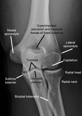
The ulna is one of the two long bones in the forearm, the other being the radius. It runs from the elbow to the wrist and is located on the medial side of the forearm, next to the bone called the humerus in the upper arm. The ulna plays a crucial role in the movement of the forearm and also serves as an attachment site for various muscles.
A bone fracture is a medical condition in which there is a partial or complete break in the continuity of a bone due to external or internal forces. Fractures can occur in any bone in the body and can vary in severity from a small crack to a shattered bone. The symptoms of a bone fracture typically include pain, swelling, bruising, deformity, and difficulty moving the affected limb. Treatment for a bone fracture may involve immobilization with a cast or splint, surgery to realign and stabilize the bone, or medication to manage pain and prevent infection. The specific treatment approach will depend on the location, type, and severity of the fracture.
Fracture healing is the natural process by which a broken bone repairs itself. When a fracture occurs, the body responds by initiating a series of biological and cellular events aimed at restoring the structural integrity of the bone. This process involves the formation of a hematoma (a collection of blood) around the fracture site, followed by the activation of inflammatory cells that help to clean up debris and prepare the area for repair.
Over time, specialized cells called osteoblasts begin to lay down new bone matrix, or osteoid, along the edges of the broken bone ends. This osteoid eventually hardens into new bone tissue, forming a bridge between the fracture fragments. As this process continues, the callus (a mass of newly formed bone and connective tissue) gradually becomes stronger and more compact, eventually remodeling itself into a solid, unbroken bone.
The entire process of fracture healing can take several weeks to several months, depending on factors such as the severity of the injury, the patient's age and overall health, and the location of the fracture. In some cases, medical intervention may be necessary to help promote healing or ensure proper alignment of the bone fragments. This may include the use of casts, braces, or surgical implants such as plates, screws, or rods.
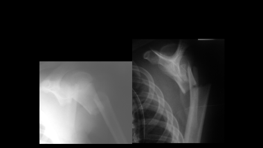
A hip fracture is a medical condition referring to a break in the upper part of the femur (thigh) bone, which forms the hip joint. The majority of hip fractures occur due to falls or direct trauma to the area. They are more common in older adults, particularly those with osteoporosis, a condition that weakens bones and makes them more prone to breaking. Hip fractures can significantly impact mobility and quality of life, often requiring surgical intervention and rehabilitation.
A femoral fracture is a medical term that refers to a break in the thigh bone, which is the longest and strongest bone in the human body. The femur extends from the hip joint to the knee joint and is responsible for supporting the weight of the upper body and allowing movement of the lower extremity. Femoral fractures can occur due to various reasons such as high-energy trauma, low-energy trauma in individuals with weak bones (osteoporosis), or as a result of a direct blow to the thigh.
Femoral fractures can be classified into different types based on their location, pattern, and severity. Some common types of femoral fractures include:
1. Transverse fracture: A break that occurs straight across the bone.
2. Oblique fracture: A break that occurs at an angle across the bone.
3. Spiral fracture: A break that occurs in a helical pattern around the bone.
4. Comminuted fracture: A break that results in multiple fragments of the bone.
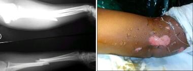
5. Open or compound fracture: A break in which the bone pierces through the skin.
6. Closed or simple fracture: A break in which the bone does not pierce through the skin.
Femoral fractures can cause severe pain, swelling, bruising, and difficulty walking or bearing weight on the affected leg. Diagnosis typically involves a physical examination, medical history, and imaging tests such as X-rays or CT scans. Treatment may involve surgical intervention, including the use of metal rods, plates, or screws to stabilize the bone, followed by rehabilitation and physical therapy to restore mobility and strength.
A spinal fracture, also known as a vertebral compression fracture, is a break in one or more bones (vertebrae) of the spine. This type of fracture often occurs due to weakened bones caused by osteoporosis, but it can also result from trauma such as a car accident or a fall.
In a spinal fracture, the front part of the vertebra collapses, causing the height of the vertebra to decrease, while the back part of the vertebra remains intact. This results in a wedge-shaped deformity of the vertebra. Multiple fractures can lead to a hunched forward posture known as kyphosis or dowager's hump.
Spinal fractures can cause pain, numbness, tingling, or weakness in the back, legs, or arms, depending on the location and severity of the fracture. In some cases, spinal cord compression may occur, leading to more severe symptoms such as paralysis or loss of bladder and bowel control.
Fracture fixation, internal, is a surgical procedure where a fractured bone is fixed using metal devices such as plates, screws, or rods that are implanted inside the body. This technique helps to maintain the alignment and stability of the broken bone while it heals. The implants may be temporarily or permanently left inside the body, depending on the nature and severity of the fracture. Internal fixation allows for early mobilization and rehabilitation, which can result in a faster recovery and improved functional outcome.
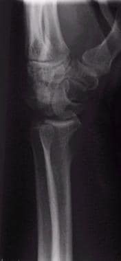
A radius fracture is a break in the bone that runs from the wrist to the elbow, located on the thumb side of the forearm. Radius fractures can occur as a result of a fall, direct blow to the forearm, or a high-energy collision such as a car accident. There are various types of radius fractures, including:
1. Distal radius fracture: A break at the end of the radius bone, near the wrist joint, which is the most common type of radius fracture.
2. Radial shaft fracture: A break in the middle portion of the radius bone.
3. Radial head and neck fractures: Breaks in the upper part of the radius bone, near the elbow joint.
4. Comminuted fracture: A complex radius fracture where the bone is broken into multiple pieces.
5. Open (compound) fracture: A radius fracture with a wound or laceration in the skin, allowing for communication between the outside environment and the fractured bone.
6. Intra-articular fracture: A radius fracture that extends into the wrist joint or elbow joint.
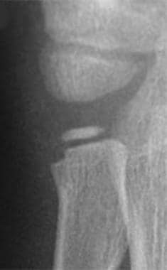
7. Torus (buckle) fracture: A stable fracture where one side of the bone is compressed, causing it to buckle or bend, but not break completely through.
Symptoms of a radius fracture may include pain, swelling, tenderness, bruising, deformity, limited mobility, and in some cases, numbness or tingling in the fingers. Treatment options depend on the type and severity of the fracture but can range from casting to surgical intervention with implant fixation.
A comminuted fracture is a type of bone break where the bone is shattered into three or more pieces. This type of fracture typically occurs after high-energy trauma, such as a car accident or a fall from a great height. Commminuted fractures can also occur in bones that are weakened by conditions like osteoporosis or cancer. Because of the severity and complexity of comminuted fractures, they often require extensive treatment, which may include surgery to realign and stabilize the bone fragments using metal screws, plates, or rods.
Fracture fixation is a surgical procedure in orthopedic trauma surgery where a fractured bone is stabilized using various devices and techniques to promote proper healing and alignment. The goal of fracture fixation is to maintain the broken bone ends in correct anatomical position and length, allowing for adequate stability during the healing process.
There are two main types of fracture fixation:
1. Internal fixation: In this method, metal implants like plates, screws, or intramedullary rods are inserted directly into the bone to hold the fragments in place. These implants can be either removed or left in the body once healing is complete, depending on the type and location of the fracture.
2. External fixation: This technique involves placing pins or screws through the skin and into the bone above and below the fracture site. These pins are then connected to an external frame that maintains alignment and stability. External fixators are typically used when there is significant soft tissue damage, infection, or when internal fixation is not possible due to the complexity of the fracture.
The choice between internal and external fixation depends on various factors such as the type and location of the fracture, patient's age and overall health, surgeon's preference, and potential complications. Both methods aim to provide a stable environment for bone healing while minimizing the risk of malunion, nonunion, or deformity.
Osteoporotic fractures are breaks or cracks in bones that occur as a result of osteoporosis, a condition characterized by weak and brittle bones. Osteoporosis causes bones to lose density and strength, making them more susceptible to fractures, even from minor injuries or falls.
The most common types of osteoporotic fractures are:
1. Hip fractures: These occur when the upper part of the thigh bone (femur) breaks, often due to a fall. Hip fractures can be serious and may require surgery and hospitalization.
2. Vertebral compression fractures: These occur when the bones in the spine (vertebrae) collapse, causing height loss, back pain, and deformity. They are often caused by everyday activities, such as bending or lifting.
3. Wrist fractures: These occur when the bones in the wrist break, often due to a fall. Wrist fractures are common in older adults with osteoporosis.
4. Other fractures: Osteoporotic fractures can also occur in other bones, such as the pelvis, ribs, and humerus (upper arm bone).
Prevention is key in managing osteoporosis and reducing the risk of osteoporotic fractures. This includes getting enough calcium and vitamin D, engaging in regular weight-bearing exercise, avoiding smoking and excessive alcohol consumption, and taking medications as prescribed by a healthcare provider.
Stress fractures are defined as small cracks or severe bruising in bones that occur from repetitive stress or overuse. They most commonly occur in weight-bearing bones, such as the legs and feet, but can also occur in the arms, hips, and back. Stress fractures differ from regular fractures because they typically do not result from a single, traumatic event. Instead, they are caused by repeated stress on the bone that results in microscopic damage over time. Athletes, military personnel, and individuals who engage in high-impact activities or have weak bones (osteoporosis) are at increased risk of developing stress fractures. Symptoms may include pain, swelling, tenderness, and difficulty walking or bearing weight on the affected bone.
Spontaneous fractures are bone breaks that occur without any identifiable trauma or injury. They are typically caused by underlying medical conditions that weaken the bones, making them more susceptible to breaking under normal stress or weight. The most common cause of spontaneous fractures is osteoporosis, a condition characterized by weak and brittle bones. Other potential causes include various bone diseases, certain cancers, long-term use of corticosteroids, and genetic disorders affecting bone strength.
It's important to note that while the term "spontaneous" implies that the fracture occurred without any apparent cause, it is usually the result of an underlying medical condition. Therefore, if you experience a spontaneous fracture, seeking medical attention is crucial to diagnose and manage the underlying cause to prevent future fractures and related complications.
A femoral neck fracture is a type of hip fracture that occurs in the narrow, vertical section of bone just below the ball of the femur (thigh bone) that connects to the hip socket. This area is called the femoral neck. Femoral neck fractures can be categorized into different types based on their location and the direction of the fractured bone.
These fractures are typically caused by high-energy trauma, such as car accidents or falls from significant heights, in younger individuals. However, in older adults, particularly those with osteoporosis, femoral neck fractures can also result from low-energy trauma, like a simple fall from standing height.
Femoral neck fractures are often serious and require prompt medical attention. Treatment usually involves surgery to realign and stabilize the broken bone fragments, followed by rehabilitation to help regain mobility and strength. Potential complications of femoral neck fractures include avascular necrosis (loss of blood flow to the femoral head), nonunion or malunion (improper healing), and osteoarthritis in the hip joint.
The radius is one of the two bones in the forearm in humans and other vertebrates. In humans, it runs from the lateral side of the elbow to the thumb side of the wrist. It is responsible for rotation of the forearm and articulates with the humerus at the elbow and the carpals at the wrist. Any medical condition or injury that affects the radius can impact the movement and function of the forearm and hand.
 Ulna fracture
Distal radius fracture
Olecranon
Colles' fracture
Smith's fracture
Galeazzi fracture
Coronoid process of the ulna
List of orthopedic implants
2023 in paleomammalogy
Tepexpan man
Monteggia fracture
Len Hutton
DeMarcus Ware
Pulled elbow
Madelung's deformity
Victoria Leonardo
Olecranon fracture
UFC on ESPN: Chiesa vs. Magny
Classification of distal radius fractures
Older's classification
Brian Givens
Distal radioulnar articulation
MS Express Aphrodite
Accessory bone
Ebb Cade
Kirschner wire
Krapina Neanderthal site
Ulnar styloid process
Willie Calhoun
Pseudofracture
Ulna fracture - Wikipedia
 Common Fractures of the Radius and Ulna | AAFP
 ICD-10 Code for Displaced fracture of left ulna styloid process, sequela- S52.612S- Codify by AAPC
Ulna Fractures | Palmetto Profiles
 Isolated ulna fracture - WikEM
 Wrist Fracture Management in the ED: Practice Essentials, Pathophysiology, Prognosis
 cpt code for orif distal radius and ulna fracture
 ulna fracture - Welcome to SYS Medtech International Pvt. Ltd.
 Monteggia lesion, transverse fracture of ulna: open reduction, compression plating
Ulna Large Left with transverse fracture U2018 - London Bone Company
 Animal Surgeries - AskMen
 Millennium Veterinary Practice - Fractured radius and ulna in a young whippet
 Neglected monteggia fracture in adult treated with open reduction and ulna lengthening using tricortical bone graft: a case...
 ICD-10-CM Diagnosis Code S52.0 - Fracture of upper end of ulna
 Fracture of Coronoid Process of Ulna Phase I | iTheraputix | Improving Health Care Through Technology
ORIF - Plating for Simple fracture of the ulna, with dislocation of proximal radioulnar joint (Monteggia)
Fracture of Combination Proximal Radius/Ulna - Left | Combination Proximal Radius/Ulna | Elbow | Trauma | RFx Showroom
Histological evaluation of transverse fracture healing in pigeon (Columba livia) ulna - Universiti Putra Malaysia...
 Ulnar nerve palsy following a closed fracture of shaft of radius and ulna in a child
 Olecranon Fractures - Orthopaedic Web Links
 Accident Search Results Page | Occupational Safety and Health Administration osha.gov
Bariatric Surgery Linked to Fracture Risk
Single dorsal incision approach for Plate fixation of Radius ulna midshaft fracture; Interesting case report - Journal of...
 Management of compound fracture of the radius and ulna using pop and splints in a Nigerian Indigenous breed of dog: A case...
 View of Radius-ulna shaft fracture with distal radioulnar joint instability in a case of ipsilateral malunited colles fracture:...
 A Love Supreme | Discover Magazine
S52.265B - Nondisplaced segmental fracture of shaft of ulna, left arm, initial encounter for open fracture type I or II - ICD...
 Massachusetts General Hospital
 OTA Fracturebook: Current Practice of Trauma and Fracture Management | Orthopaedic Trauma Association (OTA)
 Thieme E-Books & E-Journals - Orthopedic Trauma Directions / Most Read
Bone32
- An ulna fracture is a break in the ulna bone, one of the two bones in the forearm. (wikipedia.org)
- It is often associated with a fracture of the other forearm bone, the radius. (wikipedia.org)
- The ulna bone can also break after falling on the forearm or falling on an outstretched arm. (wikipedia.org)
- Fractures of the ulna can occur at different levels of the bone: near the wrist, in the middle or near the elbow. (wikipedia.org)
- When there is a displaced fracture and also when the radioulnar joints are involved an operation is often performed, using either flexible rods or screws and plates in order to reduce the fracture and immobilise the bone. (wikipedia.org)
- Fractures of the larger bone of the forearm. (musc.edu)
- Vitamin D receptor genotype and the risk of bone fractures in women. (musc.edu)
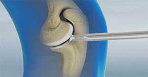
- As the load screw is tightened, the tension generated in the plate compresses the fracture evenly across the full diameter of the bone. (aofoundation.org)
- In pediatric fractures there is often a combination of patterns of bone failure. (aofoundation.org)
- Provided the alignment of the bone is anatomical and overall reduction is stable it is not necessary to perfectly reduce the entire fracture. (aofoundation.org)
- A small bone lever can be used to reduce transverse, or short oblique, fractures as illustrated. (aofoundation.org)
- In children, interdigitation of the fracture fragments may be prevented by plastic deformity of the bone ends. (aofoundation.org)
- A plate was secured over the radius fracture, after it was reduced, and held in place with a number of bone screws. (millenniumvets.co.uk)
- The bone of the distal radius, ulna and carpus was re-absorbed. (orthopaedicweblinks.com)
- Previous studies involving premenopausal women have found a risk of bone mineral density loss and fracture with bariatric surgery , but little was known about the risk among men. (medscape.com)
- The purpose of the new study "was to see if we see the same risk of fracture in veterans who are older men, so kind of the opposite of the typical bariatric patient," said Koh, who presented the research at the American Society for Bone and Mineral Research (ASBMR) 2023 Annual Meeting. (medscape.com)
- Radius and Ulna shaft fractures, also known as both bone forearm fractures, are common fractures caused due to direct or indirect trauma. (jtojournal.com)
- Conventionally, such fractures are managed using either external fixatives such as Ilizarov apparatus, Octopod external fixator, and Taylor spatial frame or internal fixatives such as intramedullary pin, bone plates, and cerclage wires. (veterinarypaper.com)
- This is a stable fracture, meaning that the broken pieces of bone are still in position and have not separated apart (displaced). (bmc.org)
- The fracture is across the upper or lower portion of the shaft of the bone and does not affect the growth plate. (bmc.org)
- The fracture extends through a portion of the bone, causing it to bend on the other side. (bmc.org)
- Because the growth plate helps determine the future length and shape of the mature bone, this type of fracture requires prompt attention. (bmc.org)
- A broken bone, fracture, ligament tear, or other elbow injury comes with heavy medical costs. (ehlinelaw.com)
- Some fractures will heal quickly without complication but others, such as comminuted or displaced fractures, may result in an incomplete union of the bone, and perhaps involve surgery to insert metalwork to support and help unite the fracture. (clarkewillmott.com)
- Osteoporosis is characterized by compromised bone strength due to loss of bone mass and deterioration of bone quality, resulting in increased fracture risk. (ajmc.com)
- Wrist Fractures Wrist fractures may involve the lower end of one or both of the forearm bones (radius or ulna) or, less often, a bone in the base of the hand. (msdmanuals.com)
- Fractures of the Upper Forearm Elbow fractures may involve the upper arm bone (humerus) near the elbow-called lower (distal) humeral fractures-or one of the upper forearm bones (radius or ulna)-called radial head fractures. (msdmanuals.com)
- Diagnosis A fracture is a crack or break in a bone. (msdmanuals.com)
- Most fractures result from force applied to a bone. (msdmanuals.com)
- The areas of each carpal bone and epiphyses of the ulna and radius were measured, and these measurements were added together to obtain the bone area (Bo). (bvsalud.org)
- Periostin-Like-Factor (PLF) is expressed in developing bone and is up regulated in adult bones that undergo remodeling due to fractures or repeated high force activity which leads to inflammation followed by injury of the bone. (cdc.gov)
- Loaded bone responds by remodeling and under extreme situations stress fractures and resorptive spaces develop. (cdc.gov)
Fixation14
- Cannulated screw fixation of refractory olecranon stress fractures with and without associated injuries allows a return to baseball. (musc.edu)
- Anterior approach for operative fixation of coronoid fractures in complex elbow instability. (musc.edu)
- Obtain immediate consultation with a hand specialist or orthopedic surgeon for open or unstable fractures and those requiring fixation. (medscape.com)
- CPT 25608 indicates fractures requiring fixation of one or two segments and Code 25609 indicates fractures requiring fixation of three or more fractures. (namokarhealthcare.com)
- ORIF distal radius CPT fractures can get complicated because of new technology (e.g., fracture specific fixation, fixed-angle plate fixation), and the recognition of distal radial fractures requires accurate repair. (namokarhealthcare.com)
- Plating of pediatric forearm shaft fractures follows the technique for plate fixation in adults. (aofoundation.org)
- After anatomical restoration and stable fixation of the ulnar fracture, relocation of the radial head will usually result. (aofoundation.org)
- In Monteggia fracture-dislocations, anatomical reduction and stable fixation of the ulna are mandatory, to ensure stable relocation of the radial head. (aofoundation.org)
- Once operative fixation of the ulna has been completed, the surgeon must ensure the stability of the reduced radial head, preferably under image intensification. (aofoundation.org)
- Anatomic reduction and fixation of the ulna is achieved first, through a standard posterior approach . (aofoundation.org)
- In cases of persisting radial head instability after anatomical fixation of the ulna, interposed annular ligament or the torn joint capsule is usually the cause and should be extracted from the joint and sutured. (aofoundation.org)
- After fixation of the ulna, check the position of the radial head, which reduces in most cases spontaneously. (aofoundation.org)
- The fracture was fixed with external skeletal fixation and all operations performed under Isoflurane anaesthesia. (upm.edu.my)
- The purpose of this study was to find out the histological assessment of healing of ulna fracture stabilized with external skeletal fixation in bird model. (upm.edu.my)
Injuries8
- Other causes of ulna fractures include sporting injuries, road traffic incidents, falls from a height, and conditions such as osteoporosis and potentially both primary and secondary cancer. (wikipedia.org)
- Osteology of the coronoid process with clinical correlation to coronoid fractures in terrible triad injuries. (musc.edu)
- Fractures of the distal radius and ulna account for three fourths of wrist injuries. (medscape.com)
- An eighty years old female had fractures of both radius and Ulna shaft with Gustilo Anderson type two open injuries. (jtojournal.com)
- The principles of multiple coding of injuries should be followed in coding fractures. (icdlist.com)
- Whilst the majority of injuries involving fractures will affect only one or bones, in the case of very serious accidents such as high speed road collisions or falls from height, a large number of bones might be broken. (clarkewillmott.com)
- Some companies may cover routine cleanings but require additional fees for any accidents or injuries such as a broken/fractured tooth or a tooth root abscess. (petmd.com)
- Fractures usually result from injuries or overuse. (msdmanuals.com)
Transverse fracture3
- A reduced transverse fracture cannot be maintained with reduction forceps alone. (aofoundation.org)
- Ulna Large Left - With an olecranon transverse fracture. (londonboneco.com)
- A transverse fracture was created at mid shat of left ulna. (upm.edu.my)
Types of forearm fractures1
- What are the different types of forearm fractures in children? (bmc.org)
Left ulna1
- Radiographs revealed that the bird's left ulna was fractured. (wildlifecenter.org)
Multiple fractures4
- Multiple fractures and broken bones can have a significant impact on your ability to live your life independently. (clarkewillmott.com)
- We have a long history of acting for clients who have claimed compensation for broken bones and multiple fractures sustained in motorcycle accidents, car accident, at work and generally as a result of the negligence of others. (clarkewillmott.com)
- Ipsilateral multiple fractures in children often result from high energy trauma and are associated with complications. (nepjol.info)
- OBJECTIVES: This study aimed to detect single or multiple fractures in the ulna or radius using deep learning techniques fed on upper-extremity radiographs. (bvsalud.org)
Angulation5
- Depending on the degree of angulation, buckle and greenstick fractures can be managed with immobilization. (aafp.org)
- These fractures are treated with immobilization or surgery, depending on the degree of displacement and angulation. (aafp.org)
- The authors present a case of a 14-year-old boy who developed ulnar nerve palsy following a left radius and ulna midshaft fracture with significant angulation and displacement. (orthocasereports.com)
- 50% of fracture opposition and 10 to 15 degrees of angulation are considered stable and can be treated conservatively. (clinicaladvisor.com)
- Alteration in the shape of the trochlea changes the angle of articulation with the ulna, resulting in increased elbow angulation. (slideshare.net)
Olecranon2
- Hume fracture - a fracture of the olecranon with an associated anterior dislocation of the radial head. (wikipedia.org)
- Fractures of the olecranon are common and are usually detected easily. (orthopaedicweblinks.com)
Radius Fractures4
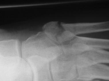
- Distal ulna fractures typically occur along with distal radius fractures. (wikipedia.org)
- In adults, distal radius fractures are the most common forearm fractures and are typically caused by a fall onto an outstretched hand. (aafp.org)
- 1 - 3 The rate of distal radius fractures is highest (nearly double) in people younger than 18 years and older than 65 years. (aafp.org)
- Patients aged 6-10 years and those aged 60-69 years have the greatest frequency of distal radius fractures. (medscape.com)
Shaft6
- For transverse forearm shaft fractures, interfragmentary compression can be achieved with a compression plate. (aofoundation.org)
- In short oblique forearm shaft fractures an empty plate hole may be necessary at the level of the fracture. (aofoundation.org)
- S52.265B is a billable ICD-10 code used to specify a medical diagnosis of nondisplaced segmental fracture of shaft of ulna, left arm, initial encounter for open fracture type i or ii. (icdlist.com)
- S52.265B is an initial encounter code, includes a 7th character and should be used while the patient is receiving active treatment for a condition like nondisplaced segmental fracture of shaft of ulna left arm for open fracture type i or ii. (icdlist.com)
- The isolated fracture of the ulnar shaft. (clinicaladvisor.com)
- Ulna/ulnar shaft fracture. (clinicaladvisor.com)
Reduction8
- Urgent reduction of fractures may be necessary when neurovascular status has been compromised. (medscape.com)
- Exaggeration of the fracture deformity may be required to loosen the periosteum and allow gentle reduction. (aofoundation.org)
- Residual plastic deformity may prevent anatomical reduction of some of the fracture edges. (aofoundation.org)
- Reduce the fracture anatomically, using a reduction forceps on each main fragment. (aofoundation.org)
- However, transverse fractures are usually dentate and are intrinsically stable after anatomical reduction. (aofoundation.org)
- Be aware that malreduction of the ulna will lead to insufficient spontaneous anatomical reduction and/or instability of the radial head. (aofoundation.org)
- Another study found a reduction of fracture risk associated with sleeve gastrectomy and no difference between RYGB and nonsurgical matched control patients in a Medicare population. (medscape.com)
- Depending on the type of fracture, reduction may or may not involve surgery. (msdmanuals.com)
Monteggia Fracture4
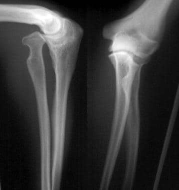
- Monteggia fracture - a fracture of the near to elbow end of the ulna with the dislocation of the head of the radius at the elbow joint. (wikipedia.org)
- Monteggia Fracture (fracture of proximal ulna) Galeazzi facture (displaced fracture of the radius) If the fracture is not displaced, is stable, and is not associated with another fracture, it may be treated with a cast for around five to six weeks. (wikipedia.org)
- In Monteggia fracture-dislocations, the ulnar fracture is associated with a dislocation of the radial head. (aofoundation.org)
- Monteggia fracture. (bmc.org)
Fall onto an outstretched hand2
- A fall onto an outstretched hand is the most common mechanism of injury for fractures of the radius and ulna. (aafp.org)
- Causes of wrist fracture include fall onto an outstretched hand and direct trauma. (medscape.com)
Midshaft2
- Isolated midshaft ulna (nightstick) fractures are often caused by a direct blow to the forearm. (aafp.org)
- We found that under specific circumstances such as open injury where two separate incisions for radius and ulna are inappropriate, a single dorsal curvilinear incision for radius and Ulna midshaft fractures is a safe and effective alternative method. (jtojournal.com)
Trauma3
- The diagnosis of an ulna fracture is made after taking the persons history, which usually includes a history of forearm pain following trauma, and then examining the injured forearm followed by an x-ray of the relevant part. (wikipedia.org)
- Radial head fractures may be difficult to visualize on initial imaging but should be suspected when there are limitations of elbow extension and supination following trauma. (aafp.org)
- A case of compound fracture of both radius and ulnar which occurs from trauma as a result of a rodent's trap was presented to the small animal unit of the Veterinary Teaching Hospital, the University of Maiduguri with the chief complaint of trauma and severe bleeding which was haphazardly managed before the presentation. (veterinarypaper.com)
Humerus Fracture1
- Simultaneous segmental humerus fracture with ipsilateral forearm is an uncommon injury and scarcely mentioned in the literature. (nepjol.info)
Radial4
- Treatment of radial head fractures depends on the specific characteristics of the fracture using the Mason classification. (aafp.org)
- of radius AND ulna 25600 Closed treatment of distal radial fracture (e.g. (namokarhealthcare.com)
- Hardware removal rates due to infection increased in all fractures except radial/ulnar fractures. (namokarhealthcare.com)
- In recent years, the open treatment of a distal radial fracture has become very complicated. (namokarhealthcare.com)
Surgical5
- Combined fractures involving both the ulna and radius generally require surgical correction. (aafp.org)
- We've shown here that especially men who are on the older side, who go through surgical weight loss, do have a higher risk of fracture compared to those who are similarly obese but have not had the operation," said Schafer, a professor of medicine at the University of California, San Francisco, and chief of endocrinology and metabolism at the San Francisco VA Medical Center. (medscape.com)
- The reasons for increased fracture risk following surgical weight loss remains unknown, according to Paccou, but they could include mechanical unloading, loss of lean mass, and hormone and nutrition changes. (medscape.com)
- Surgical exposure of the ulna and proximal third of the radius through one incision. (jtojournal.com)
- Care for complications of surgical treatment for fracture repairs during the healing or recovery phase should be coded with the appropriate complication codes. (icdlist.com)
FEMUR1
- The open fracture designations in the assignment of the 7th character for fractures of the forearm, femur and lower leg, including ankle are based on the Gustilo open fracture classification. (icdlist.com)
Carpal2
- The aim of the present study was to develop a specific formula by measuring the developing teeth, carpal bones, and epiphyses of the ulna and radius to determine the chronological age in Turkish children. (bvsalud.org)
- The carpal area (Ca), covering the epiphyses of ulna and radius and the carpal bones, was measured on the X-rays of left hand. (bvsalud.org)
High risk of fracture2
- The investigators excluded individuals who were at high risk of fracture because of another condition, such as organ transplantation or dialysis. (medscape.com)
- These findings highlight the need for effective fracture prevention strategies in patients at high risk of fracture. (ajmc.com)
Upper extremity2
- Fractures of the radius and ulna are the most common fractures of the upper extremity, with distal fractures occurring more often than proximal fractures. (aafp.org)
- MATERIALS AND METHODS: The data set used in the retrospective study consisted of different types of upper extremity radiographs obtained from an open-source dataset, with 4,480 images with fractures and 4,383 images without fractures. (bvsalud.org)
Fragility fractures1
- The current findings suggest a need for better management of fragility fractures to reduce osteoporosis cost of illness. (ajmc.com)
Type of fracture1
- In most cases, this type of fracture occurs in the growth plate of the radius near the wrist. (bmc.org)
Classification1
- When the Gustilo classification type is not specified for an open fracture, the 7th character for open fracture type I or II should be assigned (B, E, H, M, Q). (icdlist.com)
Wrist fractures2
- Wrist fractures may also be caused by hyperflexion mechanisms and by direct blows to the wrist. (medscape.com)
- Fractures of the distal radius account for one sixth of all fractures seen and treated in the ED. Although there is ittle or no risk of death associated with isolated wrist fractures, the potential does exist for substantial morbidity, including primarily arthritis, chronic pain, limitation of motion, and physical deformity. (medscape.com)
Bones6
- The forearm is made up of a combination of two bones- the ulna and the radius. (sysmedtechint.com)
- If a fracture happens in any one of the bones then it is known as a forearm fracture. (sysmedtechint.com)
- A child's bones heal more quickly than an adult's, so it is important to treat a fracture promptly-before healing begins-to avoid future problems. (bmc.org)
- Forearm fractures involve the middle of one or both of the forearm bones (radius and ulna). (msdmanuals.com)
- Usually one of the bones (radius or ulna) in the forearm is broken from a direct blow. (msdmanuals.com)
- For a simple fracture, if the bones are realigned without needing surgery, a splint is used to hold the bones in place. (msdmanuals.com)
Diagnosis1
- Even though doctors can usually identify forearm fractures based on a physical examination, diagnosis includes x-rays taken to pinpoint the fracture's location and determine the extent of the injury. (msdmanuals.com)
Galeazzi2
- Galeazzi fracture - not a fracture of the ulna but a displaced fracture of the radius accompanied by a dislocation of the ulna at the wrist, where the radius and ulna come together. (wikipedia.org)
- Galeazzi fracture. (bmc.org)
Anatomical1
- It is located on the lateral side of the forearm parallel to the ulna (in anatomical position with arms hanging at the sides of the body, palms facing forward) between the thumb and the elbow. (namokarhealthcare.com)
Nightstick3
- An ulna fracture can be a single break as in a so called nightstick fracture, which can be caused by someone being hit on the inside of the forearm often by a stick, notably when they are holding their arm up to protect their head from injury. (wikipedia.org)
- Nightstick fracture is a fracture of the middle portion of the ulna without other fractures. (wikipedia.org)
- The term "nightstick fracture" originated from the notion that a person hit by a police truncheon would hold their arms up to protect their heads from injury. (wikipedia.org)
Distal humerus1
- The injury pattern consists of ipsilateral supracondylar fracture humerus with distal humerus and ipsilateral distal forearm fracture. (nepjol.info)
Torus3
Scaphoid1
- Distal radius, scaphoid, and lunate fractures usually are the result of a fall on an outstretched hand. (medscape.com)
Pediatric forearm1
- Clinical and radiographic comparison of single-sugar-tong splint to long-arm cast immobilization for pediatric forearm fractures. (musc.edu)
Typically1
- [ 7 ] The distal radius and ulna typically demonstrate rachitic lesions early on radiographs. (medscape.com)
Complications2
- without manipulation 25606 Percutaneous When coding a closed fracture, coders must add one of the following seventh characters to each code: Compressive neuropathy is one of the most important complications of Colles fractures and usually involves the median nerve. (namokarhealthcare.com)
- Care of complications of fractures, such as malunion and nonunion, should be reported with the appropriate 7th character for subsequent care with nonunion (K, M, N,) or subsequent care with malunion (P, Q, R). (icdlist.com)
Occur4
- About three out of four forearm fractures in children occur at the wrist end of the radius. (bmc.org)
- Forearm fractures often occur when children are doing activities like playing or participating in sports. (bmc.org)
- Children love to run, hop, skip, jump and tumble, all of which are activities that could potentially result in a fracture to the forearm should an unexpected fall occur. (bmc.org)
- Isolated ulnar diaphyseal fractures are known as "night stick" fractures as the injury may occur when the patient blocks overhead contact from a blunt object. (clinicaladvisor.com)
Greenstick2
Tibia1
- What is the CPT code for ORIF tibia fracture? (namokarhealthcare.com)
Radioulnar2
- In order to test the stability of the distal radioulnar joint, the ulna is compressed against the radius. (aofoundation.org)
- 2. Bauer G, Arand M, Mutschler W. Post-traumatic radioulnar synostosis after forearm fracture osteosynthesis. (jtojournal.com)
.jpg)