Upper Extremity Deep Vein Thrombosis
Upper Extremity
Lower Extremity
Subclavian Vein
Femoral Vein
Characterizing resolution of catheter-associated upper extremity deep venous thrombosis. (1/26)
(+info)Upper-extremity deep vein thrombosis related to central venous port systems implanted in cancer patients. (2/26)
(+info)Playing games with a thrombus: a dangerous match. Paradoxical embolism from a huge central venous cathether thrombus: a case report. (3/26)
(+info)A comprehensive review of Paget-Schroetter syndrome. (4/26)
(+info)Preoperative thrombolysis and venoplasty affords no benefit in patency following first rib resection and scalenectomy for subacute and chronic subclavian vein thrombosis. (5/26)
(+info)Internal jugular vein thrombosis: outcome and risk factors. (6/26)
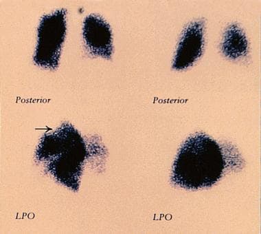
(+info)Upper extremity deep vein thrombosis in a 25 year old apparently healthy man. (7/26)
This case of upper extremity deep vein thrombosis is selected for case report as it is a rare form of deep vein thrombosis without a very well established treatment modality and prognosis. The objective of this study was to report the outcome of a 25 years old male patient with idiopathic upper extremity deep vein thrombosis treated conservatively with low molecular weight heparin (LMWH) and oral warfarin. The data sources used were patient interview, laboratory and radiology investigation results and patient charts. The patient had no apparent recurrence or complication for 3 years except the presence of occasional dull pain over the affected left upper extremity. (+info)Right intra-atrial catheter insertion at the end stage of peripheral vascular access for dialysis. (8/26)
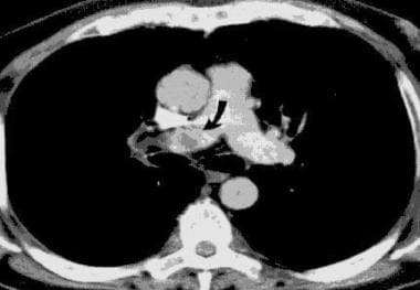
(+info)Upper extremity deep vein thrombosis (UEDVT) is a medical condition that refers to the formation of a blood clot (thrombus) in the deep veins located in the arm or shoulder. This condition can occur due to various reasons, including trauma, surgery, cancer, certain medications, and underlying medical conditions that increase the risk of blood clotting.
The deep veins are larger vessels that run through the body's muscles and are surrounded by fascia, a connective tissue. UEDVT can cause partial or complete blockage of blood flow in the affected vein, leading to swelling, pain, redness, warmth, and decreased function in the arm or hand. In some cases, the clot can break off and travel to the lungs, causing a potentially life-threatening condition called pulmonary embolism (PE).
Diagnosis of UEDVT typically involves a physical exam, medical history, and imaging tests such as ultrasound, CT scan, or MRI. Treatment may include anticoagulant medications to prevent the clot from growing or breaking off, thrombolytic therapy to dissolve the clot, or surgical intervention in severe cases. Compression stockings or other devices may also be used to help improve blood flow and reduce swelling.
Venous thrombosis is a medical condition characterized by the formation of a blood clot (thrombus) in the deep veins, often in the legs (deep vein thrombosis or DVT), but it can also occur in other parts of the body such as the arms, pelvis, or lungs (pulmonary embolism).
The formation of a venous thrombus can be caused by various factors, including injury to the blood vessel wall, changes in blood flow, and alterations in the composition of the blood. These factors can lead to the activation of clotting factors and platelets, which can result in the formation of a clot that blocks the vein.
Symptoms of venous thrombosis may include swelling, pain, warmth, and redness in the affected area. In some cases, the clot can dislodge and travel to other parts of the body, causing potentially life-threatening complications such as pulmonary embolism.
Risk factors for venous thrombosis include advanced age, obesity, smoking, pregnancy, use of hormonal contraceptives or hormone replacement therapy, cancer, recent surgery or trauma, prolonged immobility, and a history of previous venous thromboembolism. Treatment typically involves the use of anticoagulant medications to prevent further clotting and dissolve existing clots.
The term "upper extremity" is used in the medical field to refer to the portion of the upper limb that extends from the shoulder to the hand. This includes the arm, elbow, forearm, wrist, and hand. The upper extremity is responsible for various functions such as reaching, grasping, and manipulating objects, making it an essential part of a person's daily activities.
The term "lower extremity" is used in the medical field to refer to the portion of the human body that includes the structures below the hip joint. This includes the thigh, lower leg, ankle, and foot. The lower extremities are responsible for weight-bearing and locomotion, allowing individuals to stand, walk, run, and jump. They contain many important structures such as bones, muscles, tendons, ligaments, nerves, and blood vessels.
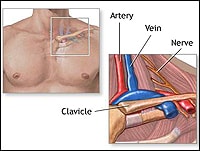
The subclavian vein is a large venous structure that carries deoxygenated blood from the upper limb and part of the thorax back to the heart. It forms when the axillary vein passes through the narrow space between the first rib and the clavicle (collarbone), becoming the subclavian vein.
On the left side, the subclavian vein joins with the internal jugular vein to form the brachiocephalic vein, while on the right side, the subclavian vein directly merges with the internal jugular vein to create the brachiocephalic vein. These brachiocephalic veins then unite to form the superior vena cava, which drains blood into the right atrium of the heart.
The subclavian vein is an essential structure for venous access in various medical procedures and interventions, such as placing central venous catheters or performing blood tests.
A pulmonary embolism (PE) is a medical condition that occurs when a blood clot, often formed in the deep veins of the legs (deep vein thrombosis), breaks off and travels to the lungs, blocking one or more pulmonary arteries. This blockage can lead to various symptoms such as shortness of breath, chest pain, rapid heart rate, and coughing up blood. In severe cases, it can cause life-threatening complications like low oxygen levels, hypotension, and even death if not promptly diagnosed and treated with anticoagulant medications or thrombolytic therapy to dissolve the clot.
Thrombophlebitis is a medical condition characterized by the inflammation and clotting of blood in a vein, usually in the legs. The term thrombophlebitis comes from two words: "thrombo" which means blood clot, and "phlebitis" which refers to inflammation of the vein.
The condition can occur in superficial or deep veins. Superficial thrombophlebitis affects the veins just below the skin's surface, while deep vein thrombophlebitis (DVT) occurs in the deeper veins. DVT is a more serious condition as it can lead to complications such as pulmonary embolism if the blood clot breaks off and travels to the lungs.
Symptoms of thrombophlebitis may include redness, warmth, pain, swelling, or discomfort in the affected area. In some cases, there may be visible surface veins that are hard, tender, or ropy to touch. If left untreated, thrombophlebitis can lead to chronic venous insufficiency and other long-term complications. Treatment typically involves medications such as anticoagulants, antiplatelet agents, or thrombolytics, along with compression stockings and other supportive measures.
The femoral vein is the large vein that runs through the thigh and carries oxygen-depleted blood from the lower limbs back to the heart. It is located in the femoral triangle, along with the femoral artery and nerve. The femoral vein begins at the knee as the popliteal vein, which then joins with the deep vein of the thigh to form the femoral vein. As it moves up the leg, it is joined by several other veins, including the great saphenous vein, before it becomes the external iliac vein at the inguinal ligament in the groin.
The iliac veins are a pair of large veins in the human body that carry deoxygenated blood from the lower extremities and the pelvic area back to the heart. They are formed by the union of the common iliac veins, which receive blood from the lower abdomen and legs, at the level of the fifth lumbar vertebra.
The combined iliac vein is called the inferior vena cava, which continues upward to the right atrium of the heart. The iliac veins are located deep within the pelvis, lateral to the corresponding iliac arteries, and are accompanied by the iliac lymphatic vessels.
The left common iliac vein is longer than the right because it must cross the left common iliac artery to join the right common iliac vein. The external and internal iliac veins are the two branches of the common iliac vein, with the external iliac vein carrying blood from the lower limbs and the internal iliac vein carrying blood from the pelvic organs.
It is essential to maintain proper blood flow in the iliac veins to prevent deep vein thrombosis (DVT), a condition that can lead to serious complications such as pulmonary embolism.
 Upper extremity deep vein thrombosis<...
 ICD-10-CM Code I82.62 - Acute embolism and thrombosis of deep veins of upper extremity
 Upper, extremity deep vein thrombosis and chronic pulmonary embolism resulting in pulmonary artery hypertension - Fingerprint
...
Upper, extremity deep vein thrombosis and chronic pulmonary embolism resulting in pulmonary artery hypertension - Fingerprint
... Deep vein thrombosis - Wikipedia
 A clinical decision rule and D-dimer testing to rule out upper extremity deep vein thrombosis in high-risk patients
 Upper Extremity Deep Vein Thrombosis; Effort-Induced Upper Extremity Deep Vein Thrombosis; Effort-Related Upper Extremity Deep...
 Sternoclavicular Joint Injury in Emergency Medicine: Practice Essentials, Pathophysiology, Epidemiology
 Superior Vena Cava Obstruction
 Where Can Blood Clots Form? Symptoms, Treatments, and More
 Venous thromboembolism - wikidoc
 Catheter-associated superior vena cava syndrome | CMAJ
 Menstrual Disorders in Adolescents: Review of Current Practice | Hormone Research in Paediatrics | Karger Publishers
 Classifications for Combined Hormonal Contraceptives | CDC
 Wanxing Chai-Ho, MD - Hematologic Malignancy - Bowyer Oncology Center | UCLA Health
 My Right Arm Was Double the Size of My Left: Robyn's Story - Blood Clots
 Sammons v. Social Security Administration, Commissioner, No. 4:2013cv00348 - Document 10 (N.D. Ala. 2013) :: Justia
 BestBets: In patients with isolated upper extremity injury, does the use of temporary immobilisation increase the short term...
 Upper extremity vein anatomy. The Veins of the Upper Extremity and Thorax. 2022-10-21
 Upper Extremity DVT | Cedars-Sinai