Upper Extremity Deep Vein Thrombosis
Upper Extremity
Lower Extremity
Subclavian Vein
Femoral Vein
Characterizing resolution of catheter-associated upper extremity deep venous thrombosis. (1/26)
(+info)Upper-extremity deep vein thrombosis related to central venous port systems implanted in cancer patients. (2/26)
(+info)Playing games with a thrombus: a dangerous match. Paradoxical embolism from a huge central venous cathether thrombus: a case report. (3/26)
(+info)A comprehensive review of Paget-Schroetter syndrome. (4/26)
(+info)Preoperative thrombolysis and venoplasty affords no benefit in patency following first rib resection and scalenectomy for subacute and chronic subclavian vein thrombosis. (5/26)
(+info)Internal jugular vein thrombosis: outcome and risk factors. (6/26)
(+info)Upper extremity deep vein thrombosis in a 25 year old apparently healthy man. (7/26)
This case of upper extremity deep vein thrombosis is selected for case report as it is a rare form of deep vein thrombosis without a very well established treatment modality and prognosis. The objective of this study was to report the outcome of a 25 years old male patient with idiopathic upper extremity deep vein thrombosis treated conservatively with low molecular weight heparin (LMWH) and oral warfarin. The data sources used were patient interview, laboratory and radiology investigation results and patient charts. The patient had no apparent recurrence or complication for 3 years except the presence of occasional dull pain over the affected left upper extremity. (+info)Right intra-atrial catheter insertion at the end stage of peripheral vascular access for dialysis. (8/26)
(+info)Upper extremity deep vein thrombosis (UEDVT) is a medical condition that refers to the formation of a blood clot (thrombus) in the deep veins located in the arm or shoulder. This condition can occur due to various reasons, including trauma, surgery, cancer, certain medications, and underlying medical conditions that increase the risk of blood clotting.
The deep veins are larger vessels that run through the body's muscles and are surrounded by fascia, a connective tissue. UEDVT can cause partial or complete blockage of blood flow in the affected vein, leading to swelling, pain, redness, warmth, and decreased function in the arm or hand. In some cases, the clot can break off and travel to the lungs, causing a potentially life-threatening condition called pulmonary embolism (PE).
Diagnosis of UEDVT typically involves a physical exam, medical history, and imaging tests such as ultrasound, CT scan, or MRI. Treatment may include anticoagulant medications to prevent the clot from growing or breaking off, thrombolytic therapy to dissolve the clot, or surgical intervention in severe cases. Compression stockings or other devices may also be used to help improve blood flow and reduce swelling.
Venous thrombosis is a medical condition characterized by the formation of a blood clot (thrombus) in the deep veins, often in the legs (deep vein thrombosis or DVT), but it can also occur in other parts of the body such as the arms, pelvis, or lungs (pulmonary embolism).
The formation of a venous thrombus can be caused by various factors, including injury to the blood vessel wall, changes in blood flow, and alterations in the composition of the blood. These factors can lead to the activation of clotting factors and platelets, which can result in the formation of a clot that blocks the vein.
Symptoms of venous thrombosis may include swelling, pain, warmth, and redness in the affected area. In some cases, the clot can dislodge and travel to other parts of the body, causing potentially life-threatening complications such as pulmonary embolism.
Risk factors for venous thrombosis include advanced age, obesity, smoking, pregnancy, use of hormonal contraceptives or hormone replacement therapy, cancer, recent surgery or trauma, prolonged immobility, and a history of previous venous thromboembolism. Treatment typically involves the use of anticoagulant medications to prevent further clotting and dissolve existing clots.
The term "upper extremity" is used in the medical field to refer to the portion of the upper limb that extends from the shoulder to the hand. This includes the arm, elbow, forearm, wrist, and hand. The upper extremity is responsible for various functions such as reaching, grasping, and manipulating objects, making it an essential part of a person's daily activities.
The term "lower extremity" is used in the medical field to refer to the portion of the human body that includes the structures below the hip joint. This includes the thigh, lower leg, ankle, and foot. The lower extremities are responsible for weight-bearing and locomotion, allowing individuals to stand, walk, run, and jump. They contain many important structures such as bones, muscles, tendons, ligaments, nerves, and blood vessels.
The subclavian vein is a large venous structure that carries deoxygenated blood from the upper limb and part of the thorax back to the heart. It forms when the axillary vein passes through the narrow space between the first rib and the clavicle (collarbone), becoming the subclavian vein.
On the left side, the subclavian vein joins with the internal jugular vein to form the brachiocephalic vein, while on the right side, the subclavian vein directly merges with the internal jugular vein to create the brachiocephalic vein. These brachiocephalic veins then unite to form the superior vena cava, which drains blood into the right atrium of the heart.
The subclavian vein is an essential structure for venous access in various medical procedures and interventions, such as placing central venous catheters or performing blood tests.
A pulmonary embolism (PE) is a medical condition that occurs when a blood clot, often formed in the deep veins of the legs (deep vein thrombosis), breaks off and travels to the lungs, blocking one or more pulmonary arteries. This blockage can lead to various symptoms such as shortness of breath, chest pain, rapid heart rate, and coughing up blood. In severe cases, it can cause life-threatening complications like low oxygen levels, hypotension, and even death if not promptly diagnosed and treated with anticoagulant medications or thrombolytic therapy to dissolve the clot.
Thrombophlebitis is a medical condition characterized by the inflammation and clotting of blood in a vein, usually in the legs. The term thrombophlebitis comes from two words: "thrombo" which means blood clot, and "phlebitis" which refers to inflammation of the vein.
The condition can occur in superficial or deep veins. Superficial thrombophlebitis affects the veins just below the skin's surface, while deep vein thrombophlebitis (DVT) occurs in the deeper veins. DVT is a more serious condition as it can lead to complications such as pulmonary embolism if the blood clot breaks off and travels to the lungs.
Symptoms of thrombophlebitis may include redness, warmth, pain, swelling, or discomfort in the affected area. In some cases, there may be visible surface veins that are hard, tender, or ropy to touch. If left untreated, thrombophlebitis can lead to chronic venous insufficiency and other long-term complications. Treatment typically involves medications such as anticoagulants, antiplatelet agents, or thrombolytics, along with compression stockings and other supportive measures.
The femoral vein is the large vein that runs through the thigh and carries oxygen-depleted blood from the lower limbs back to the heart. It is located in the femoral triangle, along with the femoral artery and nerve. The femoral vein begins at the knee as the popliteal vein, which then joins with the deep vein of the thigh to form the femoral vein. As it moves up the leg, it is joined by several other veins, including the great saphenous vein, before it becomes the external iliac vein at the inguinal ligament in the groin.
The iliac veins are a pair of large veins in the human body that carry deoxygenated blood from the lower extremities and the pelvic area back to the heart. They are formed by the union of the common iliac veins, which receive blood from the lower abdomen and legs, at the level of the fifth lumbar vertebra.
The combined iliac vein is called the inferior vena cava, which continues upward to the right atrium of the heart. The iliac veins are located deep within the pelvis, lateral to the corresponding iliac arteries, and are accompanied by the iliac lymphatic vessels.
The left common iliac vein is longer than the right because it must cross the left common iliac artery to join the right common iliac vein. The external and internal iliac veins are the two branches of the common iliac vein, with the external iliac vein carrying blood from the lower limbs and the internal iliac vein carrying blood from the pelvic organs.
It is essential to maintain proper blood flow in the iliac veins to prevent deep vein thrombosis (DVT), a condition that can lead to serious complications such as pulmonary embolism.
 Upper extremity deep vein thrombosis<...
 ICD-10-CM Code I82.62 - Acute embolism and thrombosis of deep veins of upper extremity
 Upper, extremity deep vein thrombosis and chronic pulmonary embolism resulting in pulmonary artery hypertension - Fingerprint
...
Upper, extremity deep vein thrombosis and chronic pulmonary embolism resulting in pulmonary artery hypertension - Fingerprint
...
 Deep vein thrombosis - Wikipedia
 A clinical decision rule and D-dimer testing to rule out upper extremity deep vein thrombosis in high-risk patients
Risk of pulmonary emboli after removal of an upper extremity central catheter associated with a deep vein thrombosis<...
 Upper Extremity Deep Vein Thrombosis; Effort-Induced Upper Extremity Deep Vein Thrombosis; Effort-Related Upper Extremity Deep...
 Sternoclavicular Joint Injury in Emergency Medicine: Practice Essentials, Pathophysiology, Epidemiology
Enfermedad tromboembólica venosa
 Where Can Blood Clots Form? Symptoms, Treatments, and More
Inferior Vena Cava Filters: Products, Design Features, Indications
 Superior Vena Cava Obstruction
 Venous thromboembolism - wikidoc
 Classifications for Combined Hormonal Contraceptives | CDC
 Catheter-associated superior vena cava syndrome | CMAJ
 Menstrual Disorders in Adolescents: Review of Current Practice | Hormone Research in Paediatrics | Karger Publishers
Three Cases of Neurologic Syndrome Caused by Donor-Derived Microsporidiosis - Volume 23, Number 3-March 2017 - Emerging...
 Wanxing Chai-Ho, MD - Hematologic Malignancy - Bowyer Oncology Center | UCLA Health
 My Right Arm Was Double the Size of My Left: Robyn's Story - Blood Clots
 Sammons v. Social Security Administration, Commissioner, No. 4:2013cv00348 - Document 10 (N.D. Ala. 2013) :: Justia
 BestBets: In patients with isolated upper extremity injury, does the use of temporary immobilisation increase the short term...
 Upper extremity vein anatomy. The Veins of the Upper Extremity and Thorax. 2022-10-21
 Upper Extremity DVT | Cedars-Sinai
SASD Accredited Vascular Lab - San Diego Vascular Center
HuGE Navigator|Genopedia|PHGKB
Michael T. Bollinger, MD | Healdsburg, CA
Notes from Dr. RW: March 2019
Iliac vein. Medical search
Incidence9
- 1. The incidence of cancer-associated thrombosis is increasing over time. (nih.gov)
- 10. Ovarian Vein Thrombosis: Incidence of Recurrent Venous Thromboembolism and Survival. (nih.gov)
- 11. Incidence of Upper Extremity Deep Vein Thrombosis in Acute Leukemia and Effect on Mortality. (nih.gov)
- Lower incidence of thrombosis in non-Caucasians may be related to a lower prevalence of disorders like Factor V Leiden or Prothrombin 20210A mutation . (wikidoc.org)
- The incidence of clinically overt catheter-related thrombosis (CRT) varies between 0.3% and 28.3%, whilst the incidence of clinically overt pulmonary embolism (PE) within CRT patients ranges from 15% to 25% [2]. (fortuneonline.org)
- Incidence and prevalence - Deep-vein thrombosis and pulmonary emboli are common and often "silent" and thus go undiagnosed or are only picked up at autopsy. (rxharun.com)
- The incidence of deep venous thrombosis (DVT) and pulmonary embolism (PE) associated with central venous catheter has been reported between 2% and 67% [ 1 ]. (hindawi.com)
- A small study found that fixed dose of warfarin 1 mg once daily reduced the incidence of upper extremity DVT at the 90th day of venography [ 9 ]. (hindawi.com)
- In two large studies, the administration of a prophylactic dose of low molecular weight heparin (LMWH) during at least 6 weeks after the catheter insertion did not reduce significantly the incidence of upper limb DVT compared to placebo [ 14 , 15 ]. (hindawi.com)
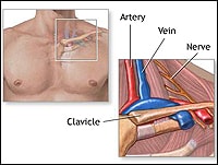
AXILLARY VEIN2
- In particular, the axillary vein is anterosuperior to the artery, and the brachial plexus is posterolateral (Fig. 2.3). (pediagenosis.com)
- Growth and extension of the clot down the subclavian and into the axillary vein can then result in further obstruction of critical collateral veins, resulting in sudden symptoms that include substantial arm swelling and cyanotic (bluish) discoloration. (wustl.edu)
Subclavian veins2
- One of the patients had two thrombosis sites on both femoral and subclavian veins. (journalofoncology.org)
- Effort thrombosis of the subclavian vein in venous TOS is due to occlusion of the axillary and subclavian veins (easily demonstrated by a venogram), which results in swelling and bluish discoloration of the arm. (wustl.edu)
Right upper extremity1
- So a diagnosis of a catheter-related deep vein thrombosis of the right upper extremity is made. (globaloncologyacademy.org)
Anticoagulation14
- This study appears to be the first to compare clinical outcomes in upper extremity DVT for patients treated with apixaban or rivaroxaban compared with the traditional anticoagulation treatment of LMWH and/or warfarin. (hematologyadvisor.com)
- Inferior vena cava (IVC) filter placement is most commonly indicated for deep venous thrombosis (DVT) or pulmonary embolism (PE) when anticoagulation therapy is contraindicated. (medscape.com)
- In the absence of a history of thrombosis, long-term prophylactic anticoagulation is not routinely recommended for asymptomatic Leiden variant heterozygotes. (nih.gov)
- A short course of prophylactic anticoagulation when circumstantial risk factors are present may prevent initial thrombosis in Leiden variant heterozygotes. (nih.gov)
- A catheter related thrombosis is associated with a number of clinically relevant complications, including catheter dysfunction, recurrent deep venous thrombosis, post thrombotic syndrome, anticoagulation-associated bleeding and pulmonary embolism. (fortuneonline.org)
- Despite growing evidence from observational studies that withholding anticoagulation may be a safe option in selected patients with isolated SSPE (i.e., those without concomitant deep vein thrombosis, cancer, etc.), most patients with isolated SSPE receive anticoagulant treatment, which is associated with an increased risk of bleeding. (clinicaltrials.gov)
- [6] Patients with superficial venous thromboses such as the long saphenous and short saphenous are at risk of developing a DV T, especially in patients who have a history of prior DVT although management with anticoagulation is controversial. (wikem.org)
- The recommendations that we have on the modalities and the therapeutic anticoagulation are mostly based on extrapolation of data from clinical trials on cancer-associated deep vein thrombosis and pulmonary embolism, and on observational studies of patients with catheter-related thrombosis. (globaloncologyacademy.org)
- If the catheter is functional, well positioned, and if there are no signs of infection, and if there is a good resolution of symptoms of the deep vein thrombosis of the upper extremity, so you can keep the catheter in place while anticoagulation therapy is administered. (globaloncologyacademy.org)
- A prospective cohort study of catheter-related thrombosis in cancer patients treated with 1 month of anticoagulation after catheter removal. (versiti.org)
- Conservative treatment of subclavian vein effort thrombosis has traditionally consisted of chronic anticoagulation (anti-clotting) therapies, intermittent arm elevation, long-term restrictions in arm activity, and the use of compression sleeves. (wustl.edu)
- Unlike lower extremity DVT, the proper duration of anticoagulation treatment for subclavian vein effort thrombosis is not known. (wustl.edu)
- Because this condition is caused by repetitive mechanical compression of the vein rather than a disorder of blood clotting, in the absence of an alteration in the anatomy many physicians recommend lifelong anticoagulation. (wustl.edu)
- Superficial venous thrombosis or phlebitis affects the superficial veins of the upper or lower extremity and only require anticoagulation in specific situations, and may be treated with anti-inflammatory pain relief only. (atozwiki.com)
Distal deep vein throm2
- 16. Long-term outcomes of cancer-related isolated distal deep vein thrombosis: the OPTIMEV study. (nih.gov)
- Furthermore, distal deep vein thrombosis in one of the lower extremities and death due to VTE were among the primary study endpoints. (esanum.com)
Recurrent thrombosis3
- Evidence suggests that heterozygosity for the Leiden variant has at most a modest effect on risk for recurrent thrombosis after initial treatment of a first VTE. (nih.gov)
- Factor V Leiden thrombophilia is suspected in individuals with a history of venous thromboembolism (VTE) manifest as deep vein thrombosis (DVT) or pulmonary embolism, especially in women with a history of VTE during pregnancy or in association with use of estrogen-containing contraceptives, and in individuals with a personal or family history of recurrent thrombosis. (nih.gov)
- Even in patients who do not get pulmonary emboli, recurrent thrombosis and "post-thrombotic syndrome" are a major cause of morbidity. (rxharun.com)
Ovarian Vein Thrombosis1
- Other rarer forms include retinal vein thrombosis , mesenteric vein thrombosis (affecting veins draining blood from the gastrointestinal organs), cerebral venous sinus thrombosis , renal vein thrombosis , and ovarian vein thrombosis. (atozwiki.com)
Embolism22
- SAN FRANCISCO - For patients with deep vein thrombosis, mortality rates and the prevalence of hemodynamically unstable pulmonary embolism are significantly higher when the upper extremities are involved than when lower extremities are involved, a large study of hospitalized patients demonstrates. (medscape.com)
- Interestingly, the location of the upper-extremity deep vein thrombosis did not affect progression to pulmonary embolism. (medscape.com)
- Venous thromboembolism (VTE) encompasses two interrelated conditions that are part of the same spectrum, deep vein thrombosis (DVT) and pulmonary embolism (PE) (see the image below). (medscape.com)
- A pulmonary embolism (PE) occurs when a blood clot from a deep vein (a DVT) detaches from a vein (embolizes), travels through the right side of the heart, and becomes lodged as an embolus in a pulmonary artery that supplies deoxygenated blood to the lungs for oxygenation. (wikipedia.org)
- 12. The risk and trend of pulmonary embolism and deep vein thrombosis in rheumatoid arthritis: a general population-based study. (nih.gov)
- Venous thromboembolism (VTE) may be classified into deep vein thrombosis ( DVT ) and pulmonary embolism ( PE ). (wikidoc.org)
- Pulmonary embolism may arise as a consequence of deep vein thrombosis as a result of embolization of the clot from deep veins of the legs. (wikidoc.org)
- Most often, pulmonary embolism is due to a venous thrombosis ( blood clot from a vein ), which has been dislodged from its site of formation in the lower extremities . (wikidoc.org)
- The primary objectives for the treatment of deep venous thrombosis (DVT) are to prevent pulmonary embolism (PE) , reduce morbidity, and prevent or minimize the risk of developing the postthrombotic syndrome (PTS). (medscape.com)
- Once a catheter-related thrombosis is formed, the risk of concomitant pulmonary embolism is regularly underestimated. (fortuneonline.org)
- 2020 review from JAMA [8] recommend treat calf DVT if "severe symptoms or risk factors for pulmonary embolism or extension to proximal veins (such as hospitalization, history of VTE, and cancer). (wikem.org)
- 452 Portal vein thrombosis 453 Other venous embolism and thrombosis 453.4 Deep vein thrombosis, unspec. (wikipedia.org)
- 453.41 Deep vein thrombosis, proximal 453.42 Deep vein thrombosis, distal 453.9 Venous embolism, unspec. (wikipedia.org)
- Researchers sought to determine whether early central venous catheter removal would be linked to pulmonary embolism risk in patients with catheter-related upper extremity deep vein thrombosis. (medicalbag.com)
- The primary endpoint identified by the researchers was a combination of proximal deep thromboembolism of the lower extremities, pulmonary embolism and symptomatic deep vein thrombosis of the upper extremities. (esanum.com)
- Venous thromboembolism was defined as deep vein thrombosis in the upper or lower extremities or in association with peripherally inserted central catheters or ports, pulmonary embolism, or concurrent deep vein thrombosis and pulmonary embolism. (bmj.com)
- Pulmonary embolism may also occur, particularly with motion of the arm, but this is infrequent compared to deep vein thrombosis, or DVT, in the lower extremities. (wustl.edu)
- There are other less common forms of venous thrombosis, some of which can also lead to pulmonary embolism. (atozwiki.com)
- Nevertheless, they can progress to the deep veins through the perforator veins or, they can be responsible for a lung embolism mainly if the head of the clot is poorly attached to the vein wall and is situated near the sapheno-femoral junction . (atozwiki.com)
- The abbreviation DVT/PE refers to a VTE where a deep vein thrombosis (DVT) has moved to the lungs (PE or pulmonary embolism). (atozwiki.com)
- Catheter-related VTE were defined as deep venous thrombosis in the arm, with or without pulmonary embolism (PE), or isolated PE. (hindawi.com)
- Catheter-related VTE may be limited to asymptomatic radiological findings but may also lead to significant clinical burden with upper limb postthrombotic syndrome reported in 5 to 28% [ 6 , 7 ] and respiratory failure in case of pulmonary embolism. (hindawi.com)
Varicose veins7
- In Hyderabad the cost ranges from Open Surgery Starting Rs 15,000 to Rs 40,000 depending on the number of Veins to be removed , In most cases, correction of Varicose veins is May be eligible for insurance coverage but various finance agencies provide easy ( Available EMI - Zero EMI PAY MONTHLY ) facility as well. (removingvaricoseveins.com)
- What is Varicose Veins Treatment Exactly? (removingvaricoseveins.com)
- Varicose veins (Treatment) are enlarged , swelling , and twisting veins , often appearing blue/dark purple. (removingvaricoseveins.com)
- When these valves are not functioning correctly, blood can pool in the veins, leading to varicose veins or even deep vein thrombosis. (e-adventure.net)
- This condition is called varicose veins. (e-adventure.net)
- Varicose veins are more common in women than men, and they can lead to complications like blood clots. (e-adventure.net)
- Szabo and colleagues suggest that "EVRF is appropriate for the treatment of teleangiectasias, reticular veins with the hand piece and K3i or K6i needles (face or lower leg), small varicose veins or tributaries 2-5mm with the hand catheters and for saphenous truncs and perforators 4-15mm in diameter with the CR45i catheter. (venousnews.com)
Diagnosis4
- Diagnosis is most commonly confirmed by ultrasound of the suspected veins. (wikipedia.org)
- See "Overview of the causes of venous thrombosis" and "Deep vein thrombosis in pregnancy: Epidemiology, pathogenesis, and diagnosis", section on 'Epidemiology' . (medilib.ir)
- CT scan is not a routine test for the diagnosis of deep vein thrombosis (DVT). (wikidoc.org)
- Blood coagulation testing is often performed in patients with upper extremity DVT, but these tests are usually negative and add little to the initial diagnosis or management of effort thrombosis. (wustl.edu)
Cerebral1
- A deep-vein thrombosis (DVT) is a blood clot that forms within the deep veins usually of the leg but can occur in the veins of the arms and the mesenteric and cerebral veins. (rxharun.com)
Thrombus4
- Superficial vein thrombosis, also known as superficial thrombophlebitis, is the formation of a blood clot (thrombus) in a vein close to the skin. (wikipedia.org)
- Deep vein thrombosis (also known as deep venous thrombosis or DVT and colloquially referred to as " economy class syndrome ") is the formation of a blood clot (" thrombus ") in a deep vein . (wikidoc.org)
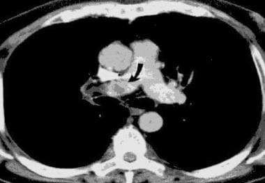
- The first image below demonstrates an IVC of normal size without a thrombus, but there is a circumaortic left renal vein and an inflow defect at the origin of the right renal vein. (medscape.com)
- Venous thrombosis is blockage of a vein caused by a thrombus (blood clot). (atozwiki.com)
Pelvic2
- However, emboli also can originate from the pelvic veins, the inferior vena cava, and the upper extremities. (medscape.com)
- Superficial venous thromboses cause discomfort but generally not serious consequences, as do the deep vein thromboses (DVTs) that form in the deep veins of the legs or in the pelvic veins. (atozwiki.com)
Asymptomatic2
- CVC-related deep vein thrombosis involving the upper extremity is usually asymptomatic but can result in post-thrombotic syndrome (swelling, pain, skin changes, and functional impairment). (elsevierpure.com)
- The initial phase of venous TOS is usually asymptomatic, involving compression injury of the vein between the clavicle and first rib, scar tissue formation, contraction around the outside of the vein, and fibrosis and wall thickening within the wall of the vein itself. (wustl.edu)
Occurs when a blood clot1
- DVT occurs when a blood clot forms in a vein, usually in the legs, and obstructs blood flow. (e-adventure.net)
Molecular weight heparin4
- Apixaban or rivaroxaban may be as safe and effective at treating upper extremity deep vein thrombosis as low molecular weight heparin and warfarin. (hematologyadvisor.com)
- Treatment of upper extremity deep vein thrombosis (DVT) with apixaban or rivaroxaban appears to be as safe and effective as low molecular weight heparin (LMWH) and/or warfarin, according to study results published in the American Journal of Hematology . (hematologyadvisor.com)
- Long-term safety and efficacy data on childhood venous thrombosis treated with a low molecular weight heparin: an open label pilot study of once-daily versus twice daily enoxaparin administration. (nih.gov)
- The international guidelines from various societies and institutions recommend anticoagulant treatment of catheter-related thrombosis with symptoms for at least 3 months, and as long as the central venous catheter is in place, and low-molecular-weight heparin are suggested in the setting. (globaloncologyacademy.org)
Lower16
- We focus more on the lower extremities. (medscape.com)
- They used International Classification of Diseases, Ninth Revision codes to assess risk factors and outcomes in 30,137 patients with upper-extremity deep vein thrombosis and 151,535 patients with lower-extremity deep vein thrombosis. (medscape.com)
- Upper-extremity deep vein thrombosis is less prevalent than lower-extremity deep vein thrombosis (0.19 vs 0.96 per 100,000 hospitalizations), but the outcomes are worse, Dr Williams reported. (medscape.com)
- Therefore, we aimed to evaluate the prevalence of thrombophilia in patients with UE-DVT compared to patients with lower extremity deep vein thrombosis (LE-DVT). (nih.gov)
- In 1856, Virchow demonstrated that 90% of all clinically important PEs result from DVT occurring in the deep veins of the lower extremities, proximal to and including the popliteal veins. (medscape.com)
- As well as lower extremity deep vein thrombosis (LEDVT), upper extremity deep vein thrombosis (UEDVT), which occurs in the subclavian, axillary, internal jugular, and/or brachial veins, cannot be negligible, accounting for up to 10% of all documented DVTs [ 7 ]. (biomedcentral.com)
- Our CPT® Advisors (physician volunteers representing multiple radiology societies) believe there are many clinical scenarios where it would be reasonable to perform a bilateral upper extremity deep vein thrombosis (DVT) exam on the same day as a bilateral lower extremity DVT exam, or a unilateral DVT exam of an arm on the same day as a unilateral DVT exam of a leg. (acr.org)
- For these reasons, the ACR and other radiology societies strongly advocated for an MUE unit of "2" so that both upper and lower extremities examinations can be reported during the same session or on the same day, when performed. (acr.org)
- The lower extremity vascular assessment should also be thorough to prepare for a potential femoral artery cannulation. (pediagenosis.com)
- A previous history of lower or upper extremity deep vein thrombosis must be elicited. (pediagenosis.com)
- Background: The current study was conducted to demonstrate that catheter-directed thrombolysis for upper and lower extremity deep vein thrombosis is equally safe in patients with and without cancer. (johnshopkins.edu)
- Some of the most common areas where valves are found are in the veins of the lower extremities, namely the legs and feet. (e-adventure.net)
- For example, in the lower extremities, valves create compartments that prevent the pooling of blood in the veins. (e-adventure.net)
- When the valves in the veins, especially the lower extremities, become damaged, blood can pool in the vein and form a bulging, twisted, and sometimes painful vein. (e-adventure.net)
- CVI is a condition caused by damaged valves in the lower extremity veins. (e-adventure.net)
- For smaller veins, more than 5,000 treatment sessions for teleangiectasias and spider veins on the face and lower limbs were completed. (venousnews.com)
Catheters5
- Although less prevalent than in LE-DVT patients, thrombophilia is a common finding in patients with UE-DVT, especially in those with thrombosis that is unrelated to venous catheters. (nih.gov)
- Central venous catheters and upper extremity deep vein thrombosis in medical inpatients: the Medical Inpatients and Thrombosis (MITH) Study. (nih.gov)
- and JUGULAR VEINS ) that develops as a complication of upper extremity central venous catheters and pacemaker uses, or cancer. (nih.gov)
- Deep vein thrombosis occurs in up to 50% of children with tunneled central venous catheters (CVCs). (elsevierpure.com)
- Catheters are at high risk for local venous thrombosis. (fortuneonline.org)
Catheter-associated2
- Catheter-associated deep vein thrombosis of the upper extremity in cancer patients: Guidance from the SSC of the ISTH. (journalofoncology.org)
- At least eight randomized controlled trials have evaluated antithrombotic therapy versus placebo in the prevention of central venous catheter-associated thrombosis [ 9 - 16 ]. (hindawi.com)
Patients22
- He added that it might be that patients with upper-extremity deep vein thrombosis tend to be sicker. (medscape.com)
- Hereditary and acquired thrombophilia in patients with upper extremity deep-vein thrombosis. (nih.gov)
- The prevalence of coagulation disorders in patients with upper extremity deep-vein thrombosis (UE-DVT) is unknown due to only a few observational studies of limited size reporting varying results. (nih.gov)
- One hundred fifty consecutive patients (15 to 91 years of age) with UE-DVT were recruited from the MAISTHRO (MAin-ISar-THROmbosis) registry. (nih.gov)
- however, LMWH has been the recommended treatment for patients with cancer who have catheter-related upper extremity DVT. (hematologyadvisor.com)
- This data, in combination with existing studies, provides reassuring information that the treatment of [upper extremity] DVT with apixaban or rivaroxaban can be considered a reasonable alternative to traditional anticoagulants in appropriate patients," concluded the authors. (hematologyadvisor.com)
- 3. High Prevalence and Mortality Associated with Upper Extremity Deep Venous Thrombosis in Hospitalized Patients at a Tertiary Care Center. (nih.gov)
- Objective We sought to determine the risk factors for subsequent bleeding and recurrent venous thromboembolism (VTE) events following isolated noncatheter-associated upper extremity deep venous thrombosis (non-CA-UEDVT) to better inform future treatment decisions for this group of patients. (uninsubria.it)
- Most patients with confirmed proximal vein DVT may be safely treated on an outpatient basis. (medscape.com)
- Objective: Patients with solid cancers frequently suffer from thrombosis, which is associated with considerable cost, morbidity, and mortality. (journalofoncology.org)
- Results: There were 113 patients with IP locations on subclavian and 7 on femoral veins. (journalofoncology.org)
- IP thromboses were detected in 10 patients. (journalofoncology.org)
- Bertoglio S, DiSomma C, Meszaros P, Gipponi M, Cafiero F, Percivale P. Long-term femoral vein central venous access in cancer patients. (journalofoncology.org)
- So here also, we have guidance from the international guidelines and in patients with catheter-related thrombosis. (globaloncologyacademy.org)
- Methods: A retrospective cohort of consecutive patients with acute iliofemoral or brachiosubclavian deep vein thrombosis treated with catheter-directed thrombolysis was identified. (johnshopkins.edu)
- Conclusion: Percutaneous catheter-directed thrombolysis is equally safe for patients with and without cancer who have acute symptomatic deep vein thrombosis. (johnshopkins.edu)
- Dr. Baumann Kreuziger is interested in the underlying mechanisms of thrombosis and optimal treatment strategies for patients with thrombosis. (versiti.org)
- In one of two patients with thrombosis of the deep veins of the upper extremity the venographic defect resolved completely. (mcmaster.ca)
- Six patients (21%) experienced adverse events considered to be related to the pacemaker or the implantation procedure, including a pericardial effusion requiring drainage, one case of tricuspid regurgitation thought to be caused by a pacing lead, an upper-extremity deep vein thrombosis, and three pocket incision-site reactions. (healthproblemsnews.com)
- DEEP VEIN THROMBOSIS - clinical features Leg pain, swelling, tenderness, palpable cord (thrombosed vessel), phlegmasia cerulea dolens in occasional patients. (pdfslide.us)
- Szabo and colleagues evaluated the effectiveness of EVRF treatment and analysed the three-year results of using the device for endovenous radiofrequency ablation and spider vein treatments from July 2011 to March 2015 to treat 751 saphenous reflux and varicosity patients. (venousnews.com)
- The three-year occlusion rate was 96.6% (two of 59 patients had an open vein segment). (venousnews.com)
Clot10
- Deep vein thrombosis (DVT) is a type of venous thrombosis involving the formation of a blood clot in a deep vein, most commonly in the legs or pelvis. (wikipedia.org)
- The most common life-threatening concern with DVT is the potential for a clot to embolize (detach from the veins), travel as an embolus through the right side of the heart, and become lodged in a pulmonary artery that supplies blood to the lungs. (wikipedia.org)
- A PE is a blockage that occurs when a clot or any part of a clot from a DVT breaks off within a major vein and travels to the lungs. (healthline.com)
- Treatment with thrombolysis can often clear much of the subclavian vein of clot, revealing an underlying stenosis at the level of the first rib. (wustl.edu)
- Thrombosis is the formation of blood clot inside of a blood vessel. (wustl.edu)
- A blood clot does eventually form in the narrowed subclavian vein, leading to thrombotic occlusion. (wustl.edu)
- Venous Thrombosis / Deep vein thrombosis is a blood clot that develops in a vein deep in the body. (rxharun.com)
- The clot may partially or completely block blood flow through the vein. (rxharun.com)
- A common form of venous thrombosis is deep vein thrombosis (DVT), when a blood clot forms in the deep veins. (atozwiki.com)
- Since the veins return blood to the heart , if a piece of a blood clot formed in a vein breaks off it can be transported to the right side of the heart, and from there into the lungs . (atozwiki.com)
DVTs1
- Symptoms can include pain, swelling, redness, and enlarged veins in the affected area, but some DVTs have no symptoms. (wikipedia.org)
Acute1
- The first acute thrombosis is treated according to standard guidelines. (nih.gov)
Symptoms2
- Additional signs and symptoms include tenderness, pitting edema (see image), dilation of surface veins, warmth, discoloration, a "pulling sensation", and even cyanosis (a blue or purplish discoloration) with fever. (wikipedia.org)
- Well, they can also be symptoms of deep vein thrombosis (DVT) , a condition that certain types of hormonal birth control increase the risk of. (healthline.com)
Calf1
- It commonly affects the deep leg veins (such as the calf veins, femoral vein, or popliteal vein) or the deep veins of the pelvis. (rxharun.com)
Cancer6
- 2. Racial disparities in cancer-associated thrombosis. (nih.gov)
- 7. Cancer - Associated Thrombosis - A Study of Cases. (nih.gov)
- Hello, my name is Cihan Ay, and I am from the Medical University of Vienna, and it's my pleasure to welcome you to this session on the Treatment of a Patient with Cancer and with Catheter-Related Thrombosis. (globaloncologyacademy.org)
- The question now is: How should we treat this patient with cancer and a catheter-related thrombosis? (globaloncologyacademy.org)
- For instance, a systematic review on the treatment of cancer-related thrombosis has shown a large variation in the treatment modalities and the treatment duration. (globaloncologyacademy.org)
- Held N, Jung B, Baumann Kreuziger L. Management of cancer-associated thrombosis with thrombocytopenia: Impact of the ISTH guidance statement. (versiti.org)
JUGULAR VEINS3
- and JUGULAR VEINS ). (nih.gov)
- Veins in the neck, called jugular veins, also have valves. (e-adventure.net)
- The jugular veins are responsible for draining blood from the head and neck regions back to the heart. (e-adventure.net)
Thrombolytic Therapy1
- In some cases, balloon angioplasty may be used at the same time as thrombolytic therapy in attempting to reduce the degree of stenosis in the subclavian vein. (wustl.edu)
Clinical2
- Investigators noted large clinical trials that lead to the approval of direct oral anticoagulants to treat venous thromboembolism (VTE) had not included data on upper extremity DVT. (hematologyadvisor.com)
- Antithrombotic therapy and prevention of thrombosis, 9th ed: American College of Chest Physicians evidence-based clinical practice guidelines. (journalofoncology.org)
Superficial2
- Superficial) femoral vein is part of the deep system, not the superficial system as the name suggests! (wikem.org)
- Venous thromboembolism and superficial vein thrombosis account for about 90% of venous thrombosis. (atozwiki.com)
Arterial Thrombosis1
- Red Blood Transfusion Does Not Increase Risk of Venous or Arterial Thrombosis. (versiti.org)
Etiology1
- f) Generalized extremity swelling - determination of the etiology of the swelling. (acr.org)
Symptomatic1
- The primary effectiveness and safety outcomes were symptomatic, recurrent VTE (any site), and major bleeding (according to the International Society of Thrombosis and Hemostasis) at 3 months of follow up. (hematologyadvisor.com)
Risk Factors2
- Risk factors for venous thrombosis in medical inpatients: validation of a thrombosis risk score. (nih.gov)
- Anticoagulant therapy is recommended for 3-12 months depending on site of thrombosis and on the ongoing presence of risk factors. (medscape.com)
Renal veins2
- If the infrarenal segment of the inferior vena cava is too short for a filter placement, the filter should be placed above the renal veins. (medscape.com)
- In addition, the inflow from the renal veins can be seen at the level of the L1 vertebral body. (medscape.com)
Chronic1
- To measure changes in blood vessel size or changes in gas volume in the lungs to assist in diagnosing diseases such as deep vein thrombosis (DVT), chronic obstructive pulmonary disease (COPD), and some peripheral vascular disorders. (unboundmedicine.com)
Occlusion of the axillary1
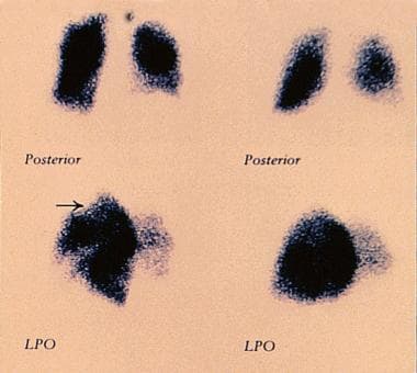
- And this ultrasound shows an obstruction and occlusion of the axillary and the subclavian vein. (globaloncologyacademy.org)
Venous system1
- Now that we know which veins have valves, let's talk about their function in the venous system. (e-adventure.net)
Femoral veins1
- When filter design allows placement through the jugular or femoral veins, the filter is specially packaged to ensure that it is deployed in the correct orientation. (medscape.com)
Outcomes2
- Cite this: Outcomes Worse for Upper-Extremity Deep Vein Thrombosis - Medscape - May 16, 2016. (medscape.com)
- It has also become clear over the past decade that placement of stents within the subclavian vein frequently leads to poor outcomes and should be avoided. (wustl.edu)
Primary1
- It is associated with mechanical factors (Upper Extremity Deep Vein Thrombosis, Primary) secondary to other anatomic factors (Upper Extremity Deep Vein Thrombosis, Secondary). (nih.gov)
Ultrasound4
- In these situations, a duplex ultrasound of all four extremities may be medically necessary and requested. (acr.org)
- A thrombosis of the right internal vein was detected with compressive ultrasound imaging. (fortuneonline.org)
- To investigate the cause of the misplacement, an ultrasound imaging was performed that showed thrombosis within the right internal and external jugular vein (Figure 2). (fortuneonline.org)
- Imaging studies, such as magnetic resonance angiography or catheter-based venograms, provide more definitive information on the location and extent of axillary-subclavian vein thrombosis than venous Duplex (ultrasound) studies. (wustl.edu)