Fetal urinoma: two new cases and a review of the literature. (1/4)
OBJECTIVE: To evaluate the functional prognosis of kidneys affected prenatally by urinomas. METHODS: This was a retrospective review of cases of fetal urinoma reported in the literature, as well as two of our own cases. RESULTS: Twenty-three patients with a prenatal diagnosis of urinoma (five bilateral) were included in the analysis. Postnatal ipsilateral renal function was observed in only six of the 28 renal units (i.e. around 20%). CONCLUSIONS: Although the precise causes of urinomas are still unknown, this review shows that in the event of a fetal urinoma, the probability of a non-functional dysplastic ipsilateral kidney lies at around 80%. In-utero puncture only appears to be justified in cases where fluid accumulation has mass effects on adjacent major structures. (+info)Urinothorax: an unusual cause of pleural effusion. (2/4)
Urinothorax refers to the presence of urine in the pleural space secondary to obstructive uropathy, and is an unusual cause of pleural effusion. The importance of recognising this entity lies in the fact that the condition is completely reversible following relief of urinary tract obstruction. We describe a 35-year-old man who developed urinothorax following a percutaneous nephrolithotomy for renal calculi. We also reviewed the literature for reported cases between 1968 and 2006. (+info)Glue ablation of a late-presentation urinary fistula after partial nephrectomy. (3/4)
(+info)Urinothorax as an unusual type of pleural effusion - clinical report and review. (4/4)
(+info)A urinoma is a medical term that refers to a collection or pooling of urine in a confined space or area within the body, usually due to a leakage from the urinary system. This condition most commonly occurs as a result of trauma, surgery, or injury to the urinary tract, such as the kidneys or ureters.
The accumulation of urine in the surrounding tissues can lead to inflammation, infection, and other complications if left untreated. Symptoms of a urinoma may include abdominal or back pain, fever, nausea, vomiting, and decreased urine output. Diagnosis is typically made through imaging tests such as CT scans or ultrasounds, and treatment may involve surgical repair of the damaged urinary tract, placement of a catheter to drain the urinoma, or administration of antibiotics to prevent infection.
 Abdominal trauma
 Struvite and Staghorn Calculi Treatment & Management: Medical Therapy, Surgical Therapy, Preoperative Details
 Fetal Urinoma Due to Circulatory Disorders in an Umbilical Artery: Case Report<...
 Yair Blumenfeld - Stanford Medicine Children's Health
 Bladder augmentation in children: current problems and experimental strategies for reconstruction | springermedizin.at
 Urofacial syndrome: A subset of neurogenic bladder dysfunction syndromes? - PubMed
 DeCS
DeCS MeSH Browser
 Hsi-Yang Wu's Profile | Stanford Profiles
 External validation of a model for tailoring the operative approach to minimally invasive partial nephrectomy<...
 Urinoma
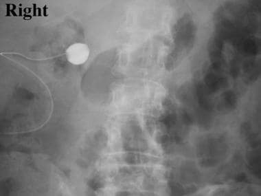
 Touro | Antegrade Pyelogram
 Spontaneous bilateral renal pelvis rupture during CT in the absence of urinary tract obstruction: case report | BMC Urology |...
 International Neurourology Journal (INJ)
 Mr Gokul Vignesh Kandaswamy | Urological Surgeon Glamorgan | Prostate Cancer Pembrokeshire
 Makale Arama, perforasyon | Selçuk Tıp Dergisi
 CT Scanner facility in Near Shimla | Himachal Pradesh APEX
 Black Maternal Health Week | CHRISTUS Children's | Improving Maternal Outcomes for our moms
 What Part Of The Kidney Attaches To The Ureter - HealthyKidneyClub.com