Uterine Prolapse
Rectal Prolapse
Cystocele
White Muscle Disease
Mitral Valve Prolapse
Pelvic Organ Prolapse
Ligaments
Aortic Valve Prolapse
Pelvic Floor
Tricuspid Valve Prolapse
Pessaries
Blood gases and sex hormones in women with and without genital descensus. (1/196)
BACKGROUND: Abnormalities in connective tissue and spirometric disorders have previously been found in women with genital descensus. OBJECTIVE: To evaluate the association of descensus and respiratory function. METHODS: The blood gases and sex hormones were measured in 130 women scheduled for surgical correction of descensus and 60 matched women without descensus. All subjects were nonsmokers and without past or present cardiorespiratory disease. RESULTS: Women with descensus had a lower pH (7.39+/-0.04 vs. 7.41+/-0.04, p = 0.01), lower arterial tensions of oxygen (12.7+/-12. vs. 14.1+/-0.9 kPa, p = 0.003) and carbon dioxide (5.1+/-0.4 vs. 5.3+/-0.3 kPa) but a higher hemoglobin concentration (141+/-11 vs. 132+/-9 g/l) and a higher serum progesterone in the follicular phase of the cycle (3.1+/-4 vs. 1.5+/-1 ng/ml, p = 0.03). In 39 (30%) women with descensus, the arterial carbon dioxide tension was below 4.9 kPa. All subjects ventilated more in the luteal compared to the follicular phase of the cycle. In women with descensus, the hemoglobin concentration increased with decreasing arterial oxygen tension (p = 10(-4)) and with decreasing pH (p<10(-3)). CONCLUSION: Women with descensus frequently hyperventilate and, compared with women without descensus, have a lower arterial oxygen tension, increased hemoglobin concentration and slightly lower pH. (+info)Extension of extramammary Paget disease of the vulva to the cervix. (2/196)
Extramammary Paget disease of the vulva was found in association with vulval adenocarcinoma in an elderly woman who also had a uterine prolapse. The characteristic histological appearances of extramammary Paget disease were masked by striking reactive changes in the squamous epithelium. Primary excision of both the intraepithelial and invasive disease appeared complete. However, a subsequent hysterectomy with repair of the prolapse revealed extramammary Paget disease in the upper vaginal mucosa and cervix, a finding which is very rarely described. Pathogenesis and diagnosis of extramammary Paget disease is discussed, with differential diagnosis and reference to immunohistochemical methods. (+info)Rupture of the rectosigmoid colon with evisceration of the small bowel through the anus. (3/196)
Spontaneous rupture of the rectosigmoid colon and herniation of the small intestine through the rupture site and eventual evisceration through the anus is a very rare event. In the literature, only 42 cases have been reported. The majority of them occurred in patients with rectal prolapse and one case was reported in association with a third-degree uterine prolapse. We experienced an 81-year-old female patient with rectal prolapse and second-degree uterine prolapse complicated by spontaneous perforation of the rectosigmoid colon and anal evisceration of the small intestine. Segmental resection of the nonviable small intestine, primary repair of the ruptured rectosigmoid colon, and sigmoid loop colostomy were performed, and the patient recovered well. In our patient, both rectal and uterine prolapses cooperatively damaged the anterior wall of the rectosigmoid colon and resulted in perforation. So, rectal and uterine prolapses should be treated before the complication develops. In this patient, uterine prolapse should be treated because of the recurrence of this rare episode. (+info)Practical use of the pessary. (4/196)
The pessary is an effective tool in the management of a number of gynecologic problems. The pessary is most commonly used in the management of pelvic support defects such as cystocele and rectocele. Pessaries can also be used in the treatment of stress urinary incontinence. The wide variety of pessary styles may cause confusion for physicians during the initial selection of the pessary. However, an understanding of the different styles and their uses will enable physicians to make an appropriate choice. Complications can be minimized with simple vaginal hygiene and regular follow-up visits. (+info)Sprengels deformity: anaesthesia management. (5/196)
A 28 years old lady presented with Sprengels deformity and hemivertebrae for Fothergills surgery. Clinically there were no anomalies of the nervous, renal or the cardiovascular systems. She had a short neck and score on modified Mallapati test was grade 2. She was successfully anaesthetised using injection Propofol as a total intravenous anaesthetic agent after adequate premedication with injection Midazolam and injection Pentazocine. Patient had an uneventful intraoperative and postoperative course. (+info)Vaginal vault suspension and enterocele repair by Richardson-Saye laparoscopic technique: description of training technique and results. (6/196)
OBJECTIVES: To describe the Richardson-Saye technique for laparoscopic vaginal vault suspension and enterocele repair (vaginal apex reconstruction) and the appropriate training needed for performance of this technique. METHODS: Before using this technique, Drs Carter, Winter, and Mendelsohn first received training by observation of skilled surgeons performing the procedure, attending courses, and finally being tutored and proctored by Dr Saye on the appropriate performance of the technique. They then used this technique to surgically treat eight patients, 42 to 85 years of age, mean age 62 years, between March and September of 1999. RESULTS: We included eight patients in this study who underwent the Saye-Richardson vaginal vault suspension and enterocele repair (apical vaginal vault reconstruction) by the suture technique. In all patients at six-month follow-up, the vaginal apex remains intact and well supported. We describe here the entire vaginal vault suspension and enterocele repair procedure with all its relevant details. CONCLUSION: Laparoscopic reconstruction of the disrupted vaginal apex followed by reattachment to the previously broken uterosacral ligament with the use of permanent suture provides a secure and anatomically correct vault suspension. Before performing this technique, physicians should undergo proper training, including observation, courses, tutoring, and proctorship by a surgeon experienced in performing this technique. (+info)Sexual life after gynaecological operations--II.(7/196)
(+info)The standardization of terminology for researchers in female pelvic floor disorders. (8/196)
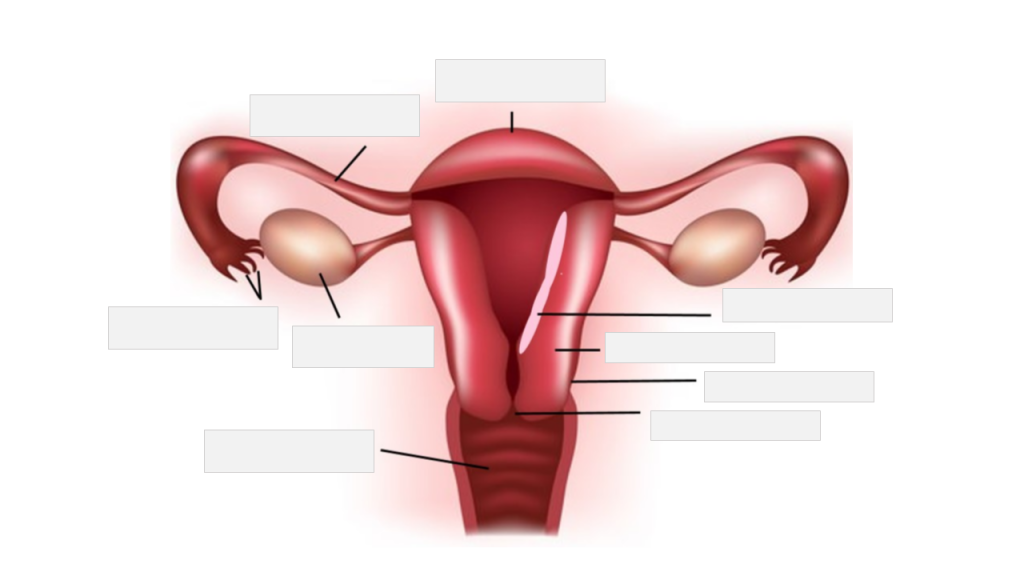
The lack of standardized terminology in pelvic floor disorders (pelvic organ prolapse, urinary incontinence, and fecal incontinence) is a major obstacle to performing and interpreting research. The National Institutes of Health convened the Terminology Workshop for Researchers in Female Pelvic Floor Disorders to: (1) agree on standard terms for defining conditions and outcomes; (2) make recommendations for minimum data collection for research; and (3) identify high priority issues for future research. Pelvic organ prolapse was defined by physical examination staging using the International Continence Society system. Stress urinary incontinence was defined by symptoms and testing; 'cure' was defined as no stress incontinence symptoms, negative testing, and no new problems due to intervention. Overactive bladder was defined as urinary frequency and urgency, with and without urge incontinence. Detrusor instability was defined by cystometry. For all urinary symptoms, defining 'improvement' after intervention was identified as a high priority. For fecal incontinence, more research is needed before recommendations can be made. A standard terminology for research on pelvic floor disorders is presented and areas of high priority for future research are identified. (+info)Uterine prolapse is a condition where the uterus descends or slips down from its normal position in the pelvic cavity into or through the cervix and sometimes even outside the vaginal opening. This occurs due to the weakening of the muscles and ligaments that support the uterus, often as a result of childbirth, aging, menopause, obesity, or prior hysterectomy. Uterine prolapse can lead to various symptoms such as a feeling of heaviness in the pelvis, difficulty in urinating or having bowel movements, and uncomfortable sexual intercourse. The severity of the condition may vary from mild to severe, and treatment options range from lifestyle changes and physical therapy to surgery.
A vaginal hysterectomy is a surgical procedure in which the uterus is removed through the vagina. During this type of hysterectomy, an incision is made in the vagina, and the uterus is separated from its attachments and then removed. The cervix may also be removed during a vaginal hysterectomy. This procedure is typically used to treat conditions such as uterine fibroids, endometriosis, pelvic support problems, and gynecologic cancer.
It's important to note that after a hysterectomy, a woman will no longer have menstrual periods or be able to become pregnant. Additionally, the removal of the ovaries (oophorectomy) during a hysterectomy can lead to surgical menopause and associated symptoms such as hot flashes, vaginal dryness, and mood changes.
As with any surgery, there are risks involved with vaginal hysterectomy, including infection, bleeding, injury to surrounding organs, and reactions to anesthesia. It is important for patients to discuss the potential benefits and risks of this procedure with their healthcare provider before making a decision about treatment.
A prolapse is a medical condition where an organ or tissue in the body slips from its normal position and drops down into a lower part of the body. This usually occurs when the muscles and ligaments that support the organ become weak or stretched. The most common types of prolapses include:
* Uterine prolapse: When the uterus slips down into or protrudes out of the vagina.
* Rectal prolapse: When the rectum (the lower end of the colon) slips outside the anus.
* Bladder prolapse (cystocele): When the bladder drops into the vagina.
* Small bowel prolapse (enterocele): When the small intestine bulges into the vagina.
Prolapses can cause various symptoms, such as discomfort, pain, pressure, and difficulty with urination or bowel movements. Treatment options depend on the severity of the prolapse and may include lifestyle changes, physical therapy, medication, or surgery.
A rectocele is a type of pelvic organ prolapse, which occurs when the rectum (the lower end of the colon) bulges into the back wall of the vagina. This condition most commonly affects women who have gone through childbirth, although it can also occur in older women or those with long-term constipation or other conditions that put pressure on the pelvic floor muscles.
Rectoceles can cause a variety of symptoms, including difficulty having bowel movements, feeling like something is sticking out of the vagina, and pain during sexual intercourse. In some cases, rectoceles may not cause any symptoms at all. Treatment options for rectoceles include pelvic floor physical therapy, lifestyle changes (such as avoiding heavy lifting or straining), and in severe cases, surgery.
The exact medical definition of a rectocele is: "A herniation of the rectal wall into the vaginal wall, often associated with disruption of the rectovaginal septum." This means that there is a protrusion or bulge of the rectal wall into the vaginal wall, which can be caused by a weakening or tearing of the tissue that separates the two structures.
Rectal prolapse is a medical condition where the rectum, which is the lower end of the colon, slips outside the anus, the opening through which stool leaves the body. This usually occurs due to weakened muscles and supporting structures in the pelvic area, often as a result of aging, childbirth, or long-term constipation or diarrhea.
The rectal prolapse can be partial, where only a small portion of the rectum slips outside the anus, or complete, where the entire rectum protrudes. This condition can cause discomfort, pain, bleeding, and difficulty with bowel movements. Treatment options may include dietary changes, medication, or surgical intervention.
A cystocele is a type of pelvic organ prolapse that occurs when the wall between the bladder and the vagina weakens and allows the bladder to bulge into the vagina. This condition is also sometimes referred to as a "prolapsed bladder." Cystoceles can cause various symptoms, including urinary incontinence, difficulty emptying the bladder completely, and discomfort or pain during sexual activity. The severity of a cystocele can vary, and treatment options may include lifestyle changes, pelvic floor exercises, or surgery.
White muscle disease is not a formal medical term, but it is a condition commonly referred to in veterinary medicine, particularly in the context of livestock and wildlife. It's also known as nutritional muscular dystrophy or enzootic muscular dystrophy.
The term "white muscle disease" refers to a group of conditions characterized by degeneration and necrosis (death) of skeletal and cardiac muscle tissue, primarily caused by deficiencies in certain nutrients, particularly selenium and vitamin E. These nutrients play crucial roles in the antioxidant defense system within the body, protecting cells from oxidative damage.
In affected animals, the lack of these essential nutrients leads to muscle damage, which can result in various clinical signs, such as:
1. Weakness
2. Stiffness
3. Reluctance to move
4. Difficulty swallowing or breathing (in severe cases)
5. Sudden death (often due to heart failure)
White muscle disease is most commonly observed in ruminants like cattle, sheep, and goats, as well as certain species of swine, poultry, and wild animals. It can be prevented through dietary supplementation with selenium and vitamin E or by providing these nutrients through mineral-rich soil and forage. In some cases, treatment may involve administering selenium and vitamin E injections to help support muscle recovery and prevent further damage.
Mitral valve prolapse (MVP) is a heart condition where the mitral valve, which separates the left atrium and left ventricle in the heart, doesn't function properly. In MVP, one or both of the mitral valve flaps (known as leaflets) bulge or billow into the left atrium during the contraction of the left ventricle. This prolapse can cause a leakage of blood back into the atrium, known as mitral regurgitation. In many cases, MVP is asymptomatic and doesn't require treatment, but in some instances, it may lead to complications such as infective endocarditis or arrhythmias. The exact causes of MVP are not fully understood, but it can be associated with certain genetic factors, connective tissue disorders, and mitral valve abnormalities present at birth.
Pelvic Organ Prolapse (POP) is a medical condition where the supporting muscles and ligaments in a woman's pelvis weaken, causing one or more of the pelvic organs - including the bladder, uterus, rectum, or small intestine - to drop or press into or out of the vagina. This can result in various symptoms such as a feeling of heaviness or fullness in the pelvis, pressure or pain in the lower back, painful intercourse, and problems with urination or bowel movements. POP is often associated with childbirth, menopause, aging, and certain medical conditions that increase abdominal pressure, like obesity or chronic coughing. Treatment options can range from lifestyle changes and physical therapy to surgery.
The sacrococcygeal region is the lower part of the back where the spine ends, specifically referring to the area where the sacrum (a triangular bone at the base of the spine formed by the fusion of several vertebrae) meets the coccyx (also known as the tailbone). This region is located at the very bottom of the spine and is susceptible to injury or trauma due to its position and role in supporting the body's weight. It is also a common site for birth defects, particularly in newborns.
Ligaments are bands of dense, fibrous connective tissue that surround joints and provide support, stability, and limits the range of motion. They are made up primarily of collagen fibers arranged in a parallel pattern to withstand tension and stress. Ligaments attach bone to bone, and their function is to prevent excessive movement that could cause injury or dislocation.
There are two main types of ligaments: extracapsular and intracapsular. Extracapsular ligaments are located outside the joint capsule and provide stability to the joint by limiting its range of motion. Intracapsular ligaments, on the other hand, are found inside the joint capsule and help maintain the alignment of the joint surfaces.
Examples of common ligaments in the body include the anterior cruciate ligament (ACL) and posterior cruciate ligament (PCL) in the knee, the medial collateral ligament (MCL) and lateral collateral ligament (LCL) in the elbow, and the coracoacromial ligament in the shoulder.
Injuries to ligaments can occur due to sudden trauma or overuse, leading to sprains, strains, or tears. These injuries can cause pain, swelling, bruising, and limited mobility, and may require medical treatment such as immobilization, physical therapy, or surgery.
Dystocia is a medical term used to describe difficult or abnormal labor or delivery in animals, including humans. It refers to a situation where the natural process of childbirth is hindered or obstructed, making it difficult for the fetus to pass through the birth canal. This condition can be caused by various factors such as the size and position of the fetus, maternal pelvic size or shape, hormonal imbalances, or other medical conditions that affect the mother's ability to give birth.
Dystocia can lead to serious complications for both the mother and the fetus if not treated promptly and appropriately. Prolonged labor can result in fetal distress, hypoxia (lack of oxygen), or even death. In addition, maternal injuries such as uterine rupture, cervical trauma, or infection can occur during a difficult delivery.
The treatment for dystocia depends on the underlying cause and severity of the condition. In some cases, manual assistance or manipulation of the fetus may be sufficient to facilitate delivery. However, in more severe cases, medical intervention such as cesarean section (C-section) may be necessary to ensure the safety of both the mother and the fetus.
It is important for pregnant individuals to receive regular prenatal care from a qualified healthcare provider to monitor their pregnancy and identify any potential risk factors for dystocia or other complications. Prompt medical attention should be sought if any signs of difficult labor or delivery are observed.
Aortic valve prolapse is a cardiac condition in which the aortic valve leaflets bulge or billow into the left ventricle during systole, the phase of the heart cycle when the ventricles contract to pump blood out of the heart. The aortic valve typically has three leaflets that open and close to regulate the flow of blood between the left ventricle and the aorta. In aortic valve prolapse, one or more of these leaflets become floppy, allowing blood to leak back into the left ventricle, a condition known as aortic regurgitation.
Aortic valve prolapse can be congenital or acquired. Some people are born with abnormalities in the aortic valve that make it more prone to prolapse, while others may develop the condition due to degenerative changes in the valve tissue over time. Certain factors, such as Marfan syndrome, bicuspid aortic valve, and infective endocarditis, can increase the risk of aortic valve prolapse.
The symptoms of aortic valve prolapse can vary depending on the severity of the condition. Mild cases may not cause any noticeable symptoms, while more severe cases can lead to shortness of breath, fatigue, chest pain, and irregular heart rhythms. Treatment for aortic valve prolapse may include monitoring, medication, or surgical repair or replacement of the aortic valve.
Visceral prolapse, also known as pelvic organ prolapse, is a medical condition where one or more of the pelvic organs (such as the bladder, uterus, rectum, or small intestine) descends from their normal position and bulges into or out of the vagina. This can occur due to weakened or damaged muscles and tissues that support these organs, often as a result of childbirth, aging, menopause, obesity, or certain medical conditions.
Visceral prolapse is classified based on the organ involved and the degree of descent. The most common types include cystocele (bladder prolapse), rectocele (rectum prolapse), enterocele (small intestine prolapse), and uterine or vaginal vault prolapse. Symptoms can range from mild discomfort, pressure, or a feeling of fullness in the pelvic area to more severe issues like urinary or fecal incontinence, difficulty emptying the bladder or bowels, and painful intercourse. Treatment options may include lifestyle changes, physical therapy, pessaries (vaginal support devices), or surgery.
Gynecologic surgical procedures refer to the operations that are performed on the female reproductive system and related organs. These surgeries can be either minimally invasive or open procedures, depending on the condition and the patient's health status.
The indications for gynecologic surgical procedures may include but are not limited to:
1. Diagnosis and treatment of various benign and malignant conditions such as uterine fibroids, ovarian cysts, endometriosis, and cancers of the reproductive organs.
2. Management of abnormal uterine bleeding, pelvic pain, and infertility.
3. Treatment of ectopic pregnancies and miscarriages.
4. Pelvic organ prolapse repair.
5. Sterilization procedures such as tubal ligation.
6. Investigation and treatment of suspicious lesions or abnormal Pap smears.
Some common gynecologic surgical procedures include hysterectomy (removal of the uterus), oophorectomy (removal of the ovary), salpingectomy (removal of the fallopian tube), cystectomy (removal of a cyst), myomectomy (removal of fibroids while preserving the uterus), and endometrial ablation (destruction of the lining of the uterus).
Minimally invasive surgical techniques such as laparoscopy and hysteroscopy have gained popularity in recent years due to their advantages over traditional open surgeries, including smaller incisions, less postoperative pain, quicker recovery times, and reduced risk of complications.
The pelvic floor is a group of muscles, ligaments, and connective tissues that form a sling or hammock across the bottom of the pelvis. It supports the organs in the pelvic cavity, including the bladder, rectum, and uterus or prostate. The pelvic floor helps control urination, defecation, and sexual function by relaxing and contracting to allow for the release of waste and during sexual activity. It also contributes to postural stability and balance. Weakness or damage to the pelvic floor can lead to various health issues such as incontinence, pelvic organ prolapse, and sexual dysfunction.
Tricuspid valve prolapse is a cardiac condition where the tricuspid valve, located between the right atrium and right ventricle of the heart, doesn't close properly due to one or more of its leaflets (flaps) bulging or billowing into the right atrium during contraction of the right ventricle. This allows the backflow of blood from the right ventricle into the right atrium, known as tricuspid regurgitation. In some cases, tricuspid valve prolapse may not cause any symptoms and can be an incidental finding on echocardiography. However, if severe tricuspid regurgitation occurs, it can lead to right-sided heart failure, atrial arrhythmias, and other complications. The condition is often associated with mitral valve prolapse or other connective tissue disorders.
A pessary is a medical device that is inserted into the vagina to provide support for the uterus, vaginal vault, or bladder. It is often used in the management of pelvic organ prolapse, urinary incontinence, and other gynecological conditions. Pessaries come in various shapes and sizes, and they are typically made of silicone, rubber, or plastic. They can be worn for extended periods of time and are usually removable and cleanable. The selection and fitting of a pessary should be performed by a healthcare professional, such as a gynecologist or nurse midwife.
Surgical mesh is a medical device that is used in various surgical procedures, particularly in reconstructive surgery, to provide additional support to weakened or damaged tissues. It is typically made from synthetic materials such as polypropylene or polyester, or from biological materials such as animal tissue or human cadaveric tissue.
The mesh is designed to be implanted into the body, where it can help to reinforce and repair damaged tissues. For example, it may be used in hernia repairs to support the weakened abdominal wall, or in pelvic floor reconstruction surgery to treat conditions such as pelvic organ prolapse or stress urinary incontinence.
Surgical mesh can come in different forms, including sheets, plugs, and patches, and may be either absorbable or non-absorbable. The choice of mesh material and type will depend on the specific surgical indication and the patient's individual needs. It is important for patients to discuss the risks and benefits of surgical mesh with their healthcare provider before undergoing any surgical procedure that involves its use.
 Uterine prolapse - Wikipedia
 Uterine prolapse: MedlinePlus Medical Encyclopedia
 Uterine Prolapse: Background, History of the Procedure, Problem
 Uterine Prolapse
 Effectiveness of Structured Teaching Programme on knowledge regarding preventive measures of Uterine Prolapse among mothers -...
 Subjects: Uterine Prolapse - Digital Collections - National Library of Medicine Search Results
 Uterine Prolapse | Health Information Center
 Best Uterine Prolapse Surgery Surgeons in Chennai, Top 10 Uterine Prolapse Surgery Doctors in Chennai
 Best Vaginoplasty Surgery in Noida | Uterine Prolapse
 Bilateral uterine prolapse in queen: case study
 What Are The Stages Of Uterine Prolapse?
 Uterine Prolapse | Causes, Symptoms, and Diagnosis: | FemiCushion
 uterine prolapse Archives - Hudson Valley Physical Therapy
 Uterine Prolapse. Women's Health.
- The Eco Woman
Uterine Prolapse. Women's Health.
- The Eco Woman Uterine Prolapse - Designs by Duvet Days Anatomy Illustrations
 Treating Uterine Prolapse Archives - University OB/GYN Associates
 A Guide to Understanding & Treating Uterine Prolapse - Style & Beauty blog
 Uterine And Bladder Prolapse? Causes, Symptoms, Diagnosis, Treatments | Melody Jacob
 How to Avoid Your Uterine Prolapse Symptoms Worsening - Pelvic Exercises
 Uterine and Apical Prolapse - Gynecology and Obstetrics - MSD Manual Professional Edition
 Case Report of Uterine Prolapse in Cattle
| Jurnal Agribisnis Peternakan (JINAK)
Case Report of Uterine Prolapse in Cattle
| Jurnal Agribisnis Peternakan (JINAK)
 An old lady with uterine prolapse gets fucked by her kneading therapist
 Vaginal Vault Prolapse, Enterocele, Uterine Prolapse Trial in Barcelona (Elevate mesh, Sacrocolpopexy) | Clincosm
 Acute prolapse of giant submucosal fibroid polyp mimicking uterine inversion- A rare case report