Uveomeningoencephalitic Syndrome
Nervous System Autoimmune Disease, Experimental
Autoimmune Diseases
High incidence of glucose intolerance in Vogt-Koyanagi-Harada disease. (1/85)
AIMS: To evaluate glucose tolerance of patients with Vogt-Koyanagi-Harada (VKH) disease before systemic corticosteroid therapy, and to assess changes brought on by treatment. METHODS: 20 VKH patients with acute bilateral panuveitis were studied. 20 healthy adults and 11 Behcet's disease patients with active uveoretinitis served as controls. A 75 g oral glucose tolerance test (OGTT) was given in the acute stage of ocular inflammation before systemic corticosteroid therapy. The OGTT was repeated in the convalescent stage of VKH disease in the patients with glucose intolerance before treatment. Insulin response was examined at the same time as the OGTT when possible. RESULTS: 55% of VKH patients (11/20) showed glucose intolerance but no apparent insulin secretion deficiency was detected. Four of seven patients in the convalescent stage showed improvement of glucose tolerance. None of the normal controls or disease controls showed glucose intolerance. CONCLUSION: A high incidence of glucose intolerance was found in the acute stage of VKH disease. However, glucose intolerance improved in most cases after systemic corticosteroid therapy. It is possible that glucose intolerance seen in VKH patients may be related to the autoimmune inflammatory process of this disease. (+info)Subtraction ICG angiography in Harada's disease. (2/85)
BACKGROUND/AIM: The significance of indocyanine green (ICG) angiography (ICGA) in Harada's disease still awaits clarification in many respects. This study investigates the details of choroidal lesions observed in Harada's disease by the subtraction method. METHODS: Eight patients with Harada's disease were followed with ICGA. ICG angiograms were obtained with a Topcon high resolution digital fundus camera and processed with a Topcon IMAGEnet computer system. Image subtraction was conducted for analysing serial angiograms taken at about 2 second intervals during the dye transit phase and those taken in the early and middle phases of angiography. RESULTS: Standard ICG images of acute stage disease showed delayed choroidal filling in the early phase. Mid phase angiograms showed areas with bright fluorescence of variable intensity, indicating intrachoroidal ICG leakage. With image subtraction of angiograms with an interval of seconds the choroidal vessels could be imaged sequentially, with the choroidal arteries visualised first, followed by the definition of the choriocapillaris and then the choroidal veins. The choroidal veins with delayed filling were visualised as positive images in serial subtraction angiograms. Subtraction with an interval of minutes showed uneven background fluorescence and bright fluorescence corresponding to the areas of intrachoroidal ICG leakage. After the disease subsided with steroid therapy, angiography revealed an improvement in delayed choroidal filling. Image subtraction by the second allowed a clear visualisation of improved choroidal venous filling, while subtraction by the minute showed homogeneous background fluorescence, eliminating brighter areas. CONCLUSION: Subtraction ICGA demonstrated that delayed filling of the choroidal veins of varying severity occurs in association with hyperpermeability of the choroidal vessels in the course of Harada's disease. (+info)Eye disease in a patient with rheumatoid arthritis. (3/85)
We report the case of a 40-year-old woman with diffuse uveitis, sensorineural hearing loss and cerebrospinal fluid pleocytosis as features of Vogt-Koyanagi-Harada syndrome who developed symmetric polyarthritis and stiffness of small and large joints, in addition to rheumatoid arthritis. Although their target tissues are distinct, both diseases have a possible autoimmune origin strongly associated with HLA-DRB4. (+info)Tyrosinase family proteins are antigens specific to Vogt-Koyanagi-Harada disease. (4/85)
Vogt-Koyanagi-Harada (VKH) disease (and sympathetic ophthalmia) is an ocular inflammatory disease that is considered to be a cell-mediated autoimmune disease against melanocytes. The purpose of this study was to determine the Ags specific to VKH disease and to develop an animal model of VKH disease. We found that exposure of lymphocytes from patients with VKH disease to peptides (30-mer) derived from the tyrosinase family proteins led to significant proliferation of the lymphocytes. Immunization of these peptides into pigmented rats induced ocular and extraocular changes that highly resembled human VKH disease, and we suggest that an experimental VKH disease was induced in these rats. We conclude that VKH disease is an autoimmune disease against the tyrosinase family proteins. (+info)Untoward effects associated with practolol administration: oculomucocutaneous syndrome. (5/85)
Keratoconjunctivitis sicca, conjunctival scarring, fibrosis, metaplasia, and shrinkage developed in 27 patients as an adverse reaction to practolol. Rashes, nasal and mucosal ulceration, fibrous or plastic peritonitis, pleurisy, cochlear damage, and secretory otitis media also occurred in some cases. Three patients suffered profound visual loss though most retained good vision. Symptoms and signs improved on withdrawal of the drug, but reduction of tear secretion persisted in most patients. (+info)Identification of autoreactive T cells in Vogt-Koyanagi-Harada disease. (6/85)
PURPOSE: To determine the finer specificity and immunologic features of autoreactive T cells in Vogt-Koyanagi-Harada (VKH) disease. METHODS: T-cell clones (TCCs ) specific to tyrosinase family proteins were raised from the peripheral blood mononuclear cells (PBMCs) of patients with VKH disease, and the response of the TCCs to 30-mer peptides was determined. The TCCs that were reactive to the peptides with strong binding sites for HLA DRB1*0405 were initially tested. Then, a finer specificity of these TCCs against 12- to 14-mer peptides was determined. The cytokine production of these clones was measured by ELISA. RESULTS: A total of 62 stable TCCs were established from the PBMCs of five patients with VKH (28 clones against tyrosinase, 34 clones against tyrosinase-related protein [TRP]1). Five of 28 TCCs for tyrosinase and 2 of 34 for TRP1 were reactive to the 30-mer peptides with strong binding sites for HLA DRB1*0405. These seven clones showed proliferative responses to one or more of the 12- to 14-mer peptides that match the motif of the strong binding site for HLADRB1*0405. Five of seven of the TCCs may be T-helper (Th) type 1, one of the remaining TCCs may be Th0, and the other may be Th2. CONCLUSIONS: The autoreactive T cells against tyrosinase and/or TRP1 may contribute to the development of VKH disease. (+info)Human S-antigen determinant recognition in uveitis. (7/85)
PURPOSE: Soluble antigen (S-Ag) is a member of the arrestin family of protein with which it shares a high level of homology. It is an immunologically privileged retinal antigen that can elicit experimental autoimmune uveitis (EAU) and is thought to be a target for ocular inflammatory diseases. This study was conducted to identify in humans, the immunogenic determinants of human S-Ag and to establish whether a specific response profile occurs in particular ocular inflammatory conditions. METHODS: Peripheral blood lymphocyte responses were measured against a panel of 40 overlapping synthetic peptides of human S-Ag in patients with chronic uveitis and compared with control subjects. Patients with Behcet disease, sarcoidosis, Vogt-Koyanagi-Harada, and sympathetic ophthalmia were tested. RESULTS: A limited number of immunodominant determinants were identified for Behcet disease and sarcoidosis. These were all located at sites of limited homology with other known arrestins. In addition, several individual patients had prominent proliferative responses to multiple determinants well above that of control subjects. This determinant spread was observed in all disease entities except sympathetic ophthalmia, which did not show any immunoreactivity to S-Ag. Significant response shifts were also noted over time in two patients. CONCLUSIONS: The results indicate that there are specific immunodominant determinants to human S-Ag in patients with certain forms of uveitis. However, in individual patients, response is not limited to these determinants. In the chronic stage of disease, response is spread over many determinants. (+info)Ultrasound biomicroscopic study of ciliary body changes in the post-treatment phase of Vogt-Koyanagi-Harada disease. (8/85)
AIMS: To investigate the usefulness of ultrasound biomicroscopy for evaluating changes in the ciliary body in patients with Vogt-Koyanagi-Harada disease. METHODS: Ultrasound biomicroscopy was used to evaluate 14 eyes of seven patients diagnosed with Vogt-Koyanagi-Harada disease. Cross sectional images of the ciliary body and thickness of the pars plana 3.0 mm posterior to the scleral spur were examined. Predicted thickness of the pars plana was obtained by multiple linear regression analysis of thickness in the acute phase and in the remission phase. RESULTS: In the active phase, the cross sectional images showed a shallow anterior chamber in eight of the 14 eyes, ciliochoroidal detachment in five eyes, and a thickened ciliary body in all 14 eyes. Internal reflectivity of the ciliary stroma was low, with ciliary processes being unclear in 13 eyes. One month after steroid treatment, slit lamp examination findings were normal in 14 eyes. 10 eyes of five patients were examined by ultrasound biomicroscopy at this stage. Ciliochoroidal detachment was no longer seen in any eye. Internal reflection of the ciliary stroma became relatively homogeneous, and the ciliary processes were seen, though not clearly. However, the pars plana remained thickened. The actual thickness was greater at 1 month after steroid treatment than the predicted thickness for the remission phase. In the remission phase, the internal reflection was homogeneous and the ciliary processes were delineated clearly in all 14 eyes. CONCLUSION: Objective, quantitative evaluation of the ciliary body is possible with ultrasound biomicroscopy during the course of Vogt-Koyanagi-Harada disease. Ultrasound biomicroscopy is useful in determining disease activity in the anterior segment and in monitoring the clinical course, and it may improve evaluation of the efficacy of treatment. (+info)I'm sorry for any confusion, but "Uveomeningoencephalitic Syndrome" is not a widely recognized or established medical term. It seems to be a combination of different terms from various medical conditions.
1. Uveitis refers to inflammation of the uvea, the middle layer of the eye between the retina and the white of the eye.
2. Meningoencephalitis refers to inflammation of both the meninges (the protective membranes covering the brain and spinal cord) and the brain itself (encephalitis).
If you're looking for information on a specific medical condition that you think might be related to these terms, I would recommend consulting a healthcare professional or searching for information on conditions like uveitis or meningoencephalitis.
A syndrome, in medical terms, is a set of symptoms that collectively indicate or characterize a disease, disorder, or underlying pathological process. It's essentially a collection of signs and/or symptoms that frequently occur together and can suggest a particular cause or condition, even though the exact physiological mechanisms might not be fully understood.
For example, Down syndrome is characterized by specific physical features, cognitive delays, and other developmental issues resulting from an extra copy of chromosome 21. Similarly, metabolic syndromes like diabetes mellitus type 2 involve a group of risk factors such as obesity, high blood pressure, high blood sugar, and abnormal cholesterol or triglyceride levels that collectively increase the risk of heart disease, stroke, and diabetes.
It's important to note that a syndrome is not a specific diagnosis; rather, it's a pattern of symptoms that can help guide further diagnostic evaluation and management.
A nervous system autoimmune disease, experimental, refers to a type of disorder in which the immune system mistakenly attacks healthy nerves or tissues in the nervous system. This category includes conditions that are currently being researched and have not yet been fully proven or accepted by the medical community as definitive diseases.
In an autoimmune disease, the body's immune system produces antibodies and activates immune cells (such as T-cells) to attack and destroy foreign substances, such as bacteria and viruses. However, in an experimental nervous system autoimmune disease, the immune system mistakenly identifies normal nerves or nerve tissues as harmful and attacks them. This can lead to damage or destruction of the nerves, resulting in various neurological symptoms.
Examples of experimental nervous system autoimmune diseases may include conditions such as MOG antibody-associated disease (MOGAD) or anti-NMDA receptor encephalitis, which are still being studied and have not yet been fully recognized by the medical community. It is important to note that while these conditions are considered experimental, they can still cause significant harm and should be treated with appropriate medical interventions.
Autoimmune diseases are a group of disorders in which the immune system, which normally protects the body from foreign invaders like bacteria and viruses, mistakenly attacks the body's own cells and tissues. This results in inflammation and damage to various organs and tissues in the body.
In autoimmune diseases, the body produces autoantibodies that target its own proteins or cell receptors, leading to their destruction or malfunction. The exact cause of autoimmune diseases is not fully understood, but it is believed that a combination of genetic and environmental factors contribute to their development.
There are over 80 different types of autoimmune diseases, including rheumatoid arthritis, lupus, multiple sclerosis, type 1 diabetes, Hashimoto's thyroiditis, Graves' disease, psoriasis, and inflammatory bowel disease. Symptoms can vary widely depending on the specific autoimmune disease and the organs or tissues affected. Treatment typically involves managing symptoms and suppressing the immune system to prevent further damage.
The Central Nervous System (CNS) is the part of the nervous system that consists of the brain and spinal cord. It is called the "central" system because it receives information from, and sends information to, the rest of the body through peripheral nerves, which make up the Peripheral Nervous System (PNS).
The CNS is responsible for processing sensory information, controlling motor functions, and regulating various autonomic processes like heart rate, respiration, and digestion. The brain, as the command center of the CNS, interprets sensory stimuli, formulates thoughts, and initiates actions. The spinal cord serves as a conduit for nerve impulses traveling to and from the brain and the rest of the body.
The CNS is protected by several structures, including the skull (which houses the brain) and the vertebral column (which surrounds and protects the spinal cord). Despite these protective measures, the CNS remains vulnerable to injury and disease, which can have severe consequences due to its crucial role in controlling essential bodily functions.
 List of MeSH codes (C10)
List of MeSH codes (C11)
List of MeSH codes (C20)
Uveomeningoencephalitic Syndrome | Profiles RNS
 Vogt-Koyanagi-Harada (VKH) Disease: Background, Etiology, Epidemiology
Vogt-Koyanagi-Harada (VKH) Disease: Background, Etiology, Epidemiology
List of MeSH codes (C10) - Wikipedia
 Bilateral Retinal Detachments Presenting With Acute Angle Closure.
HuGE Navigator|Genopedia|PHGKB
DeCS 2010 - February 12, 2010 version
DeCS 2014 - December 16, 2014 version
 DeCS 2013 - December 17, 2013 version
DeCS 2016 - July 28, 2016 version
DeCS 2008 - versión 17 de Marzo de 2008
DeCS 2012 - February 22, 2012 version
DeCS 2010 - February 12, 2010 version
DeCS 2008 - versión 17 de Marzo de 2008
DeCS 2014 - October 20, 2014 version
DeCS 2012 - February 22, 2012 version
DeCS 2009 - February 20, 2009 version
DeCS 2009 - February 20, 2009 version
Body
HuGE Navigator|Genopedia|PHGKB
 Ligia Alejandra De la Torre Cifuentes - Fingerprint
- Universidad del Rosario
Ligia Alejandra De la Torre Cifuentes - Fingerprint
- Universidad del Rosario
University of Nebraska Medical Center - Research output - Research Nebraska
Vogt-Koyanagi-Harada Disease: Background, Etiology, Epidemiology
Vogt-Koyanagi-Harada Disease: Background, Etiology, Epidemiology
Immunocytochemical diagnosis as inflammation by vitrectomy cell blocks in patients with vitreous opacity<...
Human Genome Epidemiology Literature Finder|Home|PHGKB
 Polyradiculoneuropathy | Profiles RNS
Nervous System Autoimmune Disease, Experimental | Profiles RNS
DeCS 2011 - January 06, 2011 version
 Search | Global Index Medicus
Uveitis1
- A syndrome characterized by bilateral granulomatous UVEITIS with IRITIS and secondary GLAUCOMA, premature ALOPECIA, symmetrical VITILIGO, poliosis circumscripta (a strand of depigmented hair), HEARING DISORDERS, and meningeal signs (neck stiffness and headache). (ouhsc.edu)
Disorder1
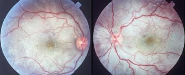
- Despite differences in their patients, the manifestations appeared to represent a spectrum of disease, and several authors suggested that the disorder should be termed Vogt-Koyanagi-Harada syndrome (see the image below). (medscape.com)
Termed Vogt-Koyanagi1
- Despite differences in their patients, the manifestations appeared to represent a spectrum of disease, and several authors suggested that the disorder should be termed Vogt-Koyanagi-Harada syndrome (see the image below). (medscape.com)
Bilateral1
- A syndrome characterized by bilateral granulomatous UVEITIS with IRITIS and secondary GLAUCOMA , premature ALOPECIA , symmetrical VITILIGO , poliosis circumscripta (a strand of depigmented hair), HEARING DISORDERS , and meningeal signs (neck stiffness and headache). (nih.gov)