Vasculitis, Leukocytoclastic, Cutaneous
Vasculitis
Purpura, Schoenlein-Henoch
Skin Diseases, Vascular
Cryoglobulins
Skin Diseases
Systemic Vasculitis
Retinal Vasculitis
Biopsy
Anti-Neutrophil Cytoplasmic Antibody-Associated Vasculitis
Skin
Vasculitis, Central Nervous System
Encyclopedias as Topic
Antibodies, Antineutrophil Cytoplasmic
Microscopic Polyangiitis
Inflammation
Polychondritis, Relapsing
Ear Deformities, Acquired
Tracheobronchomalacia
Ear, Inner
Sweet Syndrome
Alternating antineutrophil cytoplasmic antibody specificity: drug-induced vasculitis in a patient with Wegener's granulomatosis. (1/116)
We describe a patient who presented with Wegener's granulomatosis associated with antineutrophil cytoplasmic antibodies (ANCA) directed against proteinase 3 (PR3) with a cytoplasmic immunofluorescence pattern (cANCA), whose ANCA type changed to antimyeloperoxidase antibodies with a perinuclear immunofluorescence pattern (pANCA) when treated with propylthiouracil, and changed back to anti-PR3 antibodies with cANCA after the medication was discontinued. The patient developed flares of vasculitis symptoms associated with rises in either type of ANCA. Tests for antimyeloperoxidase ANCA were repeatedly negative before the drug was started, strongly implicating the drug as the cause of the episode. This case demonstrates that patients with idiopathic ANCA-positive vasculitis may quickly develop a superimposed drug-associated ANCA-positive vasculitis. Iatrogenic vasculitis should be suspected when a patient with idiopathic vasculitis with one type of ANCA develops the other type of ANCA. (+info)Microscopic polyangiitis: clinical and laboratory findings in eighty-five patients. (2/116)
OBJECTIVE: To retrospectively analyze the clinical symptoms, laboratory findings, and outcomes in patients with microscopic polyangiitis (MPA) who were enrolled in various clinical trials conducted by the French Vasculitis Study Group. METHODS: A cohort of 85 patients meeting the Chapel Hill criteria for MPA participated in the study. Seventy-one of them were included in prospective therapeutic trials. Eighty-one diagnoses were biopsy proven. In the other patients, diagnosis was based on clinical findings. RESULTS: Forty-seven men and 38 women, with a mean +/- SD age of 56.8 +/- 14.6 years, met the criteria for MPA. Their main clinical symptoms were renal manifestations (78.8%), weight loss (72.9%), skin involvement (62.4%), fever (55.3%), mononeuritis multiplex (57.6%), arthralgias (50.6%), myalgias (48.2%), hypertension (34.1%), lung involvement (24.7%; alveolar hemorrhage 11.8%), and cardiac failure (17.6%). The mean +/- SD serum creatinine level before treatment was 2.59 +/- 2.96 mg/dl; 47 patients had renal insufficiency (serum creatinine > 1.36 mg/dl). Eight patients underwent dialysis at the time of diagnosis, and long-term dialysis was necessary for 10 patients. Antineutrophil cytoplasmic antibodies (ANCA) were present in 38 of 51 patients (74.5%), of whom 33 had a perinuclear staining pattern (pANCA) and 5 had a cytoplasmic pattern. Antibodies to proteinase 3 were present in 4 patients and antibodies to myeloperoxidase were detected in 31, as determined by enzyme-linked immunosorbent assay. Of the 30 patients who underwent renal and celiac angiography, 4 had microaneurysms. Of the 29 patients (34.1%) who had relapses, 8 died during or after the relapse. During followup, 28 of the 85 patients (32.9%) died. The mean +/- SD duration of followup of the group was 69.9 +/- 60.6 months. Deaths were less frequent when patients had been treated with steroids and immunosuppressive drugs (13 patients [24.1%]) than with steroids alone (15 patients [48.4%]) (P < 0.01). The 5-year survival rate was 74%. CONCLUSION: This study demonstrated that MPA is a multisystemic disease in which renal symptoms are frequent, but the disease is also associated with general symptoms, arthritis, mononeuritis multiplex, and other manifestations that are also seen in various vasculitides. The rarity of abnormal angiogram findings and the high frequency of pANCA are characteristic of MPA. In most cases, the outcome is comparable with those of other systemic vasculitides, but relapses are frequent. (+info)Immune complexes from vasculitis patients bind to endothelial Fc receptors independent of the allelic polymorphism of FcgammaRIIa. (3/116)
Cutaneous leukocytoclastic vasculitis is characterized by the deposition of circulating immune complexes, neutrophil extravasation, and vessel destruction, but mechanisms of circulating immune complexes capture within postcapillary venules are unknown. We demonstrate that circulating immune complexes from sera of vasculitis patients bind to cultured endothelium in an Fc gamma receptor IIa-dependent fashion. In lesional skin, endothelial cells bind immunoglobulin G2 > immunoglobulin G3 and immunoglobulin G4, but not immunoglobulin G1, even before obvious neutrophil transmigration and vessel damage. As the human Fc gamma receptor IIa proteins exist in two allotypes (one with a histidine at position 131, which binds immunoglobulin G1, 2, 3 and the other with an arginine at position 131, which binds immunoglobulin G1, and 3, but is unable to bind immunoglobulin G2), we expected an altered prevalence of histidine 131 forms in vasculitis patients. Sequence analysis, however, revealed an equal distribution of allotypes in patients and controls. In conclusion, circulating immune complex binding to endothelial Fc gamma receptor IIa is among the initial steps in the development of vasculitis. Although immunoglobulin G2 is the predominant subtype precipitated at endothelial surfaces, it is not required for fixing circulating immune complexes to endothelium, because patients homozygote for Fc gamma receptor IIa-arginine 131 equally develop leukocytoclastic vasculitis as those bearing the Fc gamma receptor IIa-histidine 131 allele. As immunoglobulin G1 is virtually absent in leukocytoclastic vasculitis lesions and immunoglobulin G4 does not bind to both Fc gamma receptor IIa alleles, these complexes, in addition to immunoglobulin G2, should contain immunoglobulin G3 in order to fix to vascular Fc gamma receptor IIa, at least in persons homozygous for Fc gamma receptor IIa-arginine 131. KEYWORDS: CD32/immunoglobulin G subtypes/leukocytoclastic vasculitis/microvessels. (+info)Early vasculitis in the mercuric chloride induced Brown Norway rat model is neutrophil independent. (4/116)
In the Brown Norway rat, mercuric chloride (HgCl2) induces an autoimmune syndrome characterized by necrotizing vasculitis, predominantly affecting the caecum, and a polyclonal B-cell response. The time course of vasculitis is biphasic, with an alphabeta T-cell independent phase occurring within 24 h, and a T-cell and neutrophil dependent phase, maximal at two weeks. The pathogenesis of the early phase of vasculitis is unclear, and this study aims to examine the role of neutrophils. Rat neutrophils were depleted using cyclophosphamide. RP3, an antirat neutrophil monoclonal antibody, inhibited neutrophil leucocytosis but did not deplete neutrophils. Vasculitis was induced by subcutaneous HgCl2 injection. Serial measurements of peripheral blood leucocyte count were made. Rats were killed after 24 or 72 h. The macroscopic appearance of the caecum was scored by an experienced observer, and samples taken for histological examination. Caecums were excised and myeloperoxidase, a marker enzyme for neutrophil infiltration, assayed. Cyclophosphamide induced marked neutropaenia whereas RP3 inhibited the neutrophilia observed after HgCl2 injection. Vasculitis was present in both treated and control animals, with no significant differences in macroscopic or microscopic scores between the groups. Tissue myeloperoxidase activity was low in all animals and did not differ significantly between groups. The data do not support a role for neutrophils in the initial pathogenesis of vasculitis in this model. (+info)A prospective study of vasculitis patients collected in a five year period: evaluation of the Chapel Hill nomenclature. (5/116)
OBJECTIVE: To test the usefulness of the Chapel Hill nomenclature, supplemented with surrogate parameters, as diagnostic criteria for primary vasculitides. METHODS: To prospectively evaluate vasculitis patients according to a standardised clinical and para-clinical programme. In accordance with the Chapel Hill publication surrogate parameters were used: proteinuria, haematuria and red blood cell casts (glomerulonephritis), angiographic or ultrasonic demonstration of aneurysms or stenoses (arteritis), radiological lung infiltrates or cavitations of more than one month's duration (granuloma in the lungs), bloody nasal discharge or crusts, chronic sinusitis, otitis and/or mastoiditis, bone and/or cartilage destruction, and acute hearing loss (granuloma in upper airways). RESULTS: The following entities were diagnosed: giant cell arteritis (n=14), Takayasu arteritis (n=1), polyarteritis nodosa (n=2), Wegener's granulomatosis (n=27), Churg-Strauss syndrome (n=2), microscopic polyangiitis (n=12), Henoch-Schonlein purpura (n=2), cutaneous leucocytoclastic angiitis (n=37), and secondary vasculitis (n=21). Giant cell arteritis and cutaneous leucocytoclastic angiitis were in all cases diagnosed by biopsy. Using the Chapel Hill nomenclature supplemented with surrogate parameters, only 8 of 27 patients were diagnosed with Wegener's granulomatosis, and 3 of 12 cases with microscopic polyangiitis. The number of patients in the remaining diagnostic entities were considered to few to evaluate. CONCLUSIONS: The Chapel Hill nomenclature, supplemented with surrogate parameters, failed to act as diagnostic criteria in Wegener's granulomatosis and microscopic polyangiitis. The following diagnostic criteria are proposed for Wegener's granulomatosis: (1) Biopsy or surrogate parameter for granulomatous inflammation in the respiratory system and (2) Biopsy verified necrotising vasculitis in small to medium sized vessels or biopsy/surrogate parameter for glomerulonephritis or positive PR3-ANCA test and (3) Lack of eosinophilia in blood and biopsy samples. The following diagnostic criteria are proposed for microscopic polyangiitis: (1) Biopsy verified necrotising vasculitis in small vessels and/or glomerulonephritis with few or no immune deposits and (2) Involvement of more than one organ system as indicated by biopsy verified vasculitis in small to medium sized vessels or surrogate parameter for glomerulonephritis and (3) Lack of biopsy and surrogate parameter for granulomatous inflammation in the respiratory system. Using these criteria all Wegener's patients and 9 of 12 patients with microscopic polyangiitis could be diagnosed. (+info)Unusual manifestations of type II cryoglobulinaemia associated with Waldenstrom's macroglobulinaemia. (6/116)
Cryoglobulinaemia in association with Waldenstrom's macroglobulinaemia is relatively common, ranging from 8% to 18% of cases; however, < 5% have symptoms or complications. We describe a patient with a history of cutaneous, peritoneal, and fallopian tube vasculitis related to type II cryoglobulinaemia associated with Waldenstrom's macroglobulinaemia. Cytotoxic treatment was initiated (cyclophosphamide, vincristine, and prednisone) and had a good initial response. However, after the third course of chemotherapy, the patient presented with septic shock and died. Even though cryoglobulinaemia is a model of systemic vasculitis, peritoneal and fallopian tube vasculitis associated with type II cryoglobulinemia has not been described previously. (+info)Leukocytoclastic vasculitis as a complication of infected total hip prosthesis. (7/116)
We present a case of leukocytoclastic vasculitis, associated with an exacerbation of chronic osteomyelitis in a pseudoarthrosis of the hip. Skin lesions of the lower limb were the most prominent features. (+info)Follicular dendritic cell tumor of the liver: a clinicopathologic and Epstein-Barr virus study of two cases. (8/116)
Two cases of hepatic follicular dendritic cell (FDC) tumor are described. Both patients were female, aged 57 and 51 years. They presented with epigastralgia or abdominal fullness and weight loss. The first patient refused surgical resection. She developed progressive polyclonal gammopathy and then bilateral purpura over the legs. Skin biopsy revealed leukocytoclastic vasculitis with granular vascular deposits of IgA and C3. The second patient had marked peripheral blood and tissue eosinophilia. The histological diagnosis was confirmed by positive staining for CD21 and CD23. The stromal lymphocytes were predominantly composed of CD3(+)and CD8(+) cells. In situ hybridization for EBER showed a positive nuclear signal in tumor cells but not in inflammatory cells. Polymerase chain reaction amplification for Exon 3 of the latent membrane protein-1 (LMP-1) gene showed a characteristic 30-bp deletion between nucleotides 168282 and 168253, corresponding to the B95-8 sequence. The unique clinicopathological features of our cases have not been reported for FDC tumors before. The clinical significance of the 30-bp deletion in Exon 3 of the LMP-1 gene in FDC tumor of the liver warrants further investigation. (+info)Leukocytoclastic vasculitis, cutaneous is a type of vasculitis that is limited to the skin. Vasculitis refers to inflammation of the blood vessels, which can cause damage to the vessel walls and impair blood flow to various tissues in the body. In leukocytoclastic vasculitis, the small blood vessels (capillaries and venules) in the skin become inflamed, leading to damage and destruction of the vessel walls.
The term "leukocytoclastic" refers to the presence of nuclear debris from white blood cells (leukocytes) that have been destroyed within the affected blood vessels. This type of vasculitis is often associated with the deposition of immune complexes (formed by the interaction between antibodies and antigens) in the walls of the blood vessels, which triggers an inflammatory response.
Cutaneous leukocytoclastic vasculitis typically presents as palpable purpura (small to large, raised, purple or red spots on the skin), usually located on the lower extremities, but can also affect other areas of the body. Other symptoms may include burning or itching sensations in the affected area, and in some cases, ulcers or necrosis (tissue death) may occur.
The diagnosis of cutaneous leukocytoclastic vasculitis is typically made based on clinical presentation, laboratory tests, and histopathological examination of a skin biopsy specimen. Treatment usually involves addressing any underlying causes or triggers, as well as managing symptoms with medications such as corticosteroids or immunosuppressive agents.
Vasculitis is a group of disorders characterized by inflammation of the blood vessels, which can cause changes in the vessel walls including thickening, narrowing, or weakening. These changes can restrict blood flow, leading to organ and tissue damage. The specific symptoms and severity of vasculitis depend on the size and location of the affected blood vessels and the extent of inflammation. Vasculitis can affect any organ system in the body, and its causes can vary, including infections, autoimmune disorders, or exposure to certain medications or chemicals.
Henoch-Schönlein purpura (HSP) is a type of small vessel vasculitis, which is a condition characterized by inflammation of the blood vessels. HSP primarily affects children, but it can occur in adults as well. It is named after two German physicians, Eduard Heinrich Henoch and Johann Schönlein, who first described the condition in the mid-19th century.
The main feature of HSP is a purpuric rash, which is a type of rash that appears as small, red or purple spots on the skin. The rash is caused by leakage of blood from the small blood vessels (capillaries) beneath the skin. In HSP, this rash typically occurs on the legs and buttocks, but it can also affect other parts of the body, such as the arms, face, and trunk.
In addition to the purpuric rash, HSP is often accompanied by other symptoms, such as joint pain and swelling, abdominal pain, nausea, vomiting, and diarrhea. In severe cases, it can also affect the kidneys, leading to hematuria (blood in the urine) and proteinuria (protein in the urine).
The exact cause of HSP is not known, but it is thought to be related to an abnormal immune response to certain triggers, such as infections or medications. Treatment typically involves supportive care, such as pain relief and fluid replacement, as well as medications to reduce inflammation and suppress the immune system. In most cases, HSP resolves on its own within a few weeks or months, but it can lead to serious complications in some individuals.
Vascular skin diseases are a group of medical conditions that affect the blood vessels in the skin. These disorders can be caused by problems with the structure or function of the blood vessels, which can lead to various symptoms such as redness, discoloration, pain, itching, and ulcerations. Some examples of vascular skin diseases include:
1. Rosacea: a chronic skin condition that causes redness, flushing, and visible blood vessels in the face.
2. Eczema: a group of inflammatory skin conditions that can cause redness, itching, and dryness. Some types of eczema, such as varicose eczema, are associated with problems with the veins.
3. Psoriasis: an autoimmune condition that causes red, scaly patches on the skin. Some people with psoriasis may also develop psoriatic arthritis, which can affect the blood vessels in the skin and joints.
4. Vasculitis: a group of conditions that cause inflammation of the blood vessels. This can lead to symptoms such as redness, pain, and ulcerations.
5. Livedo reticularis: a condition that causes a net-like pattern of discoloration on the skin, usually on the legs. It is caused by abnormalities in the small blood vessels.
6. Henoch-Schönlein purpura: a rare condition that causes inflammation of the small blood vessels, leading to purple spots on the skin and joint pain.
7. Raynaud's phenomenon: a condition that affects the blood vessels in the fingers and toes, causing them to become narrow and restrict blood flow in response to cold temperatures or stress.
Treatment for vascular skin diseases depends on the specific condition and its severity. It may include medications, lifestyle changes, and in some cases, surgery.
Cryoglobulins are immunoglobulins (a type of antibody) that precipitate or become insoluble at reduced temperatures, typically below 37°C (98.6°F), and re-dissolve when rewarmed. They can be found in various clinical conditions such as infections, inflammatory diseases, and lymphoproliferative disorders.
The presence of cryoglobulins in the blood can lead to a variety of symptoms, including purpura (a type of skin rash), arthralgias (joint pain), neuropathy (nerve damage), and glomerulonephritis (kidney inflammation). The diagnosis of cryoglobulinemia is made by detecting the presence of cryoglobulins in the serum, which requires special handling and processing of the blood sample. Treatment of cryoglobulinemia depends on the underlying cause and may include medications such as corticosteroids, immunosuppressive agents, or targeted therapies.
Skin diseases, also known as dermatological conditions, refer to any medical condition that affects the skin, which is the largest organ of the human body. These diseases can affect the skin's function, appearance, or overall health. They can be caused by various factors, including genetics, infections, allergies, environmental factors, and aging.
Skin diseases can present in many different forms, such as rashes, blisters, sores, discolorations, growths, or changes in texture. Some common examples of skin diseases include acne, eczema, psoriasis, dermatitis, fungal infections, viral infections, bacterial infections, and skin cancer.
The symptoms and severity of skin diseases can vary widely depending on the specific condition and individual factors. Some skin diseases are mild and can be treated with over-the-counter medications or topical creams, while others may require more intensive treatments such as prescription medications, light therapy, or even surgery.
It is important to seek medical attention if you experience any unusual or persistent changes in your skin, as some skin diseases can be serious or indicative of other underlying health conditions. A dermatologist is a medical doctor who specializes in the diagnosis and treatment of skin diseases.
Systemic vasculitis is a group of disorders characterized by inflammation of the blood vessels (vasculitis) that can affect various organs and systems throughout the body. This condition can cause damage to the walls of the blood vessels, leading to narrowing, blockage, or weakening of the vessel walls, which can further result in reduced blood flow, tissue damage, and organ dysfunction.
The symptoms of systemic vasculitis depend on the severity and location of the affected blood vessels. They may include fever, fatigue, weight loss, joint pain, skin rashes or lesions, muscle weakness, nerve damage, and organ dysfunction such as kidney failure, lung disease, or gastrointestinal bleeding.
Systemic vasculitis can be caused by various factors, including infections, autoimmune diseases, medications, and underlying medical conditions. The diagnosis of systemic vasculitis typically involves a combination of physical examination, laboratory tests, imaging studies, and sometimes biopsy of the affected tissue. Treatment may include corticosteroids, immunosuppressive drugs, and other medications to control inflammation and prevent organ damage.
Retinal vasculitis is a medical condition characterized by inflammation of the blood vessels in the retina, which is the light-sensitive tissue located at the back of the eye. This condition can cause damage to the retina and may lead to vision loss if not treated promptly. The inflammation can affect both the small and large blood vessels in the retina and can occur as a result of various systemic diseases or infections, including autoimmune disorders, tuberculosis, syphilis, and toxoplasmosis. In some cases, retinal vasculitis may also be associated with uveitis, which is inflammation of the middle layer of the eye. Treatment typically involves addressing the underlying cause of the inflammation and may include corticosteroids or other immunosuppressive therapies to reduce inflammation and prevent further damage to the retina.
A biopsy is a medical procedure in which a small sample of tissue is taken from the body to be examined under a microscope for the presence of disease. This can help doctors diagnose and monitor various medical conditions, such as cancer, infections, or autoimmune disorders. The type of biopsy performed will depend on the location and nature of the suspected condition. Some common types of biopsies include:
1. Incisional biopsy: In this procedure, a surgeon removes a piece of tissue from an abnormal area using a scalpel or other surgical instrument. This type of biopsy is often used when the lesion is too large to be removed entirely during the initial biopsy.
2. Excisional biopsy: An excisional biopsy involves removing the entire abnormal area, along with a margin of healthy tissue surrounding it. This technique is typically employed for smaller lesions or when cancer is suspected.
3. Needle biopsy: A needle biopsy uses a thin, hollow needle to extract cells or fluid from the body. There are two main types of needle biopsies: fine-needle aspiration (FNA) and core needle biopsy. FNA extracts loose cells, while a core needle biopsy removes a small piece of tissue.
4. Punch biopsy: In a punch biopsy, a round, sharp tool is used to remove a small cylindrical sample of skin tissue. This type of biopsy is often used for evaluating rashes or other skin abnormalities.
5. Shave biopsy: During a shave biopsy, a thin slice of tissue is removed from the surface of the skin using a sharp razor-like instrument. This technique is typically used for superficial lesions or growths on the skin.
After the biopsy sample has been collected, it is sent to a laboratory where a pathologist will examine the tissue under a microscope and provide a diagnosis based on their findings. The results of the biopsy can help guide further treatment decisions and determine the best course of action for managing the patient's condition.
Anti-Neutrophil Cytoplasmic Antibody (ANCA)-Associated Vasculitis (AAV) is a group of autoimmune diseases characterized by inflammation and damage to small blood vessels, particularly capillaries, venules, and arterioles. The condition is named after the presence of ANCAs in the patient's serum, which are autoantibodies that target specific proteins in the neutrophil cytoplasm.
AAV includes several subtypes, including:
1. Granulomatosis with Polyangiitis (GPA, formerly known as Wegener's granulomatosis) - a form of AAV that typically affects the respiratory tract and kidneys, characterized by the presence of granulomas (clusters of inflammatory cells).
2. Microscopic Polyangiitis (MPA) - a form of AAV that primarily affects small vessels in various organs, such as the kidneys, lungs, and skin.
3. Eosinophilic Granulomatosis with Polyangiitis (EGPA, formerly known as Churg-Strauss syndrome) - a form of AAV that involves asthma, allergies, and eosinophilia (an increased number of eosinophils in the blood), along with vasculitis affecting various organs.
The exact cause of ANCA-Associated Vasculitis is not fully understood, but it is believed to involve an interplay between genetic factors, environmental triggers, and dysregulation of the immune system. The condition can lead to a wide range of symptoms depending on which organs are affected, including fever, fatigue, weight loss, joint pain, skin rashes, cough, shortness of breath, nosebleeds, and kidney problems. Treatment typically involves immunosuppressive medications to control inflammation and prevent further damage to the affected organs.
In medical terms, the skin is the largest organ of the human body. It consists of two main layers: the epidermis (outer layer) and dermis (inner layer), as well as accessory structures like hair follicles, sweat glands, and oil glands. The skin plays a crucial role in protecting us from external factors such as bacteria, viruses, and environmental hazards, while also regulating body temperature and enabling the sense of touch.
Vasculitis, Central Nervous System (CNS), refers to a group of disorders characterized by inflammation of blood vessels within the brain and/or spinal cord. This inflammation can cause damage to the blood vessel walls, leading to narrowing, blocking or weakening of the vessels, and in some cases, formation of aneurysms or rupture of the vessels.
The causes of CNS vasculitis are varied and can include infections, autoimmune diseases, medications, and unknown factors. The symptoms of CNS vasculitis depend on the severity and location of the inflammation, and may include headache, seizures, stroke-like symptoms (such as weakness or numbness in the face, arms, or legs), cognitive changes, and in severe cases, coma.
Diagnosis of CNS vasculitis typically involves a combination of clinical evaluation, imaging studies (such as MRI or angiography), and laboratory tests (including blood tests and analysis of cerebrospinal fluid). Treatment may involve corticosteroids, immunosuppressive medications, and/or other therapies aimed at reducing inflammation and preventing further damage to the blood vessels.
An encyclopedia is a comprehensive reference work containing articles on various topics, usually arranged in alphabetical order. In the context of medicine, a medical encyclopedia is a collection of articles that provide information about a wide range of medical topics, including diseases and conditions, treatments, tests, procedures, and anatomy and physiology. Medical encyclopedias may be published in print or electronic formats and are often used as a starting point for researching medical topics. They can provide reliable and accurate information on medical subjects, making them useful resources for healthcare professionals, students, and patients alike. Some well-known examples of medical encyclopedias include the Merck Manual and the Stedman's Medical Dictionary.
Antineutrophil cytoplasmic antibodies (ANCAs) are a type of autoantibody that specifically target certain proteins in the cytoplasm of neutrophils, which are a type of white blood cell. These antibodies are associated with several types of vasculitis, which is inflammation of the blood vessels.
There are two main types of ANCAs: perinuclear ANCAs (p-ANCAs) and cytoplasmic ANCAs (c-ANCAs). p-ANCAs are directed against myeloperoxidase, a protein found in neutrophil granules, while c-ANCAs target proteinase 3, another protein found in neutrophil granules.
The presence of ANCAs in the blood can indicate an increased risk for developing certain types of vasculitis, such as granulomatosis with polyangiitis (GPA), eosinophilic granulomatosis with polyangiitis (EGPA), and microscopic polyangiitis (MPA). ANCA testing is often used in conjunction with other clinical findings to help diagnose and manage these conditions.
It's important to note that while the presence of ANCAs can indicate an increased risk for vasculitis, not everyone with ANCAs will develop the condition. Additionally, ANCAs can also be found in some individuals without any associated disease, so their presence should be interpreted in the context of other clinical findings.
Microscopic Polyangiitis (MPA) is a rare type of vasculitis, which is a group of disorders that cause inflammation in the blood vessels. In MPA, the small blood vessels in various organs become inflamed and damaged, leading to symptoms that can affect multiple organ systems.
The term "microscopic" refers to the fact that the diagnosis of this condition typically requires examination of tissue samples under a microscope to see the characteristic patterns of inflammation and damage in the small blood vessels.
MPA is an autoimmune disorder, which means that the body's immune system mistakenly attacks its own tissues and organs. In MPA, the immune system produces abnormal antibodies called ANCA (antineutrophil cytoplasmic antibodies) that target certain proteins in the white blood cells, leading to their activation and subsequent damage to the blood vessels.
The symptoms of MPA can vary widely depending on which organs are affected, but they may include fever, fatigue, weight loss, joint pain, skin rashes, cough, shortness of breath, and kidney problems such as proteinuria and hematuria. Treatment typically involves the use of immunosuppressive medications to suppress the overactive immune system and reduce inflammation in the blood vessels.
"Foreign bodies" refer to any object or substance that is not normally present in a particular location within the body. These can range from relatively harmless items such as splinters or pieces of food in the skin or gastrointestinal tract, to more serious objects like bullets or sharp instruments that can cause significant damage and infection.
Foreign bodies can enter the body through various routes, including ingestion, inhalation, injection, or penetrating trauma. The location of the foreign body will determine the potential for harm and the necessary treatment. Some foreign bodies may pass through the body without causing harm, while others may require medical intervention such as removal or surgical extraction.
It is important to seek medical attention if a foreign body is suspected, as untreated foreign bodies can lead to complications such as infection, inflammation, and tissue damage.
Inflammation is a complex biological response of tissues to harmful stimuli, such as pathogens, damaged cells, or irritants. It is characterized by the following signs: rubor (redness), tumor (swelling), calor (heat), dolor (pain), and functio laesa (loss of function). The process involves the activation of the immune system, recruitment of white blood cells, and release of inflammatory mediators, which contribute to the elimination of the injurious stimuli and initiation of the healing process. However, uncontrolled or chronic inflammation can also lead to tissue damage and diseases.
Relapsing polychondritis is a rare autoimmune disease characterized by inflammation and damage to the cartilaginous structures in the body. The condition can affect multiple organs and tissues, including the ears, nose, trachea, bronchi, joints, and cardiovascular system. It is called "relapsing" because it tends to involve recurring episodes of inflammation and damage, followed by periods of remission.
The hallmark symptom of relapsing polychondritis is pain and swelling in the ears, nose, or airways. Other symptoms may include:
* Redness, tenderness, and warmth in affected areas
* Hearing loss or tinnitus (ringing in the ears)
* Nasal congestion, runny nose, or nosebleeds
* Hoarseness or difficulty speaking
* Wheezing, shortness of breath, or coughing
* Joint pain, stiffness, or swelling
* Skin rashes or sores
* Eye inflammation or dryness
* Heart murmurs or other cardiovascular symptoms
The exact cause of relapsing polychondritis is not known, but it is thought to involve an abnormal immune response in which the body's own antibodies attack and damage cartilage and other tissues. The diagnosis of relapsing polychondritis is typically based on a combination of clinical symptoms, laboratory tests, and imaging studies.
There is no cure for relapsing polychondritis, but treatment can help manage the symptoms and prevent complications. Treatment may include corticosteroids, immunosuppressive drugs, and other medications to reduce inflammation and suppress the immune system. In severe cases, surgery may be necessary to repair or replace damaged tissues.
Ear cartilage, also known as auricular cartilage, refers to the flexible connective tissue that makes up the structural framework of the external ear or pinna. The ear cartilage provides support and shape to the ear, helping to direct sound waves into the ear canal and towards the eardrum.
The ear cartilage is composed of type II collagen fibers and proteoglycans, which give it its flexibility and resiliency. It is covered by a thin layer of skin on both sides and contains no bones. Instead, the ear cartilage is shaped and maintained by the surrounding muscles and connective tissue.
There are three main parts of the ear cartilage: the helix, the antihelix, and the tragus. The helix is the outer rim of the ear, while the antihelix is the curved ridge that runs parallel to the helix. The tragus is the small piece of cartilage that projects from the front of the ear canal.
Ear cartilage can be affected by various conditions, including trauma, infection, and degenerative changes associated with aging. In some cases, surgical procedures may be required to reshape or reconstruct damaged ear cartilage.
Acquired ear deformities refer to abnormal shapes or structures of the ear that result from injury, infection, inflammation, or other external factors after birth. These deformities can affect the appearance and function of the ear, causing symptoms such as hearing loss or discomfort. Examples of acquired ear deformities include:
1. Cauliflower ear: a condition characterized by swelling, thickening, and distortion of the ear caused by repeated trauma or injury to the ear cartilage.
2. Microtia: a congenital ear abnormality that can become worse over time due to infection, inflammation, or trauma, resulting in an underdeveloped or absent ear.
3. Macrotia: an abnormally large ear that may result from injury or other external factors.
4. Stenosis: a narrowing of the ear canal that can result from chronic inflammation, infection, or scarring.
5. Hematoma: a collection of blood in the ear tissue caused by trauma or injury, which can lead to deformity if not treated promptly.
6. Keloids: overgrowths of scar tissue that can form after injury or surgery and distort the shape of the ear.
Treatment for acquired ear deformities may include surgical reconstruction, splinting, or other interventions depending on the severity and underlying cause of the condition.
Tracheobronchomalacia is a medical condition that refers to the abnormal softening and weakness of the tracheal and bronchial walls, leading to their collapse or narrowing during breathing, particularly during expiration. This collapse can cause symptoms such as wheezing, shortness of breath, coughing, and recurrent respiratory infections. The condition can be congenital or acquired, with common causes including aging, chronic obstructive pulmonary disease (COPD), and long-term intubation. In severe cases, tracheobronchomalacia may require surgical intervention to stabilize the airway and improve breathing.
The inner ear is the innermost part of the ear that contains the sensory organs for hearing and balance. It consists of a complex system of fluid-filled tubes and sacs called the vestibular system, which is responsible for maintaining balance and spatial orientation, and the cochlea, a spiral-shaped organ that converts sound vibrations into electrical signals that are sent to the brain.
The inner ear is located deep within the temporal bone of the skull and is protected by a bony labyrinth. The vestibular system includes the semicircular canals, which detect rotational movements of the head, and the otolith organs (the saccule and utricle), which detect linear acceleration and gravity.
Damage to the inner ear can result in hearing loss, tinnitus (ringing in the ears), vertigo (a spinning sensation), and balance problems.
Sweet syndrome, also known as acute febrile neutrophilic dermatosis, is a skin condition characterized by the rapid onset of painful, red, and swollen skin lesions. The lesions are often accompanied by fever and elevated white blood cell count, particularly an increase in neutrophils.
The medical definition of Sweet syndrome includes the following criteria:
1. Abrupt onset of painful, erythematous (red), and edematous (swollen) papules, plaques, or nodules.
2. Fever greater than 38°C (100.4°F).
3. Leukocytosis with a predominance of neutrophils in the peripheral blood.
4. Histopathological evidence of a dense dermal infiltrate of neutrophils without evidence of vasculitis.
5. Rapid response to systemic corticosteroids.
Sweet syndrome can be associated with various medical conditions, such as infections, malignancies, and inflammatory diseases, or it can occur without an identifiable underlying cause (idiopathic).
 Cutaneous small-vessel vasculitis
Systemic vasculitis
Lymphedema
Vasculitis
Bilateral lower extremity inflammatory lymphedema
Granulomatosis with polyangiitis
Gluten-related disorders
Henoch-Schönlein purpura
Polyarteritis nodosa
DOCK8 deficiency
List of skin conditions
Multisystem inflammatory syndrome in children
 Cutaneous Signs of Vascular Disorders: Idiopathic Leukocytoclastic Vasculitis | Consultant360
 Relapsing Polychondritis: Practice Essentials, Background, Pathophysiology
Cutaneous small-vessel vasculitis - Wikipedia
 Vasculitis: Treatment, symptoms, causes, and types
Relapsing Polychondritis Clinical Presentation: History, Physical Examination
Relapsing Polychondritis Treatment & Management: Medical Care, Surgical Care, Consultations
Dermatologic Manifestations of Renal Disease: Overview, Dermatologic Manifestations of Diseases Associated With ESRD,...
Relapsing Polychondritis: Practice Essentials, Background, Pathophysiology
 Cutaneous Vasculitis - Musculoskeletal and Connective Tissue Disorders - MSD Manual Professional Edition
 Tagrisso (Osimertinib Tablets): Uses, Dosage, Side Effects, Interactions, Warning
 A transgenic mouse model of autoi... | Archive ouverte UNIGE
Prolia (Denosumab Injection): Uses, Dosage, Side Effects, Interactions, Warning
KoreaMed
 Constitutively active Lyn kinase causes a cutaneous small vessel vasculitis and liver fibrosis syndrome | Nature Communications
 DailyMed - ETODOLAC tablet
 Sweet Syndrome - Symptoms, Causes, Treatment | NORD
DailyMed - FLUOXETINE capsule
 Expression of COX-2 and bcl-2 in oral lichen planus lesions and lichenoid reactions - ecancer
Rare Juvenile Primary Systemic Vasculitis
Plus it
 Mammalian target of rapamycin (mTOR) inhibitors in dermatology - Indian Journal of Dermatology, Venereology and Leprology
Leukocytoclastic Vasculitis: Practice Essentials, Pathophysiology, Epidemiology
 Necrotizing cutaneous vasculitis with massive gastrointestinal bleeding following naproxen treatment<...
1 - Forex trading automated
 Dusky bullae on the lower abdomen - Clinical Advisor
Wegener's Granulomatosis - CheckOrphan
 DeCS 2010 - Deleted terms
DeCS 2010 - Deleted terms
DeCS 2010 - Changed terms
DeCS 2010 - Deleted terms
Angiitis6
- 831 The condition is also known as hypersensitivity vasculitis, cutaneous leukocytoclastic vasculitis, hypersensitivity angiitis, cutaneous leukocytoclastic angiitis, cutaneous necrotizing vasculitis and cutaneous necrotizing venulitis, It is the most common form of vasculitis seen in clinical practice, usually caused by inflammation of post-capillary venules in the dermis). (wikipedia.org)
- Vasculitis refers to a large group of diseases, also known as angiitis, that damage blood vessels by causing inflammation. (medicalnewstoday.com)
- Vasculitis is also called angiitis and arteritis. (medicalnewstoday.com)
- CNS vasculitis has been reported under a variety of descriptive terms including isolated CNS angiitis, idiopathic angiitis of the CNS, and primary angiitis or vasculitis of the CNS. (fortuneonline.org)
- other causes of pulmonary-renal syndrome (see Granulomatosis With Polyangiitis ), cutaneous leukocytoclastic angiitis. (empendium.com)
- Granulomatosis with polyangiitis is the third most common type of vasculitis in Iran after Behcet's disease and cutaneous leukocytoclastic angiitis [6]. (rheumres.org)
Hypersensitivity7
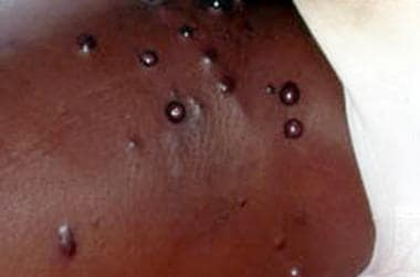
- Leukocytoclastic, or hypersensitivity, vasculitis is a neutrophilic inflammation of small blood vessels. (consultant360.com)
- Any primary or secondary vasculitis can affect the skin, including that due to serum sickness, infections (eg, hepatitis C), cancers, rheumatologic or other autoimmune disorders, and hypersensitivity to drugs. (msdmanuals.com)
- However, hypersensitivity vasculitis is sometimes used to refer to CSVV caused by a known drug or infection. (msdmanuals.com)
- Martinez-Taboada VM, Blanco R, Garcia-Fuentes M, Rodriguez-Valverde V. Clinical features and outcome of 95 patients with hypersensitivity vasculitis. (medscape.com)
- Comparative clinical and epidemiological study of hypersensitivity vasculitis versus Henoch-Schonlein purpura in adults. (medscape.com)
- Another theory is that deposition of immune complexes within dermal vessels leads to vasculitis, a type III (Arthus type) hypersensitivity reaction. (clinicaladvisor.com)
- Hypersensitivity vasculitis is inflammation of the vessel wall which is usually due to a hypersensitivity reaction to a known drug, auto-antigens or infectious agents such as bacteria. (wikidoc.org)
Acute10
- Vasculitis can be acute and short-term or chronic and long-term. (medicalnewstoday.com)
- Mice implanted with hybridoma secreting 6-19 IgG3 anti-IgG2a rheumatoid factor (RF) with cryoglobulin activity develop acute glomerulonephritis and cutaneous leukocytoclastic vasculitis. (unige.ch)
- These renal and vascular changes were very different from those observed in the acute cryoglobulinemia, characterized by mainly "wire-loop" glomerular lesions and a cutaneous leukocytoclastic form of vasculitis. (unige.ch)
- It accounts for approximately 10% of acute cutaneous vasculitis. (koreamed.org)
- Some of the acute primary vasculitides are quite common paediatric diseases (e.g. (printo.it)
- Cutaneous necrotizing vasculitis is usually induced by an acute infection or exposure to a drug. (bgu.ac.il)
- CNS vasculitis can cause brain damage with reversible and or irreversible neurologic involvement, including acute ischemic attack, progressive cognitive decline and seizures often with intractable pattern. (fortuneonline.org)
- Acute and or chronic inflammatory course of vasculitis may causes severe neurological impairment or also death. (fortuneonline.org)
- Acute hemorrhagic edema of infancy (AHEI) is a cutaneous leukocytoclastic vasculitis affecting children under 3 years old. (c3m-nice.fr)
- The vast majority of cases of cutaneous Leukocytoclastic Vasculitis, also referred to as Small-Vessel Vasculitis (LCV) follow an acute infection or exposure to a new medication. (acadderm.com)
Form of vasculitis2
- One exception is a very recently described form of vasculitis, called "DADA2", but this is very rare. (printo.it)
- Wegener's granulomatosis is a form of vasculitis that affects the lungs, kidneys and other organs. (checkorphan.org)
Vessel leukocytoclastic vasculitis1
- Piette WW, Stone MS. A cutaneous sign of IgA-associated small dermal vessel leukocytoclastic vasculitis in adults (Henoch-Schonlein purpura). (medscape.com)
Biopsy6
- If the patient has renal failure or cutaneous vasculitis, these are the most logical organs to obtain a biopsy from. (checkorphan.org)
- On histopathological examination, a biopsy will show leukocytoclastic vasculitis with necrotic changes and granulomatous inflammation (clumps of typically arranged white blood cells) on microscopy. (checkorphan.org)
- Skin biopsy of the lesions reveal inflammation of the small vessels, termed leukocytoclastic vasculitis, which is most prominent in postcapillary venules. (wikidoc.org)
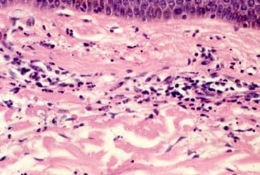
- the red pulp indicated congestion and ill-defined nodules, varying in size and comprising macrophages, polymorphonuclear neutrophils, and necrotic cells ( Figure , panels A , B ). Skin biopsy of the macular eruption on day 2 demonstrated a leukocytoclastic vasculitis with nonocclusive luminal thrombi in the dermal capillaries ( Figure , panel C ). (cdc.gov)
- Skin biopsy revealed small and intermediate vessel vasculitis. (scielo.org.za)
- Vasculitis was reported as the cause of death on postmortem biopsy. (scielo.org.za)
Polyarteritis nodosa4
- Polyarteritis nodosa, granulomatosis with polyangiitis, Henoch-Schönlein purpura, scleroderma, and otherwise nonspecified vasculitides also were reported to have caused ESRD during this period. (medscape.com)
- Medium vessel vasculitis typically affects arteries supplying the kidneys, bowels, brain or heart (e.g. polyarteritis nodosa, Kawasaki disease). (printo.it)
- Specifically, patients with VEXAS syndrome can meet diagnostic or classification criteria for one or more diseases, including relapsing polychondritis, polyarteritis nodosa, giant-cell arteritis, Sweet's syndrome, ANCA-associated vasculitis, systemic lupus erythematosus, and adult-onset Still's disease. (nejm.org)
- [6] Takayasu's arteritis is an example of a large-vessel vasculitis in childhood, whereas Kawasaki disease and polyarteritis nodosa are examples of medium-vessel vasculitis disorders. (scielo.org.za)
Manifestations7
- A high prevalence of cutaneous disorders is expected, because most patients with ESRD have an underlying disease process with cutaneous manifestations. (medscape.com)
- These systemic disorders and the associated renal diseases and cutaneous manifestations are tabulated in Table 1, below. (medscape.com)
- We present a case of small and medium-vessel vasculitis on the lower extremity with cutaneous manifestations, without an identifiable cause. (faoj.org)
- Cutaneous vasculitis may present with varying clinical manifestations and may be caused by systemic disease or secondarily due to an underlying disorder, drug reaction, or infection [1-2]. (faoj.org)
- Fever and cutaneous manifestations that reflect neutrophilic dermatosis, leukocytoclastic vasculitis, or medium-vessel vasculitis are common in patients with VEXAS syndrome. (nejm.org)
- We report a rare case of neonatal vasculitis presenting with skin manifestations similar to infection-based cellulitis. (scielo.org.za)
- RÉSUMÉ Nous avons passé en revue les manifestations dermatologiques liées à l'infection chronique par le virus de l'hépatite C (VHC) et leur rapport avec l'état hépatique. (who.int)
Symptoms10
- Additional symptoms depend on the cause of the vasculitis and if other organ systems are involved. (wikipedia.org)
- Symptoms of vasculitis can include fever , tiredness , and joint pain. (medicalnewstoday.com)
- Treatment for vasculitis depends on several factors, including what type of vasculitis the person has, the severity of their symptoms, their age, and their general health. (medicalnewstoday.com)
- Signs and symptoms of vasculitis vary depending on which blood vessels are affected, and which organs are damaged, if any. (medicalnewstoday.com)
- If skin involvement is secondary to a systemic vasculitis, symptoms may also include fever, arthralgias, other organ involvement, or a combination. (msdmanuals.com)
- Widespread (systemic) vasculitis is usually accompanied by extensive release of inflammatory molecules, causing general symptoms like fever, malaise, as well as abnormal laboratory tests detecting inflammation: erythrocyte sedimentation rate (ESR) and C- reactive protein (CRP). (printo.it)
- ANCAs can be positive after the use of certain drugs and other forms of vasculitis can present with very similar symptoms. (checkorphan.org)
- arteritis risk if you don't treat temporal (giant cell) arteritis granulomatous vasculitis that involves aortic arch at branch points age group of takayasu arteritis younger than 50 - Asian females symptoms of takayasu arteritis 1. (symptoma.com)
- 18 years of age were enrolled this study with diagnosis of cPACNS if they had: a clinical symptoms compatible with primary CNS vasculitis, and MRA findings demonstrating arterial stenosis and or aneurism that are not attributable to other disease and background. (fortuneonline.org)
- The patient remains well and free from any cutaneous or systemic symptoms 9 months after initial consultation. (cdlib.org)
Idiopathic3
- CSVV sometimes refers to small-vessel vasculitis of unknown cause (also called idiopathic cutaneous small-vessel vasculitis). (msdmanuals.com)
- In a large number of cases cutaneous vasculitis may present as an idiopathic condition and affect both small and medium sized vessels. (faoj.org)
- Cutaneous small-vessel vasculitis is often idiopathic , but all patients should be evaluated for potential underlying causes, including infections (e.g. (amboss.com)
ANCA6
- These may include tests to check for inflammation, organ involvement, immune complex formation and deposition, and ANCA-related vasculitis. (cohencenters.com)
- MPA and the clinically similar and similarly treated granulomatosis with polyangiitis ( GPA ) are classified as antineutrophil cytoplasmic autoantibody ( ANCA )-associated vasculitides ( AAV s). (empendium.com)
- Granulomatosis with polyangiitis (Wegener's granulomatosis) is a type of vasculitis which can be categorized as a sub-branch of anti-neutrophil cytoplasmic antibody (ANCA)-associated vasculitides. (rheumres.org)
- Granulomatosis with polyangiitis (GPA), previously known as Wegener's granulomatosis, is a type of vasculitis which can be categorized as a sub-branch of anti-neutrophil cytoplasmic antibody (ANCA)-associated vasculitides [1]. (rheumres.org)
- Cutaneous vasculopathy associated with levamisole-adulterated (contaminated) cocaine is an emerging syndrome characterised by a retiform purpura around the ears, the presence of anti- neutrophil cytoplasmic autoantibody ( ANCA ), and leukopenia [3,4]. (dermnetnz.org)
- Group of systemic vasculitis with a strong association with ANCA . (nih.gov)
Diagnosis5
- Microscopically, both sites revealed small vascular structures, primarily capillaries, surrounded by abundant polymorphonuclear leukocytes and few eosinophils, evidence that confirmed the diagnosis of leukocytoclastic vasculitis. (consultant360.com)
- A diagnosis of vasculitis limited to the skin requires a complete history and physical examination. (msdmanuals.com)
- Accordingly, diagnosis of cutaneous vasculitis, identification of etiological factors, follow-up for systemic involvement and treatment are important. (bgu.ac.il)
- A diagnosis of cutaneous mucinous carcinoma was made. (cdlib.org)
- The final diagnosis was primary cutaneous mucinous carcinoma and definitive treatment was carried out via wide local excision. (cdlib.org)
Lesions3
- Vasculitis affecting the small vessels of the skin (eg, arterioles, capillaries, postcapillary venules) tends to cause lesions such as purpura, petechiae, and possibly shallow ulcers. (msdmanuals.com)
- Many cutaneous side effects have been reported with their use in rheumatoid arthritis including psoriasis , dermatitis , leukocytoclastic vasculitis, lichenoid drug eruptions , and non-infectious cutaneous granulomatous reactions, such as disseminated granuloma annulare , sarcoidosis-like lesions, and interstitial granulomatous dermatitis. (arthritisdaily.net)
- Cutaneous lesions are excluded from the study. (who.int)
Findings7
- The purpose of this article is to integrate renal and cutaneous aspects of disease as well as highlight some important, although frequently underappreciated, clinical or laboratory findings that ally renal and skin diseases. (medscape.com)
- The clinical and histological findings were consistent with cutaneous necrotizing leukocytoclastic vasculitis. (bgu.ac.il)
- 2,9 However, biopsies of affected skin generally do not demonstrate findings of vasculitis. (clinicaladvisor.com)
- Others postulate that the skin findings are the result of cutaneous trauma caused by repeated, improperly performed injections, or that heparin may be poorly absorbed due to decreased vasculature in adipose tissue. (clinicaladvisor.com)
- findings include leukocytoclastic vasculitis . (amboss.com)
- The findings were reviewed by a pathologist at the University of Iowa Hospitals and Clinics and were found to be suggestive of leukocytoclastic vasculitis. (eyerounds.org)
- 5 Disposition is variable and largely depends on the severity of vasculitis and resulting skin findings. (jetem.org)
Vessels16
- Cutaneous small-vessel vasculitis (CSVV), is inflammation of small blood vessels, usually accompanied by small lumps beneath the skin. (wikipedia.org)
- Leukocytoclastic" refers to the damage caused by nuclear debris from infiltrating neutrophils in and around the vessels. (wikipedia.org)
- Vasculitis means inflammation of the blood vessels. (medicalnewstoday.com)
- Cutaneous vasculitis refers to vasculitis affecting small- or medium-sized vessels in the skin and subcutaneous tissue but not the internal organs. (msdmanuals.com)
- Overview of Vasculitis Vasculitis is inflammation of blood vessels, often with ischemia, necrosis, and organ inflammation. (msdmanuals.com)
- Vasculitis can affect the small- or medium-sized vessels of the skin. (msdmanuals.com)
- Livedo reticularis, nodules, and deep ulcers are usually caused by vasculitis of deeper, medium or large vessels. (msdmanuals.com)
- The classification of vasculitides depends mainly on the size and type of blood vessels involved. (printo.it)
- Although Wegener's granulomatosis affects small and medium-sized vessels, it is formally classified as one of the small vessel vasculitides in the Chapel Hill system. (checkorphan.org)
- Vasculitis is an uncommon condition characterized by inflammation and necrosis of blood vessels and impaired blood flow. (cohencenters.com)
- Vasculitis is a rare, inflammatory condition of the blood vessels whereby excess leukocytes within the vessel leads to a loss of structural integrity, and possible destruction. (faoj.org)
- Vasculitis is a heterogeneous group of blood vessels disorders which characterized by inflammation, necrosis and the obstruction of the inflamed vessels [1]. (fortuneonline.org)
- In a range of inflammatory vascular process, vasculitis may affect the CNS vessels. (fortuneonline.org)
- Microscopic polyangiitis ( MPA ) is a necrotizing vasculitis with few or no immunologic deposits, which usually affects small vessels (arterioles, capillaries, venules) and may involve small- and middle-sized arteries. (empendium.com)
- Vasculitis is a direct result of inflamed blood vessels. (acadderm.com)
- Vasculitis is defined as inflammation of the wall of blood vessels. (scielo.org.za)
Purpura5
- For example, if the vasculitis is a manifestation of Henoch-Schönlein purpura, individuals may also experience abdominal pain or blood in the urine. (wikipedia.org)
- In cases where a cause can be determined, medications and infectious pathogens are most common in adults, while IgA vasculitis (Henoch-Schönlein purpura) frequently affects children. (wikipedia.org)
- Henoch-Schönlein purpura, granulomatosis with polyangiitis or GPA, eosinophilic granulomatosis with polyangiitis or EGPA, previously referred to as Churg-Strauss syndrome), cutaneous leukocytoclastic vasculitis, microscopic polyangiitis). (printo.it)
- Physical exam Palpable purpura is pathognomonic for vasculitis. (symptoma.com)
- Palpable, normally painful, petechiae or purpura (skin vasculitis). (wikidoc.org)
Necrosis4
- Other clinical entities that can present similarly to heparin-induced skin necrosis include calciphylaxis, pyoderma gangrenosum, disseminated intravascular coagulation (DIC), leukocytoclastic vasculitis, and other bullous disorders. (clinicaladvisor.com)
- Its use was discontinued when patients began exhibiting serious side effects including agranulocytosis, thrombocytopenia, arthritis, vasculitis and skin necrosis. (jetem.org)
- 4 The most common complication is the need for surgical consultation and debridement if the vasculitis leads to skin necrosis. (jetem.org)
- Aurora N, Janin T, Bhanot R, Natesan S. Levamisole-induced leukocytoclastic vasculitis and neutropenia in a patient with cocaine use: an extensive case with necrosis of skin, soft tissue and cartilage. (jetem.org)
Disorders5
- In addition, uremia and conditions associated with renal replacement therapy are fraught with numerous and, often, relatively unique cutaneous disorders. (medscape.com)
- The image below illustrates several uremia-related cutaneous disorders. (medscape.com)
- Several uremia-related cutaneous disorders are visible. (medscape.com)
- Many cutaneous disorders experienced by patients undergoing dialysis have little to do with the uremic syndrome and are related to the same underlying pathologic process that caused the renal disease. (medscape.com)
- Skin eruptions may be a symptom of systemic disorders other than infections, and may be prominent in patients with vasculitis disorders. (scielo.org.za)
Lichen planus1
- Recent data suggest that OLR present a greater percentage of malignant transformation than OLP and, although the association between cancer and OLP has been documented in scientific reports, there is no association between squamous cell carcinoma and cutaneous lichen planus [ 4 - 6 ]. (ecancer.org)
Diseases4
- This tends to occur when the vasculitis is associated with chronic conditions such as connective tissue diseases. (wikipedia.org)
- Vasculitis has a number of potential causes including infections and immunologic diseases. (medicalnewstoday.com)
- Vasculitides include a wide group of diseases. (printo.it)
- The pathological mechanism that causes cutaneous small-vessel vasculitis can also cause vasculitides in organs other than the skin , which are categorized as distinct diseases. (amboss.com)
Infections3
- Cutaneous vasculitis can have various causes including but not limited to medications, bacterial and viral infections or allergens. (wikipedia.org)
- Recurrent Infections and CNS Vasculitis,44. (booksdo.com)
- Known causes of vasculitis include the following: infections (hepatitis B and Streptococcus spp. (scielo.org.za)
Inflammation of the blood1
- Vasculitis is inflammation of the blood vessel walls. (printo.it)
Glomerulonephritis1
- An exhaustive search ought to be performed for signals of systemic disease such as for example sinusitis, cutaneous leukocytoclastic vasculitis, iridocyclitis, synovitis, and glomerulonephritis [9]. (angiogenesis-blog.com)
Renal1
- The clinical spectrum of primary renal vasculitis. (medscape.com)
Examination1
- Cutaneous examination of patients with ESRD has shown that 50-100% of patients have at least 1 dermatologic condition. (medscape.com)
Clinical2
- The spectrum of paraneoplastic cutaneous vasculitis in a defined population: incidence and clinical features. (medscape.com)
- Clinical, histopathologic, and laboratory evaluation are imperative to appropriately diagnose cutaneous vasculitis. (faoj.org)
Large-vessel Vasculitis1
- Large vessel vasculitis, like Takayasu arteritis, affects the aorta and its major branches. (printo.it)
Organs1
- This term describes vasculitis that affects the skin but not the internal organs. (msdmanuals.com)
Patients2
- Retrospective analysis of adult patients with cutaneous leukocytoclastic vasculitis. (medscape.com)
- Primary systemic vasculitis is uncommon, accounting for only four percent of cases in patients with cutaneous vasculitis. (faoj.org)
Disorder4
- Cutaneous vasculitis may be limited to the skin, or may be a component of a systemic primary or secondary vasculitic disorder. (msdmanuals.com)
- Vasculitis may be due to primary disease or secondary due to an underlying disorder, drug reaction, or infection. (faoj.org)
- Central nervous system (CNS) vasculitis of childhood is a novel recognized autoimmune brain disorder with significant diagnostic and therapeutic challenges [2,3]. (fortuneonline.org)
- Vasculitis is a rare disorder during the neonatal period. (scielo.org.za)
Rheumatoid2
- Detection of cytomegalovirus antigens in phagocytosed serum complexes from a patient with rheumatoid arthritis, vasculitis, peripheral neuropathy, cutaneous ulceration, and digital gangrene . (symptoma.com)
- There are many new and emerging treatments for rheumatoid arthritis including biological treatments , Janus kinase inhibitors, rituximab , tocilizumab with their associated cutaneous side effects. (arthritisdaily.net)
Cause of the vasculitis1
- The most important part of the treatment is to eliminate the cause of the vasculitis, if at all possible. (wikidoc.org)
Organ2
- In this review, cases are divided between localised disease, non-organ threatening, generalized organ-threatening disease and severe kidney vasculitis and immediately life-threatening disease. (checkorphan.org)
- If the vasculitis is damaging organ systems such as the kidneys, immunosuppressive agents are indicated. (wikidoc.org)
Severe2
- Cutaneous vasculitis may precede severe systemic involvement, and may end in death. (bgu.ac.il)
- Systemic vasculitis complicating lupus will often present with severe skin rashes which may be necrotic leading to gangrene . (symptoma.com)
Chronic1
- We report an early case of primary cutaneous mucinous carcinoma in a male patient with chronic actinic damage. (cdlib.org)
Types of vasculitis1
- 2.1 What are the types of vasculitis? (printo.it)
Small4
- Considering the wide range of potential causes leading to cutaneous small vessel vasculitis, there are subtle variations in the underlying pathophysiology for each cause. (wikipedia.org)
- Drug-induced vasculitis is an inflammation of small-sized blood vessel caused by the use of drugs. (koreamed.org)
- Here we identified three unrelated boys with perinatal-onset of neutrophilic cutaneous small vessel vasculitis and systemic inflammation. (nature.com)
- all presented with systemic inflammation and recurrent neutrophilic small vessel vasculitis. (nature.com)
Forms of vasculitis1
- There are many forms of vasculitis, ranging from mild to potentially life-threatening. (printo.it)
Syndrome1
- Vasculitis as a paraneoplastic syndrome. (medscape.com)
Bullous1
- Cutaneous: Livedo reticularis, skin ulcers, tender erythematous nodules, bullous or vesicular eruptions, infarction and gangrene of fingers or toes, or a combination may occur. (symptoma.com)
Pathophysiology1
- Its pathophysiology is normally alveolar microcirculation damage, and the reason may Cefsulodin sodium be generalized (such as systemic vasculitis) or lung-specific disease [as in diffuse alveolar harm (Father) or an infection] [17, 18]. (angiogenesis-blog.com)
Immune1
- A cutaneous expression of immune complex disease. (medscape.com)
Blood vessel4
- Vasculitis can affect any blood vessel anywhere in the body. (medicalnewstoday.com)
- 1.4 What happens to the blood vessel in vasculitis? (printo.it)
- Vasculitis classification in children is based on the size of the blood vessel involved. (printo.it)
- Vasculitis varies and may be classified based on the size of the blood vessel involved. (faoj.org)
Diagnostic1
- The diagnostic testing for vasculitis should be guided by the patient's history and physical exam. (wikipedia.org)