Wet Macular Degeneration
Macular Degeneration
Choroidal Neovascularization
Retinal Drusen
Retinal Pigment Epithelium
Retinal Degeneration
Geographic Atrophy
Fluorescein Angiography
Choroid
Visual Acuity
Nerve Degeneration
Complement Factor H
Bruch Membrane
Pigment Epithelium of Eye
Intravitreal Injections
Retina
Macula Lutea
Lipofuscin
Optic Disk Drusen
Fundus Oculi
Wallerian Degeneration
Tomography, Optical Coherence
Choroid Diseases
Lutein
Fovea Centralis
Photoreceptor Cells, Vertebrate
Angiogenesis Inhibitors
Retinal Neovascularization
Exudates and Transudates
Photochemotherapy
Eye Proteins
Intervertebral Disc Degeneration
Subretinal Fluid
Photography
Scotoma
Retinal Diseases
Aging
Antibodies, Monoclonal, Humanized
Retinal Detachment
Vision Disorders
Vision, Low
Complement Factor B
Indocyanine Green
Polymorphism, Single Nucleotide
Laser Coagulation
Genotype
Disease Models, Animal
Blindness
Vascular Endothelial Growth Factor A
Complement C2
Retinitis Pigmentosa
Eye Diseases, Hereditary
Photosensitizing Agents
Frontotemporal Lobar Degeneration
Cataract
Retinoids
Retinal Pigments
Porphyrins
Atrophy
Light Coagulation
Retinal Photoreceptor Cell Outer Segment
Genetic Predisposition to Disease
Vitreous Body
Case-Control Studies
Pyridinium Compounds
Visual Field Tests
Diagnostic Techniques, Ophthalmological
Protective effect of paraoxonase 1 gene variant Gln192Arg in age-related macular degeneration. (1/82)
(+info)Evaluation of CXCR4 inhibition in the prevention and intervention model of laser-induced choroidal neovascularization. (2/82)
(+info)Integrin activation or alpha 9 expression allows retinal pigmented epithelial cell adhesion on Bruch's membrane in wet age-related macular degeneration. (3/82)
(+info)Pegaptanib sodium as maintenance therapy in neovascular age-related macular degeneration: the LEVEL study. (4/82)
(+info)Patient selection criteria for pilot studies on amelioration of non-neovascular age-related macular degeneration. (5/82)
(+info)A randomized pilot study of systemic immunosuppression in the treatment of age-related macular degeneration with choroidal neovascularization. (6/82)
(+info)Clinical application of therapies targeting VEGF. (7/82)
(+info)Quantification of the therapeutic response of intraretinal, subretinal, and subpigment epithelial compartments in exudative AMD during anti-VEGF therapy. (8/82)
(+info)Wet macular degeneration, also known as neovascular or exudative age-related macular degeneration (AMD), is a medical condition that affects the central part of the retina called the macula. It's characterized by the growth of new blood vessels (neovascularization) from the choroid layer behind the retina into the macula, which is not typical in healthy eyes. These abnormal blood vessels are fragile and prone to leakage, leading to the accumulation of fluid or blood in the macula, causing distortion or loss of central vision.
The wet form of AMD can progress rapidly and often leads to more severe visual loss compared to the dry form. It's essential to diagnose and treat wet AMD promptly to preserve as much vision as possible. Common treatments include anti-vascular endothelial growth factor (VEGF) injections, photodynamic therapy, or thermal laser treatment, depending on the specific case and individual patient factors.
Macular degeneration, also known as age-related macular degeneration (AMD), is a medical condition that affects the central part of the retina, called the macula. The macula is responsible for sharp, detailed vision, which is necessary for activities such as reading, driving, and recognizing faces.
In AMD, there is a breakdown or deterioration of the macula, leading to gradual loss of central vision. There are two main types of AMD: dry (atrophic) and wet (exudative). Dry AMD is more common and progresses more slowly, while wet AMD is less common but can cause rapid and severe vision loss if left untreated.
The exact causes of AMD are not fully understood, but risk factors include age, smoking, family history, high blood pressure, obesity, and exposure to sunlight. While there is no cure for AMD, treatments such as vitamin supplements, laser therapy, and medication injections can help slow its progression and reduce the risk of vision loss.
Choroidal neovascularization (CNV) is a medical term that refers to the growth of new, abnormal blood vessels in the choroid layer of the eye, which is located between the retina and the sclera. This condition typically occurs as a complication of age-related macular degeneration (AMD), although it can also be caused by other eye diseases or injuries.
In CNV, the new blood vessels that grow into the choroid layer are fragile and can leak fluid or blood, which can cause distortion or damage to the retina, leading to vision loss. Symptoms of CNV may include blurred or distorted vision, a blind spot in the center of the visual field, or changes in color perception.
Treatment for CNV typically involves medications that are designed to stop the growth of new blood vessels, such as anti-VEGF drugs, which target a protein called vascular endothelial growth factor (VEGF) that is involved in the development of new blood vessels. Laser surgery or photodynamic therapy may also be used in some cases to destroy the abnormal blood vessels and prevent further vision loss.
Retinal drusen are yellow-white, deposits of extracellular material that accumulate beneath the retina, most commonly in the macula. They are a common age-related finding and can also be seen in various other conditions such as inherited retinal diseases. Drusen can vary in size and number, and their presence is often associated with an increased risk of developing age-related macular degeneration (AMD), a leading cause of vision loss in older adults. However, not all individuals with drusen will develop AMD, and the significance of drusen depends on factors such as size, number, and location. It's important to monitor drusen and have regular eye examinations to assess any changes or progression that may indicate a higher risk for developing AMD.
The retinal pigment epithelium (RPE) is a single layer of cells located between the photoreceptor cells of the retina and the choroid, which is a part of the eye containing blood vessels. The RPE plays a crucial role in maintaining the health and function of the photoreceptors by providing them with nutrients, removing waste products, and helping to regulate the light-sensitive visual pigments within the photoreceptors.
The RPE cells contain pigment granules that absorb excess light to prevent scattering within the eye and improve visual acuity. They also help to form the blood-retina barrier, which restricts the movement of certain molecules between the retina and the choroid, providing an important protective function for the retina.
Damage to the RPE can lead to a variety of eye conditions, including age-related macular degeneration (AMD), which is a leading cause of vision loss in older adults.
Retinal degeneration is a broad term that refers to the progressive loss of photoreceptor cells (rods and cones) in the retina, which are responsible for converting light into electrical signals that are sent to the brain. This process can lead to vision loss or blindness. There are many different types of retinal degeneration, including age-related macular degeneration, retinitis pigmentosa, and Stargardt's disease, among others. These conditions can have varying causes, such as genetic mutations, environmental factors, or a combination of both. Treatment options vary depending on the specific type and progression of the condition.
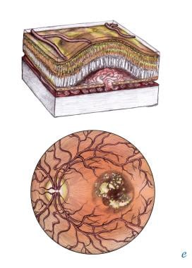
Geographic atrophy is a medical term used to describe a specific pattern of degeneration of the retinal pigment epithelium (RPE) and the underlying choroidal tissue in the eye. This condition is often associated with age-related macular degeneration (AMD), which is a leading cause of vision loss in older adults.
In geographic atrophy, there are well-defined areas of RPE and choroidal atrophy that appear as pale, irregularly shaped patches in the central part of the retina known as the macula. These patches can grow larger over time and may lead to progressive vision loss. The exact cause of geographic atrophy is not fully understood, but it is thought to be related to oxidative stress, inflammation, and other age-related changes in the eye.
Currently, there are no effective treatments for geographic atrophy, although research is ongoing to find new ways to slow or halt its progression. Regular eye exams and monitoring by an ophthalmologist are important for people with AMD or geographic atrophy to help detect any changes in their vision and manage their condition effectively.
Fluorescein angiography is a medical diagnostic procedure used in ophthalmology to examine the blood flow in the retina and choroid, which are the inner layers of the eye. This test involves injecting a fluorescent dye, Fluorescein, into a patient's arm vein. As the dye reaches the blood vessels in the eye, a specialized camera takes rapid sequences of photographs to capture the dye's circulation through the retina and choroid.
The images produced by fluorescein angiography can help doctors identify any damage to the blood vessels, leakage, or abnormal growth of new blood vessels. This information is crucial in diagnosing and managing various eye conditions such as age-related macular degeneration, diabetic retinopathy, retinal vein occlusions, and inflammatory eye diseases.
It's important to note that while fluorescein angiography is a valuable diagnostic tool, it does carry some risks, including temporary side effects like nausea, vomiting, or allergic reactions to the dye. In rare cases, severe adverse reactions can occur, so patients should discuss these potential risks with their healthcare provider before undergoing the procedure.
The choroid is a layer of the eye that contains blood vessels that supply oxygen and nutrients to the outer layers of the retina. It lies between the sclera (the white, protective coat of the eye) and the retina (the light-sensitive tissue at the back of the eye). The choroid is essential for maintaining the health and function of the retina, particularly the photoreceptor cells that detect light and transmit visual signals to the brain. Damage to the choroid can lead to vision loss or impairment.
Visual acuity is a measure of the sharpness or clarity of vision. It is usually tested by reading an eye chart from a specific distance, such as 20 feet (6 meters). The standard eye chart used for this purpose is called the Snellen chart, which contains rows of letters that decrease in size as you read down the chart.
Visual acuity is typically expressed as a fraction, with the numerator representing the testing distance and the denominator indicating the smallest line of type that can be read clearly. For example, if a person can read the line on the eye chart that corresponds to a visual acuity of 20/20, it means they have normal vision at 20 feet. If their visual acuity is 20/40, it means they must be as close as 20 feet to see what someone with normal vision can see at 40 feet.
It's important to note that visual acuity is just one aspect of overall vision and does not necessarily reflect other important factors such as peripheral vision, depth perception, color vision, or contrast sensitivity.
Nerve degeneration, also known as neurodegeneration, is the progressive loss of structure and function of neurons, which can lead to cognitive decline, motor impairment, and various other symptoms. This process occurs due to a variety of factors, including genetics, environmental influences, and aging. It is a key feature in several neurological disorders such as Alzheimer's disease, Parkinson's disease, Huntington's disease, and multiple sclerosis. The degeneration can affect any part of the nervous system, leading to different symptoms depending on the location and extent of the damage.
Complement Factor H is a protein involved in the regulation of the complement system, which is a part of the immune system that helps to clear pathogens and damaged cells from the body. Specifically, Complement Factor H helps to regulate the activation and deactivation of the complement component C3b, preventing excessive or unwanted activation of the complement system and protecting host tissues from damage.
Complement Factor H is a crucial protein in maintaining the balance between the protective effects of the complement system and the potential for harm to the body's own cells and tissues. Deficiencies or mutations in Complement Factor H have been associated with several diseases, including age-related macular degeneration (AMD), atypical hemolytic uremic syndrome (aHUS), and C3 glomerulopathy.
The Bruch membrane is a thin, layered structure that separates the retina from the choroid in the eye. It is composed of five layers: the basement membrane of the retinal pigment epithelium (RPE), the inner collagenous layer, the elastic layer, the outer collagenous layer, and the basement membrane of the choriocapillaris. The Bruch membrane provides structural support to the RPE and serves as a barrier between the retina and the choroid, allowing for the selective transport of nutrients and waste products. It also plays a role in maintaining the health of the photoreceptors in the retina. Damage to the Bruch membrane is associated with age-related macular degeneration (AMD), a leading cause of vision loss in older adults.
The pigment epithelium of the eye, also known as the retinal pigment epithelium (RPE), is a layer of cells located between the photoreceptor cells of the retina and the choroid, which is the vascular layer of the eye. The RPE plays a crucial role in maintaining the health and function of the photoreceptors by providing them with nutrients, removing waste products, and helping to regulate the light that enters the eye.
The RPE cells contain pigment granules that absorb excess light, preventing it from scattering within the eye and improving visual acuity. They also help to create a barrier between the retina and the choroid, which is important for maintaining the proper functioning of the photoreceptors. Additionally, the RPE plays a role in the regeneration of visual pigments in the photoreceptor cells, allowing us to see in different light conditions.
Damage to the RPE can lead to various eye diseases and conditions, including age-related macular degeneration (AMD), which is a leading cause of vision loss in older adults.
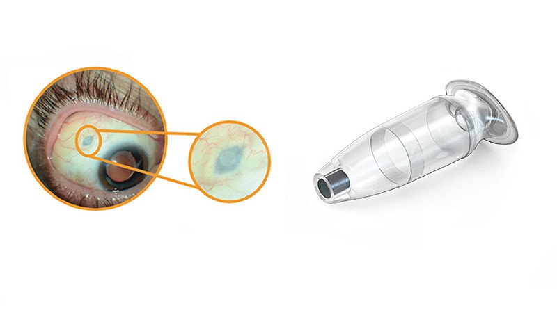
An intravitreal injection is a medical procedure in which medication is delivered directly into the vitreous cavity of the eye, which is the clear, gel-like substance that fills the space between the lens and the retina. This type of injection is typically used to treat various eye conditions such as age-related macular degeneration, diabetic retinopathy, retinal vein occlusion, and uveitis. The medication administered in intravitreal injections can help to reduce inflammation, inhibit the growth of new blood vessels, or prevent the formation of abnormal blood vessels in the eye.
Intravitreal injections are usually performed in an outpatient setting, and the procedure typically takes only a few minutes. Before the injection, the eye is numbed with anesthetic drops to minimize discomfort. The medication is then injected into the vitreous cavity using a small needle. After the injection, patients may experience some mild discomfort or a scratchy sensation in the eye, but this usually resolves within a few hours.
While intravitreal injections are generally safe, there are some potential risks and complications associated with the procedure, including infection, bleeding, retinal detachment, and increased intraocular pressure. Patients who undergo intravitreal injections should be closely monitored by their eye care provider to ensure that any complications are promptly identified and treated.
The retina is the innermost, light-sensitive layer of tissue in the eye of many vertebrates and some cephalopods. It receives light that has been focused by the cornea and lens, converts it into neural signals, and sends these to the brain via the optic nerve. The retina contains several types of photoreceptor cells including rods (which handle vision in low light) and cones (which are active in bright light and are capable of color vision).
In medical terms, any pathological changes or diseases affecting the retinal structure and function can lead to visual impairment or blindness. Examples include age-related macular degeneration, diabetic retinopathy, retinal detachment, and retinitis pigmentosa among others.
The macula lutea, often simply referred to as the macula or fovea centralis, is a part of the eye that is responsible for central vision and color perception. It's located in the center of the retina, the light-sensitive tissue at the back of the eye. The macula contains a high concentration of pigments called xanthophylls, which give it a yellowish color and protect the photoreceptor cells in this area from damage by blue light.
The central part of the macula is called the fovea, which is a small depression that contains only cones, the photoreceptor cells responsible for color vision and high visual acuity. The fovea is surrounded by the parafovea and the perifovea, which contain both cones and rods, the photoreceptor cells responsible for low-light vision and peripheral vision.
Damage to the macula can result in a loss of central vision and color perception, a condition known as age-related macular degeneration (AMD), which is a leading cause of blindness in older adults. Other conditions that can affect the macula include macular edema, macular holes, and macular pucker.
Lipofuscin is a type of pigment that accumulates in the lysosomes (membrane-bound organelles found inside cells) of various tissues, particularly in nerve cells and heart muscle cells. It consists of cross-linked proteins and lipids that are resistant to degradation by enzymes. The accumulation of lipofuscin is a normal part of aging but can also be associated with certain diseases such as neurodegenerative disorders.
It's often referred to as "age pigment" because it tends to increase in amount with age, and its presence in tissues has been linked to oxidative stress and cellular damage caused by free radicals. Lipofuscin is autofluorescent, meaning that it emits light when excited by certain wavelengths of light, which can be useful for its detection and quantification in research and diagnostic settings.
Optic disk drusen are small, calcified deposits that form within the optic nerve head, also known as the optic disc. They are made up of protein and calcium salts and can vary in size and number. These deposits can be seen on ophthalmic examination using an instrument called an ophthalmoscope.
Optic disk drusen are typically asymptomatic and are often discovered during routine eye examinations. However, in some cases, they may cause visual disturbances or even vision loss if they compress the optic nerve fibers. They can also increase the risk of developing other eye conditions such as glaucoma.
Optic disk drusen are more commonly found in individuals with a family history of the condition and tend to occur in younger people, typically before the age of 40. While there is no cure for optic disk drusen, regular eye examinations can help monitor any changes in the condition and manage any associated visual symptoms or complications.
"Fundus Oculi" is a medical term that refers to the back part of the interior of the eye, including the optic disc, macula, fovea, retinal vasculature, and peripheral retina. It is the area where light is focused and then transmitted to the brain via the optic nerve, forming visual images. Examinations of the fundus oculi are crucial for detecting various eye conditions such as diabetic retinopathy, macular degeneration, glaucoma, and other retinal diseases. The examination is typically performed using an ophthalmoscope or a specialized camera called a retinal camera.
Wallerian degeneration is a process that occurs following damage to the axons of neurons (nerve cells). After an axon is severed or traumatically injured, it undergoes a series of changes including fragmentation and removal of the distal segment of the axon, which is the part that is separated from the cell body. This process is named after Augustus Waller, who first described it in 1850.
The degenerative changes in the distal axon are characterized by the breakdown of the axonal cytoskeleton, the loss of myelin sheath (the fatty insulating material that surrounds and protects the axon), and the infiltration of macrophages to clear away the debris. These events lead to the degeneration of the distal axon segment, which is necessary for successful regeneration of the injured nerve.
Wallerian degeneration is a crucial process in the nervous system's response to injury, as it enables the regrowth of axons and the reestablishment of connections between neurons. However, if the regenerative capacity of the neuron is insufficient or the environment is not conducive to growth, functional recovery may be impaired, leading to long-term neurological deficits.
Optical coherence tomography (OCT) is a non-invasive imaging technique that uses low-coherence light to capture high-resolution cross-sectional images of biological tissues, particularly the retina and other ocular structures. OCT works by measuring the echo time delay of light scattered back from different depths within the tissue, creating a detailed map of the tissue's structure. This technique is widely used in ophthalmology to diagnose and monitor various eye conditions such as macular degeneration, diabetic retinopathy, and glaucoma.
The choroid is a part of the eye located between the retina and the sclera, which contains a large number of blood vessels that supply oxygen and nutrients to the outer layers of the retina. Choroid diseases refer to various medical conditions that affect the health and function of the choroid. Here are some examples:
1. Choroidal neovascularization (CNV): This is a condition where new blood vessels grow from the choroid into the retina, leading to fluid accumulation, bleeding, and scarring. CNV can cause vision loss and is often associated with age-related macular degeneration, myopia, and inflammatory eye diseases.
2. Chorioretinitis: This is an infection or inflammation of the choroid and retina, which can be caused by various microorganisms such as bacteria, viruses, fungi, or parasites. Symptoms may include blurred vision, floaters, light sensitivity, and eye pain.
3. Choroidal hemorrhage: This is a rare but serious condition where there is bleeding into the choroid, often caused by trauma, high blood pressure, or blood clotting disorders. It can lead to sudden vision loss and requires urgent medical attention.
4. Choroideremia: This is a genetic disorder that affects the choroid, retina, and optic nerve, leading to progressive vision loss. It is caused by mutations in the CHM gene and primarily affects males.
5. Central serous retinopathy (CSR): This is a condition where fluid accumulates under the retina, often in the macula, causing distortion or blurring of vision. While the exact cause is unknown, CSR is thought to be related to stress, steroid use, and other factors that affect the choroid's ability to regulate fluid.
6. Polypoidal choroidal vasculopathy (PCV): This is a condition where abnormal blood vessels form in the choroid, leading to serous or hemorrhagic detachment of the retina. PCV is often associated with age-related macular degeneration and can cause vision loss if left untreated.
These are just a few examples of choroidal disorders that can affect vision. If you experience any sudden changes in your vision, it's important to seek medical attention promptly.
Lutein is a type of carotenoid, specifically a xanthophyll, that is naturally present in many fruits and vegetables. It is considered a dietary antioxidant with potential health benefits for the eyes. Lutein is not a vitamin, but it is often grouped with vitamins and minerals because of its importance to human health.
In the eye, lutein is selectively accumulated in the macula, a small area in the center of the retina responsible for sharp, detailed vision. It helps filter harmful blue light and protects the eye from oxidative damage, which may help maintain eye health and reduce the risk of age-related macular degeneration (AMD), a leading cause of blindness in older adults.
It is important to note that lutein is not produced by the human body and must be obtained through dietary sources or supplements. Foods rich in lutein include dark leafy greens, such as spinach and kale, as well as other fruits and vegetables, such as corn, orange pepper, and egg yolk.
The fovea centralis, also known as the macula lutea, is a small pit or depression located in the center of the retina, an light-sensitive tissue at the back of the eye. It is responsible for sharp, detailed vision (central vision) and color perception. The fovea contains only cones, the photoreceptor cells that are responsible for color vision and high visual acuity. It has a higher concentration of cones than any other area in the retina, allowing it to provide the greatest detail and color discrimination. The center of the fovea is called the foveola, which contains the highest density of cones and is avascular, meaning it lacks blood vessels to avoid interfering with the light passing through to the photoreceptor cells.
Photoreceptor cells in vertebrates are specialized types of neurons located in the retina of the eye that are responsible for converting light stimuli into electrical signals. These cells are primarily responsible for the initial process of vision and have two main types: rods and cones.
Rods are more numerous and are responsible for low-light vision or scotopic vision, enabling us to see in dimly lit conditions. They do not contribute to color vision but provide information about the shape and movement of objects.
Cones, on the other hand, are less numerous and are responsible for color vision and high-acuity vision or photopic vision. There are three types of cones, each sensitive to different wavelengths of light: short (S), medium (M), and long (L) wavelengths, which correspond to blue, green, and red, respectively. The combination of signals from these three types of cones allows us to perceive a wide range of colors.
Both rods and cones contain photopigments that consist of a protein called opsin and a light-sensitive chromophore called retinal. When light hits the photopigment, it triggers a series of chemical reactions that ultimately lead to the generation of an electrical signal that is transmitted to the brain via the optic nerve. This process enables us to see and perceive our visual world.
Angiogenesis inhibitors are a class of drugs that block the growth of new blood vessels (angiogenesis). They work by targeting specific molecules involved in the process of angiogenesis, such as vascular endothelial growth factor (VEGF) and its receptors. By blocking these molecules, angiogenesis inhibitors can prevent the development of new blood vessels that feed tumors, thereby slowing or stopping their growth.
Angiogenesis inhibitors are used in the treatment of various types of cancer, including colon, lung, breast, kidney, and ovarian cancer. They may be given alone or in combination with other cancer treatments, such as chemotherapy or radiation therapy. Some examples of angiogenesis inhibitors include bevacizumab (Avastin), sorafenib (Nexavar), sunitinib (Sutent), and pazopanib (Votrient).
It's important to note that while angiogenesis inhibitors can be effective in treating cancer, they can also have serious side effects, such as high blood pressure, bleeding, and damage to the heart or kidneys. Therefore, it's essential that patients receive careful monitoring and management of these potential side effects while undergoing treatment with angiogenesis inhibitors.
Retinal neovascularization is a medical condition characterized by the growth of new, abnormal blood vessels on the surface of the retina, which is the light-sensitive tissue located at the back of the eye. This condition typically occurs in response to an insufficient supply of oxygen and nutrients to the retina, often due to damage or disease, such as diabetic retinopathy or retinal vein occlusion.
The new blood vessels that form during neovascularization are fragile and prone to leakage, which can cause fluid and protein to accumulate in the retina, leading to distorted vision, hemorrhages, and potentially blindness if left untreated. Retinal neovascularization is a serious eye condition that requires prompt medical attention and management to prevent further vision loss.
Electroretinography (ERG) is a medical test used to evaluate the functioning of the retina, which is the light-sensitive tissue located at the back of the eye. The test measures the electrical responses of the retina to light stimulation.
During the procedure, a special contact lens or electrode is placed on the surface of the eye to record the electrical activity generated by the retina's light-sensitive cells (rods and cones) and other cells in the retina. The test typically involves presenting different levels of flashes of light to the eye while the electrical responses are recorded.
The resulting ERG waveform provides information about the overall health and function of the retina, including the condition of the photoreceptors, the integrity of the inner retinal layers, and the health of the retinal ganglion cells. This test is often used to diagnose and monitor various retinal disorders, such as retinitis pigmentosa, macular degeneration, and diabetic retinopathy.
Exudates and transudates are two types of bodily fluids that can accumulate in various body cavities or tissues as a result of injury, inflammation, or other medical conditions. Here are the medical definitions:
1. Exudates: These are fluids that accumulate due to an active inflammatory process. Exudates contain high levels of protein, white blood cells (such as neutrophils and macrophages), and sometimes other cells like red blood cells or cellular debris. They can be yellow, green, or brown in color and may have a foul odor due to the presence of dead cells and bacteria. Exudates are often seen in conditions such as abscesses, pneumonia, pleurisy, or wound infections.
Examples of exudative fluids include pus, purulent discharge, or inflammatory effusions.
2. Transudates: These are fluids that accumulate due to increased hydrostatic pressure or decreased oncotic pressure within the blood vessels. Transudates contain low levels of protein and cells compared to exudates. They are typically clear and pale yellow in color, with no odor. Transudates can be found in conditions such as congestive heart failure, liver cirrhosis, or nephrotic syndrome.
Examples of transudative fluids include ascites, pleural effusions, or pericardial effusions.
It is essential to differentiate between exudates and transudates because their underlying causes and treatment approaches may differ significantly. Medical professionals often use various tests, such as fluid analysis, to determine whether a fluid sample is an exudate or transudate.
Photochemotherapy is a medical treatment that combines the use of drugs and light to treat various skin conditions. The most common type of photochemotherapy is PUVA (Psoralen + UVA), where the patient takes a photosensitizing medication called psoralen, followed by exposure to ultraviolet A (UVA) light.
The psoralen makes the skin more sensitive to the UVA light, which helps to reduce inflammation and suppress the overactive immune response that contributes to many skin conditions. This therapy is often used to treat severe cases of psoriasis, eczema, and mycosis fungoides (a type of cutaneous T-cell lymphoma). It's important to note that photochemotherapy can increase the risk of skin cancer and cataracts, so it should only be administered under the close supervision of a healthcare professional.
Eye proteins, also known as ocular proteins, are specific proteins that are found within the eye and play crucial roles in maintaining proper eye function and health. These proteins can be found in various parts of the eye, including the cornea, iris, lens, retina, and other structures. They perform a wide range of functions, such as:
1. Structural support: Proteins like collagen and elastin provide strength and flexibility to the eye's tissues, enabling them to maintain their shape and withstand mechanical stress.
2. Light absorption and transmission: Proteins like opsins and crystallins are involved in capturing and transmitting light signals within the eye, which is essential for vision.
3. Protection against damage: Some eye proteins, such as antioxidant enzymes and heat shock proteins, help protect the eye from oxidative stress, UV radiation, and other environmental factors that can cause damage.
4. Regulation of eye growth and development: Various growth factors and signaling molecules, which are protein-based, contribute to the proper growth, differentiation, and maintenance of eye tissues during embryonic development and throughout adulthood.
5. Immune defense: Proteins involved in the immune response, such as complement components and immunoglobulins, help protect the eye from infection and inflammation.
6. Maintenance of transparency: Crystallin proteins in the lens maintain its transparency, allowing light to pass through unobstructed for clear vision.
7. Neuroprotection: Certain eye proteins, like brain-derived neurotrophic factor (BDNF), support the survival and function of neurons within the retina, helping to preserve vision.
Dysfunction or damage to these eye proteins can contribute to various eye disorders and diseases, such as cataracts, age-related macular degeneration, glaucoma, diabetic retinopathy, and others.
Intervertebral disc degeneration is a physiological and biochemical process that occurs in the spinal discs, which are located between each vertebra in the spine. These discs act as shock absorbers and allow for movement and flexibility of the spine.
The degenerative process involves changes in the structure and composition of the disc, including loss of water content, decreased production of proteoglycans (which help to maintain the disc's elasticity), and disorganization of the collagen fibers that make up the disc's outer layer (annulus fibrosus). These changes can lead to a decrease in the disc's height and mobility, as well as the development of tears or cracks in the annulus fibrosus.
In advanced stages of degeneration, the disc may herniate or bulge outward, causing pressure on nearby nerves and potentially leading to pain, numbness, tingling, or weakness in the affected area. It's worth noting that while intervertebral disc degeneration is a normal part of aging, certain factors such as injury, smoking, obesity, and repetitive stress can accelerate the process.
Ophthalmoscopy is a medical examination technique used by healthcare professionals to observe the interior structures of the eye, including the retina, optic disc, and vitreous humor. This procedure typically involves using an ophthalmoscope, a handheld device that consists of a light and magnifying lenses. The healthcare provider looks through the ophthalmoscope and directly observes the internal structures of the eye by illuminating them.
There are several types of ophthalmoscopy, including direct ophthalmoscopy, indirect ophthalmoscopy, and slit-lamp biomicroscopy. Each type has its own advantages and disadvantages, and they may be used in different situations depending on the specific clinical situation and the information needed.
Ophthalmoscopy is an important diagnostic tool for detecting and monitoring a wide range of eye conditions, including diabetic retinopathy, glaucoma, age-related macular degeneration, and other retinal disorders. It can also provide valuable information about the overall health of the individual, as changes in the appearance of the retina or optic nerve may indicate the presence of systemic diseases such as hypertension or diabetes.
Xanthophylls are a type of pigment known as carotenoids, which are naturally occurring in various plants and animals. They are characterized by their yellow to orange color and play an important role in photosynthesis. Unlike other carotenoids, xanthophylls contain oxygen in their chemical structure.
In the context of human health, xanthophylls are often studied for their potential antioxidant properties and their possible role in reducing the risk of age-related macular degeneration (AMD), a leading cause of vision loss in older adults. The two main dietary sources of xanthophylls are lutein and zeaxanthin, which are found in green leafy vegetables, such as spinach and kale, as well as in other fruits and vegetables.
It's important to note that while a healthy diet rich in fruits and vegetables has many benefits for overall health, including eye health, more research is needed to fully understand the specific role of xanthophylls in preventing or treating diseases.
Subretinal fluid (SRF) refers to the abnormal accumulation of fluid between the neurosensory retina and the pigment epithelium of the eye. This can occur due to various conditions such as age-related macular degeneration, central serous chorioretinopathy, or retinal detachment. The presence of subretinal fluid can distort vision and may require medical intervention depending on the underlying cause and severity of the condition.
I'm sorry for any confusion, but "photography" is not a term typically used in medical definitions. Photography refers to the art, application, or process of creating images by recording light or other electromagnetic radiation, either electronically by means of an image sensor, or chemically by means of a light-sensitive material such as photographic film.
If you're looking for a medical term related to imaging, there are several terms that might be relevant, such as:
1. Radiography: This is a technique using X-rays to visualize the internal structures of the body.
2. Ultrasonography: Also known as ultrasound, this is a diagnostic imaging technique using high-frequency sound waves to create images of the inside of the body.
3. Computed Tomography (CT): A type of imaging that uses X-rays to create detailed cross-sectional images of the body.
4. Magnetic Resonance Imaging (MRI): A type of imaging that uses magnetic fields and radio waves to create detailed images of the organs and tissues within the body.
5. Nuclear Medicine: This is a branch of medical imaging that uses small amounts of radioactive material to diagnose and treat diseases.
If you have any questions related to medical definitions or topics, feel free to ask!
A scotoma is a blind spot or area of reduced vision within the visual field. It's often surrounded by an area of less distinct vision and can be caused by various conditions such as eye diseases, neurological disorders, or brain injuries. A scotoma may be temporary or permanent, depending on its underlying cause.
There are different types of scotomas, including:
1. Central scotoma - a blind spot in the center of the visual field, often associated with conditions like age-related macular degeneration and diabetic retinopathy.
2. Paracentral scotoma - a blind spot located slightly away from the center of the visual field, which can be caused by optic neuritis or other optic nerve disorders.
3. Peripheral scotoma - a blind spot in the peripheral vision, often associated with retinal diseases like retinitis pigmentosa.
4. Absolute scotoma - a complete loss of vision in a specific area of the visual field.
5. Relative scotoma - a partial loss of vision in which some details can still be perceived, but not as clearly or vividly as in normal vision.
It is essential to consult an eye care professional if you experience any changes in your vision or notice a scotoma, as early detection and treatment can help prevent further vision loss.
Retinal diseases refer to a group of conditions that affect the retina, which is the light-sensitive tissue located at the back of the eye. The retina is responsible for converting light into electrical signals that are sent to the brain and interpreted as visual images. Retinal diseases can cause vision loss or even blindness, depending on their severity and location in the retina.
Some common retinal diseases include:
1. Age-related macular degeneration (AMD): A progressive disease that affects the central part of the retina called the macula, causing blurred or distorted vision.
2. Diabetic retinopathy: A complication of diabetes that can damage the blood vessels in the retina, leading to vision loss.
3. Retinal detachment: A serious condition where the retina becomes separated from its underlying tissue, requiring immediate medical attention.
4. Macular edema: Swelling or thickening of the macula due to fluid accumulation, which can cause blurred vision.
5. Retinitis pigmentosa: A group of inherited eye disorders that affect the retina's ability to respond to light, causing progressive vision loss.
6. Macular hole: A small break in the macula that can cause distorted or blurry vision.
7. Retinal vein occlusion: Blockage of the retinal veins that can lead to bleeding, swelling, and potential vision loss.
Treatment for retinal diseases varies depending on the specific condition and its severity. Some treatments include medication, laser therapy, surgery, or a combination of these options. Regular eye exams are essential for early detection and treatment of retinal diseases.
Aging is a complex, progressive and inevitable process of bodily changes over time, characterized by the accumulation of cellular damage and degenerative changes that eventually lead to increased vulnerability to disease and death. It involves various biological, genetic, environmental, and lifestyle factors that contribute to the decline in physical and mental functions. The medical field studies aging through the discipline of gerontology, which aims to understand the underlying mechanisms of aging and develop interventions to promote healthy aging and extend the human healthspan.
Monoclonal antibodies are laboratory-produced proteins that mimic the immune system's ability to fight off harmful antigens such as viruses and cancer cells. They are created by fusing a single B cell (the type of white blood cell responsible for producing antibodies) with a tumor cell, resulting in a hybrid cell called a hybridoma. This hybridoma can then be cloned to produce a large number of identical cells, all producing the same antibody, hence "monoclonal."
Humanized monoclonal antibodies are a type of monoclonal antibody that have been genetically engineered to include human components. This is done to reduce the risk of an adverse immune response in patients receiving the treatment. In this process, the variable region of the mouse monoclonal antibody, which contains the antigen-binding site, is grafted onto a human constant region. The resulting humanized monoclonal antibody retains the ability to bind to the target antigen while minimizing the immunogenicity associated with murine (mouse) antibodies.
In summary, "antibodies, monoclonal, humanized" refers to a type of laboratory-produced protein that mimics the immune system's ability to fight off harmful antigens, but with reduced immunogenicity due to the inclusion of human components in their structure.
Eye diseases are a range of conditions that affect the eye or visual system, causing damage to vision and, in some cases, leading to blindness. These diseases can be categorized into various types, including:
1. Refractive errors: These include myopia (nearsightedness), hyperopia (farsightedness), astigmatism, and presbyopia, which affect the way light is focused on the retina and can usually be corrected with glasses or contact lenses.
2. Cataracts: A clouding of the lens inside the eye that leads to blurry vision, glare, and decreased contrast sensitivity. Cataract surgery is the most common treatment for this condition.
3. Glaucoma: A group of diseases characterized by increased pressure in the eye, leading to damage to the optic nerve and potential blindness if left untreated. Treatment includes medications, laser therapy, or surgery.
4. Age-related macular degeneration (AMD): A progressive condition that affects the central part of the retina called the macula, causing blurry vision and, in advanced stages, loss of central vision. Treatment may include anti-VEGF injections, laser therapy, or nutritional supplements.
5. Diabetic retinopathy: A complication of diabetes that affects the blood vessels in the retina, leading to bleeding, leakage, and potential blindness if left untreated. Treatment includes laser therapy, anti-VEGF injections, or surgery.
6. Retinal detachment: A separation of the retina from its underlying tissue, which can lead to vision loss if not treated promptly with surgery.
7. Amblyopia (lazy eye): A condition where one eye does not develop normal vision, often due to a misalignment or refractive error in childhood. Treatment includes correcting the underlying problem and encouraging the use of the weaker eye through patching or other methods.
8. Strabismus (crossed eyes): A misalignment of the eyes that can lead to amblyopia if not treated promptly with surgery, glasses, or other methods.
9. Corneal diseases: Conditions that affect the transparent outer layer of the eye, such as keratoconus, Fuchs' dystrophy, and infectious keratitis, which can lead to vision loss if not treated promptly.
10. Uveitis: Inflammation of the middle layer of the eye, which can cause vision loss if not treated promptly with anti-inflammatory medications or surgery.
Retinal detachment is a serious eye condition that occurs when the retina, a thin layer of tissue at the back of the eye responsible for processing light and sending visual signals to the brain, pulls away from its normal position. This can lead to significant vision loss or even blindness if not promptly treated. Retinal detachment can be caused by various factors such as aging, trauma, eye disease, or an inflammatory condition. Symptoms of retinal detachment may include sudden flashes of light, floaters, a shadow in the peripheral vision, or a curtain-like covering over part of the visual field. Immediate medical attention is necessary to prevent further damage and preserve vision.
A retinal hemorrhage is a type of bleeding that occurs in the blood vessels of the retina, which is the light-sensitive tissue located at the back of the eye. This condition can result from various underlying causes, including diabetes, high blood pressure, age-related macular degeneration, or trauma to the eye. Retinal hemorrhages can be categorized into different types based on their location and appearance, such as dot and blot hemorrhages, flame-shaped hemorrhages, or subhyaloid hemorrhages. Depending on the severity and cause of the hemorrhage, treatment options may vary from monitoring to laser therapy, medication, or even surgery. It is essential to consult an ophthalmologist for a proper evaluation and management plan if you suspect a retinal hemorrhage.
Vision disorders refer to a wide range of conditions that affect the visual system and result in various symptoms, such as blurry vision, double vision, distorted vision, impaired depth perception, and difficulty with visual tracking or focusing. These disorders can be categorized into several types, including:
1. Refractive errors: These occur when the shape of the eye prevents light from focusing directly on the retina, resulting in blurry vision. Examples include myopia (nearsightedness), hyperopia (farsightedness), astigmatism, and presbyopia (age-related loss of near vision).
2. Strabismus: Also known as crossed eyes or walleye, strabismus is a misalignment of the eyes where they point in different directions, which can lead to double vision or loss of depth perception.
3. Amblyopia: Often called lazy eye, amblyopia is a condition where one eye has reduced vision due to lack of proper visual development during childhood. It may be caused by strabismus, refractive errors, or other factors that interfere with normal visual development.
4. Accommodative disorders: These involve problems with the focusing ability of the eyes, such as convergence insufficiency (difficulty focusing on close objects) and accommodative dysfunction (inability to maintain clear vision at different distances).
5. Binocular vision disorders: These affect how the eyes work together as a team, leading to issues like poor depth perception, eye strain, and headaches. Examples include convergence insufficiency, divergence excess, and suppression.
6. Ocular motility disorders: These involve problems with eye movement, such as nystagmus (involuntary eye movements), strabismus, or restricted extraocular muscle function.
7. Visual processing disorders: These affect the brain's ability to interpret and make sense of visual information, even when the eyes themselves are healthy. Symptoms may include difficulty with reading, recognizing shapes and objects, and understanding spatial relationships.
8. Low vision: This term refers to significant visual impairment that cannot be fully corrected with glasses, contact lenses, medication, or surgery. It includes conditions like macular degeneration, diabetic retinopathy, glaucoma, and cataracts.
9. Blindness: Complete loss of sight in both eyes, which can be caused by various factors such as injury, disease, or genetic conditions.
Low vision is a term used to describe significant visual impairment that cannot be corrected with standard glasses, contact lenses, medication or surgery. It is typically defined as visual acuity of less than 20/70 in the better-seeing eye after best correction, or a visual field of less than 20 degrees in the better-seeing eye.
People with low vision may have difficulty performing everyday tasks such as reading, recognizing faces, watching television, driving, or simply navigating their environment. They may also experience symptoms such as sensitivity to light, glare, or contrast, and may benefit from the use of visual aids, assistive devices, and rehabilitation services to help them maximize their remaining vision and maintain their independence.
Low vision can result from a variety of causes, including eye diseases such as macular degeneration, diabetic retinopathy, glaucoma, or cataracts, as well as congenital or inherited conditions, brain injuries, or aging. It is important for individuals with low vision to receive regular eye examinations and consult with a low vision specialist to determine the best course of treatment and management.
Complement Factor B is a protein that plays a crucial role in the complement system, which is a part of the immune system that helps to eliminate pathogens and damaged cells from the body. Specifically, Factor B is a component of the alternative pathway of the complement system, which provides a rapid and amplified response to microbial surfaces.
Factor B is cleaved by another protease called Factor D into two fragments, Ba and Bb. The formation of the C3 convertase (C3bBb) is essential for the activation of the alternative pathway. This complex can cleave and activate more C3 molecules, leading to a cascade of reactions that result in the formation of the membrane attack complex (MAC), which forms pores in the membranes of target cells, causing their lysis and elimination.
Deficiencies or mutations in Complement Factor B can lead to various complement-mediated diseases, such as atypical hemolytic uremic syndrome (aHUS) and age-related macular degeneration (AMD).
Indocyanine green (ICG) is a sterile, water-soluble, tricarbocyanine dye that is used as a diagnostic agent in medical imaging. It is primarily used in ophthalmology for fluorescein angiography to examine blood flow in the retina and choroid, and in cardiac surgery to assess cardiac output and perfusion. When injected into the body, ICG binds to plasma proteins and fluoresces when exposed to near-infrared light, allowing for visualization of various tissues and structures. It is excreted primarily by the liver and has a half-life of approximately 3-4 minutes in the bloodstream.
Single Nucleotide Polymorphism (SNP) is a type of genetic variation that occurs when a single nucleotide (A, T, C, or G) in the DNA sequence is altered. This alteration must occur in at least 1% of the population to be considered a SNP. These variations can help explain why some people are more susceptible to certain diseases than others and can also influence how an individual responds to certain medications. SNPs can serve as biological markers, helping scientists locate genes that are associated with disease. They can also provide information about an individual's ancestry and ethnic background.
Laser coagulation, also known as laser photocoagulation, is a medical procedure that uses a laser to seal or destroy abnormal blood vessels or tissue. The laser produces a concentrated beam of light that can be precisely focused on the target area. When the laser energy is absorbed by the tissue, it causes the temperature to rise, which leads to coagulation (the formation of a clot) or destruction of the tissue.
In ophthalmology, laser coagulation is commonly used to treat conditions such as diabetic retinopathy, age-related macular degeneration, and retinal tears or holes. The procedure can help to seal leaking blood vessels, reduce fluid leakage, and prevent further vision loss. It is usually performed as an outpatient procedure and may be repeated if necessary.
In other medical specialties, laser coagulation may be used to control bleeding, destroy tumors, or remove unwanted tissue. The specific technique and parameters of the laser treatment will depend on the individual patient's needs and the condition being treated.
Genotype, in genetics, refers to the complete heritable genetic makeup of an individual organism, including all of its genes. It is the set of instructions contained in an organism's DNA for the development and function of that organism. The genotype is the basis for an individual's inherited traits, and it can be contrasted with an individual's phenotype, which refers to the observable physical or biochemical characteristics of an organism that result from the expression of its genes in combination with environmental influences.
It is important to note that an individual's genotype is not necessarily identical to their genetic sequence. Some genes have multiple forms called alleles, and an individual may inherit different alleles for a given gene from each parent. The combination of alleles that an individual inherits for a particular gene is known as their genotype for that gene.
Understanding an individual's genotype can provide important information about their susceptibility to certain diseases, their response to drugs and other treatments, and their risk of passing on inherited genetic disorders to their offspring.
Animal disease models are specialized animals, typically rodents such as mice or rats, that have been genetically engineered or exposed to certain conditions to develop symptoms and physiological changes similar to those seen in human diseases. These models are used in medical research to study the pathophysiology of diseases, identify potential therapeutic targets, test drug efficacy and safety, and understand disease mechanisms.
The genetic modifications can include knockout or knock-in mutations, transgenic expression of specific genes, or RNA interference techniques. The animals may also be exposed to environmental factors such as chemicals, radiation, or infectious agents to induce the disease state.
Examples of animal disease models include:
1. Mouse models of cancer: Genetically engineered mice that develop various types of tumors, allowing researchers to study cancer initiation, progression, and metastasis.
2. Alzheimer's disease models: Transgenic mice expressing mutant human genes associated with Alzheimer's disease, which exhibit amyloid plaque formation and cognitive decline.
3. Diabetes models: Obese and diabetic mouse strains like the NOD (non-obese diabetic) or db/db mice, used to study the development of type 1 and type 2 diabetes, respectively.
4. Cardiovascular disease models: Atherosclerosis-prone mice, such as ApoE-deficient or LDLR-deficient mice, that develop plaque buildup in their arteries when fed a high-fat diet.
5. Inflammatory bowel disease models: Mice with genetic mutations affecting intestinal barrier function and immune response, such as IL-10 knockout or SAMP1/YitFc mice, which develop colitis.
Animal disease models are essential tools in preclinical research, but it is important to recognize their limitations. Differences between species can affect the translatability of results from animal studies to human patients. Therefore, researchers must carefully consider the choice of model and interpret findings cautiously when applying them to human diseases.
Blindness is a condition of complete or near-complete vision loss. It can be caused by various factors such as eye diseases, injuries, or birth defects. Total blindness means that a person cannot see anything at all, while near-complete blindness refers to having only light perception or the ability to perceive the direction of light, but not able to discern shapes or forms. Legal blindness is a term used to define a certain level of visual impairment that qualifies an individual for government assistance and benefits; it usually means best corrected visual acuity of 20/200 or worse in the better eye, or a visual field no greater than 20 degrees in diameter.
Vision tests are a series of procedures used to assess various aspects of the visual system, including visual acuity, accommodation, convergence, divergence, stereopsis, color vision, and peripheral vision. These tests help healthcare professionals diagnose and manage vision disorders, such as nearsightedness, farsightedness, astigmatism, amblyopia, strabismus, and eye diseases like glaucoma, cataracts, and macular degeneration. Common vision tests include:
1. Visual acuity test (Snellen chart or letter chart): Measures the sharpness of a person's vision at different distances.
2. Refraction test: Determines the correct lens prescription for glasses or contact lenses by assessing how light is bent as it passes through the eye.
3. Color vision test: Evaluates the ability to distinguish between different colors and color combinations, often using pseudoisochromatic plates or Ishihara tests.
4. Stereopsis test: Assesses depth perception and binocular vision by presenting separate images to each eye that, when combined, create a three-dimensional effect.
5. Cover test: Examines eye alignment and the presence of strabismus (crossed eyes or turned eyes) by covering and uncovering each eye while observing eye movements.
6. Ocular motility test: Assesses the ability to move the eyes in various directions and coordinate both eyes during tracking and convergence/divergence movements.
7. Accommodation test: Evaluates the ability to focus on objects at different distances by using lenses, prisms, or dynamic retinoscopy.
8. Pupillary response test: Examines the size and reaction of the pupils to light and near objects.
9. Visual field test: Measures the peripheral (side) vision using automated perimetry or manual confrontation techniques.
10. Slit-lamp examination: Inspects the structures of the front part of the eye, such as the cornea, iris, lens, and anterior chamber, using a specialized microscope.
These tests are typically performed by optometrists, ophthalmologists, or other vision care professionals during routine eye examinations or when visual symptoms are present.
Vascular Endothelial Growth Factor A (VEGFA) is a specific isoform of the vascular endothelial growth factor (VEGF) family. It is a well-characterized signaling protein that plays a crucial role in angiogenesis, the process of new blood vessel formation from pre-existing vessels. VEGFA stimulates the proliferation and migration of endothelial cells, which line the interior surface of blood vessels, thereby contributing to the growth and development of new vasculature. This protein is essential for physiological processes such as embryonic development and wound healing, but it has also been implicated in various pathological conditions, including cancer, age-related macular degeneration, and diabetic retinopathy. The regulation of VEGFA expression and activity is critical to maintaining proper vascular function and homeostasis.
Complement C2 is a protein that plays a crucial role in the complement system, which is a part of the immune system that helps to eliminate pathogens and damaged cells from the body. Specifically, C2 is a component of the classical complement pathway, which is activated by the binding of antibodies to antigens on the surface of foreign particles or cells.
When the classical pathway is activated, C2 is cleaved into two fragments: C2a and C2b. C2a then binds to C4b to form the C3 convertase (C4b2a), which cleaves C3 into C3a and C3b. C3b can then go on to form the membrane attack complex, which creates a pore in the membrane of the target cell, leading to its lysis.
In summary, Complement C2 is a protein that helps to activate the complement system and destroy foreign particles or cells through the formation of the C3 convertase and the membrane attack complex.
Retinitis pigmentosa (RP) is a group of rare, genetic disorders that involve a breakdown and loss of cells in the retina - a light-sensitive tissue located at the back of the eye. The retina converts light into electrical signals which are then sent to the brain and interpreted as visual images.
In RP, the cells that detect light (rods and cones) degenerate more slowly than other cells in the retina, leading to a progressive loss of vision. Symptoms typically begin in childhood with night blindness (difficulty seeing in low light), followed by a gradual narrowing of the visual field (tunnel vision). Over time, this can lead to significant vision loss and even blindness.
The condition is usually inherited and there are several different genes that have been associated with RP. The diagnosis is typically made based on a combination of genetic testing, family history, and clinical examination. Currently, there is no cure for RP, but researchers are actively working to develop new treatments that may help slow or stop the progression of the disease.
Hereditary eye diseases refer to conditions that affect the eyes and are passed down from parents to their offspring through genetics. These diseases are caused by mutations or changes in an individual's DNA that are inherited from their parents. The mutations can occur in any of the genes associated with eye development, function, or health.
There are many different types of hereditary eye diseases, some of which include:
1. Retinitis Pigmentosa - a group of rare, genetic disorders that involve a breakdown and loss of cells in the retina.
2. Macular Degeneration - a progressive disease that damages the central portion of the retina, impairing vision.
3. Glaucoma - a group of eye conditions that damage the optic nerve, often caused by an increase in pressure inside the eye.
4. Cataracts - clouding of the lens inside the eye, which can lead to blurry vision and blindness.
5. Keratoconus - a progressive eye disease that causes the cornea to thin and bulge outward into a cone shape.
6. Color Blindness - a condition where an individual has difficulty distinguishing between certain colors.
7. Optic Neuropathy - damage to the optic nerve, which can result in vision loss.
The symptoms and severity of hereditary eye diseases can vary widely depending on the specific condition and the individual's genetic makeup. Some conditions may be present at birth or develop in early childhood, while others may not appear until later in life. Treatment options for these conditions may include medication, surgery, or lifestyle changes, and are often most effective when started early.
Photosensitizing agents are substances that, when exposed to light, particularly ultraviolet or visible light, can cause chemical reactions leading to the production of reactive oxygen species. These reactive oxygen species can interact with biological tissues, leading to damage and a variety of phototoxic or photoallergic adverse effects.
Photosensitizing agents are used in various medical fields, including dermatology and oncology. In dermatology, they are often used in the treatment of conditions such as psoriasis and eczema, where a photosensitizer is applied to the skin and then activated with light to reduce inflammation and slow the growth of skin cells.
In oncology, photosensitizing agents are used in photodynamic therapy (PDT), a type of cancer treatment that involves administering a photosensitizer, allowing it to accumulate in cancer cells, and then exposing the area to light. The light activates the photosensitizer, which produces reactive oxygen species that damage the cancer cells, leading to their death.
Examples of photosensitizing agents include porphyrins, chlorophyll derivatives, and certain antibiotics such as tetracyclines and fluoroquinolones. It is important for healthcare providers to be aware of the potential for photosensitivity when prescribing these medications and to inform patients of the risks associated with exposure to light.
Frontotemporal lobar degeneration (FTLD) is a group of disorders characterized by the progressive degeneration of the frontal and temporal lobes of the brain. These areas of the brain are involved in decision-making, behavior, emotion, and language. FTLD can be divided into several subtypes based on the specific clinical features and the underlying protein abnormalities.
The three main subtypes of FTLD are:
1. Behavioral variant frontotemporal dementia (bvFTD): This subtype is characterized by changes in personality, behavior, and judgment. People with bvFTD may lose their social inhibitions, become impulsive, or develop compulsive behaviors. They may also have difficulty with emotional processing and empathy.
2. Primary progressive aphasia (PPA): This subtype is characterized by the gradual deterioration of language skills. People with PPA may have difficulty speaking, understanding spoken or written language, or both. There are three subtypes of PPA: nonfluent/agrammatic variant, semantic variant, and logopenic variant.
3. Motor neuron disease (MND) with FTLD: This subtype is characterized by the degeneration of motor neurons, which are the nerve cells responsible for controlling voluntary muscle movements. People with MND with FTLD may develop symptoms of amyotrophic lateral sclerosis (ALS), such as muscle weakness, stiffness, and twitching, as well as cognitive and behavioral changes associated with FTLD.
The underlying protein abnormalities in FTLD include:
1. Tau protein: In some forms of FTLD, the tau protein accumulates and forms clumps called tangles inside nerve cells. This is also seen in Alzheimer's disease.
2. TDP-43 protein: In other forms of FTLD, the TDP-43 protein accumulates and forms clumps inside nerve cells.
3. Fused in sarcoma (FUS) protein: In a small number of cases, the FUS protein accumulates and forms clumps inside nerve cells.
FTLD is typically a progressive disorder, meaning that symptoms worsen over time. There is currently no cure for FTLD, but there are treatments available to help manage symptoms and improve quality of life.
A cataract is a clouding of the natural lens in the eye that affects vision. This clouding can cause vision to become blurry, faded, or dim, making it difficult to see clearly. Cataracts are a common age-related condition, but they can also be caused by injury, disease, or medication use. In most cases, cataracts develop gradually over time and can be treated with surgery to remove the cloudy lens and replace it with an artificial one.
Retinoids are a class of chemical compounds that are derivatives of vitamin A. They are widely used in dermatology for the treatment of various skin conditions, including acne, psoriasis, and photoaging. Retinoids can help to reduce inflammation, improve skin texture and tone, and stimulate collagen production.
Retinoids work by binding to specific receptors in the skin cells, which triggers a series of biochemical reactions that regulate gene expression and promote cell differentiation and turnover. This can help to unclog pores, reduce the appearance of fine lines and wrinkles, and improve the overall health and appearance of the skin.
There are several different types of retinoids used in skincare products, including retinoic acid, retinaldehyde, and retinol. Retinoic acid is the most potent form of retinoid and is available by prescription only. Retinaldehyde and retinol are weaker forms of retinoid that can be found in over-the-counter skincare products.
While retinoids can be highly effective for treating various skin conditions, they can also cause side effects such as dryness, irritation, and sensitivity to the sun. It is important to use retinoids as directed by a healthcare professional and to follow proper sun protection measures when using these products.
Retinal pigments refer to the light-sensitive chemicals found in the retina, specifically within the photoreceptor cells called rods and cones. The main types of retinal pigments are rhodopsin (also known as visual purple) in rods and iodopsins in cones. These pigments play a crucial role in the process of vision by absorbing light and initiating a series of chemical reactions that ultimately trigger nerve impulses, which are then transmitted to the brain and interpreted as visual images. Rhodopsin is more sensitive to lower light levels and is responsible for night vision, while iodopsins are sensitive to specific wavelengths of light and contribute to color vision.
Porphyrins are complex organic compounds that contain four pyrrole rings joined together by methine bridges (=CH-). They play a crucial role in the biochemistry of many organisms, as they form the core structure of various heme proteins and other metalloproteins. Some examples of these proteins include hemoglobin, myoglobin, cytochromes, and catalases, which are involved in essential processes such as oxygen transport, electron transfer, and oxidative metabolism.
In the human body, porphyrins are synthesized through a series of enzymatic reactions known as the heme biosynthesis pathway. Disruptions in this pathway can lead to an accumulation of porphyrins or their precursors, resulting in various medical conditions called porphyrias. These disorders can manifest as neurological symptoms, skin lesions, and gastrointestinal issues, depending on the specific type of porphyria and the site of enzyme deficiency.
It is important to note that while porphyrins are essential for life, their accumulation in excessive amounts or at inappropriate locations can result in pathological conditions. Therefore, understanding the regulation and function of porphyrin metabolism is crucial for diagnosing and managing porphyrias and other related disorders.
Atrophy is a medical term that refers to the decrease in size and wasting of an organ or tissue due to the disappearance of cells, shrinkage of cells, or decreased number of cells. This process can be caused by various factors such as disuse, aging, degeneration, injury, or disease.
For example, if a muscle is immobilized for an extended period, it may undergo atrophy due to lack of use. Similarly, certain medical conditions like diabetes, cancer, and heart failure can lead to the wasting away of various tissues and organs in the body.
Atrophy can also occur as a result of natural aging processes, leading to decreased muscle mass and strength in older adults. In general, atrophy is characterized by a decrease in the volume or weight of an organ or tissue, which can have significant impacts on its function and overall health.
"Light coagulation," also known as "laser coagulation," is a medical term that refers to the use of laser technology to cauterize (seal or close) tissue. This procedure uses heat generated by a laser to cut, coagulate, or destroy tissue. In light coagulation, the laser beam is focused on the blood vessels in question, causing the blood within them to clot and the vessels to seal. This can be used for various medical purposes, such as stopping bleeding during surgery, destroying abnormal tissues (like tumors), or treating eye conditions like diabetic retinopathy and age-related macular degeneration.
It's important to note that this is a general definition, and the specific use of light coagulation may vary depending on the medical specialty and the individual patient's needs. As always, it's best to consult with a healthcare professional for more detailed information about any medical procedure or treatment.
The retinal photoreceptor cells, namely rods and cones, are specialized neurons in the retina responsible for converting light into electrical signals that can be processed by the brain. The outer segment of a retinal photoreceptor cell is the portion of the cell where phototransduction primarily occurs. It contains stacks of disc-like structures filled with the visual pigment rhodopsin, which absorbs light and initiates the conversion process.
The outer segment is continuously renewed through a process called shedding and phagocytosis, in which the oldest discs at the base of the outer segment are shed, engulfed by the adjacent retinal pigment epithelium (RPE) cells, and degraded. This turnover helps maintain the sensitivity and functionality of the photoreceptor cells.
In summary, the retinal photoreceptor cell outer segment is a highly specialized compartment where light absorption and initial signal transduction occur in rods and cones, supported by continuous renewal through shedding and phagocytosis.
Genetic predisposition to disease refers to an increased susceptibility or vulnerability to develop a particular illness or condition due to inheriting specific genetic variations or mutations from one's parents. These genetic factors can make it more likely for an individual to develop a certain disease, but it does not guarantee that the person will definitely get the disease. Environmental factors, lifestyle choices, and interactions between genes also play crucial roles in determining if a genetically predisposed person will actually develop the disease. It is essential to understand that having a genetic predisposition only implies a higher risk, not an inevitable outcome.
An Eye Bank is an organization that collects, stores, and distributes donated human eyes for corneal transplantation and other ocular medical research purposes. The eye bank's primary function is to ensure the quality of the donated tissue and make it available for those in need of sight-restoring procedures.
The cornea, the clear front part of the eye, can be surgically transplanted from a deceased donor to a recipient with corneal damage or disease, thereby improving or restoring their vision. The eye bank's role includes obtaining consent for donation, retrieving the eyes from the donor, evaluating the tissue for suitability, preserving it properly, and then allocating it to surgeons for transplantation.
Eye banks follow strict medical guidelines and adhere to ethical standards to ensure the safety and quality of the donated tissues. The process involves screening potential donors for infectious diseases and other conditions that may affect the quality or safety of the cornea. Once deemed suitable, the corneas are carefully removed, preserved in specific solutions, and stored until they are needed for transplantation.
In addition to corneal transplants, eye banks also support research and education in ophthalmology by providing human eye tissues for various studies aimed at advancing our understanding of eye diseases and developing new treatments.
Eye color is a characteristic determined by variations in a person's genes. The color of the eyes depends on the amount and type of pigment called melanin found in the eye's iris.
There are three main types of eye colors: brown, blue, and green. Brown eyes have the most melanin, while blue eyes have the least. Green eyes have a moderate amount of melanin combined with a golden tint that reflects light to give them their unique color.
Eye color is a polygenic trait, which means it is influenced by multiple genes. The two main genes responsible for eye color are OCA2 and HERC2, both located on chromosome 15. These genes control the production, transport, and storage of melanin in the iris.
It's important to note that eye color can change during infancy and early childhood due to the development of melanin in the iris. Additionally, some medications or medical conditions may also cause changes in eye color over time.
The vitreous body, also known simply as the vitreous, is the clear, gel-like substance that fills the space between the lens and the retina in the eye. It is composed mainly of water, but also contains collagen fibers, hyaluronic acid, and other proteins. The vitreous helps to maintain the shape of the eye and provides a transparent medium for light to pass through to reach the retina. With age, the vitreous can become more liquefied and may eventually separate from the retina, leading to symptoms such as floaters or flashes of light.
A case-control study is an observational research design used to identify risk factors or causes of a disease or health outcome. In this type of study, individuals with the disease or condition (cases) are compared with similar individuals who do not have the disease or condition (controls). The exposure history or other characteristics of interest are then compared between the two groups to determine if there is an association between the exposure and the disease.
Case-control studies are often used when it is not feasible or ethical to conduct a randomized controlled trial, as they can provide valuable insights into potential causes of diseases or health outcomes in a relatively short period of time and at a lower cost than other study designs. However, because case-control studies rely on retrospective data collection, they are subject to biases such as recall bias and selection bias, which can affect the validity of the results. Therefore, it is important to carefully design and conduct case-control studies to minimize these potential sources of bias.
Intraocular injections are a type of medical procedure where medication is administered directly into the eye. This technique is often used to deliver drugs that treat various eye conditions, such as age-related macular degeneration, diabetic retinopathy, and endophthalmitis. The most common type of intraocular injection is an intravitreal injection, which involves injecting medication into the vitreous cavity, the space inside the eye filled with a clear gel-like substance called the vitreous humor. This procedure is typically performed by an ophthalmologist in a clinical setting and may be repeated at regular intervals depending on the condition being treated.
Pyridinium compounds are organic salts that contain a positively charged pyridinium ion. Pyridinium is a type of cation that forms when pyridine, a basic heterocyclic organic compound, undergoes protonation. The nitrogen atom in the pyridine ring accepts a proton (H+) and becomes positively charged, forming the pyridinium ion.
Pyridinium compounds have the general structure of C5H5NH+X-, where X- is an anion or negatively charged ion. These compounds are often used in research and industry, including as catalysts, intermediates in chemical synthesis, and in pharmaceuticals. Some pyridinium compounds have been studied for their potential therapeutic uses, such as in the treatment of bacterial infections or cancer. However, it is important to note that some pyridinium compounds can also be toxic or reactive, so they must be handled with care.
A visual field test is a method used to measure an individual's entire scope of vision, which includes what can be seen straight ahead and in peripheral (or side) vision. During the test, the person being tested is asked to focus on a central point while gradually identifying the appearance of objects moving into their peripheral vision. The visual field test helps detect blind spots (scotomas) or gaps in the visual field, which can be caused by various conditions such as glaucoma, brain injury, optic nerve damage, or retinal disorders. It's an essential tool for diagnosing and monitoring eye-related diseases and conditions.
Diagnostic techniques in ophthalmology refer to the various methods and tests used by eye specialists (ophthalmologists) to examine, evaluate, and diagnose conditions related to the eyes and visual system. Here are some commonly used diagnostic techniques:
1. Visual Acuity Testing: This is a basic test to measure the sharpness of a person's vision. It typically involves reading letters or numbers from an eye chart at a specific distance.
2. Refraction Test: This test helps determine the correct lens prescription for glasses or contact lenses by measuring how light is bent as it passes through the cornea and lens.
3. Slit Lamp Examination: A slit lamp is a microscope that allows an ophthalmologist to examine the structures of the eye, including the cornea, iris, lens, and retina, in great detail.
4. Tonometry: This test measures the pressure inside the eye (intraocular pressure) to detect conditions like glaucoma. Common methods include applanation tonometry and non-contact tonometry.
5. Retinal Imaging: Several techniques are used to capture images of the retina, including fundus photography, fluorescein angiography, and optical coherence tomography (OCT). These tests help diagnose conditions like macular degeneration, diabetic retinopathy, and retinal detachments.
6. Color Vision Testing: This test evaluates a person's ability to distinguish between different colors, which can help detect color vision deficiencies or neurological disorders affecting the visual pathway.
7. Visual Field Testing: This test measures a person's peripheral (or side) vision and can help diagnose conditions like glaucoma, optic nerve damage, or brain injuries.
8. Pupillary Reactions Tests: These tests evaluate how the pupils respond to light and near objects, which can provide information about the condition of the eye's internal structures and the nervous system.
9. Ocular Motility Testing: This test assesses eye movements and alignment, helping diagnose conditions like strabismus (crossed eyes) or nystagmus (involuntary eye movement).
10. Corneal Topography: This non-invasive imaging technique maps the curvature of the cornea, which can help detect irregularities, assess the fit of contact lenses, and plan refractive surgery procedures.
The eye is the organ of sight, primarily responsible for detecting and focusing on visual stimuli. It is a complex structure composed of various parts that work together to enable vision. Here are some of the main components of the eye:
1. Cornea: The clear front part of the eye that refracts light entering the eye and protects the eye from harmful particles and microorganisms.
2. Iris: The colored part of the eye that controls the amount of light reaching the retina by adjusting the size of the pupil.
3. Pupil: The opening in the center of the iris that allows light to enter the eye.
4. Lens: A biconvex structure located behind the iris that further refracts light and focuses it onto the retina.
5. Retina: A layer of light-sensitive cells (rods and cones) at the back of the eye that convert light into electrical signals, which are then transmitted to the brain via the optic nerve.
6. Optic Nerve: The nerve that carries visual information from the retina to the brain.
7. Vitreous: A clear, gel-like substance that fills the space between the lens and the retina, providing structural support to the eye.
8. Conjunctiva: A thin, transparent membrane that covers the front of the eye and the inner surface of the eyelids.
9. Extraocular Muscles: Six muscles that control the movement of the eye, allowing for proper alignment and focus.
The eye is a remarkable organ that allows us to perceive and interact with our surroundings. Various medical specialties, such as ophthalmology and optometry, are dedicated to the diagnosis, treatment, and management of various eye conditions and diseases.
 Frank A. Sloan
Macular degeneration
Geographic atrophy
Roche
HTRA1
Pegaptanib
Anecortave acetate
Anti-VEGF
Macular scarring
Robert Devenyi
Angiogenesis
Adverum Biotechnologies
Genentech
Angiogenesis inhibitor
IOLVIP
Daniel Keys Moran
Stem cell therapy for macular degeneration
Elizabeth Rakoczy
Novartis
Age-Related Eye Disease Study
AMD Alliance International
Maculopathy
Aflibercept
Conbercept
Acute visual loss
Gene therapy of the human retina
Ranibizumab
January-March 2021 in science
Bruch's membrane
Squalamine
 Exudative (Wet) Age-Related Macular Degeneration (AMD): Practice Essentials, Background, Pathophysiology
 Chapter - Wet Age-Related Macular Degeneration
| Bentham Science
Chapter - Wet Age-Related Macular Degeneration
| Bentham Science
Dexamethasone intravitreal implant as adjunct therapy for patients with wet age-related macular degeneration with incomplete...
 Preliminary Results of Aflibercept in Treatment-Naive Choroidal Neovascularization of Wet Age-Related Macular Degeneration ...
Effectiveness and safety of intravitreal aflibercept in patients with wet age-related macular degeneration treated in routine...
 Age-related macular degeneration (AMD): Dry type
 Eye Diseases Macular Degeneration
 Wet Age-Related Macular Degeneration
 Wet AMD | Age-Related Macular Degeneration | FLEI
 Dry Age-Related Macular Degeneration: Symptoms, Causes
Macular Degeneration - Wet or Dry (ARMD) Recovery - EyeSight
 Upcoming patient information session on wet macular degeneration
 Age-Related Macular Degeneration (AMD) | Pharmaceuticals | Bayer
 Dr. Marni Feldmann, MD - Ophthalmology Specialist in Bloomington, MN | Healthgrades
 Pharmaceutics | Free Full-Text | Ultrasound and Microbubbles for the Treatment of Ocular Diseases: From Preclinical Research...
 Signs and Symptoms of Wet Age-Related Macular Degeneration
 Susvimo (Ranibizumab Injection): Uses, Dosage, Side Effects, Interactions, Warning
 Factors associated with the response of age-related macular degeneration to intravitreal ranibizumab treatment
 Global Wet Age-Related Macular Degeneration Clinical Trial Pipeline Highlights - Fore Pharma
 Antiangiogenic Drugs Are Stopping Neovascularization in Wet Macular Degeneration - Living Well With Low Vision
Predictors of anti-VEGF treatment response in neovascular age-related macular degeneration
The Macular Degeneration Community
 Macular degeneration - AMD | Barraquer
Positive Preclinical Data for KIN001 in wet Age-Related Macular Degeneration - Plans for Clinical Development - Kinarus
Review and prospect of anti-vascular endothelial growth factor treatment for wet age-related macular degeneration
 Wet Age-related Macular Degeneration - Active - No Submacular Hemorrhage - Lucentis - Treat and Extend - Eye Know More
 Treatment of wet and dry types of age-related macular degeneration
| Hong Kong Journal of Ophthalmology
Treatment of wet and dry types of age-related macular degeneration
| Hong Kong Journal of Ophthalmology
 Dry Macular Degeneration - American Macular Degeneration Foundation
 Learn About Age-Related Macular Degeneration | CDC
 Age-Related Macular Degeneration - What You Need to Know - Articles of Eye Conditions and Diseases by Nora
Macula21
- With wet AMD, leaky blood vessels develop under the macula, which speeds up the damage. (medicalnewstoday.com)
- Macular degeneration occurs when a part of the retina called the macula degrades. (lensshopper.com)
- Dry, or non-neovascular macular degeneration occurs when aging macular issues begin to thin out, or pigment begins to build up in the macula. (lensshopper.com)
- Wet AMD happens when abnormal blood vessels grow in the back of the eye and damage the macula, while dry AMD occurs when the cells in the macula begin to deteriorate and die. (healthline.com)
- In age-related macular degeneration, the nerve cells are destroyed in the macula - the point of sharpest vision on the retina. (bayer.com)
- Age-related macular degeneration (AMD) is a degenerative disease of the centermost part of the retina known as the "macula. (bensnaturalhealth.com)
- Wet age-related macular degeneration affects the retinal macula, important for central vision. (kinarus.com)
- Atrophy of the macula in the context of its wet, age-related degeneration: an inescapable consequence of anti-VEGF therapy? (shsmu.edu.cn)
- In the "dry" type of macular degeneration, the deterioration of the retina is associated with the formation of small yellow deposits, known as drusen , under the macula. (macular.org)
- The early stage of dry age-related macular degeneration is associated with minimal visual impairment and is characterized by large drusen and pigmentary abnormalities in the macula (5). (macular.org)
- Age-related macular degeneration (AMD) is an irreversible destruction of the central area of the retina, called the macula. (brightfocus.org)
- Wet macular degeneration occurs when abnormal blood vessels grow behind the macula (called choroidal neovascularization ) as retinal pigment epithelial cells (RPE) and photoreceptor cells die. (brightfocus.org)
- 4 In wet AMD, new blood vessels grow under the macula and leak fluid into the eye causing scarring of the macula. (novartis.com)
- By limiting the growth of abnormal blood vessels under the macula, photodynamic therapy may help prevent the progression of wet AMD. (cigna.com)
- This is macular degeneration, or damage to the macula, an ultrasensitive part of your retina that gives you sharp focus in the center of your vision. (blackdoctor.org)
- Macular degeneration (MD) usually affects older adults and results in a loss of vision in the center of the visual field (the macula) because of damage to the retina. (patientslikeme.com)
- the wet form is due to leakage of blood or protein below the macula. (patientslikeme.com)
- If untreated, chronic macular edema can lead to irreversible damage of the macula and permanent vision loss. (tmcnet.com)
- Age-related macular degeneration (AMD) occurs when the part of the retina called the macula becomes damaged. (sweye.com)
- Wet AMD occurs when new blood vessels grow below the macula. (sweye.com)
- Age-related macular degeneration (AMD) is an eye disease that causes the macula to lose its normal structure (called degeneration). (msdmanuals.com)
Retina8
- In the wet, or exudative, form of age-related macular degeneration (AMD or ARMD), pathologic choroidal neovascular membranes (CNVM) develop under the retina. (medscape.com)
- Wet AMD is much more serious, as neovascularization, or the development of new blood vessels, occurs beneath the retina, leaking blood that permanently damages retinal cells. (lensshopper.com)
- Exacerbation of choroidal and retinal pigment epithelial atrophy after anti-vascular endothelial growth factor treatment in neovascular age-related macular degeneration[J]. Retina (Philadelphia, Pa), 2014, 34(7): 1308-1315. (shsmu.edu.cn)
- Wet macular degeneration does involve retina capillary leakage. (naturalnews.com)
- Your ophthalmologist will have to look at your retina to confirm a case of macular degeneration. (blackdoctor.org)
- If you have the wet form, the ophthalmologist will be able to detect blood vessels growing under your retina. (blackdoctor.org)
- AMD affects the macular, center portion of the retina, that is responsible for central vision and color perception. (tmcnet.com)
- In patients with wet AMD vision loss is caused by abnormal blood vessels leaking fluid and blood into and beneath the retina. (tmcnet.com)
Types of macular degeneration3
- Physicians have traditionally recognized 2 types of macular degeneration: dry and wet. (medscape.com)
- There are two main types of macular degeneration: dry macular degeneration and wet macular degeneration. (lensshopper.com)
- The two types of macular degeneration are dry and wet . (brightfocus.org)
Diabetic retinopathy1
- Her focus is on ophthalmic indications, such as age-related macular degeneration, diabetic retinopathy, and diabetic macular edema, although she has authored reports in a number of other indications, including multiple sclerosis and epilepsy. (clarivate.com)
American Macular Degeneration Foundation3
- The American Macular Degeneration Foundation has created a video depicting what someone with AMD sees . (healthline.com)
- Dry AMD is also more common than wet AMD: About 85 to 90% of age-related macular degeneration diagnoses are dry AMD, according to the American Macular Degeneration Foundation . (healthline.com)
- Example: Yes, I would like to receive emails from American Macular Degeneration Foundation. (macular.org)
Drusen4
- One of the first signs of dry macular degeneration is when drusen, yellowish deposits from deteriorating tissue, appears on the eye, accompanied with gradual loss of central vision. (lensshopper.com)
- Only eyes with large drusen are at risk for late age-related macular degeneration (8). (macular.org)
- 6. NM Bressler et al, Clinicopathological correlation of drusen and retinal pigment epithelial abnormalities in age-related macular degeneration. (macular.org)
- Drusen are often found in the eyes of older people, but an increase in the size and number of these deposits is frequently the first sign of macular degeneration. (brightfocus.org)
Detect macular degeneration2
- Regular and comprehensive eye exams can detect macular degeneration before it causes vision changes. (medicalnewstoday.com)
- A simple at-home test can help you detect macular degeneration before symptoms set in. (blackdoctor.org)
Geographic atrophy4
- Risk of geographic atrophy in age-related macular degeneration patients treated with intravitreal anti-VEGF agents[J]. Eye (Lond), 2017, 31(1): 1-9. (shsmu.edu.cn)
- Emerging treatments for dry agerelated macular degeneration with geographic atrophy: a systematic review. (hkjo.hk)
- Geographic atrophy is an advanced form of age-related macular degeneration. (webmd.com)
- Diagnosed with geographic atrophy and wet age-related macular degeneration. (webmd.com)
Symptoms4
- What are the symptoms of dry age-related macular degeneration? (healthline.com)
- You'll experience no symptoms, but a doctor will be able to see the macular degeneration during an eye exam. (healthline.com)
- At the intermediate stage, you might experience mild symptoms of macular degeneration. (healthline.com)
- Whether a case is dry or wet, the early symptoms of macular degeneration are the same. (blackdoctor.org)
Edema2
- COSTA MESA, Calif., May 22, 2023 (GLOBE NEWSWIRE) -- AiViva Biopharma Inc., a clinical-stage biotechnology company, announced that it has begun a Phase 1 trial and completed dosing the first cohort of patients diagnosed with wet age-related macular degeneration (wAMD), and/or diabetic macular edema (DME). (tmcnet.com)
- Diabetic macular edema is manifested as retinal thickening and swelling caused by accumulation of intraretinal fluid usually due to blood sugar levels being consistently high. (tmcnet.com)
Glaucoma2
- Cataracts, glaucoma and macular degeneration are all common conditions that may affect your vision as you age. (sweye.com)
- If you have been diagnosed with or believe you may have cataracts, glaucoma or macular degeneration, the best thing to do is consult your eye doctor on treatments and steps you can take to manage the progression of these conditions. (sweye.com)
Ranibizumab9
- Most published observational studies in wet age-related macular degeneration (AMD) have largely been in patients treated with ranibizumab, mainly due to its earlier European approval in 2007. (bmj.com)
- Susvimo (ranibizumab injection) is a vascular endothelial growth factor (VEGF) inhibitor indicated for the treatment of patients with Neovascular (wet) Age-related Macular Degeneration (AMD) who have previously responded to at least two intravitreal injections of a VEGF inhibitor. (rxlist.com)
- To investigate factors affecting patient response to intravitreal ranibizumab treatment for age-related macular degeneration (AMD). (nih.gov)
- Genentech, Inc. announced on May 23, 2005 that a Phase III clinical study of the investigational drug Lucentis (ranibizumab) met its primary efficacy endpoint of maintaining vision in patients with wet AMD. (preventblindness.org)
- Wet or exudative AMD must be treated with intravitreal injections of antiangiogenic drug (Ranibizumab, Aflibercept or in selected cases, Bevacizumab). (barraquer.com)
- Anti-vascular endothelial growth factor (VEGF) has been the first choice in the treatment of wet age-related macular degeneration (AMD), since ranibizumab went on sale in 2006. (shsmu.edu.cn)
- Evidence-based recommendations on ranibizumab (Lucentis) and pegaptanib (Macugen) for treating wet age-related macular oedema in adults. (nice.org.uk)
- Antoszyk AN, Tuomi L, Chung CY, Singh A. Ranibizumab combined with verteporfin photodynamic therapy in neovascular age-related macular degeneration (FOCUS): year 2 results. (medscape.com)
- Susvimo (ranibizumab intravitreal implant) - Indicated for treatment of neovascular (wet) age-related macular degeneration (ARMD) in adults who have previously responded to at least 2 intravitreal injections of a vascular endothelial growth factor (VEGF) inhibitor. (medscape.com)
ARMD2
- NaturalNews) Age-related macular degeneration (ARMD) is the primary cause of older age onset partial or sometimes total blindness. (naturalnews.com)
- Indicated for neovascular (wet) age-related macular degeneration (ARMD). (medscape.com)
Intravitreal7
- Background/aims To monitor treatment-naïve patients with wet age-related macular degeneration (wet AMD) receiving intravitreal aflibercept (IVT-AFL) in France. (bmj.com)
- Methods RAINBOW ( Real life use of intravitreal Aflibercept In FraNce - oBservatiOnal study in Wet age-related macular degeneration) is an ongoing, observational, retrospective and prospective 4-year study to assess visual (primary), anatomical and safety outcomes following IVT-AFL treatment in wet AMD patients. (bmj.com)
- This observational study of patients with wet AMD treated with intravitreal aflibercept in France showed that visual and anatomical outcomes were improved over a 12-month period. (bmj.com)
- All treatments for wet macular degeneration are administered by an in office procedure called an intravitreal injection. (eyeknowmore.com)
- Triple therapy for neovascular age-related macular degeneration using single-session photodynamic therapy combined with intravitreal bevacizumab and triamcinolone. (medscape.com)
- Kaiser PK, Boyer DS, Garcia R, Hao Y, Hughes MS. Verteporfin photodynamic therapy combined with intravitreal bevacizumab for neovascular age-related macular degeneration. (medscape.com)
- Efficacy, durability, and safety of intravitreal faricimab up to every 16 weeks for neovascular age-related macular degeneration (TENAYA and LUCERNE): two randomised, double-masked, phase 3, non-inferiority trials. (medscape.com)
Affects4
- One common condition is dry age-related macular degeneration (AMD), which affects your central eyesight. (healthline.com)
- The most common age-related macular degeneration is dry or atrophic which affects almost 85% of cases. (barraquer.com)
- Macular degeneration mostly affects central vision, forcing people to rely more on less distinct peripheral vision to recognize objects and faces. (naturalnews.com)
- Macular degeneration affects more than 1.75 million Americans and is the leading cause of legal blindness in older Americans. (blackdoctor.org)
Exudative2
- On the left: wet or exudative AMD. (barraquer.com)
- Wet (neovascular or exudative) macular degeneration accounts for approximately 10 percent of the cases of age-related macular degeneration. (brightfocus.org)
Patients23
- Allergan, Irvine, California, USA) as adjunctive therapy for patients with refractory wet age-related macular degeneration (AMD). (bmj.com)
- Patients with wet age-related macular degeneration (AMD) and their carers, family members and friends are invited to a special information session about treatment tips and the support services offered by the Macular Disease Foundation Australia. (visioneyeinstitute.com.au)
- However, it is well known nowadays that dry AMD progresses to wet AMD as a complication of the disease in some patients. (bensnaturalhealth.com)
- In the early stages of age-related macular degeneration, patients are often asymptomatic. (bensnaturalhealth.com)
- Data showed that, for the second time in a large, Phase III study, Lucentis improved vision in patients with wet AMD. (preventblindness.org)
- Kinarus has received approval to conduct a Phase 2 clinical trial of KIN001 in wet AMD patients in Germany and Switzerland. (kinarus.com)
- Age-related macular degeneration (AMD) refers to specific age-related changes in patients 50 years and older. (eyeknowmore.com)
- Therefore, even patients who do experience worsening of macular degeneration are able to see, just not clearly in the center of their vision. (eyeknowmore.com)
- Patients who have both eyes affected by wet AMD do not need to take eye vitamins. (eyeknowmore.com)
- Patients with only one eye affected by wet AMD will benefit from taking eye vitamins. (eyeknowmore.com)
- Monitoring vision is recommended for patients who have one eye with dry macular degeneration. (eyeknowmore.com)
- Dorval, Quebec, December 17, 2021 - Novartis Pharmaceuticals Canada Inc. (Novartis) is pleased to announce that Pr Beovu ® (brolucizumab injection) is now listed on the Ontario Drug Benefit Formulary and the New Brunswick Drug Plan for the treatment of neovascular (wet) age-related macular degeneration (AMD) for eligible patients. (novartis.com)
- Given wet AMD can be aggressive in its progression and has no cure, it can have a devastating impact on patients and their vision if patients don't adhere to the regular treatment that is required," said Dr. Peter Kertes, MD, ophthalmologist and retinal specialist, Sunnybrook Hospital, Toronto. (novartis.com)
- As someone who knows how important extended treatment intervals can be for patients, I welcome the news that Beovu ® is now publicly funded in these provinces, providing a new treatment option for wet AMD. (novartis.com)
- Anti-vascular endothelial growth factor (anti-VEGF) therapy is widely regarded as the standard of care for patients with wet AMD. (novartis.com)
- What are the main obstacles holding physicians back from administering Beovu to more of their wet AMD patients? (clarivate.com)
- The study involved 200 patients with choroidal neovascularisation (CNV) secondary to age-related macular degeneration in one eye but with no signs of wet AMD in the other eye. (visivite.com)
- NEW ORLEANS - Retinal pigment epithelial (RPE) cells derived from human embryonic stem cells can be safely transplanted into the eyes of patients with retinal degeneration, with early signs of vision gain, according to pioneers in the field. (medscape.com)
- Patients had the dry form of age-related macular degeneration (AMD) or Stargardt disease and received injections of human embryonic stem-cell (hESC)-derived RPE cells. (medscape.com)
- In both studies presented here, human embryonic stem cells were turned into RPE cells and injected into the subretinal space of patients with retinal degeneration at a dose of 50,000 to 200,000 cells. (medscape.com)
- This scientific research paper investigates factors related to extended injection intervals in patients with neovascular age-related macular degeneration (nAMD) who switched to brolucizumab treatment. (cgmhoph.org)
- Data from patients with macular degeneration, who have ever reported treatments, ordered by their reporting frequency during the last 5 years. (patientslikeme.com)
- This prospective, randomized, double-masked, phase 2 multicenter study compared the efficacy and safety of brolucizumab with that of aflibercept in treatment-naive patients with neovascular age-related macular degeneration (AMD). (medscape.com)
Photodynamic therapy3
- Another treatment, called photodynamic therapy (PDT), works on wet macular degeneration sufferers by injecting a drug (Visudyne) into the arm. (lensshopper.com)
- Photodynamic therapy (PDT) is a treatment for wet age-related macular degeneration (wet AMD). (cigna.com)
- The wet form of the disease can be treated with laser surgery, photodynamic therapy, or injections. (blackdoctor.org)
Ophthalmology3
- Wet Age-Related Macular Degeneration, Ophthalmology: Current and Future Developments Diagnostic Atlas of Retinal Diseases (2016) 1: 168. (benthamscience.com)
- Thus far, ophthalmology has little to offer as a remedy for macular degeneration. (naturalnews.com)
- Anti-vascular endothelial growth factor pharmacotherapy for age-related macular degeneration: a report by the American Academy of Ophthalmology. (medscape.com)
Treatment23
- What is the Treatment for Macular Degeneraton? (lensshopper.com)
- More and more medicines that inhibit a specific growth factor known as VEGF (vascular endothelial growth factor) are used today in the treatment of wet age-related macular degeneration. (bayer.com)
- In 2013, Bayer's anti-VEGF therapy was first approved for the treatment of wet age-related macular degeneration (wet AMD) in Canada. (bayer.com)
- Currently available evidence on predictors of anti-vascular endothelial growth factor (VEGF) treatment response in neovascular age-related macular degeneration was reviewed. (nih.gov)
- Kinarus AG, a Swiss clinical-stage biopharmaceutical company announced today preclinical data supporting the potential effectiveness of its lead clinical candidate, KIN001, as an oral treatment for wet age-related macular degeneration (wet AMD). (kinarus.com)
- These preclinical data strongly support the potential of KIN001 to be an effective treatment for wet AMD and other ocular diseases," said Matthew Wright PhD, Kinarus Head of Research. (kinarus.com)
- This article reviews the progress of anti-VEGF in the treatment of wet AMD. (shsmu.edu.cn)
- Review and prospect of anti-vascular endothelial growth factor treatment for wet age-related macular degeneration[J]. JOURNAL OF SHANGHAI JIAOTONG UNIVERSITY (MEDICAL SCIENCE), 2021, 41(4): 530-534. (shsmu.edu.cn)
- Lucentis is FDA approved for the treatment of wet AMD. (eyeknowmore.com)
- The goal of treatment in wet AMD is three fold: maximize vision in the affected eye, prevent the development of wet AMD in the other eye, and early detection of wet AMD in the other eye should it occur. (eyeknowmore.com)
- Having said all of the above, if you suffer with age-related macular degeneration you do need to see your medical practitioner, GP, or doctor as without treatment, the condition can worsen. (nora.cc)
- If you suspect you have age-related macular degeneration, it is best to visit your GP or medical practitioner and get referred to an optometrist for further investigation and treatment. (nora.cc)
- Access to this next generation wet AMD treatment will make a difference to the vision health of Canadians and represents another step towards Novartis' goal of eradicating preventable blindness," said Andrea Marazzi, Country Pharma Organization Head, Novartis Pharmaceuticals Canada Inc. (novartis.com)
- Regular treatment is key to preserving sight and maintaining the independence of people living with wet AMD," said Doug Earle, president and CEO, Fighting Blindness Canada. (novartis.com)
- The Governments of Ontario and New Brunswick's decision to add Beovu ® to their provincial formularies offers the opportunity to reduce the burden of frequent injections and will be meaningful to provide treatment options for people living with wet AMD and their loved ones. (novartis.com)
- Beovu ® received Health Canada approval on March 12, 2020 for the treatment of neovascular (wet) age-related macular degeneration (AMD) based on findings from the pivotal HAWK and HARRIER Phase III studies. (novartis.com)
- Novartis's Beovu (brolucizumab-dbll) is the fourth vascular endothelial growth factor ( VEGF ) inhibitor to launch for the treatment of wet age-related macular degeneration ( AMD ) in the United States and the first product to launch for this indication since Eylea in 2011. (clarivate.com)
- What do we currently know about the safety of recently approved drugs and devices for the treatment of neovascular age-related macular degeneration? (medscape.com)
- The main treatment for wet AMD is injections of anti-VEGF medicines into the eye. (cigna.com)
- For some types of wet AMD, the treatment has no detectable benefit. (cigna.com)
- Learn about a treatment option for Wet AMD. (aao.org)
- Shah GK, Sang DN, Hughes MS. Verteporfin combination regimens in the treatment of neovascular age-related macular degeneration. (medscape.com)
- Gass J, Donald M. Stereoscopic Atlas of Macular Diseases: Diagnosis and Treatment . (medscape.com)
Occurs1
- Macular degeneration is considered age-related when it occurs in people over 55 years of age. (healthline.com)
20211
- The latest research from Fore Pharma, Global Wet Age-Related Macular Degeneration Clinical Trial Pipeline Highlights - 2021, provides most up-to-date information on key pipeline products in the global Wet Age-Related Macular Degeneration market. (forepharma.com)
20201
- Recommendations of the treat-and-extend regimen for neovascular agerelated macular degeneration: 2020 updates. (hkjo.hk)
Blindness4
- However, an estimated 196 million people globally live with age-related macular degeneration, 10.4 million of whom (5,3%) have moderate or severe distance vision impairment or blindness from more severe forms of the condition. (bayer.com)
- This substance can prevent blindness in people with wet age-related macular degeneration (AMD). (bayer.com)
- Macular degeneration is a leading cause of vision loss and irreversible blindness in Americans age 60 years and older and advanced AMD is a leading cause of irreversible blindness and visual impairment in the world. (brightfocus.org)
- It's important to know, though, that macular degeneration almost never causes complete blindness. (blackdoctor.org)
Abnormal2
- Wet age-related macular degeneration is characterized by the formation of undesirable abnormal new blood vessels. (eyeknowmore.com)
- It works by stopping abnormal blood vessel growth and leakage in the eye(s) that may cause vision loss in people with wet AMD. (medlineplus.gov)
Sufferers3
- A drug called Lucentis has showed some promise in improving the vision of macular degeneration sufferers. (lensshopper.com)
- However, recent human clinical research in Italy and Australia has discovered a non-pharmaceutical approach that proved efficacious for improving eyesight with macular degeneration sufferers safely. (naturalnews.com)
- The study involved 25 macular degeneration sufferers. (naturalnews.com)
Vision16
- Conclusions DXI appears to be effective in vision stabilisation, decreasing IRF/SRF and improvement of CRT in eyes with refractory wet AMD. (bmj.com)
- Age-related macular degeneration (AMD) is a condition that causes progressive vision loss. (medicalnewstoday.com)
- Macular degeneration or AMD (age-related macular degeneration) is a vision loss disease that is the primary cause of vision loss in Americans who are 65 and over. (lensshopper.com)
- The "wet" form of macular degeneration (about 10 to 15 percent of all AMD cases) causes much more serious restrictions of vision. (bayer.com)
- When someone develops wet AMD, they usually present with acute visual distortion or sudden loss of central vision. (bensnaturalhealth.com)
- Wet AMD comes in a spectrum from those who have driving level vision upon being diagnosed to others who are legally blind. (eyeknowmore.com)
- You can take many steps to reduce your risk of vision loss by learning about age-related macular degeneration (AMD). (cdc.gov)
- Most dry forms of AMD do not progress to the wet form, but the wet form can lead to severe and permanent loss of central vision. (cdc.gov)
- Having a healthy lifestyle and lowering cholesterol can help lower your risk for AMD and also help prevent the dry form of the disease from progressing to the wet form, which can cause permanent vision loss. (cdc.gov)
- Macular degeneration leads to loss of the sharp, fine detail, "straight-ahead" vision required for reading, driving, recognizing faces, and seeing the world in color, for example. (brightfocus.org)
- Age-related macular degeneration (AMD) is the leading cause of vision loss in people over the age of 60. (brightfocus.org)
- In macular degeneration, cells in the macular region begin to die, causing blind spots and distorted central vision. (brightfocus.org)
- Dry macular degeneration may advance and cause vision loss with or without turning into the wet type of the disease. (brightfocus.org)
- While wet age-related macular degeneration (AMD) represents 10 to 20 per cent of all AMD cases, it is the most severe form of AMD and is responsible for 90 per cent of vision loss associated with this disease. (novartis.com)
- Dysfunction and degeneration of RPE cells contribute to vision loss in AMD. (medscape.com)
- Singerman LJ, Masonson H, Patel M, Adamis AP, Buggage R, Cunningham E. Pegaptanib sodium for neovascular age-related macular degeneration: third-year safety results of the VEGF Inhibition Study in Ocular Neovascularisation (VISION) trial. (medscape.com)
Neovascularization1
- Freund KB, Zweifel SA, Engelbert M. Do we need a new classification for choroidal neovascularization in age-related macular degeneration? (medscape.com)
Treatments for dry1
- There are no proven successful treatments for dry macular degeneration other than nutritional supplements. (lensshopper.com)
Older adults1
- Visual impairment by macular degeneration is a major health problem for older adults. (bensnaturalhealth.com)
Leakage1
- 1.) "Dry" age-related macular degeneration does not involve any leakage of blood or serum. (macular.org)
Choroidal1
- On the other hand, Wet AMD lesions are characterized by the growth of new blood vessels into the subretinal space, usually from the choroidal circulation. (bensnaturalhealth.com)
Stages4
- It covers emerging therapies for Wet Age-Related Macular Degeneration in active clinical development stages including early and late stage clinical trials. (forepharma.com)
- Products by Clinical Trial Stages: Wet Age-Related Macular Degeneration pipeline products by clinical trial stages including both early and late stage development - phase 3 clinical trials, phase 2 clinical trials, phase 1 clinical trials, preclinical research, and discovery stage. (forepharma.com)
- Do wet and dry age-related macular degeneration (AMD) have early, intermediate and advanced stages? (brightfocus.org)
- As many as 20 million Americans have some form of macular degeneration, including both early and later stages of the wet and dry forms. (brightfocus.org)
Increases the risk2
- Smoking: Smoking increases the risk of dry and wet AMD. (bensnaturalhealth.com)
- Smoking increases the risk of macular degeneration worsening by 3 times compared to a non-smoker. (eyeknowmore.com)
Distortion1
- One of the earliest changes with wet AMD is a distortion of straight lines. (bensnaturalhealth.com)
Pegaptanib1
- Pegaptanib controls wet AMD, but does not cure it. (medlineplus.gov)
Signs1
- On occasions, we undertake a fluoroscein angiography (the injection of an endovenous contrast dye that helps us to study the location and size of the neovascular membrane in greater detail) to confirm the diagnosis and an optical coherence tomography (a scan of the macular to that shows indirect signs of the disease's activity) to help us during follow-up. (barraquer.com)
Indications1
- Further indications of macular degeneration can emerge when reading, for example. (bayer.com)
Common6
- Dry" age-related macular degeneration (AMD) is the most common form of the disease. (bayer.com)
- What is the most common type of age-related macular degeneration? (barraquer.com)
- This form of macular degeneration is much more common than the "wet" type of macular degeneration, and it tends to progress more slowly than the "wet" type. (macular.org)
- Age-related macular degeneration is an eye-condition that is very common. (nora.cc)
- Although most common in adults over 50, macular degeneration can occur at any age, though rarely among those under 50. (naturalnews.com)
- Dry macular degeneration is the most common and least severe. (naturalnews.com)
Suffers1
- Hal Lasko suffers from wet macular degeneration. (segmation.com)
People2
- In addition, as it is closely linked to ageing, it is estimated that the number of people with macular degeneration could double in the next 20 years due to the increase in the life expectancy of the population. (barraquer.com)
- Later, some people with dry AMD may develop wet AMD. (msdmanuals.com)
Remedy1
- There's no proven remedy for dry macular degeneration once it reaches the advanced stage. (blackdoctor.org)
Form3
- The wet type is the more advanced and damaging form. (cdc.gov)
- Researchers at the University of Melbourne have found a link between reticular pseudodrusen (RPD) and an increased risk of developing the wet form of macular degeneration. (visivite.com)
- The wet form of macular degeneration is much more serious. (blackdoctor.org)
Severe1
- A view inside the eye of someone with severe age-related macular degeneration. (medicalnewstoday.com)
Risk8
- Researchers have found several genes that link with macular degeneration risk. (medicalnewstoday.com)
- Who is at risk of developing dry age-related macular degeneration? (healthline.com)
- Eating green leafy vegetables and fish on a regular basis decreases the risk of experiencing worsening macular degeneration. (eyeknowmore.com)
- A diet rich in omega-3 vitamins may further decrease the risk of macular degeneration worsening. (eyeknowmore.com)
- Maintaining a healthy weight can also decrease the risk of macular degeneration worsening. (eyeknowmore.com)
- Who is at risk for macular degeneration? (blackdoctor.org)
- Age is by far the biggest risk factor for macular degeneration, although some rare forms of the disease can strike at any age. (blackdoctor.org)
- However, recent studies suggest that a specific high-dose formulation of antioxidants and zinc can significantly reduce the risk of dry macular degeneration progressing to the advanced stage. (blackdoctor.org)
Obesity1
- The condition is also linked to high blood pressure, obesity, smoking and a family history of age-related macular degeneration. (nora.cc)
Eyes1
- You can get age-related macular degeneration in one eye or both eyes. (nora.cc)
Slowly2
- Dry AMD is a subtype, and it typically progresses more slowly than wet AMD. (medicalnewstoday.com)
- Dry age-related macular degeneration differs from wet AMD in that dry AMD happens more slowly. (healthline.com)