Abdominal Abscess
Abscess
Brain Abscess
Liver Abscess
Lung Abscess
Epidural Abscess
Psoas Abscess
Liver Abscess, Amebic
Liver Abscess, Pyogenic
Periapical Abscess
Enteral nutritional supplementation with key nutrients in patients with critical illness and cancer: a meta-analysis of randomized controlled clinical trials. (1/198)
OBJECTIVE: To conduct a meta-analysis of 11 randomized controlled trials comparing enteral nutritional support supplemented with key nutrients versus standard enteral nutritional support to determine effects on morbidity and mortality rates and hospital stay. BACKGROUND DATA: Recent studies have shown that malnutrition occurs in up to 30% of patients undergoing gastrointestinal surgery, resulting in an increased risk of postoperative complications and death. With the realization that key nutrients can modulate inflammatory, metabolic, and immune processes, enteral nutritional regimens (supplemented with large amounts of key nutrients) have been developed for clinical use. METHODS: Eleven prospective, randomized controlled trials evaluating 1009 patients treated with combinations of key nutrients (Impact, Immun-Aid) were evaluated. Outcome measures examined were the incidences of pneumonia, infectious complications, and death, and length of hospital stay. Meta-analyses were undertaken to obtain the odds ratio and 95% confidence interval for incidences of infectious complications, pneumonia, and death, and the weighted mean difference and 95% confidence interval for length of hospital stay. RESULTS: The provision of nutritional support supplemented with key nutrients to patients with critical illness resulted in a decrease in infectious complications when compared with patients receiving standard nutritional support and a significant reduction in overall hospital stay. Similar results were documented in patients with gastrointestinal cancer. However, there were no differences between patient groups for either pneumonia or death. CONCLUSIONS: This meta-analysis has demonstrated that nutritional support supplemented with key nutrients results in a significant reduction in the risk of developing infectious complications and reduces the overall hospital stay in patients with critical illness and in patients with gastrointestinal cancer. However, there is no effect on death. These data have important implications for the management of such patients. (+info)Imaging experimental intraabdominal abscesses with 99mTc-PEG liposomes and 99mTc-HYNIC IgG. (2/198)
OBJECTIVE: To evaluate the accuracy of technetium-99m-labeled polyethylene glycol-coated liposomes (99mTc-PEG liposomes) and technetium-99m-labeled nonspecific human immunoglobulin G (99mTc-HYNIC IgG) for the scintigraphic detection of experimental intraabdominal abscesses in comparison with that of a standard agent, gallium-67 citrate. BACKGROUND: Scintigraphic imaging techniques can be very useful for the rapid and accurate localization of intraabdominal abscesses. Two newly developed radiolabeled agents, 99mTc-PEG liposomes and 99mTc-HYNIC IgG, have shown to be excellent agents for imaging experimental focal infection, but have not yet been studied in the detection of abdominal abscesses. METHODS: Intraabdominal abscesses were induced in 42 rats using the cecal ligation and puncture technique. Seven days later, randomized groups of rats received 99mTc-PEG liposomes, 99mTc-HYNIC IgG, or 67Ga citrate intravenously. The rats were imaged up to 24 hours after the injection. The biodistribution of the radiolabel was determined by counting dissected tissues ex vivo. Macroscopic intraabdominal abnormalities and focal uptake on the images were independently scored on a semiquantitative scale. RESULTS: 99mTc-PEG liposomes provided the earliest scintigraphic visualization of the abscess (as soon as 2 hours after the injection vs. 4 hours for the other two agents). Liposomes, IgG, and gallium all showed similarly high absolute uptake in the abscess. Focal uptake of liposomes and gallium correlated best with the extent of the macroscopic abnormalities. CONCLUSIONS: 99mTc-PEG liposomes and 99mTc-HYNIC IgG performed at least as well as the standard agent, 67Ga citrate, in the detection of experimental intraabdominal abscesses, with obvious advantages such as lower radiation exposure and more favorable physical properties. Of the two technetium agents, the liposomes seemed to be superior, providing the earliest diagnostic image and the best correlation with the inflammatory abnormalities. In addition, the preferential localization of radiolabeled PEG liposomes holds promise for targeted delivery of liposome-encapsulated drugs. (+info)IL-2 mediates protection against abscess formation in an experimental model of sepsis. (3/198)
Little is known regarding the mechanism by which T cells control intraabdominal abscess formation. Treating animals with polysaccharide A (PS A) from Bacteroides fragilis shortly before or after challenge protects against abscess formation subsequent to challenge with different abscess-inducing bacteria. Although bacterial polysaccharides are considered to be T cell-independent Ags, T cells from PS A-treated animals mediate this protective activity. In the present study, we demonstrate that CD4+ T cells transfer PS A-mediated protection against abscess formation, and that a soluble mediator produced by these cells confers this activity. Cytokine mRNA analysis showed that T cells from PS A-treated animals produced transcript for IL-2, IFN-gamma, and IL-10, but not for IL-4. The addition of IL-2-specific Ab to T cell lysates taken from PS A-treated animals abrogated the ability to transfer protection, whereas the addition of Abs specific for IFN-gamma and IL-10 did not affect protection. Finally, administration of rIL-2 to animals at the time of bacterial challenge prevented abscess formation in a dose-dependent manner. These data demonstrate that PS A-mediated protection against abscess formation is dependent upon a CD4+ T cell-dependent response, and that IL-2 is essential to this immune mechanism. (+info)Appendix abscess: a surgical giant presenting as a geriatric giant. (4/198)
CASE REPORT: A women aged 102 years presented with falls and was found to have an atypical presentation of appendicitis. CONCLUSION: This illustrates the non-specific presentation of disease in old age and the importance of a careful medical assessment of people who have fallen. (+info)A meta-analysis of laparoscopic versus open appendectomy in patients suspected of having acute appendicitis. (5/198)
OBJECTIVE: To determine if any significant differences exist between laparoscopic appendectomy (LA) and open appendectomy (OA). DESIGN: A meta-analysis of randomized controlled trials (RCTs) comparing LA to OA. DATA SOURCES: An extensive literature search was conducted for appropriate articles published between January 1990 and March 1997. Articles were initially retrieved through MEDLINE with MeSH terms "appendicitis" or "appendectomy" and "laparoscopy". Additional methods included cross-referencing bibliographics of retrieved articles, hand searching abstracts from relevant meetings and consultation with a content expert. STUDY SELECTION: Only RCTs published in English in which patients had a preoperative diagnosis of acute appendicitis were included. DATA EXTRACTION: The outcomes of interest included operating time, hospital stay, readmission rates, return to normal activity and complications. The Cochrane Collaboration Review Manager 3.0 was used to calculate odds ratios (OR), weighted mean differences (WMD) and 95% confidence intervals (CI). The random-effects model was used for statistical analysis. DATA SYNTHESIS: Twelve trials met the inclusion criteria. Because there were insufficient data in some trials, operating time, hospitalization and return to work were assessed in only 8 trials. Mean operating time was significantly longer with LA (WMD 18.10 minutes, 95% CI 12.87 to 23.15 minutes). There were fewer wound infections in LA (OR 0.40, 95% CI 0.24 to 0.69), but no significant differences in intra-abdominal abscess rates (OR 1.94, 95% CI 0.68 to 5.58). There was no significant difference in the mean length of hospital stay (WMD -0.16 days, 95% CI -0.44 to 0.15 days) or readmission rates (OR 1.16, 95% CI 0.54 to 2.48). However, the return to normal activity was significantly earlier with LA (WMD -5.79 days, 95% CI -7.38 to -4.21 days). Sensitivity analyses did not affect the results. CONCLUSION: This meta-analysis suggests that operating room time is significantly longer, hospital stay is unchanged but return to normal activities is significantly earlier with LA. (+info)Inflammatory processes in a murine model of intra-abdominal abscess formation. (6/198)
Abscess formation has been viewed as a host defense strategy to contain the spread of infection. However, abscesses are also serious and life-threatening manifestations of persisting microbial infection. The initiation of abscess formation, both clinically and experimentally, involves the release of bacteria and an abscess-potentiating agent (e.g., fecal fiber or an analog) into a sterile site, with host defense mechanisms being unable to eliminate the infecting organisms. Abscess formation is aided by a combination of factors that share a common feature: impairment of phagocytic killing and hence clearance of microorganisms. These include bacterial virulence factors (e.g., capsule formation, succinic acid production); complement activation by the abscess potentiating agent; fibrin deposition; and microbial sequestration within abscess neutrophils. Recruitment of cells into the peritoneal cavity follows mast cell activation in the pathogenesis of infection: histamine and tumor necrosis factor alpha can be detected in the peritoneal cavity within minutes of challenge with an abscess-inducing mixture. However, the role of mast cells in host defense is made less clear by the finding of diminished abscess formation (but no mortality or increased morbidity) in mast-cell-depleted mice. This may indicate that mast cell products have a role in not only the initiation of an inflammatory response but also the promotion of fibrin deposition and abscess formation. (+info)Activities of the oxazolidinones linezolid and eperezolid in experimental intra-abdominal abscess due to Enterococcus faecalis or vancomycin-resistant Enterococcus faecium. (7/198)
The in vivo effectiveness of oxazolidinones eperezolid (U-100592) and linezolid (U-100766) against one strain each of Enterococcus faecalis and vancomycin-resistant Enterococcus faecium was examined in a rat model of intra-abdominal abscess. MICs of both drugs were 2 microg/ml for each strain. At doses of 25 mg/kg of body weight twice daily intravenously or orally, linezolid produced small but statistically significant reductions in abscess bacterial density for E. faecalis. The reduction in viable cells observed would not likely be clinically relevant. Eperezolid was ineffective at this dose. At a dosage of 100 mg/kg/day, linezolid treatment led to an approximately 100-fold reduction in viable cells per gram of abscess. Against E. faecium infections, intravenous eperezolid and oral linezolid were effective, reducing densities approximately 2 log(10) CFU/g. Both oxazolidinones demonstrated activity against enterococci in this model. However, results were modest with the dosing regimens employed. (+info)Effect of molecular size on the ability of zwitterionic polysaccharides to stimulate cellular immunity. (8/198)
The large-molecular-sized zwitterionic capsular polysaccharide of the anaerobe Bacteroides fragilis NCTC 9343, designated polysaccharide (PS) A, stimulates T cell proliferation in vitro and induces T cell-dependent protection against abscess formation in vivo. In the present study, we utilized a modification of a recently developed ozonolytic method for depolymerizing polysaccharides to examine the influence of the molecular size of PS A on cell-mediated immunity. Ozonolysis successfully depolymerized PS A into structurally intact fragments. PS A with average molecular sizes of 129.0 (native), 77.8, 46.9, and 17.1 kDa stimulated CD4+-cell proliferation in vitro to the same degree, whereas the 5.0-kDa fragment was much less stimulatory than the control 129.0-kDa PS A. Rats treated with 129.0-kDa, 46.9-kDa, and 17.1-kDa PS A molecules, but not those treated with the 5.0-kDa molecule, were protected against intraabdominal abscesses induced by challenge with viable B. fragilis. These results demonstrate that a zwitterionic polysaccharide as small as 22 repeating units (88 monosaccharides) elicits a T cell-dependent immune response. These findings clearly distinguish zwitterionic T cell-dependent polysaccharides from T cell-independent polysaccharides and give evidence of the existence of a novel mechanism for a polysaccharide-induced immune response. (+info)An abdominal abscess is a localized collection of pus in the abdominal cavity, caused by an infection. It can occur as a result of complications from surgery, trauma, or inflammatory conditions such as appendicitis or diverticulitis. Symptoms may include abdominal pain, fever, and tenderness at the site of the abscess. Abdominal abscesses can be serious and require medical treatment, which may include antibiotics, drainage of the abscess, or surgery.
An abscess is a localized collection of pus caused by an infection. It is typically characterized by inflammation, redness, warmth, pain, and swelling in the affected area. Abscesses can form in various parts of the body, including the skin, teeth, lungs, brain, and abdominal organs. They are usually treated with antibiotics to eliminate the infection and may require drainage if they are large or located in a critical area. If left untreated, an abscess can lead to serious complications such as sepsis or organ failure.
The abdomen refers to the portion of the body that lies between the thorax (chest) and the pelvis. It is a musculo-fascial cavity containing the digestive, urinary, and reproductive organs. The abdominal cavity is divided into several regions and quadrants for medical description and examination purposes. These include the upper and lower abdomen, as well as nine quadrants formed by the intersection of the midline and a horizontal line drawn at the level of the umbilicus (navel).
The major organs located within the abdominal cavity include:
1. Stomach - muscular organ responsible for initial digestion of food
2. Small intestine - long, coiled tube where most nutrient absorption occurs
3. Large intestine - consists of the colon and rectum; absorbs water and stores waste products
4. Liver - largest internal organ, involved in protein synthesis, detoxification, and metabolism
5. Pancreas - secretes digestive enzymes and hormones such as insulin
6. Spleen - filters blood and removes old red blood cells
7. Kidneys - pair of organs responsible for filtering waste products from the blood and producing urine
8. Adrenal glands - sit atop each kidney, produce hormones that regulate metabolism, immune response, and stress response
The abdomen is an essential part of the human body, playing a crucial role in digestion, absorption, and elimination of food and waste materials, as well as various metabolic processes.
A brain abscess is a localized collection of pus in the brain that is caused by an infection. It can develop as a result of a bacterial, fungal, or parasitic infection that spreads to the brain from another part of the body or from an infection that starts in the brain itself (such as from a head injury or surgery).
The symptoms of a brain abscess may include headache, fever, confusion, seizures, weakness or numbness on one side of the body, and changes in vision, speech, or behavior. Treatment typically involves antibiotics to treat the infection, as well as surgical drainage of the abscess to relieve pressure on the brain.
It is a serious medical condition that requires prompt diagnosis and treatment to prevent potentially life-threatening complications such as brain herniation or permanent neurological damage.
A liver abscess is a localized collection of pus within the liver tissue caused by an infection. It can result from various sources such as bacterial or amebic infections that spread through the bloodstream, bile ducts, or directly from nearby organs. The abscess may cause symptoms like fever, pain in the upper right abdomen, nausea, vomiting, and weight loss. If left untreated, a liver abscess can lead to serious complications, including sepsis and organ failure. Diagnosis typically involves imaging tests like ultrasound or CT scan, followed by drainage of the pus and antibiotic treatment.
A lung abscess is a localized collection of pus in the lung parenchyma caused by an infectious process, often due to bacterial infection. It's characterized by necrosis and liquefaction of pulmonary tissue, resulting in a cavity filled with purulent material. The condition can develop as a complication of community-acquired or nosocomial pneumonia, aspiration of oral secretions containing anaerobic bacteria, septic embolism, or contiguous spread from a nearby infected site.
Symptoms may include cough with foul-smelling sputum, chest pain, fever, weight loss, and fatigue. Diagnosis typically involves imaging techniques such as chest X-ray or CT scan, along with microbiological examination of the sputum to identify the causative organism(s). Treatment often includes antibiotic therapy tailored to the identified pathogen(s), as well as supportive care such as bronchoscopy, drainage, or surgery in severe cases.
An epidural abscess is a localized collection of pus (abscess) in the epidural space, which is the potential space between the dura mater (the outermost membrane covering the brain and spinal cord) and the vertebral column. The infection typically occurs as a result of bacterial invasion into this space and can cause compression of the spinal cord or nerves, leading to serious neurological deficits if not promptly diagnosed and treated.
Epidural abscesses can occur in any part of the spine but are most commonly found in the lumbar region. They may develop as a complication of a nearby infection, such as a skin or soft tissue infection, or as a result of hematogenous spread (spread through the bloodstream) from a distant site of infection. Risk factors for developing an epidural abscess include diabetes, intravenous drug use, spinal surgery, and spinal instrumentation.
Symptoms of an epidural abscess may include back pain, fever, neck stiffness, weakness or numbness in the limbs, and bladder or bowel dysfunction. Diagnosis typically involves imaging studies such as MRI or CT scans, along with laboratory tests to identify the causative organism. Treatment usually consists of surgical drainage of the abscess and administration of antibiotics to eliminate the infection. In some cases, corticosteroids may be used to reduce inflammation and prevent further neurological damage.
A psoas abscess is a localized collection of pus (infectious material) in the iliopsoas muscle compartment, which consists of the psoas major and iliacus muscles. These muscles are located in the lower back and pelvis, responsible for flexing the hip joint.
Psoas abscesses can be classified as primary or secondary:
1. Primary psoas abscess: This type is caused by hematogenous spread (dissemination through the blood) of a bacterial infection from a distant site, often involving the gastrointestinal tract, genitourinary system, or skin. It is less common and typically seen in individuals with compromised immune systems.
2. Secondary psoas abscess: This type is caused by direct extension of an infection from a nearby anatomical structure, such as the spine, vertebral column, or retroperitoneal space (the area behind the peritoneum, the lining of the abdominal cavity). Common causes include spinal osteomyelitis (spinal bone infection), discitis (infection of the intervertebral disc), or a perforated viscus (a hole in an organ like the bowel).
Symptoms of a psoas abscess may include lower back pain, hip pain, fever, chills, and difficulty walking. Diagnosis typically involves imaging studies such as CT scans or MRIs, which can confirm the presence and extent of the abscess. Treatment usually consists of antibiotic therapy and drainage of the abscess, often through a percutaneous (through the skin) approach guided by imaging. In some cases, surgical intervention may be necessary for adequate drainage and management.
Amebic liver abscess is a medical condition characterized by the presence of a pus-filled cavity (abscess) in the liver caused by the infection of the amoeba Entamoeba histolytica. This parasite typically enters the body through contaminated food or water and makes its way to the liver, where it can cause tissue damage and abscess formation. The abscess is usually solitary and contains necrotic debris and inflammatory cells, primarily composed of neutrophils. Symptoms may include fever, right upper quadrant pain, and tender hepatomegaly (enlarged liver). If left untreated, amebic liver abscess can lead to serious complications such as perforation of the liver, bacterial superinfection, or spread of the infection to other organs.
A pyogenic liver abscess is a localized collection of pus within the liver parenchyma caused by an infectious process. It's typically characterized by the presence of a purulent material, which can be composed of white blood cells (neutrophils), necrotic debris, and microorganisms. The infection usually spreads to the liver through the hepatic blood vessels from a primary focus of infection elsewhere in the body, such as the gastrointestinal tract, lungs, or dental sources.
The most common causative organisms are Escherichia coli, Klebsiella pneumoniae, and Streptococcus species; however, anaerobes and fungi can also be responsible in certain populations. The clinical presentation of pyogenic liver abscess may include fever, chills, right upper quadrant abdominal pain, nausea, vomiting, and signs of systemic infection. Diagnosis is usually confirmed with imaging techniques such as ultrasound or CT scan, followed by aspiration and culture of the pus for identification of the causative organism(s) and antibiogram-guided antimicrobial therapy. Drainage of the abscess, either percutaneously or surgically, might be required in specific cases to ensure resolution and prevent recurrence.
A periapical abscess is a localized infection that occurs at the tip of the tooth's root, specifically in the periapical tissue. This tissue surrounds the end of the tooth's root and helps anchor the tooth to the jawbone. The infection is usually caused by bacteria that enter the pulp chamber of the tooth as a result of dental caries (tooth decay), periodontal disease, or trauma that damages the tooth's protective enamel layer.
The infection leads to pus accumulation in the periapical tissue, forming an abscess. The symptoms of a periapical abscess may include:
1. Pain and tenderness in the affected tooth, which can be throbbing or continuous
2. Swelling in the gums surrounding the tooth
3. Sensitivity to hot, cold, or pressure on the tooth
4. Fever, general malaise, or difficulty swallowing (in severe cases)
5. A foul taste in the mouth or bad breath
6. Tooth mobility or loosening
7. Formation of a draining sinus tract (a small opening in the gums that allows pus to drain out)
Periapical abscesses require dental treatment, which typically involves removing the infected pulp tissue through root canal therapy and cleaning, shaping, and sealing the root canals. In some cases, antibiotics may be prescribed to help control the infection, but they do not replace the necessary dental treatment. If left untreated, a periapical abscess can lead to severe complications, such as the spread of infection to other parts of the body or tooth loss.
A retropharyngeal abscess is a deep neck infection involving the potential space between the buccopharyngeal fascia and the alar fascia, primarily located in the retropharyngeal space. This space extends from the base of the skull to the mediastinum and contains loose connective tissue, fat, and lymph nodes. The infection usually originates from an upper respiratory tract infection or a penetrating injury to the posterior pharyngeal wall.
The abscess can cause swelling and compression of surrounding structures, leading to potentially serious complications such as airway obstruction, mediastinitis, or sepsis if left untreated. Symptoms may include neck pain, difficulty swallowing, fever, drooling, and decreased appetite. Diagnosis is typically made through a combination of clinical examination, imaging studies (such as CT or MRI scans), and laboratory tests. Treatment usually involves surgical drainage of the abscess and antibiotic therapy to manage the infection.
 Tubo-ovarian abscess
Pelvic abscess
Damage control surgery
Pancreatic abscess
Biloma
Appendicitis
Abdominal cavity
Pinworm (parasite)
Appendectomy
Dennis Kasper
Actinomyces
Peptostreptococcus
Anthrax
Entamoeba histolytica
Clonorchis sinensis
John F. Kennedy
John Adam Rittinger
Liver abscess
Postpartum infections
Gastrointestinal perforation
Amoebic liver abscess
Citrobacter koseri
Abdominal trauma
Abdominal tuberculosis
Gastroenterocolitis
Brucella
Buried bumper syndrome
Parvimonas micra
Acupuncture
Amoebic brain abscess
 Abdominal Abscess: Background, Anatomy, Pathophysiology
 Intra-abdominal abscess | health.am
 Intra-abdominal abscess - References | BMJ Best Practice US
 Intraabdominal abscess after penetrating abdominal trauma
HIE Multimedia - Intra-abdominal abscess - CT scan
 Intra-abdominal Desmoid-Type Fibromatosis Mimicking Diverticulitis With Abscess: A Case Report - The Desmoid Tumor Research...
 WebmedCentral.com :: Non-operative Management Of Intra-abdominal Abscess Following Laparoscopic Cholecystectomy
 Amebic liver abscess: MedlinePlus Medical Encyclopedia
Table 1 - Hypervirulent Klebsiella pneumoniae in Cryptogenic Liver Abscesses, Paris, France - Volume 24, Number 2-February 2018...
Abdominal Abscess: Background, Anatomy, Pathophysiology
 Intra-Abdominal Abscess
 abdominal abscess Archives - MBBCH Health Encyclopedia
 Intra-abdominal Abscess | Johns Hopkins ABX Guide
 Abdominal Abscesses - Digestive Disorders - MSD Manual Consumer Version
 Abdominal Abscess - Medical Illustration, Human Anatomy Drawing, Anatomy Illustration
 Perforated duodenal ulcer: Risks and more
 Dr. Jeffrey Fronza, MD - Gastroenterological Surgery Specialist in Wheaton, IL | Healthgrades
Tubo-ovarian abscess - Wikipedia
Javier Herrera, MD | Edmonds, WA
 Alpha | Upstate Patient Care | SUNY Upstate
 Andrew Smith, MD | Cleveland Clinic
Alpha | Upstate Patient Care | SUNY Upstate
Standardized algorithms for management of anastomotic leaks and related abdominal and pelvic abscesses after colorectal surgery...
 Dr. Elizabeth Onyeaso, MD, Infectious Disease Specialist - Fayetteville, NC | Sharecare
 Portia de Rossi's Appendicitis | MedPage Today
Dr. Matthew Bloom, MD, General Surgery Specialist - Los Angeles, CA | Sharecare
Intra-abdominal abscess17
- Intra-abdominal abscess continues to be an important and serious problem in surgical practice. (medscape.com)
- A better understanding of intra-abdominal abscess pathophysiology and a high clinical index of suspicion should allow earlier recognition, definitive treatment, and reduced morbidity and mortality. (medscape.com)
- Parameshwarappa S, Rodrigues G, Prabhu R, Jayasundera M. Non-operative Management Of Intra-abdominal Abscess Following Laparoscopic Cholecystectomy. (webmedcentral.com)
- We present a case of an intra-abdominal abscess following LC secondary to a dropped surgical clip. (webmedcentral.com)
- What is an intra-abdominal abscess? (saintpetershcs.com)
- An intra-abdominal abscess is a collection of pus or infected fluid that is surrounded by inflamed tissue inside the belly. (saintpetershcs.com)
- Who is at risk for an intra-abdominal abscess? (saintpetershcs.com)
- Abdominal surgery or trauma can put you at risk for an intra-abdominal abscess. (saintpetershcs.com)
- What are the symptoms of an intra-abdominal abscess? (saintpetershcs.com)
- If you've recently had surgery or trauma to an abdominal organ and have other risk factors, such as diabetes or inflammatory bowel disease, watch for signs of an intra-abdominal abscess. (saintpetershcs.com)
- How is an intra-abdominal abscess diagnosed? (saintpetershcs.com)
- How is an intra-abdominal abscess treated? (saintpetershcs.com)
- Antibiotics may help treat an infection that could lead to an intra-abdominal abscess. (saintpetershcs.com)
- An intra-abdominal abscess may be caused by bacteria. (saintpetershcs.com)
- Intra-abdominal abscess may also develop after primary peritonitis (spontaneous bacterial peritonitis). (hopkinsguides.com)
- Indications for reoperation included anastomotic dehiscence (n=6), followed by intra-abdominal abscess (n=3), and infected hematoma (n=1). (sages.org)
- There were two readmissions and two reoperations for persistent intra-abdominal abscess. (sages.org)
Cavity11
- Intra-abdominal abscesses are localized collections of pus that are confined in the peritoneal cavity by an inflammatory barrier. (medscape.com)
- The phagocytic activity of these cells degrades cellular and bacterial debris, creating a hypertonic milieu that expands and enlarges the abscess cavity in response to osmotic forces. (medscape.com)
- Intra-abdominal abscesses - either single or multiple - are infected pockets of fluid (collections of pus) that occur within the abdominal cavity. (health.am)
- The abscess may rupture into the abdominal cavity, the lining of the lungs, the lungs, or the sac around the heart. (medlineplus.gov)
- Many times, a drainage catheter is left in the abscess cavity after it is drained. (saintpetershcs.com)
- Abdominal abscesses may form below the diaphragm, in the middle of the abdomen, in the pelvis, or behind the abdominal cavity. (msdmanuals.com)
- Abscesses behind the abdominal cavity (called retroperitoneal abscesses) lie behind the peritoneum, the membrane that lines the abdominal cavity and organs. (msdmanuals.com)
- If free perforation occurs, bowel contents leak freely through the perforation into the abdominal cavity, which leads to an infection known as peritonitis . (medicalnewstoday.com)
- Surgical complications can develop and include:[citation needed] Allergic shock due to anesthetics A paradoxical reaction to a drug Infection The development of TOA is thought to begin with the pathogens spreading from the cervix to the endometrium, through the salpinx, into the peritoneal cavity and forming the tubo-ovarian abscess with (in some cases) pelvic peritonitis. (wikipedia.org)
- A CT scan of the abdomen can show the organs, blood vessels, and bones in your abdominal cavity. (healthline.com)
- Volume of the hernia sac in relation to the total peritoneal volume, also called "loss of domain", in order to determine whether there is enough space to reduce all herniated contents into the abdominal cavity without risk of recurrence or ventilatory restriction. (radiologyassistant.nl)
Appendicitis8
- Abdominal drainage to prevent intra-peritoneal abscess after appendectomy for complicated appendicitis. (bmj.com)
- Despite diagnostic and therapeutic advancement in medicine, appendicitis remains a clinical emergency and is one of the more common causes of acute abdominal pain. (medscape.com)
- In the U.S., appendicitis is the most common cause of acute abdominal pain requiring surgery, and the lifetime risk is 8.6% for males and 6.7% for females. (medpagetoday.com)
- 1,2 ) The most common surgical cause of abdominal pain is appendicitis, affecting 7% of people during their lifetime. (ahrq.gov)
- 2,3 ) Of all ED patients with abdominal pain, however, only 1%-3% will have acute appendicitis, many of which will present atypically. (ahrq.gov)
- For example, only a minority of patients with appendicitis will present with the classic history of abdominal discomfort migrating from the epigastrium to the periumbilical region on to the right lower quadrant. (ahrq.gov)
- 6,7 ) The presence of pain in the right lower quadrant, abdominal rigidity, and migration of pain from the periumbilical region to the right lower quadrant increases the likelihood of appendicitis. (ahrq.gov)
- When abdominal tenderness is present, a computed tomography (CT) scan can enhance the diagnostic accuracy of appendicitis. (ahrq.gov)
Complications4
- Intra-abdominal abscesses secondary to intra-operatively dropped calculi and clips are relatively uncommon complications following LC. (webmedcentral.com)
- are more likely when infections are complications of prior intra-abdominal operations or procedures. (hopkinsguides.com)
- A tubo-ovarian abscess (TOA) is one of the late complications of pelvic inflammatory disease (PID) and can be life-threatening if the abscess ruptures and results in sepsis. (wikipedia.org)
- Keep reading to learn why your doctor may order an abdominal CT scan, how to prepare for your procedure, and any possible risks and complications. (healthline.com)
Nausea3
- The patient is a 44-year-old male with a past medical history significant for intravenous (IV) drug use, candidal endocarditis status (post bioprosthetic aortic valve replacement, 9 years prior to presentation), now in remission who presented with generalized abdominal pain, which started 6 days prior, with associated nausea and vomiting. (journalmc.org)
- This diagnosis should not be made unless the patient clearly exhibits symptoms of diarrhea, vomiting, nausea, crampy abdominal pain, and/or fever, which did not appear to be true for this patient. (ahrq.gov)
- Acute gastroenteritis- It is due to infection in the gastrointestinal tract and may present with cramping abdominal pain, loose stools, fever, nausea and vomiting. (maxhealthcare.in)
Drain the abscess4
- Another way to drain the abscess is with surgery. (saintpetershcs.com)
- Your doctor will drain the abscess by applying local anesthetic and giving a small incision. (differencebetween.net)
- It may be possible to drain the abscess through the skin (percutaneous). (limamemorial.org)
- Surgery to drain the abscess and remove dead tissue is often needed. (limamemorial.org)
Infection10
- The Surgical Infection Society revised guidelines on the management of intra-abdominal infection. (bmj.com)
- Diagnosis and management of complicated intra-abdominal infection in adults and children: guidelines by the Surgical Infection Society and the Infectious Diseases Society of America. (bmj.com)
- Abdominal abscesses can be caused by a bacterial infection. (saintpetershcs.com)
- Blood may be drawn to look for signs of infection or an abscess. (saintpetershcs.com)
- An abscess is a pocket of pus, usually caused by a bacterial infection. (msdmanuals.com)
- These abscesses are found most commonly in reproductive age women and typically result from upper genital tract infection. (wikipedia.org)
- In cases where there is an abscess or advanced infection, the open approach (laparotomy) may be needed. (medpagetoday.com)
- An abscess is a collection of pus a result of chronic inflammation or infection. (differencebetween.net)
- Hematoma is accumulation of blood around the injured blood vessels while an abscess is accumulation of pus and bacteria at the site of infection. (differencebetween.net)
- Urinary tract infection-Abdominal pain with burning sensation while passing urine. (maxhealthcare.in)
Collection of pus1
- Amebic liver abscess is a collection of pus in the liver in response to an intestinal parasite called Entamoeba histolytica . (medlineplus.gov)
Pain20
- In rare cases, the abscess may need to be drained using a catheter or surgery to relieve some of the abdominal pain and to increase chances of treatment success. (medlineplus.gov)
- Most people have constant abdominal pain and a fever. (msdmanuals.com)
- Abdominal pain, often severe, usually accompanies. (msdmanuals.com)
- Specific symptoms of abdominal abscesses depend on the location of the abscess, but most people have constant discomfort or pain, feel generally sick (malaise), and often have a fever. (msdmanuals.com)
- Symptoms may include abdominal pain, diarrhea caused by intestinal irritation, and an urgent or frequent need to urinate caused by bladder irritation. (msdmanuals.com)
- 103 Symptoms typically include fever, an elevated white blood cell count, lower abdominal-pelvic pain, and/or vaginal discharge. (wikipedia.org)
- Physicians are able to make the diagnosis if the abscess ruptures when the woman begins to have lower abdominal pain that then begins to spread. (wikipedia.org)
- Actress Portia de Rossi was hospitalized in March with severe abdominal pain. (medpagetoday.com)
- Initial vitals were stable and pertinent physical exam findings included open abdominal wall ulcers with multiple scratch marks and lower abdominal pain on palpation. (journalmc.org)
- A 37-year-old woman with no past medical history went to the emergency department (ED) complaining of vomiting and periumbilical abdominal pain for 6 hours. (ahrq.gov)
- Abdominal pain is a common chief complaint in emergency departments, accounting for more than 6% of the approximately 100 million ED visits in the United States each year. (ahrq.gov)
- In very mild cases (ie, patients without fever, abdominal pain, or leukocytosis), in patients who do not have other comorbidities, cessation of causative antibiotics may be the only treatment necessary. (medscape.com)
- We report a case of a patient with longstanding Crohn's disease with septic iliopsoas bursitis, presenting as worsening right-sided hip pain secondary to psoas abscess and fistula formation to the iliopsoas bursa as a rare complication of Crohn's disease. (ispub.com)
- Patients typically present with chronic diarrhea, abdominal pain, and weight loss, but may present with more acute abdominal symptoms. (ispub.com)
- We report a patient with a long standing history of Crohn's disease, who presented to the Emergency Department with an 8 month history progressive right hip pain, and difficulty ambulating, secondary to a right psoas abscess with fistulization to the right iliopsoas bursa, as a complication of Crohn's disease. (ispub.com)
- What is Acute Abdominal Pain & Why Not Ignore It? (maxhealthcare.in)
- Almost everyone will experience abdominal pain at some time or the other in their lives. (maxhealthcare.in)
- Acute attack of pain in the region bounded above by ribs and diaphragm, below by pubic bones and on the sides by flanks is known as acute abdominal pain. (maxhealthcare.in)
- Acute intestinal obstruction - Abdominal pain, vomiting, abdominal distension, inability to pass flatus and stools. (maxhealthcare.in)
- I made my way to the bathroom floor with intense abdominal pain that took my breath away. (cdc.gov)
Adhesions2
- Subsequent secondary peritonitis then becomes walled off by inflammatory adhesions, loops of intestine, mesentery or omentum, and other abdominal viscera. (hopkinsguides.com)
- Other sequelae associated with PID include dyspareunia, pyosalpinx, tubo- ovarian abscess, and pelvic adhesions (6). (cdc.gov)
Fever2
- Always seek medical help immediately if abscess is accompanied by fever. (differencebetween.net)
- Hematoma presents as discolored, tender bruise on the surface of skin with no fever and rarely swelling while abscess is a raised, soft mass with center of liquefied pus, debris and bacteria which can be accompanied by fever. (differencebetween.net)
Symptoms1
- The signs and symptoms of tubo-ovarian abscess (TOA) are the same as with pelvic inflammatory disease (PID) with the exception that the abscess can be found with magnetic resonance imaging (MRI), sonography and x-ray. (wikipedia.org)
Location of the abscess1
- Derangement of liver enzymes, elevated alkaline phosphatase or hypergammaglobulinemia depending on duration and location of the abscess (e.g., liver, gallbladder, etc. (hopkinsguides.com)
Surgery9
- Surgery was then performed because the abscess was refractory. (dtrf.org)
- Surgery may also involve fixing the condition that caused the abscess in the first place, such as a bowel perforation. (saintpetershcs.com)
- due to cancer, ulcer, or injury, and sometimes abscesses form after injury to the abdomen or after surgery on the abdomen. (msdmanuals.com)
- Treatment for TOA differs from PID in that some clinicians recommend patients with tubo-ovarian abscesses have at least 24 hours of inpatient parenteral treatment with antibiotics, and that they may require surgery. (wikipedia.org)
- If surgery becomes necessary, pre-operative administration of broad-spectrum antibiotics is started and removal of the abscess, the affected ovary and fallopian tube is done. (wikipedia.org)
- Due to increased complex abdominal wall surgery, pre-operative CT planning with abdominal wall mapping has gained increasing attention. (radiologyassistant.nl)
- As the linea alba is an avascular area, it is frequently used as point of entrance for open abdominal surgery. (radiologyassistant.nl)
- Because abdominal wall hernias are defects of the fascia of the abdominal wall, these fascia layers need to be brought together during surgery. (radiologyassistant.nl)
- Pelvic abscess is a common complication of abdominal surgery or intestinal or gynecological diseases . (bvsalud.org)
Anorectal abscess1
- Inflammatory bowel disease, particularly Crohn's disease, increase the risk of intra-abdominal and anorectal abscess and increased rates of recurrence. (hopkinsguides.com)
Pelvic Abscess2
- A recent history of abdominal or pelvic abscess, which is controlled, does not exclude the subject. (childrenshospital.org)
- To evaluate safety and efficacy of EUS-guided transrectal pelvic abscess drainage in a single center. (bvsalud.org)
Retroperitoneal abscesses1
- Aerobic and anaerobic microbiology of retroperitoneal abscesses. (bmj.com)
Diverticulitis3
- In computed tomography, the mesenteric side of the ascending colon demonstrated air and fluid collections, suggesting diverticulitis with abscess. (dtrf.org)
- Non-visceral abscesses develop after gastrointestinal perforation from local disease ( diverticulitis , etc.), trauma or surgical intervention. (hopkinsguides.com)
- Abscess is known to occur in up to 40% of cases of acute complicated diverticulitis . (hopkinsguides.com)
Peritonitis2
- In patients who were treated empirically or who had candidemia or other Candida infections (intra-abdominal abscesses, peritonitis, or pleural space infections), a similar modest effect of age was seen in older patients relative to younger patients. (theodora.com)
- It protects the abdominal viscera and aids in the localization and resolution of peritonitis. (vin.com)
Drainage of abscess1
- An excellent response to image guided percutaneous drainage of abscess without the need for formal surgical retrieval of the clip was achieved. (webmedcentral.com)
Abdomen5
- A contrast CT abdomen and pelvis showed a post cholecystectomy status with a well defined peripherally enhancing loculated hypodense collection (abscess) with few internal septations in the gall bladder fossa. (webmedcentral.com)
- The abdomen is usually painful in the area of the abscess. (msdmanuals.com)
- An abscess in the lower abdomen may track down into the thigh or the area around the rectum (called the perirectal fossa). (msdmanuals.com)
- Pelvic abscesses can result from the same disorders that cause abscesses in the mid-abdomen or from gynecologic infections. (msdmanuals.com)
- Contrast enhanced CT of the abdomen and pelvis was obtained which revealed a large right iliopsoas abscess (figure 2) with fistulous communication to the distal small bowel as well as fistulization between the iliopsoas abscess and the iliopsoas bursa. (ispub.com)
Liver abscess3
- Antibiotics such as metronidazole (Flagyl) or tinidazole (Tindamax) are the usual treatment for liver abscess. (medlineplus.gov)
- Entamoeba species, including amebic colitis and liver abscess. (medlineplus.gov)
- Amoebic liver abscess-Occur after amoebic dysentery. (maxhealthcare.in)
Pancreatic abscess2
- A pancreatic abscess is an area filled with pus within the pancreas. (limamemorial.org)
- Draining a pancreatic pseudocyst may help prevent some cases of pancreatic abscess. (limamemorial.org)
Omentum3
Percutaneous drainage2
- Factors affecting the successful management of intra-abdominal abscesses with antibiotics and the need for percutaneous drainage. (bmj.com)
- In the presence of an abscess from a ruptured appendix, a percutaneous drainage procedure may be performed, thus allowing a decrease in inflammation and a lower surgical risk for a later appendectomy. (medpagetoday.com)
Catheter1
- The patient was admitted for antibiotic therapy and CT guided drainage catheter placement within the iliopsoas abscess. (ispub.com)
Secondary3
- For example, abscesses in the lesser sac may develop secondary to severe pancreatitis, or periappendiceal abscesses from a perforated appendix may develop in the right lower quadrant. (medscape.com)
- Antibiotics fail to prevent abscess formation secondary to bacteria trapped in fibrin clots. (bmj.com)
- An abscess secondary to a dropped surgical clip is an uncommon event. (webmedcentral.com)
Rupture1
- Without treatment, the abscess may break open (rupture) and spread into other organs, leading to death. (medlineplus.gov)
Resection2
Sepsis1
- 8. Berger D, Buttenschoen K. Management of abdominal sepsis. (bmj.com)
Aspiration1
- Aspiration of the abscess revealed a purulent polymycrobial fluid with isolates of two morphotypes of Klebsiella pneumoniae, Enterococcus faecium, and Candida albicans. (ispub.com)
Diagnosis5
- Appropriate treatment is often delayed because of the obscure nature of many conditions resulting in abscess formation, which can make diagnosis and localization difficult. (medscape.com)
- Diagnosis and treatment of intra-abdominal abscesses. (bmj.com)
- Although rare, DF should be included in the preoperative differential diagnosis of intra-abdominal abscesses. (dtrf.org)
- The unusual sites of abscess formation coupled with the lack of awareness of a previous LC can lead to an inaccurate or delayed diagnosis. (webmedcentral.com)
- Abdominal hernias are usually a clinical diagnosis and have been considered a simple problem to be repaired. (radiologyassistant.nl)
Infections3
- For patient education resources, see the Infections Center , as well as Abscess and Antibiotics . (medscape.com)
- Intra-abdominal infections. (bmj.com)
- In the presence of certain risk factors Lactobacilli have been responsible for serious infections such as bacteremia, infective endocarditis, and intra-abdominal abscesses [ 3 - 5 ]. (journalmc.org)
Severe1
- The type of antibiotic will depend on how severe your abscess is, your age, and any other health problems you may have. (saintpetershcs.com)
Antibiotics4
- 18. Brand M, Grieve A. Prophylactic antibiotics for penetrating abdominal trauma. (bmj.com)
- But once the abscess has developed, antibiotics don't work as well. (saintpetershcs.com)
- But often antibiotics are given along with draining the abscess. (saintpetershcs.com)
- Treatment involves draining pus from the abscess and taking antibiotics. (msdmanuals.com)
Scan4
- CT scan of the pelvis showing a large intra-abdominal mass. (adam.com)
- The best imaging test to check for an abscess is usually a CT scan of the belly. (saintpetershcs.com)
- A typical abdominal CT scan takes from 10 to 30 minutes. (healthline.com)
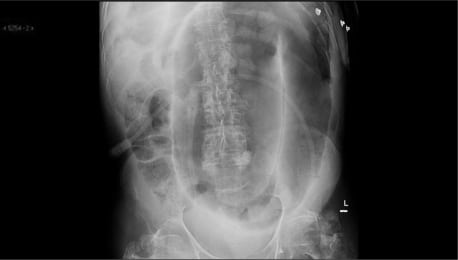
- I was too sick to ingest the barium required for a CAT scan, so the attending physician ordered an abdominal x-ray. (cdc.gov)
Pelvis1
- The abscess extended superiorly into the subdiaphragmatic region, indenting the diaphragmatic surface of liver, inferiorly under the right anterior abdominal wall into the paracolic gutter and up to the dome of the urinary bladder, extending into the pelvis. (webmedcentral.com)
Laparoscopy1
- Laparoscopy and other imaging tools can visualize the abscess. (wikipedia.org)
Postoperative1
- In 57 (2.4 percent) of 2,416 patients undergoing laparotomy for penetrating abdominal trauma from 1977 to 1980, an intraabdominal abscess developed in the postoperative period. (nih.gov)
Perforation2
- Over 80 percent of penetrating wounds leading to abscesses occurred in the upper quadrants, and common risk factors included multiple intraabdominal solid organ injuries requiring open drainage, coupled with gastrointestinal tract perforation. (nih.gov)
- The incidence of bile leak and loss of gallstones into the peritoneum due to gall bladder perforation during LC has been reported to be between 3% and 33% and can lead to the formation of intra-abdominal abscesses. (webmedcentral.com)
Ultrasound3
- Repeat ultrasound revealed no residual abscess and patient made a good recovery. (webmedcentral.com)
- Abscess drainage can be done through an endoscope using endoscopic ultrasound (EUS) in some cases. (limamemorial.org)
- Over the last decades, endoscopic ultrasound (EUS)-guided drainage has emerged as a minimally invasive alternative to percutaneous or surgical treatment of pelvic abscesses . (bvsalud.org)
Disease2
- Neisseria gonorrhoeae and chlamydial species are the most common organisms involved in pelvic abscesses in females as part of pelvic inflammatory disease. (medscape.com)
- Crohn's disease is a recurrent transmural inflammatory condition of the bowel, leading to bowel wall thickening, fixation of bowel loops, frequently complicated by stricture, fistula and abscess formation. (ispub.com)
Inflammatory1
- The center of the abscess contains liquefied mass of pus and bacterial debris, with tension in surrounding tissues due to inflammatory response. (differencebetween.net)
Anastomotic1
- Methods: The medical literature from 1973 to 2007 was reviewed using PubMed for papers relating to anastomotic leaks and abdominal abscess, with a specific emphasis on predisposing factors, prevention strategies, and treatment approaches. (elsevierpure.com)
Cancer2
- If an abscess unrelated to a surgical procedure or other obvious source is encountered, must consider the possibility of an underlying necrotic cancer. (hopkinsguides.com)
- Hematoma is caused due to trauma or injury, use of medicines like blood thinners, and reduced platelets count while abscess is caused due to compromised immune system due to diabetes, steroid therapy, cancer treatment, and other autoimmune diseases. (differencebetween.net)
Organs3
- Untreated abscesses can grow and damage nearby blood vessels and organs. (msdmanuals.com)
- An abscess below the diaphragm may form when infected fluid, for example, from a ruptured appendix, is moved upward by the pressure of abdominal organs and by the suction created when the diaphragm moves during breathing. (msdmanuals.com)
- Important muscular structures that surround the abdominal organs are shown in this figure. (radiologyassistant.nl)
Blood vessels1
- Hematoma is the result of blood cells that pool around the injured blood vessels as a result of trauma while abscess is a result of body's immune response that leads to accumulation of pus, bacteria and debris. (differencebetween.net)
Complication2
- Although the overall complication rate after LC is less than the traditional open approach, intra-abdominal abscesses following LC are rarely encountered. (webmedcentral.com)
- The complication rate of percutaneous abscess drainage is low (~2.5%) but should be considered when managing patients. (hopkinsguides.com)
Laparotomy1
- During laparotomy, the scar tissue of the abscess was found to be attached to the lateral wall of the ascending colon. (dtrf.org)
Treatment3
- This treatment can usually wait until after the abscess has been treated. (medlineplus.gov)
- The right early treatment can greatly improve the outcome for people who have intra-abdominal abscesses. (saintpetershcs.com)
- This case series contributes to the cumulative evidence that, in expert hands , EUS-guided drainage should be considered as first-line approach for treatment of pelvic abscesses . (bvsalud.org)
Bacteria2
- The abscesses usually contain a mixture of aerobic and anaerobic bacteria from the gastrointestinal (GI) tract. (medscape.com)
- The type and density of aerobic and anaerobic bacteria isolated from intra-abdominal abscesses depend upon the nature of the microflora associated with the diseased or injured organ. (medscape.com)
Hernia1
- The Spigelian hernia is an uncommon hernia at a weak spot between the oblique abdominal muscles and the rectus abdominis. (radiologyassistant.nl)