Adamantinoma
Fibrous Dysplasia of Bone
Ameloblastoma
Bone Diseases, Developmental
Tibia
Fibula
Allograft reconstruction for bone sarcoma of the tibia in the growing child. (1/12)
The outcome of tibial allograft reconstruction after resection of a tumour is inconsistent and has a high rate of failure. There are few reports on the use of tibial allografts in children with open growth plates. We performed 21 allograft reconstructions (16 osteoarticular, five intercalary) in 19 consecutive patients between seven and 17 years of age. Two had Ewing's sarcoma, one an adamantinoma and 16 osteosarcoma, one with multifocal disease. Five patients have died; the other 14 were free from disease at the time of follow-up. Six surviving patients (eight allograft reconstructions) continue to have good or excellent function at a mean of 59 months (14 to 132). One patient has poor function at 31 months. The other seven patients have a good or excellent function after additional procedures including exchange of the allograft and resurfacing or revision to an endoprosthesis at a mean of 101 months (43 to 198). The additional operations were performed at a mean of 47 months (20 to 84) after the first reconstruction. With the use of allograft reconstruction in growing children, joints and growth plates may be preserved, at least partially. Although our results remain inconsistent, tibial allograft reconstruction in selected patients may restore complete and durable function of the limb. (+info)A classic adamantinoma arising from osteofibrous dysplasia-like adamantinoma in the lower leg: a case report and review of the literature. (2/12)
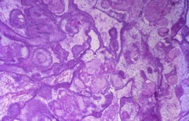
Adamantinoma is known as a low-grade malignant biphasic tumor. Classic adamantinoma is in general characterized by admixture of both epithelial and osteofibrous components that are associated with various proportions and differentiation patterns. Osteofibrous dysplasia (OFD) is a self-limited benign fibro-osseous lesion of bone during infancy and childhood. OFD-like adamantinoma is characterized by predominance of osteofibrous tissues, in which small groups of epithelial cells are only detected by careful search or immunohistochemistry. There have been controversies as to the potential correlation among OFD, OFD-like adamantinoma and classic adamantinoma. We report an unusual case of adamantinoma arising in the tibia, with an extensive review of the literature. The present findings suggest a direct correlation between OFD-like adamantinoma and classic adamantinoma. At the age of 12 years, the tibial biopsy lesion was diagnosed as OFD. At the age of 23, the lesion became larger and more destructive on x-ray films. The biopsy lesion was diagnosed as classic adamantinoma. Wide excision was performed. The primary lesion was retrospectively diagnosed as OFD-like adamantinoma because of presence of keratin-positive epithelial cells within the stroma. At five years after surgery, the patient was free from recurrence or metastasis. The retrospective histological findings of OFD-like adamantinoma in the original biopsy and of a classic adamantinoma in all sections of the later resection specimen raised the possibility of an unusual progression of OFD-like adamantinoma to a classic adamantinoma. The present case and the literature review suggest that an OFD-like adamantinoma may be a precursor lesion of classic adamantinoma. Therefore, the possibility of progression of OFD-like adamantinoma to a classic adamantinoma should be kept in mind, particularly when the destructive changes are seen radiologically. (+info)Osteofibrous dysplasia of the tibia. Is there a need for a radical surgical approach? (3/12)
Osteofibrous dysplasia is an unusual developmental condition of childhood, which almost exclusively affects the tibia. It is thought to follow a slowly progressive course and to stabilise after skeletal maturity. The possible link with adamantinoma is controversial and some authors believe that they are part of one histological process. We retrospectively reviewed 16 patients who were diagnosed as having osteofibrous dysplasia initially or on the final histological examination. Their management was diverse, depending on the severity of symptoms and the extent of the lesion. Definitive (extraperiosteal) surgery was localised "shark-bite" excision for small lesions in five patients. Extensive lesions were treated by segmental excision and fibular autograft in six patients, external fixation and bone transport in four and proximal tibial replacement in one. One patient who had a fibular autograft required further excision and bone transport for recurrence. Six initially underwent curettage and all had recurrence. There were no recurrences after localised extraperiosteal excision or bone transport. There were three confirmed cases of adamantinoma. The relevant literature is reviewed. We recommend extraperiosteal excision in all cases of osteofibrous dysplasia, with segmental excision and reconstruction in more extensive lesions. (+info)Fibular osteoadiposal flap for treatment of tibial adamantinoma: a case report. (4/12)
We treated a case with left tibial adamantinoma by use of a contralateral fibular osteoadiposal flap. The donor site of conventional fibular osteocutaneous flap must be covered with a skin graft because if we close the donor skin defect directly, compartment syndrome might occur. We were able to close the donor skin defect because this combined type flap included only a small monitoring skin paddle. We present herein the utility of the osteoadiposal flap and show the value of a skin-sparing approach with a minimal aesthetic defect. (+info)Preliminary report of expression of bax in oral cavity pathologies. (5/12)
Bax is considered one of major effectors of apoptosis--programmed cell death. Immunohistochemical analysis of in vitro patterns of bax expression was mostly investigated in mammalian cell lines and tissues. The present study is the first in vivo molecular analysis of bax expression in oral cavity pathologies. The study population consisted of 45 patients with hyperplasia, neoplasm in situ malignancy, and carcinoma. Biopsies were taken from incision line, tumour section, and healthy tissue. bax expression was investigated depending on the site of biopsy material sampling and final histopathology result. No statistically significant difference was demonstrated in bax expression between four hyperplasia subgroups. However, statistically significant differences in bax expression were found between the three basic study groups (P = 0.001). Statistically significant differences in bax expression were demonstrated depending on tissue collection site (P = 0.0002). We conclude that differences in bax expression may play a role in the pathogenesis of neoplastic disease. (+info)Adamantinoma of the tibia with late skeletal metastasis: an unusual presentation. (6/12)
Adamantinoma is a rare tumour of long bones that occurs most commonly in the tibia. Its pathogenesis is unknown. It is locally aggressive and recurrences are common after resection. Metastases have been reported in 10% to 20% of cases, most commonly in the lungs and rarely in the lymph nodes. We report a patient who developed a skeletal metastasis four years after resection of the primary tumour. There was no evidence of recurrence at the primary site or of secondary deposits in the lungs. (+info)Treatment of osteofibrous dysplasia and associated lesions. (7/12)
PURPOSE: To report long term treatment outcomes of osteofibrous dysplasia and association with adamantinoma. PATIENTS AND METHODS: From January 1984 to July 2001, 14 patients with osteofibrous dysplasia were followed for an average of 108 months (78 to 260 months). Our patient group consisted of 6 men and 8 women, with a mean age of 13.9 years (2 to 65 years). We reviewed the clinical and pathological features of all 14 patients. RESULTS: Thirteen patients had a lesion in the tibia, while one patient had lesions in both the tibia and the fibula. Initial treatments were observation after biopsy (6 patients), curettage with or without a bone graft (3 patients), resection followed by a free vascularized fibular bone graft (4 patients), or resection and regeneration with the Ilizarov external fixation (1 patient). Curettage was performed on 6 patients due to recurrence or progression after the initial treatment. Among these patients, one was diagnosed with AD from the biopsy of the recurrent lesion. This patient was further treated by segmental resection and pasteurization. After the initial pathology slides of the 13 patients were reviewed with immunohistochemical cytokeratin staining, one patient diagnosis was changed from osteofibrous dysplasia to osteofibrous dysplasia-like adamantinoma. CONCLUSION: Some patients with osteofibrous dysplasia require close observation because of the high association risk between osteofibrous dysplasia and adamantinoma, Immunohistochemical staining may be helpful in differentiating these two diagnoses. (+info)Analysis of stromal cells in osteofibrous dysplasia and adamantinoma of long bones. (8/12)
(+info)Adamantinoma is a rare, slow-growing malignant (cancerous) tumor that typically develops in the tibia or fibula bones of the lower leg. It primarily affects young adults and can be difficult to diagnose due to its rarity and nonspecific symptoms.
The name "adamantinoma" comes from its microscopic appearance, which resembles that of a type of skin cancer called adamantinoma of the skin or adamantoblastoma. However, they are not related.
Adamantinomas are characterized by the presence of epithelial cells (cells that line the outer surface of the body and internal organs) within the bone tissue. These tumors tend to be locally aggressive, meaning they can invade surrounding tissues and bones. In some cases, adamantinomas may metastasize (spread) to other parts of the body, such as the lungs or lymph nodes.
Treatment for adamantinoma usually involves surgical removal of the tumor, along with a portion of the affected bone. In some cases, reconstruction or limb-sparing surgery may be necessary to maintain function and appearance. Radiation therapy and chemotherapy are not typically effective against adamantinomas, but they might be used in specific situations or as part of clinical trials.
Regular follow-up appointments with a healthcare provider are essential for monitoring the patient's condition and detecting any potential recurrence or metastasis early on.
Fibrous Dysplasia of Bone is a rare, benign bone disorder that is characterized by the replacement of normal bone tissue with fibrous (scar-like) and immature bone tissue. This results in weakened bones that are prone to fractures, deformities, and pain. The condition can affect any bone in the body but most commonly involves the long bones of the legs, arms, and skull. It can occur as an isolated finding or as part of a genetic disorder called McCune-Albright syndrome. The exact cause of fibrous dysplasia is not fully understood, but it is believed to result from a genetic mutation that occurs during early bone development. There is no cure for fibrous dysplasia, and treatment typically focuses on managing symptoms and preventing complications.
Ameloblastoma is a slow-growing, non-cancerous tumor that develops in the jawbone, typically in the lower jaw. It originates from the cells that form the enamel (the hard, outer surface of the teeth). This tumor can cause swelling, pain, and displacement or loosening of teeth. In some cases, it may also lead to fractures of the jawbone.
There are different types of ameloblastomas, including solid or multicystic, unicystic, and peripheral ameloblastoma. Treatment usually involves surgical removal of the tumor, with careful monitoring to ensure that it does not recur. In rare cases, more aggressive treatment may be necessary if the tumor is large or has invaded surrounding tissues.
It's important to note that while ameloblastomas are generally benign, they can still cause significant morbidity and should be treated promptly by an oral and maxillofacial surgeon or other qualified healthcare professional.
Developmental bone diseases are a group of medical conditions that affect the growth and development of bones. These diseases are present at birth or develop during childhood and adolescence, when bones are growing rapidly. They can result from genetic mutations, hormonal imbalances, or environmental factors such as poor nutrition.
Some examples of developmental bone diseases include:
1. Osteogenesis imperfecta (OI): Also known as brittle bone disease, OI is a genetic disorder that affects the body's production of collagen, a protein necessary for healthy bones. People with OI have fragile bones that break easily and may also experience other symptoms such as blue sclerae (whites of the eyes), hearing loss, and joint laxity.
2. Achondroplasia: This is the most common form of dwarfism, caused by a genetic mutation that affects bone growth. People with achondroplasia have short limbs and a large head relative to their body size.
3. Rickets: A condition caused by vitamin D deficiency or an inability to absorb or use vitamin D properly. This leads to weak, soft bones that can bow or bend easily, particularly in children.
4. Fibrous dysplasia: A rare bone disorder where normal bone is replaced with fibrous tissue, leading to weakened bones and deformities.
5. Scoliosis: An abnormal curvature of the spine that can develop during childhood or adolescence. While not strictly a developmental bone disease, scoliosis can be caused by various underlying conditions such as cerebral palsy, muscular dystrophy, or spina bifida.
Treatment for developmental bone diseases varies depending on the specific condition and its severity. Treatment may include medication, physical therapy, bracing, or surgery to correct deformities and improve function. Regular follow-up with a healthcare provider is essential to monitor growth, manage symptoms, and prevent complications.
The tibia, also known as the shin bone, is the larger of the two bones in the lower leg and part of the knee joint. It supports most of the body's weight and is a major insertion point for muscles that flex the foot and bend the leg. The tibia articulates with the femur at the knee joint and with the fibula and talus bone at the ankle joint. Injuries to the tibia, such as fractures, are common in sports and other activities that put stress on the lower leg.
The fibula is a slender bone located in the lower leg of humans and other vertebrates. It runs parallel to the larger and more robust tibia, and together they are known as the bones of the leg or the anterior tibial segment. The fibula is the lateral bone in the leg, positioned on the outside of the tibia.
In humans, the fibula extends from the knee joint proximally to the ankle joint distally. Its proximal end, called the head of the fibula, articulates with the lateral condyle of the tibia and forms part of the inferior aspect of the knee joint. The narrowed portion below the head is known as the neck of the fibula.
The shaft of the fibula, also called the body of the fibula, is a long, thin structure that descends from the neck and serves primarily for muscle attachment rather than weight-bearing functions. The distal end of the fibula widens to form the lateral malleolus, which is an important bony landmark in the ankle region. The lateral malleolus articulates with the talus bone of the foot and forms part of the ankle joint.
The primary functions of the fibula include providing attachment sites for muscles that act on the lower leg, ankle, and foot, as well as contributing to the stability of the ankle joint through its articulation with the talus bone. Fractures of the fibula can occur due to various injuries, such as twisting or rotational forces applied to the ankle or direct trauma to the lateral aspect of the lower leg.
Bone neoplasms are abnormal growths or tumors that develop in the bone. They can be benign (non-cancerous) or malignant (cancerous). Benign bone neoplasms do not spread to other parts of the body and are rarely a threat to life, although they may cause problems if they grow large enough to press on surrounding tissues or cause fractures. Malignant bone neoplasms, on the other hand, can invade and destroy nearby tissue and may spread (metastasize) to other parts of the body.
There are many different types of bone neoplasms, including:
1. Osteochondroma - a benign tumor that develops from cartilage and bone
2. Enchondroma - a benign tumor that forms in the cartilage that lines the inside of the bones
3. Chondrosarcoma - a malignant tumor that develops from cartilage
4. Osteosarcoma - a malignant tumor that develops from bone cells
5. Ewing sarcoma - a malignant tumor that develops in the bones or soft tissues around the bones
6. Giant cell tumor of bone - a benign or occasionally malignant tumor that develops from bone tissue
7. Fibrosarcoma - a malignant tumor that develops from fibrous tissue in the bone
The symptoms of bone neoplasms vary depending on the type, size, and location of the tumor. They may include pain, swelling, stiffness, fractures, or limited mobility. Treatment options depend on the type and stage of the tumor but may include surgery, radiation therapy, chemotherapy, or a combination of these treatments.
 Adamantinoma
Ameloblastoma
Bone Cancer Research Trust
Osteofibrous dysplasia
List of MeSH codes (C05)
List of MeSH codes (C04)
Adamantine
International Classification of Diseases for Oncology
Sarcoma
Index of trauma and orthopaedics articles
Dysplasia-like adamantinoma1
- BACKGROUND: Osteofibrous dysplasia-like adamantinoma (OFD-AD) and classic adamantinoma (AD) are rare, neoplastic diseases with only limited data supporting current treatment protocols. (ox.ac.uk)
Osteofibrous Dysplasia1
- citation needed] Benign osteofibrous dysplasia may be a precursor of adamantinoma or a regressive phase of adamantinoma. (wikipedia.org)
Tumor4
- Adamantinoma is a rare biphasic tumor, and its origin remains controversial. (medscape.com)
- A variety of tumors and tumor-like lesions can mimic an adamantinoma. (medscape.com)
- Radiofrequency Ablation (RFA) may be a safe and reliable option for treatment of OFD like adamantinoma, without vital tumor spill. (easychair.org)
- Local tumor ablation with RFA has the potential to become a safe and effective treatment alternative in OFD like adamantinoma in a child. (easychair.org)
Tibia5
- Adamantinoma of Tibia" by Ali Kemal UZUNLAR, Ahmet KAPUKAYA et al. (tubitak.gov.tr)
- Osteo Fibrous Dysplasia (OFD) like adamantinoma is considered a benign condition that is usually located in the tibia. (easychair.org)
- Two young patients with OFD like adamantinoma of the tibia were treated with CAS guided open intra-operative RFA in our institution. (easychair.org)
- i had adamantinoma too, I had surgery in 2003 where they took out 7 inches of my tibia and replaced it with cadaver bone + 2 metal rods and held it together with 15 screws. (cancer.org)
- Long-Term Follow-Up of Adamantinoma of the Tibia Complicated by Metastases and a Second Unrelated Primary Cancer: A Case Report and Literature Review. (uc.edu)
Bone5
- Adamantinoma (from the Greek word adamantinos, meaning "very hard") is a rare bone cancer, making up less than 1% of all bone cancers. (wikipedia.org)
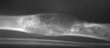
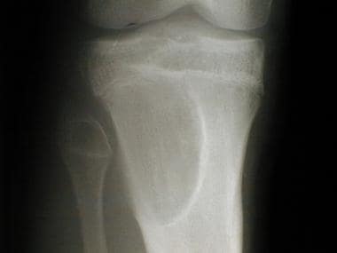
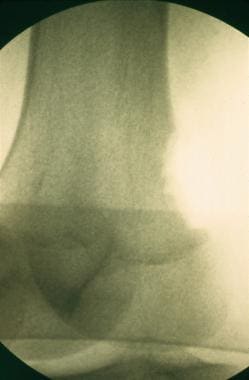
- In its early stages, an adamantinoma appears as an elongated, linear lucency on plain radiographs, and no periosteal reaction is noted in the surrounding bone. (medscape.com)
- The condition is mainly found in children and has an association with classical adamantinoma, a malignancy of bone. (easychair.org)
- Reid lives in Canada and was diagnosed with adamantinoma in his shin bone when he was 9 years old. (bcrt.org.uk)
- Tsistektomiya s zapolneniem kostnoy polosti poristo-pronitsaemym nikelid-titanom, obogashchyonnym trombotsitarnoy massoy [Cystektomy with filling the bone cavity by porous-permeable nikelid of titanium, en-riched with thrombocytic mass]. (vestnik-avicenna.tj)
Fibroma1
- All dogs affected by one oral tumour of 7 types: 5 epulides, 3 melanomas, 4 squamous cell carcinomas, 1 fibrosarcoma, 1 fibroma, 1 adamantinoma and 1 lymphoma. (vin.com)
Malignancy2
- Adamantinoma-like Ewing sarcoma (ALES) is a rare malignancy currently considered a variant of Ewing sarcoma with most known cases harboring EWSR1 rearrangements. (bvsalud.org)
- INTRODUCTION: Adamantinoma-like Ewing sarcoma (ALES) is a rare aggressive malignancy occasionally diagnosed in the thyroid gland. (bvsalud.org)
Neoplasm1
- In 1885, this kind of odontogenic neoplasm was designated as an adamantinoma by Malassez. (wikipedia.org)
Epithelioid1
- Epithelioid sarcoma, chordoma, and adamantinoma show strong positivity corresponding to that of simple epithelia (with antibodies against CK8, CK18 and CK19). (scytek.com)
Characteristics2
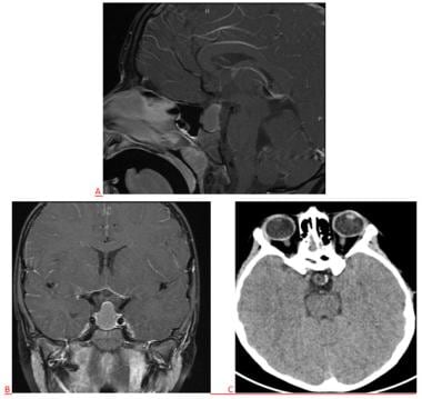
- Many pathologic conditions that are as rare as or more common than adamantinoma demonstrate similar characteristics on plain radiographs, as well as CT scans and MRIs. (medscape.com)
- Describe the radiographic characteristics of adamantinoma of the long bones. (elsevierpure.com)
Describe1
- Some authors still confusingly misuse the term adamantinoma to describe ameloblastomas. (wikipedia.org)
Diagnosis1
- In December 2020, we finally had a correct diagnosis of adamantinoma . (bcrt.org.uk)
Long2
- Limitations of plain-film radiography include the relatively long list of differential diagnoses for adamantinoma. (medscape.com)
- 3. Discuss treatment option for adamantinoma of the long bones. (elsevierpure.com)
Surgical1
- Treatment options for adamantinoma are surgical and include either marginal or en bloc resection. (medscape.com)
Treatment2
- CAS guided RFA can be a safe and effective treatment in OFD like adamantinoma in a child. (easychair.org)
- But Forsythia supplements or Forsythia extracts offer less benefit if on Radiation treatment for Primary Adamantinoma compared to Caffeine. (addon.life)
Patients2
- Most commonly, patients are in their second or third decade, but adamantinoma can occur over a wide age range. (wikipedia.org)
- We believe that our retrospective multicenter cohort study is the largest analysis of patients with adamantinoma to date. (ox.ac.uk)
Doctor1
- I sent this off to our doctor, who then asked for another biopsy where they tested for adamantinoma, and it came back positive. (bcrt.org.uk)
Tibia11
- The experts in our Bone and Soft Tissue Tumors Program have extensive experience caring for children and adolescents with adamantinoma of the tibia and other bone and soft tissue disorders. (dana-farber.org)
- My son is 12 and is going through a similar experience: OFD-life adamantinoma on the left tibia. (cancer.org)
- Osteo Fibrous Dysplasia (OFD) like adamantinoma is considered a benign condition that is usually located in the tibia. (easychair.org)
- Two young patients with OFD like adamantinoma of the tibia were treated with CAS guided open intra-operative RFA in our institution. (easychair.org)
- 21. Adamantinoma of tibia: a study of 12 cases. (nih.gov)
- 29. Multiple recurrences and late metastasis of adamantinoma in the tibia: a case report. (nih.gov)
- 30. Osteofibrous Dysplasia-like Adamantinoma of the Tibia in a 15-Year-Old Girl. (nih.gov)
- 31. Dedifferentiated classic adamantinoma of the tibia: a report of a case with eventual complete revertant mesenchymal phenotype. (nih.gov)
- 36. Pulmonary metastases of a tibia adamantinoma. (nih.gov)
- Suele localizarse en la TIBIA. (bvsalud.org)
- When FD in the tibia is considered, adamantinoma should be in the differential diagnosis. (250grados.net)
Dysplasia4
- citation needed] Benign osteofibrous dysplasia may be a precursor of adamantinoma or a regressive phase of adamantinoma. (wikipedia.org)
- 22. Does osteofibrous dysplasia progress to adamantinoma and how should they be treated? (nih.gov)
- Journal of Clinical and Experimental Orthopaedics considers articles on clinical orthopaedics related issues such as Adamantinoma, Aneurysmal Bone Cysts ,Chondrosarcoma Chordomas Cryosurgery Enchondroma , Fibrous Dysplasia, Giant Cell Tumor of Bone, Malignant Osteoid ,Metastatic Bone Cancer Multilobular Tumour of Bone Orthopaedic Oncology Osteocartilaginous Exostosis Osteochondrodysplasia Osteoma Osteonecrosis Osteosarcoma Primary Bone Tumors Secondary Bone Tumours Tumours of Bone. (imedpub.com)
- According to the AAOS, adamantinoma and osteofibrous dysplasia (OFD) are rare forms of bone tumors that often begin growing in the shinbone. (orthosportsmed.com)
Chondrosarcoma1
- 32. Dedifferentiated chondrosarcoma with "adamantinoma-like" features: A case report and review of literature. (nih.gov)
Ameloblastoma2
- Ameloblastoma, adamantinoma. (nih.gov)
- Other tumors in this area may include ameloblastoma or adamantinoma, rare tumors that may arise in the bones of the jaw. (marystolfacancerfoundation.org)
Long bones2
Histologically1
- What does a craniopharyngioma (also known as adamantinoma) resemble histologically? (rahulgladwin.com)
Malignant1
- Other Malignant Tumors 79 (1) Myeloma 79 (2) Endothelioma 81 (3) Glioma 82 (4) Neurocytoma 84 (5) Hypernephroma 84 (6) Adamantinoma 85 (7) Chorioma 87 (8) Mixed Tumors 88 (9) Teratoma 90 VII. (nih.gov)
Fibula1
- 8. MohlerDG, CunninghamDC.Adamantinoma arising in the distal fibula treated with distal fibulectomy: A case report and review of the literature.Foot Ankle Int1997;18:746-51. (jbstjournal.com)
Diagnoses1
- Limitations of plain-film radiography include the relatively long list of differential diagnoses for adamantinoma. (medscape.com)
Occur2
- Most commonly, patients are in their second or third decade, but adamantinoma can occur over a wide age range. (wikipedia.org)
- Adamantinoma may appear over a short time or may occur for six months or more. (dana-farber.org)
Chordoma1
- Epithelioid sarcoma, chordoma, and adamantinoma show strong positivity corresponding to that of simple epithelia (with antibodies against CK8, CK18 and CK19). (teomics.com)
Dentigerous1
- 5. Cahn I. R. The dentigerous cyst is a potencial adamantinoma. (bvsalud.org)
Lower leg1
- Most of the time, adamantinoma grows in the lower leg. (handlebar-online.com)
Rare3
- Adamantinoma (from the Greek word adamantinos, meaning "very hard") is a rare bone cancer, making up less than 1% of all bone cancers. (wikipedia.org)
- Many pathologic conditions that are as rare as or more common than adamantinoma demonstrate similar characteristics on plain radiographs, as well as CT scans and MRIs. (medscape.com)
- Adamantinoma is a rare, slow-growing bone cancer. (dana-farber.org)
Doctors1
- Doctors typically recommend surgery to treat adamantinoma. (dana-farber.org)
Cases1
- 24. Adamantinoma in childhood: report of six cases and review of the literature. (nih.gov)
Children3
- For most children with adamantinoma, the long-term outlook is positive. (dana-farber.org)
- When treated with surgery, the majority of children with adamantinoma have a positive long-term outlook. (dana-farber.org)
- The condition is mainly found in children and has an association with classical adamantinoma, a malignancy of bone. (easychair.org)
Term2
- Some authors still confusingly misuse the term adamantinoma to describe ameloblastomas. (wikipedia.org)
- 25. Adamantinoma of bone: Long-term follow-up of 46 consecutive patients. (nih.gov)
Time1
- About 20 percent of the time, adamantinoma can spread to other parts of the body, including the lungs. (dana-farber.org)