Adrenal Cortex
Adrenal Glands
Adrenal Medulla
Zona Reticularis
Adrenocorticotropic Hormone
Zona Fasciculata
Cerebral Cortex
Adrenal Insufficiency
Prefrontal Cortex
Visual Cortex
Zona Glomerulosa
Steroid 11-beta-Hydroxylase
Motor Cortex
Adrenocortical Adenoma
Adrenal Cortex Function Tests
Adrenal Cortex Hormones
Adrenocortical Carcinoma
Auditory Cortex
Adrenal Hyperplasia, Congenital
Somatosensory Cortex
Cosyntropin
Aldosterone
Adrenodoxin
Hydrocortisone
Corticosterone
Aldosterone Synthase
Cholesterol Side-Chain Cleavage Enzyme
Steroidogenic Factor 1
Cattle
Steroid 21-Hydroxylase
Cushing Syndrome
Adrenocortical Hyperfunction
Addison Disease
Steroid 17-alpha-Hydroxylase
Entorhinal Cortex
Adosterol
Pregnenolone
Kidney Cortex
Ferredoxin-NADP Reductase
17-Hydroxycorticosteroids
Brain Mapping
Hyperaldosteronism
Pituitary-Adrenal System
Steroids
Chromaffin Cells
Dehydroepiandrosterone
Receptor, Melanocortin, Type 2
Cerebellar Cortex
Pheochromocytoma
Magnetic Resonance Imaging
Hypothalamo-Hypophyseal System
Hypophysectomy
RNA, Messenger
Mitochondria
Adrenarche
Progesterone Reductase
Rats, Sprague-Dawley
Dehydroepiandrosterone Sulfate
Brain
Neurons
Glucocorticoids
Metyrapone
Rats, Inbred Strains
Immunohistochemistry
Steroid Hydroxylases
Cytochrome P-450 Enzyme System
Frontal Lobe
Rats, Wistar
Photic Stimulation
Angiotensin II
Organ Specificity
Fetus
Mineralocorticoids
Chromaffin System
Microscopy, Electron
Tissue Distribution
Functional Laterality
Parietal Lobe
Gyrus Cinguli
Molecular Sequence Data
Fushi Tarazu Transcription Factors
Stress, Physiological
Autoradiography
Histocytochemistry
Hyperplasia
Cyclic AMP
In Situ Hybridization
Endocrine Glands
Pituitary Gland
Hyperandrogenism
Base Sequence
Restraint, Physical
Image Processing, Computer-Assisted
Cyanoketone
Ovary
Chromaffin Granules
Psychomotor Performance
Cats
Renin
Nerve Net
Bucladesine
Vibrissae
Hormones
Temporal Lobe
Cells, Cultured
Microsomes
Adrenal Rest Tumor
Androgens
Stimulation, Chemical
Freeze Etching
Nuclear bodies are usual constituents in tissues of hibernating dormice. (1/1201)
In previous studies we demonstrated in several tissues of the hazel dormouse Muscardinus avellanarius that during hibernation cell nuclei contain particular structural constituents absent in euthermia. In the present study we examine the same tissues in euthermic and hibernating individuals of the edible dormouse Glis glis in order to investigate possible modifications of nuclear structural constituents occurring during hibernation in this species. Edible dormice were captured in the wild and maintained in an external animal house. Samples of liver, pancreas, brown adipose tissue and adrenal cortex were taken from three hibernating and three euthermic animals and processed for resin embedding. Ultrastructural and immunocytochemical studies were carried out on cell nuclei of these tissues. The most evident feature of cell nuclei of hibernating dormice was the presence of several nuclear bodies, namely fibro-granular material, amorphous bodies, coiled bodies, perichromatin granule-like granules and nucleoplasmic fibrils, the distribution of which was peculiar to each tissue. No one of these constituents was detectable during euthermia. Immunocytochemical analyses revealed that they contain some splicing factors. Apart from some differences, maybe due to the different characteristics of lethargy, the nuclear bodies found in edible dormice were morphologically and immunocytochemically similar to those previously described in the same tissues of hazel dormice. They therefore seem to be strictly correlated to the hibernating state. If they represent storage and/or assembly sites of splicing factors to be rapidly used upon arousal, they could represent a usual structural feature in cells of hibernating species. (+info)Natural killer cell activity in the peripheral blood of patients with Cushing's syndrome. (2/1201)
BACKGROUND: Natural killer (NK) cells are CD3(-)CD16(+)CD56(+) bone-marrow-derived lymphocytes mediating first-line defence by direct cytotoxicity against various types of target cells without prior immunization. NK cell activity is positively regulated by immune interferon (IFN-gamma); among hormones, glucocorticoids are potent in vitro and in vivo inhibitors, whereas ACTH and beta-endorphin in many experimental circumstances enhance NK cytotoxicity. DESIGN: We measured NK cytotoxicity of peripheral blood mononuclear cells (PBMC) obtained at 0800h and 2000h from 26 patients with Cushing's syndrome (12 pituitary-dependent, 12 adrenal-dependent and two dependent on ectopic ACTH secretion). In vitro responsiveness to IFN-gamma or cortisol was also tested. METHODS: NK activity was measured in a 4-h direct cytotoxicity assay using K562 cells as targets. Plasma ACTH, serum and urinary free cortisol were concomitantly measured with commercially available kits. RESULTS: Spontaneous activity and responsiveness to IFN-gamma or cortisol were significantly greater in 15 age- and sex-matched controls than in Cushing's patients at 0800h. In pituitary-dependent Cushing's patients, plasma ACTH correlated positively with mean levels of spontaneous NK activity (r=0.64, P<0.05) and negatively with cortisol-dependent percentage inhibition (r=-0.69, P<0.02). In adrenal-dependent Cushing's patients, a negative correlation was observed between levels of spontaneous NK activity and urinary free cortisol (r=-0.67, P<0.02). CONCLUSIONS: Our data indicate that excess endogenous glucocorticoids affect spontaneous NK cell activity and responsiveness to exogenous IFN-gamma or cortisol. The differential patterns observed between pituitary-dependent and adrenal-dependent groups are compatible with a positive immunomodulatory role of pituitary pro-opiomelanocortin-derived peptides that effectively counterbalance, at least partially, glucocorticoid immunosuppression. (+info)Lipid requirement of membrane-bound 3-oxosteroid delta4-delta5-isomerase. Studies on beef adrenocortical microsomes. (3/1201)
The role of phospholipid in the beef adrenal microsomal 3-oxosteroid delta4-delta5-isomerase (EC 5.3.1.1) has been investigated with the use of phospholipase A to alter the microsomal phospholipids. The byproducts of phospholipase A digestion have been removed with a wash solution containing bovine serum albumin. Removal of 80-85% of the phospholipid leads to loss of 80-90% of the 3-oxosteroid delta4-delta5-isomerase activity. Reconstitution experiments have been performed by introduction of lipid aqueous dispersions in the enzymatic assay. Asolectin, a commercially available preparation of soy phosphatides, is able to stimulate the enzymatic activity but does not restore the 3-oxosteroid delta4-delta5-isomerase activity in phospholipase-A-treated membranes. In contrast, the introduction of aqueous dispersions of microsomal total lipid mixtures in the enzymatic assay brings about a complete restoration of the 3-oxosteroid delta4-delta5-isomerase activity in the lipid-depleted membranes. It is concluded that the bovine adrenal microsomal 3-oxosteroid delta4-delta5-isomerase requires phospholipid(s) to exhibit its full catalytic activity. (+info)Local renin-angiotensin system is involved in K+-induced aldosterone secretion from human adrenocortical NCI-H295 cells. (4/1201)
NCI-H295, a human adrenocarcinoma cell line, has been proposed as a model system to define the role of the renin-angiotensin system in the regulation of aldosterone production in humans. Because the precise cellular localization of the components of the renin-angiotensin system in human adrenal cortical cells remains unclear, we investigated their localization in this defined cell system. NCI-H295 cells expressed both angiotensinogen and renin as shown by reverse transcriptase polymerase chain reaction and immunohistochemistry. Human angiotensin-converting enzyme (ACE) was not detectable by immunocytochemistry, ACE binding, or reverse transcriptase polymerase chain reaction. However, 3.5 mmol/L K+ stimulated the formation of both angiotensin I and angiotensin II 1. 9- and 2.5-fold, respectively, and increased aldosterone release 3. 0-fold. The K+-induced stimulation of aldosterone release was decreased by captopril and enalaprilat (24% and 26%, respectively) and by the angiotensin type 1 (AT1)-receptor antagonist losartan (28%). Angiotensin II-induced stimulation of aldosterone release was abolished by losartan treatment. Specific [125I]Sar1-angiotensin II binding was detected by receptor autoradiography. The binding of [125I]Sar1-angiotensin II was completely displaced by the AT1 antagonist losartan but not by the AT2 receptor ligand PD 123319, confirming the expression of angiotensin II AT1 receptors in NCI-H295 cells. Our results demonstrate that NCI-H295 cells express most of the components of the renin-angiotensin system. Our failure to detect ACE, however, suggests that the production of angiotensin II in NCI-H295 cells may be ACE independent. NCI-H295 cells are able to produce angiotensin II, and K+ increases aldosterone secretion in part through an angiotensin-mediated pathway. The production of angiotensin II in NCI-H295 cells demonstrates that this human cell line can be useful to characterize the role of locally produced angiotensin II in the regulation of aldosterone release. (+info)Comparison of expression and regulation of the high-density lipoprotein receptor SR-BI and the low-density lipoprotein receptor in human adrenocortical carcinoma NCI-H295 cells. (5/1201)
In rodents, cholesterol for adrenal steroidogenesis is derived mainly from high-density lipoproteins (HDL) via the HDL receptor, scavenger receptor-BI (SR-BI). In humans cholesterol for steroidogenesis is considered to be derived from the low-density lipoprotein (LDL) receptor pathway, and the contribution of SR-BI to that is unknown. In the present study SR-BI expression and regulation by steroidogenic stimuli was analysed in human adrenocortical cells and compared with LDL receptor expression. In addition, the functional contribution of both receptors for cholesteryl ester delivery to human adrenocortical cells was compared. Northern blot and reverse transcription-PCR amplification and sequence analysis demonstrated the presence of SR-BI mRNA in foetal and adult human adrenal cortex. Furthermore, SR-BI mRNA was expressed to similar levels in human primary adrenocortical and adrenocortical carcinoma NCI-H295 cells, indicating its presence in the steroid-producing cells. Treatment of NCI-H295 cells with 8Br-cAMP, a stimulator of glucocorticoid synthesis via the protein kinase A second messenger signal transduction pathway, resulted in an increase of both SR-BI and LDL receptor mRNA levels in a time- and dose-dependent manner. The induction of SR-BI and LDL receptor by cAMP was independent of ongoing protein synthesis and occurred at the transcriptional level. Ligand blot experiments indicated that a protein of similar size to SR-BI is the major HDL-binding protein in NCI-H295 cells. Western blot analysis demonstrated that cAMP treatment increased the levels of LDL receptor and, to a lesser extent, SR-BI protein in NCI-H295 cells. Binding and uptake of cholesterol was quantitatively smaller from HDL than from LDL, both in basal as well as in cAMP-stimulated cells. Scatchard analysis under basal conditions indicated that NCI-H295 cells express twice as many specific binding sites for LDL than for HDL. Dissociation constant values (Kd; in nm) were approximately five times higher for HDL than for LDL, indicating a lower affinity of HDL compared with LDL. The combined effects of these two parameters and the low cholesteryl ester content of HDL subfraction 3 (HDL3) contributes to a lower cholesteryl ester uptake from HDL than from LDL by the NCI-H295 cells. In conclusion, both the SR-BI and LDL receptor genes are expressed in the human adrenal cortex and coordinately regulated by activators of glucocorticoid synthesis. In contrast to rodents, in human adrenocortical cells the HDL pathway of cholesterol delivery appears to be of lesser importance than the LDL pathway. Nevertheless, the SR-BI pathway may become of major importance in conditions of functional defects in the LDL receptor pathway. (+info)The expression of inhibin/activin subunits in the human adrenal cortex and its tumours. (6/1201)
Inhibins and activins are dimeric proteins of the transforming growth factor-beta superfamily which have been shown to be expressed in the adrenal cortex. Recent studies have suggested a role for these peptides in the pathogenesis and/or function of adrenal tumours. To investigate further their physiological and pathological roles, we have documented immunoreactivity for inhibin alpha, betaA and betaB subunits in normal adult and fetal human adrenals, in hyperplastic adrenals and in adrenal tumours. In the normal and hyperplastic adult gland, diffuse immunopositivity was demonstrated for beta subunits, suggesting that activins (beta beta dimers) can be expressed in all zones. Inhibin alpha was limited to the zona reticularis and the innermost zona fasciculata in the normal gland, extending centripetally into the zona fasciculata in hyperplasia, supporting a role for ACTH in the regulation of expression, and suggesting that expression of inhibins (alpha beta dimers) is restricted. Immunopositivity for all three subunits was seen in both fetal and definitive zones of the fetal cortex, indicating that both inhibins and activins could be expressed in both. Immunopositivity for all three subunits was seen in most adrenocortical tumours. Loss of immunopositivity for inhibin alpha in a subgroup of carcinomas might indicate a role in tumour progression. The greater intensity of staining for inhibin alpha in tumours associated with Cushing's syndrome again suggests a link with cortisol production. (+info)Influences of long-term administration of 24R, 25-dihydroxyvitamin D3, a vitamin D3 derivative, in rats. (7/1201)
In order to examine the influences by long-term feeding of 24R, 25 dihydroxyvitamin D3[24R, 25(OH)2D3], an active form of vitamin D, Wistar rats (14-week-old, male, 20 rats/group) were fed a powder diet containing 0 or 5 ppm 24R, 25(OH)2D3 for 57 weeks. Final body weights and total food consumption were comparable between the groups. Urinary calcium levels were significantly (p < 0.05 or 0.01) increased by the administration of 24R, 25(OH)2D3 at weeks 3, 22 and 56, although the levels of serum calcium did not differ between the groups at the termination of week 57. In the 24R, 25(OH)2D3 group, weights of the adrenals and femurs were significantly (p < 0.01) increased. Histopathologically, this was found due to thickening of cortical bone in the femurs, and medullary hyperplasia and pheochromocytoma of the adrenals. Immunohistochemically, proliferating cell nuclear antigen (PCNA)-labeling indices for intact adrenal medulla, medullary hyperplasia and pheochromocytoma in the 24R, 25(OH)2D3 group were respectively 1.82 +/- 1.21, 5.88 +/- 4.13 and 16, all higher than that for the adrenal medulla in the control group (0.87 +/- 0.67). These results indicate that 24R, 25(OH)2D3 at a dose with which serum calcium is not chronically increased causes thickening of the cortex of the femur, and development of adrenal proliferative lesions, suggesting that rats may be too sensitive for results to be relevant to human risk assessment. (+info)Calcium and reactive oxygen species as messengers in endotoxin action on adrenocortical cells. (8/1201)
The effect of Escherichia coli 0111:B4 endotoxin (lipopolysaccharide, LPS) on the intracellular Ca2+ and reactive oxygen metabolite content of both rat isolated fasciculata-reticularis and glomerulosa cells was evaluated by flow cytometry to know the role of these mechanisms in the initiation of cell injury produced by LPS on adrenocortical cells during endotoxic shock. A rapid increase of intracellular calcium was induced by endotoxin in both cell types. In fasciculata-reticularis cells, this [Ca2+]i increase was mainly due to an important mobilization of intracellular stores. Dose-dependent increases in [Ca2+]i were also observed when both cell types were incubated with LPS for 20 min in the presence of extracellular calcium. This treatment abolished the increase in intracellular calcium induced by ACTH and angiotensin II. On the other hand, the endotoxin produced a fast and dose-dependent increase in reactive oxygen species in both cell types, higher in glomerulosa than in fasciculata-reticularis cells. LPS-pretreated cells showed more susceptibility to the oxidative stress induced by Fe2+. These results can be related to functional alterations previously described showing the involvement of calcium and reactive oxygen species as messengers in the endotoxin action on adrenocortical cells. (+info)The adrenal cortex is the outer portion of the adrenal gland, which is located on top of the kidneys. It plays a crucial role in producing hormones that are essential for various bodily functions. The adrenal cortex is divided into three zones:
1. Zona glomerulosa: This outermost zone produces mineralocorticoids, primarily aldosterone. Aldosterone helps regulate sodium and potassium balance and thus influences blood pressure by controlling the amount of fluid in the body.
2. Zona fasciculata: The middle layer is responsible for producing glucocorticoids, with cortisol being the most important one. Cortisol regulates metabolism, helps manage stress responses, and has anti-inflammatory properties. It also plays a role in blood sugar regulation and maintaining the body's response to injury and illness.
3. Zona reticularis: The innermost zone produces androgens, primarily dehydroepiandrosterone (DHEA) and its sulfate form (DHEAS). These androgens are weak compared to those produced by the gonads (ovaries or testes), but they can be converted into more potent androgens or estrogens in peripheral tissues.
Disorders related to the adrenal cortex can lead to hormonal imbalances, affecting various bodily functions. Examples include Addison's disease (insufficient adrenal cortical hormone production) and Cushing's syndrome (excessive glucocorticoid levels).
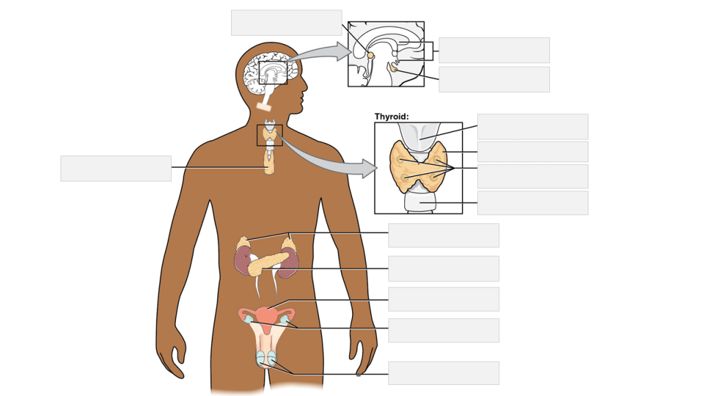
The adrenal glands are a pair of endocrine glands that are located on top of the kidneys. Each gland has two parts: the outer cortex and the inner medulla. The adrenal cortex produces hormones such as cortisol, aldosterone, and androgens, which regulate metabolism, blood pressure, and other vital functions. The adrenal medulla produces catecholamines, including epinephrine (adrenaline) and norepinephrine (noradrenaline), which help the body respond to stress by increasing heart rate, blood pressure, and alertness.
Adrenal cortex neoplasms refer to abnormal growths (tumors) in the adrenal gland's outer layer, known as the adrenal cortex. These neoplasms can be benign or malignant (cancerous). Benign tumors are called adrenal adenomas, while cancerous tumors are called adrenocortical carcinomas.
Adrenal cortex neoplasms can produce various hormones, leading to different clinical presentations. For instance, they may cause Cushing's syndrome (characterized by excessive cortisol production), Conn's syndrome (caused by aldosterone excess), or virilization (due to androgen excess). Some tumors may not produce any hormones and are discovered incidentally during imaging studies for unrelated conditions.
The diagnosis of adrenal cortex neoplasms typically involves a combination of imaging techniques, such as CT or MRI scans, and hormonal assessments to determine if the tumor is functional or non-functional. In some cases, a biopsy may be necessary to confirm the diagnosis and differentiate between benign and malignant tumors. Treatment options depend on the type, size, location, and hormonal activity of the neoplasm and may include surgical excision, radiation therapy, chemotherapy, or a combination of these approaches.
The adrenal medulla is the inner part of the adrenal gland, which is located on top of the kidneys. It is responsible for producing and releasing hormones such as epinephrine (also known as adrenaline) and norepinephrine (also known as noradrenaline). These hormones play a crucial role in the body's "fight or flight" response, preparing the body for immediate action in response to stress.
Epinephrine increases heart rate, blood pressure, and respiratory rate, while also increasing blood flow to muscles and decreasing blood flow to the skin and digestive system. Norepinephrine has similar effects but is generally less potent than epinephrine. Together, these hormones help to prepare the body for physical activity and increase alertness and focus.
Disorders of the adrenal medulla can lead to a variety of symptoms, including high blood pressure, rapid heart rate, anxiety, and tremors. Some conditions that affect the adrenal medulla include pheochromocytoma, a tumor that causes excessive production of epinephrine and norepinephrine, and neuroblastoma, a cancerous tumor that arises from immature nerve cells in the adrenal gland.
The zona reticularis is a layer of the adrenal cortex, which is the outer part of the adrenal gland. These glands are located on top of the kidneys and are responsible for producing several important hormones. The adrenal cortex itself has three distinct layers: the zona glomerulosa, the zona fasciculata, and the zona reticularis.
The zona reticularis is the innermost layer of the adrenal cortex. It is responsible for producing and releasing certain steroid hormones, particularly androgens such as dehydroepiandrosterone (DHEA) and its sulfate (DHEAS). These androgens are precursor hormones that can be converted into more potent androgens or estrogens in other parts of the body. The zona reticularis plays a crucial role in sexual development and function, as well as maintaining overall health and well-being.
Disorders related to the zona reticularis may result in abnormal hormone production, leading to conditions such as congenital adrenal hyperplasia, Cushing's syndrome, or Addison's disease. Proper diagnosis and treatment of these disorders typically involve endocrinologists, healthcare professionals specializing in hormonal and metabolic disorders.
Adrenal cortex diseases refer to a group of conditions that affect the adrenal glands, which are small glands located on top of the kidneys. The adrenal glands consist of two parts: the outer adrenal cortex and the inner medulla. The adrenal cortex is responsible for producing hormones such as cortisol, aldosterone, and androgens that regulate various bodily functions, including metabolism, blood pressure, and sexual development.
Diseases of the adrenal cortex can result from an overproduction or underproduction of these hormones. Some common adrenal cortex diseases include:
1. Addison's disease: a condition characterized by insufficient production of hormones by the adrenal glands, leading to symptoms such as fatigue, weight loss, low blood pressure, and darkening of the skin.
2. Cushing's syndrome: a condition caused by an excess of cortisol in the body, which can result from taking high doses of corticosteroid medications or from a tumor in the pituitary gland or adrenal glands. Symptoms include weight gain, particularly around the trunk and face, thinning of the skin, easy bruising, muscle weakness, and mood changes.
3. Congenital adrenal hyperplasia: a group of inherited disorders that affect the production of hormones by the adrenal glands. Depending on the specific type of congenital adrenal hyperplasia, symptoms can range from ambiguous genitalia in newborns to precocious puberty, short stature, and infertility in older children and adults.
4. Adrenal tumors: benign or cancerous growths that develop in the adrenal glands and can cause hormonal imbalances. Symptoms depend on the type of tumor and the hormones it produces.
Treatment for adrenal cortex diseases depends on the specific condition and its underlying cause. Treatment options may include medication, surgery, or radiation therapy.
Adrenal gland neoplasms refer to abnormal growths or tumors in the adrenal glands. These glands are located on top of each kidney and are responsible for producing hormones that regulate various bodily functions such as metabolism, blood pressure, and stress response. Adrenal gland neoplasms can be benign (non-cancerous) or malignant (cancerous).
Benign adrenal tumors are called adenomas and are usually small and asymptomatic. However, some adenomas may produce excessive amounts of hormones, leading to symptoms such as high blood pressure, weight gain, and mood changes.
Malignant adrenal tumors are called adrenocortical carcinomas and are rare but aggressive cancers that can spread to other parts of the body. Symptoms of adrenocortical carcinoma may include abdominal pain, weight loss, and hormonal imbalances.
It is important to diagnose and treat adrenal gland neoplasms early to prevent complications and improve outcomes. Diagnostic tests may include imaging studies such as CT scans or MRIs, as well as hormone level testing and biopsy. Treatment options may include surgery, radiation therapy, chemotherapy, or a combination of these approaches.
Adrenal gland diseases refer to a group of medical conditions that affect the function or structure of the adrenal glands. The adrenal glands are small, triangular-shaped glands located on top of each kidney. They are responsible for producing several essential hormones, including cortisol, aldosterone, and adrenaline (epinephrine).
There are various types of adrenal gland diseases, some of which include:
1. Adrenal Insufficiency: A condition where the adrenal glands do not produce enough hormones, particularly cortisol and aldosterone. This can lead to symptoms such as fatigue, weight loss, low blood pressure, and skin hyperpigmentation.
2. Cushing's Syndrome: A condition characterized by an excess of cortisol in the body. It can be caused by a tumor in the pituitary gland or adrenal glands, or it can result from long-term use of steroid medications.
3. Adrenal Cancer: A rare type of cancer that affects the adrenal glands. Symptoms may include abdominal pain, weight loss, and high blood pressure.
4. Pheochromocytoma: A tumor that develops in the adrenal glands and causes an overproduction of adrenaline (epinephrine) and noradrenaline (norepinephrine). Symptoms may include high blood pressure, headaches, sweating, and anxiety.
5. Adrenal Hemorrhage: A condition where bleeding occurs in the adrenal glands, often as a result of severe trauma or infection. This can lead to adrenal insufficiency and other complications.
6. Congenital Adrenal Hyperplasia: An inherited disorder that affects the production of cortisol and other hormones in the adrenal glands. Symptoms may include ambiguous genitalia, precocious puberty, and short stature.
Treatment for adrenal gland diseases varies depending on the specific condition and its severity. Treatment options may include medication, surgery, or radiation therapy.
Adrenocorticotropic Hormone (ACTH) is a hormone produced and released by the anterior pituitary gland, a small endocrine gland located at the base of the brain. ACTH plays a crucial role in the regulation of the body's stress response and has significant effects on various physiological processes.
The primary function of ACTH is to stimulate the adrenal glands, which are triangular-shaped glands situated on top of the kidneys. The adrenal glands consist of two parts: the outer cortex and the inner medulla. ACTH specifically targets the adrenal cortex, where it binds to specific receptors and initiates a series of biochemical reactions leading to the production and release of steroid hormones, primarily cortisol (a glucocorticoid) and aldosterone (a mineralocorticoid).
Cortisol is involved in various metabolic processes, such as regulating blood sugar levels, modulating the immune response, and helping the body respond to stress. Aldosterone plays a vital role in maintaining electrolyte and fluid balance by promoting sodium reabsorption and potassium excretion in the kidneys.
ACTH release is controlled by the hypothalamus, another part of the brain, which produces corticotropin-releasing hormone (CRH). CRH stimulates the anterior pituitary gland to secrete ACTH, which in turn triggers cortisol production in the adrenal glands. This complex feedback system helps maintain homeostasis and ensures that appropriate amounts of cortisol are released in response to various physiological and psychological stressors.
Disorders related to ACTH can lead to hormonal imbalances, resulting in conditions such as Cushing's syndrome (excessive cortisol production) or Addison's disease (insufficient cortisol production). Proper diagnosis and management of these disorders typically involve assessing the function of the hypothalamic-pituitary-adrenal axis and addressing any underlying issues affecting ACTH secretion.
The Zona Fasciculata is a region within the adrenal gland, which is a small gland located on top of the kidneys. It plays an essential role in endocrine function. The adrenal gland is divided into two main parts: the outer cortex and the inner medulla. The cortex itself is further divided into three zones: the Zona Glomerulosa, the Zona Fasciculata, and the Zona Reticularis.
The Zona Fasciculata is the middle layer of the adrenal cortex. It is primarily responsible for producing and releasing steroid hormones, particularly glucocorticoids such as cortisol. Cortisol helps regulate metabolism, immune response, and stress response, among other functions. The Zona Fasciculata contains large, column-shaped cells called fasciculated cells that contain lipid droplets filled with cholesterol esters. These cells convert cholesterol into pregnenolone, which is then converted into cortisol through a series of enzymatic reactions.
In summary, the Zona Fasciculata is a crucial region within the adrenal gland that produces and releases cortisol, a vital glucocorticoid hormone involved in various physiological processes.
The cerebral cortex is the outermost layer of the brain, characterized by its intricate folded structure and wrinkled appearance. It is a region of great importance as it plays a key role in higher cognitive functions such as perception, consciousness, thought, memory, language, and attention. The cerebral cortex is divided into two hemispheres, each containing four lobes: the frontal, parietal, temporal, and occipital lobes. These areas are responsible for different functions, with some regions specializing in sensory processing while others are involved in motor control or associative functions. The cerebral cortex is composed of gray matter, which contains neuronal cell bodies, and is covered by a layer of white matter that consists mainly of myelinated nerve fibers.
Adrenal insufficiency is a condition in which the adrenal glands do not produce adequate amounts of certain hormones, primarily cortisol and aldosterone. Cortisol helps regulate metabolism, respond to stress, and suppress inflammation, while aldosterone helps regulate sodium and potassium levels in the body to maintain blood pressure.
Primary adrenal insufficiency, also known as Addison's disease, occurs when there is damage to the adrenal glands themselves, often due to autoimmune disorders, infections, or certain medications. Secondary adrenal insufficiency occurs when the pituitary gland fails to produce enough adrenocorticotropic hormone (ACTH), which stimulates the adrenal glands to produce cortisol.
Symptoms of adrenal insufficiency may include fatigue, weakness, weight loss, decreased appetite, nausea, vomiting, diarrhea, abdominal pain, low blood pressure, dizziness, and darkening of the skin. Treatment typically involves replacing the missing hormones with medications taken orally or by injection.
The prefrontal cortex is the anterior (frontal) part of the frontal lobe in the brain, involved in higher-order cognitive processes such as planning complex cognitive behavior, personality expression, decision making, and moderating social behavior. It also plays a significant role in working memory and executive functions. The prefrontal cortex is divided into several subregions, each associated with specific cognitive and emotional functions. Damage to the prefrontal cortex can result in various impairments, including difficulties with planning, decision making, and social behavior regulation.
The visual cortex is the part of the brain that processes visual information. It is located in the occipital lobe, which is at the back of the brain. The visual cortex is responsible for receiving and interpreting signals from the retina, which are then transmitted through the optic nerve and optic tract.
The visual cortex contains several areas that are involved in different aspects of visual processing, such as identifying shapes, colors, and movements. These areas work together to help us recognize and understand what we see. Damage to the visual cortex can result in various visual impairments, such as blindness or difficulty with visual perception.
Zona glomerulosa is a region of the adrenal gland, specifically the outer portion of the adrenal cortex. It is responsible for producing mineralocorticoids, with the principal one being aldosterone. Aldosterone helps regulate electrolyte and fluid balance in the body by increasing the reabsorption of sodium ions and water in the distal nephron of the kidney while promoting the excretion of potassium ions. This process assists in maintaining blood pressure and volume within normal ranges. The zona glomerulosa's function is primarily under the control of the renin-angiotensin-aldosterone system (RAAS).
Steroid 11-beta-hydroxylase is a crucial enzyme involved in the steroidogenesis pathway, specifically in the synthesis of cortisol and aldosterone, which are vital hormones produced by the adrenal glands. This enzyme is encoded by the CYP11B1 gene in humans.
The enzyme's primary function is to catalyze the conversion of 11-deoxycortisol to cortisol and 11-deoxycorticosterone to aldosterone through the process of hydroxylation at the 11-beta position of the steroid molecule. Cortisol is a critical glucocorticoid hormone that helps regulate metabolism, immune response, and stress response, while aldosterone is a mineralocorticoid hormone responsible for maintaining electrolyte and fluid balance in the body.
Deficiencies or mutations in the CYP11B1 gene can lead to various disorders, such as congenital adrenal hyperplasia (CAH), which may result in impaired cortisol and aldosterone production, causing hormonal imbalances and associated symptoms.
The motor cortex is a region in the frontal lobe of the brain that is responsible for controlling voluntary movements. It is involved in planning, initiating, and executing movements of the limbs, body, and face. The motor cortex contains neurons called Betz cells, which have large cell bodies and are responsible for transmitting signals to the spinal cord to activate muscles. Damage to the motor cortex can result in various movement disorders such as hemiplegia or paralysis on one side of the body.
An adrenocortical adenoma is a benign tumor that arises from the cells of the adrenal cortex, which is the outer layer of the adrenal gland. These tumors can produce and release various hormones, such as cortisol, aldosterone, or androgens, depending on the type of cells they originate from.
Most adrenocortical adenomas are nonfunctioning, meaning that they do not secrete excess hormones and may not cause any symptoms. However, some functioning adenomas can produce excessive amounts of hormones, leading to a variety of clinical manifestations. For example:
* Cortisol-secreting adenomas can result in Cushing's syndrome, characterized by weight gain, muscle wasting, thin skin, easy bruising, and mood changes.
* Aldosterone-producing adenomas can cause Conn's syndrome, marked by hypertension (high blood pressure), hypokalemia (low potassium levels), and metabolic alkalosis.
* Androgen-secreting adenomas may lead to hirsutism (excessive hair growth) or virilization (development of male secondary sexual characteristics) in women.
The diagnosis of an adrenocortical adenoma typically involves imaging tests, such as CT or MRI scans, and hormonal evaluations to determine if the tumor is functioning or not. Treatment usually consists of surgical removal of the tumor, especially if it is causing hormonal imbalances or growing in size.
Adrenal cortex function tests are a group of diagnostic tests that evaluate the proper functioning of the adrenal cortex, which is the outer layer of the adrenal glands. These glands are located on top of each kidney and are responsible for producing several essential hormones. The adrenal cortex produces hormones such as cortisol, aldosterone, and androgens.
There are several types of adrenal cortex function tests, including:
1. Cortisol testing: This test measures the levels of cortisol in the blood or urine to determine if the adrenal glands are producing adequate amounts of this hormone. Cortisol helps regulate metabolism, immune response, and stress response.
2. ACTH (adrenocorticotropic hormone) stimulation test: This test measures the adrenal gland's response to ACTH, a hormone produced by the pituitary gland that stimulates the adrenal glands to produce cortisol. The test involves administering synthetic ACTH and measuring cortisol levels before and after administration.
3. Aldosterone testing: This test measures the levels of aldosterone in the blood or urine to determine if the adrenal glands are producing adequate amounts of this hormone. Aldosterone helps regulate electrolyte balance and blood pressure.
4. Dexamethasone suppression test: This test involves administering dexamethasone, a synthetic corticosteroid, to suppress cortisol production. The test measures cortisol levels before and after administration to determine if the adrenal glands are overproducing cortisol.
5. Androgen testing: This test measures the levels of androgens, such as testosterone and dehydroepiandrosterone (DHEA), in the blood or urine to determine if the adrenal glands are producing excessive amounts of these hormones.
Abnormal results from adrenal cortex function tests may indicate conditions such as Addison's disease, Cushing's syndrome, congenital adrenal hyperplasia, and pheochromocytoma.
The adrenal cortex hormones are a group of steroid hormones produced and released by the outer portion (cortex) of the adrenal glands, which are located on top of each kidney. These hormones play crucial roles in regulating various physiological processes, including:
1. Glucose metabolism: Cortisol helps control blood sugar levels by increasing glucose production in the liver and reducing its uptake in peripheral tissues.
2. Protein and fat metabolism: Cortisol promotes protein breakdown and fatty acid mobilization, providing essential building blocks for energy production during stressful situations.
3. Immune response regulation: Cortisol suppresses immune function to prevent overactivation and potential damage to the body during stress.
4. Cardiovascular function: Aldosterone regulates electrolyte balance and blood pressure by promoting sodium reabsorption and potassium excretion in the kidneys.
5. Sex hormone production: The adrenal cortex produces small amounts of sex hormones, such as androgens and estrogens, which contribute to sexual development and function.
6. Growth and development: Cortisol plays a role in normal growth and development by influencing the activity of growth-promoting hormones like insulin-like growth factor 1 (IGF-1).
The main adrenal cortex hormones include:
1. Glucocorticoids: Cortisol is the primary glucocorticoid, responsible for regulating metabolism and stress response.
2. Mineralocorticoids: Aldosterone is the primary mineralocorticoid, involved in electrolyte balance and blood pressure regulation.
3. Androgens: Dehydroepiandrosterone (DHEA) and its sulfate derivative (DHEAS) are the most abundant adrenal androgens, contributing to sexual development and function.
4. Estrogens: Small amounts of estrogens are produced by the adrenal cortex, mainly in women.
Disorders related to impaired adrenal cortex hormone production or regulation can lead to various clinical manifestations, such as Addison's disease (adrenal insufficiency), Cushing's syndrome (hypercortisolism), and congenital adrenal hyperplasia (CAH).
Adrenocortical carcinoma (ACC) is a rare cancer that develops in the outer layer of the adrenal gland, known as the adrenal cortex. The adrenal glands are small hormone-producing glands located on top of each kidney. They produce important hormones such as cortisol, aldosterone, and sex steroids.
ACC is a malignant tumor that can invade surrounding tissues and organs and may metastasize (spread) to distant parts of the body. Symptoms of ACC depend on the size and location of the tumor and whether it produces excess hormones. Common symptoms include abdominal pain, a mass in the abdomen, weight loss, and weakness. Excessive production of hormones can lead to additional symptoms such as high blood pressure, Cushing's syndrome, virilization (excessive masculinization), or feminization.
The exact cause of ACC is not known, but genetic factors, exposure to certain chemicals, and radiation therapy may increase the risk of developing this cancer. Treatment options for ACC include surgery, chemotherapy, radiation therapy, and targeted therapy. The prognosis for ACC varies depending on the stage and extent of the disease at diagnosis, as well as the patient's overall health.
The auditory cortex is the region of the brain that is responsible for processing and analyzing sounds, including speech. It is located in the temporal lobe of the cerebral cortex, specifically within the Heschl's gyrus and the surrounding areas. The auditory cortex receives input from the auditory nerve, which carries sound information from the inner ear to the brain.
The auditory cortex is divided into several subregions that are responsible for different aspects of sound processing, such as pitch, volume, and location. These regions work together to help us recognize and interpret sounds in our environment, allowing us to communicate with others and respond appropriately to our surroundings. Damage to the auditory cortex can result in hearing loss or difficulty understanding speech.
Congenital Adrenal Hyperplasia (CAH) is a group of inherited genetic disorders that affect the adrenal glands, which are triangular-shaped glands located on top of the kidneys. The adrenal glands are responsible for producing several essential hormones, including cortisol, aldosterone, and androgens.
CAH is caused by mutations in genes that code for enzymes involved in the synthesis of these hormones. The most common form of CAH is 21-hydroxylase deficiency, which affects approximately 90% to 95% of all cases. Other less common forms of CAH include 11-beta-hydroxylase deficiency and 3-beta-hydroxysteroid dehydrogenase deficiency.
The severity of the disorder can vary widely, depending on the degree of enzyme deficiency. In severe cases, the lack of cortisol production can lead to life-threatening salt wasting and electrolyte imbalances in newborns. The excess androgens produced due to the enzyme deficiency can also cause virilization, or masculinization, of female fetuses, leading to ambiguous genitalia at birth.
In milder forms of CAH, symptoms may not appear until later in childhood or even adulthood. These may include early puberty, rapid growth followed by premature fusion of the growth plates and short stature, acne, excessive hair growth, irregular menstrual periods, and infertility.
Treatment for CAH typically involves replacing the missing hormones with medications such as hydrocortisone, fludrocortisone, and/or sex hormones. Regular monitoring of hormone levels and careful management of medication doses is essential to prevent complications such as adrenal crisis, growth suppression, and osteoporosis.
In severe cases of CAH, early diagnosis and treatment can help prevent or minimize the risk of serious health problems and improve quality of life. Genetic counseling may also be recommended for affected individuals and their families to discuss the risks of passing on the disorder to future generations.
The somatosensory cortex is a part of the brain located in the postcentral gyrus of the parietal lobe, which is responsible for processing sensory information from the body. It receives and integrates tactile, proprioceptive, and thermoception inputs from the skin, muscles, joints, and internal organs, allowing us to perceive and interpret touch, pressure, pain, temperature, vibration, position, and movement of our body parts. The somatosensory cortex is organized in a map-like manner, known as the sensory homunculus, where each body part is represented according to its relative sensitivity and density of innervation. This organization allows for precise localization and discrimination of tactile stimuli across the body surface.
Cosyntropin is a synthetic form of adrenocorticotropic hormone (ACTH) that is used in medical testing to assess the function of the adrenal glands. ACTH is a hormone produced and released by the pituitary gland that stimulates the production and release of cortisol, a steroid hormone produced by the adrenal glands.
Cosyntropin is typically administered as an injection, and its effects on cortisol production are measured through blood tests taken at various time points after administration. This test, known as a cosyntropin stimulation test or ACTH stimulation test, can help diagnose conditions that affect the adrenal glands, such as Addison's disease or adrenal insufficiency.
It is important to note that while cosyntropin is a synthetic form of ACTH, it is not identical to the natural hormone and may have slightly different effects on the body. Therefore, it should only be used under the supervision of a healthcare professional.
Aldosterone is a hormone produced by the adrenal gland. It plays a key role in regulating sodium and potassium balance and maintaining blood pressure through its effects on the kidneys. Aldosterone promotes the reabsorption of sodium ions and the excretion of potassium ions in the distal tubules and collecting ducts of the nephrons in the kidneys. This increases the osmotic pressure in the blood, which in turn leads to water retention and an increase in blood volume and blood pressure.
Aldosterone is released from the adrenal gland in response to a variety of stimuli, including angiotensin II (a peptide hormone produced as part of the renin-angiotensin-aldosterone system), potassium ions, and adrenocorticotropic hormone (ACTH) from the pituitary gland. The production of aldosterone is regulated by a negative feedback mechanism involving sodium levels in the blood. High sodium levels inhibit the release of aldosterone, while low sodium levels stimulate its release.
In addition to its role in maintaining fluid and electrolyte balance and blood pressure, aldosterone has been implicated in various pathological conditions, including hypertension, heart failure, and primary hyperaldosteronism (a condition characterized by excessive production of aldosterone).
Adrenalectomy is a surgical procedure in which one or both adrenal glands are removed. The adrenal glands are small, triangular-shaped glands located on top of each kidney that produce hormones such as cortisol, aldosterone, and adrenaline (epinephrine).
There are several reasons why an adrenalectomy may be necessary. For example, the procedure may be performed to treat tumors or growths on the adrenal glands, such as pheochromocytomas, which can cause high blood pressure and other symptoms. Adrenalectomy may also be recommended for patients with Cushing's syndrome, a condition in which the body is exposed to too much cortisol, or for those with adrenal cancer.
During an adrenalectomy, the surgeon makes an incision in the abdomen or back and removes the affected gland or glands. In some cases, laparoscopic surgery may be used, which involves making several small incisions and using specialized instruments to remove the gland. After the procedure, patients may need to take hormone replacement therapy to compensate for the loss of adrenal gland function.
Adrenodoxin is a small iron-sulfur protein that plays a crucial role in the steroidogenesis process within the mitochondria of cells. It functions as an electron carrier in the final steps of steroid hormone biosynthesis, specifically during the conversion of cholesterol to pregnenolone. This conversion is catalyzed by the cytochrome P450 side-chain cleavage enzyme (P450scc), which requires adrenodoxin to donate electrons for its activity. Adrenodoxin itself receives electrons from another protein, adrenodoxin reductase, in a series of redox reactions. Proper adrenodoxin function is essential for the production of various steroid hormones, including cortisol, aldosterone, and sex hormones.
Hydrocortisone is a synthetic glucocorticoid, which is a class of steroid hormones. It is identical to the naturally occurring cortisol, a hormone produced by the adrenal gland that helps regulate metabolism and helps your body respond to stress. Hydrocortisone has anti-inflammatory effects and is used to treat various inflammatory conditions such as allergies, skin disorders, and autoimmune diseases. It works by suppressing the immune system's response to reduce swelling, redness, itching, and other symptoms caused by inflammation.
Hydrocortisone is available in different forms, including oral tablets, topical creams, lotions, gels, and ointments, as well as injectable solutions. The specific use and dosage depend on the condition being treated and the individual patient's medical history and current health status.
As with any medication, hydrocortisone can have side effects, especially when used in high doses or for extended periods. Common side effects include increased appetite, weight gain, mood changes, insomnia, and skin thinning. Long-term use of hydrocortisone may also increase the risk of developing osteoporosis, diabetes, cataracts, and other health problems. Therefore, it is essential to follow your healthcare provider's instructions carefully when using this medication.
Corticosterone is a hormone produced by the adrenal gland in many animals, including humans. It is a type of glucocorticoid steroid hormone that plays an important role in the body's response to stress, immune function, metabolism, and regulation of inflammation. Corticosterone helps to regulate the balance of sodium and potassium in the body and also plays a role in the development and functioning of the nervous system. It is the primary glucocorticoid hormone in rodents, while cortisol is the primary glucocorticoid hormone in humans and other primates.
Aldosterone synthase is a steroidogenic enzyme that is primarily responsible for the production of the hormone aldosterone in the adrenal gland. It is encoded by the CYP11B2 gene and is located within the mitochondria of the zona glomerulosa cells in the adrenal cortex.
Aldosterone synthase catalyzes two key reactions in the biosynthesis of aldosterone: the conversion of corticosterone to 18-hydroxycorticosterone and the subsequent conversion of 18-hydroxycorticosterone to aldosterone. These reactions involve the sequential addition of hydroxyl groups at the C18 position of the steroid molecule, which is a critical step in the synthesis of aldosterone.
Aldosterone plays an important role in regulating blood pressure and electrolyte balance by increasing the reabsorption of sodium and water in the distal nephron of the kidney, while promoting the excretion of potassium. Disorders of aldosterone synthase can lead to conditions such as primary hyperaldosteronism, which is characterized by excessive production of aldosterone and can result in hypertension and hypokalemia.
The Cholesterol Side-Chain Cleavage Enzyme, also known as Steroidogenic Acute Regulatory (StAR) protein or P450scc, is a complex enzymatic system that plays a crucial role in the production of steroid hormones. It is located in the inner mitochondrial membrane of steroid-producing cells, such as those found in the adrenal glands, gonads, and placenta.
The Cholesterol Side-Chain Cleavage Enzyme is responsible for converting cholesterol into pregnenolone, which is the first step in the biosynthesis of all steroid hormones, including cortisol, aldosterone, sex hormones, and vitamin D. This enzymatic complex consists of two components: a flavoprotein called NADPH-cytochrome P450 oxidoreductase, which provides electrons for the reaction, and a cytochrome P450 protein called CYP11A1, which catalyzes the actual cleavage of the cholesterol side chain.
Defects in the Cholesterol Side-Chain Cleavage Enzyme can lead to various genetic disorders, such as congenital lipoid adrenal hyperplasia (CLAH), a rare autosomal recessive disorder characterized by impaired steroidogenesis and accumulation of cholesteryl esters in the adrenal glands and gonads.
Steroidogenic Factor 1 (SF-1 or NR5A1) is a nuclear receptor protein that functions as a transcription factor, playing a crucial role in the development and regulation of the endocrine system. It is involved in the differentiation and maintenance of steroidogenic tissues such as the adrenal glands, gonads (ovaries and testes), and the hypothalamus and pituitary glands in the brain.
SF-1 regulates the expression of genes that are essential for steroid hormone biosynthesis, including enzymes involved in the production of cortisol, aldosterone, and sex steroids (androgens, estrogens). Mutations in the SF-1 gene can lead to various disorders related to sexual development, adrenal function, and fertility.
In summary, Steroidogenic Factor 1 is a critical transcription factor that regulates the development and function of steroidogenic tissues and the biosynthesis of steroid hormones.
"Cattle" is a term used in the agricultural and veterinary fields to refer to domesticated animals of the genus *Bos*, primarily *Bos taurus* (European cattle) and *Bos indicus* (Zebu). These animals are often raised for meat, milk, leather, and labor. They are also known as bovines or cows (for females), bulls (intact males), and steers/bullocks (castrated males). However, in a strict medical definition, "cattle" does not apply to humans or other animals.
Steroid 21-hydroxylase, also known as CYP21A2, is a crucial enzyme involved in the synthesis of steroid hormones in the adrenal gland. Specifically, it catalyzes the conversion of 17-hydroxyprogesterone to 11-deoxycortisol and progesterone to deoxycorticosterone in the glucocorticoid and mineralocorticoid pathways, respectively.
Deficiency or mutations in this enzyme can lead to a group of genetic disorders called congenital adrenal hyperplasia (CAH), which is characterized by impaired cortisol production and disrupted hormonal balance. Depending on the severity of the deficiency, CAH can result in various symptoms such as ambiguous genitalia, precocious puberty, sexual infantilism, infertility, and increased risk of adrenal crisis.
Cushing syndrome is a hormonal disorder that occurs when your body is exposed to high levels of the hormone cortisol for a long time. This can happen due to various reasons such as taking high doses of corticosteroid medications or tumors that produce cortisol or adrenocorticotropic hormone (ACTH).
The symptoms of Cushing syndrome may include:
* Obesity, particularly around the trunk and upper body
* Thinning of the skin, easy bruising, and purple or red stretch marks on the abdomen, thighs, breasts, and arms
* Weakened bones, leading to fractures
* High blood pressure
* High blood sugar
* Mental changes such as depression, anxiety, and irritability
* Increased fatigue and weakness
* Menstrual irregularities in women
* Decreased fertility in men
Cushing syndrome can be diagnosed through various tests, including urine and blood tests to measure cortisol levels, saliva tests, and imaging tests to locate any tumors. Treatment depends on the cause of the condition but may include surgery, radiation therapy, chemotherapy, or adjusting medication dosages.
Adrenocortical hyperfunction, also known as Cushing's syndrome, is a condition characterized by the overproduction of cortisol hormone from the adrenal glands. The adrenal glands are located on top of the kidneys and are responsible for producing several essential hormones, including cortisol. Cortisol helps regulate metabolism, blood pressure, and the body's response to stress.
In Adrenocortical hyperfunction, the adrenal glands produce too much cortisol, leading to a range of symptoms such as weight gain, particularly around the trunk and face, thinning of the skin, easy bruising, muscle weakness, mood changes, and high blood pressure. The condition can be caused by several factors, including tumors in the pituitary gland or adrenal glands, long-term use of corticosteroid medications, or genetic disorders that affect the adrenal glands.
Treatment for Adrenocortical hyperfunction depends on the underlying cause of the condition and may include surgery to remove tumors, medication to reduce cortisol production, or radiation therapy. It is essential to diagnose and treat this condition promptly, as long-term exposure to high levels of cortisol can lead to serious health complications such as diabetes, osteoporosis, and heart disease.
Addison disease, also known as primary adrenal insufficiency or hypocortisolism, is a rare endocrine disorder characterized by the dysfunction and underproduction of hormones produced by the adrenal glands, specifically cortisol and aldosterone. The adrenal glands are located on top of the kidneys and play a crucial role in regulating various bodily functions such as metabolism, blood pressure, stress response, and immune system function.
The primary cause of Addison disease is the destruction of more than 90% of the adrenal cortex, which is the outer layer of the adrenal glands responsible for hormone production. This damage can be due to an autoimmune disorder where the body's immune system mistakenly attacks and destroys the adrenal gland tissue, infections such as tuberculosis or HIV, cancer, genetic disorders, or certain medications.
The symptoms of Addison disease often develop gradually and may include fatigue, weakness, weight loss, decreased appetite, low blood pressure, darkening of the skin, and mood changes. In some cases, an acute crisis known as acute adrenal insufficiency or Addisonian crisis can occur, which is a medical emergency characterized by sudden and severe symptoms such as extreme weakness, confusion, dehydration, vomiting, diarrhea, low blood sugar, and coma.
Diagnosis of Addison disease typically involves blood tests to measure hormone levels, imaging studies such as CT scans or MRIs to assess the adrenal glands' size and structure, and stimulation tests to evaluate the adrenal glands' function. Treatment usually involves replacing the missing hormones with medications such as hydrocortisone, fludrocortisone, and sometimes mineralocorticoids. With proper treatment and management, individuals with Addison disease can lead normal and productive lives.
Steroid 17-alpha-hydroxylase, also known as CYP17A1, is a cytochrome P450 enzyme that plays a crucial role in steroid hormone biosynthesis. It is located in the endoplasmic reticulum of cells in the adrenal glands and gonads. This enzyme catalyzes the 17-alpha-hydroxylation and subsequent lyase cleavage of pregnenolone and progesterone, converting them into dehydroepiandrosterone (DHEA) and androstenedione, respectively. These steroid intermediates are essential for the biosynthesis of both glucocorticoids and sex steroids, including cortisol, aldosterone, estrogens, and testosterone.
Defects in the CYP17A1 gene can lead to several disorders, such as congenital adrenal hyperplasia (CAH) due to 17-alpha-hydroxylase deficiency, which is characterized by decreased production of cortisol and sex steroids and increased mineralocorticoid levels. This condition results in sexual infantilism, electrolyte imbalances, and hypertension.
The entorhinal cortex is a region in the brain that is located in the medial temporal lobe and is part of the limbic system. It plays a crucial role in memory, navigation, and the processing of sensory information. The entorhinal cortex is closely connected to the hippocampus, which is another important structure for memory and spatial cognition.
The entorhinal cortex can be divided into several subregions, including the lateral, medial, and posterior sections. These subregions have distinct connectivity patterns and may contribute differently to various cognitive functions. One of the most well-known features of the entorhinal cortex is the presence of "grid cells," which are neurons that fire in response to specific spatial locations and help to form a cognitive map of the environment.
Damage to the entorhinal cortex has been linked to several neurological and psychiatric conditions, including Alzheimer's disease, epilepsy, and schizophrenia.
I'm sorry for any confusion, but "Adosterol" is not a recognized term in medical or scientific literature. It's possible that there may be a spelling mistake or it could be a term used only within a specific context, such as a code name in a research study. If you have more information about where this term was used, I might be able to provide a more accurate response. However, without additional context, I can't provide a medical definition for "Adosterol".
Pregnenolone is defined as a steroid hormone produced in the body from cholesterol. It's often referred to as the "mother hormone" since many other hormones, including cortisol, aldosterone, progesterone, testosterone, and estrogen, are synthesized from it.
Pregnenolone is primarily produced in the adrenal glands but can also be produced in smaller amounts in the brain, skin, and sex organs (ovaries and testes). It plays a crucial role in various physiological processes such as maintaining membrane fluidity, acting as an antioxidant, and contributing to cognitive function.
However, it's important to note that while pregnenolone is a hormone, over-the-counter supplements containing this compound are not approved by the FDA for any medical use or condition. As always, consult with a healthcare provider before starting any new supplement regimen.
The kidney cortex is the outer region of the kidney where most of the functional units called nephrons are located. It plays a crucial role in filtering blood and regulating water, electrolyte, and acid-base balance in the body. The kidney cortex contains the glomeruli, proximal tubules, loop of Henle, and distal tubules, which work together to reabsorb necessary substances and excrete waste products into the urine.
Ferredoxin-NADP Reductase (FDNR) is an enzyme that catalyzes the electron transfer from ferredoxin to NADP+, reducing it to NADPH. This reaction plays a crucial role in several metabolic pathways, including photosynthesis and nitrogen fixation.
In photosynthesis, FDNR is located in the stroma of chloroplasts and receives electrons from ferredoxin, which is reduced by photosystem I. The enzyme then transfers these electrons to NADP+, generating NADPH, which is used in the Calvin cycle for carbon fixation.
In nitrogen fixation, FDNR is found in the nitrogen-fixing bacteria and receives electrons from ferredoxin, which is reduced by nitrogenase. The enzyme then transfers these electrons to NADP+, generating NADPH, which is used in the reduction of nitrogen gas (N2) to ammonia (NH3).
FDNR is a flavoprotein that contains a FAD cofactor and an iron-sulfur cluster. The enzyme catalyzes the electron transfer through a series of conformational changes that bring ferredoxin and NADP+ in close proximity, allowing for efficient electron transfer.
17-Hydroxycorticosteroids are a class of steroid hormones that are produced in the adrenal gland. They are formed from the metabolism of cortisol, which is a hormone that helps regulate metabolism, immune response, and stress response. 17-Hydroxycorticosteroids include compounds such as cortisone and corticosterone.
These hormones have various functions in the body, including:
* Regulation of carbohydrate, fat, and protein metabolism
* Suppression of the immune system
* Modulation of the stress response
* Influence on blood pressure and electrolyte balance
Abnormal levels of 17-hydroxycorticosteroids can indicate problems with the adrenal gland or pituitary gland, which regulates adrenal function. They are often measured in urine or blood tests to help diagnose conditions such as Cushing's syndrome (overproduction of cortisol) and Addison's disease (underproduction of cortisol).
Brain mapping is a broad term that refers to the techniques used to understand the structure and function of the brain. It involves creating maps of the various cognitive, emotional, and behavioral processes in the brain by correlating these processes with physical locations or activities within the nervous system. Brain mapping can be accomplished through a variety of methods, including functional magnetic resonance imaging (fMRI), positron emission tomography (PET) scans, electroencephalography (EEG), and others. These techniques allow researchers to observe which areas of the brain are active during different tasks or thoughts, helping to shed light on how the brain processes information and contributes to our experiences and behaviors. Brain mapping is an important area of research in neuroscience, with potential applications in the diagnosis and treatment of neurological and psychiatric disorders.
Hyperaldosteronism is a medical condition characterized by the overproduction of aldosterone, a hormone produced by the adrenal glands. Aldosterone helps regulate sodium and potassium balance and blood pressure by promoting sodium retention and potassium excretion in the kidneys.
There are two types of hyperaldosteronism: primary and secondary. Primary hyperaldosteronism is caused by an overproduction of aldosterone from an abnormality within the adrenal gland, such as a tumor (Conn's syndrome) or hyperplasia. Secondary hyperaldosteronism occurs when there is an excess production of renin, a hormone produced by the kidneys, which then stimulates the adrenal glands to produce more aldosterone. This can be caused by various conditions that affect kidney function, such as renal artery stenosis or heart failure.
Symptoms of hyperaldosteronism may include high blood pressure, low potassium levels (hypokalemia), muscle weakness, and frequent urination. Diagnosis typically involves measuring aldosterone and renin levels in the blood, as well as other tests to determine the underlying cause. Treatment depends on the type and cause of hyperaldosteronism but may include medications, surgery, or lifestyle changes.
The pituitary-adrenal system, also known as the hypothalamic-pituitary-adrenal (HPA) axis, is a complex set of interactions between the hypothalamus, the pituitary gland, and the adrenal glands. This system plays a crucial role in the body's response to stress through the release of hormones that regulate various physiological processes.
The hypothalamus, located within the brain, receives information from the nervous system about the internal and external environment and responds by releasing corticotropin-releasing hormone (CRH) and vasopressin. These hormones then travel to the anterior pituitary gland, where they stimulate the release of adrenocorticotropic hormone (ACTH).
ACTH is transported through the bloodstream to the adrenal glands, which are located on top of the kidneys. The adrenal glands consist of two parts: the outer cortex and the inner medulla. ACTH specifically targets the adrenal cortex, causing it to release cortisol and other glucocorticoids, as well as androgens such as dehydroepiandrosterone (DHEA).
Cortisol has numerous effects on metabolism, immune function, and cardiovascular regulation. It helps regulate blood sugar levels, suppresses the immune system, and aids in the breakdown of fats, proteins, and carbohydrates to provide energy during stressful situations. DHEA can be converted into male and female sex hormones (androgens and estrogens) in various tissues throughout the body.
The pituitary-adrenal system is tightly regulated through negative feedback mechanisms. High levels of cortisol, for example, inhibit the release of CRH and ACTH from the hypothalamus and pituitary gland, respectively, thereby limiting further cortisol production. Dysregulation of this system has been implicated in several medical conditions, including Cushing's syndrome (overproduction of cortisol) and Addison's disease (underproduction of cortisol).
Steroids, also known as corticosteroids, are a type of hormone that the adrenal gland produces in your body. They have many functions, such as controlling the balance of salt and water in your body and helping to reduce inflammation. Steroids can also be synthetically produced and used as medications to treat a variety of conditions, including allergies, asthma, skin conditions, and autoimmune disorders.
Steroid medications are available in various forms, such as oral pills, injections, creams, and inhalers. They work by mimicking the effects of natural hormones produced by your body, reducing inflammation and suppressing the immune system's response to prevent or reduce symptoms. However, long-term use of steroids can have significant side effects, including weight gain, high blood pressure, osteoporosis, and increased risk of infections.
It is important to note that anabolic steroids are a different class of drugs that are sometimes abused for their muscle-building properties. These steroids are synthetic versions of the male hormone testosterone and can have serious health consequences when taken in large doses or without medical supervision.
3-Hydroxysteroid dehydrogenases (3-HSDs) are a group of enzymes that play a crucial role in steroid hormone biosynthesis. These enzymes catalyze the conversion of 3-beta-hydroxy steroids to 3-keto steroids, which is an essential step in the production of various steroid hormones, including progesterone, cortisol, aldosterone, and sex hormones such as testosterone and estradiol.
There are several isoforms of 3-HSDs that are expressed in different tissues and have distinct substrate specificities. For instance, 3-HSD type I is primarily found in the ovary and adrenal gland, where it catalyzes the conversion of pregnenolone to progesterone and 17-hydroxyprogesterone to 17-hydroxycortisol. On the other hand, 3-HSD type II is mainly expressed in the testes, adrenal gland, and placenta, where it catalyzes the conversion of dehydroepiandrosterone (DHEA) to androstenedione and androstenedione to testosterone.
Defects in 3-HSDs can lead to various genetic disorders that affect steroid hormone production and metabolism, resulting in a range of clinical manifestations such as adrenal insufficiency, ambiguous genitalia, and sexual development disorders.
Chromaffin cells are specialized neuroendocrine cells that are responsible for the synthesis and release of catecholamines, which are hormones such as adrenaline (epinephrine) and noradrenaline (norepinephrine). These cells are located in the medulla of the adrenal gland and in some autonomic ganglia outside the central nervous system. Chromaffin cells contain secretory granules that stain brown with chromium salts, hence their name. They play a crucial role in the body's response to stress by releasing catecholamines into the bloodstream, which helps prepare the body for the "fight or flight" response.
Dehydroepiandrosterone (DHEA) is a steroid hormone produced by the adrenal glands. It serves as a precursor to other hormones, including androgens such as testosterone and estrogens such as estradiol. DHEA levels typically peak during early adulthood and then gradually decline with age.
DHEA has been studied for its potential effects on various health conditions, including aging, cognitive function, sexual dysfunction, and certain chronic diseases. However, the evidence supporting its use for these purposes is generally limited and inconclusive. As with any supplement or medication, it's important to consult with a healthcare provider before taking DHEA to ensure safety and effectiveness.
Catecholamines are a group of hormones and neurotransmitters that are derived from the amino acid tyrosine. The most well-known catecholamines are dopamine, norepinephrine (also known as noradrenaline), and epinephrine (also known as adrenaline). These hormones are produced by the adrenal glands and are released into the bloodstream in response to stress. They play important roles in the "fight or flight" response, increasing heart rate, blood pressure, and alertness. In addition to their role as hormones, catecholamines also function as neurotransmitters, transmitting signals in the nervous system. Disorders of catecholamine regulation can lead to a variety of medical conditions, including hypertension, mood disorders, and neurological disorders.
A melanocortin type 2 receptor (MC2R) is a G protein-coupled receptor that binds melanocortin peptides such as adrenocorticotropic hormone (ACTH). It is primarily expressed in the adrenal gland, specifically in the zona fasciculata of the cortex. Upon activation by ACTH, MC2R stimulates the production and release of steroid hormones, particularly cortisol, through the cAMP signaling pathway. Dysfunction in this receptor can lead to various endocrine disorders such as congenital adrenal hyperplasia and Cushing's disease.
The cerebellar cortex is the outer layer of the cerebellum, which is a part of the brain that plays a crucial role in motor control, balance, and coordination of muscle movements. The cerebellar cortex contains numerous small neurons called granule cells, as well as other types of neurons such as Purkinje cells, basket cells, and stellate cells. These neurons are organized into distinct layers and microcircuits that process information related to motor function and possibly other functions such as cognition and emotion. The cerebellar cortex receives input from various sources, including the spinal cord, vestibular system, and cerebral cortex, and sends output to brainstem nuclei and thalamus, which in turn project to the cerebral cortex. Damage to the cerebellar cortex can result in ataxia, dysmetria, dysdiadochokinesia, and other motor symptoms.
Pheochromocytoma is a rare type of tumor that develops in the adrenal glands, which are triangular-shaped glands located on top of each kidney. These tumors produce excessive amounts of hormones called catecholamines, including adrenaline and noradrenaline. This can lead to a variety of symptoms such as high blood pressure, sweating, headaches, rapid heartbeat, and anxiety.
Pheochromocytomas are typically slow-growing and can be benign or malignant (cancerous). While the exact cause of these tumors is not always known, some genetic factors have been identified that may increase a person's risk. Treatment usually involves surgical removal of the tumor, along with medications to manage symptoms and control blood pressure before and after surgery.
An adenoma is a benign (noncancerous) tumor that develops from glandular epithelial cells. These types of cells are responsible for producing and releasing fluids, such as hormones or digestive enzymes, into the surrounding tissues. Adenomas can occur in various organs and glands throughout the body, including the thyroid, pituitary, adrenal, and digestive systems.
Depending on their location, adenomas may cause different symptoms or remain asymptomatic. Some common examples of adenomas include:
1. Colorectal adenoma (also known as a polyp): These growths occur in the lining of the colon or rectum and can develop into colorectal cancer if left untreated. Regular screenings, such as colonoscopies, are essential for early detection and removal of these polyps.
2. Thyroid adenoma: This type of adenoma affects the thyroid gland and may result in an overproduction or underproduction of hormones, leading to conditions like hyperthyroidism (overactive thyroid) or hypothyroidism (underactive thyroid).
3. Pituitary adenoma: These growths occur in the pituitary gland, which is located at the base of the brain and controls various hormonal functions. Depending on their size and location, pituitary adenomas can cause vision problems, headaches, or hormonal imbalances that affect growth, reproduction, and metabolism.
4. Liver adenoma: These rare benign tumors develop in the liver and may not cause any symptoms unless they become large enough to press on surrounding organs or structures. In some cases, liver adenomas can rupture and cause internal bleeding.
5. Adrenal adenoma: These growths occur in the adrenal glands, which are located above the kidneys and produce hormones that regulate stress responses, metabolism, and blood pressure. Most adrenal adenomas are nonfunctioning, meaning they do not secrete excess hormones. However, functioning adrenal adenomas can lead to conditions like Cushing's syndrome or Conn's syndrome, depending on the type of hormone being overproduced.
It is essential to monitor and manage benign tumors like adenomas to prevent potential complications, such as rupture, bleeding, or hormonal imbalances. Treatment options may include surveillance with imaging studies, medication to manage hormonal issues, or surgical removal of the tumor in certain cases.
Medical Definition:
Magnetic Resonance Imaging (MRI) is a non-invasive diagnostic imaging technique that uses a strong magnetic field and radio waves to create detailed cross-sectional or three-dimensional images of the internal structures of the body. The patient lies within a large, cylindrical magnet, and the scanner detects changes in the direction of the magnetic field caused by protons in the body. These changes are then converted into detailed images that help medical professionals to diagnose and monitor various medical conditions, such as tumors, injuries, or diseases affecting the brain, spinal cord, heart, blood vessels, joints, and other internal organs. MRI does not use radiation like computed tomography (CT) scans.
The Hypothalamo-Hypophyseal system, also known as the hypothalamic-pituitary system, is a crucial part of the endocrine system that regulates many bodily functions. It consists of two main components: the hypothalamus and the pituitary gland.
The hypothalamus is a region in the brain that receives information from various parts of the body and integrates them to regulate vital functions such as body temperature, hunger, thirst, sleep, and emotional behavior. It also produces and releases neurohormones that control the secretion of hormones from the pituitary gland.
The pituitary gland is a small gland located at the base of the brain, just below the hypothalamus. It consists of two parts: the anterior pituitary (also called adenohypophysis) and the posterior pituitary (also called neurohypophysis). The anterior pituitary produces and releases several hormones that regulate various bodily functions such as growth, metabolism, reproduction, and stress response. The posterior pituitary stores and releases hormones produced by the hypothalamus, including antidiuretic hormone (ADH) and oxytocin.
The hypothalamo-hypophyseal system works together to maintain homeostasis in the body by regulating various physiological processes through hormonal signaling. Dysfunction of this system can lead to several endocrine disorders, such as diabetes insipidus, pituitary tumors, and hypothalamic-pituitary axis disorders.
Hypophysectomy is a surgical procedure that involves the removal or partial removal of the pituitary gland, also known as the hypophysis. The pituitary gland is a small endocrine gland located at the base of the brain, just above the nasal cavity, and is responsible for producing and secreting several important hormones that regulate various bodily functions.
Hypophysectomy may be performed for therapeutic or diagnostic purposes. In some cases, it may be used to treat pituitary tumors or other conditions that affect the function of the pituitary gland. It may also be performed as a research procedure in animal models to study the effects of pituitary hormone deficiency on various physiological processes.
The surgical approach for hypophysectomy may vary depending on the specific indication and the patient's individual anatomy. In general, however, the procedure involves making an incision in the skull and exposing the pituitary gland through a small opening in the bone. The gland is then carefully dissected and removed or partially removed as necessary.
Potential complications of hypophysectomy include damage to surrounding structures such as the optic nerves, which can lead to vision loss, and cerebrospinal fluid leaks. Additionally, removal of the pituitary gland can result in hormonal imbalances that may require long-term management with hormone replacement therapy.
Messenger RNA (mRNA) is a type of RNA (ribonucleic acid) that carries genetic information copied from DNA in the form of a series of three-base code "words," each of which specifies a particular amino acid. This information is used by the cell's machinery to construct proteins, a process known as translation. After being transcribed from DNA, mRNA travels out of the nucleus to the ribosomes in the cytoplasm where protein synthesis occurs. Once the protein has been synthesized, the mRNA may be degraded and recycled. Post-transcriptional modifications can also occur to mRNA, such as alternative splicing and addition of a 5' cap and a poly(A) tail, which can affect its stability, localization, and translation efficiency.
Mitochondria are specialized structures located inside cells that convert the energy from food into ATP (adenosine triphosphate), which is the primary form of energy used by cells. They are often referred to as the "powerhouses" of the cell because they generate most of the cell's supply of chemical energy. Mitochondria are also involved in various other cellular processes, such as signaling, differentiation, and apoptosis (programmed cell death).
Mitochondria have their own DNA, known as mitochondrial DNA (mtDNA), which is inherited maternally. This means that mtDNA is passed down from the mother to her offspring through the egg cells. Mitochondrial dysfunction has been linked to a variety of diseases and conditions, including neurodegenerative disorders, diabetes, and aging.
Adrenarche is a phase of development in which the adrenal glands begin to produce androgens, specifically DHEA (dehydroepiandrosterone) and its sulfate form DHEAS. This process usually begins between the ages of 6-8 in children, although it can vary. The androgens produced during adrenarche contribute to the development of secondary sexual characteristics such as pubic and underarm hair, but do not play a significant role in the growth spurt or reproductive function. It is important to note that adrenarche is separate from puberty, which is initiated by the hypothalamus and pituitary gland and involves the release of gonadotropins that stimulate the gonads to produce sex steroids.
Aminoglutethimide is a medication that is primarily used to treat hormone-sensitive cancers such as breast cancer and prostate cancer. It works by blocking the production of certain hormones in the body, including estrogen and cortisol. Aminoglutethimide is an inhibitor of steroid synthesis, specifically targeting the enzymes involved in the conversion of cholesterol to steroid hormones.
The medication is available in oral form and is typically taken 2-3 times a day. Common side effects include drowsiness, dizziness, dry mouth, skin rash, and changes in appetite or weight. More serious side effects may include liver damage, severe allergic reactions, and changes in heart rhythm.
It's important to note that aminoglutethimide can interact with other medications, so it's crucial to inform your healthcare provider about all the drugs you are currently taking before starting this medication. Additionally, regular monitoring of liver function and hormone levels may be necessary during treatment with aminoglutethimide.
Progesterone reductase is not a widely recognized or used term in medical literature. However, based on the terms "progesterone" and "reductase," it can be inferred that progesterone reductase might refer to an enzyme responsible for reducing or converting progesterone into another form through a reduction reaction.
Progesterone is a steroid hormone involved in the menstrual cycle, pregnancy, and embryogenesis. Reductases are enzymes that catalyze the transfer of electrons from a donor to an acceptor, often resulting in the reduction of a substrate. In this context, progesterone reductase could potentially refer to an enzyme responsible for reducing progesterone into a different steroid hormone or metabolite.
However, it is essential to note that there is no widely accepted or established definition of "progesterone reductase" in medical literature. If you are looking for information on a specific enzyme related to progesterone metabolism, I would recommend consulting primary scientific literature or seeking guidance from a medical professional.
Sprague-Dawley rats are a strain of albino laboratory rats that are widely used in scientific research. They were first developed by researchers H.H. Sprague and R.C. Dawley in the early 20th century, and have since become one of the most commonly used rat strains in biomedical research due to their relatively large size, ease of handling, and consistent genetic background.
Sprague-Dawley rats are outbred, which means that they are genetically diverse and do not suffer from the same limitations as inbred strains, which can have reduced fertility and increased susceptibility to certain diseases. They are also characterized by their docile nature and low levels of aggression, making them easier to handle and study than some other rat strains.
These rats are used in a wide variety of research areas, including toxicology, pharmacology, nutrition, cancer, and behavioral studies. Because they are genetically diverse, Sprague-Dawley rats can be used to model a range of human diseases and conditions, making them an important tool in the development of new drugs and therapies.
Dehydroepiandrosterone sulfate (DHEA-S) is a steroid hormone that is produced by the adrenal glands. It is a modified form of dehydroepiandrosterone (DHEA), which is converted to DHEA-S in the body for storage and later conversion back to DHEA or other steroid hormones, such as testosterone and estrogen. DHEA-S is often measured in the blood as a marker of adrenal function. It is also available as a dietary supplement, although its effectiveness for any medical purpose is not well established.
The brain is the central organ of the nervous system, responsible for receiving and processing sensory information, regulating vital functions, and controlling behavior, movement, and cognition. It is divided into several distinct regions, each with specific functions:
1. Cerebrum: The largest part of the brain, responsible for higher cognitive functions such as thinking, learning, memory, language, and perception. It is divided into two hemispheres, each controlling the opposite side of the body.
2. Cerebellum: Located at the back of the brain, it is responsible for coordinating muscle movements, maintaining balance, and fine-tuning motor skills.
3. Brainstem: Connects the cerebrum and cerebellum to the spinal cord, controlling vital functions such as breathing, heart rate, and blood pressure. It also serves as a relay center for sensory information and motor commands between the brain and the rest of the body.
4. Diencephalon: A region that includes the thalamus (a major sensory relay station) and hypothalamus (regulates hormones, temperature, hunger, thirst, and sleep).
5. Limbic system: A group of structures involved in emotional processing, memory formation, and motivation, including the hippocampus, amygdala, and cingulate gyrus.
The brain is composed of billions of interconnected neurons that communicate through electrical and chemical signals. It is protected by the skull and surrounded by three layers of membranes called meninges, as well as cerebrospinal fluid that provides cushioning and nutrients.
Neurons, also known as nerve cells or neurocytes, are specialized cells that constitute the basic unit of the nervous system. They are responsible for receiving, processing, and transmitting information and signals within the body. Neurons have three main parts: the dendrites, the cell body (soma), and the axon. The dendrites receive signals from other neurons or sensory receptors, while the axon transmits these signals to other neurons, muscles, or glands. The junction between two neurons is called a synapse, where neurotransmitters are released to transmit the signal across the gap (synaptic cleft) to the next neuron. Neurons vary in size, shape, and structure depending on their function and location within the nervous system.
Glucocorticoids are a class of steroid hormones that are naturally produced in the adrenal gland, or can be synthetically manufactured. They play an essential role in the metabolism of carbohydrates, proteins, and fats, and have significant anti-inflammatory effects. Glucocorticoids suppress immune responses and inflammation by inhibiting the release of inflammatory mediators from various cells, such as mast cells, eosinophils, and lymphocytes. They are frequently used in medical treatment for a wide range of conditions, including allergies, asthma, rheumatoid arthritis, dermatological disorders, and certain cancers. Prolonged use or high doses of glucocorticoids can lead to several side effects, such as weight gain, mood changes, osteoporosis, and increased susceptibility to infections.
Metyrapone is a medication that is primarily used in the diagnosis and treatment of Cushing's syndrome, a condition characterized by excessive levels of cortisol hormone in the body. It works as an inhibitor of steroidogenesis, specifically blocking the enzyme 11-beta-hydroxylase, which is involved in the production of cortisol in the adrenal gland.
By inhibiting this enzyme, metyrapone prevents the formation of cortisol and leads to an accumulation of its precursor, 11-deoxycortisol. This can help restore the balance of hormones in the body and alleviate symptoms associated with Cushing's syndrome.
It is important to note that metyrapone should only be used under the supervision of a healthcare professional, as it can have significant side effects and interactions with other medications.
"Inbred strains of rats" are genetically identical rodents that have been produced through many generations of brother-sister mating. This results in a high degree of homozygosity, where the genes at any particular locus in the genome are identical in all members of the strain.
Inbred strains of rats are widely used in biomedical research because they provide a consistent and reproducible genetic background for studying various biological phenomena, including the effects of drugs, environmental factors, and genetic mutations on health and disease. Additionally, inbred strains can be used to create genetically modified models of human diseases by introducing specific mutations into their genomes.
Some commonly used inbred strains of rats include the Wistar Kyoto (WKY), Sprague-Dawley (SD), and Fischer 344 (F344) rat strains. Each strain has its own unique genetic characteristics, making them suitable for different types of research.
Immunohistochemistry (IHC) is a technique used in pathology and laboratory medicine to identify specific proteins or antigens in tissue sections. It combines the principles of immunology and histology to detect the presence and location of these target molecules within cells and tissues. This technique utilizes antibodies that are specific to the protein or antigen of interest, which are then tagged with a detection system such as a chromogen or fluorophore. The stained tissue sections can be examined under a microscope, allowing for the visualization and analysis of the distribution and expression patterns of the target molecule in the context of the tissue architecture. Immunohistochemistry is widely used in diagnostic pathology to help identify various diseases, including cancer, infectious diseases, and immune-mediated disorders.
Steroid hydroxylases are enzymes that catalyze the addition of a hydroxyl group (-OH) to a steroid molecule. These enzymes are located in the endoplasmic reticulum and play a crucial role in the biosynthesis of various steroid hormones, such as cortisol, aldosterone, and sex hormones. The hydroxylation reaction catalyzed by these enzymes increases the polarity and solubility of steroids, allowing them to be further metabolized and excreted from the body.
The most well-known steroid hydroxylases are part of the cytochrome P450 family, specifically CYP11A1, CYP11B1, CYP11B2, CYP17A1, CYP19A1, and CYP21A2. Each enzyme has a specific function in steroid biosynthesis, such as converting cholesterol to pregnenolone (CYP11A1), hydroxylating the 11-beta position of steroids (CYP11B1 and CYP11B2), or performing multiple hydroxylation reactions in the synthesis of sex hormones (CYP17A1, CYP19A1, and CYP21A2).
Defects in these enzymes can lead to various genetic disorders, such as congenital adrenal hyperplasia, which is characterized by impaired steroid hormone biosynthesis.
The Cytochrome P-450 (CYP450) enzyme system is a group of enzymes found primarily in the liver, but also in other organs such as the intestines, lungs, and skin. These enzymes play a crucial role in the metabolism and biotransformation of various substances, including drugs, environmental toxins, and endogenous compounds like hormones and fatty acids.
The name "Cytochrome P-450" refers to the unique property of these enzymes to bind to carbon monoxide (CO) and form a complex that absorbs light at a wavelength of 450 nm, which can be detected spectrophotometrically.
The CYP450 enzyme system is involved in Phase I metabolism of xenobiotics, where it catalyzes oxidation reactions such as hydroxylation, dealkylation, and epoxidation. These reactions introduce functional groups into the substrate molecule, which can then undergo further modifications by other enzymes during Phase II metabolism.
There are several families and subfamilies of CYP450 enzymes, each with distinct substrate specificities and functions. Some of the most important CYP450 enzymes include:
1. CYP3A4: This is the most abundant CYP450 enzyme in the human liver and is involved in the metabolism of approximately 50% of all drugs. It also metabolizes various endogenous compounds like steroids, bile acids, and vitamin D.
2. CYP2D6: This enzyme is responsible for the metabolism of many psychotropic drugs, including antidepressants, antipsychotics, and beta-blockers. It also metabolizes some endogenous compounds like dopamine and serotonin.
3. CYP2C9: This enzyme plays a significant role in the metabolism of warfarin, phenytoin, and nonsteroidal anti-inflammatory drugs (NSAIDs).
4. CYP2C19: This enzyme is involved in the metabolism of proton pump inhibitors, antidepressants, and clopidogrel.
5. CYP2E1: This enzyme metabolizes various xenobiotics like alcohol, acetaminophen, and carbon tetrachloride, as well as some endogenous compounds like fatty acids and prostaglandins.
Genetic polymorphisms in CYP450 enzymes can significantly affect drug metabolism and response, leading to interindividual variability in drug efficacy and toxicity. Understanding the role of CYP450 enzymes in drug metabolism is crucial for optimizing pharmacotherapy and minimizing adverse effects.
The frontal lobe is the largest lobes of the human brain, located at the front part of each cerebral hemisphere and situated in front of the parietal and temporal lobes. It plays a crucial role in higher cognitive functions such as decision making, problem solving, planning, parts of social behavior, emotional expressions, physical reactions, and motor function. The frontal lobe is also responsible for what's known as "executive functions," which include the ability to focus attention, understand rules, switch focus, plan actions, and inhibit inappropriate behaviors. It is divided into five areas, each with its own specific functions: the primary motor cortex, premotor cortex, Broca's area, prefrontal cortex, and orbitofrontal cortex. Damage to the frontal lobe can result in a wide range of impairments, depending on the location and extent of the injury.
"Wistar rats" are a strain of albino rats that are widely used in laboratory research. They were developed at the Wistar Institute in Philadelphia, USA, and were first introduced in 1906. Wistar rats are outbred, which means that they are genetically diverse and do not have a fixed set of genetic characteristics like inbred strains.
Wistar rats are commonly used as animal models in biomedical research because of their size, ease of handling, and relatively low cost. They are used in a wide range of research areas, including toxicology, pharmacology, nutrition, cancer, cardiovascular disease, and behavioral studies. Wistar rats are also used in safety testing of drugs, medical devices, and other products.
Wistar rats are typically larger than many other rat strains, with males weighing between 500-700 grams and females weighing between 250-350 grams. They have a lifespan of approximately 2-3 years. Wistar rats are also known for their docile and friendly nature, making them easy to handle and work with in the laboratory setting.
Photic stimulation is a medical term that refers to the exposure of the eyes to light, specifically repetitive pulses of light, which is used as a method in various research and clinical settings. In neuroscience, it's often used in studies related to vision, circadian rhythms, and brain function.
In a clinical context, photic stimulation is sometimes used in the diagnosis of certain medical conditions such as seizure disorders (like epilepsy). By observing the response of the brain to this light stimulus, doctors can gain valuable insights into the functioning of the brain and the presence of any neurological disorders.
However, it's important to note that photic stimulation should be conducted under the supervision of a trained healthcare professional, as improper use can potentially trigger seizures in individuals who are susceptible to them.
Neural pathways, also known as nerve tracts or fasciculi, refer to the highly organized and specialized routes through which nerve impulses travel within the nervous system. These pathways are formed by groups of neurons (nerve cells) that are connected in a series, creating a continuous communication network for electrical signals to transmit information between different regions of the brain, spinal cord, and peripheral nerves.
Neural pathways can be classified into two main types: sensory (afferent) and motor (efferent). Sensory neural pathways carry sensory information from various receptors in the body (such as those for touch, temperature, pain, and vision) to the brain for processing. Motor neural pathways, on the other hand, transmit signals from the brain to the muscles and glands, controlling movements and other effector functions.
The formation of these neural pathways is crucial for normal nervous system function, as it enables efficient communication between different parts of the body and allows for complex behaviors, cognitive processes, and adaptive responses to internal and external stimuli.
Angiotensin II is a potent vasoactive peptide hormone that plays a critical role in the renin-angiotensin-aldosterone system (RAAS), which is a crucial regulator of blood pressure and fluid balance in the body. It is formed from angiotensin I through the action of an enzyme called angiotensin-converting enzyme (ACE).
Angiotensin II has several physiological effects on various organs, including:
1. Vasoconstriction: Angiotensin II causes contraction of vascular smooth muscle, leading to an increase in peripheral vascular resistance and blood pressure.
2. Aldosterone release: Angiotensin II stimulates the adrenal glands to release aldosterone, a hormone that promotes sodium reabsorption and potassium excretion in the kidneys, thereby increasing water retention and blood volume.
3. Sympathetic nervous system activation: Angiotensin II activates the sympathetic nervous system, leading to increased heart rate and contractility, further contributing to an increase in blood pressure.
4. Thirst regulation: Angiotensin II stimulates the hypothalamus to increase thirst, promoting water intake and helping to maintain intravascular volume.
5. Cell growth and fibrosis: Angiotensin II has been implicated in various pathological processes, such as cell growth, proliferation, and fibrosis, which can contribute to the development of cardiovascular and renal diseases.
Angiotensin-converting enzyme inhibitors (ACEIs) and angiotensin receptor blockers (ARBs) are two classes of medications commonly used in clinical practice to target the RAAS by blocking the formation or action of angiotensin II, respectively. These drugs have been shown to be effective in managing hypertension, heart failure, and chronic kidney disease.
Organ specificity, in the context of immunology and toxicology, refers to the phenomenon where a substance (such as a drug or toxin) or an immune response primarily affects certain organs or tissues in the body. This can occur due to various reasons such as:
1. The presence of specific targets (like antigens in the case of an immune response or receptors in the case of drugs) that are more abundant in these organs.
2. The unique properties of certain cells or tissues that make them more susceptible to damage.
3. The way a substance is metabolized or cleared from the body, which can concentrate it in specific organs.
For example, in autoimmune diseases, organ specificity describes immune responses that are directed against antigens found only in certain organs, such as the thyroid gland in Hashimoto's disease. Similarly, some toxins or drugs may have a particular affinity for liver cells, leading to liver damage or specific drug interactions.
A fetus is the developing offspring in a mammal, from the end of the embryonic period (approximately 8 weeks after fertilization in humans) until birth. In humans, the fetal stage of development starts from the eleventh week of pregnancy and continues until childbirth, which is termed as full-term pregnancy at around 37 to 40 weeks of gestation. During this time, the organ systems become fully developed and the body grows in size. The fetus is surrounded by the amniotic fluid within the amniotic sac and is connected to the placenta via the umbilical cord, through which it receives nutrients and oxygen from the mother. Regular prenatal care is essential during this period to monitor the growth and development of the fetus and ensure a healthy pregnancy and delivery.
Mineralocorticoids are a class of steroid hormones that primarily regulate electrolyte and fluid balance in the body. The most important mineralocorticoid is aldosterone, which is produced by the adrenal gland in response to signals from the renin-angiotensin system. Aldosterone acts on the distal tubules and collecting ducts of the nephrons in the kidneys to increase the reabsorption of sodium ions (Na+) and water into the bloodstream, while promoting the excretion of potassium ions (K+) and hydrogen ions (H+) into the urine. This helps maintain blood pressure and volume, as well as ensuring a proper balance of electrolytes in the body. Other mineralocorticoids include cortisol and corticosterone, which have weak mineralocorticoid activity and play a more significant role as glucocorticoids, regulating metabolism and immune response.
The chromaffin system is a part of the autonomic nervous system that consists of specialized cells called chromaffin cells. These cells are found in two main locations: the adrenal medulla, which is the inner portion of the adrenal glands located on top of the kidneys; and scattered throughout various nerve ganglia along the sympathetic trunk, a chain of ganglia that runs parallel to the spinal cord.
Chromaffin cells are responsible for synthesizing, storing, and releasing catecholamines, which are hormones and neurotransmitters that help regulate various bodily functions such as heart rate, blood pressure, and metabolism. The most well-known catecholamines are adrenaline (epinephrine) and noradrenaline (norepinephrine), which are released in response to stress or excitement.
The term "chromaffin" refers to the ability of these cells to take up chromium salts and produce a brown coloration, which is why they are called chromaffin cells. The chromaffin system plays an important role in the body's fight-or-flight response, helping to prepare the body for immediate action in response to perceived threats or stressors.
Dexamethasone is a type of corticosteroid medication, which is a synthetic version of a natural hormone produced by the adrenal glands. It is often used to reduce inflammation and suppress the immune system in a variety of medical conditions, including allergies, asthma, rheumatoid arthritis, and certain skin conditions.
Dexamethasone works by binding to specific receptors in cells, which triggers a range of anti-inflammatory effects. These include reducing the production of chemicals that cause inflammation, suppressing the activity of immune cells, and stabilizing cell membranes.
In addition to its anti-inflammatory effects, dexamethasone can also be used to treat other medical conditions, such as certain types of cancer, brain swelling, and adrenal insufficiency. It is available in a variety of forms, including tablets, liquids, creams, and injectable solutions.
Like all medications, dexamethasone can have side effects, particularly if used for long periods of time or at high doses. These may include mood changes, increased appetite, weight gain, acne, thinning skin, easy bruising, and an increased risk of infections. It is important to follow the instructions of a healthcare provider when taking dexamethasone to minimize the risk of side effects.
Organ size refers to the volume or physical measurement of an organ in the body of an individual. It can be described in terms of length, width, and height or by using specialized techniques such as imaging studies (like CT scans or MRIs) to determine the volume. The size of an organ can vary depending on factors such as age, sex, body size, and overall health status. Changes in organ size may indicate various medical conditions, including growths, inflammation, or atrophy.
Electron microscopy (EM) is a type of microscopy that uses a beam of electrons to create an image of the sample being examined, resulting in much higher magnification and resolution than light microscopy. There are several types of electron microscopy, including transmission electron microscopy (TEM), scanning electron microscopy (SEM), and reflection electron microscopy (REM).
In TEM, a beam of electrons is transmitted through a thin slice of the sample, and the electrons that pass through the sample are focused to form an image. This technique can provide detailed information about the internal structure of cells, viruses, and other biological specimens, as well as the composition and structure of materials at the atomic level.
In SEM, a beam of electrons is scanned across the surface of the sample, and the electrons that are scattered back from the surface are detected to create an image. This technique can provide information about the topography and composition of surfaces, as well as the structure of materials at the microscopic level.
REM is a variation of SEM in which the beam of electrons is reflected off the surface of the sample, rather than scattered back from it. This technique can provide information about the surface chemistry and composition of materials.
Electron microscopy has a wide range of applications in biology, medicine, and materials science, including the study of cellular structure and function, disease diagnosis, and the development of new materials and technologies.
Tissue distribution, in the context of pharmacology and toxicology, refers to the way that a drug or xenobiotic (a chemical substance found within an organism that is not naturally produced by or expected to be present within that organism) is distributed throughout the body's tissues after administration. It describes how much of the drug or xenobiotic can be found in various tissues and organs, and is influenced by factors such as blood flow, lipid solubility, protein binding, and the permeability of cell membranes. Understanding tissue distribution is important for predicting the potential effects of a drug or toxin on different parts of the body, and for designing drugs with improved safety and efficacy profiles.
In the field of medicine, "time factors" refer to the duration of symptoms or time elapsed since the onset of a medical condition, which can have significant implications for diagnosis and treatment. Understanding time factors is crucial in determining the progression of a disease, evaluating the effectiveness of treatments, and making critical decisions regarding patient care.
For example, in stroke management, "time is brain," meaning that rapid intervention within a specific time frame (usually within 4.5 hours) is essential to administering tissue plasminogen activator (tPA), a clot-busting drug that can minimize brain damage and improve patient outcomes. Similarly, in trauma care, the "golden hour" concept emphasizes the importance of providing definitive care within the first 60 minutes after injury to increase survival rates and reduce morbidity.
Time factors also play a role in monitoring the progression of chronic conditions like diabetes or heart disease, where regular follow-ups and assessments help determine appropriate treatment adjustments and prevent complications. In infectious diseases, time factors are crucial for initiating antibiotic therapy and identifying potential outbreaks to control their spread.
Overall, "time factors" encompass the significance of recognizing and acting promptly in various medical scenarios to optimize patient outcomes and provide effective care.
Functional laterality, in a medical context, refers to the preferential use or performance of one side of the body over the other for specific functions. This is often demonstrated in hand dominance, where an individual may be right-handed or left-handed, meaning they primarily use their right or left hand for tasks such as writing, eating, or throwing.
However, functional laterality can also apply to other bodily functions and structures, including the eyes (ocular dominance), ears (auditory dominance), or legs. It's important to note that functional laterality is not a strict binary concept; some individuals may exhibit mixed dominance or no strong preference for one side over the other.
In clinical settings, assessing functional laterality can be useful in diagnosing and treating various neurological conditions, such as stroke or traumatic brain injury, where understanding any resulting lateralized impairments can inform rehabilitation strategies.
The parietal lobe is a region of the brain that is located in the posterior part of the cerebral cortex, covering the upper and rear portions of the brain. It is involved in processing sensory information from the body, such as touch, temperature, and pain, as well as spatial awareness and perception, visual-spatial cognition, and the integration of different senses.
The parietal lobe can be divided into several functional areas, including the primary somatosensory cortex (which receives tactile information from the body), the secondary somatosensory cortex (which processes more complex tactile information), and the posterior parietal cortex (which is involved in spatial attention, perception, and motor planning).
Damage to the parietal lobe can result in various neurological symptoms, such as neglect of one side of the body, difficulty with spatial orientation, problems with hand-eye coordination, and impaired mathematical and language abilities.
The gyrus cinguli, also known as the cingulate gyrus, is a structure located in the brain. It forms part of the limbic system and plays a role in various functions such as emotion, memory, and perception of pain. The gyrus cinguli is situated in the medial aspect of the cerebral hemisphere, adjacent to the corpus callosum, and curves around the frontal portion of the corpus callosum, forming a C-shaped structure. It has been implicated in several neurological and psychiatric conditions, including depression, anxiety disorders, and chronic pain syndromes.
Reaction time, in the context of medicine and physiology, refers to the time period between the presentation of a stimulus and the subsequent initiation of a response. This complex process involves the central nervous system, particularly the brain, which perceives the stimulus, processes it, and then sends signals to the appropriate muscles or glands to react.
There are different types of reaction times, including simple reaction time (responding to a single, expected stimulus) and choice reaction time (choosing an appropriate response from multiple possibilities). These measures can be used in clinical settings to assess various aspects of neurological function, such as cognitive processing speed, motor control, and alertness.
However, it is important to note that reaction times can be influenced by several factors, including age, fatigue, attention, and the use of certain medications or substances.
Molecular sequence data refers to the specific arrangement of molecules, most commonly nucleotides in DNA or RNA, or amino acids in proteins, that make up a biological macromolecule. This data is generated through laboratory techniques such as sequencing, and provides information about the exact order of the constituent molecules. This data is crucial in various fields of biology, including genetics, evolution, and molecular biology, allowing for comparisons between different organisms, identification of genetic variations, and studies of gene function and regulation.
Fushi Tarazu (FTZ) transcription factors are a family of proteins that regulate gene expression during development in various organisms, including insects and mammals. The name "Fushi Tarazu" comes from the phenotype observed in Drosophila melanogaster (fruit fly) mutants, which have segmentation defects resembling a "broken rosary bead" or "incomplete abdomen."
FTZ transcription factors contain a zinc finger DNA-binding domain and are involved in the regulation of homeotic genes, which control body pattern formation during development. They play crucial roles in establishing and maintaining proper segmentation and regional identity along the anterior-posterior axis of the organism. In mammals, FTZ transcription factors have been implicated in various processes, including neurogenesis, adipogenesis, and energy metabolism.
Physiological stress is a response of the body to a demand or threat that disrupts homeostasis and activates the autonomic nervous system and hypothalamic-pituitary-adrenal (HPA) axis. This results in the release of stress hormones such as adrenaline, cortisol, and noradrenaline, which prepare the body for a "fight or flight" response. Increased heart rate, rapid breathing, heightened sensory perception, and increased alertness are some of the physiological changes that occur during this response. Chronic stress can have negative effects on various bodily functions, including the immune, cardiovascular, and nervous systems.
Autoradiography is a medical imaging technique used to visualize and localize the distribution of radioactively labeled compounds within tissues or organisms. In this process, the subject is first exposed to a radioactive tracer that binds to specific molecules or structures of interest. The tissue is then placed in close contact with a radiation-sensitive film or detector, such as X-ray film or an imaging plate.
As the radioactive atoms decay, they emit particles (such as beta particles) that interact with the film or detector, causing chemical changes and leaving behind a visible image of the distribution of the labeled compound. The resulting autoradiogram provides information about the location, quantity, and sometimes even the identity of the molecules or structures that have taken up the radioactive tracer.
Autoradiography has been widely used in various fields of biology and medical research, including pharmacology, neuroscience, genetics, and cell biology, to study processes such as protein-DNA interactions, gene expression, drug metabolism, and neuronal connectivity. However, due to the use of radioactive materials and potential hazards associated with them, this technique has been gradually replaced by non-radioactive alternatives like fluorescence in situ hybridization (FISH) or immunofluorescence techniques.
Histochemistry is the branch of pathology that deals with the microscopic localization of cellular or tissue components using specific chemical reactions. It involves the application of chemical techniques to identify and locate specific biomolecules within tissues, cells, and subcellular structures. This is achieved through the use of various staining methods that react with specific antigens or enzymes in the sample, allowing for their visualization under a microscope. Histochemistry is widely used in diagnostic pathology to identify different types of tissues, cells, and structures, as well as in research to study cellular and molecular processes in health and disease.
Hyperplasia is a medical term that refers to an abnormal increase in the number of cells in an organ or tissue, leading to an enlargement of the affected area. It's a response to various stimuli such as hormones, chronic irritation, or inflammation. Hyperplasia can be physiological, like the growth of breast tissue during pregnancy, or pathological, like in the case of benign or malignant tumors. The process is generally reversible if the stimulus is removed. It's important to note that hyperplasia itself is not cancerous, but some forms of hyperplasia can increase the risk of developing cancer over time.
Cyclic adenosine monophosphate (cAMP) is a key secondary messenger in many biological processes, including the regulation of metabolism, gene expression, and cellular excitability. It is synthesized from adenosine triphosphate (ATP) by the enzyme adenylyl cyclase and is degraded by the enzyme phosphodiesterase.
In the body, cAMP plays a crucial role in mediating the effects of hormones and neurotransmitters on target cells. For example, when a hormone binds to its receptor on the surface of a cell, it can activate a G protein, which in turn activates adenylyl cyclase to produce cAMP. The increased levels of cAMP then activate various effector proteins, such as protein kinases, which go on to regulate various cellular processes.
Overall, the regulation of cAMP levels is critical for maintaining proper cellular function and homeostasis, and abnormalities in cAMP signaling have been implicated in a variety of diseases, including cancer, diabetes, and neurological disorders.
In situ hybridization (ISH) is a molecular biology technique used to detect and localize specific nucleic acid sequences, such as DNA or RNA, within cells or tissues. This technique involves the use of a labeled probe that is complementary to the target nucleic acid sequence. The probe can be labeled with various types of markers, including radioisotopes, fluorescent dyes, or enzymes.
During the ISH procedure, the labeled probe is hybridized to the target nucleic acid sequence in situ, meaning that the hybridization occurs within the intact cells or tissues. After washing away unbound probe, the location of the labeled probe can be visualized using various methods depending on the type of label used.
In situ hybridization has a wide range of applications in both research and diagnostic settings, including the detection of gene expression patterns, identification of viral infections, and diagnosis of genetic disorders.
Endocrine glands are ductless glands in the human body that release hormones directly into the bloodstream, which then carry the hormones to various tissues and organs in the body. These glands play a crucial role in regulating many of the body's functions, including metabolism, growth and development, tissue function, sexual function, reproduction, sleep, and mood.
Examples of endocrine glands include the pituitary gland, thyroid gland, parathyroid glands, adrenal glands, pineal gland, pancreas, ovaries, and testes. Each of these glands produces specific hormones that have unique effects on various target tissues in the body.
The endocrine system works closely with the nervous system to regulate many bodily functions through a complex network of feedback mechanisms. Disorders of the endocrine system can result in a wide range of symptoms and health problems, including diabetes, thyroid disease, growth disorders, and sexual dysfunction.
The pituitary gland is a small, endocrine gland located at the base of the brain, in the sella turcica of the sphenoid bone. It is often called the "master gland" because it controls other glands and makes the hormones that trigger many body functions. The pituitary gland measures about 0.5 cm in height and 1 cm in width, and it weighs approximately 0.5 grams.
The pituitary gland is divided into two main parts: the anterior lobe (adenohypophysis) and the posterior lobe (neurohypophysis). The anterior lobe is further divided into three zones: the pars distalis, pars intermedia, and pars tuberalis. Each part of the pituitary gland has distinct functions and produces different hormones.
The anterior pituitary gland produces and releases several important hormones, including:
* Growth hormone (GH), which regulates growth and development in children and helps maintain muscle mass and bone strength in adults.
* Thyroid-stimulating hormone (TSH), which controls the production of thyroid hormones by the thyroid gland.
* Adrenocorticotropic hormone (ACTH), which stimulates the adrenal glands to produce cortisol and other steroid hormones.
* Follicle-stimulating hormone (FSH) and luteinizing hormone (LH), which regulate reproductive function in both males and females.
* Prolactin, which stimulates milk production in pregnant and lactating women.
The posterior pituitary gland stores and releases two hormones that are produced by the hypothalamus:
* Antidiuretic hormone (ADH), which helps regulate water balance in the body by controlling urine production.
* Oxytocin, which stimulates uterine contractions during childbirth and milk release during breastfeeding.
Overall, the pituitary gland plays a critical role in maintaining homeostasis and regulating various bodily functions, including growth, development, metabolism, and reproductive function.
Hyperandrogenism is a medical condition characterized by excessive levels of androgens (male sex hormones) in the body. This can lead to various symptoms such as hirsutism (excessive hair growth), acne, irregular menstrual periods, and infertility in women. It can be caused by conditions like polycystic ovary syndrome (PCOS), congenital adrenal hyperplasia, and tumors in the ovaries or adrenal glands. Proper diagnosis and management of hyperandrogenism is important to prevent complications and improve quality of life.
A base sequence in the context of molecular biology refers to the specific order of nucleotides in a DNA or RNA molecule. In DNA, these nucleotides are adenine (A), guanine (G), cytosine (C), and thymine (T). In RNA, uracil (U) takes the place of thymine. The base sequence contains genetic information that is transcribed into RNA and ultimately translated into proteins. It is the exact order of these bases that determines the genetic code and thus the function of the DNA or RNA molecule.
In the context of medicine and pharmacology, "kinetics" refers to the study of how a drug moves throughout the body, including its absorption, distribution, metabolism, and excretion (often abbreviated as ADME). This field is called "pharmacokinetics."
1. Absorption: This is the process of a drug moving from its site of administration into the bloodstream. Factors such as the route of administration (e.g., oral, intravenous, etc.), formulation, and individual physiological differences can affect absorption.
2. Distribution: Once a drug is in the bloodstream, it gets distributed throughout the body to various tissues and organs. This process is influenced by factors like blood flow, protein binding, and lipid solubility of the drug.
3. Metabolism: Drugs are often chemically modified in the body, typically in the liver, through processes known as metabolism. These changes can lead to the formation of active or inactive metabolites, which may then be further distributed, excreted, or undergo additional metabolic transformations.
4. Excretion: This is the process by which drugs and their metabolites are eliminated from the body, primarily through the kidneys (urine) and the liver (bile).
Understanding the kinetics of a drug is crucial for determining its optimal dosing regimen, potential interactions with other medications or foods, and any necessary adjustments for special populations like pediatric or geriatric patients, or those with impaired renal or hepatic function.
Physical restraint, in a medical context, refers to the use of physical force or equipment to limit a person's movements or access to their own body. This is typically done to prevent harm to the individual themselves or to others. It can include various devices such as wrist restraints, vest restraints, or bed rails. The use of physical restraints should be a last resort and must be in accordance with established guidelines and regulations to ensure the safety and rights of the patient are respected.
"Newborn animals" refers to the very young offspring of animals that have recently been born. In medical terminology, newborns are often referred to as "neonates," and they are classified as such from birth until about 28 days of age. During this time period, newborn animals are particularly vulnerable and require close monitoring and care to ensure their survival and healthy development.
The specific needs of newborn animals can vary widely depending on the species, but generally, they require warmth, nutrition, hydration, and protection from harm. In many cases, newborns are unable to regulate their own body temperature or feed themselves, so they rely heavily on their mothers for care and support.
In medical settings, newborn animals may be examined and treated by veterinarians to ensure that they are healthy and receiving the care they need. This can include providing medical interventions such as feeding tubes, antibiotics, or other treatments as needed to address any health issues that arise. Overall, the care and support of newborn animals is an important aspect of animal medicine and conservation efforts.
Computer-assisted image processing is a medical term that refers to the use of computer systems and specialized software to improve, analyze, and interpret medical images obtained through various imaging techniques such as X-ray, CT (computed tomography), MRI (magnetic resonance imaging), ultrasound, and others.
The process typically involves several steps, including image acquisition, enhancement, segmentation, restoration, and analysis. Image processing algorithms can be used to enhance the quality of medical images by adjusting contrast, brightness, and sharpness, as well as removing noise and artifacts that may interfere with accurate diagnosis. Segmentation techniques can be used to isolate specific regions or structures of interest within an image, allowing for more detailed analysis.
Computer-assisted image processing has numerous applications in medical imaging, including detection and characterization of lesions, tumors, and other abnormalities; assessment of organ function and morphology; and guidance of interventional procedures such as biopsies and surgeries. By automating and standardizing image analysis tasks, computer-assisted image processing can help to improve diagnostic accuracy, efficiency, and consistency, while reducing the potential for human error.
I'm sorry for any confusion, but "cyanoketone" is not a recognized medical term or a compound with a specific medical relevance. The term "cyanoketone" does not appear in standard biochemical or medical databases such as PubChem, ChemSpider, or Medline.
The prefix "cyano-" refers to the cyano group (CN), which is a functional group consisting of a carbon atom bonded to a nitrogen atom by a triple bond (:C::N:-). Ketones are compounds containing a carbonyl group (>C=O). However, without more specific information about the structure and context, it's challenging to provide a medical definition or relevance for "cyanoketone."
If you have more context or details regarding this term, I would be happy to help further.
An ovary is a part of the female reproductive system in which ova or eggs are produced through the process of oogenesis. They are a pair of solid, almond-shaped structures located one on each side of the uterus within the pelvic cavity. Each ovary measures about 3 to 5 centimeters in length and weighs around 14 grams.
The ovaries have two main functions: endocrine (hormonal) function and reproductive function. They produce and release eggs (ovulation) responsible for potential fertilization and development of an embryo/fetus during pregnancy. Additionally, they are essential in the production of female sex hormones, primarily estrogen and progesterone, which regulate menstrual cycles, sexual development, and reproduction.
During each menstrual cycle, a mature egg is released from one of the ovaries into the fallopian tube, where it may be fertilized by sperm. If not fertilized, the egg, along with the uterine lining, will be shed, leading to menstruation.
Chromaffin granules are membrane-bound organelles found in the cytoplasm of chromaffin cells, which are a type of neuroendocrine cell. These cells are located in the adrenal medulla and some sympathetic ganglia and play a crucial role in the body's stress response.
Chromaffin granules contain a variety of substances, including catecholamines such as epinephrine (adrenaline) and norepinephrine (noradrenaline), as well as proteins and other molecules. When the chromaffin cell is stimulated, the granules fuse with the cell membrane and release their contents into the extracellular space, where they can bind to receptors on nearby cells and trigger a variety of physiological responses.
The name "chromaffin" comes from the fact that these granules contain enzymes that can react with chromium salts to produce a brown color, which is why they are also sometimes referred to as "black-brown granules."
Psychomotor performance refers to the integration and coordination of mental processes (cognitive functions) with physical movements. It involves the ability to perform complex tasks that require both cognitive skills, such as thinking, remembering, and perceiving, and motor skills, such as gross and fine motor movements. Examples of psychomotor performances include driving a car, playing a musical instrument, or performing surgical procedures.
In a medical context, psychomotor performance is often used to assess an individual's ability to perform activities of daily living (ADLs) and instrumental activities of daily living (IADLs), such as bathing, dressing, cooking, cleaning, and managing medications. Deficits in psychomotor performance can be a sign of neurological or psychiatric disorders, such as dementia, Parkinson's disease, or depression.
Assessment of psychomotor performance may involve tests that measure reaction time, coordination, speed, precision, and accuracy of movements, as well as cognitive functions such as attention, memory, and problem-solving skills. These assessments can help healthcare professionals develop appropriate treatment plans and monitor the progression of diseases or the effectiveness of interventions.
"Cat" is a common name that refers to various species of small carnivorous mammals that belong to the family Felidae. The domestic cat, also known as Felis catus or Felis silvestris catus, is a popular pet and companion animal. It is a subspecies of the wildcat, which is found in Europe, Africa, and Asia.
Domestic cats are often kept as pets because of their companionship, playful behavior, and ability to hunt vermin. They are also valued for their ability to provide emotional support and therapy to people. Cats are obligate carnivores, which means that they require a diet that consists mainly of meat to meet their nutritional needs.
Cats are known for their agility, sharp senses, and predatory instincts. They have retractable claws, which they use for hunting and self-defense. Cats also have a keen sense of smell, hearing, and vision, which allow them to detect prey and navigate their environment.
In medical terms, cats can be hosts to various parasites and diseases that can affect humans and other animals. Some common feline diseases include rabies, feline leukemia virus (FeLV), feline immunodeficiency virus (FIV), and toxoplasmosis. It is important for cat owners to keep their pets healthy and up-to-date on vaccinations and preventative treatments to protect both the cats and their human companions.
Renin is a medically recognized term and it is defined as:
"A protein (enzyme) that is produced and released by specialized cells (juxtaglomerular cells) in the kidney. Renin is a key component of the renin-angiotensin-aldosterone system (RAAS), which helps regulate blood pressure and fluid balance in the body.
When the kidney detects a decrease in blood pressure or a reduction in sodium levels, it releases renin into the bloodstream. Renin then acts on a protein called angiotensinogen, converting it to angiotensin I. Angiotensin-converting enzyme (ACE) subsequently converts angiotensin I to angiotensin II, which is a potent vasoconstrictor that narrows blood vessels and increases blood pressure.
Additionally, angiotensin II stimulates the adrenal glands to release aldosterone, a hormone that promotes sodium reabsorption in the kidneys and increases water retention, further raising blood pressure.
Therefore, renin plays a critical role in maintaining proper blood pressure and electrolyte balance in the body."
A nerve net, also known as a neural net or neuronal network, is not a medical term per se, but rather a concept in neuroscience and artificial intelligence (AI). It refers to a complex network of interconnected neurons that process and transmit information. In the context of the human body, the nervous system can be thought of as a type of nerve net, with the brain and spinal cord serving as the central processing unit and peripheral nerves carrying signals to and from various parts of the body.
In the field of AI, artificial neural networks are computational models inspired by the structure and function of biological nerve nets. These models consist of interconnected nodes or "neurons" that process information and learn patterns through a process of training and adaptation. They have been used in a variety of applications, including image recognition, natural language processing, and machine learning.
I apologize for the confusion, but "Bucladesine" is not a recognized medical term or a medication in current use in medicine. It's possible that there may be some mistake or typo in the spelling. If you have any more context about where you encountered this term, I might be able to provide a more accurate and helpful response.
Vibrissae are stiff, tactile hairs that are highly sensitive to touch and movement. They are primarily found in various mammals, including humans (in the form of eyelashes and eyebrows), but they are especially prominent in certain animals such as cats, rats, and seals. These hairs are deeply embedded in skin and have a rich supply of nerve endings that provide the animal with detailed information about its environment. They are often used for detecting nearby objects, navigating in the dark, and maintaining balance.
Hormones are defined as chemical messengers that are produced by endocrine glands or specialized cells and are transported through the bloodstream to tissues and organs, where they elicit specific responses. They play crucial roles in regulating various physiological processes such as growth, development, metabolism, reproduction, and mood. Examples of hormones include insulin, estrogen, testosterone, adrenaline, and thyroxine.
The temporal lobe is one of the four main lobes of the cerebral cortex in the brain, located on each side of the head roughly level with the ears. It plays a major role in auditory processing, memory, and emotion. The temporal lobe contains several key structures including the primary auditory cortex, which is responsible for analyzing sounds, and the hippocampus, which is crucial for forming new memories. Damage to the temporal lobe can result in various neurological symptoms such as hearing loss, memory impairment, and changes in emotional behavior.
"Cells, cultured" is a medical term that refers to cells that have been removed from an organism and grown in controlled laboratory conditions outside of the body. This process is called cell culture and it allows scientists to study cells in a more controlled and accessible environment than they would have inside the body. Cultured cells can be derived from a variety of sources, including tissues, organs, or fluids from humans, animals, or cell lines that have been previously established in the laboratory.
Cell culture involves several steps, including isolation of the cells from the tissue, purification and characterization of the cells, and maintenance of the cells in appropriate growth conditions. The cells are typically grown in specialized media that contain nutrients, growth factors, and other components necessary for their survival and proliferation. Cultured cells can be used for a variety of purposes, including basic research, drug development and testing, and production of biological products such as vaccines and gene therapies.
It is important to note that cultured cells may behave differently than they do in the body, and results obtained from cell culture studies may not always translate directly to human physiology or disease. Therefore, it is essential to validate findings from cell culture experiments using additional models and ultimately in clinical trials involving human subjects.
Microsomes are subcellular membranous vesicles that are obtained as a byproduct during the preparation of cellular homogenates. They are not naturally occurring structures within the cell, but rather formed due to fragmentation of the endoplasmic reticulum (ER) during laboratory procedures. Microsomes are widely used in various research and scientific studies, particularly in the fields of biochemistry and pharmacology.
Microsomes are rich in enzymes, including the cytochrome P450 system, which is involved in the metabolism of drugs, toxins, and other xenobiotics. These enzymes play a crucial role in detoxifying foreign substances and eliminating them from the body. As such, microsomes serve as an essential tool for studying drug metabolism, toxicity, and interactions, allowing researchers to better understand and predict the effects of various compounds on living organisms.
An Adrenal Rest Tumor is a rare, benign (non-cancerous) growth that occurs in the adrenal glands. These tumors are made up of cells called "adrenal rests," which are small clusters of adrenal tissue that can be found outside of the adrenal glands.
Adrenal rest tumors are typically asymptomatic and are often discovered incidentally during imaging studies performed for other medical reasons. However, in some cases, these tumors may produce hormones such as cortisol or aldosterone, leading to symptoms associated with hormonal imbalances, such as Cushing's syndrome or Conn's syndrome.
Treatment for adrenal rest tumors typically involves surgical removal of the tumor. In cases where the tumor is producing hormones, medication may be used to manage the hormonal imbalance before and after surgery. It is important to monitor patients with adrenal rest tumors for recurrence, as these tumors can grow back over time.
Androgens are a class of hormones that are primarily responsible for the development and maintenance of male sexual characteristics and reproductive function. Testosterone is the most well-known androgen, but other androgens include dehydroepiandrosterone (DHEA), androstenedione, and dihydrotestosterone (DHT).
Androgens are produced primarily by the testes in men and the ovaries in women, although small amounts are also produced by the adrenal glands in both sexes. They play a critical role in the development of male secondary sexual characteristics during puberty, such as the growth of facial hair, deepening of the voice, and increased muscle mass.
In addition to their role in sexual development and function, androgens also have important effects on bone density, mood, and cognitive function. Abnormal levels of androgens can contribute to a variety of medical conditions, including infertility, erectile dysfunction, acne, hirsutism (excessive hair growth), and prostate cancer.
A chemical stimulation in a medical context refers to the process of activating or enhancing physiological or psychological responses in the body using chemical substances. These chemicals can interact with receptors on cells to trigger specific reactions, such as neurotransmitters and hormones that transmit signals within the nervous system and endocrine system.
Examples of chemical stimulation include the use of medications, drugs, or supplements that affect mood, alertness, pain perception, or other bodily functions. For instance, caffeine can chemically stimulate the central nervous system to increase alertness and decrease feelings of fatigue. Similarly, certain painkillers can chemically stimulate opioid receptors in the brain to reduce the perception of pain.
It's important to note that while chemical stimulation can have therapeutic benefits, it can also have adverse effects if used improperly or in excessive amounts. Therefore, it's essential to follow proper dosing instructions and consult with a healthcare provider before using any chemical substances for stimulation purposes.
Freeze etching is not a medical term per se, but it is a technique used in scientific research and analysis, including some medical fields such as microbiology and cell biology. Here's a brief explanation:
Freeze etching (also known as freeze-fracture replication) is a preparation technique for electron microscopy that allows the observation of biological specimens at high resolution. This method involves rapid freezing of a sample to preserve its natural structure, followed by fracturing it at low temperatures to expose internal surfaces. The exposed surface is then etched, or lightly bombarded with ions to remove thin layers of ice and reveal more detail. A layer of metal (usually platinum or gold) is then evaporated onto the surface at an oblique angle, creating a replica of the surface structure. This replica can be examined in a transmission electron microscope (TEM).
This technique is particularly useful for studying cell membranes and their associated structures, as it allows researchers to observe the distribution and organization of proteins and lipids within these membranes at high resolution.
Electric stimulation, also known as electrical nerve stimulation or neuromuscular electrical stimulation, is a therapeutic treatment that uses low-voltage electrical currents to stimulate nerves and muscles. It is often used to help manage pain, promote healing, and improve muscle strength and mobility. The electrical impulses can be delivered through electrodes placed on the skin or directly implanted into the body.
In a medical context, electric stimulation may be used for various purposes such as:
1. Pain management: Electric stimulation can help to block pain signals from reaching the brain and promote the release of endorphins, which are natural painkillers produced by the body.
2. Muscle rehabilitation: Electric stimulation can help to strengthen muscles that have become weak due to injury, illness, or surgery. It can also help to prevent muscle atrophy and improve range of motion.
3. Wound healing: Electric stimulation can promote tissue growth and help to speed up the healing process in wounds, ulcers, and other types of injuries.
4. Urinary incontinence: Electric stimulation can be used to strengthen the muscles that control urination and reduce symptoms of urinary incontinence.
5. Migraine prevention: Electric stimulation can be used as a preventive treatment for migraines by applying electrical impulses to specific nerves in the head and neck.
It is important to note that electric stimulation should only be administered under the guidance of a qualified healthcare professional, as improper use can cause harm or discomfort.
Myelolipoma is a type of benign tumor that occurs in the adrenal gland, which is located on top of each kidney. This tumor is composed of both fatty tissue (lipoma) and cells that are similar to those found in the bone marrow (myeloid). Myelolipomas are usually small and asymptomatic, but they can grow larger and cause symptoms such as abdominal pain or discomfort, depending on their size and location.
Myelolipomas are rare tumors that typically affect middle-aged to older adults, with a slight female predominance. They are usually discovered incidentally during imaging studies performed for other medical conditions. In most cases, myelolipomas do not require treatment unless they cause symptoms or grow large enough to pose a risk of bleeding or rupture. Surgical removal is the standard treatment for symptomatic or complicated myelolipomas.
 Adrenal cortex
Hypoadrenocorticism in dogs
Pain in amphibians
Estradiol 6beta-monooxygenase
11-Deoxycortisol
Lumleian Lectures
Cuthbert Leslie Cope
Hypothalamic-pituitary-adrenal axis
Long-chain-alcohol oxidase
Homeostasis
Posterior pituitary
Neuroendocrine cell
Mitotane
Pituitary pars intermedia dysfunction
Prednisolone acetate
Steroid 11beta-monooxygenase
Adrenal gland
Stressor
MEN1
Cyanoketone
Insulin tolerance test
Cytochrome P450
Broda Otto Barnes
Endocrine gland
3β-Hydroxysteroid dehydrogenase
Neural top-down control of physiology
Howard Florey
Luigi Castaldi
Corticosteroid
Development of the endocrine system
Adrenal cortex - Wikipedia
JCI - THE RÔLE OF THE ADRENAL CORTEX IN ACUTE ANOXIA
Ebook Adrenal Cortex
 Adrenal Gland, Cortex, X-Zone - Atrophy - Nonneoplastic Lesion Atlas
91285009 - Ectopic adrenal cortex - SNOMED CT
 DailyMed - TOTAL ANDRO-BAL- damiana, lecithinum,phosphoricum acidum,sabal serrata, chosesterinum,dna,hypothalamus,adrenal...
 Journal of Diabetes and Endocrinology - assessment of adrenal cortex function in a group of hiv infected patients in sub...
Fullscript | Adrenal Cortex 250 mg
Adrenal Cortex Neoplasms | Colorado PROFILES
 Age-Related Changes in the Adrenal Cortex | Endocrine Society
 adrenal cortex - MEDizzy
 Adrenal Cortex - DocsBay
 Oncocytic Lesions of the Thyroid, Kidney, Salivary Glands, Adrenal Cortex, and Parathyroid Glands
 Adrenal Cortex vs. Adrenal Medulla
Adrenal Cortex Ex (DHEA) - Juventee
 Nutricology, Adrenal Cortex Glandular - 100 Vegan Capsules
 Adrenal Cortex--Thorne 60 caps - Spinning Moon Apothecary
 Adrenal Cortex 100 Vegicaps by Allergy Research Group
 Adrenal Cortex Extract This natural glandular extract contains...
 The 5 Best Vitamin Brands for Safety and Quality | livestrong
Difference Between Adrenal Cortex And Adrenal Medulla - Relationship Between
 Thorne Adrenal Cortex | Adrenal Cortex Bovine - The Natural Athletes Clinic
 Thorne Research Adrenal Cortex 60 vegcaps - Chronic Lyme Disease Help
adrenal cortex antibody,ACA ELISA kit | Technique alternative | 01014596060 - RnaGrade
VINTAGE MEDICINE BOTTLE schieffelin company New York Adrenal Cortex EST. 1794
 Adrenal Cortex Glandular Supplement-increases mental and physical ener - Essential Therapeutics
 Light and electron microscopic study on the effect of immobilization stress on adrenal cortex of adult rats and possible...
 Endocrine Glands | MindMeister Mind Map
Upstate Patient Care | SUNY Upstate Medical University
 Acute adrenal crisis: MedlinePlus Medical Encyclopedia
Glands39
- Adrenocortical adenoma encapsulated, expansile, yellow tumors, lipid-rich cells, adjacent adrenal cortex and contralateral adrenal glands are atrophic b/c of suppression of endogenous ACTH by high cortisol levels. (docsbay.net)
- Adrenal glands are the paired structures that are located on the top of the kidneys. (difference.wiki)
- Adrenal extract is a chemical that is made from the adrenal glands of slaughtered cows, pigs, and sheep. (vladcentral.ru)
- Adrenal glands make certain hormones. (vladcentral.ru)
- Adrenal extracts are derived from the adrenal glands of bovine (beef) sources. (vladcentral.ru)
- Adrenal extracts are made from the adrenal glands of cows, pigs, or other animals. (vladcentral.ru)
- One such part is the adrenal glands, which play an important role in regulating hormones and keeping the body's stress response in check. (relationshipbetween.com)
- While often thought of as one entity, the adrenal glands actually consist of two distinct parts - the adrenal cortex and the adrenal medulla. (relationshipbetween.com)
- The adrenal glands are a pair of small organs that sit atop the kidneys and are an essential part of the endocrine system. (relationshipbetween.com)
- The adrenal glands consist of two distinct parts: the adrenal cortex and the adrenal medulla. (relationshipbetween.com)
- The adrenal glands are two small, triangular-shaped glands located just above the kidneys. (relationshipbetween.com)
- The difference between the adrenal cortex and adrenal medulla is one of the most important distinctions when it comes to how the adrenal glands function. (relationshipbetween.com)
- The adrenal cortex is the outer layer of the adrenal glands, and it is responsible for producing hormones like cortisol, aldosterone, and androgens. (relationshipbetween.com)
- The adrenal medulla, on the other hand, is the inner layer of the adrenal glands and is responsible for producing hormones like adrenaline and noradrenaline. (relationshipbetween.com)
- The varying functions of these two layers of the adrenal glands can have a significant impact on overall adrenal gland function. (relationshipbetween.com)
- Extracts from adrenal cortex tissue can play a valuable role in supporting healthy adrenal glands and help maintain immune function and promote energy. (chroniclymediseasehelp.com)
- Thorne's Adrenal Cortex provides extracts of adrenal cortex tissue to help repair the adrenal glands. (chroniclymediseasehelp.com)
- Many cells in the adrenal glands of CUMS-exposed rats showed deeply stained nuclei, indicating that they underwent apoptosis that was confirmed by caspase-3 immunohistochemical staining. (scite.ai)
- while they were studying the effect of immobilization stress on adrenal glands of rats. (scite.ai)
- Adrenal glands were processed for histopathological and immunohistochemical assessment. (scite.ai)
- There are 2 adrenal glands. (epnet.com)
- This is a hormone produced by the adrenal glands . (medlineplus.gov)
- The adrenal glands are small hormone-releasing organs located on top of each kidney. (medlineplus.gov)
- This is a hormone that causes the adrenal glands to release cortisol. (medlineplus.gov)
- Abstract The surgically removed adrenal glands of an 83-year-old woman with ectopic adrenocorticotropic hormone syndrome were studied by light microscopy, transmission and scanning electron microscopy, and flow-cytometric and image analyses for DNA ploidy. (invisionzone.com)
- The animals had elevated CRH, ACTH and corticosterone blood levels, involuted thymuses and markedly enlarged adrenal glands with prominent lipid-depleted cortices and dilated congested capillaries, similar to those of animals treated with ACTH. (tau.ac.il)
- Endocrine glands are glands of the endocrine system that secrete their products hormones directly into interstitial spaces and then absorbed into blood rather than through a duct.The major glands of the endocrine system include the pineal gland pituitary gland pancreas ovaries testes thyroid gland parathyroid gland hypothalamus and adrenal glands. (answersmcq.com)
- These two triangular adrenal (pronounced: uh-DREE-nul) glands sit on top of each kidney. (kidshealth.org)
- It is also measured to evaluate how well the pituitary and adrenal glands are working. (ucsfhealth.org)
- It is an important test that helps check the function of the pituitary and adrenal glands. (ucsfhealth.org)
- The adrenal glands are located at the top of each kidney and produce hormones that help regulate metabolism, blood sodium and potassium levels, blood pressure, response to stressors, immune function, and other essential functions. (health.mil)
- 1 The adrenal glands produce cortisol, aldosterone, catecholamines (epinephrine, norepinephrine, and dopamine), and small amounts of androgens (hormones with testosterone-like function). (health.mil)
- The adrenal glands are situated just above each kidney . (medicalnewstoday.com)
- The adrenal glands consist of two layers: the medulla, which is the interior layer, and the cortex, the outer layer. (medicalnewstoday.com)
- Addison's disease occurs when damage to the adrenal cortex disrupts hormone production in the adrenal glands. (medicalnewstoday.com)
- This is the outer layer of the adrenal glands. (medicalnewstoday.com)
- The pituitary gland produces adrenocorticotropic hormone (ACTH), which stimulates the adrenal glands to produce other hormones. (medicalnewstoday.com)
- Consequently, the adrenal glands will also produce lower levels of hormones. (medicalnewstoday.com)
- In Addison's disease, the immune system attacks cells of the adrenal glands, and they gradually stop working. (medicalnewstoday.com)
Hormones24
- The adrenal cortex comprises three main zones, or layers that are regulated by distinct hormones as noted below. (wikipedia.org)
- Subsequent steps to generate aldosterone and cortisol, however, primarily occur in the adrenal cortex: Progesterone → (hydroxylation at C21) → 11-Deoxycorticosterone → (two further hydroxylations at C11 and C18) → Aldosterone Progesterone → (hydroxylation at C17) → 17-alpha-hydroxyprogesterone → (hydroxylation at C21) → 11-Deoxycortisol → (hydroxylation at C11) → Cortisol The adrenal cortex produces a number of different corticosteroid hormones. (wikipedia.org)
- This process affects all organs including the adrenal cortex, which normally functions to produce essential steroid hormones including mineralocorticoids, glucocorticoids, and androgens. (endocrine.org)
- The adrenal cortex releases three types of hormones which include the mineralocorticoids, the glucocorticoids, and the androgens, whereas, adrenal medulla releases two major hormones which include the adrenaline (epinephrine) and the noradrenaline (norepinephrine). (difference.wiki)
- The ACTH of the anterior lobe of the pituitary gland is responsible for the stimulation of adrenal cortex to release its hormones, on the other hand, the nerve impulses which are reaching through the sympathetic nerve fibers are responsible for stimulating the adrenal medulla to release its hormones. (difference.wiki)
- The ACTH of the anterior lobe of the pituitary gland is responsible for the stimulation of adrenal cortex to release its hormones. (difference.wiki)
- The nerve impulses which are reaching through the sympathetic nerve fibers are responsible for stimulating the adrenal medulla to release its hormones. (difference.wiki)
- The outer adrenal cortex releases three types of hormones, i.e., the mineralocorticoids, the glucocorticoids, and the androgens. (difference.wiki)
- The adrenal cortex and the adrenal medulla are two distinct parts of the adrenal gland that work together to regulate hormones in the body. (relationshipbetween.com)
- The adrenal cortex is the outermost layer and produces hormones that are essential for life, such as cortisol and aldosterone. (relationshipbetween.com)
- While both parts of the adrenal gland are important for regulating hormones, they each have different functions and play different roles in the body. (relationshipbetween.com)
- The adrenal medulla is the inner layer of the gland and is responsible for secreting hormones such as epinephrine and norepinephrine. (relationshipbetween.com)
- The difference between the adrenal cortex and the adrenal medulla is that the cortex produces hormones that are essential for metabolism, while the medulla produces hormones that help the body respond to stress. (relationshipbetween.com)
- The adrenal cortex is the outer layer of the adrenal gland and is responsible for producing hormones that regulate salt and water balance, blood pressure, and other metabolic functions. (relationshipbetween.com)
- In short, the adrenal cortex produces hormones that help regulate salt and water balance, blood pressure, metabolism, and other metabolic functions, while the adrenal medulla produces hormones that help regulate heart rate, blood pressure, and other functions in response to stress. (relationshipbetween.com)
- The cortex section of the adrenal gland is the control center for many hormones. (ondietandhealth.com)
- The Adrenal Cortex also secretes androgens (sex hormones), important for women after menopause. (ondietandhealth.com)
- The adrenal cortex synthesizes and secretes steroid hormones essential for adaptive responses to stress (glucocorticoid) and mineral balance (mineralocorticoid) and for some direct and indirect androgenic functions, particularly in women (adrenal androgens). (mhmedical.com)
- Alteration of the hypothalamic-pituitary-adrenal (HPA) axis hormones has been associated with a range of chronic metabolic and cardiovascular health conditions. (cdc.gov)
- The medulla produces epinephrine-like hormones, while the cortex secretes corticosteroids . (medicalnewstoday.com)
- Damage to the cortex can disrupt the production of hormones such as cortisol, aldosterone, and androgens. (medicalnewstoday.com)
- Most adrenal deficiency syndromes affect output of all adrenocortical hormones. (msdmanuals.com)
- Deoxycorticosterone (DOC) is a C-21 (21 carbon atoms) steroid hormones synthesized in the zona fasciculata (ZF) and zona glomerulosa (ZG) of the adrenal gland and is a precursor for the synthesis of cortisol and aldosterone (see the image below). (medscape.com)
- Depressive affect and endocrine function: blood levels of adrenal cortex and thyroid hormones in patients suffering from depressive reactions. (bvsalud.org)
ACTH4
- While synthetic derivatives of ACTH are used commonly in the diagnostic assessment of adrenal cortex function, synthetic corticosteroids, rather than ACTH, are used therapeutically. (mhmedical.com)
- Cortisol is a glucocorticoid (steroid) hormone released from the adrenal gland in response to adrenocorticotropic hormone (ACTH). (ucsfhealth.org)
- 2,3 A diagnosis of primary adrenal insufficiency is established on the basis of a poor response to the adrenocorticotropic hormone (ACTH) stimulation test and an elevated blood ACTH level.4 Adrenal insufficiency is rare with prevalence estimates in Western countries ranging from 82 to 280 cases per million population. (health.mil)
- Secondary adrenal deficiency can also develop when a person uses steroid drugs that affect the hypothalamus and the pituitary gland and prevent the body from producing ACTH. (medicalnewstoday.com)
Portion of the adrenal gland4
- Provides tissue from the cortex portion of the adrenal gland. (fullscript.com)
- The outermost portion of the adrenal gland is called adrenal cortex, whereas, the inner or middle part of the adrenal gland is called adrenal medulla. (difference.wiki)
- It is the outer rigid portion of the adrenal gland. (difference.wiki)
- 1 Adrenal insufficiency occurs when the outer portion of the adrenal gland (adrenal cortex) does not produce an adequate amount of cortisol. (health.mil)
Androgens5
- Situated between the glomerulosa and reticularis, the cells of the zona fasciculata synthesize and secrete glucocorticoids (such as 11-deoxycorticosterone, corticosterone, and cortisol), as well as small amounts of adrenal androgens and estrogens. (wikipedia.org)
- The inner most cortical layer, the zona reticularis produces adrenal androgens, as well as small amounts of estrogens and some glucocorticoids. (wikipedia.org)
- Hormonally, a substantial reduction in adrenal androgens is a key feature of aging, which is accompanied by modest changes in aldosterone and cortisol. (endocrine.org)
- Adrenal androgens' chief physiologic activity occurs after conversion to testosterone and dihydrotestosterone. (msdmanuals.com)
- Adrenal Virilism Adrenal virilism is a syndrome in which excessive adrenal androgens cause virilization. (msdmanuals.com)
Insufficiency17
- The relationship between adrenal insufficiency and the HIV infection have been less investigated. (academicjournals.org)
- Only 5% of the HIV-infected patients in stage 4 had absolute adrenal insufficiency. (academicjournals.org)
- Many people with primary or secondary adrenal insufficiency have an emergency syringe with hydrocortisone at home that a family member can inject into muscle if they are too sick or too weak to take their hydrocortisone pills. (medlineplus.gov)
- Parents should learn to do this for their children who have adrenal insufficiency. (medlineplus.gov)
- Always carry medical ID (card, bracelet, or necklace) that says you have adrenal insufficiency. (medlineplus.gov)
- These agents are used as physiological replacement therapy when endogenous production is impaired, as in adrenal insufficiency. (mhmedical.com)
- During 2002-2017, the most common incident adrenal gland disorder among male and female service members was adrenal insufficiency and the least common was adrenomedullary hyperfunction. (health.mil)
- Adrenal insufficiency was diagnosed among 267 females (crude overall incidence rate: 8.2 cases per 100,000 person-years [p-yrs]) and 729 males (3.9 per 100,000 p-yrs). (health.mil)
- In both sexes, overall rates of other disorders of adrenal gland and Cushing's syndrome were lower than for adrenal insufficiency but higher than for hyperaldosteronism, adrenogenital disorders, and adrenomedullary hyperfunction. (health.mil)
- 1 for adrenal insufficiency to 5.5 for androgenital disorders and Cushing's syndrome. (health.mil)
- The highest overall rates of adrenal insufficiency for males and females were among non-Hispanic white service members. (health.mil)
- In both sexes, the annual rates of adrenal insufficiency and other disorders of adrenal gland increased slightly during the 16-year period. (health.mil)
- The most common type of adrenal disorder was adrenal insufficiency. (health.mil)
- 3 Both primary and secondary adrenal insufficiency occur more frequently in adult women than in adult men and can be life threatening if untreated. (health.mil)
- Addison's disease, or adrenal insufficiency, is usually an autoimmune disease, resulting from a faulty immune response. (medicalnewstoday.com)
- A person may have primary or secondary adrenal insufficiency. (medicalnewstoday.com)
- People with certain genetic features and conditions have a higher risk of adrenal insufficiency. (medicalnewstoday.com)
Adrenocortical3
- Adrenocortical carcinoma is cancer that starts in the adrenal cortex. (epnet.com)
- It really is unclear whether various other factors such as for example Pod1 and Pref1 that are implicated in stem/progenitor cell biology within the adrenal and/or various other organs may also be implicated in the etiology of adrenocortical carcinoma. (gasyblog.com)
- 2010 Steroidogenic Factor 1 in Normal and Neoplastic Adrenocortical Growth The expression of the orphan nuclear receptor Sf1 defines the adrenogonadal lineages during development as Apaziquone evidenced by gonadal and adrenal aplasia in Sf1 knockout mice and patients with loss-of-function mutations in the Sf1 gene. (gasyblog.com)
Carcinoma5
- We report the light microscopic, transmission and scanning electron microscopic features of the adrenal cortices in rats bearing a medullary thyroid carcinoma cell line transfected with a corticotrophin-releasing hormone (CRH) cDNA expression vector. (tau.ac.il)
- This case first appeared as Performance Improvement Program in Surgical Pathology (PIP) 2018, Case 23, and is an adrenal cortical carcinoma of the adrenal gland. (cap.org)
- These findings are consistent with adrenal cortical carcinoma (ACC). (cap.org)
- Primary Aldosteronism Primary aldosteronism is aldosteronism caused by autonomous production of aldosterone by the adrenal cortex (due to hyperplasia, adenoma, or carcinoma). (msdmanuals.com)
- Use in control of cortisol production when the adrenal carcinoma is inoperable or removal is incomplete. (medscape.com)
Glucocorticoids1
- In-vitro studies with isolated SA progenitor cells had suggested that chromaffin cell differentiation depends crucially on glucocorticoids provided by adrenal cortical cells. (silverchair.com)
Aldosterone3
- aldosterone secreting adenoma in one adrenal gland. (docsbay.net)
- A mutation or deletion of any of the genes that code for enzymes involved in cortisol or aldosterone synthesis results in congenital adrenal hyperplasia. (medscape.com)
- Deoxycorticosterone (DOC) is a C-21 (21 carbon atoms) steroid hormone synthesized in zona fasciculata (ZF) and zona glomerulosa (ZG) of the adrenal gland and is a precursor for the synthesis of cortisol and aldosterone (see the image below). (medscape.com)
Tumors7
- Tumors or cancers of the ADRENAL CORTEX. (ucdenver.edu)
- One of the most notable changes with adrenal aging is the increased incidence of adrenal tumors, which is sex dimorphic with a higher prevalence in females. (endocrine.org)
- Novel antiaging strategies offer a promising avenue to mitigate adrenal aging and alleviate age-associated pathologies, including adrenal tumors. (endocrine.org)
- Certainly Sf1 is extremely upregulated in ACC and mice with overexpression of Sf1 develop adrenal tumors produced from proliferating subcapsular cells (Almeida et al. (gasyblog.com)
- Of primary adrenal tumors, ACC is less common than cortical adenoma and pheochromocytoma. (cap.org)
- Advanced imaging techniques often reveal adrenal tumors in asymptomatic patients. (cap.org)
- It is important to distinguish the histological variants of conventional ACC as they share similar features to secondary tumors that not infrequently involve the adrenal gland. (cap.org)
Congenital adrenal hyp1
- Congenital adrenal hyperplasia (CAH) due to 21-hydroxylase deficiency, 3-beta-steroid dehydrogenase deficiency, or steroidogenic acute regulatory protein (StAR) deficiency. (medscape.com)
Bovine8
- This natural glandular extract contains the outer cortex of a bovine adrenal gland which is rich in substrates necessary to support human adrenal function. (vladcentral.ru)
- Adrenal Cortex delivers 50 mg of bovine adrenal cortex per capsule, freeze-dried using standardized procedures to preserve biological activity. (vladcentral.ru)
- Natural, Maximum Adrenal Support with AC Adrenal Cortex (USDA approved bovine extract) · Positive mood* · Sound sleep* · Reduced carbohydrate cravings* · Normal. (vladcentral.ru)
- Made from bovine adrenal cortex tissue, Adrenal Cortex is a glandular extract that can help support healthy levels of cortisol, improve stamina and energy. (naturalathleteclinic.com)
- Thorne Adrenal Cortex contains an ingredient derived from a bovine (beef) source. (naturalathleteclinic.com)
- Thorne Research Adrenal Cortex, made from bovine adrenal cortex tissue, supports healthy adrenal function by improving stamina and promoting an energetic feeling. (chroniclymediseasehelp.com)
- Adrenal Cortex, made from bovine adrenal cortex tissue, supports healthy adrenal function and helps combat adrenal fatigue. (chroniclymediseasehelp.com)
- 50 mg. pure bovine adrenal cortex per capsule, 60 capsules. (ondietandhealth.com)
Cancer of the1
- Parathion caused cancer of the adrenal cortex in rats. (cdc.gov)
Capsule5
- A fibrous capsule surrounds the adrenal cortex. (difference.wiki)
- Conversely, the adrenal medulla is not covered by a fibrous capsule. (difference.wiki)
- Fibrous capsule does not cover the adrenal medulla. (difference.wiki)
- Physique 2 Hypothesized Stem/progenitor cell Populations in the Adrenal Capsule and Subcapsular Cortex. (gasyblog.com)
- stem/progenitor cells residing in the capsule have been shown to differentiate into cells of the cortex (King et al. (gasyblog.com)
Tissue3
- Increased adrenal tumorigenesis with age is likely driven by both an increase in genetic mutations as well as remodeling of the tissue microenvironment. (endocrine.org)
- Adrenal cortex tissue extract is the most direct way to stimulate adrenal gland function. (naturalathleteclinic.com)
- Provides tissue from the entire adrenal gland, both medulla and cortex portions. (nutricology.com)
Hormone3
- The adrenal cortex in ectopic adrenocorticotropic hormone syndrome: A morphological study with histology, transmission and scanning electron. (invisionzone.com)
- Cortisol is a steroid (glucocorticoid or corticosteroid) hormone produced by the adrenal gland . (ucsfhealth.org)
- Adrenal disorders can be caused by the production of too much or too little of a particular adrenal hormone. (health.mil)
Tumor5
- Surgery- Adrenalectomy to remove the adrenal tumor. (epnet.com)
- While open laparotomy for adrenalectomy represents the standard of care, several reports suggest a role for laparoscopic resection if the adrenal tumor is small and there is no preoperative evidence of metastatic disease. (medscape.com)
- Although vascular and capsular invasion cannot be assessed, cytologic features on fine needle aspiration (FNA) such as hypercellularity, necrosis, and cellular dyscohesion, in conjunction with radiologic features of tumor size (greater than 4.0 cm), heterogeneity, and enhancement, favor ACC over other adrenal neoplasms. (cap.org)
- Pheochromocytoma A pheochromocytoma is a catecholamine-secreting tumor of chromaffin cells typically located in the adrenals. (msdmanuals.com)
- Therefore, elevated serum DOC levels are clinically important in only a few disease states that involve enzymatic deficiencies, generalized adrenal hyperfunction, or direct adrenal tumor production. (medscape.com)
Thyroid1
- In this article, you will learn about the impact of Covid-19 infection on children's adrenals and thyroid. (internationaljournals.co.in)
Neoplasms4
- 1970. Normal and pathologic anatomy of the adrenal gland of the mouse, including neoplasms. (nih.gov)
- Adrenal Cortex Neoplasms" is a descriptor in the National Library of Medicine's controlled vocabulary thesaurus, MeSH (Medical Subject Headings) . (ucdenver.edu)
- This graph shows the total number of publications written about "Adrenal Cortex Neoplasms" by people in this website by year, and whether "Adrenal Cortex Neoplasms" was a major or minor topic of these publications. (ucdenver.edu)
- Below are the most recent publications written about "Adrenal Cortex Neoplasms" by people in Profiles. (ucdenver.edu)
Chromaffin cells2
- The diversification of neural-crest-derived sympathoadrenal (SA) progenitor cells into sympathetic neurons and neuroendocrine adrenal chromaffin cells was thought to be largely understood. (silverchair.com)
- However, analysis of mice lacking the glucocorticoid receptor gene had revealed that adrenal chromaffin cells develop mostly normally in these mice. (silverchair.com)
Outer4
- The adrenal cortex is the outer region and also the largest part of the adrenal gland. (wikipedia.org)
- The adrenal cortex is the outer rigid part of the adrenal gland, while, the adrenal medulla is the central soft part of the adrenal gland. (difference.wiki)
- They are made up of an outer portion called the cortex, and an inner portion called the medulla. (medlineplus.gov)
- The outer part is the adrenal cortex . (kidshealth.org)
Androgen1
- These precursors can be converted to adrenal androgen leading to virilization in females and premature adrenarche in both genders. (medscape.com)
HYPOTHALAMUS2
- Western analysis and reverse transcription polymerase chain reaction confirmed that the GR- beta isoform is not present or expressed at extremely low levels in the fetal adrenal, hypothalamus , pituitary, and placenta . (bvsalud.org)
- or secondary (due to lack of adrenal stimulation by the pituitary or hypothalamus). (msdmanuals.com)
Secretes1
- The pituitary also secretes endorphins (pronounced: en-DOR-fins), chemicals that act on the nervous system and reduce feelings of pain. (kidshealth.org)
Adenoma3
Natural Glandular1
- Adrenal Cortex Extract This natural glandular extract contains. (vladcentral.ru)
Atrophy1
- Decreases production of cortisol by causing adrenal atrophy and affecting mitochondria in adrenal cortical cells. (medscape.com)
Normal adrenal function2
- They were considered as having normal adrenal function. (academicjournals.org)
- Adrenal cortex extracts help repair and restore normal adrenal function. (vladcentral.ru)
Corticosteroids1
- DOC is an intermediate in the biosynthesis of the major corticosteroids and not a major adrenal secretory product. (medscape.com)
Progesterone1
- Adrenal 20α-hydroxysteroid dehydrogenase in the mouse catabolizes progesterone and 11-deoxycorticosterone and is restricted to the X-zone. (nih.gov)
Endocrine3
- There are basically two endocrine organs which are present in the adrenal gland, in which one surrounds the other. (difference.wiki)
- The endocrine (pronounced: EN-duh-krin) system influences almost every cell, organ, and function of our bodies. (kidshealth.org)
- The adrenal cortex and adrenal medulla each have separate endocrine functions. (msdmanuals.com)
Glandulars1
- I've taken cortex-only glandulars long-term while seeking to repair my own adrenal failure. (ondietandhealth.com)
Adjacent adrenal1
- The mass grossly invades the adjacent adrenal gland. (cap.org)
Function7
- The aim of this study was to assess the adrenal cortex function according to the stage of infection in a group of HIV-infected patients in sub-Saharan Africa. (academicjournals.org)
- Adrenal function seems to be preserved in the majority of HIV-infected patients. (academicjournals.org)
- However, patients at the late stage of the disease could have an impairment of adrenal function and therefore should be placed on cortisol supplementation. (academicjournals.org)
- With increasing age, features such as reduced adrenal cortex size, altered zonation, and increased myeloid immune cell infiltration substantially alter the structure and function of the adrenal cortex. (endocrine.org)
- 2007 These data together with the plethora of data describing the function of Sf1 because the obligate activator of all steroidogeneic enzymes within the adrenal cortex facilitates the essential function of Sf1 both in proliferation and differentiation (steroidogenesis) from the adult gland and anticipate unique systems of Sf1 activation that preferentially employ transcription of genes that regulate proliferation versus differentiation. (gasyblog.com)
- These data indicate that LTH alters GR-alpha function in the fetal adrenal cortex and suggest that GR-beta is not expressed in sheep . (bvsalud.org)
- The adrenal cortex : physiological function and disease / Don H. Nelson. (who.int)
Incidence of adrenal1
- This is the first MSMR report of the incidence of adrenal disorders in the U.S. Armed Forces. (health.mil)
Glucocorticoid receptor1
- This study was designed to determine if long-term hypoxia (LTH) alters adrenal glucocorticoid receptor (GR) expression in the ovine fetal adrenal cortex . (bvsalud.org)
Synthesis1
- Decreased serum DOC levels occur in enzymatic deficiencies "upstream" from DOC synthesis or generalized adrenal hypofunction. (medscape.com)
Healthy adrenal1
- Licorice extract helps to maintain healthy cortisol metabolism, while calcium pantothenate promotes healthy adrenal gland activity.3 Eleutherococcus senticosus. (vladcentral.ru)
Hypofunction1
- Addison disease presents as adrenal cortical hypofunction along with splotchy or generalized bronzing of the mucosa and skin. (medscape.com)
Zona glomerulosa1
- Immunohistochemistry demonstrated dense GR staining in the zona glomerulosa with minimal staining in the zona fasciculata in the control group , while dense staining was observed throughout the cortex in LTH. (bvsalud.org)
Occur1
- Many of these hallmark features of adrenal cortex aging occur both in males and females, yet are more enhanced in males. (endocrine.org)
Symptoms2
- Go to the emergency room or call the local emergency number (such as 911) if you develop symptoms of acute adrenal crisis. (medlineplus.gov)
- It causes various symptoms, including hypotension and hyperpigmentation, and can lead to adrenal crisis. (msdmanuals.com)
Rats2
- Light and electron microscopic study on the effect of immobilization stress on adrenal cortex of adult rats and possible ameliorative role of vitamin E. (scite.ai)
- Objectives: This study aimed to investigate the effects of immobilization stress on the adrenal cortex of rats and the ameliorative effect of vitamin E. Materials and Methods: Forty adult male rats were used in this study and were divided equally into 4 groups. (scite.ai)
Extracts2
- Commercially available adrenal extracts are made using the whole gland (whole. (vladcentral.ru)
- Supporters say adrenal extracts boost energy and memory, and provide natural stress relief. (vladcentral.ru)
Primary1
- Incidentally found adrenal masses can be further classified as benign or malignant and primary adrenal versus metastatic using radiologic-cytologic or pathologic correlation. (cap.org)
Fetal2
- Expression and distribution of glucocorticoid receptors in the ovine fetal adrenal cortex: effect of long-term hypoxia. (bvsalud.org)
- Ewes were maintained at 3820 m from approximately 30 to 138 to 140 days' gestation , and fetal adrenals were collected. (bvsalud.org)
Metabolism1
- The adrenal cortex is involved in maintaining the body's balance of electrolytes, regulating metabolism and blood pressure, and responding to stress, while the adrenal medulla is responsible for the body's fight-or-flight response. (relationshipbetween.com)
Cortisol levels1
- Adrenal Cortex Extract is an additional TIER 1 supportive supplement for TRIAD 1 Adrenal stress including low or trending low cortisol levels. (vladcentral.ru)
![Recombinant Anti-p53 antibody [E26] KO Tested (ab32389) | Abcam](https://www.abcam.com/ps/products/32/ab32389/Images/ab32389-389819-antip53antibodyimmunocytochemistryimmunofluorescencehap1human.jpg)