Angioplasty, Balloon
Angioplasty, Balloon, Coronary
Angioplasty
Angioplasty, Laser
Stents
Constriction, Pathologic
Coronary Disease
Coronary Angiography
Treatment Outcome
Follow-Up Studies
Myocardial Infarction
Arterial Occlusive Diseases
Iliac Artery
Atherectomy, Coronary
Popliteal Artery
Coronary Artery Bypass
Angina Pectoris
Thrombolytic Therapy
Prospective Studies
Carotid Stenosis
Tunica Intima
Aortic Coarctation
Ischemia
Renal Artery Obstruction
Retreatment
Retrospective Studies
Atherectomy
Catheterization
Coronary Restenosis
Intermittent Claudication
Platelet Aggregation Inhibitors
Ultrasonography, Interventional
Endarterectomy, Carotid
Hirudin Therapy
Coronary Thrombosis
Intracranial Arteriosclerosis
Myocardial Revascularization
Reoperation
Peripheral Vascular Diseases
Limb Salvage
Arteriosclerosis
Postoperative Complications
Hyperplasia
Ultrasonography, Doppler, Duplex
Myocardial Reperfusion
Cardiac Catheterization
Immunoglobulin Fab Fragments
Coronary Artery Disease
Arteriovenous Shunt, Surgical
Electrocardiography
Treatment Failure
Risk Factors
Lower Extremity
Carotid Artery Injuries
Fibromuscular Dysplasia
Heparin
Blood Vessel Prosthesis Implantation
Carotid Arteries
Alloys
Feasibility Studies
Embolism
Collateral Circulation
Subclavian Steal Syndrome
Tibial Arteries
Vertebrobasilar Insufficiency
Hospital Mortality
Endarterectomy
Life Tables
Polytetrafluoroethylene
Thrombectomy
Blood Vessel Prosthesis
Vascular Access Devices
Patient Transfer
Angiography, Digital Subtraction
Emergencies
Survival Analysis
Myocardial Ischemia
Carotid Artery, Internal
Blood Flow Velocity
Risk Assessment
Aspirin
Hirudins
Combined Modality Therapy
Patient Selection
Severity of Illness Index
Survival Rate
Cerebral Angiography
Platelet Glycoprotein GPIIb-IIIa Complex
Carotid Artery, Common
Subclavian Artery
Radial Artery
Tissue Plasminogen Activator
Ventricular Function, Left
Premedication
Intracranial Embolism
Brachytherapy
Intraoperative Complications
Randomized Controlled Trials as Topic
Cardiovascular Agents
Registries
Radiology, Interventional
Chi-Square Distribution
Rabbits
Peripheral Arterial Disease
Embolism, Cholesterol
Streptokinase
Swine, Miniature
Prognosis
Predictive Value of Tests
Radiography, Interventional
Stroke Volume
Drug-Eluting Stents
Multivariate Analysis
Swine
Stroke
Safety
Catheterization, Peripheral
Neointima
Vascular Diseases
Papaverine
Proportional Hazards Models
Analysis of Variance
Phlebography
Double-Blind Method
Ischemic Attack, Transient
Budd-Chiari Syndrome
Incidence
Multicenter Studies as Topic
Laser Therapy
Nicorandil
Coronary Aneurysm
Actuarial Analysis
Kaplan-Meier Estimate
Brachiocephalic Trunk
Takayasu Arteritis
Diabetes Complications
Hospital Charges
Regression Analysis
Logistic Models
Vertebral Artery
Pulmonary Veno-Occlusive Disease
Echocardiography
Dipyridamole
Mesenteric Vascular Occlusion
Coated Materials, Biocompatible
Exercise Test
Costs and Cost Analysis
Ticlopidine
Equipment Reuse
Disease-Free Survival
Hemodynamics
Carotid Artery Diseases
Polyethylene Terephthalates
Hospital Costs
Renal Dialysis
Evaluation Studies as Topic
Pulmonary Valve Stenosis
The endovascular management of blue finger syndrome. (1/1210)
OBJECTIVES: To review our experience of the endovascular management of upper limb embolisation secondary to an ipsilateral proximal arterial lesion. DESIGN: A retrospective study. MATERIALS AND METHODS: Over 3 years, 17 patients presented with blue fingers secondary to an ipsilateral proximal vascular lesion. These have been managed using transluminal angioplasty (14) and arterial stenting (five), combined with embolectomy (two) and anticoagulation (three)/anti-platelet therapy (14). RESULTS: All the patients were treated successfully. There have been no further symptomatic embolic episodes originating from any of the treated lesions, and no surgical amputations. Complications were associated with the use of brachial arteriotomy for vascular access. CONCLUSIONS: Endovascular techniques are safe and effective in the management of upper limb embolic phenomena associated with an ipsilateral proximal focal vascular lesion. (+info)Infrainguinal revascularisation in the era of vein-graft surveillance--do clinical factors influence long-term outcome? (2/1210)
OBJECTIVES: To investigate the variables affecting the long-term outcome of infrainguinal vein bypass grafts that have undergone postoperative surveillance. DESIGN: A retrospective analysis. PATIENTS AND METHODS: Details of 299 consecutive infrainguinal vein grafts performed in 275 patients from a single university hospital were collected and analysed. All grafts underwent postoperative duplex surveillance. Factors affecting patency, limb salvage and survival rates were examined. These factors were gender, diabetes, hypertension, aspirin, warfarin, ischaemic heart disease, run-off, graft type, early thrombectomy, level of anastomoses and indication for surgery. RESULTS: The 6-year primary, primary assisted and secondary patency rates were 23, 47, and 57%, respectively. Six-year limb salvage and patient survival were 68 and 45%, respectively. Primary patency was adversely influenced by the use of composite vein grafts. Early thrombectomy was the only factor that significantly influenced secondary patency. Limb salvage was worse in diabetic limbs, limbs with poor run-off and in grafts that required early thrombectomy. Postoperative survival was better in males, claudicants and in patients who took aspirin. CONCLUSIONS: Although co-morbid factors did not influence graft patency rates, diabetes did adversely effect limb salvage. This study, like others before it, confirms that aspirin significantly reduces long-term mortality in patients undergoing infrainguinal revascularisation. (+info)Endothelial implants inhibit intimal hyperplasia after porcine angioplasty. (3/1210)
The perivascular implantation of tissue-engineered endothelial cells around injured arteries offers an opportunity to study fundamental vascular physiology as well as restore and improve tissue function. Cell source is an important issue because the ability to implant either xenogeneic or allogeneic cells would greatly enhance the clinical applications of tissue-engineered grafts. We investigated the biological and immunological responses to endothelial cell xenografts and allografts in pigs 4 weeks after angioplasty of the carotid arteries. Porcine or bovine aortic endothelial cells were cultured within Gelfoam matrices and implanted in the perivascular space of 42 injured arteries. Both porcine and bovine endothelial cell grafts reduced the restenosis index compared with control by 54% and 46%, respectively. Perivascular heparin release devices, formulated to release heparin at twice the rate of release of heparan sulfate proteoglycan from endothelial cell implants, produced no significant reduction in the restenosis index. Endothelial cell implants also reduced occlusive thrombosis compared with control and heparin release devices. Host immune responses to endothelial implants were investigated by immunohistochemical examination of explanted devices and by immunocytochemistry of serum samples. The bovine cell grafts displayed infiltration of leukocytes, consisting primarily of lymphocytes, and caused an increase in antibodies detected in serum samples. Reduced cellular infiltration and no humoral response were detected in animals that received allografts. Despite the difference in immune response, the biological effects of xenografts or allografts did not differ significantly. (+info)Long-term functional status and quality of life after lower extremity revascularization. (4/1210)
OBJECTIVE: The objective of this study was to assess the longer term (up to 7 years) functional status and quality of life outcomes from lower extremity revascularization. METHODS: This study was designed as a cross-sectional telephone survey and chart review at the University of Minnesota Hospital. The subjects were patients who underwent their first lower extremity revascularization procedure or a primary amputation for vascular disease between January 1, 1989, and January 31, 1995, who had granted consent or had died. The main outcome measures were ability to walk, SF-36 physical function, SF-12, subsequent amputation, and death. RESULTS: The medical records for all 329 subjects were reviewed after the qualifying procedures for details of the primary procedure (62.6% arterial bypass graft, 36.8% angioplasty, 0.6% atherectomy), comorbidities (64% diabetics), severity of disease, and other vascular risk factors. All 166 patients who were living were surveyed by telephone between June and August 1996. At 7 years after the qualifying procedure, 73% of the patients who were alive still had the qualifying limb, although 63% of the patients had died. Overall, at the time of the follow-up examination (1 to 7.5 years after the qualifying procedure), 65% of the patients who were living were able to walk independently and 43% had little or no limitation in walking several blocks. In a multiple regression model, patients with diabetes and patients who were older were less likely to be able to walk at follow-up examination and had a worse functional status on the SF-36 and a lower physical health on the SF-12. Number of years since the procedure was not a predictor in any of the analyses. CONCLUSION: Although the long-term mortality rate is high in the population that undergoes lower limb revascularization, the survivors are likely to retain their limb over time and have good functional status. (+info)Economics of myocardial perfusion imaging in Europe--the EMPIRE Study. (5/1210)
BACKGROUND: Physicians use myocardial perfusion imaging to a variable extent in patients presenting with possible coronary artery disease. There are few clinical data on the most cost-effective strategy although computer models predict that routine use of myocardial perfusion imaging is cost-effective. OBJECTIVES: To measure the cost-effectiveness of four diagnostic strategies in patients newly presenting with possible coronary artery disease, and to compare cost-effectiveness in centres that routinely use myocardial perfusion imaging with those that do not. METHODS: We have studied 396 patients presenting to eight hospitals for the diagnosis of coronary artery disease. The hospitals were regular users or non-users of myocardial perfusion imaging with one of each in four countries (France, Germany, Italy, United Kingdom). Information was gathered retrospectively on presentation, investigations, complications, and clinical management, and patients were followed-up for 2 years in order to assess outcome. Pre- and post-test probabilities of coronary artery disease were computed for diagnostic tests and each test was also assigned as diagnostic or part of management. Diagnostic strategies defined were: 1: Exercise electrocardiogram/coronary angiography, 2: exercise electrocardiogram/myocardial perfusion imaging/coronary angiography, 3: myocardial perfusion imaging/coronary angiography, 4: coronary angiography. Primary outcome measures were the cost and accuracy of diagnosis, the cost of subsequent management, and clinical outcome. Secondary measures included prognostic power, normal angiography rate, and rate of angiography not followed by revascularization. RESULTS: Mean diagnostic costs per patient were: strategy 1: 490 Pounds, 2: 409 Pounds, 3: 460 Pounds, 4: 1253 Pounds (P < 0.0001). Myocardial perfusion imaging users: 529 Pounds, non-users 667 Pounds (P = 0.006). Mean probability of the presence of coronary artery disease when the final clinical diagnosis was coronary artery disease present were, strategy 1: 0.85, 2: 0.82, 3: 0.97, 4: 1.0 (P < 0.0001), users 0.93, non-users 0.88 (P = 0.02), and when coronary artery disease was absent, 1: 0.26, 2: 0.22, 3: 0.16, 4: 0.0 (P < 0.0001), users 0.21, non-users 0.20 (P = ns). Total 2-year costs (coronary artery disease present/absent) were: strategy 1: 4453 Pounds/710 Pounds, 2: 3842 Pounds/478 Pounds, 3: 3768 Pounds/574 Pounds, 4: 5599 Pounds/1475 Pounds (P < 0.05/0.0001), users: 5563 Pounds/623 Pounds, non-users: 5428 Pounds/916 Pounds (P = ns/0.001). Prognostic power at diagnosis was higher (P < 0.0001) and normal coronary angiography rate lower (P = 0.07) in the scintigraphic centres and strategies. Numbers of soft and hard cardiac events over 2 years and final symptomatic status did not differ between strategy or centre. CONCLUSION: Investigative strategies using myocardial perfusion imaging are cheaper and equally effective when compared with strategies that do not use myocardial perfusion imaging, both for cost of diagnosis and for overall 2 year management costs. Two year patient outcome is the same. (+info)Health-related quality of life after angioplasty and stent placement in patients with iliac artery occlusive disease: results of a randomized controlled clinical trial. The Dutch Iliac Stent Trial Study Group. (6/1210)
BACKGROUND: To assess the quality of life in patients with iliac artery occlusive disease, we compared primary stent placement versus primary angioplasty followed by selective stent placement in a multicenter randomized controlled trial. METHODS AND RESULTS: Quality-of-life assessments were completed by 254 patients in a telephone interview. Assessment measures consisted of the RAND 36-Item Health Survey 1.0, time tradeoff, standard gamble, rating scale, health utilities index, and EuroQol-5D. The interviews were performed before treatment and after 1, 3, 12, and 24 months. When the 2 treatments were compared, no significant difference was observed (P>0.05). All measurements showed a significant improvement in the quality of life after treatment (P<0.05). The RAND 36-Item Health Survey measures physical functioning, role limitations caused by physical problems, and bodily pain and the EuroQol-5D were the most sensitive to the impact of revascularization. CONCLUSIONS: Health-related quality of life improves equally after primary stent placement and primary angioplasty with selective stent placement in the treatment of intermittent claudication caused by iliac artery occlusive disease. (+info)Isolated inferior mesenteric artery revascularization for chronic visceral ischemia. (7/1210)
PURPOSE: Complete visceral artery revascularization is recommended for the treatment of chronic visceral ischemia. However, in rare cases, it may not be possible to revascularize either the celiac or superior mesenteric (SMA) arteries. We have managed a series of patients with isolated revascularization of the inferior mesenteric artery (IMA) and now report our experience gained over a period of three decades. METHODS: Records were reviewed from 11 patients with chronic visceral ischemia who underwent isolated IMA revascularization (n = 8) or who, because of failure of concomitant celiac or SMA repairs, were functionally left with an isolated IMA revascularization (n = 3). All the patients had symptomatic chronic visceral ischemia documented with arteriography. Five patients had recurrent visceral ischemia after failed visceral revascularization, and two patients had undergone resection of ischemic bowel. The celiac or the SMA was unsuitable for revascularization in five cases, and extensive adhesions precluded safe exposure of the celiac or the SMA in five cases. IMA revascularization techniques included: bypass grafting (n = 4), transaortic endarterectomy (n = 4), reimplantation (n = 2), and patch angioplasty (n = 1). RESULTS: There was one perioperative death, and the remaining 10 patients had cured or improved conditions at discharge. One IMA repair thrombosed acutely but was successfully revascularized at reoperation. The median follow-up period was 6 years (range, 1 month to 13 years). Two patients had recurrent symptoms develop despite patent IMA repairs and required subsequent visceral revascularization; interruption of collateral circulation by prior bowel resection may have contributed to recurrence in both patients. Objective follow-up examination with arteriography or duplex scanning was available for eight patients at least 1 year after IMA revascularization, and all underwent patent IMA repairs. There were no late deaths as a result of bowel infarction. CONCLUSION: Isolated IMA revascularization may be useful when revascularization of other major visceral arteries cannot be performed and a well-developed, intact IMA collateral circulation is present. In this select subset of patients with chronic visceral ischemia, isolated IMA revascularization can achieve relief of symptoms and may be a lifesaving procedure. (+info)Impact of race on the treatment for peripheral arterial occlusive disease. (8/1210)
PURPOSE: The purpose of this study was to determine the impact of race on the treatment of peripheral artery occlusive disease (PAOD) and to examine the role of access to care and disease distribution on the observed racial disparity. METHODS: The study was performed as a retrospective analysis of hospital discharge abstracts from 1992 to 1995 in 202 non-federal, acute-care hospitals in the state of Florida. The subjects were patients older than 44 years of age who underwent major lower extremity amputation or revascularization (bypass grafting or angioplasty) for PAOD. The main outcome measures were incidence of intervention, incidence per demographic group, multivariate predictors of amputation versus revascularization, multivariate predictors of amputation versus revascularization among those patients with access to sophisticated care (hospital with arteriographic capabilities), and multivariate predictors of surgical bypass graft type (aortoiliac vs infrainguinal). RESULTS: A total of 51,819 procedures (9.1 per 10,000 population) were performed for PAOD during the study period and included 15,579 major lower extremity amputations (30.1%) and 36,240 revascularizations (69.9%). Although the incidence of a procedure for PAOD was comparable between African Americans and whites (9.0 vs 9.6 per 10, 000 demographic group), the incidence of amputation (5.0 vs 2.5 per 10,000 demographic group) was higher and the incidence of revascularization (4.0 vs 7.1 per 10,000 demographic group) was lower among African Americans. Furthermore, multivariate analysis results showed that African Americans (odds ratio, 3.79; 95% confidence interval [CI], 3.34 to 4.30) were significantly more likely than whites to undergo amputation as opposed to revascularization. The secondary multivariate analyses results revealed that African Americans (odds ratio, 2.29; 95% CI, 1.58 to 3. 33) were more likely to undergo amputation among those patients (n = 9193) who underwent arteriography during the procedural admission and to undergo infrainguinal bypass grafting (odds ratio, 2.00; 95% CI, 1.48 to 2.71) among those patients (n = 27,796) who underwent surgical bypass grafting. CONCLUSION: There is a marked racial disparity in the treatment of patients with PAOD that may be caused in part by differences in the severity of disease or disease distribution. (+info)Angioplasty, balloon refers to a medical procedure used to widen narrowed or obstructed blood vessels, particularly the coronary arteries that supply blood to the heart muscle. This procedure is typically performed using a catheter-based technique, where a thin, flexible tube called a catheter is inserted into an artery, usually through the groin or wrist, and guided to the site of the narrowing or obstruction in the coronary artery.
Once the catheter reaches the affected area, a small balloon attached to the tip of the catheter is inflated, which compresses the plaque against the artery wall and stretches the artery, thereby restoring blood flow. The balloon is then deflated and removed, along with the catheter.
Balloon angioplasty is often combined with the placement of a stent, a small metal mesh tube that helps to keep the artery open and prevent it from narrowing again. This procedure is known as percutaneous coronary intervention (PCI) or coronary angioplasty and stenting.
Overall, balloon angioplasty is a relatively safe and effective treatment for coronary artery disease, although complications such as bleeding, infection, or re-narrowing of the artery can occur in some cases.
Coronary balloon angioplasty is a minimally invasive medical procedure used to widen narrowed or obstructed coronary arteries (the blood vessels that supply oxygen-rich blood to the heart muscle) and improve blood flow to the heart. This procedure is typically performed in conjunction with the insertion of a stent, a small mesh tube that helps keep the artery open.
During coronary balloon angioplasty, a thin, flexible catheter with a deflated balloon at its tip is inserted into a blood vessel, usually through a small incision in the groin or arm. The catheter is then guided to the narrowed or obstructed section of the coronary artery. Once in position, the balloon is inflated to compress the plaque against the artery wall and widen the lumen (the inner space) of the artery. This helps restore blood flow to the heart muscle.
The procedure is typically performed under local anesthesia and conscious sedation to minimize discomfort. Coronary balloon angioplasty is a relatively safe and effective treatment for many people with coronary artery disease, although complications such as bleeding, infection, or re-narrowing of the artery (restenosis) can occur in some cases.
Angioplasty is a medical procedure used to open narrowed or blocked blood vessels, often referred to as coronary angioplasty when it involves the heart's blood vessels (coronary arteries). The term "angio" refers to an angiogram, which is a type of X-ray image that reveals the inside of blood vessels.
The procedure typically involves the following steps:
1. A thin, flexible catheter (tube) is inserted into a blood vessel, usually through a small incision in the groin or arm.
2. The catheter is guided to the narrowed or blocked area using real-time X-ray imaging.
3. Once in place, a tiny balloon attached to the tip of the catheter is inflated to widen the blood vessel and compress any plaque buildup against the artery walls.
4. A stent (a small mesh tube) may be inserted to help keep the blood vessel open and prevent it from narrowing again.
5. The balloon is deflated, and the catheter is removed.
Angioplasty helps improve blood flow, reduce symptoms such as chest pain or shortness of breath, and lower the risk of heart attack in patients with blocked arteries. It's important to note that angioplasty is not a permanent solution for coronary artery disease, and lifestyle changes, medications, and follow-up care are necessary to maintain long-term cardiovascular health.
Angioplasty, laser is a medical procedure that uses laser energy to open up narrowed or blocked blood vessels. The term "angioplasty" refers to the general class of procedures used to restore blood flow through a narrowed or obstructed blood vessel, typically by inflating a small balloon within the vessel to widen it. In laser angioplasty, a thin catheter with a laser fiber at its tip is inserted into the affected blood vessel and guided to the site of the blockage. The laser is then used to vaporize or break up the blockage, allowing blood to flow more freely through the vessel. This procedure may be used to treat conditions such as peripheral artery disease (PAD), coronary artery disease (CAD), and carotid artery stenosis.
A stent is a small mesh tube that's used to treat narrow or weak arteries. Arteries are blood vessels that carry blood away from your heart to other parts of your body. A stent is placed in an artery as part of a procedure called angioplasty. Angioplasty restores blood flow through narrowed or blocked arteries by inflating a tiny balloon inside the blocked artery to widen it.
The stent is then inserted into the widened artery to keep it open. The stent is usually made of metal, but some are coated with medication that is slowly and continuously released to help prevent the formation of scar tissue in the artery. This can reduce the chance of the artery narrowing again.
Stents are also used in other parts of the body, such as the neck (carotid artery) and kidneys (renal artery), to help maintain blood flow and prevent blockages. They can also be used in the urinary system to treat conditions like ureteropelvic junction obstruction or narrowing of the urethra.
Recurrence, in a medical context, refers to the return of symptoms or signs of a disease after a period of improvement or remission. It indicates that the condition has not been fully eradicated and may require further treatment. Recurrence is often used to describe situations where a disease such as cancer comes back after initial treatment, but it can also apply to other medical conditions. The likelihood of recurrence varies depending on the type of disease and individual patient factors.
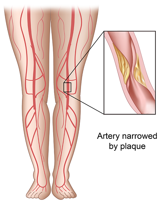
Pathological constriction refers to an abnormal narrowing or tightening of a body passage or organ, which can interfere with the normal flow of blood, air, or other substances through the area. This constriction can occur due to various reasons such as inflammation, scarring, or abnormal growths, and can affect different parts of the body, including blood vessels, airways, intestines, and ureters. Pathological constriction can lead to a range of symptoms and complications depending on its location and severity, and may require medical intervention to correct.
Coronary artery disease, often simply referred to as coronary disease, is a condition in which the blood vessels that supply oxygen-rich blood to the heart become narrowed or blocked due to the buildup of fatty deposits called plaques. This can lead to chest pain (angina), shortness of breath, or in severe cases, a heart attack.
The medical definition of coronary artery disease is:
A condition characterized by the accumulation of atheromatous plaques in the walls of the coronary arteries, leading to decreased blood flow and oxygen supply to the myocardium (heart muscle). This can result in symptoms such as angina pectoris, shortness of breath, or arrhythmias, and may ultimately lead to myocardial infarction (heart attack) or heart failure.
Risk factors for coronary artery disease include age, smoking, high blood pressure, high cholesterol, diabetes, obesity, physical inactivity, and a family history of the condition. Lifestyle changes such as quitting smoking, exercising regularly, eating a healthy diet, and managing stress can help reduce the risk of developing coronary artery disease. Medical treatments may include medications to control blood pressure, cholesterol levels, or irregular heart rhythms, as well as procedures such as angioplasty or bypass surgery to improve blood flow to the heart.
Coronary angiography is a medical procedure that uses X-ray imaging to visualize the coronary arteries, which supply blood to the heart muscle. During the procedure, a thin, flexible catheter is inserted into an artery in the arm or groin and threaded through the blood vessels to the heart. A contrast dye is then injected through the catheter, and X-ray images are taken as the dye flows through the coronary arteries. These images can help doctors diagnose and treat various heart conditions, such as blockages or narrowing of the arteries, that can lead to chest pain or heart attacks. It is also known as coronary arteriography or cardiac catheterization.
Treatment outcome is a term used to describe the result or effect of medical treatment on a patient's health status. It can be measured in various ways, such as through symptoms improvement, disease remission, reduced disability, improved quality of life, or survival rates. The treatment outcome helps healthcare providers evaluate the effectiveness of a particular treatment plan and make informed decisions about future care. It is also used in clinical research to compare the efficacy of different treatments and improve patient care.
Follow-up studies are a type of longitudinal research that involve repeated observations or measurements of the same variables over a period of time, in order to understand their long-term effects or outcomes. In medical context, follow-up studies are often used to evaluate the safety and efficacy of medical treatments, interventions, or procedures.
In a typical follow-up study, a group of individuals (called a cohort) who have received a particular treatment or intervention are identified and then followed over time through periodic assessments or data collection. The data collected may include information on clinical outcomes, adverse events, changes in symptoms or functional status, and other relevant measures.
The results of follow-up studies can provide important insights into the long-term benefits and risks of medical interventions, as well as help to identify factors that may influence treatment effectiveness or patient outcomes. However, it is important to note that follow-up studies can be subject to various biases and limitations, such as loss to follow-up, recall bias, and changes in clinical practice over time, which must be carefully considered when interpreting the results.
Myocardial infarction (MI), also known as a heart attack, is a medical condition characterized by the death of a segment of heart muscle (myocardium) due to the interruption of its blood supply. This interruption is most commonly caused by the blockage of a coronary artery by a blood clot formed on the top of an atherosclerotic plaque, which is a buildup of cholesterol and other substances in the inner lining of the artery.
The lack of oxygen and nutrients supply to the heart muscle tissue results in damage or death of the cardiac cells, causing the affected area to become necrotic. The extent and severity of the MI depend on the size of the affected area, the duration of the occlusion, and the presence of collateral circulation.
Symptoms of a myocardial infarction may include chest pain or discomfort, shortness of breath, nausea, lightheadedness, and sweating. Immediate medical attention is necessary to restore blood flow to the affected area and prevent further damage to the heart muscle. Treatment options for MI include medications, such as thrombolytics, antiplatelet agents, and pain relievers, as well as procedures such as percutaneous coronary intervention (PCI) or coronary artery bypass grafting (CABG).
Arterial occlusive diseases are medical conditions characterized by the blockage or narrowing of the arteries, which can lead to a reduction in blood flow to various parts of the body. This reduction in blood flow can cause tissue damage and may result in serious complications such as tissue death (gangrene), organ dysfunction, or even death.
The most common cause of arterial occlusive diseases is atherosclerosis, which is the buildup of plaque made up of fat, cholesterol, calcium, and other substances in the inner lining of the artery walls. Over time, this plaque can harden and narrow the arteries, restricting blood flow. Other causes of arterial occlusive diseases include blood clots, emboli (tiny particles that travel through the bloodstream and lodge in smaller vessels), inflammation, trauma, and certain inherited conditions.
Symptoms of arterial occlusive diseases depend on the location and severity of the blockage. Common symptoms include:
* Pain, cramping, or fatigue in the affected limb, often triggered by exercise and relieved by rest (claudication)
* Numbness, tingling, or weakness in the affected limb
* Coldness or discoloration of the skin in the affected area
* Slow-healing sores or wounds on the toes, feet, or legs
* Erectile dysfunction in men
Treatment for arterial occlusive diseases may include lifestyle changes such as quitting smoking, exercising regularly, and eating a healthy diet. Medications to lower cholesterol, control blood pressure, prevent blood clots, or manage pain may also be prescribed. In severe cases, surgical procedures such as angioplasty, stenting, or bypass surgery may be necessary to restore blood flow.
The iliac arteries are major branches of the abdominal aorta, the large artery that carries oxygen-rich blood from the heart to the rest of the body. The iliac arteries divide into two branches, the common iliac arteries, which further bifurcate into the internal and external iliac arteries.
The internal iliac artery supplies blood to the lower abdomen, pelvis, and the reproductive organs, while the external iliac artery provides blood to the lower extremities, including the legs and feet. Together, the iliac arteries play a crucial role in circulating blood throughout the body, ensuring that all tissues and organs receive the oxygen and nutrients they need to function properly.
Vascular patency is a term used in medicine to describe the state of a blood vessel (such as an artery or vein) being open, unobstructed, and allowing for the normal flow of blood. It is an important concept in the treatment and management of various cardiovascular conditions, such as peripheral artery disease, coronary artery disease, and deep vein thrombosis.
Maintaining vascular patency can help prevent serious complications like tissue damage, organ dysfunction, or even death. This may involve medical interventions such as administering blood-thinning medications to prevent clots, performing procedures to remove blockages, or using devices like stents to keep vessels open. Regular monitoring of vascular patency is also crucial for evaluating the effectiveness of treatments and adjusting care plans accordingly.
Atherectomy, coronary, is a medical procedure used to treat narrowed or blocked coronary arteries due to the buildup of plaque (atherosclerosis). The goal of coronary atherectomy is to improve blood flow to the heart muscle by removing the obstructive material within the vessel.
During the procedure, a specialized catheter with a cutting device on its tip is inserted into a peripheral artery, usually in the groin or arm, and advanced to the affected coronary artery. The cutting device can be a rotating blade, a high-speed spinning burr, or a laser fiber that is used to shave, drill, or vaporize the plaque, respectively. The removed material is collected in a chamber within the catheter or washed away by blood flow.
There are different types of coronary atherectomy devices, including:
1. Directional atherectomy (DCA): A rotating blade cuts and removes the plaque in a targeted direction.
2. Rotational atherectomy (Rotablator): A high-speed spinning burr is used to abrade and pulverize the plaque into tiny particles that can be safely carried away by blood flow.
3. Laser atherectomy: A laser fiber is used to vaporize or break down the plaque into gaseous or small particle form.
Coronary atherectomy is typically performed in conjunction with angioplasty and stenting, as it helps prepare the narrowed artery for these procedures by creating a larger lumen and reducing the risk of complications like dissections or restenosis (re-narrowing). However, its use may be limited to specific cases due to the potential risks, such as vessel trauma, distal embolization, or perforation.
It is essential to consult with a medical professional for detailed information and personalized treatment recommendations regarding coronary atherectomy.
Coronary vessels refer to the network of blood vessels that supply oxygenated blood and nutrients to the heart muscle, also known as the myocardium. The two main coronary arteries are the left main coronary artery and the right coronary artery.
The left main coronary artery branches off into the left anterior descending artery (LAD) and the left circumflex artery (LCx). The LAD supplies blood to the front of the heart, while the LCx supplies blood to the side and back of the heart.
The right coronary artery supplies blood to the right lower part of the heart, including the right atrium and ventricle, as well as the back of the heart.
Coronary vessel disease (CVD) occurs when these vessels become narrowed or blocked due to the buildup of plaque, leading to reduced blood flow to the heart muscle. This can result in chest pain, shortness of breath, or a heart attack.
The femoral artery is the major blood vessel that supplies oxygenated blood to the lower extremity of the human body. It is a continuation of the external iliac artery and becomes the popliteal artery as it passes through the adductor hiatus in the adductor magnus muscle of the thigh.
The femoral artery is located in the femoral triangle, which is bound by the sartorius muscle anteriorly, the adductor longus muscle medially, and the biceps femoris muscle posteriorly. It can be easily palpated in the groin region, making it a common site for taking blood samples, measuring blood pressure, and performing surgical procedures such as femoral artery catheterization and bypass grafting.
The femoral artery gives off several branches that supply blood to the lower limb, including the deep femoral artery, the superficial femoral artery, and the profunda femoris artery. These branches provide blood to the muscles, bones, skin, and other tissues of the leg, ankle, and foot.
Graft occlusion in the context of vascular surgery refers to the complete or partial blockage of a blood vessel that has been surgically replaced or repaired with a graft. The graft can be made from either synthetic materials or autologous tissue (taken from another part of the patient's body).
Graft occlusion can occur due to various reasons, including:
1. Thrombosis: Formation of a blood clot within the graft, which can obstruct blood flow.
2. Intimal hyperplasia: Overgrowth of the inner lining (intima) of the graft or the adjacent native vessel, causing narrowing of the lumen and reducing blood flow.
3. Atherosclerosis: Deposition of cholesterol and other substances in the walls of the graft, leading to hardening and narrowing of the vessel.
4. Infection: Bacterial or fungal infection of the graft can cause inflammation, weakening, and ultimately occlusion of the graft.
5. Mechanical factors: Kinking, twisting, or compression of the graft can lead to obstruction of blood flow.
Graft occlusion is a significant complication following vascular surgery, as it can result in reduced perfusion to downstream tissues and organs, leading to ischemia (lack of oxygen supply) and potential tissue damage or loss.
In the field of medicine, "time factors" refer to the duration of symptoms or time elapsed since the onset of a medical condition, which can have significant implications for diagnosis and treatment. Understanding time factors is crucial in determining the progression of a disease, evaluating the effectiveness of treatments, and making critical decisions regarding patient care.
For example, in stroke management, "time is brain," meaning that rapid intervention within a specific time frame (usually within 4.5 hours) is essential to administering tissue plasminogen activator (tPA), a clot-busting drug that can minimize brain damage and improve patient outcomes. Similarly, in trauma care, the "golden hour" concept emphasizes the importance of providing definitive care within the first 60 minutes after injury to increase survival rates and reduce morbidity.
Time factors also play a role in monitoring the progression of chronic conditions like diabetes or heart disease, where regular follow-ups and assessments help determine appropriate treatment adjustments and prevent complications. In infectious diseases, time factors are crucial for initiating antibiotic therapy and identifying potential outbreaks to control their spread.
Overall, "time factors" encompass the significance of recognizing and acting promptly in various medical scenarios to optimize patient outcomes and provide effective care.
The popliteal artery is the continuation of the femoral artery that passes through the popliteal fossa, which is the area behind the knee. It is the major blood vessel that supplies oxygenated blood to the lower leg and foot. The popliteal artery divides into the anterior tibial artery and the tibioperoneal trunk at the lower border of the popliteus muscle. Any damage or blockage to this artery can result in serious health complications, including reduced blood flow to the leg and foot, which may lead to pain, cramping, numbness, or even tissue death (gangrene) if left untreated.
Coronary artery bypass surgery, also known as coronary artery bypass grafting (CABG), is a surgical procedure used to improve blood flow to the heart in patients with severe coronary artery disease. This condition occurs when the coronary arteries, which supply oxygen-rich blood to the heart muscle, become narrowed or blocked due to the buildup of fatty deposits, called plaques.
During CABG surgery, a healthy blood vessel from another part of the body is grafted, or attached, to the coronary artery, creating a new pathway for oxygen-rich blood to flow around the blocked or narrowed portion of the artery and reach the heart muscle. This bypass helps to restore normal blood flow and reduce the risk of angina (chest pain), shortness of breath, and other symptoms associated with coronary artery disease.
There are different types of CABG surgery, including traditional on-pump CABG, off-pump CABG, and minimally invasive CABG. The choice of procedure depends on various factors, such as the patient's overall health, the number and location of blocked arteries, and the presence of other medical conditions.
It is important to note that while CABG surgery can significantly improve symptoms and quality of life in patients with severe coronary artery disease, it does not cure the underlying condition. Lifestyle modifications, such as regular exercise, a healthy diet, smoking cessation, and medication therapy, are essential for long-term management and prevention of further progression of the disease.
Angiography is a medical procedure in which an x-ray image is taken to visualize the internal structure of blood vessels, arteries, or veins. This is done by injecting a radiopaque contrast agent (dye) into the blood vessel using a thin, flexible catheter. The dye makes the blood vessels visible on an x-ray image, allowing doctors to diagnose and treat various medical conditions such as blockages, narrowing, or malformations of the blood vessels.
There are several types of angiography, including:
* Cardiac angiography (also called coronary angiography) - used to examine the blood vessels of the heart
* Cerebral angiography - used to examine the blood vessels of the brain
* Peripheral angiography - used to examine the blood vessels in the limbs or other parts of the body.
Angiography is typically performed by a radiologist, cardiologist, or vascular surgeon in a hospital setting. It can help diagnose conditions such as coronary artery disease, aneurysms, and peripheral arterial disease, among others.
Angina pectoris is a medical term that describes chest pain or discomfort caused by an inadequate supply of oxygen-rich blood to the heart muscle. This condition often occurs due to coronary artery disease, where the coronary arteries become narrowed or blocked by the buildup of cholesterol, fatty deposits, and other substances, known as plaques. These blockages can reduce blood flow to the heart, causing ischemia (lack of oxygen) and leading to angina symptoms.
There are two primary types of angina: stable and unstable. Stable angina is predictable and usually occurs during physical exertion or emotional stress when the heart needs more oxygen-rich blood. The pain typically subsides with rest or after taking prescribed nitroglycerin medication, which helps widen the blood vessels and improve blood flow to the heart.
Unstable angina, on the other hand, is more severe and unpredictable. It can occur at rest, during sleep, or with minimal physical activity and may not be relieved by rest or nitroglycerin. Unstable angina is considered a medical emergency, as it could indicate an imminent heart attack.
Symptoms of angina pectoris include chest pain, pressure, tightness, or heaviness that typically radiates to the left arm, neck, jaw, or back. Shortness of breath, nausea, sweating, and fatigue may also accompany angina symptoms. Immediate medical attention is necessary if you experience chest pain or discomfort, especially if it's new, severe, or persistent, as it could be a sign of a more serious condition like a heart attack.
Thrombolytic therapy, also known as thrombolysis, is a medical treatment that uses medications called thrombolytics or fibrinolytics to dissolve or break down blood clots (thrombi) in blood vessels. These clots can obstruct the flow of blood to vital organs such as the heart, lungs, or brain, leading to serious conditions like myocardial infarction (heart attack), pulmonary embolism, or ischemic stroke.
The goal of thrombolytic therapy is to restore blood flow as quickly and efficiently as possible to prevent further damage to the affected organ and potentially save lives. Commonly used thrombolytic drugs include alteplase (tPA), reteplase, and tenecteplase. It's essential to administer these medications as soon as possible after the onset of symptoms for optimal treatment outcomes. However, there are risks associated with thrombolytic therapy, such as an increased chance of bleeding complications, which must be carefully weighed against its benefits in each individual case.
Prospective studies, also known as longitudinal studies, are a type of cohort study in which data is collected forward in time, following a group of individuals who share a common characteristic or exposure over a period of time. The researchers clearly define the study population and exposure of interest at the beginning of the study and follow up with the participants to determine the outcomes that develop over time. This type of study design allows for the investigation of causal relationships between exposures and outcomes, as well as the identification of risk factors and the estimation of disease incidence rates. Prospective studies are particularly useful in epidemiology and medical research when studying diseases with long latency periods or rare outcomes.
Carotid stenosis is a medical condition that refers to the narrowing or constriction of the lumen (inner space) of the carotid artery. The carotid arteries are major blood vessels that supply oxygenated blood to the head and neck. Carotid stenosis usually results from the buildup of plaque, made up of fat, cholesterol, calcium, and other substances, on the inner walls of the artery. This process is called atherosclerosis.
As the plaque accumulates, it causes the artery to narrow, reducing blood flow to the brain. Severe carotid stenosis can increase the risk of stroke, as a clot or debris from the plaque can break off and travel to the brain, blocking a smaller blood vessel and causing tissue damage or death.
Carotid stenosis is typically diagnosed through imaging tests such as ultrasound, CT angiography, or MRI angiography. Treatment options may include lifestyle modifications (such as quitting smoking, controlling blood pressure, and managing cholesterol levels), medications to reduce the risk of clots, or surgical procedures like endarterectomy or stenting to remove or bypass the blockage.
Tunica intima, also known as the intima layer, is the innermost layer of a blood vessel, including arteries and veins. It is in direct contact with the flowing blood and is composed of simple squamous endothelial cells that form a continuous, non-keratinized, stratified epithelium. These cells play a crucial role in maintaining vascular homeostasis by regulating the passage of molecules and immune cells between the blood and the vessel wall, as well as contributing to the maintenance of blood fluidity and preventing coagulation.
The tunica intima is supported by a thin layer of connective tissue called the basement membrane, which provides structural stability and anchorage for the endothelial cells. Beneath the basement membrane lies a loose network of elastic fibers and collagen, known as the internal elastic lamina, that separates the tunica intima from the middle layer, or tunica media.
In summary, the tunica intima is the innermost layer of blood vessels, primarily composed of endothelial cells and a basement membrane, which regulates various functions to maintain vascular homeostasis.
Aortic coarctation is a narrowing of the aorta, the largest blood vessel in the body that carries oxygen-rich blood from the heart to the rest of the body. This condition usually occurs in the part of the aorta that is just beyond where it arises from the left ventricle and before it divides into the iliac arteries.
In aortic coarctation, the narrowing can vary from mild to severe, and it can cause a variety of symptoms depending on the severity of the narrowing and the age of the individual. In newborns and infants with severe coarctation, symptoms may include difficulty breathing, poor feeding, and weak or absent femoral pulses (located in the groin area). Older children and adults with mild to moderate coarctation may not experience any symptoms until later in life, when high blood pressure, headaches, nosebleeds, leg cramps, or heart failure develop.
Aortic coarctation is typically diagnosed through physical examination, imaging tests such as echocardiography, CT angiography, or MRI, and sometimes cardiac catheterization. Treatment options include surgical repair or balloon dilation (also known as balloon angioplasty) to open the narrowed section of the aorta. If left untreated, aortic coarctation can lead to serious complications such as high blood pressure, heart failure, stroke, and rupture or dissection of the aorta.
Ischemia is the medical term used to describe a lack of blood flow to a part of the body, often due to blocked or narrowed blood vessels. This can lead to a shortage of oxygen and nutrients in the tissues, which can cause them to become damaged or die. Ischemia can affect many different parts of the body, including the heart, brain, legs, and intestines. Symptoms of ischemia depend on the location and severity of the blockage, but they may include pain, cramping, numbness, weakness, or coldness in the affected area. In severe cases, ischemia can lead to tissue death (gangrene) or organ failure. Treatment for ischemia typically involves addressing the underlying cause of the blocked blood flow, such as through medication, surgery, or lifestyle changes.
Renal artery obstruction is a medical condition that refers to the blockage or restriction of blood flow in the renal artery, which is the main vessel that supplies oxygenated and nutrient-rich blood to the kidneys. This obstruction can be caused by various factors, such as blood clots, atherosclerosis (the buildup of fats, cholesterol, and other substances in and on the artery walls), emboli (tiny particles or air bubbles that travel through the bloodstream and lodge in smaller vessels), or compressive masses like tumors.
The obstruction can lead to reduced kidney function, hypertension, and even kidney failure in severe cases. Symptoms may include high blood pressure, proteinuria (the presence of protein in the urine), hematuria (blood in the urine), and a decrease in kidney function as measured by serum creatinine levels. Diagnosis typically involves imaging studies like Doppler ultrasound, CT angiography, or magnetic resonance angiography to visualize the renal artery and assess the extent of the obstruction. Treatment options may include medications to control blood pressure and reduce kidney damage, as well as invasive procedures like angioplasty and stenting or surgical intervention to remove the obstruction and restore normal blood flow to the kidneys.
In medical terms, "retreatment" refers to the process of providing additional treatment or courses of therapy to an individual who has previously undergone a medical intervention but has not achieved the desired outcomes or has experienced a recurrence of symptoms. This may apply to various medical conditions and treatments, including dental procedures, cancer therapies, mental health treatments, and more.
In the context of dentistry, specifically endodontics (root canal treatment), retreatment is the process of repeating the root canal procedure on a tooth that has already been treated before. This may be necessary if the initial treatment was not successful in eliminating infection or if reinfection has occurred. The goal of retreatment is to preserve the natural tooth and alleviate any persistent pain or discomfort.
Retrospective studies, also known as retrospective research or looking back studies, are a type of observational study that examines data from the past to draw conclusions about possible causal relationships between risk factors and outcomes. In these studies, researchers analyze existing records, medical charts, or previously collected data to test a hypothesis or answer a specific research question.
Retrospective studies can be useful for generating hypotheses and identifying trends, but they have limitations compared to prospective studies, which follow participants forward in time from exposure to outcome. Retrospective studies are subject to biases such as recall bias, selection bias, and information bias, which can affect the validity of the results. Therefore, retrospective studies should be interpreted with caution and used primarily to generate hypotheses for further testing in prospective studies.
Atherectomy is a medical procedure in which the accumulated plaque or deposits in the inner lining of the artery (the endothelium) are removed using a specialized catheter with a cutting device on its tip. The goal of this procedure is to improve blood flow through the artery by physically removing the obstruction, as opposed to other procedures like angioplasty and stenting which use balloons and/or metal scaffolds to open up the artery.
There are several types of atherectomy devices available, including:
1. Directional atherectomy (DA): A rotating blade cuts and removes plaque from the artery wall into a collection chamber within the catheter.
2. Rotational atherectomy (RA): A high-speed burr-like device abrades and pulverizes the plaque, which is then carried away by blood flow.
3. Laser atherectomy: A laser beam vaporizes the plaque, turning it into gas that is absorbed or removed through irrigation.
4. Orbital atherectomy: A high-speed spinning diamond-coated crown abrades and removes plaque while minimizing the risk of damaging the artery wall.
Atherectomy can be an effective treatment option for peripheral arterial disease (PAD) and coronary artery disease (CAD), particularly in cases where angioplasty and stenting are not feasible or have failed. However, like any medical procedure, atherectomy carries certain risks, such as bleeding, infection, perforation of the artery, and distal embolization (the release of plaque particles downstream). Proper patient selection, careful technique, and close follow-up are essential for successful outcomes.
Catheterization is a medical procedure in which a catheter (a flexible tube) is inserted into the body to treat various medical conditions or for diagnostic purposes. The specific definition can vary depending on the area of medicine and the particular procedure being discussed. Here are some common types of catheterization:
1. Urinary catheterization: This involves inserting a catheter through the urethra into the bladder to drain urine. It is often performed to manage urinary retention, monitor urine output in critically ill patients, or assist with surgical procedures.
2. Cardiac catheterization: A procedure where a catheter is inserted into a blood vessel, usually in the groin or arm, and guided to the heart. This allows for various diagnostic tests and treatments, such as measuring pressures within the heart chambers, assessing blood flow, or performing angioplasty and stenting of narrowed coronary arteries.
3. Central venous catheterization: A catheter is inserted into a large vein, typically in the neck, chest, or groin, to administer medications, fluids, or nutrition, or to monitor central venous pressure.
4. Peritoneal dialysis catheterization: A catheter is placed into the abdominal cavity for individuals undergoing peritoneal dialysis, a type of kidney replacement therapy.
5. Neurological catheterization: In some cases, a catheter may be inserted into the cerebrospinal fluid space (lumbar puncture) or the brain's ventricular system (ventriculostomy) to diagnose or treat various neurological conditions.
These are just a few examples of catheterization procedures in medicine. The specific definition and purpose will depend on the medical context and the particular organ or body system involved.
Coronary restenosis is the re-narrowing or re-occlusion of a coronary artery after a previous successful procedure to open or widen the artery, such as angioplasty or stenting. This narrowing is usually caused by the excessive growth of scar tissue or smooth muscle cells in the artery lining, which can occur spontaneously or as a response to the initial procedure. Restenosis can lead to recurrent symptoms of coronary artery disease, such as chest pain or shortness of breath, and may require additional medical intervention.
Intermittent claudication is a medical condition characterized by pain or cramping in the legs, usually in the calf muscles, that occurs during exercise or walking and is relieved by rest. This symptom is caused by insufficient blood flow to the working muscles due to peripheral artery disease (PAD), a narrowing or blockage of the arteries in the limbs. As the individual walks, the muscle demands for oxygen and nutrients increase, but the restricted blood supply cannot meet these demands, leading to ischemia (lack of oxygen) and pain. The pain typically subsides after a few minutes of rest, as the muscle's demand for oxygen decreases, allowing the limited blood flow to compensate. Regular exercise and medications may help improve symptoms and reduce the risk of complications associated with PAD.
Coronary circulation refers to the circulation of blood in the coronary vessels, which supply oxygenated blood to the heart muscle (myocardium) and drain deoxygenated blood from it. The coronary circulation system includes two main coronary arteries - the left main coronary artery and the right coronary artery - that branch off from the aorta just above the aortic valve. These arteries further divide into smaller branches, which supply blood to different regions of the heart muscle.
The left main coronary artery divides into two branches: the left anterior descending (LAD) artery and the left circumflex (LCx) artery. The LAD supplies blood to the front and sides of the heart, while the LCx supplies blood to the back and sides of the heart. The right coronary artery supplies blood to the lower part of the heart, including the right ventricle and the bottom portion of the left ventricle.
The veins that drain the heart muscle include the great cardiac vein, the middle cardiac vein, and the small cardiac vein, which merge to form the coronary sinus. The coronary sinus empties into the right atrium, allowing deoxygenated blood to enter the right side of the heart and be pumped to the lungs for oxygenation.
Coronary circulation is essential for maintaining the health and function of the heart muscle, as it provides the necessary oxygen and nutrients required for proper contraction and relaxation of the myocardium. Any disruption or blockage in the coronary circulation system can lead to serious consequences, such as angina, heart attack, or even death.
Platelet aggregation inhibitors are a class of medications that prevent platelets (small blood cells involved in clotting) from sticking together and forming a clot. These drugs work by interfering with the ability of platelets to adhere to each other and to the damaged vessel wall, thereby reducing the risk of thrombosis (blood clot formation).
Platelet aggregation inhibitors are often prescribed for people who have an increased risk of developing blood clots due to various medical conditions such as atrial fibrillation, coronary artery disease, peripheral artery disease, stroke, or a history of heart attack. They may also be used in patients undergoing certain medical procedures, such as angioplasty and stenting, to prevent blood clot formation in the stents.
Examples of platelet aggregation inhibitors include:
1. Aspirin: A nonsteroidal anti-inflammatory drug (NSAID) that irreversibly inhibits the enzyme cyclooxygenase, which is involved in platelet activation and aggregation.
2. Clopidogrel (Plavix): A P2Y12 receptor antagonist that selectively blocks ADP-induced platelet activation and aggregation.
3. Prasugrel (Effient): A third-generation thienopyridine P2Y12 receptor antagonist, similar to clopidogrel but with faster onset and greater potency.
4. Ticagrelor (Brilinta): A direct-acting P2Y12 receptor antagonist that does not require metabolic activation and has a reversible binding profile.
5. Dipyridamole (Persantine): An antiplatelet agent that inhibits platelet aggregation by increasing cyclic adenosine monophosphate (cAMP) levels in platelets, which leads to decreased platelet reactivity.
6. Iloprost (Ventavis): A prostacyclin analogue that inhibits platelet aggregation and causes vasodilation, often used in the treatment of pulmonary arterial hypertension.
7. Cilostazol (Pletal): A phosphodiesterase III inhibitor that increases cAMP levels in platelets, leading to decreased platelet activation and aggregation, as well as vasodilation.
8. Ticlopidine (Ticlid): An older P2Y12 receptor antagonist with a slower onset of action and more frequent side effects compared to clopidogrel or prasugrel.
In medical terms, the leg refers to the lower portion of the human body that extends from the knee down to the foot. It includes the thigh (femur), lower leg (tibia and fibula), foot, and ankle. The leg is primarily responsible for supporting the body's weight and enabling movements such as standing, walking, running, and jumping.
The leg contains several important structures, including bones, muscles, tendons, ligaments, blood vessels, nerves, and joints. These structures work together to provide stability, support, and mobility to the lower extremity. Common medical conditions that can affect the leg include fractures, sprains, strains, infections, peripheral artery disease, and neurological disorders.
Interventional ultrasonography is a medical procedure that involves the use of real-time ultrasound imaging to guide minimally invasive diagnostic and therapeutic interventions. This technique combines the advantages of ultrasound, such as its non-ionizing nature (no radiation exposure), relatively low cost, and portability, with the ability to perform precise and targeted procedures.
In interventional ultrasonography, a specialized physician called an interventional radiologist or an interventional sonographer uses high-frequency sound waves to create detailed images of internal organs and tissues. These images help guide the placement of needles, catheters, or other instruments used during the procedure. Common interventions include biopsies (tissue sampling), fluid drainage, tumor ablation, and targeted drug delivery.
The real-time visualization provided by ultrasonography allows for increased accuracy and safety during these procedures, minimizing complications and reducing recovery time compared to traditional surgical approaches. Additionally, interventional ultrasonography can be performed on an outpatient basis, further contributing to its appeal as a less invasive alternative in many clinical scenarios.
Carotid endarterectomy is a surgical procedure to remove plaque buildup (atherosclerosis) from the carotid arteries, which are the major blood vessels that supply oxygen-rich blood to the brain. The surgery involves making an incision in the neck, opening the carotid artery, and removing the plaque from the inside of the artery wall. The goal of the procedure is to restore normal blood flow to the brain and reduce the risk of stroke caused by the narrowing or blockage of the carotid arteries.
Beta particles, also known as beta rays, are a type of ionizing radiation that consist of high-energy electrons or positrons emitted from the nucleus of certain radioactive isotopes during their decay process. When a neutron in the nucleus decays into a proton, it results in an excess energy state and one electron is ejected from the atom at high speed. This ejected electron is referred to as a beta particle.
Beta particles can have both positive and negative charges, depending on the type of decay process. Negative beta particles (β−) are equivalent to electrons, while positive beta particles (β+) are equivalent to positrons. They possess kinetic energy that varies in range, with higher energies associated with greater penetrating power.
Beta particles can cause ionization and excitation of atoms and molecules they encounter, leading to chemical reactions and potential damage to living tissues. Therefore, appropriate safety measures must be taken when handling materials that emit beta radiation.
Hirudin therapy, also known as leech therapy, is a type of treatment that uses the saliva of medicinal leeches (Hirudo medicinalis) to alleviate symptoms and promote healing. The saliva of these leeches contains various bioactive compounds, including hirudin, which is a potent anticoagulant that prevents blood clotting.
In hirudin therapy, leeches are applied to specific areas of the body, usually on congested tissues or sites of stasis, where they feed on the patient's blood and release their saliva into the bite site. The hirudin in the saliva helps to dissolve blood clots, improve circulation, reduce swelling, and relieve pain.
Hirudin therapy is used in various medical conditions, such as arterial and venous insufficiency, skin ulcers, joint diseases, and post-surgical recovery, particularly after reconstructive surgery or organ transplantation. It can also be used to treat thrombophlebitis, varicose veins, and other circulatory disorders.
It is essential to note that hirudin therapy should only be performed by trained medical professionals in a controlled environment due to the potential risks associated with infection transmission and bleeding complications.
Coronary thrombosis is a medical condition that refers to the formation of a blood clot (thrombus) inside a coronary artery, which supplies oxygenated blood to the heart muscle. The development of a thrombus can partially or completely obstruct blood flow, leading to insufficient oxygen supply to the heart muscle. This can cause chest pain (angina) or a heart attack (myocardial infarction), depending on the severity and duration of the blockage.
Coronary thrombosis often results from the rupture of an atherosclerotic plaque, a buildup of cholesterol, fat, calcium, and other substances in the inner lining (endothelium) of the coronary artery. The ruptured plaque exposes the underlying tissue to the bloodstream, triggering the coagulation cascade and resulting in the formation of a thrombus.
Immediate medical attention is crucial for managing coronary thrombosis, as timely treatment can help restore blood flow, prevent further damage to the heart muscle, and reduce the risk of complications such as heart failure or life-threatening arrhythmias. Treatment options may include medications, such as antiplatelet agents, anticoagulants, and thrombolytic drugs, or interventional procedures like angioplasty and stenting to open the blocked artery. In some cases, surgical intervention, such as coronary artery bypass grafting (CABG), may be necessary.
Unstable angina is a term used in cardiology to describe chest pain or discomfort that occurs suddenly and unexpectedly, often at rest or with minimal physical exertion. It is caused by an insufficient supply of oxygen-rich blood to the heart muscle due to reduced blood flow, typically as a result of partial or complete blockage of the coronary arteries.
Unlike stable angina, which tends to occur predictably during physical activity and can be relieved with rest or nitroglycerin, unstable angina is more severe, unpredictable, and may not respond to traditional treatments. It is considered a medical emergency because it can be a sign of an impending heart attack or other serious cardiac event.
Unstable angina is often treated in the hospital with medications such as nitroglycerin, beta blockers, calcium channel blockers, and antiplatelet agents to improve blood flow to the heart and prevent further complications. In some cases, more invasive treatments such as coronary angioplasty or bypass surgery may be necessary to restore blood flow to the affected areas of the heart.
Intracranial arteriosclerosis is a medical condition characterized by the thickening and hardening of the walls of the intracranial arteries, which are the blood vessels that supply blood to the brain. This process is caused by the buildup of plaque, made up of fat, cholesterol, and other substances, within the walls of the arteries.
Intracranial arteriosclerosis can lead to a narrowing or blockage of the affected arteries, reducing blood flow to the brain. This can result in various neurological symptoms, such as headaches, dizziness, seizures, and transient ischemic attacks (TIAs) or strokes.
The condition is more common in older adults, particularly those with a history of hypertension, diabetes, smoking, and high cholesterol levels. Intracranial arteriosclerosis can be diagnosed through imaging tests such as magnetic resonance angiography (MRA) or computed tomographic angiography (CTA). Treatment typically involves managing risk factors and may include medications to control blood pressure, cholesterol levels, and prevent blood clots. In severe cases, surgical procedures such as angioplasty and stenting may be necessary to open up the affected arteries.
Myocardial revascularization is a medical term that refers to the restoration of blood flow to the heart muscle (myocardium), typically through a surgical or interventional procedure. This is often performed in patients with coronary artery disease, where the buildup of plaque in the coronary arteries restricts blood flow to the heart muscle, causing symptoms such as chest pain (angina) or shortness of breath, and increasing the risk of a heart attack (myocardial infarction).
There are two main types of myocardial revascularization:
1. Coronary artery bypass grafting (CABG): This is a surgical procedure in which a healthy blood vessel from another part of the body is used to create a detour around the blocked or narrowed coronary artery, allowing blood to flow more freely to the heart muscle.
2. Percutaneous coronary intervention (PCI), also known as angioplasty and stenting: This is a minimally invasive procedure in which a thin catheter is inserted into an artery in the groin or arm and threaded up to the blocked or narrowed coronary artery. A balloon is then inflated to widen the artery, and a stent may be placed to keep it open.
Both procedures aim to improve symptoms, reduce the risk of heart attack, and prolong survival in appropriately selected patients with coronary artery disease.
A reoperation is a surgical procedure that is performed again on a patient who has already undergone a previous operation for the same or related condition. Reoperations may be required due to various reasons, such as inadequate initial treatment, disease recurrence, infection, or complications from the first surgery. The nature and complexity of a reoperation can vary widely depending on the specific circumstances, but it often carries higher risks and potential complications compared to the original operation.
Peripheral Vascular Diseases (PVD) refer to a group of medical conditions that affect the blood vessels outside of the heart and brain. These diseases are characterized by a narrowing or blockage of the peripheral arteries, which can lead to reduced blood flow to the limbs, particularly the legs.
The primary cause of PVD is atherosclerosis, a buildup of fats, cholesterol, and other substances in and on the walls of the arteries, forming plaques that restrict blood flow. Other risk factors include smoking, diabetes, hypertension, high cholesterol levels, and a family history of vascular disease.
Symptoms of PVD can vary depending on the severity of the condition but may include leg pain or cramping during exercise (claudication), numbness or tingling in the legs, coldness or discoloration of the feet, sores or wounds that heal slowly or not at all, and in severe cases, gangrene.
PVD can increase the risk of heart attack and stroke, so it is essential to diagnose and treat the condition as early as possible. Treatment options include lifestyle changes such as quitting smoking, exercising regularly, and maintaining a healthy diet, medications to control symptoms and reduce the risk of complications, and surgical procedures such as angioplasty or bypass surgery to restore blood flow.
Limb salvage is a medical term used to describe the surgical procedures and treatments aimed at preserving and restoring the functionality of a severely injured or diseased limb, rather than amputating it. The goal of limb salvage is to improve the patient's quality of life by maintaining their mobility, independence, and overall well-being.
Limb salvage may involve various surgical techniques such as vascular reconstruction, bone realignment, muscle flap coverage, and external fixation. These procedures aim to restore blood flow, stabilize bones, cover exposed tissues, and prevent infection. Additionally, adjuvant therapies like hyperbaric oxygen treatment, physical therapy, and pain management may be employed to support the healing process and improve functional outcomes.
Limb salvage is typically considered when a limb is threatened by conditions such as severe trauma, tumors, infections, or peripheral arterial disease. The decision to pursue limb salvage over amputation depends on factors like the patient's overall health, age, and personal preferences, as well as the extent of the injury or disease, potential for recovery, and likelihood of successful rehabilitation.
Arteriosclerosis is a general term that describes the hardening and stiffening of the artery walls. It's a progressive condition that can occur as a result of aging, or it may be associated with certain risk factors such as high blood pressure, high cholesterol, diabetes, smoking, and a sedentary lifestyle.
The process of arteriosclerosis involves the buildup of plaque, made up of fat, cholesterol, calcium, and other substances, in the inner lining of the artery walls. Over time, this buildup can cause the artery walls to thicken and harden, reducing the flow of oxygen-rich blood to the body's organs and tissues.
Arteriosclerosis can affect any of the body's arteries, but it is most commonly found in the coronary arteries that supply blood to the heart, the cerebral arteries that supply blood to the brain, and the peripheral arteries that supply blood to the limbs. When arteriosclerosis affects the coronary arteries, it can lead to heart disease, angina, or heart attack. When it affects the cerebral arteries, it can lead to stroke or transient ischemic attack (TIA). When it affects the peripheral arteries, it can cause pain, numbness, or weakness in the limbs, and in severe cases, gangrene and amputation.
Postoperative complications refer to any unfavorable condition or event that occurs during the recovery period after a surgical procedure. These complications can vary in severity and may include, but are not limited to:
1. Infection: This can occur at the site of the incision or inside the body, such as pneumonia or urinary tract infection.
2. Bleeding: Excessive bleeding (hemorrhage) can lead to a drop in blood pressure and may require further surgical intervention.
3. Blood clots: These can form in the deep veins of the legs (deep vein thrombosis) and can potentially travel to the lungs (pulmonary embolism).
4. Wound dehiscence: This is when the surgical wound opens up, which can lead to infection and further complications.
5. Pulmonary issues: These include atelectasis (collapsed lung), pneumonia, or respiratory failure.
6. Cardiovascular problems: These include abnormal heart rhythms (arrhythmias), heart attack, or stroke.
7. Renal failure: This can occur due to various reasons such as dehydration, blood loss, or the use of certain medications.
8. Pain management issues: Inadequate pain control can lead to increased stress, anxiety, and decreased mobility.
9. Nausea and vomiting: These can be caused by anesthesia, opioid pain medication, or other factors.
10. Delirium: This is a state of confusion and disorientation that can occur in the elderly or those with certain medical conditions.
Prompt identification and management of these complications are crucial to ensure the best possible outcome for the patient.
Hyperplasia is a medical term that refers to an abnormal increase in the number of cells in an organ or tissue, leading to an enlargement of the affected area. It's a response to various stimuli such as hormones, chronic irritation, or inflammation. Hyperplasia can be physiological, like the growth of breast tissue during pregnancy, or pathological, like in the case of benign or malignant tumors. The process is generally reversible if the stimulus is removed. It's important to note that hyperplasia itself is not cancerous, but some forms of hyperplasia can increase the risk of developing cancer over time.
Ultrasonography, Doppler, and Duplex are diagnostic medical techniques that use sound waves to create images of internal body structures and assess their function. Here are the definitions for each:
1. Ultrasonography: Also known as ultrasound, this is a non-invasive imaging technique that uses high-frequency sound waves to produce images of internal organs and tissues. A small handheld device called a transducer is placed on the skin surface, which emits and receives sound waves. The returning echoes are then processed to create real-time visual images of the internal structures.
2. Doppler: This is a type of ultrasound that measures the velocity and direction of blood flow in the body by analyzing the frequency shift of the reflected sound waves. It can be used to assess blood flow in various parts of the body, such as the heart, arteries, and veins.
3. Duplex: Duplex ultrasonography is a combination of both gray-scale ultrasound and Doppler ultrasound. It provides detailed images of internal structures, as well as information about blood flow velocity and direction. This technique is often used to evaluate conditions such as deep vein thrombosis, carotid artery stenosis, and peripheral arterial disease.
In summary, ultrasonography is a diagnostic imaging technique that uses sound waves to create images of internal structures, Doppler is a type of ultrasound that measures blood flow velocity and direction, and duplex is a combination of both techniques that provides detailed images and information about blood flow.
Myocardial reperfusion is the restoration of blood flow to the heart muscle (myocardium), usually after a period of ischemia or reduced oxygen supply, such as during a myocardial infarction (heart attack). This can be achieved through various medical interventions, including thrombolytic therapy, percutaneous coronary intervention (PCI), or coronary artery bypass surgery (CABG). The goal of myocardial reperfusion is to salvage the jeopardized myocardium, preserve cardiac function, and reduce the risk of complications like heart failure or arrhythmias. However, it's important to note that while reperfusion is crucial for treating ischemic heart disease, it can also lead to additional injury to the heart muscle, known as reperfusion injury.
Renovascular hypertension is a type of secondary hypertension (high blood pressure) that is caused by renal artery stenosis or narrowing. This condition reduces blood flow to the kidneys, leading to the activation of the renin-angiotensin-aldosterone system (RAAS), which causes an increase in peripheral vascular resistance and blood volume, resulting in hypertension.
Renovascular hypertension is often seen in people with atherosclerosis or fibromuscular dysplasia, which are the most common causes of renal artery stenosis. Other conditions that can lead to renovascular hypertension include vasculitis, blood clots, and compression of the renal artery by nearby structures.
Diagnosis of renovascular hypertension typically involves imaging studies such as duplex ultrasound, CT angiography, or magnetic resonance angiography to visualize the renal arteries and assess for stenosis. Treatment may involve medications to control blood pressure, lifestyle modifications, and procedures such as angioplasty and stenting to open up the narrowed renal artery. In some cases, surgery may be necessary to restore blood flow to the kidney.
Fibrinolytic agents are medications that dissolve or break down blood clots by activating plasminogen, which is converted into plasmin. Plasmin is a proteolytic enzyme that degrades fibrin, the structural protein in blood clots. Fibrinolytic agents are used medically to treat conditions such as acute ischemic stroke, deep vein thrombosis, pulmonary embolism, and myocardial infarction (heart attack) by restoring blood flow in occluded vessels. Examples of fibrinolytic agents include alteplase, reteplase, and tenecteplase. It is important to note that these medications carry a risk of bleeding complications and should be administered with caution.
Cardiac catheterization is a medical procedure used to diagnose and treat cardiovascular conditions. In this procedure, a thin, flexible tube called a catheter is inserted into a blood vessel in the arm or leg and threaded up to the heart. The catheter can be used to perform various diagnostic tests, such as measuring the pressure inside the heart chambers and assessing the function of the heart valves.
Cardiac catheterization can also be used to treat certain cardiovascular conditions, such as narrowed or blocked arteries. In these cases, a balloon or stent may be inserted through the catheter to open up the blood vessel and improve blood flow. This procedure is known as angioplasty or percutaneous coronary intervention (PCI).
Cardiac catheterization is typically performed in a hospital cardiac catheterization laboratory by a team of healthcare professionals, including cardiologists, radiologists, and nurses. The procedure may be done under local anesthesia with sedation or general anesthesia, depending on the individual patient's needs and preferences.
Overall, cardiac catheterization is a valuable tool in the diagnosis and treatment of various heart conditions, and it can help improve symptoms, reduce complications, and prolong life for many patients.
Immunoglobulin (Ig) Fab fragments are the antigen-binding portions of an antibody that result from the digestion of the whole antibody molecule by enzymes such as papain. An antibody, also known as an immunoglobulin, is a Y-shaped protein produced by the immune system to identify and neutralize foreign substances like bacteria, viruses, or toxins. The antibody has two identical antigen-binding sites, located at the tips of the two shorter arms, which can bind specifically to a target antigen.
Fab fragments are formed when an antibody is cleaved by papain, resulting in two Fab fragments and one Fc fragment. Each Fab fragment contains one antigen-binding site, composed of a variable region (Fv) and a constant region (C). The Fv region is responsible for the specificity and affinity of the antigen binding, while the C region contributes to the effector functions of the antibody.
Fab fragments are often used in various medical applications, such as immunodiagnostics and targeted therapies, due to their ability to bind specifically to target antigens without triggering an immune response or other effector functions associated with the Fc region.
Coronary artery disease (CAD) is a medical condition in which the coronary arteries, which supply oxygen-rich blood to the heart muscle, become narrowed or blocked due to the buildup of cholesterol, fatty deposits, and other substances, known as plaque. Over time, this buildup can cause the arteries to harden and narrow (a process called atherosclerosis), reducing blood flow to the heart muscle.
The reduction in blood flow can lead to various symptoms and complications, including:
1. Angina (chest pain or discomfort) - This occurs when the heart muscle doesn't receive enough oxygen-rich blood, causing pain, pressure, or discomfort in the chest, arms, neck, jaw, or back.
2. Shortness of breath - When the heart isn't receiving adequate blood flow, it can't pump blood efficiently to meet the body's demands, leading to shortness of breath during physical activities or at rest.
3. Heart attack - If a piece of plaque ruptures or breaks off in a coronary artery, a blood clot can form and block the artery, causing a heart attack (myocardial infarction). This can damage or destroy part of the heart muscle.
4. Heart failure - Chronic reduced blood flow to the heart muscle can weaken it over time, leading to heart failure, a condition in which the heart can't pump blood efficiently to meet the body's needs.
5. Arrhythmias - Reduced blood flow and damage to the heart muscle can lead to abnormal heart rhythms (arrhythmias), which can be life-threatening if not treated promptly.
Coronary artery disease is typically diagnosed through a combination of medical history, physical examination, and diagnostic tests such as electrocardiograms (ECGs), stress testing, cardiac catheterization, and imaging studies like coronary computed tomography angiography (CCTA). Treatment options for CAD include lifestyle modifications, medications, medical procedures, and surgery.
Amputation is defined as the surgical removal of all or part of a limb or extremity such as an arm, leg, foot, hand, toe, or finger. This procedure is typically performed to remove damaged or dead tissue due to various reasons like severe injury, infection, tumors, or chronic conditions that impair circulation, such as diabetes or peripheral arterial disease. The goal of amputation is to alleviate pain, prevent further complications, and improve the patient's quality of life. Following the surgery, patients may require rehabilitation and prosthetic devices to help them adapt to their new physical condition.
An arteriovenous shunt is a surgically created connection between an artery and a vein. This procedure is typically performed to reroute blood flow or to provide vascular access for various medical treatments. In a surgical setting, the creation of an arteriovenous shunt involves connecting an artery directly to a vein, bypassing the capillary network in between.
There are different types of arteriovenous shunts used for specific medical purposes:
1. Arteriovenous Fistula (AVF): This is a surgical connection created between an artery and a vein, usually in the arm or leg. The procedure involves dissecting both the artery and vein, then suturing them directly together. Over time, the increased blood flow to the vein causes it to dilate and thicken, making it suitable for repeated needle punctures during hemodialysis treatments for patients with kidney failure.
2. Arteriovenous Graft (AVG): An arteriovenous graft is a synthetic tube used to connect an artery and a vein when a direct AVF cannot be created due to insufficient vessel size or poor quality. The graft can be made of various materials, such as polytetrafluoroethylene (PTFE) or Dacron. Grafts are more prone to infection and clotting compared to native AVFs but remain an essential option for patients requiring hemodialysis access.
3. Central Venous Catheter (CVC): A central venous catheter is a flexible tube inserted into a large vein, often in the neck or groin, and advanced towards the heart. CVCs can be used as temporary arteriovenous shunts for patients who require immediate hemodialysis access but do not have time to wait for an AVF or AVG to mature. However, they are associated with higher risks of infection and thrombosis compared to native AVFs and AVGs.
In summary, a surgical arteriovenous shunt is a connection between an artery and a vein established through a medical procedure. The primary purpose of these shunts is to provide vascular access for hemodialysis in patients with end-stage renal disease or to serve as temporary access when native AVFs or AVGs are not feasible.
Electrocardiography (ECG or EKG) is a medical procedure that records the electrical activity of the heart. It provides a graphic representation of the electrical changes that occur during each heartbeat. The resulting tracing, called an electrocardiogram, can reveal information about the heart's rate and rhythm, as well as any damage to its cells or abnormalities in its conduction system.
During an ECG, small electrodes are placed on the skin of the chest, arms, and legs. These electrodes detect the electrical signals produced by the heart and transmit them to a machine that amplifies and records them. The procedure is non-invasive, painless, and quick, usually taking only a few minutes.
ECGs are commonly used to diagnose and monitor various heart conditions, including arrhythmias, coronary artery disease, heart attacks, and electrolyte imbalances. They can also be used to evaluate the effectiveness of certain medications or treatments.
Treatment failure is a term used in medicine to describe the situation when a prescribed treatment or intervention is not achieving the desired therapeutic goals or objectives. This may occur due to various reasons, such as:
1. Development of drug resistance by the pathogen or disease being treated.
2. Inadequate dosage or frequency of the medication.
3. Poor adherence or compliance to the treatment regimen by the patient.
4. The presence of underlying conditions or comorbidities that may affect the efficacy of the treatment.
5. The severity or progression of the disease despite appropriate treatment.
When treatment failure occurs, healthcare providers may need to reassess the patient's condition and modify the treatment plan accordingly, which may include adjusting the dosage, changing the medication, adding new medications, or considering alternative treatments.
Medical Definition:
"Risk factors" are any attribute, characteristic or exposure of an individual that increases the likelihood of developing a disease or injury. They can be divided into modifiable and non-modifiable risk factors. Modifiable risk factors are those that can be changed through lifestyle choices or medical treatment, while non-modifiable risk factors are inherent traits such as age, gender, or genetic predisposition. Examples of modifiable risk factors include smoking, alcohol consumption, physical inactivity, and unhealthy diet, while non-modifiable risk factors include age, sex, and family history. It is important to note that having a risk factor does not guarantee that a person will develop the disease, but rather indicates an increased susceptibility.
Coronary stenosis is a medical condition that refers to the narrowing of the coronary arteries, which supply oxygen-rich blood to the heart muscle. This narrowing is typically caused by the buildup of plaque, made up of fat, cholesterol, and other substances, on the inner walls of the arteries. Over time, as the plaque hardens and calcifies, it can cause the artery to become narrowed or blocked, reducing blood flow to the heart muscle.
Coronary stenosis can lead to various symptoms and complications, including chest pain (angina), shortness of breath, irregular heart rhythms (arrhythmias), and heart attacks. Treatment options for coronary stenosis may include lifestyle changes, medications, medical procedures such as angioplasty or bypass surgery, or a combination of these approaches. Regular check-ups and diagnostic tests, such as stress testing or coronary angiography, can help detect and monitor coronary stenosis over time.
The saphenous vein is a term used in anatomical description to refer to the great or small saphenous veins, which are superficial veins located in the lower extremities of the human body.
The great saphenous vein (GSV) is the longest vein in the body and originates from the medial aspect of the foot, ascending along the medial side of the leg and thigh, and drains into the femoral vein at the saphenofemoral junction, located in the upper third of the thigh.
The small saphenous vein (SSV) is a shorter vein that originates from the lateral aspect of the foot, ascends along the posterior calf, and drains into the popliteal vein at the saphenopopliteal junction, located in the popliteal fossa.
These veins are often used as conduits for coronary artery bypass grafting (CABG) surgery due to their consistent anatomy and length.
The term "lower extremity" is used in the medical field to refer to the portion of the human body that includes the structures below the hip joint. This includes the thigh, lower leg, ankle, and foot. The lower extremities are responsible for weight-bearing and locomotion, allowing individuals to stand, walk, run, and jump. They contain many important structures such as bones, muscles, tendons, ligaments, nerves, and blood vessels.
Carotid artery injuries refer to damages or traumas that affect the carotid arteries, which are a pair of major blood vessels located in the neck that supply oxygenated blood to the head and neck. These injuries can occur due to various reasons such as penetrating or blunt trauma, iatrogenic causes (during medical procedures), or degenerative diseases.
Carotid artery injuries can be categorized into three types:
1. Blunt carotid injury (BCI): This type of injury is caused by a sudden and severe impact to the neck, which can result in intimal tears, dissection, or thrombosis of the carotid artery. BCIs are commonly seen in motor vehicle accidents, sports-related injuries, and assaults.
2. Penetrating carotid injury: This type of injury is caused by a foreign object that penetrates the neck and damages the carotid artery. Examples include gunshot wounds, stab wounds, or other sharp objects that pierce the skin and enter the neck.
3. Iatrogenic carotid injury: This type of injury occurs during medical procedures such as endovascular interventions, surgical procedures, or the placement of central lines.
Symptoms of carotid artery injuries may include:
* Stroke or transient ischemic attack (TIA)
* Neurological deficits such as hemiparesis, aphasia, or visual disturbances
* Bleeding from the neck or mouth
* Pulsatile mass in the neck
* Hypotension or shock
* Loss of consciousness
Diagnosis of carotid artery injuries may involve imaging studies such as computed tomography angiography (CTA), magnetic resonance angiography (MRA), or conventional angiography. Treatment options include endovascular repair, surgical repair, or anticoagulation therapy, depending on the severity and location of the injury.
Fibromuscular dysplasia (FMD) is a rare condition that affects the arterial walls, primarily in the medium and large-sized arteries. According to the American Heart Association, FMD is characterized by uneven growth or damage to the cells in the artery wall, leading to the formation of fibrous tissue and areas with narrowing (stenosis) or ballooning (aneurysm) of the artery.
FMD most commonly affects the renal (kidney) and carotid (neck) arteries but can also occur in other arteries, such as those in the abdomen, arms, and legs. The exact cause of FMD is unknown, but genetic factors and hormonal influences are believed to play a role.
Symptoms of FMD depend on which arteries are affected and may include high blood pressure, headaches, neck pain, dizziness, visual disturbances, or kidney problems. Diagnosis typically involves imaging tests like ultrasound, CT angiography, or magnetic resonance angiography (MRA). Treatment options for FMD include medications to manage symptoms and control high blood pressure, as well as various interventions such as angioplasty or stenting to open narrowed arteries.
Heparin is defined as a highly sulfated glycosaminoglycan (a type of polysaccharide) that is widely present in many tissues, but is most commonly derived from the mucosal tissues of mammalian lungs or intestinal mucosa. It is an anticoagulant that acts as an inhibitor of several enzymes involved in the blood coagulation cascade, primarily by activating antithrombin III which then neutralizes thrombin and other clotting factors.
Heparin is used medically to prevent and treat thromboembolic disorders such as deep vein thrombosis, pulmonary embolism, and certain types of heart attacks. It can also be used during hemodialysis, cardiac bypass surgery, and other medical procedures to prevent the formation of blood clots.
It's important to note that while heparin is a powerful anticoagulant, it does not have any fibrinolytic activity, meaning it cannot dissolve existing blood clots. Instead, it prevents new clots from forming and stops existing clots from growing larger.
Cineradiography is a medical imaging technique that combines fluoroscopy and cinematography to record moving images of the internal structures of a patient's body. It uses a special X-ray machine with a high-speed image intensifier and a movie camera or video recorder to capture real-time, dynamic visualizations of bodily functions such as swallowing, digestion, or muscle movements.
During cineradiography, a continuous X-ray beam is passed through the patient's body while the image intensifier converts the X-rays into visible light, which is then captured by the camera or video recorder. The resulting film or digital recordings can be played back in slow motion or frame by frame to analyze the movement and function of internal organs and structures.
Cineradiography has largely been replaced by newer imaging technologies such as CT and MRI, which offer higher resolution and more detailed images without the use of radiation. However, it is still used in some specialized applications where real-time, dynamic visualization is essential for diagnosis or treatment planning.
Blood vessel prosthesis implantation is a surgical procedure in which an artificial blood vessel, also known as a vascular graft or prosthetic graft, is inserted into the body to replace a damaged or diseased native blood vessel. The prosthetic graft can be made from various materials such as Dacron (polyester), PTFE (polytetrafluoroethylene), or bovine/human tissue.
The implantation of a blood vessel prosthesis is typically performed to treat conditions that cause narrowing or blockage of the blood vessels, such as atherosclerosis, aneurysms, or traumatic injuries. The procedure may be used to bypass blocked arteries in the legs (peripheral artery disease), heart (coronary artery bypass surgery), or neck (carotid endarterectomy). It can also be used to replace damaged veins for hemodialysis access in patients with kidney failure.
The success of blood vessel prosthesis implantation depends on various factors, including the patient's overall health, the location and extent of the vascular disease, and the type of graft material used. Possible complications include infection, bleeding, graft thrombosis (clotting), and graft failure, which may require further surgical intervention or endovascular treatments.
Angioscopy is a medical diagnostic procedure that uses a small fiber-optic scope, called an angioscope, to directly visualize the interior of blood vessels. The angioscope is inserted into the vessel through a small incision or catheter and allows physicians to examine the vessel walls for abnormalities such as plaque buildup, inflammation, or damage. This procedure can be used to diagnose and monitor conditions such as coronary artery disease, peripheral artery disease, and vasculitis. It can also be used during surgical procedures to assist with the placement of stents or other devices in the blood vessels.
The carotid arteries are a pair of vital blood vessels in the human body that supply oxygenated blood to the head and neck. Each person has two common carotid arteries, one on each side of the neck, which branch off from the aorta, the largest artery in the body.
The right common carotid artery originates from the brachiocephalic trunk, while the left common carotid artery arises directly from the aortic arch. As they ascend through the neck, they split into two main branches: the internal and external carotid arteries.
The internal carotid artery supplies oxygenated blood to the brain, eyes, and other structures within the skull, while the external carotid artery provides blood to the face, scalp, and various regions of the neck.
Maintaining healthy carotid arteries is crucial for overall cardiovascular health and preventing serious conditions like stroke, which can occur when the arteries become narrowed or blocked due to the buildup of plaque or fatty deposits (atherosclerosis). Regular check-ups with healthcare professionals may include monitoring carotid artery health through ultrasound or other imaging techniques.
'Alloys' is not a medical term. It is a term used in materials science and engineering to describe a mixture or solid solution composed of two or more elements, at least one of which is a metal. The components are typically present in significant amounts (>1% by weight). The properties of alloys, such as their strength, durability, and corrosion resistance, often differ from those of the constituent elements.
While not directly related to medicine, some alloys do have medical applications. For example, certain alloys are used in orthopedic implants, dental restorations, and other medical devices due to their desirable properties such as biocompatibility, strength, and resistance to corrosion.
A feasibility study is a preliminary investigation or analysis conducted to determine the viability of a proposed project, program, or product. In the medical field, feasibility studies are often conducted before implementing new treatments, procedures, equipment, or facilities. These studies help to assess the practicality and effectiveness of the proposed intervention, as well as its potential benefits and risks.
Feasibility studies in healthcare typically involve several steps:
1. Problem identification: Clearly define the problem that the proposed project, program, or product aims to address.
2. Objectives setting: Establish specific, measurable, achievable, relevant, and time-bound (SMART) objectives for the study.
3. Literature review: Conduct a thorough review of existing research and best practices related to the proposed intervention.
4. Methodology development: Design a methodology for data collection and analysis that will help answer the research questions and achieve the study's objectives.
5. Resource assessment: Evaluate the availability and adequacy of resources, including personnel, time, and finances, required to carry out the proposed intervention.
6. Risk assessment: Identify potential risks and challenges associated with the implementation of the proposed intervention and develop strategies to mitigate them.
7. Cost-benefit analysis: Estimate the costs and benefits of the proposed intervention, including direct and indirect costs, as well as short-term and long-term benefits.
8. Stakeholder engagement: Engage relevant stakeholders, such as patients, healthcare providers, administrators, and policymakers, to gather their input and support for the proposed intervention.
9. Decision-making: Based on the findings of the feasibility study, make an informed decision about whether or not to proceed with the proposed project, program, or product.
Feasibility studies are essential in healthcare as they help ensure that resources are allocated efficiently and effectively, and that interventions are evidence-based, safe, and beneficial for patients.
Thrombosis is the formation of a blood clot (thrombus) inside a blood vessel, obstructing the flow of blood through the circulatory system. When a clot forms in an artery, it can cut off the supply of oxygen and nutrients to the tissues served by that artery, leading to damage or tissue death. If a thrombus forms in the heart, it can cause a heart attack. If a thrombus breaks off and travels through the bloodstream, it can lodge in a smaller vessel, causing blockage and potentially leading to damage in the organ that the vessel supplies. This is known as an embolism.
Thrombosis can occur due to various factors such as injury to the blood vessel wall, abnormalities in blood flow, or changes in the composition of the blood. Certain medical conditions, medications, and lifestyle factors can increase the risk of thrombosis. Treatment typically involves anticoagulant or thrombolytic therapy to dissolve or prevent further growth of the clot, as well as addressing any underlying causes.
An embolism is a medical condition that occurs when a substance, such as a blood clot or an air bubble, blocks a blood vessel. This can happen in any part of the body, but it is particularly dangerous when it affects the brain (causing a stroke) or the lungs (causing a pulmonary embolism). Embolisms can cause serious harm by preventing oxygen and nutrients from reaching the tissues and organs that need them. They are often the result of underlying medical conditions, such as heart disease or deep vein thrombosis, and may require immediate medical attention to prevent further complications.
The renal artery is a pair of blood vessels that originate from the abdominal aorta and supply oxygenated blood to each kidney. These arteries branch into several smaller vessels that provide blood to the various parts of the kidneys, including the renal cortex and medulla. The renal arteries also carry nutrients and other essential components needed for the normal functioning of the kidneys. Any damage or blockage to the renal artery can lead to serious consequences, such as reduced kidney function or even kidney failure.
A smooth muscle within the vascular system refers to the involuntary, innervated muscle that is found in the walls of blood vessels. These muscles are responsible for controlling the diameter of the blood vessels, which in turn regulates blood flow and blood pressure. They are called "smooth" muscles because their individual muscle cells do not have the striations, or cross-striped patterns, that are observed in skeletal and cardiac muscle cells. Smooth muscle in the vascular system is controlled by the autonomic nervous system and by hormones, and can contract or relax slowly over a period of time.
Collateral circulation refers to the alternate blood supply routes that bypass an obstructed or narrowed vessel and reconnect with the main vascular system. These collateral vessels can develop over time as a result of the body's natural adaptation to chronic ischemia (reduced blood flow) caused by various conditions such as atherosclerosis, thromboembolism, or vasculitis.
The development of collateral circulation helps maintain adequate blood flow and oxygenation to affected tissues, minimizing the risk of tissue damage and necrosis. In some cases, well-developed collateral circulations can help compensate for significant blockages in major vessels, reducing symptoms and potentially preventing the need for invasive interventions like revascularization procedures. However, the extent and effectiveness of collateral circulation vary from person to person and depend on factors such as age, overall health status, and the presence of comorbidities.
Subclavian Steal Syndrome is a medical condition that occurs when there is a narrowing or blockage (stenosis) in the subclavian artery, usually at or near its origin from the aorta. This stenosis causes reduced blood flow to the ipsilateral upper extremity. The decreased blood supply to the arm leads to reversal of flow in the vertebral artery, which normally supplies blood to the brain and neck structures. As a result, the brain may receive insufficient blood flow, causing symptoms such as dizziness, lightheadedness, syncope (fainting), or transient ischemic attacks (TIAs or "mini-strokes").
The syndrome is called 'subclavian steal' because the vertebral artery essentially "steals" blood from the circle of Willis (the network of arteries at the base of the brain) to compensate for the reduced flow in the subclavian artery. The condition most commonly affects the left subclavian artery, but it can also occur on the right side or both sides.
Subclavian Steal Syndrome is typically diagnosed through a combination of physical examination, medical history, and imaging tests such as Doppler ultrasound, CT angiography (CTA), or magnetic resonance angiography (MRA). Treatment options include surgical bypass, endovascular stenting, or medication to manage symptoms and reduce the risk of stroke.
The tibial arteries are three major arteries that supply blood to the lower leg and foot. They are branches of the popliteal artery, which is a continuation of the femoral artery. The three tibial arteries are:
1. Anterior tibial artery: This artery runs down the front of the leg and supplies blood to the muscles in the anterior compartment of the leg, as well as to the foot. It becomes the dorsalis pedis artery as it approaches the ankle.
2. Posterior tibial artery: This artery runs down the back of the leg and supplies blood to the muscles in the posterior compartment of the leg. It then branches into the fibular (peroneal) artery and the medial and lateral plantar arteries, which supply blood to the foot.
3. Fibular (peroneal) artery: This artery runs down the outside of the leg and supplies blood to the muscles in the lateral compartment of the leg. It also provides branches that anastomose with the anterior and posterior tibial arteries, forming a network of vessels that helps ensure adequate blood flow to the foot.
Together, these arteries play a critical role in providing oxygenated blood and nutrients to the lower leg and foot, helping to maintain their health and function.
Vertebrobasilar insufficiency (VBI) is a medical condition characterized by inadequate blood flow to the vertebral and basilar arteries, which supply oxygenated blood to the brainstem and cerebellum. These arteries arise from the subclavian arteries and merge to form the basilar artery, which supplies critical structures in the posterior circulation of the brain.
VBI is often caused by atherosclerosis, or the buildup of plaque in the arterial walls, leading to narrowing (stenosis) or occlusion of these vessels. Other causes include embolism, arterial dissection, and vasculitis. The decreased blood flow can result in various neurological symptoms, such as dizziness, vertigo, imbalance, difficulty swallowing, slurred speech, visual disturbances, and even transient ischemic attacks (TIAs) or strokes.
Diagnosis of VBI typically involves a combination of clinical evaluation, imaging studies like MRA or CTA, and sometimes cerebral angiography to assess the extent and location of vascular narrowing or occlusion. Treatment options may include lifestyle modifications, medications to manage risk factors (such as hypertension, diabetes, or high cholesterol), antiplatelet therapy, or surgical interventions like endarterectomy or stenting in severe cases.
Hospital mortality is a term used to describe the number or rate of deaths that occur in a hospital setting during a specific period. It is often used as a measure of the quality of healthcare provided by a hospital, as a higher hospital mortality rate may indicate poorer care or more complex cases being treated. However, it's important to note that hospital mortality rates can be influenced by many factors, including the severity of illness of the patients being treated, patient demographics, and the availability of resources and specialized care. Therefore, hospital mortality rates should be interpreted with caution and in the context of other quality metrics.
Endarterectomy is a surgical procedure in which the inner lining of an artery (the endothelium) that has become thickened, damaged, or narrowed due to the buildup of fatty deposits, called plaques, is removed. This process helps restore normal blood flow through the artery and reduces the risk of serious complications such as stroke or limb loss.
The procedure typically involves making an incision in the affected artery, carefully removing the plaque and inner lining, and then closing the artery with sutures or a patch graft. Endarterectomy is most commonly performed on the carotid arteries in the neck, but it can also be done on other arteries throughout the body, including the femoral artery in the leg and the iliac artery in the pelvis.
Endarterectomy is usually recommended for patients with significant narrowing of their arteries who are experiencing symptoms such as pain, numbness, or weakness in their limbs, or who have a high risk of stroke due to carotid artery disease. The procedure is generally safe and effective, but like any surgery, it carries risks such as bleeding, infection, and damage to nearby nerves or tissues.
Life tables are statistical tools used in actuarial science, demography, and public health to estimate the mortality rate and survival rates of a population. They provide a data-driven representation of the probability that individuals of a certain age will die before their next birthday (the death rate) or live to a particular age (the survival rate).
Life tables are constructed using data on the number of deaths and the size of the population in specific age groups over a given period. These tables typically include several columns representing different variables, such as:
1. Age group or interval: The age range for which the data is being presented (e.g., 0-1 year, 1-5 years, 5-10 years, etc.).
2. Number of people in the population: The size of the population within each age group.
3. Number of deaths: The number of individuals who died during the study period within each age group.
4. Death rate: The probability that an individual in a given age group will die before their next birthday. It is calculated as the number of deaths divided by the size of the population for that age group.
5. Survival rate: The probability that an individual in a given age group will survive to a specific age or older. It is calculated using the death rates from earlier age groups.
6. Life expectancy: The average number of years a person is expected to live, based on their current age and mortality rates for each subsequent age group.
Life tables are essential in various fields, including insurance, pension planning, social security administration, and healthcare policy development. They help researchers and policymakers understand the health status and demographic trends of populations, allowing them to make informed decisions about resource allocation, program development, and public health interventions.
Anticoagulants are a class of medications that work to prevent the formation of blood clots in the body. They do this by inhibiting the coagulation cascade, which is a series of chemical reactions that lead to the formation of a clot. Anticoagulants can be given orally, intravenously, or subcutaneously, depending on the specific drug and the individual patient's needs.
There are several different types of anticoagulants, including:
1. Heparin: This is a naturally occurring anticoagulant that is often used in hospitalized patients who require immediate anticoagulation. It works by activating an enzyme called antithrombin III, which inhibits the formation of clots.
2. Low molecular weight heparin (LMWH): LMWH is a form of heparin that has been broken down into smaller molecules. It has a longer half-life than standard heparin and can be given once or twice daily by subcutaneous injection.
3. Direct oral anticoagulants (DOACs): These are newer oral anticoagulants that work by directly inhibiting specific clotting factors in the coagulation cascade. Examples include apixaban, rivaroxaban, and dabigatran.
4. Vitamin K antagonists: These are older oral anticoagulants that work by inhibiting the action of vitamin K, which is necessary for the formation of clotting factors. Warfarin is an example of a vitamin K antagonist.
Anticoagulants are used to prevent and treat a variety of conditions, including deep vein thrombosis (DVT), pulmonary embolism (PE), atrial fibrillation, and prosthetic heart valve thrombosis. It is important to note that anticoagulants can increase the risk of bleeding, so they must be used with caution and regular monitoring of blood clotting times may be required.
Polytetrafluoroethylene (PTFE) is not inherently a medical term, but it is a chemical compound with significant uses in the medical field. Medically, PTFE is often referred to by its brand name, Teflon. It is a synthetic fluoropolymer used in various medical applications due to its unique properties such as high resistance to heat, electrical and chemical interaction, and exceptional non-reactivity with body tissues.
PTFE can be found in medical devices like catheters, where it reduces friction, making insertion easier and minimizing trauma. It is also used in orthopedic and dental implants, drug delivery systems, and sutures due to its biocompatibility and non-adhesive nature.
A thrombectomy is a medical procedure that involves the removal of a blood clot (thrombus) from a blood vessel. This is typically performed to restore blood flow in cases where the clot is causing significant blockage, which can lead to serious complications such as tissue damage or organ dysfunction.
During a thrombectomy, a surgeon makes an incision and accesses the affected blood vessel, often with the help of imaging guidance. Specialized tools are then used to extract the clot, after which the blood vessel is usually repaired. Thrombectomies can be performed on various blood vessels throughout the body, including those in the brain, heart, lungs, and limbs.
This procedure may be recommended for patients with deep vein thrombosis (DVT), pulmonary embolism (PE), or certain types of stroke, depending on the specific circumstances and the patient's overall health. It is generally considered when anticoagulation therapy or clot-dissolving medications are not sufficient or appropriate to treat the blood clot.
A blood vessel prosthesis is a medical device that is used as a substitute for a damaged or diseased natural blood vessel. It is typically made of synthetic materials such as polyester, Dacron, or ePTFE (expanded polytetrafluoroethylene) and is designed to mimic the function of a native blood vessel by allowing the flow of blood through it.
Blood vessel prostheses are used in various surgical procedures, including coronary artery bypass grafting, peripheral arterial reconstruction, and the creation of arteriovenous fistulas for dialysis access. The choice of material and size of the prosthesis depends on several factors, such as the location and diameter of the vessel being replaced, the patient's age and overall health status, and the surgeon's preference.
It is important to note that while blood vessel prostheses can be effective in restoring blood flow, they may also carry risks such as infection, thrombosis (blood clot formation), and graft failure over time. Therefore, careful patient selection, surgical technique, and postoperative management are crucial for the success of these procedures.
Vascular surgical procedures are operations that are performed to treat conditions and diseases related to the vascular system, which includes the arteries, veins, and capillaries. These procedures can be invasive or minimally invasive and are often used to treat conditions such as peripheral artery disease, carotid artery stenosis, aortic aneurysms, and venous insufficiency.
Some examples of vascular surgical procedures include:
* Endarterectomy: a procedure to remove plaque buildup from the inside of an artery
* Bypass surgery: creating a new path for blood to flow around a blocked or narrowed artery
* Angioplasty and stenting: using a balloon to open a narrowed artery and placing a stent to keep it open
* Aneurysm repair: surgically repairing an aneurysm, a weakened area in the wall of an artery that has bulged out and filled with blood
* Embolectomy: removing a blood clot from a blood vessel
* Thrombectomy: removing a blood clot from a vein
These procedures are typically performed by vascular surgeons, who are trained in the diagnosis and treatment of vascular diseases.
Vascular access devices (VADs) are medical devices that are used to gain access to a patient's vascular system for the purpose of administering treatments, monitoring vital signs, or obtaining diagnostic samples. These devices can be categorized into short-term and long-term based on their intended duration of use.
Short-term VADs include peripheral intravenous catheters (PIVs), midline catheters, and peripherally inserted central catheters (PICCs). PIVs are thin, flexible tubes that are inserted into a vein in the arm or hand for short-term use. Midlines are similar to PIVs but are longer and can be used for up to 4 weeks. PICCs are inserted into a vein in the upper arm and threaded through to the larger veins near the heart, allowing for long-term access.
Long-term VADs include tunneled central venous catheters (CVCs), non-tunneled CVCs, and implanted ports. Tunneled CVCs are inserted into a large vein in the neck or chest and then threaded under the skin to an exit site, reducing the risk of infection. Non-tunneled CVCs are similar but do not have a tunnel, making them more prone to infection. Implanted ports are small devices that are surgically implanted under the skin, usually in the chest or arm, and connected to a catheter that is inserted into a large vein.
VADs can be used for various medical treatments such as chemotherapy, antibiotic therapy, parenteral nutrition, dialysis, and blood transfusions. Proper care and maintenance of VADs are essential to prevent complications such as infection, thrombosis, and catheter-related bloodstream infections (CRBSI).
A "patient transfer" is a medical procedure that involves moving a patient from one location, piece of medical equipment, or healthcare provider to another. This can include:
1. Transferring a patient from a bed to a stretcher, wheelchair, or other mobility device.
2. Moving a patient from a hospital bed to a surgical table or imaging machine such as an MRI or CT scanner.
3. Transporting a patient between healthcare facilities, such as from a hospital to a rehabilitation center or long-term care facility.
4. Transferring a patient between medical teams during the course of their treatment, like when they are moved from the emergency department to the intensive care unit.
Patient transfers require careful planning and execution to ensure the safety and comfort of the patient, as well as to prevent any potential injuries or complications for both the patient and the healthcare providers involved in the process. Proper techniques, equipment, and communication are essential for a successful patient transfer.
Digital subtraction angiography (DSA) is a medical imaging technique used to visualize the blood vessels and blood flow within the body. It combines the use of X-ray technology with digital image processing to produce detailed images of the vascular system.
In DSA, a contrast agent is injected into the patient's bloodstream through a catheter, which is typically inserted into an artery in the leg and guided to the area of interest using fluoroscopy. As the contrast agent flows through the blood vessels, X-ray images are taken at multiple time points.
The digital subtraction process involves taking a baseline image without contrast and then subtracting it from subsequent images taken with contrast. This allows for the removal of background structures and noise, resulting in clearer images of the blood vessels. DSA can be used to diagnose and evaluate various vascular conditions, such as aneurysms, stenosis, and tumors, and can also guide interventional procedures such as angioplasty and stenting.
Cineangiography is a medical imaging technique used to visualize the blood flow in the heart and cardiovascular system. It involves the injection of a contrast agent into the bloodstream while X-ray images are taken in quick succession, creating a movie-like sequence that shows the movement of the contrast through the blood vessels and chambers of the heart. This technique is often used to diagnose and evaluate various heart conditions, such as coronary artery disease, valvular heart disease, and congenital heart defects.
The procedure typically involves threading a catheter through a blood vessel in the arm or leg and guiding it to the heart. Once in place, the contrast agent is injected, and X-ray images are taken using a specialized X-ray machine called a fluoroscope. The images captured during cineangiography can help doctors identify areas of narrowing or blockage in the coronary arteries, abnormalities in heart valves, and other cardiovascular problems.
Cineangiography is an invasive procedure that carries some risks, such as bleeding, infection, and reactions to the contrast agent. However, it can provide valuable information for diagnosing and treating heart conditions, and may be recommended when other diagnostic tests have been inconclusive.
An emergency is a sudden, unexpected situation that requires immediate medical attention to prevent serious harm, permanent disability, or death. Emergencies can include severe injuries, trauma, cardiac arrest, stroke, difficulty breathing, severe allergic reactions, and other life-threatening conditions. In such situations, prompt medical intervention is necessary to stabilize the patient's condition, diagnose the underlying problem, and provide appropriate treatment.
Emergency medical services (EMS) are responsible for providing emergency care to patients outside of a hospital setting, such as in the home, workplace, or public place. EMS personnel include emergency medical technicians (EMTs), paramedics, and other first responders who are trained to assess a patient's condition, provide basic life support, and transport the patient to a hospital for further treatment.
In a hospital setting, an emergency department (ED) is a specialized unit that provides immediate care to patients with acute illnesses or injuries. ED staff includes physicians, nurses, and other healthcare professionals who are trained to handle a wide range of medical emergencies. The ED is equipped with advanced medical technology and resources to provide prompt diagnosis and treatment for critically ill or injured patients.
Overall, the goal of emergency medical care is to stabilize the patient's condition, prevent further harm, and provide timely and effective treatment to improve outcomes and save lives.
Survival analysis is a branch of statistics that deals with the analysis of time to event data. It is used to estimate the time it takes for a certain event of interest to occur, such as death, disease recurrence, or treatment failure. The event of interest is called the "failure" event, and survival analysis estimates the probability of not experiencing the failure event until a certain point in time, also known as the "survival" probability.
Survival analysis can provide important information about the effectiveness of treatments, the prognosis of patients, and the identification of risk factors associated with the event of interest. It can handle censored data, which is common in medical research where some participants may drop out or be lost to follow-up before the event of interest occurs.
Survival analysis typically involves estimating the survival function, which describes the probability of surviving beyond a certain time point, as well as hazard functions, which describe the instantaneous rate of failure at a given time point. Other important concepts in survival analysis include median survival times, restricted mean survival times, and various statistical tests to compare survival curves between groups.
Myocardial ischemia is a condition in which the blood supply to the heart muscle (myocardium) is reduced or blocked, leading to insufficient oxygen delivery and potential damage to the heart tissue. This reduction in blood flow typically results from the buildup of fatty deposits, called plaques, in the coronary arteries that supply the heart with oxygen-rich blood. The plaques can rupture or become unstable, causing the formation of blood clots that obstruct the artery and limit blood flow.
Myocardial ischemia may manifest as chest pain (angina pectoris), shortness of breath, fatigue, or irregular heartbeats (arrhythmias). In severe cases, it can lead to myocardial infarction (heart attack) if the oxygen supply is significantly reduced or cut off completely, causing permanent damage or death of the heart muscle. Early diagnosis and treatment of myocardial ischemia are crucial for preventing further complications and improving patient outcomes.
The internal carotid artery is a major blood vessel that supplies oxygenated blood to the brain. It originates from the common carotid artery and passes through the neck, entering the skull via the carotid canal in the temporal bone. Once inside the skull, it branches into several smaller vessels that supply different parts of the brain with blood.
The internal carotid artery is divided into several segments: cervical, petrous, cavernous, clinoid, and supraclinoid. Each segment has distinct clinical significance in terms of potential injury or disease. The most common conditions affecting the internal carotid artery include atherosclerosis, which can lead to stroke or transient ischemic attack (TIA), and dissection, which can cause severe headache, neck pain, and neurological symptoms.
It's important to note that any blockage or damage to the internal carotid artery can have serious consequences, as it can significantly reduce blood flow to the brain and lead to permanent neurological damage or even death. Therefore, regular check-ups and screening tests are recommended for individuals at high risk of developing vascular diseases.
Blood flow velocity is the speed at which blood travels through a specific part of the vascular system. It is typically measured in units of distance per time, such as centimeters per second (cm/s) or meters per second (m/s). Blood flow velocity can be affected by various factors, including cardiac output, vessel diameter, and viscosity of the blood. Measuring blood flow velocity is important in diagnosing and monitoring various medical conditions, such as heart disease, stroke, and peripheral vascular disease.
Risk assessment in the medical context refers to the process of identifying, evaluating, and prioritizing risks to patients, healthcare workers, or the community related to healthcare delivery. It involves determining the likelihood and potential impact of adverse events or hazards, such as infectious diseases, medication errors, or medical devices failures, and implementing measures to mitigate or manage those risks. The goal of risk assessment is to promote safe and high-quality care by identifying areas for improvement and taking action to minimize harm.
Aspirin is the common name for acetylsalicylic acid, which is a medication used to relieve pain, reduce inflammation, and lower fever. It works by inhibiting the activity of an enzyme called cyclooxygenase (COX), which is involved in the production of prostaglandins, hormone-like substances that cause inflammation and pain. Aspirin also has an antiplatelet effect, which means it can help prevent blood clots from forming. This makes it useful for preventing heart attacks and strokes.
Aspirin is available over-the-counter in various forms, including tablets, capsules, and chewable tablets. It is also available in prescription strengths for certain medical conditions. As with any medication, aspirin should be taken as directed by a healthcare provider, and its use should be avoided in children and teenagers with viral infections due to the risk of Reye's syndrome, a rare but serious condition that can affect the liver and brain.
Hirudin is not a medical term itself, but it is a specific substance with medical relevance. Hirudin is a naturally occurring anticoagulant that is found in the saliva of certain species of leeches (such as Hirudo medicinalis). This compound works by inhibiting the activity of thrombin, a key enzyme in the coagulation cascade, which ultimately results in preventing blood clot formation.
Medically, hirudin has been used in some research and therapeutic settings for its anticoagulant properties. For instance, recombinant hirudin (also known as lepirudin) is available for clinical use as an injectable anticoagulant to treat or prevent blood clots in specific medical conditions, such as heparin-induced thrombocytopenia (HIT).
In summary, Hirudins are a group of anticoagulant substances, primarily derived from leeches, that inhibit the activity of thrombin and have potential medical applications in preventing or treating blood clots.
Combined modality therapy (CMT) is a medical treatment approach that utilizes more than one method or type of therapy simultaneously or in close succession, with the goal of enhancing the overall effectiveness of the treatment. In the context of cancer care, CMT often refers to the combination of two or more primary treatment modalities, such as surgery, radiation therapy, and systemic therapies (chemotherapy, immunotherapy, targeted therapy, etc.).
The rationale behind using combined modality therapy is that each treatment method can target cancer cells in different ways, potentially increasing the likelihood of eliminating all cancer cells and reducing the risk of recurrence. The specific combination and sequence of treatments will depend on various factors, including the type and stage of cancer, patient's overall health, and individual preferences.
For example, a common CMT approach for locally advanced rectal cancer may involve preoperative (neoadjuvant) chemoradiation therapy, followed by surgery to remove the tumor, and then postoperative (adjuvant) chemotherapy. This combined approach allows for the reduction of the tumor size before surgery, increases the likelihood of complete tumor removal, and targets any remaining microscopic cancer cells with systemic chemotherapy.
It is essential to consult with a multidisciplinary team of healthcare professionals to determine the most appropriate CMT plan for each individual patient, considering both the potential benefits and risks associated with each treatment method.
Equipment design, in the medical context, refers to the process of creating and developing medical equipment and devices, such as surgical instruments, diagnostic machines, or assistive technologies. This process involves several stages, including:
1. Identifying user needs and requirements
2. Concept development and brainstorming
3. Prototyping and testing
4. Design for manufacturing and assembly
5. Safety and regulatory compliance
6. Verification and validation
7. Training and support
The goal of equipment design is to create safe, effective, and efficient medical devices that meet the needs of healthcare providers and patients while complying with relevant regulations and standards. The design process typically involves a multidisciplinary team of engineers, clinicians, designers, and researchers who work together to develop innovative solutions that improve patient care and outcomes.
Patient selection, in the context of medical treatment or clinical research, refers to the process of identifying and choosing appropriate individuals who are most likely to benefit from a particular medical intervention or who meet specific criteria to participate in a study. This decision is based on various factors such as the patient's diagnosis, stage of disease, overall health status, potential risks, and expected benefits. The goal of patient selection is to ensure that the selected individuals will receive the most effective and safe care possible while also contributing to meaningful research outcomes.
A Severity of Illness Index is a measurement tool used in healthcare to assess the severity of a patient's condition and the risk of mortality or other adverse outcomes. These indices typically take into account various physiological and clinical variables, such as vital signs, laboratory values, and co-morbidities, to generate a score that reflects the patient's overall illness severity.
Examples of Severity of Illness Indices include the Acute Physiology and Chronic Health Evaluation (APACHE) system, the Simplified Acute Physiology Score (SAPS), and the Mortality Probability Model (MPM). These indices are often used in critical care settings to guide clinical decision-making, inform prognosis, and compare outcomes across different patient populations.
It is important to note that while these indices can provide valuable information about a patient's condition, they should not be used as the sole basis for clinical decision-making. Rather, they should be considered in conjunction with other factors, such as the patient's overall clinical presentation, treatment preferences, and goals of care.
Medical survival rate is a statistical measure used to determine the percentage of patients who are still alive for a specific period of time after their diagnosis or treatment for a certain condition or disease. It is often expressed as a five-year survival rate, which refers to the proportion of people who are alive five years after their diagnosis. Survival rates can be affected by many factors, including the stage of the disease at diagnosis, the patient's age and overall health, the effectiveness of treatment, and other health conditions that the patient may have. It is important to note that survival rates are statistical estimates and do not necessarily predict an individual patient's prognosis.
Cerebral angiography is a medical procedure that involves taking X-ray images of the blood vessels in the brain after injecting a contrast dye into them. This procedure helps doctors to diagnose and treat various conditions affecting the blood vessels in the brain, such as aneurysms, arteriovenous malformations, and stenosis (narrowing of the blood vessels).
During the procedure, a catheter is inserted into an artery in the leg and threaded through the body to the blood vessels in the neck or brain. The contrast dye is then injected through the catheter, and X-ray images are taken to visualize the blood flow through the brain's blood vessels.
Cerebral angiography provides detailed images of the blood vessels in the brain, allowing doctors to identify any abnormalities or blockages that may be causing symptoms or increasing the risk of stroke. Based on the results of the cerebral angiography, doctors can develop a treatment plan to address these issues and prevent further complications.
The platelet glycoprotein GPIIb-IIIa complex, also known as integrin αIIbβ3 or CD41/CD61, is a heterodimeric transmembrane receptor found on the surface of platelets and megakaryocytes. It plays a crucial role in platelet aggregation and thrombus formation during hemostasis and pathological conditions such as arterial thrombosis.
The GPIIb-IIIa complex is composed of two non-covalently associated subunits, GPIIb (αIIb or CD41) and IIIa (β3 or CD61). Upon platelet activation by various agonists like ADP, thrombin, or collagen, the GPIIb-IIIa complex undergoes a conformational change that allows it to bind fibrinogen, von Willebrand factor, and other adhesive proteins. This binding event leads to platelet aggregation and the formation of a hemostatic plug or pathological thrombus.
Inhibition of the GPIIb-IIIa complex has been a target for antiplatelet therapy in the prevention and treatment of arterial thrombosis, such as myocardial infarction and stroke. Several pharmacological agents, including monoclonal antibodies and small molecule antagonists, have been developed to block this complex and reduce platelet aggregation.
The common carotid artery is a major blood vessel in the neck that supplies oxygenated blood to the head and neck. It originates from the brachiocephalic trunk or the aortic arch and divides into the internal and external carotid arteries at the level of the upper border of the thyroid cartilage. The common carotid artery is an important structure in the circulatory system, and any damage or blockage to it can have serious consequences, including stroke.
The subclavian artery is a major blood vessel that supplies the upper limb and important structures in the neck and head. It arises from the brachiocephalic trunk (in the case of the right subclavian artery) or directly from the aortic arch (in the case of the left subclavian artery).
The subclavian artery has several branches, including:
1. The vertebral artery, which supplies blood to the brainstem and cerebellum.
2. The internal thoracic artery (also known as the mammary artery), which supplies blood to the chest wall, breast, and anterior mediastinum.
3. The thyrocervical trunk, which gives rise to several branches that supply the neck, including the inferior thyroid artery, the suprascapular artery, and the transverse cervical artery.
4. The costocervical trunk, which supplies blood to the neck and upper back, including the posterior chest wall and the lower neck muscles.
The subclavian artery is a critical vessel in maintaining adequate blood flow to the upper limb, and any blockage or damage to this vessel can lead to significant morbidity, including arm pain, numbness, weakness, or even loss of function.
The radial artery is a key blood vessel in the human body, specifically a part of the peripheral arterial system. Originating from the brachial artery in the upper arm, the radial artery travels down the arm and crosses over the wrist, where it can be palpated easily. It then continues into the hand, dividing into several branches to supply blood to the hand's tissues and digits.
The radial artery is often used for taking pulse readings due to its easy accessibility at the wrist. Additionally, in medical procedures such as coronary angiography or bypass surgery, the radial artery can be utilized as a site for catheter insertion. This allows healthcare professionals to examine the heart's blood vessels and assess cardiovascular health.
Tissue Plasminogen Activator (tPA) is a thrombolytic enzyme, which means it dissolves blood clots. It is naturally produced by the endothelial cells that line the interior surface of blood vessels. tPA activates plasminogen, a zymogen, to convert it into plasmin, a protease that breaks down fibrin, the structural protein in blood clots. This enzyme is used medically as a thrombolytic drug under various brand names, such as Activase and Alteplase, to treat conditions like acute ischemic stroke, pulmonary embolism, and deep vein thrombosis by dissolving the clots and restoring blood flow.
Left ventricular function refers to the ability of the left ventricle (the heart's lower-left chamber) to contract and relax, thereby filling with and ejecting blood. The left ventricle is responsible for pumping oxygenated blood to the rest of the body. Its function is evaluated by measuring several parameters, including:
1. Ejection fraction (EF): This is the percentage of blood that is pumped out of the left ventricle with each heartbeat. A normal ejection fraction ranges from 55% to 70%.
2. Stroke volume (SV): The amount of blood pumped by the left ventricle in one contraction. A typical SV is about 70 mL/beat.
3. Cardiac output (CO): The total volume of blood that the left ventricle pumps per minute, calculated as the product of stroke volume and heart rate. Normal CO ranges from 4 to 8 L/minute.
Assessment of left ventricular function is crucial in diagnosing and monitoring various cardiovascular conditions such as heart failure, coronary artery disease, valvular heart diseases, and cardiomyopathies.
Premedication is the administration of medication before a medical procedure or surgery to prevent or manage pain, reduce anxiety, minimize side effects of anesthesia, or treat existing medical conditions. The goal of premedication is to improve the safety and outcomes of the medical procedure by preparing the patient's body in advance. Common examples of premedication include administering antibiotics before surgery to prevent infection, giving sedatives to help patients relax before a procedure, or providing medication to control acid reflux during surgery.
An intracranial embolism is a medical condition that occurs when a blood clot or other foreign material (embolus) forms elsewhere in the body and travels to the blood vessels within the brain. This embolus then blocks the flow of blood in the cerebral arteries, leading to potential damage or death of brain tissue. Common sources of intracranial emboli include heart conditions such as atrial fibrillation, valvular heart disease, or following a heart attack; or from large-vessel atherosclerosis in the carotid arteries. Symptoms can vary depending on the location and size of the obstruction, but may include sudden weakness or numbness, confusion, difficulty speaking, vision loss, severe headache, or even loss of consciousness. Immediate medical attention is required to diagnose and treat intracranial embolism, often involving anticoagulation therapy, endovascular procedures, or surgery.
Brachytherapy is a type of cancer treatment that involves placing radioactive material directly into or near the tumor site. The term "brachy" comes from the Greek word for "short," which refers to the short distance that the radiation travels. This allows for a high dose of radiation to be delivered directly to the tumor while minimizing exposure to healthy surrounding tissue.
There are two main types of brachytherapy:
1. Intracavitary brachytherapy: The radioactive material is placed inside a body cavity, such as the uterus or windpipe.
2. Interstitial brachytherapy: The radioactive material is placed directly into the tumor or surrounding tissue using needles, seeds, or catheters.
Brachytherapy can be used alone or in combination with other cancer treatments such as surgery, external beam radiation therapy, and chemotherapy. It may be recommended for a variety of cancers, including prostate, cervical, vaginal, vulvar, head and neck, and skin cancers. The specific type of brachytherapy used will depend on the size, location, and stage of the tumor.
The advantages of brachytherapy include its ability to deliver a high dose of radiation directly to the tumor while minimizing exposure to healthy tissue, which can result in fewer side effects compared to other forms of radiation therapy. Additionally, brachytherapy is often a shorter treatment course than external beam radiation therapy, with some treatments lasting only a few minutes or hours.
However, there are also potential risks and side effects associated with brachytherapy, including damage to nearby organs and tissues, bleeding, infection, and pain. Patients should discuss the benefits and risks of brachytherapy with their healthcare provider to determine if it is an appropriate treatment option for them.
Intraoperative complications refer to any unforeseen problems or events that occur during the course of a surgical procedure, once it has begun and before it is completed. These complications can range from minor issues, such as bleeding or an adverse reaction to anesthesia, to major complications that can significantly impact the patient's health and prognosis.
Examples of intraoperative complications include:
1. Bleeding (hemorrhage) - This can occur due to various reasons such as injury to blood vessels or organs during surgery.
2. Infection - Surgical site infections can develop if the surgical area becomes contaminated during the procedure.
3. Anesthesia-related complications - These include adverse reactions to anesthesia, difficulty maintaining the patient's airway, or cardiovascular instability.
4. Organ injury - Accidental damage to surrounding organs can occur during surgery, leading to potential long-term consequences.
5. Equipment failure - Malfunctioning surgical equipment can lead to complications and compromise the safety of the procedure.
6. Allergic reactions - Patients may have allergies to certain medications or materials used during surgery, causing an adverse reaction.
7. Prolonged operative time - Complications may arise if a surgical procedure takes longer than expected, leading to increased risk of infection and other issues.
Intraoperative complications require prompt identification and management by the surgical team to minimize their impact on the patient's health and recovery.
A randomized controlled trial (RCT) is a type of clinical study in which participants are randomly assigned to receive either the experimental intervention or the control condition, which may be a standard of care, placebo, or no treatment. The goal of an RCT is to minimize bias and ensure that the results are due to the intervention being tested rather than other factors. This design allows for a comparison between the two groups to determine if there is a significant difference in outcomes. RCTs are often considered the gold standard for evaluating the safety and efficacy of medical interventions, as they provide a high level of evidence for causal relationships between the intervention and health outcomes.
Cardiovascular agents are a class of medications that are used to treat various conditions related to the cardiovascular system, which includes the heart and blood vessels. These agents can be further divided into several subcategories based on their specific mechanisms of action and therapeutic effects. Here are some examples:
1. Antiarrhythmics: These drugs are used to treat abnormal heart rhythms or arrhythmias. They work by stabilizing the electrical activity of the heart and preventing irregular impulses from spreading through the heart muscle.
2. Antihypertensives: These medications are used to lower high blood pressure, also known as hypertension. There are several classes of antihypertensive drugs, including diuretics, beta-blockers, calcium channel blockers, and angiotensin-converting enzyme (ACE) inhibitors.
3. Anticoagulants: These drugs are used to prevent blood clots from forming or growing larger. They work by interfering with the coagulation cascade, which is a series of chemical reactions that lead to the formation of a blood clot.
4. Antiplatelet agents: These medications are used to prevent platelets in the blood from sticking together and forming clots. They work by inhibiting the aggregation of platelets, which are small cells in the blood that help form clots.
5. Lipid-lowering agents: These drugs are used to lower cholesterol and other fats in the blood. They work by reducing the production or absorption of cholesterol in the body or increasing the removal of cholesterol from the bloodstream. Examples include statins, bile acid sequestrants, and PCSK9 inhibitors.
6. Vasodilators: These medications are used to widen blood vessels and improve blood flow. They work by relaxing the smooth muscle in the walls of blood vessels, causing them to dilate or widen. Examples include nitrates, calcium channel blockers, and ACE inhibitors.
7. Inotropes: These drugs are used to increase the force of heart contractions. They work by increasing the sensitivity of heart muscle cells to calcium ions, which are necessary for muscle contraction.
These are just a few examples of cardiovascular medications that are used to treat various conditions related to the heart and blood vessels. It is important to note that these medications can have side effects and should be taken under the guidance of a healthcare provider.
A registry in the context of medicine is a collection or database of standardized information about individuals who share a certain condition or attribute, such as a disease, treatment, exposure, or demographic group. These registries are used for various purposes, including:
* Monitoring and tracking the natural history of diseases and conditions
* Evaluating the safety and effectiveness of medical treatments and interventions
* Conducting research and generating hypotheses for further study
* Providing information to patients, clinicians, and researchers
* Informing public health policy and decision-making
Registries can be established for a wide range of purposes, including disease-specific registries (such as cancer or diabetes registries), procedure-specific registries (such as joint replacement or cardiac surgery registries), and population-based registries (such as birth defects or cancer registries). Data collected in registries may include demographic information, clinical data, laboratory results, treatment details, and outcomes.
Registries can be maintained by a variety of organizations, including hospitals, clinics, academic medical centers, professional societies, government agencies, and industry. Participation in registries is often voluntary, although some registries may require informed consent from participants. Data collected in registries are typically de-identified to protect the privacy of individuals.
Interventional radiology (IR) is a subspecialty of radiology that uses minimally invasive image-guided procedures to diagnose and treat various medical conditions. The main goal of interventional radiology is to offer patients less invasive options for treatment, which can result in smaller incisions, reduced recovery time, and fewer complications compared to traditional open surgeries.
Interventional radiologists use a variety of imaging techniques, such as X-rays, fluoroscopy, computed tomography (CT), magnetic resonance imaging (MRI), and ultrasound, to guide catheters, wires, needles, and other small instruments through the body to target specific areas. These targeted interventions can be used for both diagnostic and therapeutic purposes, including:
1. Biopsies: Obtaining tissue samples from organs or tumors to determine a diagnosis.
2. Drainage procedures: Removing fluid from abscesses, cysts, or blocked areas to alleviate symptoms and promote healing.
3. Stent placements: Opening narrowed or obstructed blood vessels, bile ducts, or airways using small mesh tubes called stents.
4. Embolization: Blocking abnormal blood vessels or reducing blood flow to tumors, aneurysms, or other problematic areas.
5. Tumor ablation: Destroying tumors using heat (radiofrequency ablation, microwave ablation), cold (cryoablation), or other energy sources.
6. Pain management: Treating chronic pain by targeting specific nerves and blocking their transmission of pain signals.
7. Vascular access: Creating secure pathways to blood vessels for dialysis, chemotherapy, or other long-term treatments.
8. Aneurysm repair: Reinforcing weakened or bulging blood vessel walls using coils, stents, or flow diverters.
9. Vertebroplasty and kyphoplasty: Stabilizing fractured vertebrae in the spine to alleviate pain and improve mobility.
10. Uterine fibroid embolization: Reducing the size and symptoms of uterine fibroids by blocking their blood supply.
These are just a few examples of interventional radiology procedures. The field is constantly evolving, with new techniques and technologies being developed to improve patient care and outcomes. Interventional radiologists work closely with other medical specialists to provide minimally invasive treatment options for a wide range of conditions.
The Chi-square distribution is a continuous probability distribution that is often used in statistical hypothesis testing. It is the distribution of a sum of squares of k independent standard normal random variables. The resulting quantity follows a chi-square distribution with k degrees of freedom, denoted as χ²(k).
The probability density function (pdf) of the Chi-square distribution with k degrees of freedom is given by:
f(x; k) = (1/ (2^(k/2) * Γ(k/2))) \* x^((k/2)-1) \* e^(-x/2), for x > 0 and 0, otherwise.
Where Γ(k/2) is the gamma function evaluated at k/2. The mean and variance of a Chi-square distribution with k degrees of freedom are k and 2k, respectively.
The Chi-square distribution has various applications in statistical inference, including testing goodness-of-fit, homogeneity of variances, and independence in contingency tables.
I believe there may be some confusion in your question. "Rabbits" is a common name used to refer to the Lagomorpha species, particularly members of the family Leporidae. They are small mammals known for their long ears, strong legs, and quick reproduction.
However, if you're referring to "rabbits" in a medical context, there is a term called "rabbit syndrome," which is a rare movement disorder characterized by repetitive, involuntary movements of the fingers, resembling those of a rabbit chewing. It is also known as "finger-chewing chorea." This condition is usually associated with certain medications, particularly antipsychotics, and typically resolves when the medication is stopped or adjusted.
Peripheral Arterial Disease (PAD) is a medical condition characterized by the narrowing or blockage of arteries that supply blood to the extremities, most commonly the legs. This results in reduced blood flow, leading to symptoms such as leg pain, cramping, numbness, or weakness during physical activity, and in severe cases, tissue damage or gangrene. PAD is often indicative of widespread atherosclerosis, which is the hardening and narrowing of arteries due to the buildup of fatty deposits called plaques. It's important to note that early detection and management can help prevent serious complications.
Cholesterol embolism is a medical condition that occurs when cholesteral crystals or plaque debris from an atherosclerotic lesion in the aorta or its major branches dislodge and travel to smaller vessels, where they obstruct blood flow. This can lead to tissue damage or infarction in various organs, depending on the location of the embolism.
Cholesterol emboli are typically small, crystalline, and composed of cholesterol, calcium, and other debris from atherosclerotic plaques. They can cause inflammation and damage to the endothelial cells lining the blood vessels, leading to further narrowing or occlusion of the vessel lumen.
Symptoms of cholesterol embolism depend on the location and extent of the obstruction. Common sites for embolization include the kidneys, brain, eyes, skin, and extremities. Symptoms can range from mild to severe and may include sudden pain, weakness, or numbness in the affected area; skin discoloration or ulcerations; vision changes; kidney dysfunction; and stroke-like symptoms.
Cholesterol embolism is often a complication of invasive procedures such as angiography, coronary artery bypass grafting, or aortic surgery. It can also occur spontaneously in patients with advanced atherosclerosis or those who have recently undergone anticoagulation therapy.
Diagnosis of cholesterol embolism is often challenging due to its nonspecific symptoms and variable presentation. Imaging studies, such as angiography or CT scans, may be used to visualize the location and extent of the obstruction. Blood tests and biopsy of affected tissues can also provide diagnostic clues.
Treatment of cholesterol embolism is primarily supportive and aimed at managing symptoms and preventing further complications. Antiplatelet therapy, statins, and anti-inflammatory agents may be used to reduce the risk of recurrent embolization and improve outcomes. In severe cases, surgical intervention or endovascular procedures may be necessary to remove the obstruction or restore blood flow.
Streptokinase is a thrombolytic or clot-busting enzyme produced by certain strains of streptococcus bacteria. It functions by converting plasminogen to plasmin, which then degrades fibrin, a protein that forms the structural framework of blood clots. This activity helps in dissolving blood clots and restoring blood flow in areas obstructed by them. In a medical context, streptokinase is often used as a medication to treat conditions associated with abnormal blood clotting, such as heart attacks, pulmonary embolisms, and deep vein thromboses. However, its use carries the risk of bleeding complications due to excessive fibrinolysis or clot dissolution.
"Miniature Swine" is not a medical term per se, but it is commonly used in the field of biomedical research to refer to certain breeds or types of pigs that are smaller in size compared to traditional farm pigs. These miniature swine are often used as animal models for human diseases due to their similarities with humans in terms of anatomy, genetics, and physiology. Examples of commonly used miniature swine include the Yucatan, Sinclair, and Göttingen breeds. It is important to note that while these animals are often called "miniature," they can still weigh between 50-200 pounds depending on the specific breed or age.
Prognosis is a medical term that refers to the prediction of the likely outcome or course of a disease, including the chances of recovery or recurrence, based on the patient's symptoms, medical history, physical examination, and diagnostic tests. It is an important aspect of clinical decision-making and patient communication, as it helps doctors and patients make informed decisions about treatment options, set realistic expectations, and plan for future care.
Prognosis can be expressed in various ways, such as percentages, categories (e.g., good, fair, poor), or survival rates, depending on the nature of the disease and the available evidence. However, it is important to note that prognosis is not an exact science and may vary depending on individual factors, such as age, overall health status, and response to treatment. Therefore, it should be used as a guide rather than a definitive forecast.
The Predictive Value of Tests, specifically the Positive Predictive Value (PPV) and Negative Predictive Value (NPV), are measures used in diagnostic tests to determine the probability that a positive or negative test result is correct.
Positive Predictive Value (PPV) is the proportion of patients with a positive test result who actually have the disease. It is calculated as the number of true positives divided by the total number of positive results (true positives + false positives). A higher PPV indicates that a positive test result is more likely to be a true positive, and therefore the disease is more likely to be present.
Negative Predictive Value (NPV) is the proportion of patients with a negative test result who do not have the disease. It is calculated as the number of true negatives divided by the total number of negative results (true negatives + false negatives). A higher NPV indicates that a negative test result is more likely to be a true negative, and therefore the disease is less likely to be present.
The predictive value of tests depends on the prevalence of the disease in the population being tested, as well as the sensitivity and specificity of the test. A test with high sensitivity and specificity will generally have higher predictive values than a test with low sensitivity and specificity. However, even a highly sensitive and specific test can have low predictive values if the prevalence of the disease is low in the population being tested.
Diabetic angiopathies refer to a group of vascular complications that occur due to diabetes mellitus. Prolonged exposure to high blood sugar levels can damage the blood vessels, leading to various types of angiopathies such as:
1. Diabetic retinopathy: This is a condition where the small blood vessels in the retina get damaged due to diabetes, leading to vision loss or blindness if left untreated.
2. Diabetic nephropathy: In this condition, the kidneys' glomeruli (the filtering units) become damaged due to diabetes, leading to protein leakage and eventually kidney failure if not managed properly.
3. Diabetic neuropathy: This is a type of nerve damage caused by diabetes that can affect various parts of the body, including the legs, feet, and hands, causing numbness, tingling, or pain.
4. Diabetic cardiomyopathy: This is a condition where the heart muscle becomes damaged due to diabetes, leading to heart failure.
5. Diabetic peripheral arterial disease (PAD): In this condition, the blood vessels that supply the legs and feet become narrowed or blocked due to diabetes, leading to pain, cramping, or even gangrene in severe cases.
Overall, diabetic angiopathies are serious complications of diabetes that can significantly impact a person's quality of life and overall health. Therefore, it is crucial for individuals with diabetes to manage their blood sugar levels effectively and undergo regular check-ups to detect any early signs of these complications.
Interventional radiography is a subspecialty of radiology that uses imaging guidance (such as X-ray fluoroscopy, ultrasound, CT, or MRI) to perform minimally invasive diagnostic and therapeutic procedures. These procedures typically involve the insertion of needles, catheters, or other small instruments through the skin or a natural body opening, allowing for targeted treatment with reduced risk, trauma, and recovery time compared to traditional open surgeries.
Examples of interventional radiography procedures include:
1. Angiography: Imaging of blood vessels to diagnose and treat conditions like blockages, narrowing, or aneurysms.
2. Biopsy: The removal of tissue samples for diagnostic purposes.
3. Drainage: The removal of fluid accumulations (e.g., abscesses, cysts) or the placement of catheters to drain fluids continuously.
4. Embolization: The blocking of blood vessels to control bleeding, tumor growth, or reduce the size of an aneurysm.
5. Stenting and angioplasty: The widening of narrowed or blocked vessels using stents (small mesh tubes) or balloon catheters.
6. Radiofrequency ablation: The use of heat to destroy tumors or abnormal tissues.
7. Cryoablation: The use of extreme cold to destroy tumors or abnormal tissues.
Interventional radiologists are medical doctors who have completed specialized training in both diagnostic imaging and interventional procedures, allowing them to provide comprehensive care for patients requiring image-guided treatments.
Stroke volume is a term used in cardiovascular physiology and medicine. It refers to the amount of blood that is pumped out of the left ventricle of the heart during each contraction (systole). Specifically, it is the difference between the volume of blood in the left ventricle at the end of diastole (when the ventricle is filled with blood) and the volume at the end of systole (when the ventricle has contracted and ejected its contents into the aorta).
Stroke volume is an important measure of heart function, as it reflects the ability of the heart to pump blood effectively to the rest of the body. A low stroke volume may indicate that the heart is not pumping efficiently, while a high stroke volume may suggest that the heart is working too hard. Stroke volume can be affected by various factors, including heart disease, high blood pressure, and physical fitness level.
The formula for calculating stroke volume is:
Stroke Volume = End-Diastolic Volume - End-Systolic Volume
Where end-diastolic volume (EDV) is the volume of blood in the left ventricle at the end of diastole, and end-systolic volume (ESV) is the volume of blood in the left ventricle at the end of systole.
Drug-eluting stents (DES) are medical devices used in the treatment of coronary artery disease. They are small, flexible tubes that are coated with a medication that is slowly released (eluted) over time to prevent the formation of scar tissue and reduce the risk of renarrowing (restenosis) of the artery after it has been treated with angioplasty and stenting.
The stent is typically placed in a narrowed or blocked coronary artery during a percutaneous coronary intervention (PCI) procedure, such as angioplasty, to open up the blood vessel and improve blood flow to the heart muscle. The medication on the DES helps to prevent the growth of smooth muscle cells and the formation of scar tissue in the artery, which can cause restenosis and require additional treatments.
The most commonly used medications on DES are sirolimus, paclitaxel, zotarolimus, and everolimus. These drugs work by inhibiting the growth of smooth muscle cells and reducing inflammation in the artery. While DES have been shown to reduce the risk of restenosis compared to bare-metal stents, they also carry a small increased risk of late stent thrombosis (blood clots forming in the stent), which can lead to serious complications such as heart attack or stroke. Therefore, patients who receive DES are typically prescribed long-term antiplatelet therapy to reduce this risk.
Multivariate analysis is a statistical method used to examine the relationship between multiple independent variables and a dependent variable. It allows for the simultaneous examination of the effects of two or more independent variables on an outcome, while controlling for the effects of other variables in the model. This technique can be used to identify patterns, associations, and interactions among multiple variables, and is commonly used in medical research to understand complex health outcomes and disease processes. Examples of multivariate analysis methods include multiple regression, factor analysis, cluster analysis, and discriminant analysis.
"Swine" is a common term used to refer to even-toed ungulates of the family Suidae, including domestic pigs and wild boars. However, in a medical context, "swine" often appears in the phrase "swine flu," which is a strain of influenza virus that typically infects pigs but can also cause illness in humans. The 2009 H1N1 pandemic was caused by a new strain of swine-origin influenza A virus, which was commonly referred to as "swine flu." It's important to note that this virus is not transmitted through eating cooked pork products; it spreads from person to person, mainly through respiratory droplets produced when an infected person coughs or sneezes.
A stroke, also known as cerebrovascular accident (CVA), is a serious medical condition that occurs when the blood supply to part of the brain is interrupted or reduced, leading to deprivation of oxygen and nutrients to brain cells. This can result in the death of brain tissue and cause permanent damage or temporary impairment to cognitive functions, speech, memory, movement, and other body functions controlled by the affected area of the brain.
Strokes can be caused by either a blockage in an artery that supplies blood to the brain (ischemic stroke) or the rupture of a blood vessel in the brain (hemorrhagic stroke). A transient ischemic attack (TIA), also known as a "mini-stroke," is a temporary disruption of blood flow to the brain that lasts only a few minutes and does not cause permanent damage.
Symptoms of a stroke may include sudden weakness or numbness in the face, arm, or leg; difficulty speaking or understanding speech; vision problems; loss of balance or coordination; severe headache with no known cause; and confusion or disorientation. Immediate medical attention is crucial for stroke patients to receive appropriate treatment and prevent long-term complications.
In the context of healthcare, "safety" refers to the freedom from harm or injury that is intentionally designed into a process, system, or environment. It involves the prevention of adverse events or injuries, as well as the reduction of risk and the mitigation of harm when accidents do occur. Safety in healthcare aims to protect patients, healthcare workers, and other stakeholders from potential harm associated with medical care, treatments, or procedures. This is achieved through evidence-based practices, guidelines, protocols, training, and continuous quality improvement efforts.
Prosthesis design is a specialized field in medical device technology that involves creating and developing artificial substitutes to replace a missing body part, such as a limb, tooth, eye, or internal organ. The design process typically includes several stages: assessment of the patient's needs, selection of appropriate materials, creation of a prototype, testing and refinement, and final fabrication and fitting of the prosthesis.
The goal of prosthesis design is to create a device that functions as closely as possible to the natural body part it replaces, while also being comfortable, durable, and aesthetically pleasing for the patient. The design process may involve collaboration between medical professionals, engineers, and designers, and may take into account factors such as the patient's age, lifestyle, occupation, and overall health.
Prosthesis design can be highly complex, particularly for advanced devices such as robotic limbs or implantable organs. These devices often require sophisticated sensors, actuators, and control systems to mimic the natural functions of the body part they replace. As a result, prosthesis design is an active area of research and development in the medical field, with ongoing efforts to improve the functionality, comfort, and affordability of these devices for patients.
Peripheral catheterization is a medical procedure that involves the insertion of a thin, flexible tube (catheter) into a peripheral vein, which is a blood vessel located outside of the chest and abdomen. This type of catheterization is typically performed to administer medications, fluids, or nutritional support, or to monitor various physiological parameters such as central venous pressure.
Peripheral catheters are usually inserted into veins in the hands or arms, although they can also be placed in other peripheral veins. The procedure is typically performed using aseptic technique to minimize the risk of infection. Once the catheter is in place, it may be secured with a dressing or suture to prevent movement and dislodgement.
Peripheral catheterization is a relatively safe and common procedure that is routinely performed in hospitals, clinics, and other healthcare settings. However, like any medical procedure, it carries a small risk of complications such as infection, bleeding, or damage to the vein or surrounding tissues.
Diatrizoate Meglumine is a type of contrast medium that is used during X-ray examinations, such as CT scans and angiography. It is a radiopaque substance, which means that it contains atoms that absorb X-rays, making it possible to visualize the internal structures of the body on an X-ray image.
Diatrizoate Meglumine is a salt of diatrizoic acid, which is a type of ionic contrast medium. It works by increasing the contrast between different tissues and organs in the body, making them easier to distinguish on an X-ray image. This can help doctors to diagnose a wide range of medical conditions, including injuries, tumors, and vascular diseases.
Like all medications, Diatrizoate Meglumine can have side effects, including allergic reactions, kidney damage, and thyroid problems. It is important for patients to discuss any potential risks and benefits with their doctor before undergoing an X-ray examination that involves the use of this contrast medium.
Intra-arterial injection is a type of medical procedure where a medication or contrast agent is delivered directly into an artery. This technique is used for various therapeutic and diagnostic purposes.
For instance, intra-arterial chemotherapy may be used to deliver cancer drugs directly to the site of a tumor, while intra-arterial thrombolysis involves the administration of clot-busting medications to treat arterial blockages caused by blood clots. Intra-arterial injections are also used in diagnostic imaging procedures such as angiography, where a contrast agent is injected into an artery to visualize the blood vessels and identify any abnormalities.
It's important to note that intra-arterial injections require precise placement of the needle or catheter into the artery, and are typically performed by trained medical professionals using specialized equipment.
Neointima is a term used in pathology and refers to the layer of tissue that forms inside a blood vessel as part of the healing process after an injury, such as angioplasty or stenting. This new tissue is composed mainly of smooth muscle cells and extracellular matrix and can grow inward, potentially causing restenosis (re-narrowing) of the vessel lumen.
In simpler terms, Neointima is a type of scar tissue that forms inside blood vessels as part of the healing process after an injury, but its growth can sometimes cause problems by narrowing the vessel and restricting blood flow.
Cardiogenic shock is a serious condition characterized by the inability of the heart to pump enough blood to meet the body's needs. It is a type of shock that originates from a primary cardiac dysfunction, such as severe heart muscle damage (myocardial infarction or heart attack), abnormal heart rhythms (arrhythmias), or acute valvular insufficiency.
In cardiogenic shock, the low cardiac output leads to inadequate tissue perfusion and oxygenation, resulting in multiple organ dysfunction and failure. Symptoms of cardiogenic shock include severe hypotension (low blood pressure), cool extremities, decreased urine output, altered mental status, and signs of congestive heart failure such as shortness of breath, cough, and peripheral edema.
Cardiogenic shock is a medical emergency that requires prompt diagnosis and immediate treatment, which may include medications to support blood pressure and heart function, mechanical assist devices, or even emergency heart transplantation in some cases.
Hemorrhage is defined in the medical context as an excessive loss of blood from the circulatory system, which can occur due to various reasons such as injury, surgery, or underlying health conditions that affect blood clotting or the integrity of blood vessels. The bleeding may be internal, external, visible, or concealed, and it can vary in severity from minor to life-threatening, depending on the location and extent of the bleeding. Hemorrhage is a serious medical emergency that requires immediate attention and treatment to prevent further blood loss, organ damage, and potential death.
Vascular diseases are medical conditions that affect the circulatory system, specifically the blood vessels (arteries, veins, and capillaries). These diseases can include conditions such as:
1. Atherosclerosis: The buildup of fats, cholesterol, and other substances in and on the walls of the arteries, which can restrict blood flow.
2. Peripheral Artery Disease (PAD): A condition caused by atherosclerosis where there is narrowing or blockage of the peripheral arteries, most commonly in the legs. This can lead to pain, numbness, and cramping.
3. Coronary Artery Disease (CAD): Atherosclerosis of the coronary arteries that supply blood to the heart muscle. This can lead to chest pain, shortness of breath, or a heart attack.
4. Carotid Artery Disease: Atherosclerosis of the carotid arteries in the neck that supply blood to the brain. This can increase the risk of stroke.
5. Cerebrovascular Disease: Conditions that affect blood flow to the brain, including stroke and transient ischemic attack (TIA or "mini-stroke").
6. Aneurysm: A weakened area in the wall of a blood vessel that causes it to bulge outward and potentially rupture.
7. Deep Vein Thrombosis (DVT): A blood clot that forms in the deep veins, usually in the legs, which can cause pain, swelling, and increased risk of pulmonary embolism if the clot travels to the lungs.
8. Varicose Veins: Swollen, twisted, and often painful veins that have filled with an abnormal collection of blood, usually appearing in the legs.
9. Vasculitis: Inflammation of the blood vessels, which can cause damage and narrowing, leading to reduced blood flow.
10. Raynaud's Phenomenon: A condition where the small arteries that supply blood to the skin become narrowed, causing decreased blood flow, typically in response to cold temperatures or stress.
These are just a few examples of vascular conditions that fall under the umbrella term "cerebrovascular disease." Early diagnosis and treatment can significantly improve outcomes for many of these conditions.
Papaverine is defined as a smooth muscle relaxant and a non-narcotic alkaloid derived from the opium poppy. It works by blocking the phosphodiesterase enzyme, leading to an increase in cyclic adenosine monophosphate (cAMP) levels within the cells, which in turn results in muscle relaxation.
It is used medically for its vasodilatory effects to treat conditions such as cerebral or peripheral vascular spasms and occlusive diseases, Raynaud's phenomenon, and priapism. Papaverine can also be used as an anti-arrhythmic agent in the management of certain types of cardiac arrhythmias.
It is important to note that papaverine has a narrow therapeutic index, and its use should be closely monitored due to the potential for adverse effects such as hypotension, reflex tachycardia, and gastrointestinal disturbances.
Proportional hazards models are a type of statistical analysis used in medical research to investigate the relationship between covariates (predictor variables) and survival times. The most common application of proportional hazards models is in the Cox regression model, which is named after its developer, Sir David Cox.
In a proportional hazards model, the hazard rate or risk of an event occurring at a given time is assumed to be proportional to the hazard rate of a reference group, after adjusting for the covariates. This means that the ratio of the hazard rates between any two individuals remains constant over time, regardless of their survival times.
Mathematically, the hazard function h(t) at time t for an individual with a set of covariates X can be expressed as:
h(t|X) = h0(t) \* exp(β1X1 + β2X2 + ... + βpXp)
where h0(t) is the baseline hazard function, X1, X2, ..., Xp are the covariates, and β1, β2, ..., βp are the regression coefficients that represent the effect of each covariate on the hazard rate.
The assumption of proportionality is crucial in the interpretation of the results from a Cox regression model. If the assumption is violated, then the estimated regression coefficients may be biased and misleading. Therefore, it is important to test for the proportional hazards assumption before interpreting the results of a Cox regression analysis.
Gangrene is a serious and potentially life-threatening condition that occurs when there is a loss of blood flow to a specific area of the body, resulting in tissue death. It can be caused by various factors such as bacterial infections, trauma, diabetes, vascular diseases, and smoking. The affected tissues may become discolored, swollen, and emit a foul odor due to the accumulation of bacteria and toxins.
Gangrene can be classified into two main types: dry gangrene and wet (or moist) gangrene. Dry gangrene develops slowly and is often associated with peripheral arterial disease, which reduces blood flow to the extremities. The affected area turns black and shriveled as it dries out. Wet gangrene, on the other hand, progresses rapidly due to bacterial infections that cause tissue breakdown and pus formation. This type of gangrene can spread quickly throughout the body, leading to severe complications such as sepsis and organ failure if left untreated.
Treatment for gangrene typically involves surgical removal of the dead tissue (debridement), antibiotics to control infections, and sometimes revascularization procedures to restore blood flow to the affected area. In severe cases where the infection has spread or the damage is irreversible, amputation of the affected limb may be necessary to prevent further complications and save the patient's life.
Analysis of Variance (ANOVA) is a statistical technique used to compare the means of two or more groups and determine whether there are any significant differences between them. It is a way to analyze the variance in a dataset to determine whether the variability between groups is greater than the variability within groups, which can indicate that the groups are significantly different from one another.
ANOVA is based on the concept of partitioning the total variance in a dataset into two components: variance due to differences between group means (also known as "between-group variance") and variance due to differences within each group (also known as "within-group variance"). By comparing these two sources of variance, ANOVA can help researchers determine whether any observed differences between groups are statistically significant, or whether they could have occurred by chance.
ANOVA is a widely used technique in many areas of research, including biology, psychology, engineering, and business. It is often used to compare the means of two or more experimental groups, such as a treatment group and a control group, to determine whether the treatment had a significant effect. ANOVA can also be used to compare the means of different populations or subgroups within a population, to identify any differences that may exist between them.
Phlebography is a medical imaging technique used to visualize and assess the veins, particularly in the legs. It involves the injection of a contrast agent into the veins, followed by X-ray imaging to capture the flow of the contrast material through the veins. This allows doctors to identify any abnormalities such as blood clots, blockages, or malformations in the venous system.
There are different types of phlebography, including ascending phlebography (where the contrast agent is injected into a foot vein and travels up the leg) and descending phlebography (where the contrast agent is injected into a vein in the groin or neck and travels down the leg).
Phlebography is an invasive procedure that requires careful preparation and monitoring, and it is typically performed by radiologists or vascular specialists. It has largely been replaced by non-invasive imaging techniques such as ultrasound and CT angiography in many clinical settings.
The double-blind method is a study design commonly used in research, including clinical trials, to minimize bias and ensure the objectivity of results. In this approach, both the participants and the researchers are unaware of which group the participants are assigned to, whether it be the experimental group or the control group. This means that neither the participants nor the researchers know who is receiving a particular treatment or placebo, thus reducing the potential for bias in the evaluation of outcomes. The assignment of participants to groups is typically done by a third party not involved in the study, and the codes are only revealed after all data have been collected and analyzed.
A Transient Ischemic Attack (TIA), also known as a "mini-stroke," is a temporary period of symptoms similar to those you'd get if you were having a stroke. A TIA doesn't cause permanent damage and is often caused by a temporary decrease in blood supply to part of your brain, which may last as little as five minutes.
Like an ischemic stroke, a TIA occurs when a clot or debris blocks blood flow to part of your nervous system. However, unlike a stroke, a TIA doesn't leave lasting damage because the blockage is temporary.
Symptoms of a TIA can include sudden onset of weakness, numbness or paralysis in your face, arm or leg, typically on one side of your body. You could also experience slurred or garbled speech, or difficulty understanding others. Other symptoms can include blindness in one or both eyes, dizziness, or a severe headache with no known cause.
Even though TIAs usually last only a few minutes, they are a serious condition and should not be ignored. If you suspect you or someone else is experiencing a TIA, seek immediate medical attention. TIAs can be a warning sign that a full-blown stroke is imminent.
Budd-Chiari syndrome is a rare condition characterized by the obstruction of the hepatic veins, which are the blood vessels that carry blood from the liver to the heart. This obstruction can be caused by blood clots, tumors, or other abnormalities, and it can lead to a backflow of blood in the liver, resulting in various symptoms such as abdominal pain, swelling, and liver enlargement. In severe cases, Budd-Chiari syndrome can cause liver failure and other complications if left untreated. The diagnosis of this condition typically involves imaging tests such as ultrasound, CT scan, or MRI, and treatment may include anticoagulation therapy, thrombolytic therapy, or surgical intervention to remove the obstruction.
In epidemiology, the incidence of a disease is defined as the number of new cases of that disease within a specific population over a certain period of time. It is typically expressed as a rate, with the number of new cases in the numerator and the size of the population at risk in the denominator. Incidence provides information about the risk of developing a disease during a given time period and can be used to compare disease rates between different populations or to monitor trends in disease occurrence over time.
A multicenter study is a type of clinical research study that involves multiple centers or institutions. These studies are often conducted to increase the sample size and diversity of the study population, which can improve the generalizability of the study results. In a multicenter study, data is collected from participants at multiple sites and then analyzed together to identify patterns, trends, and relationships in the data. This type of study design can be particularly useful for researching rare diseases or conditions, or for testing new treatments or interventions that require a large number of participants.
Multicenter studies can be either interventional (where participants are randomly assigned to receive different treatments or interventions) or observational (where researchers collect data on participants' characteristics and outcomes without intervening). In both cases, it is important to ensure standardization of data collection and analysis procedures across all study sites to minimize bias and ensure the validity and reliability of the results.
Multicenter studies can provide valuable insights into the effectiveness and safety of new treatments or interventions, as well as contribute to our understanding of disease mechanisms and risk factors. However, they can also be complex and expensive to conduct, requiring careful planning, coordination, and management to ensure their success.
Laser therapy, also known as phototherapy or laser photobiomodulation, is a medical treatment that uses low-intensity lasers or light-emitting diodes (LEDs) to stimulate healing, reduce pain, and decrease inflammation. It works by promoting the increase of cellular metabolism, blood flow, and tissue regeneration through the process of photobiomodulation.
The therapy can be used on patients suffering from a variety of acute and chronic conditions, including musculoskeletal injuries, arthritis, neuropathic pain, and wound healing complications. The wavelength and intensity of the laser light are precisely controlled to ensure a safe and effective treatment.
During the procedure, the laser or LED device is placed directly on the skin over the area of injury or discomfort. The non-ionizing light penetrates the tissue without causing heat or damage, interacting with chromophores in the cells to initiate a series of photochemical reactions. This results in increased ATP production, modulation of reactive oxygen species, and activation of transcription factors that lead to improved cellular function and reduced pain.
In summary, laser therapy is a non-invasive, drug-free treatment option for various medical conditions, providing patients with an alternative or complementary approach to traditional therapies.
Nicorandil is a medication that belongs to a class of drugs known as potassium channel activators. It works by relaxing and widening blood vessels, which improves blood flow and reduces the workload on the heart. Nicorandil is primarily used to treat chronic stable angina, a type of chest pain caused by reduced blood flow to the heart muscle.
The medical definition of Nicorandil can be described as:
A synthetic derivative of nicotinamide with vasodilatory properties, acting as an opener of ATP-sensitive potassium channels in vascular smooth muscle and cardiomyocytes. It is used in the management of chronic stable angina, providing both antianginal and antiischemic effects through a dual mechanism that includes coronary and peripheral vasodilation. By reducing afterload and preload, Nicorandil decreases myocardial oxygen demand while increasing supply, leading to improved exercise tolerance and reduced frequency of anginal episodes.
"Length of Stay" (LOS) is a term commonly used in healthcare to refer to the amount of time a patient spends receiving care in a hospital, clinic, or other healthcare facility. It is typically measured in hours, days, or weeks and can be used as a metric for various purposes such as resource planning, quality assessment, and reimbursement. The length of stay can vary depending on the type of illness or injury, the severity of the condition, the patient's response to treatment, and other factors. It is an important consideration in healthcare management and can have significant implications for both patients and providers.
Ioxaglic acid is not a medical term or a substance used in medicine. It seems that there might be some confusion with the term "iohexol," which is a type of radiocontrast agent containing ioxaglate meglumine, used in medical imaging procedures such as CT scans to improve visualization of internal structures and tissues.
Iohexol is a non-ionic, low-osmolar contrast medium that is less likely to cause adverse reactions compared to high-osmolar contrast media. It works by increasing the X-ray absorption of the area being imaged, making it easier for radiologists to interpret the images and make accurate diagnoses.
Therefore, if you meant "iohexol" instead of "ioxaglic acid," then here is the definition:
Iohexol (trade name Omnipaque) is a radiocontrast agent used in medical imaging procedures such as CT scans to improve visualization of internal structures and tissues. It is a non-ionic, low-osmolar contrast medium that reduces the risk of adverse reactions compared to high-osmolar contrast media. Iohexol works by increasing X-ray absorption in the area being imaged, making it easier for radiologists to interpret the images and make accurate diagnoses.
A coronary aneurysm is a localized dilation or bulging of a portion of the wall of a coronary artery, which supplies blood to the muscle tissue of the heart. It's similar to a bubble or balloon-like structure that forms within the artery wall due to weakness in the arterial wall, leading to abnormal enlargement or widening.
Coronary aneurysms can vary in size and may be classified as true or false aneurysms based on their structure. True aneurysms involve all three layers of the artery wall, while false aneurysms (also known as pseudoaneurysms) only have one or two layers involved, with the remaining layer disrupted.
These aneurysms can lead to complications such as blood clots forming inside the aneurysm sac, which can then dislodge and cause blockages in smaller coronary arteries (embolism). Additionally, coronary aneurysms may rupture, leading to severe internal bleeding and potentially life-threatening situations.
Coronary aneurysms are often asymptomatic but can present with symptoms such as chest pain, shortness of breath, or palpitations, especially if the aneurysm causes a significant narrowing (stenosis) in the affected artery. They can be diagnosed through imaging techniques like coronary angiography, computed tomography (CT), or magnetic resonance imaging (MRI). Treatment options include medications to manage symptoms and prevent complications, as well as surgical interventions such as stenting or bypass grafting to repair or reroute the affected artery.
Actuarial analysis is a process used in the field of actuarial science to evaluate and manage risk, typically for financial or insurance purposes. It involves the use of statistical modeling, mathematical calculations, and data analysis to estimate the probability and potential financial impact of various events or outcomes.
In a medical context, actuarial analysis may be used to assess the risks and costs associated with different health conditions, treatments, or patient populations. For example, an actuary might use data on morbidity rates, mortality rates, and healthcare utilization patterns to estimate the expected costs of providing coverage to a group of patients with a particular medical condition.
Actuarial analysis can help healthcare organizations, insurers, and policymakers make informed decisions about resource allocation, pricing, and risk management. It can also be used to develop predictive models that identify high-risk populations or forecast future trends in healthcare utilization and costs.
The Kaplan-Meier estimate is a statistical method used to calculate the survival probability over time in a population. It is commonly used in medical research to analyze time-to-event data, such as the time until a patient experiences a specific event like disease progression or death. The Kaplan-Meier estimate takes into account censored data, which occurs when some individuals are lost to follow-up before experiencing the event of interest.
The method involves constructing a survival curve that shows the proportion of subjects still surviving at different time points. At each time point, the survival probability is calculated as the product of the conditional probabilities of surviving from one time point to the next. The Kaplan-Meier estimate provides an unbiased and consistent estimator of the survival function, even when censoring is present.
In summary, the Kaplan-Meier estimate is a crucial tool in medical research for analyzing time-to-event data and estimating survival probabilities over time while accounting for censored observations.
Veins are blood vessels that carry deoxygenated blood from the tissues back to the heart. They have a lower pressure than arteries and contain valves to prevent the backflow of blood. Veins have a thin, flexible wall with a larger lumen compared to arteries, allowing them to accommodate more blood volume. The color of veins is often blue or green due to the absorption characteristics of light and the reduced oxygen content in the blood they carry.
The brachiocephalic trunk, also known as the brachiocephalic artery or innominate artery, is a large vessel that branches off the aorta and divides into the right common carotid artery and the right subclavian artery. It supplies blood to the head, neck, and arms on the right side of the body.
Takayasu arteritis is a rare inflammatory disease that affects the large blood vessels in the body, most commonly the aorta and its main branches. It's also known as pulseless disease or aortic arch syndrome. The condition primarily affects young to middle-aged women, although it can occur in anyone at any age.
The inflammation caused by Takayasu arteritis can lead to narrowing, thickening, and weakening of the affected blood vessels' walls, which can result in reduced blood flow to various organs and tissues. This can cause a variety of symptoms depending on the severity and location of the vessel involvement.
Common symptoms include:
* Weak or absent pulses in the arms and/or legs
* High blood pressure (hypertension)
* Dizziness, lightheadedness, or fainting spells due to reduced blood flow to the brain
* Headaches
* Visual disturbances
* Fatigue
* Weight loss
* Night sweats
* Fever
Diagnosis of Takayasu arteritis typically involves a combination of medical history, physical examination, laboratory tests, and imaging studies. Treatment usually includes corticosteroids or other immunosuppressive medications to control inflammation and maintain remission. Regular follow-up with a healthcare provider is essential to monitor disease activity and adjust treatment as necessary.
Diabetes complications refer to a range of health issues that can develop as a result of poorly managed diabetes over time. These complications can affect various parts of the body and can be classified into two main categories: macrovascular and microvascular.
Macrovascular complications include:
* Cardiovascular disease (CVD): People with diabetes are at an increased risk of developing CVD, including coronary artery disease, peripheral artery disease, and stroke.
* Peripheral arterial disease (PAD): This condition affects the blood vessels that supply oxygen and nutrients to the limbs, particularly the legs. PAD can cause pain, numbness, or weakness in the legs and may increase the risk of amputation.
Microvascular complications include:
* Diabetic neuropathy: This is a type of nerve damage that can occur due to prolonged high blood sugar levels. It commonly affects the feet and legs, causing symptoms such as numbness, tingling, or pain.
* Diabetic retinopathy: This condition affects the blood vessels in the eye and can cause vision loss or blindness if left untreated.
* Diabetic nephropathy: This is a type of kidney damage that can occur due to diabetes. It can lead to kidney failure if not managed properly.
Other complications of diabetes include:
* Increased risk of infections, particularly skin and urinary tract infections.
* Slow healing of wounds, which can increase the risk of infection and amputation.
* Gum disease and other oral health problems.
* Hearing impairment.
* Sexual dysfunction.
Preventing or managing diabetes complications involves maintaining good blood sugar control, regular monitoring of blood glucose levels, following a healthy lifestyle, and receiving routine medical care.
Hospital charges refer to the total amount that a hospital charges for providing medical and healthcare services, including room and board, surgery, laboratory tests, medications, and other related expenses. These charges are typically listed on a patient's bill or invoice and can vary widely depending on the type of care provided, the complexity of the treatment, and the specific hospital or healthcare facility. It is important to note that hospital charges may not reflect the actual cost of care, as many hospitals negotiate discounted rates with insurance companies and government payers. Additionally, patients may be responsible for paying a portion of these charges out-of-pocket, depending on their insurance coverage and other factors.
Contrast media are substances that are administered to a patient in order to improve the visibility of internal body structures or processes in medical imaging techniques such as X-rays, CT scans, MRI scans, and ultrasounds. These media can be introduced into the body through various routes, including oral, rectal, or intravenous administration.
Contrast media work by altering the appearance of bodily structures in imaging studies. For example, when a patient undergoes an X-ray examination, contrast media can be used to highlight specific organs, tissues, or blood vessels, making them more visible on the resulting images. In CT and MRI scans, contrast media can help to enhance the differences between normal and abnormal tissues, allowing for more accurate diagnosis and treatment planning.
There are several types of contrast media available, each with its own specific properties and uses. Some common examples include barium sulfate, which is used as a contrast medium in X-ray studies of the gastrointestinal tract, and iodinated contrast media, which are commonly used in CT scans to highlight blood vessels and other structures.
While contrast media are generally considered safe, they can sometimes cause adverse reactions, ranging from mild symptoms such as nausea or hives to more serious complications such as anaphylaxis or kidney damage. As a result, it is important for healthcare providers to carefully evaluate each patient's medical history and individual risk factors before administering contrast media.
Hemostatic techniques refer to various methods used in medicine to stop bleeding or hemorrhage. The goal of these techniques is to promote the body's natural clotting process and prevent excessive blood loss. Some common hemostatic techniques include:
1. Mechanical compression: Applying pressure directly to the wound to physically compress blood vessels and stop the flow of blood. This can be done manually or with the use of medical devices such as clamps, tourniquets, or compression bandages.
2. Suturing or stapling: Closing a wound with stitches or staples to bring the edges of the wound together and allow the body's natural clotting process to occur.
3. Electrocautery: Using heat generated by an electrical current to seal off blood vessels and stop bleeding.
4. Hemostatic agents: Applying topical substances that promote clotting, such as fibrin glue, collagen, or gelatin sponges, to the wound site.
5. Vascular embolization: Inserting a catheter into a blood vessel and injecting a substance that blocks the flow of blood to a specific area, such as a bleeding tumor or aneurysm.
6. Surgical ligation: Tying off a bleeding blood vessel with suture material during surgery.
7. Arterial or venous repair: Repairing damaged blood vessels through surgical intervention to restore normal blood flow and prevent further bleeding.
Regression analysis is a statistical technique used in medicine, as well as in other fields, to examine the relationship between one or more independent variables (predictors) and a dependent variable (outcome). It allows for the estimation of the average change in the outcome variable associated with a one-unit change in an independent variable, while controlling for the effects of other independent variables. This technique is often used to identify risk factors for diseases or to evaluate the effectiveness of medical interventions. In medical research, regression analysis can be used to adjust for potential confounding variables and to quantify the relationship between exposures and health outcomes. It can also be used in predictive modeling to estimate the probability of a particular outcome based on multiple predictors.
Logistic models, specifically logistic regression models, are a type of statistical analysis used in medical and epidemiological research to identify the relationship between the risk of a certain health outcome or disease (dependent variable) and one or more independent variables, such as demographic factors, exposure variables, or other clinical measurements.
In contrast to linear regression models, logistic regression models are used when the dependent variable is binary or dichotomous in nature, meaning it can only take on two values, such as "disease present" or "disease absent." The model uses a logistic function to estimate the probability of the outcome based on the independent variables.
Logistic regression models are useful for identifying risk factors and estimating the strength of associations between exposures and health outcomes, adjusting for potential confounders, and predicting the probability of an outcome given certain values of the independent variables. They can also be used to develop clinical prediction rules or scores that can aid in decision-making and patient care.
The vertebral artery is a major blood vessel that supplies oxygenated blood to the brain and upper spinal cord. It arises from the subclavian artery, then ascends through the transverse processes of several cervical vertebrae before entering the skull through the foramen magnum. Inside the skull, it joins with the opposite vertebral artery to form the basilar artery, which supplies blood to the brainstem and cerebellum. The vertebral artery also gives off several important branches that supply blood to various regions of the brainstem and upper spinal cord.
Pulmonary Veno-Occlusive Disease (PVOD) is a rare form of pulmonary hypertension, characterized by the obstruction or blockage of the pulmonary veins. This obstruction can lead to increased pressure in the pulmonary circulation, ultimately causing right heart failure.
The medical definition of Pulmonary Veno-Occlusive Disease is: "A progressive and often fatal condition in which there is a selective occlusion or obliteration of the pulmonary venules and small veins, resulting in pulmonary hypertension, right ventricular failure, and death."
The obstruction of the pulmonary veins can be caused by various factors, including inflammation, fibrosis, or thrombosis. Symptoms of PVOD may include shortness of breath, fatigue, coughing up blood, and signs of right heart failure such as peripheral edema and ascites.
Diagnosis of PVOD can be challenging due to its rarity and nonspecific symptoms. Imaging studies, such as chest X-ray or CT scan, may show signs of pulmonary congestion and enlarged central pulmonary veins. A definitive diagnosis usually requires a lung biopsy.
Treatment options for PVOD are limited, and there is no cure for the disease. Currently, lung transplantation remains the only potentially curative treatment option for patients with PVOD.
Echocardiography is a medical procedure that uses sound waves to produce detailed images of the heart's structure, function, and motion. It is a non-invasive test that can help diagnose various heart conditions, such as valve problems, heart muscle damage, blood clots, and congenital heart defects.
During an echocardiogram, a transducer (a device that sends and receives sound waves) is placed on the chest or passed through the esophagus to obtain images of the heart. The sound waves produced by the transducer bounce off the heart structures and return to the transducer, which then converts them into electrical signals that are processed to create images of the heart.
There are several types of echocardiograms, including:
* Transthoracic echocardiography (TTE): This is the most common type of echocardiogram and involves placing the transducer on the chest.
* Transesophageal echocardiography (TEE): This type of echocardiogram involves passing a specialized transducer through the esophagus to obtain images of the heart from a closer proximity.
* Stress echocardiography: This type of echocardiogram is performed during exercise or medication-induced stress to assess how the heart functions under stress.
* Doppler echocardiography: This type of echocardiogram uses sound waves to measure blood flow and velocity in the heart and blood vessels.
Echocardiography is a valuable tool for diagnosing and managing various heart conditions, as it provides detailed information about the structure and function of the heart. It is generally safe, non-invasive, and painless, making it a popular choice for doctors and patients alike.
Dipyridamole is a medication that belongs to a class of drugs called antiplatelet agents. It works by preventing platelets in your blood from sticking together to form clots. Dipyridamole is often used in combination with aspirin to prevent stroke and other complications in people who have had a heart valve replacement or a type of irregular heartbeat called atrial fibrillation.
Dipyridamole can also be used as a stress agent in myocardial perfusion imaging studies, which are tests used to evaluate blood flow to the heart. When used for this purpose, dipyridamole is given intravenously and works by dilating the blood vessels in the heart, allowing more blood to flow through them and making it easier to detect areas of reduced blood flow.
The most common side effects of dipyridamole include headache, dizziness, and gastrointestinal symptoms such as diarrhea, nausea, and vomiting. In rare cases, dipyridamole can cause more serious side effects, such as allergic reactions, abnormal heart rhythms, or low blood pressure. It is important to take dipyridamole exactly as directed by your healthcare provider and to report any unusual symptoms or side effects promptly.
The postoperative period is the time following a surgical procedure during which the patient's response to the surgery and anesthesia is monitored, and any complications or adverse effects are managed. This period can vary in length depending on the type of surgery and the individual patient's needs, but it typically includes the immediate recovery phase in the post-anesthesia care unit (PACU) or recovery room, as well as any additional time spent in the hospital for monitoring and management of pain, wound healing, and other aspects of postoperative care.
The goals of postoperative care are to ensure the patient's safety and comfort, promote optimal healing and rehabilitation, and minimize the risk of complications such as infection, bleeding, or other postoperative issues. The specific interventions and treatments provided during this period will depend on a variety of factors, including the type and extent of surgery performed, the patient's overall health and medical history, and any individualized care plans developed in consultation with the patient and their healthcare team.
Mesenteric vascular occlusion refers to the blockage or obstruction of the blood vessels that supply the intestines, specifically the mesenteric arteries and veins. This condition can result in insufficient blood flow to the intestines, leading to ischemia (inadequate oxygen supply) and potential necrosis (tissue death).
There are two primary types of mesenteric vascular occlusion:
1. Mesenteric arterial occlusion: This occurs when the mesenteric artery, which carries oxygenated blood from the heart to the intestines, becomes blocked. The most common causes include atherosclerosis (plaque buildup in the arteries), embolism (a clot or particle that travels from another part of the body and lodges in the artery), and thrombosis (a blood clot forming directly in the artery).
2. Mesenteric venous occlusion: This happens when the mesenteric vein, which returns deoxygenated blood from the intestines to the heart, becomes obstructed. The most common causes include thrombophlebitis (inflammation and clot formation in the vein), tumors, or abdominal trauma.
Symptoms of mesenteric vascular occlusion may include severe abdominal pain, nausea, vomiting, diarrhea, and bloody stools. Rapid diagnosis and treatment are crucial to prevent intestinal tissue damage and potential life-threatening complications such as sepsis or shock. Treatment options typically involve surgical intervention, anticoagulation therapy, or endovascular procedures to restore blood flow.
Biocompatible coated materials refer to surfaces or substances that are treated or engineered with a layer or film designed to interact safely and effectively with living tissues or biological systems, without causing harm or adverse reactions. The coating material is typically composed of biomaterials that can withstand the conditions of the specific application while promoting a positive response from the body.
The purpose of these coatings may vary depending on the medical device or application. For example, they might be used to enhance the lubricity and wear resistance of implantable devices, reduce the risk of infection, promote integration with surrounding tissues, control drug release, or prevent the formation of biofilms.
Biocompatible coated materials must undergo rigorous testing and evaluation to ensure their safety and efficacy in various clinical settings. This includes assessing potential cytotoxicity, genotoxicity, sensitization, hemocompatibility, carcinogenicity, and other factors that could impact the body's response to the material.
Examples of biocompatible coating materials include:
1. Hydrogels: Cross-linked networks of hydrophilic polymers that can be used for drug delivery, tissue engineering, or as lubricious coatings on medical devices.
2. Self-assembling monolayers (SAMs): Organosilane or thiol-based molecules that form a stable, well-ordered film on surfaces, which can be further functionalized to promote specific biological interactions.
3. Poly(ethylene glycol) (PEG): A biocompatible polymer often used as a coating material due to its ability to reduce protein adsorption and cell attachment, making it useful for preventing biofouling or thrombosis on medical devices.
4. Bioactive glass: A type of biomaterial composed of silica-based glasses that can stimulate bone growth and healing when used as a coating material in orthopedic or dental applications.
5. Drug-eluting coatings: Biocompatible polymers impregnated with therapeutic agents, designed to release the drug over time to promote healing, prevent infection, or inhibit restenosis in various medical devices.
Whole Blood Coagulation Time (WBCT) is not a standard term used in medical literature. However, I believe you may be referring to "bleeding time" or "coagulation time" which are tests used to evaluate the function of the blood's clotting system.
Bleeding time is a measure of how long it takes for bleeding to stop after a small cut is made in the skin. It helps assess the function of the platelets and the smaller blood vessels.
Coagulation time, on the other hand, measures the time it takes for a larger clot to form in whole blood. This test is not commonly used in clinical practice.
It's important to note that these tests have largely been replaced by more specific coagulation tests, such as prothrombin time (PT) and activated partial thromboplastin time (aPTT), which provide more detailed information about the different components of the clotting system.
Arteries are blood vessels that carry oxygenated blood away from the heart to the rest of the body. They have thick, muscular walls that can withstand the high pressure of blood being pumped out of the heart. Arteries branch off into smaller vessels called arterioles, which further divide into a vast network of tiny capillaries where the exchange of oxygen, nutrients, and waste occurs between the blood and the body's cells. After passing through the capillary network, deoxygenated blood collects in venules, then merges into veins, which return the blood back to the heart.
An exercise test, also known as a stress test or an exercise stress test, is a medical procedure used to evaluate the heart's function and response to physical exertion. It typically involves walking on a treadmill or pedaling a stationary bike while being monitored for changes in heart rate, blood pressure, electrocardiogram (ECG), and sometimes other variables such as oxygen consumption or gas exchange.
During the test, the patient's symptoms, such as chest pain or shortness of breath, are also closely monitored. The exercise test can help diagnose coronary artery disease, assess the severity of heart-related symptoms, and evaluate the effectiveness of treatments for heart conditions. It may also be used to determine a person's safe level of physical activity and fitness.
There are different types of exercise tests, including treadmill stress testing, stationary bike stress testing, nuclear stress testing, and stress echocardiography. The specific type of test used depends on the patient's medical history, symptoms, and overall health status.
Costs refer to the total amount of resources, such as money, time, and labor, that are expended in the provision of a medical service or treatment. Costs can be categorized into direct costs, which include expenses directly related to patient care, such as medication, supplies, and personnel; and indirect costs, which include overhead expenses, such as rent, utilities, and administrative salaries.
Cost analysis is the process of estimating and evaluating the total cost of a medical service or treatment. This involves identifying and quantifying all direct and indirect costs associated with the provision of care, and analyzing how these costs may vary based on factors such as patient volume, resource utilization, and reimbursement rates.
Cost analysis is an important tool for healthcare organizations to understand the financial implications of their operations and make informed decisions about resource allocation, pricing strategies, and quality improvement initiatives. It can also help policymakers and payers evaluate the cost-effectiveness of different treatment options and develop evidence-based guidelines for clinical practice.
Ticlopidine is defined as a platelet aggregation inhibitor drug, which works by preventing certain types of blood cells (platelets) from sticking together to form clots. It is used to reduce the risk of stroke and heart attack in patients who have already had a stroke or have peripheral arterial disease.
Ticlopidine is a thienopyridine derivative that selectively inhibits platelet activation and aggregation by blocking the ADP (adenosine diphosphate) receptor on the platelet surface. This action prevents the formation of platelet plugs, which can lead to the development of blood clots in the arteries.
Ticlopidine is available in oral form as tablets and is typically taken twice daily. Common side effects include diarrhea, skin rash, and itching. More serious side effects, such as neutropenia (low white blood cell count), thrombotic thrombocytopenic purpura (TTP), and aplastic anemia, are rare but can be life-threatening.
Due to the risk of serious side effects, ticlopidine is usually reserved for use in patients who cannot tolerate or have failed other antiplatelet therapies, such as aspirin or clopidogrel. It is important to monitor patients taking ticlopidine closely for signs of adverse reactions and to follow the prescribing instructions carefully.
I cannot provide a medical definition for "equipment reuse" as it is not a term that is commonly used in the medical field. However, I can give you some general information about equipment reuse in healthcare settings.
Equipment reuse refers to the practice of using previously owned or disposable medical equipment and supplies multiple times after they have been sterilized or disinfected according to established protocols. This practice is often adopted by healthcare facilities as a way to reduce costs, conserve resources, and minimize waste.
Examples of medical equipment that may be reused include anesthesia breathing circuits, blood pressure cuffs, stethoscopes, and electronic thermometers. It's important to note that any reprocessed or reused medical equipment must undergo strict cleaning, disinfection, and sterilization procedures to ensure the safety of patients and healthcare workers.
Reusing medical equipment can have benefits such as reducing costs and waste, but it also carries risks if not done properly. Proper training and adherence to established protocols are crucial to ensuring that reused equipment is safe for use.
Disease-free survival (DFS) is a term used in medical research and clinical practice, particularly in the field of oncology. It refers to the length of time after primary treatment for a cancer during which no evidence of the disease can be found. This means that the patient shows no signs or symptoms of the cancer, and any imaging studies or other tests do not reveal any tumors or other indications of the disease.
DFS is often used as an important endpoint in clinical trials to evaluate the effectiveness of different treatments for cancer. By measuring the length of time until the cancer recurs or a new cancer develops, researchers can get a better sense of how well a particular treatment is working and whether it is improving patient outcomes.
It's important to note that DFS is not the same as overall survival (OS), which refers to the length of time from primary treatment until death from any cause. While DFS can provide valuable information about the effectiveness of cancer treatments, it does not necessarily reflect the impact of those treatments on patients' overall survival.
Hemodynamics is the study of how blood flows through the cardiovascular system, including the heart and the vascular network. It examines various factors that affect blood flow, such as blood volume, viscosity, vessel length and diameter, and pressure differences between different parts of the circulatory system. Hemodynamics also considers the impact of various physiological and pathological conditions on these variables, and how they in turn influence the function of vital organs and systems in the body. It is a critical area of study in fields such as cardiology, anesthesiology, and critical care medicine.
Cardiology is a branch of medicine that deals with the diagnosis and treatment of diseases and disorders of the heart and blood vessels. It encompasses the study of the normal functioning of the heart, the investigation and diagnosis of heart disease, and the treatment of various cardiovascular conditions through both surgical and non-surgical interventions. Cardiologists are medical professionals who specialize in this field, providing comprehensive care for patients with conditions such as coronary artery disease, congenital heart defects, valvular heart disease, electrophysiology disorders, and hypertension, among others. They work closely with other healthcare providers to manage cardiovascular risk factors, optimize overall cardiovascular health, and improve patients' quality of life.
An angioscope is a type of medical device that is used to visually examine the interior of blood vessels and other hollow structures within the body. It is a thin, flexible fiber-optic instrument that can be inserted into the vessel through a small incision or catheterization procedure. Once in place, the angioscope allows physicians to directly observe the internal lining of the vessel and identify any abnormalities, such as plaque buildup, inflammation, or tumors.
Angioscopes are often used in conjunction with other diagnostic tools, such as angiography or intravascular ultrasound, to provide a more complete picture of the condition of the blood vessels. They can be particularly useful in the diagnosis and treatment of conditions such as atherosclerosis, aneurysms, and vasculitis.
It's important to note that while angioscopes can provide valuable diagnostic information, they are typically used as part of a larger diagnostic workup and may not always be necessary or appropriate in every case. The decision to use an angioscope should be made by a qualified medical professional based on the individual patient's needs and circumstances.
Carotid artery diseases refer to conditions that affect the carotid arteries, which are the major blood vessels that supply oxygen-rich blood to the head and neck. The most common type of carotid artery disease is atherosclerosis, which occurs when fatty deposits called plaques build up in the inner lining of the arteries.
These plaques can cause the arteries to narrow or become blocked, reducing blood flow to the brain and increasing the risk of stroke. Other carotid artery diseases include carotid artery dissection, which occurs when there is a tear in the inner lining of the artery, and fibromuscular dysplasia, which is a condition that affects the muscle and tissue in the walls of the artery.
Symptoms of carotid artery disease may include neck pain or pulsations, transient ischemic attacks (TIAs) or "mini-strokes," and strokes. Treatment options for carotid artery disease depend on the severity and type of the condition but may include lifestyle changes, medications, endarterectomy (a surgical procedure to remove plaque from the artery), or angioplasty and stenting (procedures to open blocked arteries using a balloon and stent).
Vasodilator agents are pharmacological substances that cause the relaxation or widening of blood vessels by relaxing the smooth muscle in the vessel walls. This results in an increase in the diameter of the blood vessels, which decreases vascular resistance and ultimately reduces blood pressure. Vasodilators can be further classified based on their site of action:
1. Systemic vasodilators: These agents cause a generalized relaxation of the smooth muscle in the walls of both arteries and veins, resulting in a decrease in peripheral vascular resistance and preload (the volume of blood returning to the heart). Examples include nitroglycerin, hydralazine, and calcium channel blockers.
2. Arterial vasodilators: These agents primarily affect the smooth muscle in arterial vessel walls, leading to a reduction in afterload (the pressure against which the heart pumps blood). Examples include angiotensin-converting enzyme (ACE) inhibitors, angiotensin receptor blockers (ARBs), and direct vasodilators like sodium nitroprusside.
3. Venous vasodilators: These agents primarily affect the smooth muscle in venous vessel walls, increasing venous capacitance and reducing preload. Examples include nitroglycerin and other organic nitrates.
Vasodilator agents are used to treat various cardiovascular conditions such as hypertension, heart failure, angina, and pulmonary arterial hypertension. It is essential to monitor their use carefully, as excessive vasodilation can lead to orthostatic hypotension, reflex tachycardia, or fluid retention.
I'm sorry for any confusion, but "Polyethylene Terephthalates" is not a medical term. It is a type of polymer used in the manufacturing of various products, such as plastic bottles and textile fibers. Medically, you might encounter the abbreviation "PET" or "PET scan," which stands for "Positron Emission Tomography." A PET scan is a type of medical imaging that provides detailed pictures of the body's interior. If you have any medical terms you would like defined, I'd be happy to help!
An acute disease is a medical condition that has a rapid onset, develops quickly, and tends to be short in duration. Acute diseases can range from minor illnesses such as a common cold or flu, to more severe conditions such as pneumonia, meningitis, or a heart attack. These types of diseases often have clear symptoms that are easy to identify, and they may require immediate medical attention or treatment.
Acute diseases are typically caused by an external agent or factor, such as a bacterial or viral infection, a toxin, or an injury. They can also be the result of a sudden worsening of an existing chronic condition. In general, acute diseases are distinct from chronic diseases, which are long-term medical conditions that develop slowly over time and may require ongoing management and treatment.
Examples of acute diseases include:
* Acute bronchitis: a sudden inflammation of the airways in the lungs, often caused by a viral infection.
* Appendicitis: an inflammation of the appendix that can cause severe pain and requires surgical removal.
* Gastroenteritis: an inflammation of the stomach and intestines, often caused by a viral or bacterial infection.
* Migraine headaches: intense headaches that can last for hours or days, and are often accompanied by nausea, vomiting, and sensitivity to light and sound.
* Myocardial infarction (heart attack): a sudden blockage of blood flow to the heart muscle, often caused by a buildup of plaque in the coronary arteries.
* Pneumonia: an infection of the lungs that can cause coughing, chest pain, and difficulty breathing.
* Sinusitis: an inflammation of the sinuses, often caused by a viral or bacterial infection.
It's important to note that while some acute diseases may resolve on their own with rest and supportive care, others may require medical intervention or treatment to prevent complications and promote recovery. If you are experiencing symptoms of an acute disease, it is always best to seek medical attention to ensure proper diagnosis and treatment.
Hospital costs are the total amount of money that is expended by a hospital to provide medical and healthcare services to patients. These costs can include expenses related to:
* Hospital staff salaries and benefits
* Supplies, such as medications, medical devices, and surgical equipment
* Utilities, such as electricity, water, and heating
* Facility maintenance and renovation
* Equipment maintenance and purchase
* Administrative costs, such as billing and insurance processing
Hospital costs can also be classified into fixed and variable costs. Fixed costs are those that do not change with the volume of services provided, such as rent or depreciation of equipment. Variable costs are those that change with the volume of services provided, such as supplies and medications.
It's important to note that hospital costs can vary widely depending on factors such as the complexity of care provided, the geographic location of the hospital, and the patient population served. Additionally, hospital costs may not always align with charges or payments for healthcare services, which can be influenced by factors such as negotiated rates with insurance companies and government reimbursement policies.
Renal dialysis is a medical procedure that is used to artificially remove waste products, toxins, and excess fluids from the blood when the kidneys are no longer able to perform these functions effectively. This process is also known as hemodialysis.
During renal dialysis, the patient's blood is circulated through a special machine called a dialyzer or an artificial kidney, which contains a semi-permeable membrane that filters out waste products and excess fluids from the blood. The cleaned blood is then returned to the patient's body.
Renal dialysis is typically recommended for patients with advanced kidney disease or kidney failure, such as those with end-stage renal disease (ESRD). It is a life-sustaining treatment that helps to maintain the balance of fluids and electrolytes in the body, prevent the buildup of waste products and toxins, and control blood pressure.
There are two main types of renal dialysis: hemodialysis and peritoneal dialysis. Hemodialysis is the most common type and involves using a dialyzer to filter the blood outside the body. Peritoneal dialysis, on the other hand, involves placing a catheter in the abdomen and using the lining of the abdomen (peritoneum) as a natural filter to remove waste products and excess fluids from the body.
Overall, renal dialysis is an essential treatment option for patients with kidney failure, helping them to maintain their quality of life and prolong their survival.
"Evaluation studies" is a broad term that refers to the systematic assessment or examination of a program, project, policy, intervention, or product. The goal of an evaluation study is to determine its merits, worth, and value by measuring its effects, efficiency, and impact. There are different types of evaluation studies, including formative evaluations (conducted during the development or implementation of a program to provide feedback for improvement), summative evaluations (conducted at the end of a program to determine its overall effectiveness), process evaluations (focusing on how a program is implemented and delivered), outcome evaluations (assessing the short-term and intermediate effects of a program), and impact evaluations (measuring the long-term and broad consequences of a program).
In medical contexts, evaluation studies are often used to assess the safety, efficacy, and cost-effectiveness of new treatments, interventions, or technologies. These studies can help healthcare providers make informed decisions about patient care, guide policymakers in developing evidence-based policies, and promote accountability and transparency in healthcare systems. Examples of evaluation studies in medicine include randomized controlled trials (RCTs) that compare the outcomes of a new treatment to those of a standard or placebo treatment, observational studies that examine the real-world effectiveness and safety of interventions, and economic evaluations that assess the costs and benefits of different healthcare options.
Pulmonary Valve Stenosis is a cardiac condition where the pulmonary valve, located between the right ventricle and the pulmonary artery, has a narrowed opening. This stenosis (narrowing) can cause obstruction of blood flow from the right ventricle to the lungs. The narrowing can be caused by a fusion of the valve leaflets, thickened or calcified valve leaflets, or rarely, a dysplastic valve.
The severity of Pulmonary Valve Stenosis is classified based on the gradient pressure across the valve, which is measured during an echocardiogram. A mild stenosis has a gradient of less than 30 mmHg, moderate stenosis has a gradient between 30-59 mmHg, and severe stenosis has a gradient of 60 mmHg or higher.
Mild Pulmonary Valve Stenosis may not require treatment, while more severe cases may need to be treated with balloon valvuloplasty or surgical valve replacement. If left untreated, Pulmonary Valve Stenosis can lead to right ventricular hypertrophy, heart failure, and other complications.
Coronary occlusion is the medical term used to describe a complete blockage in one or more of the coronary arteries, which supply oxygenated blood to the heart muscle. This blockage is usually caused by the buildup of fatty deposits, called plaques, inside the artery walls, a condition known as atherosclerosis. Over time, these plaques can rupture, leading to the formation of blood clots that completely obstruct the flow of blood through the coronary artery.
Coronary occlusion can lead to serious complications, such as a heart attack (myocardial infarction), angina (chest pain), or even sudden cardiac death, depending on the severity and duration of the blockage. Immediate medical attention is required in case of coronary occlusion to restore blood flow to the affected areas of the heart and prevent further damage. Treatment options may include medications, minimally invasive procedures like angioplasty and stenting, or surgical interventions such as coronary artery bypass grafting (CABG).
A dissecting aneurysm is a serious and potentially life-threatening condition that occurs when there is a tear in the inner layer of the artery wall, allowing blood to flow between the layers of the artery wall. This can cause the artery to bulge or balloon out, leading to a dissection aneurysm.
Dissecting aneurysms can occur in any artery, but they are most commonly found in the aorta, which is the largest artery in the body. When a dissecting aneurysm occurs in the aorta, it is often referred to as a "dissecting aortic aneurysm."
Dissecting aneurysms can be caused by various factors, including high blood pressure, atherosclerosis (hardening and narrowing of the arteries), genetic disorders that affect the connective tissue, trauma, or illegal drug use (such as cocaine).
Symptoms of a dissecting aneurysm may include sudden severe chest or back pain, which can feel like ripping or tearing, shortness of breath, sweating, lightheadedness, or loss of consciousness. If left untreated, a dissecting aneurysm can lead to serious complications, such as rupture of the artery, stroke, or even death.
Treatment for a dissecting aneurysm typically involves surgery or endovascular repair to prevent further damage and reduce the risk of rupture. The specific treatment approach will depend on various factors, including the location and size of the aneurysm, the patient's overall health, and their medical history.
Intra-aortic balloon pumping (IABP) is a form of short-term mechanical circulatory support that is used in patients with cardiogenic shock or acute complications of coronary artery disease, such as acute mitral regurgitation or papillary muscle rupture. It involves the insertion of a specialized catheter into the aorta, which contains a sausage-shaped balloon at its tip.
The IABP is synchronized with the patient's ECG and inflates the balloon during diastole (when the heart relaxes) and deflates it during systole (when the heart contracts). By inflating the balloon during diastole, the IABP increases the diastolic pressure in the aorta, which improves coronary perfusion and myocardial oxygen supply. By deflating the balloon during systole, the IABP reduces afterload, which decreases the work of the left ventricle and improves cardiac output.
Overall, IABP can help to stabilize patients with acute heart failure or cardiogenic shock while more definitive treatments are being planned or implemented. However, it is not a long-term solution and carries risks such as infection, bleeding, and limb ischemia.
Ultrasonography, Doppler refers to a non-invasive diagnostic medical procedure that uses high-frequency sound waves to create real-time images of the movement of blood flow through vessels, tissues, or heart valves. The Doppler effect is used to measure the frequency shift of the ultrasound waves as they bounce off moving red blood cells, which allows for the calculation of the speed and direction of blood flow. This technique is commonly used to diagnose and monitor various conditions such as deep vein thrombosis, carotid artery stenosis, heart valve abnormalities, and fetal heart development during pregnancy. It does not use radiation or contrast agents and is considered safe with minimal risks.
The iliac veins are a pair of large veins in the human body that carry deoxygenated blood from the lower extremities and the pelvic area back to the heart. They are formed by the union of the common iliac veins, which receive blood from the lower abdomen and legs, at the level of the fifth lumbar vertebra.
The combined iliac vein is called the inferior vena cava, which continues upward to the right atrium of the heart. The iliac veins are located deep within the pelvis, lateral to the corresponding iliac arteries, and are accompanied by the iliac lymphatic vessels.
The left common iliac vein is longer than the right because it must cross the left common iliac artery to join the right common iliac vein. The external and internal iliac veins are the two branches of the common iliac vein, with the external iliac vein carrying blood from the lower limbs and the internal iliac vein carrying blood from the pelvic organs.
It is essential to maintain proper blood flow in the iliac veins to prevent deep vein thrombosis (DVT), a condition that can lead to serious complications such as pulmonary embolism.
Emission-Computed Tomography, Single-Photon (SPECT) is a type of nuclear medicine imaging procedure that generates detailed, three-dimensional images of the distribution of radioactive pharmaceuticals within the body. It uses gamma rays emitted by a radiopharmaceutical that is introduced into the patient's body, and a specialized gamma camera to detect these gamma rays and create tomographic images. The data obtained from the SPECT imaging can be used to diagnose various medical conditions, evaluate organ function, and guide treatment decisions. It is commonly used to image the heart, brain, and bones, among other organs and systems.
Iopamidol is a non-ionic, low-osmolar contrast media (LOCM) used in diagnostic imaging procedures such as X-rays, CT scans, and angiography. It is a type of radiocontrast agent that contains iodine atoms, which absorb X-rays and make the internal structures of the body visible on X-ray images. Iopamidol has a low osmolarity, which means it has fewer particles per unit volume compared to high-osmolar contrast media (HOCM). This makes it safer and more comfortable for patients as it reduces the risk of adverse reactions such as pain, vasodilation, and kidney damage. Iopamidol is elimated from the body primarily through the kidneys and excreted in the urine.
Cerebral revascularization is a surgical procedure aimed at restoring blood flow to the brain. This is often performed in cases where there is narrowing or blockage of the cerebral arteries, a condition known as cerebrovascular disease. The most common type of cerebral revascularization is called carotid endarterectomy, which involves removing plaque buildup from the carotid artery in the neck to improve blood flow to the brain. Another type is extracranial-intracranial bypass, where a new connection is created between an external carotid artery and an intracranial artery to bypass a blockage.
Cost-benefit analysis (CBA) is a systematic process used to compare the costs and benefits of different options to determine which one provides the greatest net benefit. In a medical context, CBA can be used to evaluate the value of medical interventions, treatments, or policies by estimating and monetizing all the relevant costs and benefits associated with each option.
The costs included in a CBA may include direct costs such as the cost of the intervention or treatment itself, as well as indirect costs such as lost productivity or time away from work. Benefits may include improved health outcomes, reduced morbidity or mortality, and increased quality of life.
Once all the relevant costs and benefits have been identified and quantified, they are typically expressed in monetary terms to allow for a direct comparison. The option with the highest net benefit (i.e., the difference between total benefits and total costs) is considered the most cost-effective.
It's important to note that CBA has some limitations and can be subject to various biases and assumptions, so it should be used in conjunction with other evaluation methods to ensure a comprehensive understanding of the value of medical interventions or policies.
In medical terms, suction refers to the process of creating and maintaining a partial vacuum in order to remove fluids or gases from a body cavity or wound. This is typically accomplished using specialized medical equipment such as a suction machine, which uses a pump to create the vacuum, and a variety of different suction tips or catheters that can be inserted into the area being treated.
Suction is used in a wide range of medical procedures and treatments, including wound care, surgical procedures, respiratory therapy, and diagnostic tests. It can help to remove excess fluids such as blood or pus from a wound, clear secretions from the airways during mechanical ventilation, or provide a means of visualizing internal structures during endoscopic procedures.
It is important to use proper technique when performing suctioning, as excessive or improperly applied suction can cause tissue damage or bleeding. Medical professionals are trained in the safe and effective use of suction equipment and techniques to minimize risks and ensure optimal patient outcomes.
Iatrogenic disease refers to any condition or illness that is caused, directly or indirectly, by medical treatment or intervention. This can include adverse reactions to medications, infections acquired during hospitalization, complications from surgical procedures, or injuries caused by medical equipment. It's important to note that iatrogenic diseases are unintended and often preventable with proper care and precautions.
Ultrasonography, Doppler, color is a type of diagnostic ultrasound technique that uses the Doppler effect to produce visual images of blood flow in vessels and the heart. The Doppler effect is the change in frequency or wavelength of a wave in relation to an observer who is moving relative to the source of the wave. In this context, it refers to the change in frequency of the ultrasound waves as they reflect off moving red blood cells.
In color Doppler ultrasonography, different colors are used to represent the direction and speed of blood flow. Red typically represents blood flowing toward the transducer (the device that sends and receives sound waves), while blue represents blood flowing away from the transducer. The intensity or brightness of the color is proportional to the velocity of blood flow.
Color Doppler ultrasonography is often used in conjunction with grayscale ultrasound imaging, which provides information about the structure and composition of tissues. Together, these techniques can help diagnose a wide range of conditions, including heart disease, blood clots, and abnormalities in blood flow.
Magnetic Resonance Angiography (MRA) is a non-invasive medical imaging technique that uses magnetic fields and radio waves to create detailed images of the blood vessels or arteries within the body. It is a type of Magnetic Resonance Imaging (MRI) that focuses specifically on the circulatory system.
MRA can be used to diagnose and evaluate various conditions related to the blood vessels, such as aneurysms, stenosis (narrowing of the vessel), or the presence of plaques or tumors. It can also be used to plan for surgeries or other treatments related to the vascular system. The procedure does not use radiation and is generally considered safe, although people with certain implants like pacemakers may not be able to have an MRA due to safety concerns.
A catheter is a flexible tube that can be inserted into the body to treat various medical conditions or to perform certain medical procedures. Catheters are used to drain fluids, deliver medications, or provide access to different parts of the body for diagnostic or therapeutic purposes. They come in various sizes and materials, depending on their intended use.
In a general sense, catheters can be classified into two main categories:
1. **External catheters:** These are applied to the outside of the body and are commonly used for urinary drainage. For example, a condom catheter is an external collection device that fits over the penis to drain urine into a bag. Similarly, a Texas or Foley catheter can be used in females, where a small tube is inserted into the urethra and inflated with a balloon to keep it in place.
2. **Internal catheters:** These are inserted into the body through various openings or surgical incisions. They have different applications based on their placement:
* **Urinary catheters:** Used for bladder drainage, similar to external catheters but inserted through the urethra.
* **Vascular catheters:** Inserted into veins or arteries to administer medication, fluids, or to perform diagnostic tests like angiography.
* **Cardiovascular catheters:** Used in procedures such as cardiac catheterization to diagnose and treat heart conditions.
* **Neurological catheters:** Placed in the cerebrospinal fluid spaces of the brain or spinal cord for diagnostic or therapeutic purposes, like draining excess fluid or delivering medication.
* **Gastrointestinal catheters:** Used to provide enteral nutrition, drain fluids, or perform procedures within the gastrointestinal tract.
Proper care and maintenance of catheters are crucial to prevent infection and other complications. Patients with indwelling catheters should follow their healthcare provider's instructions for cleaning, handling, and monitoring the catheter site.
X-ray computed tomography (CT or CAT scan) is a medical imaging method that uses computer-processed combinations of many X-ray images taken from different angles to produce cross-sectional (tomographic) images (virtual "slices") of the body. These cross-sectional images can then be used to display detailed internal views of organs, bones, and soft tissues in the body.
The term "computed tomography" is used instead of "CT scan" or "CAT scan" because the machines take a series of X-ray measurements from different angles around the body and then use a computer to process these data to create detailed images of internal structures within the body.
CT scanning is a noninvasive, painless medical test that helps physicians diagnose and treat medical conditions. CT imaging provides detailed information about many types of tissue including lung, bone, soft tissue and blood vessels. CT examinations can be performed on every part of the body for a variety of reasons including diagnosis, surgical planning, and monitoring of therapeutic responses.
In computed tomography (CT), an X-ray source and detector rotate around the patient, measuring the X-ray attenuation at many different angles. A computer uses this data to construct a cross-sectional image by the process of reconstruction. This technique is called "tomography". The term "computed" refers to the use of a computer to reconstruct the images.
CT has become an important tool in medical imaging and diagnosis, allowing radiologists and other physicians to view detailed internal images of the body. It can help identify many different medical conditions including cancer, heart disease, lung nodules, liver tumors, and internal injuries from trauma. CT is also commonly used for guiding biopsies and other minimally invasive procedures.
In summary, X-ray computed tomography (CT or CAT scan) is a medical imaging technique that uses computer-processed combinations of many X-ray images taken from different angles to produce cross-sectional images of the body. It provides detailed internal views of organs, bones, and soft tissues in the body, allowing physicians to diagnose and treat medical conditions.
Iridium radioisotopes are unstable isotopes or variants of the element iridium that emit radiation as they decay into more stable forms. These isotopes can be used in various medical applications, such as brachytherapy, a type of cancer treatment where a small amount of radioactive material is placed inside the body near the tumor site to deliver targeted radiation therapy.
Iridium-192 is one commonly used iridium radioisotope for this purpose. It has a half-life of 74.2 days and emits gamma rays, making it useful for treating various types of cancer, including breast, gynecological, prostate, and head and neck cancers.
It's important to note that handling and using radioisotopes requires specialized training and equipment due to the potential radiation hazards associated with them.
A puncture, in medical terms, refers to a small hole or wound that is caused by a sharp object penetrating the skin or other body tissues. This can result in damage to underlying structures such as blood vessels, nerves, or organs, and may lead to complications such as bleeding, infection, or inflammation.
Punctures can occur accidentally, such as from stepping on a nail or getting pricked by a needle, or they can be inflicted intentionally, such as during medical procedures like injections or blood draws. In some cases, puncture wounds may require medical attention to clean and close the wound, prevent infection, and promote healing.
Cell division is the process by which a single eukaryotic cell (a cell with a true nucleus) divides into two identical daughter cells. This complex process involves several stages, including replication of DNA, separation of chromosomes, and division of the cytoplasm. There are two main types of cell division: mitosis and meiosis.
Mitosis is the type of cell division that results in two genetically identical daughter cells. It is a fundamental process for growth, development, and tissue repair in multicellular organisms. The stages of mitosis include prophase, prometaphase, metaphase, anaphase, and telophase, followed by cytokinesis, which divides the cytoplasm.
Meiosis, on the other hand, is a type of cell division that occurs in the gonads (ovaries and testes) during the production of gametes (sex cells). Meiosis results in four genetically unique daughter cells, each with half the number of chromosomes as the parent cell. This process is essential for sexual reproduction and genetic diversity. The stages of meiosis include meiosis I and meiosis II, which are further divided into prophase, prometaphase, metaphase, anaphase, and telophase.
In summary, cell division is the process by which a single cell divides into two daughter cells, either through mitosis or meiosis. This process is critical for growth, development, tissue repair, and sexual reproduction in multicellular organisms.
A false aneurysm, also known as a pseudoaneurysm, is a type of aneurysm that occurs when there is a leakage or rupture of blood from a blood vessel into the surrounding tissues, creating a pulsating hematoma or collection of blood. Unlike true aneurysms, which involve a localized dilation or bulging of the blood vessel wall, false aneurysms do not have a complete covering of all three layers of the arterial wall (intima, media, and adventitia). Instead, they are typically covered by only one or two layers, such as the intima and adventitia, or by surrounding tissues like connective tissue or fascia.
False aneurysms can result from various factors, including trauma, infection, iatrogenic causes (such as medical procedures), or degenerative changes in the blood vessel wall. They are more common in arteries than veins and can occur in any part of the body. If left untreated, false aneurysms can lead to serious complications such as rupture, thrombosis, distal embolization, or infection. Treatment options for false aneurysms include surgical repair, endovascular procedures, or observation with regular follow-up imaging.
Probucol is not a medication that has a widely accepted or commonly used medical definition in the same way that many other medications do. However, probucol is a type of drug that was developed for use in treating cardiovascular disease. It is a cholesterol-lowering agent and antioxidant that was previously used in the management of hypercholesterolemia (high levels of cholesterol in the blood).
Probucol works by reducing the amount of low-density lipoprotein (LDL) or "bad" cholesterol in the body, which can help to lower the risk of heart disease and stroke. It is also believed to have antioxidant properties, which may help to protect against the damaging effects of free radicals on the body's cells.
Despite its potential benefits, probucol is not commonly used in clinical practice today due to concerns about its safety and efficacy. Some studies have suggested that probucol may be associated with an increased risk of death from heart disease, as well as other serious side effects. As a result, it is generally not recommended for use in the treatment of hypercholesterolemia or any other medical conditions.
A hematoma is defined as a localized accumulation of blood in a tissue, organ, or body space caused by a break in the wall of a blood vessel. This can result from various causes such as trauma, surgery, or certain medical conditions that affect coagulation. The severity and size of a hematoma may vary depending on the location and extent of the bleeding. Symptoms can include swelling, pain, bruising, and decreased mobility in the affected area. Treatment options depend on the size and location of the hematoma but may include observation, compression, ice, elevation, or in some cases, surgical intervention.
Coronary vasospasm refers to a sudden constriction (narrowing) of the coronary arteries, which supply oxygenated blood to the heart muscle. This constriction can reduce or block blood flow, leading to symptoms such as chest pain (angina) or, in severe cases, a heart attack (myocardial infarction). Coronary vasospasm can occur spontaneously or be triggered by various factors, including stress, smoking, and certain medications. It is also associated with conditions such as coronary artery disease and variant angina. Prolonged or recurrent vasospasms can cause damage to the heart muscle and increase the risk of cardiovascular events.
A cohort study is a type of observational study in which a group of individuals who share a common characteristic or exposure are followed up over time to determine the incidence of a specific outcome or outcomes. The cohort, or group, is defined based on the exposure status (e.g., exposed vs. unexposed) and then monitored prospectively to assess for the development of new health events or conditions.
Cohort studies can be either prospective or retrospective in design. In a prospective cohort study, participants are enrolled and followed forward in time from the beginning of the study. In contrast, in a retrospective cohort study, researchers identify a cohort that has already been assembled through medical records, insurance claims, or other sources and then look back in time to assess exposure status and health outcomes.
Cohort studies are useful for establishing causality between an exposure and an outcome because they allow researchers to observe the temporal relationship between the two. They can also provide information on the incidence of a disease or condition in different populations, which can be used to inform public health policy and interventions. However, cohort studies can be expensive and time-consuming to conduct, and they may be subject to bias if participants are not representative of the population or if there is loss to follow-up.
The brachiocephalic veins, also known as the innominate veins, are large veins in the human body. They are formed by the union of the subclavian vein and the internal jugular vein on each side of the body. The resulting vein then carries blood from the upper limbs, head, and neck to the superior vena cava, which is the large vein that returns blood to the heart.
Here's a more detailed medical definition:
The brachiocephalic veins are paired venous structures that result from the union of the subclavian vein and the internal jugular vein on each side of the body. These veins are located in the superior mediastinum, near the base of the neck, and are typically about 2 to 3 centimeters in length. The brachiocephalic veins receive blood from several sources, including the upper extremities, head, neck, and thoracic wall. They then transport this blood to the superior vena cava, which is a large vein that returns blood to the right atrium of the heart.
It's worth noting that the brachiocephalic veins are subject to various pathological conditions, including thrombosis (blood clots), stenosis (narrowing), and compression by nearby structures such as the first rib or the scalene muscles. These conditions can lead to a variety of symptoms, including swelling, pain, and difficulty breathing.
Regional blood flow (RBF) refers to the rate at which blood flows through a specific region or organ in the body, typically expressed in milliliters per minute per 100 grams of tissue (ml/min/100g). It is an essential physiological parameter that reflects the delivery of oxygen and nutrients to tissues while removing waste products. RBF can be affected by various factors such as metabolic demands, neural regulation, hormonal influences, and changes in blood pressure or vascular resistance. Measuring RBF is crucial for understanding organ function, diagnosing diseases, and evaluating the effectiveness of treatments.
Emergency Medical Services (EMS) is a system that provides immediate and urgent medical care, transportation, and treatment to patients who are experiencing an acute illness or injury that poses an immediate threat to their health, safety, or life. EMS is typically composed of trained professionals, such as emergency medical technicians (EMTs), paramedics, and first responders, who work together to assess a patient's condition, administer appropriate medical interventions, and transport the patient to a hospital or other medical facility for further treatment.
The goal of EMS is to quickly and effectively stabilize patients in emergency situations, prevent further injury or illness, and ensure that they receive timely and appropriate medical care. This may involve providing basic life support (BLS) measures such as cardiopulmonary resuscitation (CPR), controlling bleeding, and managing airway obstructions, as well as more advanced interventions such as administering medications, establishing intravenous lines, and performing emergency procedures like intubation or defibrillation.
EMS systems are typically organized and managed at the local or regional level, with coordination and oversight provided by public health agencies, hospitals, and other healthcare organizations. EMS providers may work for private companies, non-profit organizations, or government agencies, and they may be dispatched to emergencies via 911 or other emergency response systems.
In summary, Emergency Medical Services (EMS) is a critical component of the healthcare system that provides urgent medical care and transportation to patients who are experiencing acute illnesses or injuries. EMS professionals work together to quickly assess, stabilize, and transport patients to appropriate medical facilities for further treatment.
Intravenous (IV) infusion is a medical procedure in which liquids, such as medications, nutrients, or fluids, are delivered directly into a patient's vein through a needle or a catheter. This route of administration allows for rapid absorption and distribution of the infused substance throughout the body. IV infusions can be used for various purposes, including resuscitation, hydration, nutrition support, medication delivery, and blood product transfusion. The rate and volume of the infusion are carefully controlled to ensure patient safety and efficacy of treatment.
Thallium radioisotopes are radioactive isotopes or variants of the element thallium (Tl), which decays and emits radiation. Thallium has several radioisotopes, with the most commonly used being thallium-201 (^201Tl). This radioisotope is used in medical imaging, specifically in myocardial perfusion scintigraphy, to evaluate blood flow to the heart muscle. It decays by electron capture and emits gamma radiation with a half-life of 73 hours, making it suitable for diagnostic procedures.
It's important to note that handling and using radioisotopes require proper training and safety measures due to their ionizing radiation properties.
A chronic disease is a long-term medical condition that often progresses slowly over a period of years and requires ongoing management and care. These diseases are typically not fully curable, but symptoms can be managed to improve quality of life. Common chronic diseases include heart disease, stroke, cancer, diabetes, arthritis, and COPD (chronic obstructive pulmonary disease). They are often associated with advanced age, although they can also affect children and younger adults. Chronic diseases can have significant impacts on individuals' physical, emotional, and social well-being, as well as on healthcare systems and society at large.
The external carotid artery is a major blood vessel in the neck that supplies oxygenated blood to the structures of the head and neck, excluding the brain. It originates from the common carotid artery at the level of the upper border of the thyroid cartilage, then divides into several branches that supply various regions of the head and neck, including the face, scalp, ears, and neck muscles.
The external carotid artery has eight branches:
1. Superior thyroid artery: Supplies blood to the thyroid gland, larynx, and surrounding muscles.
2. Ascending pharyngeal artery: Supplies blood to the pharynx, palate, and meninges of the brain.
3. Lingual artery: Supplies blood to the tongue and floor of the mouth.
4. Facial artery: Supplies blood to the face, nose, lips, and palate.
5. Occipital artery: Supplies blood to the scalp and muscles of the neck.
6. Posterior auricular artery: Supplies blood to the ear and surrounding muscles.
7. Maxillary artery: Supplies blood to the lower face, nasal cavity, palate, and meninges of the brain.
8. Superficial temporal artery: Supplies blood to the scalp, face, and temporomandibular joint.
The external carotid artery is an essential structure for maintaining adequate blood flow to the head and neck, and any damage or blockage can lead to serious medical conditions such as stroke or tissue necrosis.
An aneurysm is a localized, balloon-like bulge in the wall of a blood vessel. It occurs when the pressure inside the vessel causes a weakened area to swell and become enlarged. Aneurysms can develop in any blood vessel, but they are most common in arteries at the base of the brain (cerebral aneurysm) and the main artery carrying blood from the heart to the rest of the body (aortic aneurysm).
Aneurysms can be classified as saccular or fusiform, depending on their shape. A saccular aneurysm is a round or oval bulge that projects from the side of a blood vessel, while a fusiform aneurysm is a dilated segment of a blood vessel that is uniform in width and involves all three layers of the arterial wall.
The size and location of an aneurysm can affect its risk of rupture. Generally, larger aneurysms are more likely to rupture than smaller ones. Aneurysms located in areas with high blood pressure or where the vessel branches are also at higher risk of rupture.
Ruptured aneurysms can cause life-threatening bleeding and require immediate medical attention. Symptoms of a ruptured aneurysm may include sudden severe headache, neck stiffness, nausea, vomiting, blurred vision, or loss of consciousness. Unruptured aneurysms may not cause any symptoms and are often discovered during routine imaging tests for other conditions.
Treatment options for aneurysms depend on their size, location, and risk of rupture. Small, unruptured aneurysms may be monitored with regular imaging tests to check for growth or changes. Larger or symptomatic aneurysms may require surgical intervention, such as clipping or coiling, to prevent rupture and reduce the risk of complications.
The axillary vein is a large vein that runs through the axilla or armpit region. It is formed by the union of the brachial vein and the basilic vein at the lower border of the teres major muscle. The axillary vein carries deoxygenated blood from the upper limb, chest wall, and breast towards the heart. As it moves proximally, it becomes continuous with the subclavian vein to form the brachiocephalic vein. It is accompanied by the axillary artery and forms part of the important neurovascular bundle in the axilla.
Fluoroscopy is a type of medical imaging that uses X-rays to obtain real-time moving images of the internal structures of the body. A continuous X-ray beam is passed through the body part being examined, and the resulting fluoroscopic images are transmitted to a monitor, allowing the medical professional to view the structure and movement of the internal organs and bones in real time.
Fluoroscopy is often used to guide minimally invasive procedures such as catheterization, stent placement, or joint injections. It can also be used to diagnose and monitor a variety of medical conditions, including gastrointestinal disorders, musculoskeletal injuries, and cardiovascular diseases.
It is important to note that fluoroscopy involves exposure to ionizing radiation, and the risks associated with this exposure should be carefully weighed against the benefits of the procedure. Medical professionals are trained to use the lowest possible dose of radiation necessary to obtain the desired diagnostic information.
Blood pressure is the force exerted by circulating blood on the walls of the blood vessels. It is measured in millimeters of mercury (mmHg) and is given as two figures:
1. Systolic pressure: This is the pressure when the heart pushes blood out into the arteries.
2. Diastolic pressure: This is the pressure when the heart rests between beats, allowing it to fill with blood.
Normal blood pressure for adults is typically around 120/80 mmHg, although this can vary slightly depending on age, sex, and other factors. High blood pressure (hypertension) is generally considered to be a reading of 130/80 mmHg or higher, while low blood pressure (hypotension) is usually defined as a reading below 90/60 mmHg. It's important to note that blood pressure can fluctuate throughout the day and may be affected by factors such as stress, physical activity, and medication use.
Balloon occlusion is a medical procedure that involves the use of a small, deflated balloon at the end of a catheter, which can be inserted into a blood vessel or other tubular structure in the body. Once the balloon is in position, it is inflated with a fluid or gas to create a blockage or obstruction in the vessel. This can be used for various medical purposes, such as:
1. Controlling bleeding: By inflating the balloon in a blood vessel, doctors can temporarily stop the flow of blood to a specific area, allowing them to treat injuries or abnormalities that are causing excessive bleeding.
2. Vessel narrowing or blockage assessment: Balloon occlusion can be used to assess the severity of narrowing or blockages in blood vessels. By inflating the balloon and measuring the pressure differences upstream and downstream, doctors can determine the extent of the obstruction and plan appropriate treatment.
3. Embolization therapy: In some cases, balloon occlusion is used to deliver embolic agents (such as coils, particles, or glue) that block off blood flow to specific areas. This can be useful in treating conditions like tumors, arteriovenous malformations, or aneurysms.
4. Temporary vessel occlusion during surgery: During certain surgical procedures, it may be necessary to temporarily stop the flow of blood to a specific area. Balloon occlusion can be used to achieve this quickly and safely.
5. Assisting in the placement of stents or other devices: Balloon occlusion can help position and deploy stents or other medical devices by providing temporary support or blocking off blood flow during the procedure.
It is important to note that balloon occlusion procedures carry potential risks, such as vessel injury, infection, or embolism (the blockage of a blood vessel by a clot or foreign material). These risks should be carefully weighed against the benefits when considering this type of treatment.
Animal disease models are specialized animals, typically rodents such as mice or rats, that have been genetically engineered or exposed to certain conditions to develop symptoms and physiological changes similar to those seen in human diseases. These models are used in medical research to study the pathophysiology of diseases, identify potential therapeutic targets, test drug efficacy and safety, and understand disease mechanisms.
The genetic modifications can include knockout or knock-in mutations, transgenic expression of specific genes, or RNA interference techniques. The animals may also be exposed to environmental factors such as chemicals, radiation, or infectious agents to induce the disease state.
Examples of animal disease models include:
1. Mouse models of cancer: Genetically engineered mice that develop various types of tumors, allowing researchers to study cancer initiation, progression, and metastasis.
2. Alzheimer's disease models: Transgenic mice expressing mutant human genes associated with Alzheimer's disease, which exhibit amyloid plaque formation and cognitive decline.
3. Diabetes models: Obese and diabetic mouse strains like the NOD (non-obese diabetic) or db/db mice, used to study the development of type 1 and type 2 diabetes, respectively.
4. Cardiovascular disease models: Atherosclerosis-prone mice, such as ApoE-deficient or LDLR-deficient mice, that develop plaque buildup in their arteries when fed a high-fat diet.
5. Inflammatory bowel disease models: Mice with genetic mutations affecting intestinal barrier function and immune response, such as IL-10 knockout or SAMP1/YitFc mice, which develop colitis.
Animal disease models are essential tools in preclinical research, but it is important to recognize their limitations. Differences between species can affect the translatability of results from animal studies to human patients. Therefore, researchers must carefully consider the choice of model and interpret findings cautiously when applying them to human diseases.
Monoclonal antibodies are a type of antibody that are identical because they are produced by a single clone of cells. They are laboratory-produced molecules that act like human antibodies in the immune system. They can be designed to attach to specific proteins found on the surface of cancer cells, making them useful for targeting and treating cancer. Monoclonal antibodies can also be used as a therapy for other diseases, such as autoimmune disorders and inflammatory conditions.
Monoclonal antibodies are produced by fusing a single type of immune cell, called a B cell, with a tumor cell to create a hybrid cell, or hybridoma. This hybrid cell is then able to replicate indefinitely, producing a large number of identical copies of the original antibody. These antibodies can be further modified and engineered to enhance their ability to bind to specific targets, increase their stability, and improve their effectiveness as therapeutic agents.
Monoclonal antibodies have several mechanisms of action in cancer therapy. They can directly kill cancer cells by binding to them and triggering an immune response. They can also block the signals that promote cancer growth and survival. Additionally, monoclonal antibodies can be used to deliver drugs or radiation directly to cancer cells, increasing the effectiveness of these treatments while minimizing their side effects on healthy tissues.
Monoclonal antibodies have become an important tool in modern medicine, with several approved for use in cancer therapy and other diseases. They are continuing to be studied and developed as a promising approach to treating a wide range of medical conditions.
Isosorbide dinitrate is a medication that belongs to a class of drugs called nitrates. It is primarily used in the prevention and treatment of angina pectoris, which is chest pain caused by reduced blood flow to the heart muscle.
The medical definition of Isosorbide dinitrate is:
A soluble nitrate ester used in the prevention and treatment of anginal attacks. It acts by dilating coronary and peripheral arteries and veins, thereby reducing cardiac workload and increasing oxygen delivery to the heart muscle. Its therapeutic effects are attributed to its conversion to nitric oxide, a potent vasodilator, in the body. Isosorbide dinitrate is available in various forms, including tablets, capsules, and oral solutions, and is typically taken 2-3 times daily for optimal effect.
Partial Thromboplastin Time (PTT) is a medical laboratory test that measures the time it takes for blood to clot. It's more specifically a measure of the intrinsic and common pathways of the coagulation cascade, which are the series of chemical reactions that lead to the formation of a clot.
The test involves adding a partial thromboplastin reagent (an activator of the intrinsic pathway) and calcium to plasma, and then measuring the time it takes for a fibrin clot to form. This is compared to a control sample, and the ratio of the two times is calculated.
The PTT test is often used to help diagnose bleeding disorders or abnormal blood clotting, such as hemophilia or disseminated intravascular coagulation (DIC). It can also be used to monitor the effectiveness of anticoagulant therapy, such as heparin. Prolonged PTT results may indicate a bleeding disorder or an increased risk of bleeding, while shortened PTT results may indicate a hypercoagulable state and an increased risk of thrombosis.
The aorta is the largest artery in the human body, which originates from the left ventricle of the heart and carries oxygenated blood to the rest of the body. It can be divided into several parts, including the ascending aorta, aortic arch, and descending aorta. The ascending aorta gives rise to the coronary arteries that supply blood to the heart muscle. The aortic arch gives rise to the brachiocephalic, left common carotid, and left subclavian arteries, which supply blood to the head, neck, and upper extremities. The descending aorta travels through the thorax and abdomen, giving rise to various intercostal, visceral, and renal arteries that supply blood to the chest wall, organs, and kidneys.
Equipment failure is a term used in the medical field to describe the malfunction or breakdown of medical equipment, devices, or systems that are essential for patient care. This can include simple devices like syringes and thermometers, as well as complex machines such as ventilators, infusion pumps, and imaging equipment.
Equipment failure can have serious consequences for patients, including delayed or inappropriate treatment, injury, or even death. It is therefore essential that medical equipment is properly maintained, tested, and repaired to ensure its safe and effective operation.
There are many potential causes of equipment failure, including:
* Wear and tear from frequent use
* Inadequate cleaning or disinfection
* Improper handling or storage
* Power supply issues
* Software glitches or bugs
* Mechanical failures or defects
* Human error or misuse
To prevent equipment failure, healthcare facilities should have established policies and procedures for the acquisition, maintenance, and disposal of medical equipment. Staff should be trained in the proper use and handling of equipment, and regular inspections and testing should be performed to identify and address any potential issues before they lead to failure.
I'm sorry for any confusion, but "pilot projects" is not a medical term per se. Instead, it is a general term used in various fields, including healthcare and medicine, to describe a small-scale initiative that is implemented on a temporary basis to evaluate its feasibility, effectiveness, or impact before deciding whether to expand or continue it.
In the context of healthcare, pilot projects might involve testing new treatment protocols, implementing innovative care models, or introducing technology solutions in a limited setting to assess their potential benefits and drawbacks. The results of these projects can help inform decisions about broader implementation and provide valuable insights for improving the quality and efficiency of healthcare services.
A rupture, in medical terms, refers to the breaking or tearing of an organ, tissue, or structure in the body. This can occur due to various reasons such as trauma, injury, increased pressure, or degeneration. A ruptured organ or structure can lead to serious complications, including internal bleeding, infection, and even death, if not treated promptly and appropriately. Examples of ruptures include a ruptured appendix, ruptured eardrum, or a ruptured disc in the spine.
The endothelium is a thin layer of simple squamous epithelial cells that lines the interior surface of blood vessels, lymphatic vessels, and heart chambers. The vascular endothelium, specifically, refers to the endothelial cells that line the blood vessels. These cells play a crucial role in maintaining vascular homeostasis by regulating vasomotor tone, coagulation, platelet activation, inflammation, and permeability of the vessel wall. They also contribute to the growth and repair of the vascular system and are involved in various pathological processes such as atherosclerosis, hypertension, and diabetes.
Prosthesis implantation is a surgical procedure where an artificial device or component, known as a prosthesis, is placed inside the body to replace a missing or damaged body part. The prosthesis can be made from various materials such as metal, plastic, or ceramic and is designed to perform the same function as the original body part.
The implantation procedure involves making an incision in the skin to create a pocket where the prosthesis will be placed. The prosthesis is then carefully positioned and secured in place using screws, cement, or other fixation methods. In some cases, tissue from the patient's own body may be used to help anchor the prosthesis.
Once the prosthesis is in place, the incision is closed with sutures or staples, and the area is bandaged. The patient will typically need to undergo rehabilitation and physical therapy to learn how to use the new prosthesis and regain mobility and strength.
Prosthesis implantation is commonly performed for a variety of reasons, including joint replacement due to arthritis or injury, dental implants to replace missing teeth, and breast reconstruction after mastectomy. The specific procedure and recovery time will depend on the type and location of the prosthesis being implanted.
Cerebral arterial diseases refer to conditions that affect the blood vessels supplying the brain. These diseases can result in reduced blood flow, blockages, or bleeding in the brain. The most common cerebral arterial diseases include:
1. Atherosclerosis: A buildup of plaque made up of fat, cholesterol, and other substances in the inner lining of an artery, which can lead to narrowing or blockage of the artery.
2. Embolism: A blood clot or other particle that forms elsewhere in the body and travels to the brain, where it blocks a cerebral artery.
3. Thrombosis: The formation of a blood clot within a cerebral artery.
4. Aneurysm: A weakened area in the wall of an artery that bulges out and can rupture, causing bleeding in the brain.
5. Arteriovenous malformation (AVM): An abnormal tangle of blood vessels in the brain that can cause bleeding or reduced blood flow to surrounding tissue.
6. Vasculitis: Inflammation of the blood vessels in the brain, which can lead to narrowing, blockage, or weakening of the vessel walls.
These conditions can lead to serious complications such as stroke, transient ischemic attack (TIA), or vascular dementia. Treatment options include medications, surgery, and lifestyle changes to manage risk factors.
A spasm is a sudden, involuntary contraction or tightening of a muscle, group of muscles, or a hollow organ such as the ureter or bronchi. Spasms can occur as a result of various factors including muscle fatigue, injury, irritation, or abnormal nerve activity. They can cause pain and discomfort, and in some cases, interfere with normal bodily functions. For example, a spasm in the bronchi can cause difficulty breathing, while a spasm in the ureter can cause severe pain and may lead to a kidney stone blockage. The treatment for spasms depends on the underlying cause and may include medication, physical therapy, or lifestyle changes.
Aortography is a medical procedure that involves taking X-ray images of the aorta, which is the largest blood vessel in the body. The procedure is usually performed to diagnose or assess various conditions related to the aorta, such as aneurysms, dissections, or blockages.
To perform an aortography, a contrast dye is injected into the aorta through a catheter that is inserted into an artery, typically in the leg or arm. The contrast dye makes the aorta visible on X-ray images, allowing doctors to see its structure and any abnormalities that may be present.
The procedure is usually performed in a hospital or outpatient setting and may require sedation or anesthesia. While aortography can provide valuable diagnostic information, it also carries some risks, such as allergic reactions to the contrast dye, damage to blood vessels, or infection. Therefore, it is typically reserved for situations where other diagnostic tests have been inconclusive or where more invasive treatment may be required.
Ultrasonography, also known as sonography, is a diagnostic medical procedure that uses high-frequency sound waves (ultrasound) to produce dynamic images of organs, tissues, or blood flow inside the body. These images are captured in real-time and can be used to assess the size, shape, and structure of various internal structures, as well as detect any abnormalities such as tumors, cysts, or inflammation.
During an ultrasonography procedure, a small handheld device called a transducer is placed on the patient's skin, which emits and receives sound waves. The transducer sends high-frequency sound waves into the body, and these waves bounce back off internal structures and are recorded by the transducer. The recorded data is then processed and transformed into visual images that can be interpreted by a medical professional.
Ultrasonography is a non-invasive, painless, and safe procedure that does not use radiation like other imaging techniques such as CT scans or X-rays. It is commonly used to diagnose and monitor conditions in various parts of the body, including the abdomen, pelvis, heart, blood vessels, and musculoskeletal system.
The Ulnar Artery is a major blood vessel that supplies the forearm, hand, and fingers with oxygenated blood. It originates from the brachial artery in the upper arm and travels down the medial (towards the body's midline) side of the forearm, passing through the Guyon's canal at the wrist before branching out to supply the hand and fingers.
The ulnar artery provides blood to the palmar aspect of the hand and the ulnar side of the little finger and half of the ring finger. It also contributes to the formation of the deep palmar arch, which supplies blood to the deep structures of the hand. The ulnar artery is an important structure in the circulatory system, providing critical blood flow to the upper limb.
The subclavian vein is a large venous structure that carries deoxygenated blood from the upper limb and part of the thorax back to the heart. It forms when the axillary vein passes through the narrow space between the first rib and the clavicle (collarbone), becoming the subclavian vein.
On the left side, the subclavian vein joins with the internal jugular vein to form the brachiocephalic vein, while on the right side, the subclavian vein directly merges with the internal jugular vein to create the brachiocephalic vein. These brachiocephalic veins then unite to form the superior vena cava, which drains blood into the right atrium of the heart.
The subclavian vein is an essential structure for venous access in various medical procedures and interventions, such as placing central venous catheters or performing blood tests.
Creatine kinase (CK) is a muscle enzyme that is normally present in small amounts in the blood. It is primarily found in tissues that require a lot of energy, such as the heart, brain, and skeletal muscles. When these tissues are damaged or injured, CK is released into the bloodstream, causing the levels to rise.
Creatine kinase exists in several forms, known as isoenzymes, which can be measured in the blood to help identify the location of tissue damage. The three main isoenzymes are:
1. CK-MM: Found primarily in skeletal muscle
2. CK-MB: Found primarily in heart muscle
3. CK-BB: Found primarily in the brain
Elevated levels of creatine kinase, particularly CK-MB, can indicate damage to the heart muscle, such as occurs with a heart attack. Similarly, elevated levels of CK-BB may suggest brain injury or disease. Overall, measuring creatine kinase levels is a useful diagnostic tool for assessing tissue damage and determining the severity of injuries or illnesses.
Ischemic preconditioning, myocardial is a phenomenon in cardiac physiology where the heart muscle (myocardium) is made more resistant to the damaging effects of a prolonged period of reduced blood flow (ischemia) or oxygen deprivation (hypoxia), followed by reperfusion (restoration of blood flow). This resistance is developed through a series of brief, controlled episodes of ischemia and reperfusion, which act as "preconditioning" stimuli, protecting the myocardium from subsequent more severe ischemic events. The adaptive responses triggered during preconditioning include the activation of various protective signaling pathways, release of protective factors, and modulation of cellular metabolism, ultimately leading to reduced infarct size, improved contractile function, and attenuated reperfusion injury in the myocardium.
Indwelling catheters, also known as Foley catheters, are medical devices that are inserted into the bladder to drain urine. They have a small balloon at the tip that is inflated with water once the catheter is in the correct position in the bladder, allowing it to remain in place and continuously drain urine. Indwelling catheters are typically used for patients who are unable to empty their bladders on their own, such as those who are bedridden or have nerve damage that affects bladder function. They are also used during and after certain surgical procedures. Prolonged use of indwelling catheters can increase the risk of urinary tract infections and other complications.
Transcranial Doppler ultrasonography is a non-invasive diagnostic technique that uses high-frequency sound waves to visualize and measure the velocity of blood flow in the cerebral arteries located in the skull. This imaging modality employs the Doppler effect, which describes the change in frequency of sound waves as they reflect off moving red blood cells. By measuring the frequency shift of the reflected ultrasound waves, the velocity and direction of blood flow can be determined.
Transcranial Doppler ultrasonography is primarily used to assess cerebrovascular circulation and detect abnormalities such as stenosis (narrowing), occlusion (blockage), or embolism (obstruction) in the intracranial arteries. It can also help monitor patients with conditions like sickle cell disease, vasospasm following subarachnoid hemorrhage, and evaluate the effectiveness of treatments such as thrombolysis or angioplasty. The procedure is typically performed by placing a transducer on the patient's skull after applying a coupling gel, and it does not involve radiation exposure or contrast agents.
Platelet activation is the process by which platelets (also known as thrombocytes) become biologically active and change from their inactive discoid shape to a spherical shape with pseudopodia, resulting in the release of chemical mediators that are involved in hemostasis and thrombosis. This process is initiated by various stimuli such as exposure to subendothelial collagen, von Willebrand factor, or thrombin during vascular injury, leading to platelet aggregation and the formation of a platelet plug to stop bleeding. Platelet activation also plays a role in inflammation, immune response, and wound healing.
Elective surgical procedures are operations that are scheduled in advance because they do not involve a medical emergency. These surgeries are chosen or "elective" based on the patient's and doctor's decision to improve the patient's quality of life or to treat a non-life-threatening condition. Examples include but are not limited to:
1. Aesthetic or cosmetic surgery such as breast augmentation, rhinoplasty, etc.
2. Orthopedic surgeries like knee or hip replacements
3. Cataract surgery
4. Some types of cancer surgeries where the tumor is not spreading or causing severe symptoms
5. Gastric bypass for weight loss
It's important to note that while these procedures are planned, they still require thorough preoperative evaluation and preparation, and carry risks and benefits that need to be carefully considered by both the patient and the healthcare provider.
Filtration in the medical context refers to a process used in various medical treatments and procedures, where a substance is passed through a filter with the purpose of removing impurities or unwanted components. The filter can be made up of different materials such as paper, cloth, or synthetic membranes, and it works by trapping particles or molecules based on their size, shape, or charge.
For example, filtration is commonly used in kidney dialysis to remove waste products and excess fluids from the blood. In this case, the patient's blood is pumped through a special filter called a dialyzer, which separates waste products and excess fluids from the blood based on size differences between these substances and the blood cells. The clean blood is then returned to the patient's body.
Filtration is also used in other medical applications such as water purification, air filtration, and tissue engineering. In each case, the goal is to remove unwanted components or impurities from a substance, making it safer or more effective for use in medical treatments and procedures.
Combination drug therapy is a treatment approach that involves the use of multiple medications with different mechanisms of action to achieve better therapeutic outcomes. This approach is often used in the management of complex medical conditions such as cancer, HIV/AIDS, and cardiovascular diseases. The goal of combination drug therapy is to improve efficacy, reduce the risk of drug resistance, decrease the likelihood of adverse effects, and enhance the overall quality of life for patients.
In combining drugs, healthcare providers aim to target various pathways involved in the disease process, which may help to:
1. Increase the effectiveness of treatment by attacking the disease from multiple angles.
2. Decrease the dosage of individual medications, reducing the risk and severity of side effects.
3. Slow down or prevent the development of drug resistance, a common problem in chronic diseases like HIV/AIDS and cancer.
4. Improve patient compliance by simplifying dosing schedules and reducing pill burden.
Examples of combination drug therapy include:
1. Antiretroviral therapy (ART) for HIV treatment, which typically involves three or more drugs from different classes to suppress viral replication and prevent the development of drug resistance.
2. Chemotherapy regimens for cancer treatment, where multiple cytotoxic agents are used to target various stages of the cell cycle and reduce the likelihood of tumor cells developing resistance.
3. Cardiovascular disease management, which may involve combining medications such as angiotensin-converting enzyme (ACE) inhibitors, beta-blockers, diuretics, and statins to control blood pressure, heart rate, fluid balance, and cholesterol levels.
4. Treatment of tuberculosis, which often involves a combination of several antibiotics to target different aspects of the bacterial life cycle and prevent the development of drug-resistant strains.
When prescribing combination drug therapy, healthcare providers must carefully consider factors such as potential drug interactions, dosing schedules, adverse effects, and contraindications to ensure safe and effective treatment. Regular monitoring of patients is essential to assess treatment response, manage side effects, and adjust the treatment plan as needed.
In the context of medicine, risk is the probability or likelihood of an adverse health effect or the occurrence of a negative event related to treatment or exposure to certain hazards. It is usually expressed as a ratio or percentage and can be influenced by various factors such as age, gender, lifestyle, genetics, and environmental conditions. Risk assessment involves identifying, quantifying, and prioritizing risks to make informed decisions about prevention, mitigation, or treatment strategies.
The thoracic aorta is the segment of the largest artery in the human body (the aorta) that runs through the chest region (thorax). The thoracic aorta begins at the aortic arch, where it branches off from the ascending aorta, and extends down to the diaphragm, where it becomes the abdominal aorta.
The thoracic aorta is divided into three parts: the ascending aorta, the aortic arch, and the descending aorta. The ascending aorta rises from the left ventricle of the heart and is about 2 inches (5 centimeters) long. The aortic arch curves backward and to the left, giving rise to the brachiocephalic trunk, the left common carotid artery, and the left subclavian artery. The descending thoracic aorta runs downward through the chest, passing through the diaphragm to become the abdominal aorta.
The thoracic aorta supplies oxygenated blood to the upper body, including the head, neck, arms, and chest. It plays a critical role in maintaining blood flow and pressure throughout the body.
Nonparametric statistics is a branch of statistics that does not rely on assumptions about the distribution of variables in the population from which the sample is drawn. In contrast to parametric methods, nonparametric techniques make fewer assumptions about the data and are therefore more flexible in their application. Nonparametric tests are often used when the data do not meet the assumptions required for parametric tests, such as normality or equal variances.
Nonparametric statistical methods include tests such as the Wilcoxon rank-sum test (also known as the Mann-Whitney U test) for comparing two independent groups, the Wilcoxon signed-rank test for comparing two related groups, and the Kruskal-Wallis test for comparing more than two independent groups. These tests use the ranks of the data rather than the actual values to make comparisons, which allows them to be used with ordinal or continuous data that do not meet the assumptions of parametric tests.
Overall, nonparametric statistics provide a useful set of tools for analyzing data in situations where the assumptions of parametric methods are not met, and can help researchers draw valid conclusions from their data even when the data are not normally distributed or have other characteristics that violate the assumptions of parametric tests.
Microcirculation is the circulation of blood in the smallest blood vessels, including arterioles, venules, and capillaries. It's responsible for the delivery of oxygen and nutrients to the tissues and the removal of waste products. The microcirculation plays a crucial role in maintaining tissue homeostasis and is regulated by various physiological mechanisms such as autonomic nervous system activity, local metabolic factors, and hormones.
Impairment of microcirculation can lead to tissue hypoxia, inflammation, and organ dysfunction, which are common features in several diseases, including diabetes, hypertension, sepsis, and ischemia-reperfusion injury. Therefore, understanding the structure and function of the microcirculation is essential for developing new therapeutic strategies to treat these conditions.
The odds ratio (OR) is a statistical measure used in epidemiology and research to estimate the association between an exposure and an outcome. It represents the odds that an event will occur in one group versus the odds that it will occur in another group, assuming that all other factors are held constant.
In medical research, the odds ratio is often used to quantify the strength of the relationship between a risk factor (exposure) and a disease outcome. An OR of 1 indicates no association between the exposure and the outcome, while an OR greater than 1 suggests that there is a positive association between the two. Conversely, an OR less than 1 implies a negative association.
It's important to note that the odds ratio is not the same as the relative risk (RR), which compares the incidence rates of an outcome in two groups. While the OR can approximate the RR when the outcome is rare, they are not interchangeable and can lead to different conclusions about the association between an exposure and an outcome.
The Surgery Department in a hospital is a specialized unit where surgical procedures are performed. It is typically staffed by surgeons, anesthesiologists, nurse anesthetists, registered nurses, surgical technologists, and other healthcare professionals who work together to provide surgical care for patients. The department may include various sub-specialties such as cardiovascular surgery, neurosurgery, orthopedic surgery, pediatric surgery, plastic surgery, and trauma surgery, among others.
The Surgery Department is responsible for the preoperative evaluation and preparation of patients, the performance of surgical procedures, and the postoperative care and management of patients. This includes ordering and interpreting diagnostic tests, developing treatment plans, obtaining informed consent from patients, performing surgeries, managing complications, providing postoperative pain control and wound care, and coordinating with other healthcare providers to ensure continuity of care.
The Surgery Department is equipped with operating rooms that contain specialized equipment and instruments necessary for performing surgical procedures. These may include microscopes, endoscopes, imaging equipment, and other technology used to assist in the performance of surgeries. The department may also have dedicated recovery areas, such as post-anesthesia care units (PACUs) or intensive care units (ICUs), where patients can be monitored and cared for immediately after surgery.
Overall, the Surgery Department plays a critical role in the delivery of healthcare services in a hospital setting, providing specialized surgical care to patients with a wide range of medical conditions and injuries.
The abdominal aorta is the portion of the aorta, which is the largest artery in the body, that runs through the abdomen. It originates from the thoracic aorta at the level of the diaphragm and descends through the abdomen, where it branches off into several smaller arteries that supply blood to the pelvis, legs, and various abdominal organs. The abdominal aorta is typically divided into four segments: the suprarenal, infrarenal, visceral, and parietal portions. Disorders of the abdominal aorta can include aneurysms, atherosclerosis, and dissections, which can have serious consequences if left untreated.
The ankle, also known as the talocrural region, is the joint between the leg and the foot. It is a synovial hinge joint that allows for dorsiflexion and plantarflexion movements. The ankle is composed of three bones: the tibia and fibula of the lower leg, and the talus of the foot. The bottom portion of the tibia and fibula, called the malleoli, form a mortise that surrounds and articulates with the talus.
The ankle joint is strengthened by several ligaments, including the medial (deltoid) ligament and lateral ligament complex. The ankle also contains important nerves and blood vessels that provide sensation and circulation to the foot.
Damage to the ankle joint, such as sprains or fractures, can result in pain, swelling, and difficulty walking. Proper care and rehabilitation are essential for maintaining the health and function of the ankle joint.
Nitroglycerin, also known as glyceryl trinitrate, is a medication used primarily for the treatment of angina pectoris (chest pain due to coronary artery disease) and hypertensive emergencies (severe high blood pressure). It belongs to a class of drugs called nitrates or organic nitrites.
Nitroglycerin works by relaxing and dilating the smooth muscle in blood vessels, which leads to decreased workload on the heart and increased oxygen delivery to the myocardium (heart muscle). This results in reduced symptoms of angina and improved cardiac function during hypertensive emergencies.
The drug is available in various forms, including sublingual tablets, sprays, transdermal patches, ointments, and intravenous solutions. The choice of formulation depends on the specific clinical situation and patient needs. Common side effects of nitroglycerin include headache, dizziness, and hypotension (low blood pressure).
"Time" is not a medical term or concept. It is a fundamental concept in physics that refers to the ongoing sequence of events taking place. While there are medical terms that include the word "time," such as "reaction time" or "pregnancy due date," these refer to specific measurements or periods within a medical context, rather than the concept of time itself.
Cilazapril is an oral antihypertensive drug, which belongs to the class of medications known as ACE (angiotensin-converting enzyme) inhibitors. It works by blocking the action of a chemical in the body called angiotensin II that causes blood vessels to narrow, thereby helping to relax and widen blood vessels, and lower blood pressure.
Cilazapril is primarily used to treat hypertension (high blood pressure), and can also be used to improve survival after a heart attack and to manage chronic heart failure. It is available under the brand name Inhibace and in generic form as well. As with any medication, it should be taken under the supervision of a healthcare provider, who will consider the individual's medical history, current medications, and other factors before prescribing it.
The mammary arteries are a set of blood vessels that supply oxygenated blood to the mammary glands, which are the structures in female breasts responsible for milk production during lactation. The largest mammary artery, also known as the internal thoracic or internal mammary artery, originates from the subclavian artery and descends along the inner side of the chest wall. It then branches into several smaller arteries that supply blood to the breast tissue. These include the anterior and posterior intercostal arteries, lateral thoracic artery, and pectoral branches. The mammary arteries are crucial in maintaining the health and function of the breast tissue, and any damage or blockage to these vessels can lead to various breast-related conditions or diseases.
"Endpoint determination" is a medical term that refers to the process of deciding when a clinical trial or study should be stopped or concluded based on the outcomes or results that have been observed. The endpoint of a study is the primary outcome or result that the study is designed to investigate and measure.
In endpoint determination, researchers use pre-specified criteria, such as statistical significance levels or safety concerns, to evaluate whether the study has met its objectives or if there are any significant benefits or risks associated with the intervention being studied. The decision to end a study early can be based on various factors, including the achievement of a predefined level of efficacy, the emergence of unexpected safety issues, or the realization that the study is unlikely to achieve its intended goals.
Endpoint determination is an important aspect of clinical trial design and conduct, as it helps ensure that studies are conducted in an ethical and scientifically rigorous manner, and that their results can be used to inform medical practice and policy.
In medical terms, the heart is a muscular organ located in the thoracic cavity that functions as a pump to circulate blood throughout the body. It's responsible for delivering oxygen and nutrients to the tissues and removing carbon dioxide and other wastes. The human heart is divided into four chambers: two atria on the top and two ventricles on the bottom. The right side of the heart receives deoxygenated blood from the body and pumps it to the lungs, while the left side receives oxygenated blood from the lungs and pumps it out to the rest of the body. The heart's rhythmic contractions and relaxations are regulated by a complex electrical conduction system.
Superior Vena Cava Syndrome (SVCS) is a medical condition characterized by the obstruction of the superior vena cava (SVC), which is the large vein that carries blood from the upper body to the heart. This obstruction can be caused by cancerous tumors, thrombosis (blood clots), or other compressive factors.
The obstruction results in the impaired flow of blood from the head, neck, arms, and upper chest, leading to a variety of symptoms such as swelling of the face, neck, and upper extremities; shortness of breath; cough; chest pain; and distended veins visible on the skin surface. In severe cases, SVCS can cause life-threatening complications like cerebral edema (swelling of the brain) or pulmonary edema (fluid accumulation in the lungs).
Immediate medical attention is required for individuals with suspected SVCS to prevent further complications and to manage the underlying cause. Treatment options may include chemotherapy, radiation therapy, anticoagulation therapy, or surgery, depending on the etiology of the obstruction.
Creatine kinase (CK) is an enzyme found in various cells throughout the body, including heart muscle cells and skeletal muscle fibers. The CK enzyme exists in different forms, depending on the type of tissue where it is found. One such form is creatine kinase MB (CK-MB), which is primarily found in cardiac muscle cells.
An increase in the levels of CK-MB in the blood can indicate damage to the heart muscle, such as that caused by a heart attack or myocardial infarction. When heart muscle cells are damaged, they release their contents, including CK-MB, into the bloodstream. Therefore, measuring CK-MB levels is a useful diagnostic tool for detecting and monitoring heart muscle damage.
It's important to note that while an elevated CK-MB level can suggest heart muscle damage, it is not specific to the heart and can also be elevated in other conditions such as skeletal muscle damage or certain muscle disorders. Therefore, CK-MB levels should be interpreted in conjunction with other clinical findings and diagnostic tests.
Paclitaxel is a chemotherapeutic agent derived from the bark of the Pacific yew tree (Taxus brevifolia). It is an antimicrotubule agent that promotes the assembly and stabilization of microtubules, thereby interfering with the normal dynamic reorganization of the microtubule network that is essential for cell division.
Paclitaxel is used in the treatment of various types of cancer including ovarian, breast, lung, and pancreatic cancers. It works by inhibiting the disassembly of microtubules, which prevents the separation of chromosomes during mitosis, leading to cell cycle arrest and apoptosis (programmed cell death).
Common side effects of paclitaxel include neutropenia (low white blood cell count), anemia (low red blood cell count), alopecia (hair loss), peripheral neuropathy (nerve damage causing numbness or tingling in the hands and feet), myalgias (muscle pain), arthralgias (joint pain), and hypersensitivity reactions.
Ergonovine is a medication that belongs to a class of drugs called ergot alkaloids. It is derived from the ergot fungus and is used in medical settings as a uterotonic agent, which means it causes the uterus to contract. Ergonovine is often used after childbirth to help the uterus return to its normal size and reduce bleeding.
Ergonovine works by binding to specific receptors in the smooth muscle of the uterus, causing it to contract. It has a potent effect on the uterus and can also cause vasoconstriction (narrowing of blood vessels) in other parts of the body. This is why ergonovine is sometimes used to treat severe bleeding caused by conditions such as uterine fibroids or ectopic pregnancy.
Like other ergot alkaloids, ergonovine can have serious side effects if not used carefully. It should be administered under the close supervision of a healthcare provider and should not be used in women with certain medical conditions, such as high blood pressure or heart disease. Ergonovine can also interact with other medications, so it's important to inform your healthcare provider of all medications you are taking before receiving this drug.
Intravenous injections are a type of medical procedure where medication or fluids are administered directly into a vein using a needle and syringe. This route of administration is also known as an IV injection. The solution injected enters the patient's bloodstream immediately, allowing for rapid absorption and onset of action. Intravenous injections are commonly used to provide quick relief from symptoms, deliver medications that are not easily absorbed by other routes, or administer fluids and electrolytes in cases of dehydration or severe illness. It is important that intravenous injections are performed using aseptic technique to minimize the risk of infection.
Coronary Care Units (CCUs) are specialized hospital wards that provide intensive care to patients with severe, life-threatening heart conditions. These units are equipped with advanced monitoring and treatment technologies to continuously monitor a patient's cardiac function and provide immediate medical interventions when necessary. Common conditions treated in CCUs include acute myocardial infarction (heart attack), unstable angina, cardiac arrhythmias, and heart failure. The primary goal of a CCU is to stabilize the patient's condition, prevent further complications, and facilitate recovery.
Batroxobin is a serine protease enzyme that is isolated from the venom of Bothrops atrox, also known as the South American fer-de-lance snake. It has thrombin-like activity and can induce fibrinogen to form fibrin, which is an important step in blood clotting. Batroxobin is used medically as a defibrinating agent to treat conditions such as snake envenomation, cerebral infarction, and arterial thrombosis. It may also be used for research purposes to study hemostasis and coagulation.
A Cardiology Service in a hospital is a specialized department that provides medical care and treatment for patients with conditions related to the heart and cardiovascular system. The service is typically staffed by cardiologists, who are doctors with additional training and expertise in diagnosing and treating heart diseases. They work closely with other healthcare professionals such as nurses, technicians, and support staff to provide comprehensive care to patients with various heart conditions, including coronary artery disease, heart failure, arrhythmias, valvular heart disease, and genetic disorders that affect the heart.
The Cardiology Service may offer a range of diagnostic tests and procedures such as electrocardiograms (ECGs), stress testing, echocardiography, cardiac catheterization, and coronary angioplasty. They may also provide interventional procedures such as implantation of pacemakers or defibrillators, as well as more invasive surgeries like coronary artery bypass grafting (CABG) or valve replacement surgery.
In addition to providing clinical care, Cardiology Services may also be involved in research and education, conducting studies to advance the understanding of heart disease and training medical students, residents, and fellows in the latest diagnostic and treatment techniques.
A leg ulcer is a chronic wound that occurs on the lower extremities, typically on the inner or outer ankle. It's often caused by poor circulation, venous insufficiency, or diabetes. Leg ulcers can also result from injury, infection, or inflammatory diseases such as rheumatoid arthritis or lupus. These ulcers can be painful, and they may take a long time to heal, making them prone to infection. Proper diagnosis, treatment, and wound care are essential for healing leg ulcers and preventing complications.
Radio waves are not a medical term, but rather a type of electromagnetic radiation with frequencies ranging from about 30 kilohertz (kHz) to 300 gigahertz (GHz). They have longer wavelengths and lower frequencies than other types of electromagnetic radiation such as microwaves, infrared light, visible light, ultraviolet light, X-rays, and gamma rays.
In the medical field, radio waves are used in various diagnostic and therapeutic applications, including:
* Diagnostic imaging: Magnetic resonance imaging (MRI) uses radio waves in combination with a strong magnetic field to generate detailed images of internal organs and tissues.
* Radiation therapy: High-energy radio waves are used to destroy cancer cells or shrink tumors in radiation therapy.
* Cardiac ablation: Radiofrequency ablation is a medical procedure that uses radio waves to destroy small areas of heart tissue that cause abnormal heart rhythms.
It's important to note that while radio waves have many medical applications, they are not themselves a medical term or condition.
The tunica media is the middle layer of the wall of a blood vessel or hollow organ in the body. It is primarily composed of smooth muscle cells and elastic fibers, which allow the vessel or organ to expand and contract. This layer helps regulate the diameter of the lumen (the inner space) of the vessel or organ, thereby controlling the flow of fluids such as blood or lymph through it. The tunica media plays a crucial role in maintaining proper organ function and blood pressure regulation.
Antithrombins are substances that prevent the formation or promote the dissolution of blood clots (thrombi). They include:
1. Anticoagulants: These are medications that reduce the ability of the blood to clot. Examples include heparin, warfarin, and direct oral anticoagulants (DOACs) such as apixaban, rivaroxaban, and dabigatran.
2. Thrombolytic agents: These are medications that break down existing blood clots. Examples include alteplase, reteplase, and tenecteplase.
3. Fibrinolytics: These are a type of thrombolytic agent that specifically target fibrin, a protein involved in the formation of blood clots.
4. Natural anticoagulants: These are substances produced by the body to regulate blood clotting. Examples include antithrombin III, protein C, and protein S.
Antithrombins are used in the prevention and treatment of various thromboembolic disorders, such as deep vein thrombosis (DVT), pulmonary embolism (PE), stroke, and myocardial infarction (heart attack). It is important to note that while antithrombins can help prevent or dissolve blood clots, they also increase the risk of bleeding, so their use must be carefully monitored.
Internal mammary-coronary artery anastomosis is a surgical procedure in which the internal mammary artery (IMA) is connected to the coronary artery of the heart. This type of surgery, also known as internal thoracic artery-coronary artery bypass grafting (ITA CABG), is performed to improve blood flow to the heart muscle and reduce symptoms of coronary artery disease such as angina and shortness of breath.
The IMA is a small artery that branches off the subclavian artery and runs along the inside of the chest wall. It has several advantages over other conduits used for bypass grafting, including its size, length, and excellent long-term patency rates. The procedure involves harvesting the IMA through a small incision in the chest wall and then sewing it to the coronary artery using fine sutures.
The internal mammary-coronary artery anastomosis can be performed as a single bypass graft or in combination with other conduits such as the saphenous vein. The choice of conduit and number of grafts depends on various factors, including the location and severity of coronary artery disease, patient's age and overall health status.
Overall, internal mammary-coronary artery anastomosis is a safe and effective surgical procedure that has been shown to improve symptoms, quality of life, and survival in patients with coronary artery disease.
Sirolimus is a medication that belongs to a class of drugs called immunosuppressants. It is also known as rapamycin. Sirolimus works by inhibiting the mammalian target of rapamycin (mTOR), which is a protein that plays a key role in cell growth and division.
Sirolimus is primarily used to prevent rejection of transplanted organs, such as kidneys, livers, and hearts. It works by suppressing the activity of the immune system, which can help to reduce the risk of the body rejecting the transplanted organ. Sirolimus is often used in combination with other immunosuppressive drugs, such as corticosteroids and calcineurin inhibitors.
Sirolimus is also being studied for its potential therapeutic benefits in a variety of other conditions, including cancer, tuberous sclerosis complex, and lymphangioleiomyomatosis. However, more research is needed to fully understand the safety and efficacy of sirolimus in these contexts.
It's important to note that sirolimus can have significant side effects, including increased risk of infections, mouth sores, high blood pressure, and kidney damage. Therefore, it should only be used under the close supervision of a healthcare provider.
The femoral vein is the large vein that runs through the thigh and carries oxygen-depleted blood from the lower limbs back to the heart. It is located in the femoral triangle, along with the femoral artery and nerve. The femoral vein begins at the knee as the popliteal vein, which then joins with the deep vein of the thigh to form the femoral vein. As it moves up the leg, it is joined by several other veins, including the great saphenous vein, before it becomes the external iliac vein at the inguinal ligament in the groin.
Cerebrovascular disorders are a group of medical conditions that affect the blood vessels of the brain. These disorders can be caused by narrowing, blockage, or rupture of the blood vessels, leading to decreased blood flow and oxygen supply to the brain. The most common types of cerebrovascular disorders include:
1. Stroke: A stroke occurs when a blood vessel in the brain becomes blocked or bursts, causing a lack of oxygen and nutrients to reach brain cells. This can lead to permanent damage or death of brain tissue.
2. Transient ischemic attack (TIA): Also known as a "mini-stroke," a TIA occurs when blood flow to the brain is temporarily blocked, often by a blood clot. Symptoms may last only a few minutes to a few hours and typically resolve on their own. However, a TIA is a serious warning sign that a full-blown stroke may occur in the future.
3. Aneurysm: An aneurysm is a weakened or bulging area in the wall of a blood vessel. If left untreated, an aneurysm can rupture and cause bleeding in the brain.
4. Arteriovenous malformation (AVM): An AVM is a tangled mass of abnormal blood vessels that connect arteries and veins. This can lead to bleeding in the brain or stroke.
5. Carotid stenosis: Carotid stenosis occurs when the carotid arteries, which supply blood to the brain, become narrowed or blocked due to plaque buildup. This can increase the risk of stroke.
6. Vertebrobasilar insufficiency: This condition occurs when the vertebral and basilar arteries, which supply blood to the back of the brain, become narrowed or blocked. This can lead to symptoms such as dizziness, vertigo, and difficulty swallowing.
Cerebrovascular disorders are a leading cause of disability and death worldwide. Risk factors for these conditions include age, high blood pressure, smoking, diabetes, high cholesterol, and family history. Treatment may involve medications, surgery, or lifestyle changes to reduce the risk of further complications.
Percutaneous Coronary Intervention (PCI), also known as coronary angioplasty, is a non-surgical procedure that opens up clogged coronary arteries to improve blood flow to the heart. It involves inserting a thin, flexible catheter into an artery in the groin or wrist and guiding it to the blocked artery in the heart. A small balloon is then inflated to widen the narrowed or blocked artery, and sometimes a stent (a tiny mesh tube) is placed to keep the artery open. This procedure helps to restore and maintain blood flow to the heart muscle, reducing symptoms of angina and improving overall cardiac function.
Trandil, also known as Trapidil, is a pharmaceutical drug that functions as a vasodilator. It works by relaxing the smooth muscles in blood vessel walls, which results in the widening of these vessels and an improvement in blood flow. This medication has been used in the treatment of various cardiovascular conditions such as hypertension (high blood pressure) and angina pectoris (chest pain due to reduced blood flow to the heart). It is important to note that Trapidil may not be widely available or commonly used, and its usage might be subject to specific regulations and prescriptions by medical professionals.
Carotid artery thrombosis is a medical condition characterized by the formation of a blood clot (thrombus) inside the carotid artery, which is one of the major blood vessels that supplies oxygenated blood to the head and neck. This condition can lead to serious complications such as a stroke or transient ischemic attack (TIA), also known as a "mini-stroke," if the clot dislodges and travels to the brain, blocking the flow of blood and oxygen.
Carotid artery thrombosis can result from various factors, including atherosclerosis (the buildup of fats, cholesterol, and other substances in the artery walls), hypertension (high blood pressure), diabetes, smoking, and genetic predisposition. Symptoms may include neck pain or stiffness, weakness or numbness in the face or limbs, difficulty speaking or understanding speech, vision problems, and sudden severe headaches. Diagnosis typically involves imaging tests such as ultrasound, CT angiography, or MRI angiography. Treatment options may include anticoagulant or antiplatelet medications, endovascular procedures to remove the clot, or surgery to clean out the artery (carotid endarterectomy).
Hypercholesterolemia is a medical term that describes a condition characterized by high levels of cholesterol in the blood. Specifically, it refers to an abnormally elevated level of low-density lipoprotein (LDL) cholesterol, also known as "bad" cholesterol, which can contribute to the development of fatty deposits in the arteries called plaques. Over time, these plaques can narrow and harden the arteries, leading to atherosclerosis, a condition that increases the risk of heart disease, stroke, and other cardiovascular complications.
Hypercholesterolemia can be caused by various factors, including genetics, lifestyle choices, and underlying medical conditions. In some cases, it may not cause any symptoms until serious complications arise. Therefore, regular cholesterol screening is essential for early detection and management of hypercholesterolemia. Treatment typically involves lifestyle modifications, such as a healthy diet, regular exercise, and weight management, along with medication if necessary.
Technetium Tc 99m Sestamibi is a radiopharmaceutical compound used in medical imaging, specifically in myocardial perfusion scintigraphy. It is a technetium-labeled isonitrile chelate that is taken up by mitochondria in cells with high metabolic activity, such as cardiomyocytes (heart muscle cells).
Once injected into the patient's body, Technetium Tc 99m Sestamibi emits gamma rays, which can be detected by a gamma camera. This allows for the creation of images that reflect the distribution and function of the radiopharmaceutical within the heart muscle. The images can help identify areas of reduced blood flow or ischemia, which may indicate coronary artery disease.
The uptake of Technetium Tc 99m Sestamibi in other organs, such as the breast and thyroid, can also be used for imaging purposes, although its primary use remains in cardiac imaging.
Dilation, also known as dilatation, refers to the process of expanding or enlarging a body passage or cavity. In medical terms, it typically refers to the widening of a bodily opening or hollow organ, allowing for increased flow or access. This can occur naturally, such as during childbirth when the cervix dilates to allow for the passage of a baby, or it can be induced through medical procedures or interventions.
For example, dilation of the pupils is a natural response to darkness or certain medications, while dilation of blood vessels is a common side effect of some drugs and can also occur in response to changes in temperature or emotional state. Dilation of the stomach or intestines may be necessary for medical procedures such as endoscopies or surgeries.
It's important to note that dilation can also refer to the abnormal enlargement of a body part, such as dilated cardiomyopathy, which refers to an enlarged and weakened heart muscle.
Radioisotope renography is a type of nuclear medicine test used to evaluate the function and anatomy of the kidneys. It involves the intravenous administration of a small amount of radioactive material, called a radiopharmaceutical or radioisotope, which is taken up by the kidneys and emits gamma rays that can be detected by a special camera.
The most commonly used radiopharmaceutical for renography is technetium-99m mercaptoacetyltriglycine (Tc-99m MAG3). The patient is positioned under the gamma camera, and images are taken at various intervals after the injection of the radioisotope.
The test provides information about the blood flow to the kidneys, the glomerular filtration rate (GFR), which measures how well the kidneys filter waste products from the blood, and the drainage of urine from the kidneys into the bladder. Renography can help diagnose conditions such as renal artery stenosis, hydronephrosis, and kidney obstruction.
It is important to note that while radioisotope renography involves exposure to a small amount of radiation, the benefits of the test in terms of diagnostic accuracy and patient management often outweigh the risks associated with the radiation exposure.
Counterpulsation is a medical treatment used in critical care medicine, particularly in the management of cardiovascular conditions. It refers to a technique that involves delivering therapies that counter or oppose the patient's own cardiac cycle. The most common form of counterpulsation is through the use of an intra-aortic balloon pump (IABP).
During IABP, a catheter with a sausage-shaped balloon at its tip is inserted into the patient's aorta, usually through the femoral artery in the groin. The balloon is then connected to a console that controls its inflation and deflation. The console is programmed to detect the patient's cardiac cycle using either the ECG or arterial pressure waveform.
During diastole (when the heart muscle relaxes and fills with blood), the balloon inflates, increasing the volume of blood in the aorta and improving coronary artery perfusion. This helps to increase oxygen delivery to the myocardium (heart muscle) and reduce its workload.
During systole (when the heart muscle contracts and ejects blood), the balloon deflates, reducing afterload (the resistance against which the heart must pump). This reduces the workload of the left ventricle, allowing it to fill more easily during diastole and improving overall cardiac output.
In summary, counterpulsation is a medical intervention that uses therapies, such as intra-aortic balloon pumps, to counter or oppose the patient's own cardiac cycle. This technique aims to improve coronary artery perfusion, reduce afterload, and enhance overall cardiac function.
The inguinal canal is a narrow passage in the lower abdominal wall. In males, it allows for the spermatic cord and blood vessels to travel from the abdomen to the scrotum. In females, it provides a pathway for the round ligament of the uterus to pass through. The inguinal canal is located in the groin region, and an inguinal hernia occurs when a portion of the intestine protrudes through this canal.
Chest pain is a discomfort or pain that you feel in the chest area. The pain can be sharp, dull, burning, crushing, heaviness, or tightness. It may be accompanied by other symptoms such as shortness of breath, sweating, nausea, dizziness, or pain that radiates to the arm, neck, jaw, or back.
Chest pain can have many possible causes, including heart-related conditions such as angina or a heart attack, lung conditions such as pneumonia or pleurisy, gastrointestinal problems such as acid reflux or gastritis, musculoskeletal issues such as costochondritis or muscle strain, and anxiety or panic attacks.
It is important to seek immediate medical attention if you experience chest pain that is severe, persistent, or accompanied by other concerning symptoms, as it may be a sign of a serious medical condition. A healthcare professional can evaluate your symptoms, perform tests, and provide appropriate treatment.
Prosthesis failure is a term used to describe a situation where a prosthetic device, such as an artificial joint or limb, has stopped functioning or failed to meet its intended purpose. This can be due to various reasons, including mechanical failure, infection, loosening of the device, or a reaction to the materials used in the prosthesis.
Mechanical failure can occur due to wear and tear, manufacturing defects, or improper use of the prosthetic device. Infection can also lead to prosthesis failure, particularly in cases where the prosthesis is implanted inside the body. The immune system may react to the presence of the foreign material, leading to inflammation and infection.
Loosening of the prosthesis can also cause it to fail over time, as the device becomes less stable and eventually stops working properly. Additionally, some people may have a reaction to the materials used in the prosthesis, leading to tissue damage or other complications that can result in prosthesis failure.
In general, prosthesis failure can lead to decreased mobility, pain, and the need for additional surgeries or treatments to correct the problem. It is important for individuals with prosthetic devices to follow their healthcare provider's instructions carefully to minimize the risk of prosthesis failure and ensure that the device continues to function properly over time.
Equipment safety in a medical context refers to the measures taken to ensure that medical equipment is free from potential harm or risks to patients, healthcare providers, and others who may come into contact with the equipment. This includes:
1. Designing and manufacturing the equipment to meet safety standards and regulations.
2. Properly maintaining and inspecting the equipment to ensure it remains safe over time.
3. Providing proper training for healthcare providers on how to use the equipment safely.
4. Implementing safeguards, such as alarms and warnings, to alert users of potential hazards.
5. Conducting regular risk assessments to identify and address any potential safety concerns.
6. Reporting and investigating any incidents or accidents involving the equipment to determine their cause and prevent future occurrences.
Radionuclide ventriculography (RVG), also known as multiple-gated acquisition scan (MUGA) or nuclear ventriculography, is a non-invasive diagnostic test used to evaluate the function and pumping efficiency of the heart's lower chambers (ventricles). The test involves the use of radioactive tracers (radionuclides) that are injected into the patient's bloodstream. A specialized camera then captures images of the distribution of the radionuclide within the heart, which allows for the measurement of ventricular volumes and ejection fraction (EF), an important indicator of cardiac function.
During the test, the patient lies on a table while the camera takes pictures of their heart as it beats. The images are captured in "gates" or intervals, corresponding to different phases of the cardiac cycle. This allows for the calculation of ventricular volumes and EF at each phase of the cycle, providing detailed information about the heart's pumping ability.
RVG is commonly used to assess patients with known or suspected heart disease, including those who have had a heart attack, heart failure, valvular heart disease, or cardiomyopathy. It can also be used to monitor the effectiveness of treatment and to evaluate changes in cardiac function over time.
I'm sorry for any confusion, but "Rhenium" is not a medical term. It is a chemical element with the symbol "Re" and atomic number 75. Rhenium is a heavy, silvery-white, metallic element that is highly resistant to corrosion and is used in high-temperature alloys and electronics.
It does not have any direct medical relevance or application as a drug, treatment, or diagnostic tool in human medicine. However, like many other elements, rhenium compounds are being studied for their potential medicinal uses, such as in cancer therapy. But it's important to note that these are still in the research phase and have not yet been approved for use in humans.
Pipicolic acid is not a term that refers to a specific medical condition or disease. Instead, it is a metabolite that is involved in the body's metabolic processes.
Pipicolic acid is a type of organic compound called a cyclic amino acid, which is derived from the amino acid lysine. It is produced in the liver and is excreted in urine. Pipicolic acid has been found to have various functions in the body, including regulating the metabolism of lipids and bile acids.
Abnormal levels of pipicolic acid in the body may be associated with certain medical conditions, such as liver disease or genetic disorders that affect amino acid metabolism. However, pipicolic acid is not typically used as a diagnostic marker for these conditions.
In summary, pipicolic acid is a cyclic amino acid produced in the liver and involved in various metabolic processes in the body. Abnormal levels of pipicolic acid may be associated with certain medical conditions but are not typically used as diagnostic markers.
 Angioplasty - Wikipedia
 Angioplasty - Multiple Languages: MedlinePlus
 Angioplasty, Information about Angioplasty
 Resorbable Scaffold Beats Angioplasty in Below-Knee CLTI
Angioplasty | BIDMC of Boston
 Coronary Angioplasty | Cigna
 Angioplasty | Heart Care | Intermountain Healthcare
 Acute anterior circulation stroke: recanalization using clot angioplasty
 angioplasty
Acute myocardial infarction: primary angioplasty | Heart
 Angioplasty Clinical Trials - Mayo Clinic Research
MAUDE Adverse Event Report: BOSTON SCIENTIFIC CORPORATION SYNERGY CATHETER, ANGIOPLASTY, PERIPHERAL, TRANSLUMINAL
 Give Plavix early, suggests angioplasty trial - PharmaTimes
 Stent with balloon angioplasty 297500 Vector Art at Vecteezy
 Peripheral angioplasty help | Medical Billing and Coding Forum - AAPC
Attempted/Failed angioplasty Coding | Medical Billing and Coding Forum - AAPC
 Convenient Access to Elective Angioplasty - Atlantic Health
Below Knee Angioplasty
The appropriateness of use of percutaneous transluminal coronary angioplasty in New York State
 NCA - Percutaneous Transluminal Angioplasty (PTA) of the Carotid Artery Concurrent with Stenting (CAG-00085R7) - Decision Memo
 Primary Angioplasty
Renal angioplasty - edren.org
 Cardiology Patient Forum on Angioplasty.Org
 Aorto-iliac angioplasty and stenting (2023 - CME) - online course - CIRSE
Anticoagulation after Coronary Angioplasty: What Test, and Which Method? | Clinical Science | Portland Press
 Best Hospitals for Angioplasty Treatment - Request Now
 Does my mother need to undergo angioplasty?
 Angioplasty and Stenting | Atrium Health Wake Forest Baptist
 Silk Road's ENROUTE Reverses Blood Flow Through Carotid Artery for Safe Angioplasty, Stenting Procedures (VIDEO)
 Coronary Angioplasty - Lahey Hospital & Medical Center, Burlington & Peabody
Arteries27
- Angioplasty, also known as balloon angioplasty and percutaneous transluminal angioplasty (PTA), is a minimally invasive endovascular procedure used to widen narrowed or obstructed arteries or veins, typically to treat arterial atherosclerosis. (wikipedia.org)
- A coronary angioplasty is a therapeutic procedure to treat the stenotic (narrowed) coronary arteries of the heart found in coronary heart disease. (wikipedia.org)
- Peripheral angioplasty refers to the use of a balloon to open a blood vessel outside the coronary arteries. (wikipedia.org)
- Angioplasty of blocked arteries in the extremities or supplying organs, suchas the kidneys is performed by a physician specializing in interventional radiologic procedures. (faqs.org)
- If you have a severe blockage in one or more arteries, you may benefit from an angioplasty, also known as percutaneous coronary intervention (PCI) or revascularization. (bidmc.org)
- An angioplasty opens blocked or narrowed coronary arteries. (bidmc.org)
- The CVI's Cardiac Catheterization Laboratory offers elective angioplasty, such as stenting, as well as round-the-clock availability of primary percutaneous transluminal coronary angioplasty (PTCA), a common procedure to reopen clogged or damaged coronary arteries. (bidmc.org)
- Angioplasty is a treatment for narrowed or blocked arteries and veins in patients with coronary artery disease (heart attack, angina), peripheral artery disease, and carotid artery disease. (intermountainhealthcare.org)
- When done in the arteries that supply the arms and legs it is called peripheral angioplasty , and carotid angioplasty when done in the arteries that supply the head and neck. (intermountainhealthcare.org)
- However, in general terms, arteries with this extent of blockage, which supply a significant portion of the heart will need some sort of intervention in the form of angioplasty. (ndtv.com)
- Angioplasty and stenting are procedures that can help widen narrowed arteries and restore good blood flow. (wakehealth.edu)
- The neck, arms, legs, kidneys, and pelvis are just a few of the body areas that can benefit from an angioplasty if they have narrow or clogged arteries. (vejthani.com)
- Angioplasty is used to treat atherosclerosis, which is a buildup of plaque consisting of fat and cholesterol in the arteries throughout the body. (vejthani.com)
- Angioplasty is a procedure used to treat atherosclerosis in the main arteries of your arms, legs, and pelvis. (vejthani.com)
- Angioplasty can help clear blocked arteries in the neck. (vejthani.com)
- For older people or those with several blocked arteries, kidney disease, or heart failure, the risk of angioplasty consequences is increased. (vejthani.com)
- How Does an Angioplasty Open Arteries in the Heart? (smartimagebase.com)
- It was noted that hospitals were successful in slashing the so-called door-to-balloon time, (average time patients suffering from a heart attack wait to be treated with angioplasty to get their clogged arteries opened) because of improved synchronization between hospital units and emergency workers. (themedguru.com)
- Balloon angioplasty is a procedure which opens arteries that have become narrowed or clogged from plaque buildup. (msdmanuals.com)
- For people living with coronary artery disease , coronary angioplasty or stents may be the solution to treat their damaged arteries. (goshenhealth.com)
- Coronary angioplasty, also called balloon angioplasty, is a minimally invasive procedure that opens narrowed or blocked arteries. (goshenhealth.com)
- Our cardiologists at Goshen Heart & Vascular Center often use stents along with angioplasty to open arteries to allow fresh, oxygenated blood to flow through the heart. (goshenhealth.com)
- Coronary angioplasty and stents are often the best treatment option for blocked arteries. (goshenhealth.com)
- TOBA II is investigating the combination of the Tack ® implant with plain angioplasty balloons and the BD Interventional Lutonix ® drug-coated balloon (DCB) in arteries above the knee and has completed its primary endpoint analysis. (digitalmedianet.com)
- TOBA II BTK has completed enrollment and is investigating the combination of the Tack implant with plain balloon angioplasty in the arteries below the knee. (digitalmedianet.com)
- As I reference in the title of my column, just as an angioplasty clears clogged arteries, I have found already that the new staff structure is indeed breaking down communication silos, fostering greater collaboration, and inviting new proposals and initiatives. (mary.org)
- In 2003, approximately 84% of the 660,000 hospitalized patients who underwent a coronary angioplasty received a stent, a wire mesh tube inserted during angioplasty to reduce future narrowing of arteries. (cdc.gov)
Procedure20
- A percutaneous coronary intervention (PCI), or coronary angioplasty with stenting, is a non-surgical procedure used to improve the blood flow to the heart. (wikipedia.org)
- The bypass versus angioplasty in severe ischemia of the leg (BASIL) trial investigated infrainguinal bypass surgery first compared to angioplasty first in select patients with severe lower limb ischemia who were candidates for either procedure. (wikipedia.org)
- citation needed] Percutaneous transluminal coronary angioplasty (PTCA) is contraindicated in patients with left main coronary artery disease, due to the risk of spasm of the left main coronary artery during the procedure. (wikipedia.org)
- Angioplasty is a medical procedure used to widen an artery that is narrowed or blocked. (faqs.org)
- The procedure is similar to coronary angioplasty. (faqs.org)
- The individual undergoing an angioplasty enters the hospital the morning of the procedure. (faqs.org)
- Coronary angioplasty is a procedure to open a blocked or narrow coronary artery. (cigna.com)
- A procedure called angioplasty can widen a narrowed or blocked coronary artery. (cigna.com)
- Clot angioplasty can potentially shorten recanalization times in well-selected patients and can be an effective complimentary procedure in patients with tPA resistant clot. (nih.gov)
- The results of the PCI-CLARITY study suggest that early use of clopidogrel - hours or days before angioplasty - is associated with a 45% reduction in death, heart attack or stroke compared to when the drug was given at the same time as the procedure, according to the researchers, from Brigham & Women's Hospital in Boston, USA. (pharmatimes.com)
- Procedure was aborted adn the angioplasty was unsuccessful since the balloon would not pass. (aapc.com)
- This minimally invasive procedure, called angioplasty, can relieve a blockage within minutes after it's discovered. (atlantichealth.org)
- Based upon the study's conclusion that cath labs like Overlook's could safely offer the minimally invasive procedure without on-site surgical backup, Gov. Phil Murphy signed legislation last year allowing qualifying hospitals to perform nonemergency angioplasty. (atlantichealth.org)
- Angioplasty is a procedure to widen blocked blood vessels while stenting prevents future narrowing or blockage. (wakehealth.edu)
- A coronary angioplasty is a procedure to open an artery in the heart that has become narrowed. (lahey.org)
- Angioplasty is a medical procedure that can be performed in response to an emergency situation, such as a heart attack. (vejthani.com)
- Angioplasty enables improved blood flow through a narrowed or plaque-blocked artery, resulting in enhanced blood supply to the organ served by that artery following the procedure. (vejthani.com)
- Serious complications following an angioplasty are uncommon, yet like any invasive procedure, there are occasional risks involved. (vejthani.com)
- Angioplasty is a minimally invasive procedure: a cardiac surgeon will use a catheter to access your heart through a small incision in the groin. (goshenhealth.com)
- Angioplasty is a minimally invasive procedure used to treat an artery which has become blocked or narrowed. (bsir.org)
Catheter5
- If the angioplasty catheter is inserted into the femoral artery in the groin, the individual is instructed to lie flat and keep the affected leg straight for at least six hours. (faqs.org)
- An angioplasty is done using a thin, soft tube called a catheter. (cigna.com)
- Further balloon angioplasty performed with a 2.5x40mm PTA catheter yielded satisfactory results (Fig. 4). (terumo-europe.com)
- During angioplasty and stenting, your doctor inserts a very small balloon attached to a thin catheter into a blood vessel through a small nick in the skin. (wakehealth.edu)
- A long, thin tube (catheter) is inserted into a blood vessel and directed to the blocked coronary artery during angioplasty. (vejthani.com)
Keep the artery open1
- Your doctor may insert a stent during your angioplasty to keep the artery open and reduce the chance of another blockage in the future. (goshenhealth.com)
Stents3
- What is the cost of an angioplasty with stents? (ptca.org)
- In this program, Dr. Krishan Soni discusses interventions such as angioplasties and stents to treat heart attacks. (uctv.tv)
- We use the latest technologies to conveniently offer interventional cardiology , including coronary angioplasty and stents, in Goshen, IN. (goshenhealth.com)
Artery open1
- In many cases, angioplasty is combined with stent placement, where a tiny tube is inserted to hold the artery open. (intermountainhealthcare.org)
Coronary artery5
- Coronary angioplasty is indicated for coronary artery disease such as unstable angina, NSTEMI, STEMI and spontaneous coronary artery perforation. (wikipedia.org)
- During angioplasty, a thin tube with a balloon on the end is threaded through a blood vessel to the narrowed or blocked coronary artery. (bidmc.org)
- Coronary angioplasty may be required for individuals diagnosed with heart attack or coronary artery disease. (vejthani.com)
- If they experience chest pain and/or a heart attack due to a narrowed or blocked coronary artery preventing their heart from receiving the oxygen it needs, coronary angioplasty, also known as percutaneous coronary intervention, may be able to help. (vejthani.com)
- In some cases, emergency coronary artery bypass graft surgery may be necessary during or shortly after angioplasty. (vejthani.com)
Balloon Pulmonary Angioplasty3
- The International Balloon Pulmonary Angioplasty (BPA) Registry is a prospective, multi-center, long-term observational project. (mayo.edu)
- Impact of Balloon Pulmonary Angioplasty on Right Ventricular Function in Patients With Chronic Thromboembolic Pulmonary Hypertension. (bvsalud.org)
- Balloon pulmonary angioplasty has been developed as a treatment option for CTEPH by systematically disrupting thrombosed vessels and improving blood flow throughout the pulmonary circulation . (bvsalud.org)
American College of1
- The American College of Cardiology recommends that chest pain patients receive angioplasty no more than 90 minutes from the time they arrive in the emergency department. (wakehealth.edu)
Risks of angioplasty1
- The risks of angioplasty are very low. (bsir.org)
Benefit from angioplasty2
- The DP was seen to supply the plantar arch and it was felt that the patient may benefit from angioplasty of the DP lesion to improve supply to the plantar arch. (terumo-europe.com)
- Physicians in our emergency department work quickly to identify chest pain patients who will benefit from angioplasty and stenting so that those patients can receive treatment quickly. (wakehealth.edu)
Blockage2
- A renal angioplasty is a way of relieving a blockage in the renal artery , the main blood vessel to the kidney, without having an operation. (edren.org)
- Oftentimes during angioplasty, a stent is inserted at the site of the blockage to help the artery from re-closing. (msdmanuals.com)
Renal angioplasty3
- Renal angioplasty is usually done to protect the kidney from further damage due to loss of the blood supply. (edren.org)
- Since its introduction in 1978, percutaneous transluminal renal angioplasty (PTRA) has emerged as a highly effective technique for the correction of renal artery stenoses. (medscape.com)
- Renal angioplasty has notable physiologic, psychological, and economic advantages over other treatment modalities, and it should now be considered the therapy of choice for renovascular HTN. (medscape.com)
Undergo angioplasty3
Peripheral angioplasty1
- Often, peripheral angioplasty is used in conjunction with guide wire, peripheral stenting and an atherectomy. (wikipedia.org)
Thrombolysis1
- Angioplasty for acute myocardial infarction was first described as a rescue treatment in the case of failed intracoronary thrombolysis, and was studied extensively as adjunctive therapy, performed immediately (within hours), early (within 1-2 days), late (after two days), or elective for inducible ischaemia and/or postinfarction angina, after intravenous thrombolytic treatment. (bmj.com)
Percutaneous transluminal6
- The primary objective of this trial is to assess the effects of Bendavia on renal blood flow and renal function in atherosclerotic renal artery stenosis (ARAS), compared with placebo in patients with ARAS who receive one dose of study drug infused 30 minutes before and 3 hours after percutaneous transluminal angioplasty of the renal artery (PTRA). (mayo.edu)
- We have decided to make no changes in coverage of patient groups for percutaneous transluminal angioplasty (PTA) of the carotid artery concurrent with stenting (Medicare NCD Manual 20.7B3 and B). We have decided to retain our existing coverage policy with a slight revision to the language regarding embolic protection devices. (cms.gov)
- Both percutaneous transluminal angioplasty (PTA) and stenting are commonly used endovascular treatment options for iliac artery occlusive disease. (cirse.org)
- Objectives To evaluate the outcome of percutaneous transluminal angioplasty (PTA) in children with renovascular hypertension (RVH) treated at a single centre over 29 years. (bmj.com)
- Percutaneous transluminal angioplasty (PTA) of the renal artery has become an increasingly widespread peripheral vascular intervention for the treatment of renovascular hypertension (HTN). (medscape.com)
- Renal arteriogram obtained after renal percutaneous transluminal angioplasty. (medscape.com)
Intracranial2
- This is a retrospective review of nine patients with symptoms of acute stroke from clot in the middle cerebral or internal carotid territories who were treated with intracranial balloon angioplasty. (nih.gov)
- As a means of preventing secondary ischaemic stroke, angioplasty and stenting are considered potentially beneficial for patients with severe intracranial atherosclerotic stenosis. (hkmj.org)
Carotid artery2
- Carotid artery stenosis can be treated with angioplasty and carotid stenting for patients at high risk for undergoing carotid endarterectomy (CEA). (wikipedia.org)
- Silk Road Medical of Sunnyvale, California won FDA clearance to introduce its ENROUTE Transcarotid Neuroprotection System that prevents embolisms by reversing the blood flow in the carotid artery during angioplasty and stenting procedures. (medgadget.com)
Stenosis3
- There is a weak recommendation for renal artery angioplasty in patients with renal artery stenosis and flash edema or congestive heart failure. (wikipedia.org)
- Angioplasty is used to treat venous stenosis affecting dialysis access, with drug-coated balloon angioplasty proving to have better 6 month and 12 month patency than conventional balloon angioplasty. (wikipedia.org)
- Angioplasty is occasionally used to treat residual subclavian vein stenosis following decompression surgery for thoracic outlet syndrome. (wikipedia.org)
Endovascular1
- Endovascular System is a first-of-its-kind dissection repair device implanted post-angioplasty to resolve dissections in patients with peripheral arterial disease (PAD). (digitalmedianet.com)
Hospital4
- After angioplasty, an observation period is required in a cardiac care unit or a hospital room for several hours up to two days. (faqs.org)
- An angioplasty is done in the hospital and can take from 30 minutes to three hours. (bidmc.org)
- You had an angioplasty done when you were in the hospital. (limamemorial.org)
- Though hospital teams have been successful in cutting the wait in treating patients with emergency angioplasty, heart attacks deaths have still remained the same. (themedguru.com)
Vascular5
- Angioplasty has come to include all manner of vascular interventions that are typically performed percutaneously. (wikipedia.org)
- Angioplasty requires an access vessel, typically the femoral or radial artery or femoral vein, to permit access to the vascular system for the wires and catheters used. (wikipedia.org)
- The stent (Esprit BTK, Abbott Vascular) was also noninferior to angioplasty in terms of safety. (medscape.com)
- Ask your doctor about getting angioplasty at Goshen Heart & Vascular Center or call (574) 537-5000 for a referral. (goshenhealth.com)
- Serving communities in Elkhart County and beyond, Goshen Heart & Vascular Center offers coronary angioplasty in Goshen, IN. (goshenhealth.com)
Complications1
- Complications from catheterization, angiogram or angioplasty using the femoral (leg/groin) approach (nerve damage, bleeding, etc. (ptca.org)
Stenting6
- Atherosclerotic obstruction of the renal artery can be treated with angioplasty with or without stenting of the renal artery. (wikipedia.org)
- Your health care provider may order a stress test, which will help determine whether you may be a candidate for cardiac catheterization and, if needed, coronary angioplasty and stenting. (atlantichealth.org)
- Possibility of doing angioplasty or stenting on a 100% total occlusion? (ptca.org)
- Balloon angioplasty and stenting have generally replaced open surgery as the first-line treatment because randomized trials have shown interventional therapy to be as effective as surgery for many arterial occlusions. (wakehealth.edu)
- In the past 7 to 10 years, a very large clinical experience in centers around the world has shown that stenting and angioplasty are preferred as a first-line treatment for more and more processes throughout the body. (wakehealth.edu)
- Both angioplasty and angioplasty with stenting may need to be repeated if the plaque buildup returns. (msdmanuals.com)
Arterial2
- He said that for infrapopliteal or below-the-knee (BTK) arterial disease, treatment with angioplasty has proven superior to bypass graft surgery, but some limitations of angioplasty are elastic recoil, dissection, and restenosis, thus limiting its durability. (medscape.com)
- The purpose of this study is to determine the amount of MSCs that adhere to predetermined areas of arterial wall following balloon angioplasty vs no intervention, and to characterize the effect of intra-arterial injection of MSCs on predetermined areas of arterial wall via transcriptome analysis. (mayo.edu)
Elective2
- Overlook's Cardiac Catheterization Lab has been performing elective angioplasties since 2006 as part of its participation in the nationwide C-PORT-E clinical trial. (atlantichealth.org)
- Additionally, when a healthcare provider has a strong suspicion that a patient may have heart disease, they may choose to perform angioplasty as an elective surgery. (vejthani.com)
Femoral artery1
- aortoiliac angiogram with bilat runoff followed by percutaneous angioplasty from a vein graft going from the common femoral artery to the below-the-knee popliteal artery, angioplasty of the common femoral artery, and angioplasty of the profunda artery. (aapc.com)
Bypass graft1
- I guess I'm questioning how to code for an angioplasty of the bypass graft more than anything. (aapc.com)
Procedures2
- The antiplatelet drug Plavix (clopidogrel) should be given before rather than at the time of coronary angioplasty procedures, according to a clinical study presented at the European Society of Cardiology meeting in Stockholm, Sweden. (pharmatimes.com)
- Worried about angioplasty failures and repeat procedures. (ptca.org)
Relieve2
- Angioplasty can be used to treat advanced peripheral artery disease to relieve the claudication, or leg pain, that is classically associated with the condition. (wikipedia.org)
- Angioplasty is performed to relieve or improve these symptoms. (bsir.org)
Patients9
- Based on the BASIL trial, the ACCF/AHA guidelines recommend balloon angioplasty only for patients with a life expectancy of 2 years or less or those who do not have an autogenous vein available. (wikipedia.org)
- San Francisco - An everolimus-eluting stent with a resorbable scaffold showed superior efficacy in a randomized multicenter trial when compared with angioplasty for the treatment of patients with chronic limb threatening ischemia (CLTI) resulting from infrapopliteal artery disease. (medscape.com)
- The trial was prospective, international multicenter, and single-blind and randomly assigned 261 patients aged 18 years or older in the ratio of 2:1 to Esprit BTK (n = 173) or to angioplasty (n = 88). (medscape.com)
- Sarwar A, Ahn E, Brennan I, Brook OR, Faintuch S, Malik R, Khwaja K, Ahmed M. Utility of liver biopsy in predicting clinical outcomes after percutaneous angioplasty for hepatic venous obstruction in liver transplant patients. (wjgnet.com)
- System, a minimal metal solution that improves angioplasty outcomes and preserves future treatment options for patients. (digitalmedianet.com)
- Patients undergoing coronary angiography and primary angioplasty. (revespcardiol.org)
- Primary angioplasty is the treatment of first choice for patients with ST-segment elevation acute myocardial infarction. (revespcardiol.org)
- Black and white angioplasty patients were equally likely to receive a stent. (cdc.gov)
- All consecutive patients with diabetes mellitus in Sweden who underwent percutaneous coronary intervention were entered into the Swedish Coronary Angiography and Angioplasty Registry (SCAAR) during 2003-06 with complete follow-up for 1-4 years (median 2.5). (lu.se)
Angiography2
- The two methods to achieve this goal are thrombolytic treatment and immediate coronary angiography followed by primary angioplasty if appropriate. (bmj.com)
- We proceeded with an on table angiography and angioplasty. (terumo-europe.com)
Blood vessel2
- Angioplasty opens a blood vessel by inflating a small balloon inside it. (intermountainhealthcare.org)
- Reconstruction or repair of a blood vessel, which includes the widening of a pathological narrowing of an artery or vein by the removal of atheromatous plaque material and/or the endothelial lining as well, or by dilatation (BALLOON ANGIOPLASTY) to compress an ATHEROMA. (bvsalud.org)
Clot1
- If the volume of clot was judged to be significant by the treating neurointerventionist, then a limited trial of tPA was administered intra-arterially followed by balloon angioplasty of persistant clot. (nih.gov)
Outcomes3
- The BASIL trial found that angioplasty was associated with less short term morbidity compared with bypass surgery, however long term outcomes favor bypass surgery. (wikipedia.org)
- Angioplasty can be performed with a very low complication rate using the technique described and may be associated with good outcomes. (nih.gov)
- The objective of this study was to determine short- and long-term outcomes of systematically implementing a primary angioplasty program at two hospitals, one of which did not have a catheterization laboratory. (revespcardiol.org)
Primary3
- The primary efficacy endpoint was superiority of Esprit BTK over angioplasty in terms of freedom of above-ankle amputation in the index limb, binary restenosis of the target lesion, and clinically driven target lesion revascularization evaluated at 1 year. (medscape.com)
- Primary angioplasty, without the use of thrombolytic treatment, was described in 1983. (bmj.com)
- however, in case of emergency, when an angioplasty is performed post heart attack, it is a primary angioplasty. (apollohospitals.com)
Vessel3
- If no access vessel of sufficient size and quality is available, angioplasty is contraindicated. (wikipedia.org)
- 0001). Another secondary endpoint, freedom from above-ankle amputation, 100% total occlusion of the target vessel, or clinically driven target lesion revascularization, occurred in 82.5% of the scaffold group vs 70.4% in the angioplasty group ( P = .0081). (medscape.com)
- Balloon angioplasty, whether performed with plain or drug-coated balloons, inherently injures the vessel wall, creating dissections that may inhibit blood flow and promote thrombosis formation. (digitalmedianet.com)
Chest pain1
- We exceed national standards for treating emergency chest pain with angioplasty. (wakehealth.edu)
Known as balloon1
- Angioplasty, also known as balloon angioplasty or percutaneous coronary intervention (PCI), a process that enlarges the lumen of an artery with accumulated plaque buildup, thus increasing its internal space to make it easier for blood to flow through them. (vejthani.com)
Cardiac3
- A cardiac monitor is used to monitor the patient's heart pattern, rate, and rhythm after coronary angioplasty. (faqs.org)
- What Is Cardiac Catheterization and Angioplasty? (atlantichealth.org)
- Should hospitals without on-site cardiac surgery should perform angioplasties? (ptca.org)
Heart4
- Coronary angioplasty is performed by a cardiologist, a physician who specializes in heart disorders. (faqs.org)
- Angioplasty helps blood flow more normally to the heart muscle. (cigna.com)
- Angioplasty can also be done during or after a heart attack. (cigna.com)
- When angioplasty is performed in the blood vessels of the heart it is called coronary angioplasty or percutaneous coronary intervention (PCI) . (intermountainhealthcare.org)
Dissection1
- Results from the Tack Optimized Balloon Angioplasty (TOBA) study demonstrate the benefits of minimal metal implants for dissection repair after angioplasty. (digitalmedianet.com)