Anterior Hypothalamic Nucleus
Dorsomedial Hypothalamic Nucleus
Neonatal handling and the expression of immunoreactivity to tyrosine hydroxylase in the hypothalamus of adult male rats. (1/22)
Neonatal handling has long-lasting effects on behavior and stress reactivity. The purpose of the present study was to investigate the effect of neonatal handling on the number of dopaminergic neurons in the hypothalamic nuclei of adult male rats as part of a series of studies that could explain the long-lasting effects of neonatal stimulation. Two groups of Wistar rats were studied: nonhandled (pups were left undisturbed, control) and handled (pups were handled for 1 min once a day during the first 10 days of life). At 75-80 days, the males were anesthetized and the brains were processed for immunohistochemistry. An anti-tyrosine hydroxylase antibody and the avidin-biotin-peroxidase method were used. Tyrosine hydroxylase-immunoreactive (TH-IR) neurons were counted bilaterally in the arcuate, paraventricular and periventricular nuclei of the hypothalamus in 30-microm sections at 120-microm intervals. Neonatal handling did not change the number of TH-IR neurons in the arcuate (1021 +/- 206, N = 6; 1020 +/- 150, N = 6; nonhandled and handled, respectively), paraventricular (584 +/- 85, N = 8; 682 +/- 62, N = 9) or periventricular (743 +/- 118, N = 7; 990 +/- 158, N = 7) nuclei of the hypothalamus. The absence of an effect on the number of dopaminergic cells in the hypothalamus indicates that the reduction in the amount of neurons induced by neonatal handling, as shown by other studies, is not a general phenomenon in the brain. (+info)Effects of microinjection of melatonin and its receptor antagonists into anterior hypothalamic area on blood pressure and heart rate in rats. (2/22)
AIM: To examine the effects of microinjection of melatonin and its receptor antagonists into the anterior hypothalamic area (AHA) on blood pressure (BP) and heart rate (HR) in normotensive and stress-induced hypertensive rats. METHODS: Melatonin and its receptor antagonists were microinjected into the AHA, then BP, mean arterial pressure (MAP), and HR were synchronously recorded. RESULTS: Microinjection of melatonin produced a fall in MAP. Prazosin, an antagonist of melatonin ML2 receptor, could not antagonize the depressive response induced by melatonin. While luzindole, a competitive antagonist of melatonin ML1 receptor, was able to almost completely prevented the depressive response induced by injection of melatonin. CONCLUSION: Melatonin acts as a hypotensive factor and the effects are mainly due to activation of ML1 receptors in rat brain, and the AHA may be one of the important central areas where melatonin can exert modulatory effects on BP and HR. (+info)Alpha2A-adrenergic receptors mediate sympathoinhibitory responses to atrial natriuretic peptide in the mouse anterior hypothalamic nucleus. (3/22)
In the rat, activation of alpha2-adrenergic receptors in the anterior hypothalamic nucleus inhibits sympathetic nervous system activity. Furthermore, local release of atrial natriuretic peptide inhibits norepinephrine release in this nucleus, blocking local activation of alpha2-adrenergic receptors, and thereby contributes to NaCl-sensitive hypertension in spontaneously hypertensive rats. To further test the specificity of this mechanism, either alpha2-adrenergic receptor agonists or atrial natriuretic peptide was microinjected into anterior hypothalamic nucleus of conscious C57BL/6 mice in which the alpha2-adrenergic receptor was functionally deleted by a single point mutation (n=10 per group). In control mice, microinjection of either clonidine or guanabenz (10-3 to 10-7 mol/L) caused a rapid fall in mean arterial pressure that lasted for several minutes. In the knockout mice there was no response to the injection of either dose of either agonist. Microinjection of atrial natriuretic peptide (10-6 to 10-7 mol/L) caused a rapid increase in mean arterial pressure (8.2+/-1.3 and 6.55+/-1.2 mm Hg, respectively) in the control mice that was similar to the responses previously observed in Wistar-Kyoto rats. In contrast, the microinjections did not significantly alter mean arterial pressure in the knockout mice. These experiments demonstrate that in the anterior hypothalamic nucleus of the mouse (and probably in the rat) alpha2A-adrenergic receptors mediate both sympathoinhibitory responses to alpha2-adrenergic receptor agonists and the action of atrial natriuretic peptide. (+info)Blocking hypothalamic AT1 receptors lowers blood pressure in salt-sensitive rats. (4/22)
Previous studies from our laboratory have shown that microinjection of DuP 753 (2-n-butyl-4-chloro-5-(hydroxymethyl)-1-[[2'-(1H-tetrazol-5-yl) biphenyl-4-yl]methyl]imidazole, potassium salt), a highly selective nonpeptide antagonist of type 1 angiotensin II receptors, into the anterior hypothalamic area produces a dose-related depressor response in salt-sensitive spontaneously hypertensive rats fed a basal (1%) salt diet. The current study tested the hypothesis that the depressor response to anterior hypothalamic type 1 angiotensin II receptor blockade with DuP 753 or its metabolite EXP 3174 is enhanced by high (8%) salt feeding in this model. DuP 753 or EXP 3174 (40 micrograms in 100 nl artificial cerebrospinal fluid vehicle) or vehicle alone was microinjected into the anterior hypothalamic area of conscious salt-sensitive spontaneously hypertensive and Wistar-Kyoto rats that had been fed 1% or 8% salt diets for 3 weeks. Both DuP 753 and EXP 3174 caused significant decreases in mean arterial pressure in spontaneously hypertensive but not in Wistar-Kyoto rats fed either diet. The magnitude and duration of the depressor responses to DuP 753 and EXP 3174 were significantly greater in the 8% salt-fed spontaneously hypertensive rats than in 1% salt-fed rats. Vehicle injections had no effect on blood pressure in either strain-diet group. Microinjection of angiotensin II (2 micrograms in 100 nl artificial cerebrospinal fluid vehicle) into the anterior hypothalamic area caused significant pressor and bradycardiac responses in all strain-diet groups; dietary salt supplementation enhanced these effects in salt-sensitive spontaneously hypertensive rats but not in Wistar-Kyoto rats. These responses were blocked by pretreatment with EXP 3174.(ABSTRACT TRUNCATED AT 250 WORDS) (+info)Prostaglandin E2-increased thermosensitivity of anterior hypothalamic neurons is associated with depressed inhibition. (5/22)
Temperature responses of anterior hypothalamic neurons are considered key elements in the regulation of the temperature setpoint of homeotherms. We have investigated the sensitivity to warming of cultured neurons of the AH from mice with electrophysiological and immunocytochemical techniques. In control experiments, only approximately 9% of the 3- to 5-week-old cells exhibited changes of their basic firing rate when the temperature was raised from 37 degrees C to 40 degrees C. This ratio was increased to 27% after the cultures were "primed" by adding prostaglandin E2 (PGE2), an endogenous pyrogen, in the extracellular medium. In these neurons the firing rate was significantly increased, and the frequency of the gamma gamma-aminobutyric acid (GABA) inhibitory postsynaptic potentials was markedly decreased. In contrast, the resting potential and membrane resistance of the recorded cells remained unchanged. PGE2 was found to decrease the level of phosphorylation of the extracellular signal-regulated kinases 1 and 2 in a subset of GABAergic neurons that express the E-prostanoid receptor type 3. Inhibition of ERK1/2 by U0126 mimicked the effects of PGE2. These data indicate that PGE2 acts primarily on the excitability of GABAergic presynaptic cells, most likely via alterations of voltage-gated K+ channels. Our results also suggest that far from being an inherent property of a specialized class of neurons, the degree of thermosensitivity can be strongly modulated by synaptic activity and is a more adaptive property of hypothalamic neurons than previously thought. (+info)Evidence that atrazine and diaminochlorotriazine inhibit the estrogen/progesterone induced surge of luteinizing hormone in female Sprague-Dawley rats without changing estrogen receptor action. (6/22)
High oral doses of atrazine (ATRA) disrupt normal neuroendocrine function, resulting in suppression of the luteinizing hormone (LH) surge in adult, ovariectomized (OVX) estrogen-primed female rats. While the mechanism by which ATRA inhibits LH secretion is not known, current data indicate that ATRA does have anti-estrogenic properties in vitro and in vivo. In the body, ATRA is rapidly converted to diaminochlorotriazine (DACT). The present study was conducted to investigate the effects of ATRA and DACT on the estradiol benzoate (EB)/progesterone (P) induced LH surge and to determine if such changes correlate with impaired estrogen receptor (ER) function. ATRA, administered by gavage for five consecutive days to adult OVX, female Sprague-Dawley rats, caused a dose-dependent suppression of the EB/P induced LH surge. Although to a lesser degree than ATRA, DACT significantly suppressed total plasma LH and peak LH surge levels in EB/P primed animals by 60 and 58%, respectively. DACT treatment also decreased release of LH from the pituitary in response to exogenous gonadotropin releasing hormone (GnRH) by 47% compared to control. Total plasma LH secretion was reduced by 37% compared to control, suggesting that in addition to potential hypothalamic dysfunction, pituitary function is altered. To further investigate the mechanism by which hypothalamic function might be altered, potential anti-estrogenicity of ATRA and DACT were assessed by evaluating ER function treated rats. Using an in vitro receptor binding assay, ATRA, but not DACT, inhibited binding of [(3)H]-estradiol to ER. In contrast, ATRA, administered to female rats under dosing conditions which suppressed the LH surge, neither changed the levels of unoccupied ER nor altered the estrogen induced up-regulation of progesterone receptor mRNA. Collectively, these results indicate that although ATRA is capable of binding ER in vitro, the suppression of LH after treatment with high doses of ATRA is not due to alterations of hypothalamic ER function. (+info)Inhibition of the preoptic area and anterior hypothalamus by tetrodotoxin alters thermoregulatory functions in exercising rats. (7/22)
We have previously demonstrated a functional role of the preoptic area and anterior hypothalamus (PO/AH) in thermoregulation in freely moving rats at various temperature conditions by using microdialysis and biotelemetry methods. In the present study, we perfused tetrodotoxin (TTX) solution into the PO/AH to investigate whether this manipulation can modify thermoregulation in exercising rats. Male Wistar rats were trained for 3 wk by treadmill running. Body core temperature (Tb), heart rate (HR), and tail skin temperature (Ttail) were measured. Rats ran for 120 min at speed of 10 m/min, with TTX (5 microM) perfused into the left PO/AH during the last 60 min of exercise through a microdialysis probe (control, n=12; TTX, n=12). Tb, HR, and Ttail increased during the first 20 min of exercise. Thereafter, Tb, HR, and Ttail were stable in both groups. Perfusion of TTX into the PO/AH evoked an additional rise in Tb (control: 38.2 +/- 0.1 degrees C, TTX: 39.3 +/- 0.2 degrees C; P <0.001) with a significant decrease in Ttail (control: 31.2 +/- 0.5 degrees C, TTX: 28.3 +/- 0.7 degrees C; P <0.01) and a significant increase in HR (control: 425.2 +/- 12 beats/min, TTX: 502.1 +/- 13 beats/min; P <0.01). These results suggest that the TTX-induced hyperthermia was the result of both an impairment of heat loss and an elevation of heat production during exercise. We therefore propose the PO/AH as an important thermoregulatory site in the brain during exercise. (+info)Analysis of in vitro glucose utilization in a circadian pacemaker model. (8/22)
An in vitro glucose utilization method, based upon 14C-2-deoxyglucose kinetics in brain slices, has been used to study circadian rhythms in hypothalamic slices containing the suprachiasmatic nucleus (SCN). Spontaneous SCN metabolic activity in vitro is similar to that observed in vivo with higher metabolic rates in subjective daytime and lower rates during subjective night. However, in vitro SCN metabolic activity during late subjective day is above that seen when glucose utilization is measured in vivo, suggesting that an inhibitory influence normally active in vivo is lost during slice isolation. Incubation of slices containing SCN in the presence of TTX exposes a TTX-insensitive component of metabolic activity in early subjective day, supporting prior suggestions that glucose utilization by the circadian oscillator continues in the absence of Na(+)-dependent action potentials. Studies with high Mg2+ concentrations are consistent with the hypothesis that most metabolic activity above the basal level observed with the glucose utilization method is related to synaptic activity. Pharmacological studies of the SCN brain slice model with radiotracers offer potential for analysis of both circadian rhythmicity and neural regulation. (+info)The anterior hypothalamic nucleus is a collection of neurons located in the rostral (front) part of the hypothalamus, a region of the brain that plays a crucial role in regulating various autonomic functions and behaviors. The anterior hypothalamic nucleus is involved in several physiological processes, including:
1. Temperature regulation: The anterior hypothalamic nucleus helps maintain body temperature within a normal range by integrating information from thermal receptors and modulating the activity of the autonomic nervous system to promote heat production or dissipation as needed.
2. Energy balance: This region is involved in regulating energy intake and expenditure through its connections with other hypothalamic nuclei, such as the arcuate nucleus, that control feeding behavior and metabolism.
3. Sleep-wake regulation: The anterior hypothalamic nucleus contains neurons that are active during wakefulness and contribute to arousal. It also contains sleep-promoting neurons that help facilitate transitions from wakefulness to sleep.
4. Stress response: The anterior hypothalamic nucleus is part of the hypothalamic-pituitary-adrenal (HPA) axis, which regulates the body's stress response. It releases corticotropin-releasing hormone (CRH), which stimulates the release of adrenocorticotropic hormone (ACTH) from the pituitary gland and ultimately leads to the production and release of cortisol, a steroid hormone involved in the stress response.
5. Emotional regulation: The anterior hypothalamic nucleus has connections with limbic structures such as the amygdala and hippocampus, which are involved in emotional processing. Dysfunction in this region has been implicated in mood disorders like depression and anxiety.
In summary, the anterior hypothalamic nucleus is a critical component of the hypothalamus that plays a significant role in regulating various physiological processes, including temperature regulation, energy balance, sleep-wake regulation, stress response, and emotional regulation.
The dorsomedial hypothalamic nucleus (DMH) is a collection of neurons located in the dorsomedial region of the hypothalamus, a part of the brain that regulates various autonomic and endocrine functions. The DMH plays a critical role in regulating several physiological processes, including feeding behavior, energy balance, body temperature, and circadian rhythms.
The neurons in the DMH release different neurotransmitters, such as glutamate, GABA, and neuropeptides, that modulate its functions. The DMH receives inputs from various brain regions, including the limbic system, which is involved in emotional processing, and the brainstem, which regulates autonomic functions.
The DMH also projects to several brain areas, such as the paraventricular hypothalamic nucleus (PVN), lateral hypothalamus, and other regions of the brainstem, forming a complex neural network that controls energy balance and feeding behavior. Dysfunction in the DMH has been implicated in various pathological conditions, including obesity, diabetes, and mood disorders.
The ventromedial hypothalamic nucleus (VMN) is a collection of neurons located in the ventromedial region of the hypothalamus, a part of the brain that regulates various autonomic and endocrine functions. The VMN plays an essential role in regulating several physiological processes, including feeding behavior, energy balance, and glucose homeostasis. It contains neurons that are sensitive to changes in nutrient status, such as leptin and insulin levels, and helps to integrate this information with other signals to modulate food intake and energy expenditure. Additionally, the VMN has been implicated in the regulation of various emotional and motivational states, including anxiety, fear, and reward processing.
 Anterior hypothalamic nucleus
Sexually dimorphic nucleus
Ghrelin
Stria medullaris of thalamus
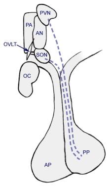
Hypophyseal portal system
Anterior nucleus
Retinohypothalamic tract
Dopaminergic cell groups
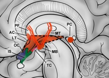
Papez circuit
Thermoregulation in humans
Vasopressin receptor 1A
Hypothalamus
Periventricular nucleus
Causes of gender incongruence
Transsexual
List of MeSH codes (A08)
Anterior pituitary
Corticotropin-releasing factor family
Neuroscience of sleep
INAH 3
Anabolic steroid
Hypothalamic-pituitary-adrenal axis
Posterior pituitary
Vasoactive intestinal peptide
Sleep onset
Clinical neurochemistry
Dopaminergic pathways
Tuberoinfundibular pathway
Emotion perception
Hippocampus anatomy
Ventromedial4
- VMH, ventromedial hypothalamic nucleus. (elifesciences.org)
- Anatomical tract-tracing and cFos mapping studies in rats exposed to cats identified a set of three interconnected medial hypothalamic nuclei, the anterior hypothalamic nucleus (AHN), dorsomedial division of the ventromedial hypothalamus (VMHdm), and dorsal premammillary nucleus (PMD), which together comprise the medial hypothalamic defensive system ( Canteras, 2002 ). (jneurosci.org)
- In particular, the two peptides were found active when injected into the paraventricular nucleus , the dorsomedial nucleus, the anterior hypothalamic area, the ventromedial nucleus and surrounding areas, and inactive when injected into the preoptic area, the caudate nucleus or the CA 1 field of the hippocampus. (baillement.com)
- In contrast, CRHR2 is virtually restricted to subcortical structures such as the lateral septum (LS),bed nucleus of the stria terminalis (BNST), the ventromedial hypothalamic Entinostat nucleus (VMH), and certain amygdaloid nuclei (medial and cortical nuclei). (cd177signaling.com)
Suprachiasmatic nucleus2
- In contrast to previous studies, modafinil did not produce statistically significant increases in Fos expression in either the suprachiasmatic nucleus or the anterior hypothalamic area. (jneurosci.org)
- We identified dopamine neurons that uniquely coexpress the Onecut3 and Nmur2 genes, and placed these in the periventricular nucleus with many synaptic afferents arising from neuromedin S + neurons of the suprachiasmatic nucleus. (nature.com)
Periventricular6
- Although the lateral region is the most voluminous, the medial and periventricular regions contain the majority of hypothalamic nuclei. (medscape.com)
- The present study shows that ACTH(1-24) and a-MSH induce stretching, yawning and penile erection in male rats when injected into the hypothalamic periventricular region surrounding, the third ventricle. (baillement.com)
- Indeed, to our knowledge this is the first report showing that ACTH(1-24) and a-MSH induce penile erection when injected into the hypothalamic periventricular area. (baillement.com)
- The behavioural responses induced by ACTH(1-24) and a-MSH injected into the hypothalamic periventricular region are indistinguishable from those induced by the two peptides given intracerebroventricularly. (baillement.com)
- This also occurs when ACTH(1-24) and a-MSH are injected into the hypothalamic periventricular region. (baillement.com)
- Schematic representation of the interactions between somatostatin (SRIH)-producing neurons in the hypothalamic periventricular nucleus (Pev) and growth hormone releasing hormone (GHRH)-producing neurons in the arcuate nucleus. (endotext.org)
Lateral6
- This low dose of modafinil also increased the number of Fos-immunoreactive (Fos-IR) neurons in the lateral subdivision of the central nucleus of the amygdala. (jneurosci.org)
- The Dorsal hypothalamic area forms part of the Lateral hypothalamic area. (unifr.ch)
- [ 2 ] Superiorly, the hypothalamus is divided from the thalamus by a groove in the lateral wall of the third ventricle, the hypothalamic sulcus. (medscape.com)
- Labeling was also found in extrahypothalamic structures such as the piriform cortex, the nucleus of the lateral olfactory tract, the central grey matter, the pars compacta of the substantia nigra, the dorsal raphe nucleus, the entorhinal cortex, the dentate gyrus and the Ammon's horn. (nih.gov)
- Vasopressin binding sites were detected in the dorsal part of the lateral septal nucleus, in midline nuclei and adjacent intralaminar nuclei of the thalamus, in the hilus of the dentate gyrus, the dorsolateral part of the basal amygdaloid nucleus and the brainstem. (shengsci.com)
- 1989). Oxytocin binding sites were also observed in the basal nucleus of Meynert, the nucleus of the vertical limb of the diagonal band of Broca, the ventral part of the lateral septal nucleus, the preoptic/anterior hypothalamic area, the posterior hypothalamic area, and variably in the globus pallidus and ventral pallidum. (shengsci.com)
Third ventricle3
- Osmoreceptors in the anterior wall of the third ventricle, near the organum vasculosum mediate the osmotic regulation of thirst, near to or even common to the osmoreceptors that regulate secretion of aqueous vasopressin (AVP). (medscape.com)
- Cross-section of the monkey hypothalamus displays two of the major hypothalamic nuclei on either side of the fluid-filled third ventricle. (cloudfront.net)
- The hypothalamus occupies the ventral diencephalon and is composed of numerous fiber tracts and nuclei situated symmetrically about the third ventricle. (medscape.com)
Supraoptic nucleus2
- In mammals, magnocellular neurosecretory cells in the paraventricular nucleus and the supraoptic nucleus of the hypothalamus produce neurohypophysial hormones , oxytocin and vasopressin . (cloudfront.net)
- Visual afferent: Afferent fibers from visual pathway pass from opticchiasma to supraoptic nucleus of hypothalamus via retinohypothalamic tract. (kypho.com)
Neurons11
- We found that 75 mg/kg modafinil increased Fos immunoreactivity in the tuberomammillary nucleus (TMN) and in orexin (hypocretin) neurons of the perifornical area, two cell groups implicated in the regulation of wakefulness. (jneurosci.org)
- Densely labeled magnocellular neurons were observed throughout the anterior and posterior magnocellular subdivisions of the hypothalamic paraventricular nucleus. (ku.edu)
- [7] Much smaller parvocellular neurosecretory cells , neurons of the paraventricular nucleus, release corticotropin-releasing hormone and other hormones into the hypophyseal portal system , where these hormones diffuse to the anterior pituitary . (cloudfront.net)
- Neurons of supraoptic and paraventricular nuclei possessing secretory functions liberate hormones vasopressin and oxytocin respectively. (kypho.com)
- Efferent for adenohypophysis (anterior pituitary): Neurons of tuberal nucleus of hypothalamus send axons to infundibulum of pituitary gland. (kypho.com)
- These axon bundles are known as tuberoinfundibular tract which transports two hormones liberated by neurons of tuberal nucleus. (kypho.com)
- Experimental chronic intoxication caused the similar changes in the neurons of studied brain areas, which were maximally expressed in the frontal cortex and the anterior hypothalamic nuclei. (jvolsu.com)
- The aim of this study was to examine the involvement of the hypothalamic oxytocin (OXT) and vasopressin (AVP) neurons in acute phase reaction using quantitative dual-labeled immunostaining with Fos and either OXT and AVP in several hypothalamic regions. (shengsci.com)
- Minireview: Regulation of Prohormone Convertases in Hypothalamic Neurons: Implications for ProThyrotropin-Releasing Hormone and Proopiomelanocortin Eduardo A. Nillni Division of Endocrinology, Department of Medicine, Brown Medical School/Rhode Island Hospital, and Department of Molecular Biology, Cell Biology, and Biochemistry, Brown University, Providence, Rhode Island 02903 Recent evidence demonstrated that posttranslational processing of neuropeptides is critical in the pathogenesis of obesity. (moam.info)
- T. belangeri of the Scandentia contained ChAT+ neurons within the nucleus of the trapezoid body as well as the superior olivary nuclear complex, which has not been described in any mammal studied to date. (wits.ac.za)
- Figure 3: Neurotransmitter phenotypes in hypothalamic neurons. (nature.com)
Thalamus4
- Axons of nucleus tractus solitarius ascend to ventroposteromedial nucleus of thalamus as solitariothalamic tract. (kypho.com)
- Afferent from thalamus: These are fibers reaching hypothalamus from dorsomedial, anterior and midline nuclei of thalamus. (kypho.com)
- Efferent to thalamus (mammillothalamic tract): These fibers pass from hypothalamic nucleus of mammillary region to anterior nucleus of thalamus. (kypho.com)
- Bilateral damage to the mediodorsal nuclei of the thalamus severely impairs recent memory and the ability to form new memories. (msdmanuals.com)
Amygdala1
- Orbitofrontal cortex, anterior cingulate cortex, insula and amygdala, were reported the most from the 19 included studies. (frontiersin.org)
Regulates2
- This study was designed to investigate whether the paraventricular hypothalamic nucleus (PVH) regulates IOP as the other nuclei do. (qxmd.com)
- The hypothalamus has a central neuroendocrine function , most notably by its control of the anterior pituitary, which in turn regulates various endocrine glands and organs. (sciencebeta.com)
Caudate nucleus1
- In rats, areas within the caudate nucleus appear to regulate water intake through norepinephrine-sensitive alpha receptors. (medscape.com)
Tuberal1
- The Subdivision of the Hypothalamic nuclei is rearranged into three Hypothalamic areas: Anterior (Chiasmatic), Middle (Tuberal) and Posterior (Mamillary). (unifr.ch)
Midline1
- L. capensis of the Lagomorpha presented vi the rodent specific rostral dorsal midline medullary nucleus (C3), while T. belangeri was lacking both the ventral and dorsal divisions of the anterior hypothalamic group (A15v and A15d), and both species were lacking the primate/Megachiropteran specific compact portion of the locus coeruleus. (wits.ac.za)
Dorsal nucleus1
- General visceral afferent: General sensations from viscera, sense of stretch, compression or distension and pain sensation due to lack of oxygen following ischemia, primarily reach the autonomic center of brain (dorsal nucleus of vagus) and spinal cord (T1 - L2 and S2 - S4 segments). (kypho.com)
Intralaminar nuclei1
- The thalamic intralaminar nuclei and brain stem reticular formation stimulate the imprinting of memories. (msdmanuals.com)
Basal ganglia1
- Network architecture of the cerebral nuclei (basal ganglia) association and commissural connectome. (neurotree.org)
Paraventricular nuclei2
- Efferent to neurohypophysis (posterior pituitary): Axons of supraoptic and paraventricular nuclei of hypothalamus extend upto posterior pituitary (neurohypophysis). (kypho.com)
- In situ hybridization analysis of apelin receptor mRNA expression in the adult rat brain showed intense labeling in the hypothalamus, especially in the supraoptic and the paraventricular nuclei. (nih.gov)
Preoptic area2
- The anterior hypothalamic region is sometimes grouped with the preoptic area. (wikipedia.org)
- Recently, we identified a sexually dimorphic nucleus (oSDN) in the sheep preoptic area-anterior hypothalamus. (oregonstate.edu)
Neuron subtypes1
- Figure 2: Hierarchical clustering of hypothalamic neuron subtypes. (nature.com)
Amygdaloid nucleus1
- The amygdaloid nucleus contributes emotional amplifications to memory. (msdmanuals.com)
Corticotropin-releasi1
- In response to a stressor, the paraventricular nucleus (PVN) of the hypothalamus releases corticotropin releasing hormone (CRH) and arginine vasopressin (AVP). (encyclopedia.pub)
Ventral1
- The three Insectivoran shrews lacked the cholinergic parabigeminal and Edinger-Westphal nuclei, had a mediodorsal arch of the cholinergic laterodorsal tegmental nucleus, lacked the catecholaminergic A4 and A15d nuclei and presented an incipient ventral division of the substantia nigra which is identical to previously studied Microchiroptera. (wits.ac.za)
Stria2
- We have shown previously that the bed nucleus of the stria terminalis (BSTc) is female in size and neuron number in male-to-female transsexual people. (transsexualitaet-ngs.de)
- Wir haben zuvor gezeigt, dass der Bed Nucleus der Stria Terminalis (BSTc) bei Mann-zu-Frau-Transsexuellen in Größe und Neuronenzahl weiblich ist. (transsexualitaet-ngs.de)
Brainstem2
- Distinct correlates of threat intensity and motor responses were found in both structures, suggesting a distributed encoding of sensory and motor features in the medial hypothalamic-brainstem instinctive network. (jneurosci.org)
- The hypothalamus receives many inputs from the brainstem , the most notable from the nucleus of the solitary tract , the locus coeruleus , and the ventrolateral medulla . (cloudfront.net)
Pituitary gland2
- It synthesizes and secretes certain neurohormones , called releasing hormones or hypothalamic hormones, and these in turn stimulate or inhibit the secretion of hormones from the pituitary gland. (cloudfront.net)
- By synthesizing and secreting neurohormones, the nuclei of the hypothalamus act as a conduit between the nervous and endocrine systems via the pituitary gland (hypophysis), regulating homeostatic functions such as hunger, thirst, body temperature, and circadian rhythms. (medscape.com)
Neuroendocrine cells1
- Thus, it is likely that 5-HT2A receptors are present on neuroendocrine cells in the hypothalamic paraventricular nucleus. (ku.edu)
Neuronal4
- Overall, our catalog of neuronal subclasses provides new understanding of hypothalamic organization and function. (nature.com)
- Figure 4: Neuropeptide associations to individual hypothalamic neuronal subtypes. (nature.com)
- The hypothalamus comprises various nuclei and neuronal subpopulations that control fundamental homeostasis and behaviors. (bnu.edu.cn)
- Furthermore, the researchers defined spatiotemporal transcriptional patterns of diverse neuronal subpopulations that would occupy specific nuclei during hypothalamic neurogenesis. (bnu.edu.cn)
Median preopti1
- MnPO, median preoptic nucleus. (elifesciences.org)
Autonomic1
- An efferent neural pathway exists between the hypothalamic nuclei and the autonomic nerve endings in the anterior chamber of the eye. (qxmd.com)
Hypophyseal3
- Through hypophyseal portal system capillaries at both ends the hormone releasing factors and hormone release inhibiting factors reach the adenohypophysis (anterior pituitary) to produce influence on the endocrine cells. (kypho.com)
- The hypothalamic and hypophyseal distribution of the receptor suggests an involvement of apelin in the control of neuro- and adenohypophyseal hormone release, whereas its presence in the pineal gland and in discrete higher brain structures points out to possible roles in the regulation of circadian rhythms and of water and food intake behavior. (nih.gov)
- In the hypothalamic-adenohypophyseal axis, releasing hormones, also known as hypophysiotropic or hypothalamic hormones, are released from the median eminence, a prolongation of the hypothalamus, into the hypophyseal portal system, which carries them to the anterior pituitary where they exert their regulatory functions on the secretion of adenohypophyseal hormones. (sciencebeta.com)
Accumbens1
- The hypothalamus has projections directly to the nucleus accumbens (NAcc). (surgicalneurologyint.com)
Subdivisions2
- These subdivisions are derived primarily from the hypothalamic blood supply. (medscape.com)
- Swanson, L.W. & Kuypers, H.G. The paraventricular nucleus of the hypothalamus: cytoarchitectonic subdivisions and organization of projections to the pituitary, dorsal vagal complex, and spinal cord as demonstrated by retrograde fluorescence double-labeling methods. (nature.com)
Regulate1
- Because biosynthesis of mature peptides in response to leptin requires prohormone processing, it is hypothesized that leptin might regulate hypothalamic PC1/3 and PC2 expression, ultimately leading to coordinated processing of prohormones into mature peptides. (moam.info)
Neuropeptide1
- To identify and delineate the nuclei and determine their volume and shape we used three different stainings throughout the nuclei in every 15th section, i.e. thionin, neuropeptide Y and synaptophysin, using an image analysis system. (transsexualitaet-ngs.de)