Arachnoiditis
Arachnoid
Iophendylate
Syringomyelia
Laminectomy
Myelography
Arachnoid Cysts
Subarachnoid Space
Meningitis
Tuberculosis, Meningeal
Spinal Cord Diseases
Dura Mater
Nutritional supply to the cauda equina in lumbar adhesive arachnoiditis in rats. (1/41)
Laminectomy-induced cauda equina adhesion has been proved by rat experiments and postoperative serial MRI in humans. A degenerative change of the cauda equina has been proved when cauda equina adhesion has been prolonged. Since it has not been reported how the nutritional supply is changed in such a condition, we evaluated the glucose supply to the adhered cauda equina in rats. Wistar rats were divided into the following three groups: the control group which received no operation, the laminectomy group which underwent L5-L6 laminectomy only, and the koalin group which received 5 mg of kaolin on the dorsal extradural space following L5-L6 laminectomy. Based on 3H-methyl-glucose uptake study, we analyzed (1) glucose transport from the intraneural vessels to the nerve tissue, and (2) glucose transport from the cerebrospinal fluid to the nerve tissue. We evaluated the relation between the severity of cauda equina adhesion and 3H uptake into the cauda equina. Cauda equina adhesion was observed in 2 of 12 rats in the control group, in 3 of 12 rats in the laminectomy group, and in 18 of 20 rats in the kaolin group. In the 3H-methyl-glucose uptake study, at 12 weeks the glucose transport to the cauda equina from the vessels increased by 44%, and that from the cerebrospinal fluid decreased by 64% in the kaolin group compared with the control group. In the condition of complete cauda equina adhesion, the glucose transport to the cauda equina from the vessels increased by 53% and that from the cerebrospinal fluid remarkably decreased by 72% compared with the normal cauda equina. Considering the greater nutritional importance of the cerebrospinal fluid in the cauda equina, it is most likely that the impairment of nutritional supply to adhered cauda equina may lead to eventual neural degeneration. (+info)Neurocysticercosis presenting as stroke. (2/41)
Stroke is a common but under recognized complication of neurocysticercosis (NCC). We report six patients having NCC who presented with stroke. All patients were young with no vascular risk factors. The arteritis which resulted in ischaemic infarct in these patients was related to the presence and severity of arachnoiditis. All patients responded well to steroids and albendazole therapy with minimal residual deficit. (+info)Intrathecal treatment of neoplastic meningitis due to breast cancer with a slow-release formulation of cytarabine. (3/41)
DepoCyte is a slow-release formulation of cytarabine designed for intrathecal administration. The goal of this multi-centre cohort study was to determine the safety and efficacy of DepoCyte for the intrathecal treatment of neoplastic meningitis due to breast cancer. DepoCyte 50 mg was injected once every 2 weeks for one month of induction therapy; responding patients were treated with an additional 3 months of consolidation therapy. All patients had metastatic breast cancer and a positive CSF cytology or neurologic findings characteristic of neoplastic meningitis. The median number of DepoCyte doses was 3, and 85% of patients completed the planned 1 month induction. Median follow up is currently 19 months. The primary endpoint was response, defined as conversion of the CSF cytology from positive to negative at all sites known to be positive, and the absence of neurologic progression at the time the cytologic conversion was documented. The response rate among the 43 evaluable patients was 28% (CI 95%: 14-41%); the intent-to-treat response rate was 21% (CI 95%: 12-34%). Median time to neurologic progression was 49 days (range 1-515(+)); median survival was 88 days (range 1-515(+)), and 1 year survival is projected to be 19%. The major adverse events were headache and arachnoiditis. When drug-related, these were largely of low grade, transient and reversible. Headache occurred on 11% of cycles; 90% were grade 1 or 2. Arachnoiditis occurred on 19% of cycles; 88% were grade 1 or 2. DepoCyte demonstrated activity in neoplastic meningitis due to breast cancer that is comparable to results reported with conventional intrathecal agents. However, this activity was achieved with one fourth as many intrathecal injections as typically required in conventional therapy. The every 2 week dose schedule is a major advantage for both patients and physicians. (+info)Tuberculous meningitis with spinal tuberculous arachnoiditis. (4/41)
This report is of a 36-year-old woman who initially presented with confusion and fever. Subsequent investigations showed tuberculous meningitis with acute hydrocephalus. Ventriculoperitoneal shunt was performed and anti-tuberculosis therapy was given. The patient was later noticed to have weakness of both lower limbs and urinary retention. Magnetic resonance imaging of the thoracic spine showed radiological features of tuberculous arachnoiditis with cord compression. Decompressive laminectomy was performed and high-dose systemic corticosteroid was given. A high level of awareness is required when diagnosing tuberculous arachnoiditis and the importance of high-dose corticosteroid in the treatment regimen is emphasised. (+info)Fluid flow in an animal model of post-traumatic syringomyelia. (5/41)
More than a quarter of patients with spinal cord injury develop syringomyelia, often with progressive neurological deficit. Treatment options remain limited and long-term failure rates are high. The current poor understanding is impeding development of improved therapies. The source and route of fluid flow into syringes has been investigated using cerebrospinal fluid (CSF) tracers. Previous work using a model of canalicular syringomyelia has shown that fluid enters the dilated central canal from perivascular spaces. The aim of this study was to determine the source and route of fluid flow in an animal model of extracanalicular (post-traumatic) syringomyelia. A model of post-traumatic syringomyelia was established in 25 Sprague-Dawley rats with intraparenchymal injections of quisqualic acid and kaolin-induced arachnoiditis. Rats survived for 6 weeks before injection of the CSF tracer horseradish peroxidase into the cisterna magna. Examination of the spatial distribution of horseradish peroxidase at 0, 3, 5, 10, or 20 min after injection was used to determine the route of fluid flow. Horseradish peroxidase rapidly spread to the ventromedian fissure, perivascular spaces, central canal, and extracanalicular syrinx. Flow occurred into the syrinx prior to significant perivascular flow in the rostral spinal cord. Preferential flow into the syrinx occurred from the perivascular spaces of the central penetrating branches of the anterior spinal artery in the grey matter. Transparenchymal flow into the syrinx was less prominent than perivascular flow. This is the first report of fluid flow within the spinal cord in a model of post-traumatic syringomyelia. Fluid from perivascular spaces moves preferentially into extracanalicular syringes and the surrounding parenchyma. Obstruction to CSF flow and loss of compliance from traumatic arachnoiditis might potentiate fluid flow in the perivascular space. (+info)Obstetric epidurals and chronic adhesive arachnoiditis. (6/41)
It has been suggested that obstetric epidurals lead to chronic adhesive arachnoiditis (CAA). CAA is a nebulous disease entity with much confusion over its symptomatology. This review outlines the pathological, clinical, and radiological features of the disease. The proposed diagnostic criteria for CAA are: back pain that increases on exertion, with or without leg pain; neurological abnormality on examination; and characteristic MRI findings. Using these criteria, there is evidence to show that epidural or subarachnoid placement of some contrast media, preservatives and possibly vasoconstrictors, may lead to CAA. No evidence was found that the preservative-free, low concentration bupivacaine with opioid mixtures or plain bupivacaine currently used in labour lead to CAA. (+info)Theoretical analysis of the pathophysiology of syringomyelia associated with adhesive arachnoiditis. (7/41)
OBJECTIVE: To apply a theoretical model to analyse the derangement of cerebrospinal fluid (CSF) dynamics in syringomyelia associated with adhesive arachnoiditis. METHODS: An electrical circuit model of CSF dynamics in the spine was used. With this model, the derangement of CSF dynamics in adhesive arachnoiditis was simulated. The effects of various surgical procedures were then analysed, such as syringo-subarachnoid shunting, subarachnoid bypass, and foramen magnum decompression. RESULTS: When CSF flow in the subarachnoid space was obstructed at a certain point, the pressure inside the spinal cord increased in the segment immediately distal to the blockage. This location of increased pressure corresponded to the preferred site of syrinx formation in adhesive arachnoiditis. Syringo-subarachnoid shunting, subarachnoid bypass, and foramen magnum decompression were all effective at reducing this pressure gradient. CONCLUSIONS: Blockage of the spinal subarachnoid CSF pathway produces a relative increase in the pressure inside the spinal cord distal to the blockage point. Repetitive formation of this pressure gradient then induces CSF leakage into the spinal parenchyma, leading to the formation of syringomyelia. Using this model, alternative surgical procedures could be suggested that might be effective in treating this disease. (+info)Thoracic arachnoiditis, arachnoid cyst and syrinx formation secondary to myelography with Myodil, 30 years previously. (8/41)
Spinal arachnoiditis can rarely occur following irritation from foreign body substances, including certain oil based contrast agents used for myelography. We describe a patient with thoracic arachnoiditis, arachnoid cyst and syringomyelia, 30 years following a myelogram with Myodil. A 62-year-old female presented with chronic thoraco-lumbar back pain, a spastic paraparesis and sphincter disturbance. She had undergone a myelogram with Myodil, 30 years previously for investigation of back pain. A MRI scan revealed evidence of arachnoiditis, thoracic syringomyelia (T6-T8) and an anteriorly placed, extramedullary, arachnoid cyst at T10-T12, compressing the cord. At surgery, T7-T10 thoracic laminectomies were carried out and syringo- and cysto-subarachnoid shunts were inserted. At 12 months follow-up, the sphincter disturbance, lower limb weakness and mobility problems had almost resolved. Although, the use of oil based contrast agents such as Myodil has been discontinued, the present case illustrates some of the rare sequelae of its use, manifesting decades later. Aggressive surgical intervention produced symptomatic benefit. (+info)Arachnoiditis is a medical condition that affects the arachnoid, one of the membranes that surround and protect the nerves of the central nervous system (the brain and spinal cord). The arachnoid becomes inflamed, often as a result of infection, direct injury, or complications from spinal surgery or chronic exposure to irritants such as steroids or contrast dyes.
The inflammation can cause the formation of scar tissue, which can lead to a variety of symptoms including:
1. Chronic pain in the back, legs, or arms
2. Numbness, tingling, or weakness in the limbs
3. Muscle cramps and spasms
4. Bladder and bowel dysfunction
5. Sexual dysfunction
In severe cases, arachnoiditis can cause permanent nerve damage and disability. Treatment typically focuses on managing symptoms and improving quality of life, as there is no cure for the condition.
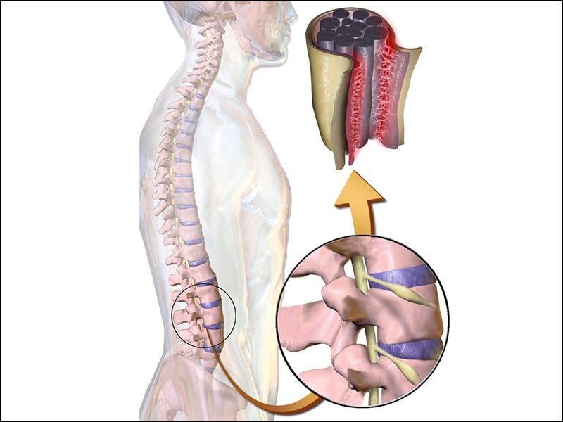
The arachnoid is one of the three membranes that cover the brain and the spinal cord, known as the meninges. It is located between the dura mater (the outermost layer) and the pia mater (the innermost layer). The arachnoid is a thin, delicate membrane that is filled with cerebrospinal fluid, which provides protection and nutrition to the central nervous system.
The arachnoid has a spider-web like appearance, hence its name, and it is composed of several layers of collagen fibers and elastic tissue. It is highly vascularized, meaning that it contains many blood vessels, and it plays an important role in regulating the flow of cerebrospinal fluid around the brain and spinal cord.
In some cases, the arachnoid can become inflamed or irritated, leading to a condition called arachnoiditis. This can cause a range of symptoms, including pain, muscle weakness, and sensory changes, and it may require medical treatment to manage.
Iophendylate is not typically referred to as a medical definition, but it is the chemical name for a contrast agent that is used in radiology procedures. It is a type of oil-based contrast medium that is injected into the cerebrospinal fluid (CSF) during myelography, which is an imaging test used to visualize the spinal cord and surrounding structures.
Iophendylate, also known as Pantopaque, is a heavy oily substance that outlines the spinal canal and nerve roots on X-ray images, allowing radiologists to diagnose various conditions such as herniated discs, spinal stenosis, or tumors. However, due to the risks associated with its use, including chemical meningitis and potential neurological complications, it has largely been replaced by water-soluble contrast agents in current clinical practice.
Syringomyelia is a medical condition characterized by the formation of a fluid-filled cavity or cavities (syrinx) within the spinal cord. This syrinx can lead to various symptoms depending on its size and location, which may include pain, muscle weakness, numbness, and stiffness in the neck, back, shoulders, arms, or legs. In some cases, it may also affect bladder and bowel function, sexual performance, and the ability to maintain normal body temperature. Syringomyelia is often associated with Chiari malformation, a condition where the lower part of the brain extends into the spinal canal. However, other conditions such as spinal cord injuries, tumors, or infections may also cause syringomyelia.
A laminectomy is a surgical procedure that involves the removal of the lamina, which is the back part of the vertebra that covers the spinal canal. This procedure is often performed to relieve pressure on the spinal cord or nerves caused by conditions such as herniated discs, spinal stenosis, or tumors. By removing the lamina, the surgeon can access the affected area and alleviate the compression on the spinal cord or nerves, thereby reducing pain, numbness, or weakness in the back, legs, or arms.
Laminectomy may be performed as a standalone procedure or in combination with other surgical techniques such as discectomy, foraminotomy, or spinal fusion. The specific approach and extent of the surgery will depend on the patient's individual condition and symptoms.
Myelography is a medical imaging technique used to examine the spinal cord and surrounding structures, such as the spinal nerves, intervertebral discs, and the spinal column. This procedure involves the injection of a contrast dye into the subarachnoid space, which is the area surrounding the spinal cord filled with cerebrospinal fluid (CSF). The dye outlines the spinal structures, making them visible on X-ray or CT scan images.
The primary purpose of myelography is to diagnose various spinal conditions, including herniated discs, spinal stenosis, tumors, infection, and traumatic injuries. It can help identify any compression or irritation of the spinal cord or nerves that may be causing pain, numbness, weakness, or other neurological symptoms.
The procedure typically requires the patient to lie flat on their stomach or side while the radiologist inserts a thin needle into the subarachnoid space, usually at the lower lumbar level. Once the contrast dye is injected, the patient will be repositioned for various X-ray views or undergo a CT scan to capture detailed images of the spine. After the procedure, patients may experience headaches, nausea, or discomfort at the injection site, but these symptoms usually resolve within a few days.
Tissue adhesions, also known as scar tissue adhesions, are abnormal bands of fibrous tissue that form between two or more internal organs, or between organs and the walls of the chest or abdominal cavity. These adhesions can develop after surgery, infection, injury, radiation, or prolonged inflammation. The fibrous bands can cause pain, restrict movement of the organs, and potentially lead to complications such as bowel obstruction. Treatment options for tissue adhesions may include medication, physical therapy, or surgical intervention to remove the adhesions.
An Arachnoid cyst is a type of abnormal fluid-filled sac that develops between the brain or spinal cord and the arachnoid membrane, which is one of the three layers that cover and protect the central nervous system. These cysts are filled with cerebrospinal fluid (CSF), which is the same fluid that surrounds and cushions the brain and spinal cord.
Arachnoid cysts can vary in size and may be present at birth or develop later in life due to trauma, infection, or other factors. While many arachnoid cysts are asymptomatic and do not cause any problems, larger cysts or those that grow or shift over time can put pressure on the brain or spinal cord, leading to a range of neurological symptoms such as headaches, seizures, hearing or vision changes, balance or coordination difficulties, and cognitive impairments.
Treatment for arachnoid cysts depends on their size, location, and associated symptoms. In some cases, observation and monitoring may be sufficient, while in others, surgical intervention may be necessary to drain the cyst or create a connection between it and the surrounding CSF space to relieve pressure.
The subarachnoid space is the area between the arachnoid mater and pia mater, which are two of the three membranes covering the brain and spinal cord (the third one being the dura mater). This space is filled with cerebrospinal fluid (CSF), which provides protection and cushioning to the central nervous system. The subarachnoid space also contains blood vessels that supply the brain and spinal cord with oxygen and nutrients. It's important to note that subarachnoid hemorrhage, a type of stroke, can occur when there is bleeding into this space.
Meningitis is a medical condition characterized by the inflammation of the meninges, which are the membranes that cover the brain and spinal cord. This inflammation can be caused by various infectious agents, such as bacteria, viruses, fungi, or parasites, or by non-infectious causes like autoimmune diseases, cancer, or certain medications.
The symptoms of meningitis may include fever, headache, stiff neck, nausea, vomiting, confusion, and sensitivity to light. In severe cases, it can lead to seizures, coma, or even death if not treated promptly and effectively. Bacterial meningitis is usually more severe and requires immediate medical attention, while viral meningitis is often less severe and may resolve on its own without specific treatment.
It's important to note that meningitis can be a serious and life-threatening condition, so if you suspect that you or someone else has symptoms of meningitis, you should seek medical attention immediately.
Meningeal tuberculosis, also known as Tuberculous meningitis, is a severe form of tuberculosis (TB) that affects the meninges, which are the membranes covering the brain and spinal cord. It is caused by the Mycobacterium tuberculosis bacterium, which can spread through the bloodstream from a primary infection site in the lungs or elsewhere in the body.
In meningeal tuberculosis, the bacteria cause inflammation and thickening of the meninges, leading to increased intracranial pressure, cerebral edema, and vasculitis. These conditions can result in various neurological symptoms such as headache, fever, stiff neck, altered mental status, seizures, and focal neurologic deficits. If left untreated, meningeal tuberculosis can lead to severe complications, including brain damage, hydrocephalus, and even death.
Diagnosis of meningeal tuberculosis typically involves a combination of clinical symptoms, cerebrospinal fluid (CSF) analysis, imaging studies, and sometimes molecular or culture-based tests to detect the presence of Mycobacterium tuberculosis in the CSF. Treatment usually involves a prolonged course of antibiotics specifically designed to target TB, such as isoniazid, rifampin, ethambutol, and pyrazinamide, often administered for six to nine months or longer. In some cases, corticosteroids may also be used to reduce inflammation and prevent complications.
Spinal cord diseases refer to a group of conditions that affect the spinal cord, which is a part of the central nervous system responsible for transmitting messages between the brain and the rest of the body. These diseases can cause damage to the spinal cord, leading to various symptoms such as muscle weakness, numbness, pain, bladder and bowel dysfunction, and difficulty with movement and coordination.
Spinal cord diseases can be congenital or acquired, and they can result from a variety of causes, including infections, injuries, tumors, degenerative conditions, autoimmune disorders, and genetic factors. Some examples of spinal cord diseases include multiple sclerosis, spina bifida, spinal cord injury, herniated discs, spinal stenosis, and motor neuron diseases such as amyotrophic lateral sclerosis (ALS).
The treatment for spinal cord diseases varies depending on the underlying cause and severity of the condition. Treatment options may include medication, physical therapy, surgery, and rehabilitation. In some cases, the damage to the spinal cord may be irreversible, leading to permanent disability or paralysis.
Dura Mater is the thickest and outermost of the three membranes (meninges) that cover the brain and spinal cord. It provides protection and support to these delicate structures. The other two layers are called the Arachnoid Mater and the Pia Mater, which are thinner and more delicate than the Dura Mater. Together, these three layers form a protective barrier around the central nervous system.
Piroxicam is a non-steroidal anti-inflammatory drug (NSAID) that is used to treat pain, inflammation, and fever. It works by inhibiting the activity of cyclooxygenase (COX) enzymes, which are involved in the production of prostaglandins, chemicals that contribute to inflammation and pain.
Piroxicam is available as a prescription medication and is used to treat conditions such as osteoarthritis, rheumatoid arthritis, and ankylosing spondylitis. It is typically taken orally in the form of tablets or capsules, and its effects can last for up to 12 hours.
Like other NSAIDs, piroxicam can cause side effects such as stomach ulcers, bleeding, and kidney problems, especially when used at high doses or for long periods of time. It is important to use piroxicam only as directed by a healthcare provider and to follow any recommended precautions.
 Arachnitis
Labdia arachnitis
Failed back syndrome
Syringomyelia
Methiodal
Epidural administration
Lillie's trichrome
Epidural blood patch
Lourdes Medical Bureau
Radicular pain
Vascular myelopathy
Chloroprocaine
Katherine Gillespie Sells
Corsia
Paul Weyrich
Ramón Carrillo
Arachnoid cyst
Tuberculoma
Coenurosis in humans
The Myelin Project
Edwin Orion Brownell
Spinal fusion
Ankle jerk reflex
Radiculopathy
Post-dural-puncture headache
List of neurological conditions and disorders
Tarlov cyst
List of inflammatory disorders
Sitting disability
Iofendylate
Arachnoiditis
Mercaptopurine
Corsiaceae
Extrapulmonary tuberculosis
Myelography
Praziquantel
Twelfth rib syndrome
Arachnoiditis - Wikipedia
 Chronic Back Pain May Be Arachnoiditis
 Arachnoiditis | National Institute of Neurological Disorders and Stroke
Arachnoiditis Imaging: Practice Essentials, Radiography, Computed Tomography
 SciELO - Brazil - ARACHNOIDITIS OSSIFICATIONS IN THE SPINE ARACHNOIDITIS OSSIFICATIONS IN THE SPINE
 Arachnitis in Luxembourgish | Translate.com
 Flexible thecoscopy for extensive spinal arachnoiditis - PubMed
Arachnoiditis Treatment | Hawthorne, NJ
 The History of Arachnoiditis
Arachnoiditis at The Medical Dictionary
 ROLE OF INTRATHECAL DETERGENTS IN PATHOGENESIS OF ADHESIVE ARACHNOIDITIS | Archives of Neurology & Psychiatry | JAMA Network
 Dr Balaji Anvekar FRCR: Focal Adhesive Arachnoiditis of Spinal Cord
 These highlights do not include all the information needed to use DEPOCYT® safely and effectively. See full prescribing...
 Can an inflammatory reaction in the meninges, caused by spinal puncture through tattooed skin, evolve into adhesive...
 Meningitis | Spinal Meningitis | MedlinePlus
 Article By Diseases
| Bentham Science
Article By Diseases
| Bentham Science
 The Neuroscience Behind Pain Education - Podcast
 Important Safety Information
 Despite Antifungal Treatment, More Woes For Some Meningitis Patients : Shots - Health News : NPR
 Sarah Woodrow, MD | Cleveland Clinic
 Multistate Outbreak of Fungal Meningitis and Other Infections - Microscopic Gallery of Pathologic Results
| HAI | CDC
Multistate Outbreak of Fungal Meningitis and Other Infections - Microscopic Gallery of Pathologic Results
| HAI | CDC
Cytosar U, DepoCyt (cytarabine) dosing, indications, interactions, adverse effects, and more
 ORDER granting 14 Motion to Reverse for Cumella v. Astrue :: Justia Dockets & Filings
Hospitalization Frequency and Charges for Neurocysticercosis, United States, 2003-2012 - Volume 21, Number 6-June 2015 -...
 Reader Questions Safety of Spinal Anesthesia - Anesthesia Patient Safety Foundation
 Medical grade calcium sulfate bone incorporated with streptomycin in the treatment of thoracolumbar spinal tuberculosis
 Zilretta (Triamcinolone Acetonide Extended-Release Injectable Suspension): Uses, Dosage, Side Effects, Interactions, Warning
 Obamacare Narrow Networks: How They Affect Doctor Specialties
 Interventional Pain: Center for Pain Relief | Carle.org
Adhesive11
- The most severe type of arachnoiditis is adhesive arachnoiditis, with scar tissue compressing the nerve roots and ultimately disrupting both blood supply and flow of cerebrospinal fluid. (medscape.com)
- Adhesive arachnoiditis can progress to arachnoiditis ossificans, or ossification of the spinal arachnoid. (medscape.com)
- MRI is preferred for adhesive arachnoiditis because it can differentiate between benign meningeal calcification and ossification of arachnoid tissue. (medscape.com)
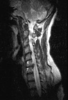
- Sagittal T1-weighted MRI of the lumbar spine in a patient with adhesive arachnoiditis who received epidural steroid injections. (medscape.com)
- Arachnoiditis ossificans is an uncommon end-stage appearance of chronic adhesive arachnoiditis. (ajnr.org)
- It can be a complication of obstetric epidurals, resulting in chronic adhesive arachnoiditis . (medjournal.com)
- In contrast to the more common benign causes of meningeal calcification, arachnoiditis ossificans results in replacement of portions of the spinal arachnoid by bone as an end-stage complication of adhesive arachnoiditis. (openneuroimagingjournal.com)
- Arachnoiditis ossificans is a rare entity characterized by ossification within the arachnoid as a result of end-stage adhesive arachnoiditis. (openneuroimagingjournal.com)
- The sad fact is that adhesive arachnoiditis remains a contentious diagnosis, which may reflect the medical profession's reluctance to acknowledge this largely iatrogenic condition. (naturalnewsblogs.com)
- and chronic adhesive arachnoiditis. (family-health-information.com)
- Postoperative spinal adhesive arachnoiditis presenting with hydrocephalus and cauda equina syndrome. (saintluc.be)
Epidural3
- Arachnoiditis following epidural blood patch-An avoidable rare complication due to blind technique: A response. (upenn.edu)
- Arachnoiditis, a complication of epidural blood patch for the treatment of low-pressure headache: A case report and systematic review. (upenn.edu)
- The investigation includes fungal meningitis (a form of meningitis that is not contagious), localized spinal or paraspinal infections, such as epidural abscess and arachnoiditis, and infections associated with injections in a peripheral joint space, such as a knee, shoulder, or ankle. (cdc.gov)
Arachnoid6
- Arachnoiditis occurs when the arachnoid is damaged. (medicalnewstoday.com)
- Arachnoiditis (say "uh-rak-noy-DY-tus") is inflammation of a membrane (called the arachnoid) that surrounds the spinal cord. (peacehealth.org)
- Arachnoiditis is an inflammation of the arachnoid, which surrounds the brain and spinal cord. (medjournal.com)
- Arachnoiditis describes a pain disorder caused by the inflammation of the arachnoid, one of the membranes that surround and protect the nerves of the spinal cord. (the-medical-dictionary.com)
- Arachnoiditis is a health condition that involves inflammation of the arachnoid membrane, which is one of three meninges (protective membranes) that surround and protect the brain and spinal cord. (painscale.com)
- Arachnoiditis can be caused by various factors that lead to inflammation and damage to the arachnoid membrane. (spineinfo.com)
Ossificans8
- For arachnoiditis ossificans, noncontrast enhanced CT has been reported to provide more sensitivity than MRI. (medscape.com)
- In cases of arachnoiditis ossificans, MRI shows irregular thickening and clumping of nerve roots of the cauda equina, and CT can show evidence of the mineral component. (medscape.com)
- Imaging features of arachnoiditis ossificans are characteristic and should be diagnosed to avoid unnecessary intervention and guide prognosis and management. (ajnr.org)
- Arachnoiditis ossificans is an important, likely under-recognized consideration in patients who present with back pain. (ajnr.org)
- Arachnoiditis ossificans is a rare cause of chronic, progressive myelopathy. (openneuroimagingjournal.com)
- We present a classic case of arachnoiditis ossificans in an elderly man who presented with progressive myelopathy and a recent fall, along with a review of the literature. (openneuroimagingjournal.com)
- The imaging in this case not only identified the characteristic findings of arachnoiditis ossificans but also identified secondary findings of the underlying causative etiology. (openneuroimagingjournal.com)
- Arachnoiditis ossificans is clinically and radiographically distinct from the far more common benign etiologies of meningeal calcification, as are seen with age-related degeneration and abnormalities in calcium metabolism. (openneuroimagingjournal.com)
Increase the risk of arachnoiditis1
- Multiple operations increase the risk of arachnoiditis, obviously. (naturalnewsblogs.com)
Type of arachnoiditis1
- Knowing the type of arachnoiditis a person has can aid in predicting symptoms. (medicalnewstoday.com)
Case of arachnoiditis1
- We present a case of arachnoiditis in an 89-year-old man who presented with long-standing myelopathy and a history of a recent fall, as well as a comprehensive literature review. (openneuroimagingjournal.com)
Treatments for arachnoiditis1
- Most treatments for arachnoiditis are focused on pain relief and the improvement of symptoms that impair daily function. (the-medical-dictionary.com)
Individuals with arachnoiditis3
- Physical therapy is a valuable treatment option for individuals with arachnoiditis. (painscale.com)
- Individuals with arachnoiditis may experience sensations of numbness, tingling, or "pins and needles" in the affected areas. (spineinfo.com)
- Some individuals with arachnoiditis may experience sexual dysfunction, including decreased libido, erectile dysfunction, or difficulties with sexual arousal or orgasm. (spineinfo.com)
Complication1
- Arachnoiditis is a complication present in some of the patients. (cdc.gov)
Chronic pain and neurological2
- Most often it is treated with medications focusing on relieving pain, since arachnoiditis typically results in chronic pain and neurological defects. (medjournal.com)
- For many, arachnoiditis is a disabling disease that causes chronic pain and neurological deficits. (the-medical-dictionary.com)
Symptoms9
- In this article, we look at the causes and types of arachnoiditis, as well as how to manage the pain and other symptoms. (medicalnewstoday.com)
- The symptoms of arachnoiditis vary from person to person and can change over time. (medicalnewstoday.com)
- A retrospective study of 28 patients with lumbar arachnoiditis found limited association between MRI findings and clinical findings, with the exception of confounding lumbar pathology (which was associated with symptom dynamics) and nerve root contour (which was associated with motor and sensory symptoms). (medscape.com)
- If arachnoiditis begins to interfere with the function of one or more of these nerves, it can cause a number of symptoms, including numbness, tingling, and a characteristic stinging and burning pain in the lower back or legs. (the-medical-dictionary.com)
- Arachnoiditis has no consistent pattern of symptoms, but it more frequently affects the nerves that supply the lower back and legs. (the-medical-dictionary.com)
- This article aims to provide a comprehensive overview of spinal arachnoiditis, including its causes, symptoms, and available treatment options. (spineinfo.com)
- What are the symptoms of arachnoiditis? (spineinfo.com)
- Arachnoiditis can present with a variety of symptoms that can vary in severity and progression. (spineinfo.com)
- The diagnosis of arachnoiditis is often based on your symptoms, physical examination findings, and your clinical history of potential triggering events. (spineinfo.com)
Corsiaceae1
- Arachnitis uniflora, the sole species in the genus Arachnitis, is a non-photosynthetic plant species in the family Corsiaceae. (wikipedia.org)
Inflammation of the meninges1
- Arachnoiditis is a broad term denoting inflammation of the meninges and subarachnoid space. (medscape.com)
Syringomyelia1
- Introduction: Post-traumatic syringomyelia typically occurs in the spinal curd adjacent to a region ol arachnoiditis. (the-medical-dictionary.com)
Spine3
- There are some reports of hereditary arachnoiditis, but most people with arachnoiditis develop the condition because of an injury to the spine. (medicalnewstoday.com)
- An MRI scan of the spine can help diagnose arachnoiditis. (peacehealth.org)
- Physical injury to the spine or spinal surgeries can result in arachnoiditis. (spineinfo.com)
Uniflora1
- Genetic diversity patterns of arbuscular mycorrhizal fungi associated with the mycoheterotroph Arachnitis uniflora Phil. (wikipedia.org)
Fungal infections1
- Bacterial, viral, or fungal infections can trigger arachnoiditis. (spineinfo.com)
Meninges1
- A rare form of arachnoiditis, this is due to genetic defects in the meninges. (medicalnewstoday.com)
Diagnosis1
- Diagnosing arachnoiditis can be challenging as there is no single definitive test for its diagnosis. (spineinfo.com)
Severe1
- Persistent and often severe pain is a hallmark symptom of arachnoiditis. (spineinfo.com)
Neurologic1
- Headache was the most common grade ≤2 neurologic event and two patients developed grade ≤2 arachnoiditis. (nih.gov)
Magnetic3
- Because of its noninvasive nature, multiplanar capabilities, and superb soft-tissue characterization, magnetic resonance imaging (MRI) is the study of choice for the diagnostic evaluation of arachnoiditis. (medscape.com)
- The primary imaging modality used for diagnosing arachnoiditis is magnetic resonance imaging (MRI) . (spineinfo.com)
- The spinal magnetic resonance images of each showed extensive arachnoiditis and a cystic structure. (elsevierpure.com)
Postoperative1
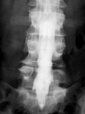
- Postoperative anteroposterior (AP) myelogram reveals thickened, clumped nerve roots in arachnoiditis. (medscape.com)
Lumbar1
- Repeated spinal procedures, such as multiple spinal surgeries or multiple lumbar punctures (spinal taps), can increase the risk of developing arachnoiditis. (spineinfo.com)
Injections1
- Can injections cause Arachnoiditis? (northshore.org)
Progresses1
- Weakness in the legs or difficulty in walking can develop as arachnoiditis progresses. (spineinfo.com)
Patients1
- We report the clinical and MRI findings of two patients with familial spinal arachnoiditis. (elsevierpure.com)
Spinal nerves1
- Arachnoiditis is an inflammation of one of the tissues that form a protective barrier around our spinal nerves. (naturalnewsblogs.com)
Nerves2
- Arachnoiditis develops when the tissues that protect the brain, the nerves, and the spinal cord are damaged, usually by a traumatic injury. (medicalnewstoday.com)
- Arachnoiditis can disrupt the normal functioning of the nerves that control bladder and bowel function. (spineinfo.com)
Disease1
- In the early stages of the disease, some people with arachnoiditis mistakenly believe they have a muscle or joint injury. (medicalnewstoday.com)
Treatment5
- The cause of arachnoiditis may influence the type a person develops, as well as the outlook and treatment plan. (medicalnewstoday.com)
- The treatment of arachnoiditis is difficult and often unsuccessful. (medjournal.com)
- Treatment options for arachnoiditis are limited. (painscale.com)
- Surgery is not a recommended treatment for arachnoiditis because pain relief tends to be short-lived, and surgery can cause additional scar tissue or adhesions to form, worsening the condition. (painscale.com)
- However, it should be done under the supervision of a doctor who specializes in arachnoiditis and with a physical therapist who is trained in the treatment of the condition. (painscale.com)
Imaging1
- There is no standard test for arachnoiditis, but imaging tests may be used. (medicalnewstoday.com)
Typically1
- Arachnoiditis typically causes shooting, stinging or burning pain in the lower back or lower limbs. (painscale.com)
Nerve roots1
- Conditions that lead to prolonged compression or irritation of the spinal cord and nerve roots can contribute to the development of arachnoiditis. (spineinfo.com)
Phil1
- On the Biology, Biogeography, and Taxonomy of Arachnitis Phil. (wikipedia.org)
Mobility1
- Arachnoiditis can also affect a person's vision, sight, and mobility. (medicalnewstoday.com)
Affects1
- Spinal arachnoiditis is a rare but debilitating condition that affects the delicate membranes surrounding the spinal cord. (spineinfo.com)
Diagnose1
- Arachnoiditis is rare, so it may require many tests and take some time to diagnose. (medicalnewstoday.com)
Pain1
- Arachnoiditis is a chronic pain disorder and while there is no known cure at this time some quality of life may be redeemed through pain management routines. (the-medical-dictionary.com)
Disorder1
- Arachnoiditis is a progressive disorder , which means that it tends to get worse over time if not treated. (medicalnewstoday.com)
Condition1
- Arachnoiditis remains a difficult condition to treat, and long-term outcomes are unpredictable. (the-medical-dictionary.com)