Plasma Volume
Hypovolemia
Erythrocyte Volume
Hematocrit
Plasma Substitutes
Magnetic Resonance Imaging
Hemodynamics
Stroke Volume
Fluid Shifts
Lung Volume Measurements
Cardiac Output
Pulmonary Diffusing Capacity
Brain
Hemodilution
Dye Dilution Technique
Blood Flow Velocity
Hemoglobins
Isotonic Solutions
Hydroxyethyl Starch Derivatives
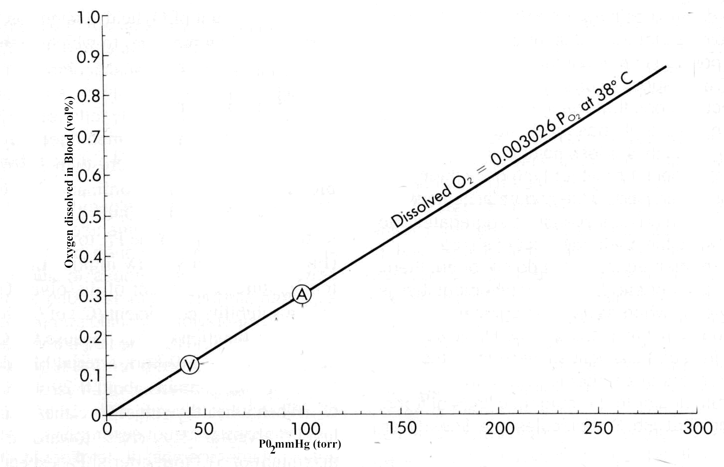
Oxygen
Spectroscopy, Near-Infrared
Reproducibility of Results
Perfusion Imaging
Image Processing, Computer-Assisted
Indocyanine Green
Magnetic Resonance Angiography
Blood Volume Determination
Dogs
Oxygen Radioisotopes
Vascular Capacitance
Gadolinium DTPA
Oxygen Consumption
Tomography, X-Ray Computed
Tomography, Emission-Computed
Hypotension
Fluid Therapy
Thorax
Vascular Resistance
Exchange Transfusion, Whole Blood
Brain Neoplasms
Image Interpretation, Computer-Assisted
Polygeline
Carbon Dioxide
Sensitivity and Specificity
Image Enhancement
Splanchnic Circulation
Imaging, Three-Dimensional
Central Venous Pressure
Water-Electrolyte Balance
Indicator Dilution Techniques
Forced Expiratory Volume
Acetazolamide
Reference Values
Diuresis
Technetium
Coloring Agents
Brain Ischemia
Carbon Monoxide
Saline Solution, Hypertonic
Pressure
Echo-Planar Imaging
Pressoreceptors
Plethysmography, Impedance
Lung
Algorithms
Capillary Permeability
Hyperventilation
Vasopressins
Oxyhemoglobins
Microvessels
Colloids
Prospective Studies
Glioma
Analysis of Variance
Residual Volume
Extravascular Lung Water
Dizziness
Albumins
Rats, Sprague-Dawley
Models, Cardiovascular
Paracentesis
Osmolar Concentration
Intracranial Pressure
Blood Transfusion
Blood Circulation Time
Lower Body Negative Pressure
Tin Polyphosphates
Thermodilution
Gadolinium
Extracellular Space
Tilt-Table Test
Disease Models, Animal
Dextrans
Sodium
Chromium Isotopes
Blood Substitutes
Hypotension, Orthostatic
Body Fluid Compartments
Monitoring, Physiologic
Predictive Value of Tests
Venous Pressure
Renin
Blood Viscosity
Models, Biological
Ultrafiltration
Kidney
Atrial Natriuretic Factor
Respiratory Mechanics
Diffusion Magnetic Resonance Imaging
Brain Mapping
Body Weight
Respiration
Anesthesia
Glycocalyx
Renal Dialysis
Treatment Outcome
Partial Pressure
Serum Albumin, Radio-Iodinated
Reduction in baroreflex cardiovascular responses due to venous infusion in the rabbit. (1/1976)
We studied reflex bradycardia and depression of mean arterial blood pressure (MAP) during left aortic nerve (LAN) stimulation before and after volume infusion in the anesthetized rabbit. Step increases in mean right atrial pressure (MRAP) to 10 mm Hg did not result in a significant change in heart rate or MAP. After volume loading, responses to LAN stimulation were not as great and the degree of attenuation was propoetional to the level of increased MRAP. A change in responsiveness was observed after elevation of MRAP by only 1 mm Hg, corresponding to less than a 10% increase in average calculated blood volume. after an increase in MRAP of 10 mm Hg, peak responses were attenuated by 44% (heart rate) and 52% (MAP), and the initial slopes (rate of change) were reduced by 46% (heart rate) and 66% (MAP). Comparison of the responses after infusion with blood and dextran solutions indicated that hemodilution was an unlikely explanation for the attenuation of the reflex responses. Total arterial baroreceptor denervation (ABD) abolished the volume-related attenuation was still present following bilateral aortic nerve section or vagotomy. It thus appears that the carotid sinus responds to changes inblood volume and influences the reflex cardiovascular responses to afferent stimulation of the LAN. On the other hand, cardiopulmonary receptors subserved by vagal afferents do not appear to be involved. (+info)Quantification of baroreceptor influence on arterial pressure changes seen in primary angiotension-induced hypertension in dogs. (2/1976)
We studied the role of the sino-aortic baroreceptors in the gradual development of hypertension induced by prolonged administration of small amounts of angiotensin II (A II) in intact dogs and dogs with denervated sino-aortic baroreceptors. Short-term 1-hour infusions of A II(1.0-100 ng/kg per min) showed that conscious denervated dogs had twice the pressor sensitivity of intact dogs. Long-term infusions of A II at 5.0 ng/kg per min (2-3 weeks) with continuous 24-hour recordings of arterial pressure showed that intact dogs required 28 hours to reach the same level of pressure attained by denervated dogs during the 1st hour of infusion. At the 28th hour the pressure in both groups was 70% of the maximum value attained by the 7th day of infusion. Both intact and denervated dogs reached nearly the same plateau level of pressure, the magnitude being directly related both the the A II infusion rate and the daily sodium intake. Cardiac output in intact dogs initially decreased after the onset of A II infusion, but by the 5th day of infusion it was 38% above control, whereas blood volume was unchanged. Heart rate returned to normal after a reduction during the 1st day of infusion in intact dogs. Plasma renin activity could not be detected after 24 hours of A II infusion in either intact or denervated dogs. The data indicate that about 35% of the hypertensive effect of A II results from its acute pressor action, and an additional 35% of the gradual increase in arterial pressure is in large measure a result of baroreceptor resetting. We conclude that the final 30% increase in pressure seems to result from increased cardiac output, the cause of which may be decreased vascular compliance. since the blood volume remains unaltered. (+info)Myocardial oxygenation during high work states in hearts with postinfarction remodeling. (3/1976)
BACKGROUND: Postinfarction left ventricular remodeling (LVR) is associated with reductions in myocardial high-energy phosphate (HEP) levels, which are more severe in animals that develop overt congestive heart failure (CHF). During high work states, further HEP loss occurs, which suggests demand-induced ischemia. This study tested the hypothesis that inadequate myocyte oxygen availability is the basis for these HEP abnormalities. METHODS AND RESULTS: Myocardial infarction was produced by left circumflex coronary artery ligation in swine. Studies were performed in 20 normal animals, 14 animals with compensated LVR, and 9 animals with CHF. Phosphocreatine (PCr)/ATP was determined with 31P NMR and deoxymyoglobin (Mb-delta) with 1H NMR in myocardium remote from the infarct. Basal PCr/ATP tended to be decreased in postinfarct hearts, and this was significant in animals with CHF. Infusion of dobutamine (20 microg x kg-1 x min-1 IV) caused doubling of the rate-pressure product in both normal and LVR hearts and resulted in comparable significant decreases of PCr/ATP in both groups. This decrease in PCr/ATP was not associated with detectable Mb-delta. In CHF hearts, rate-pressure product increased only 40% in response to dobutamine; this attenuated response also was not associated with detectable Mb-delta. CONCLUSIONS: Thus, the decrease of PCr/ATP during dobutamine infusion is not the result of insufficient myocardial oxygen availability. Furthermore, in CHF hearts, the low basal PCr/ATP and the attenuated response to dobutamine occurred in the absence of myocardial hypoxia, indicating that the HEP and contractile abnormalities were not the result of insufficient oxygen availability. (+info)Increased orthostatic tolerance following moderate exercise training in patients with unexplained syncope. (4/1976)
OBJECTIVE: To determine whether a programme of simple, moderate exercise training increases blood volume and improves orthostatic tolerance in patients with attacks of syncope or near syncope related to orthostatic stress. DESIGN: An open study in 14 patients referred with unexplained attacks of syncope, who were shown to have a low tolerance to an orthostatic stress test. Measurements were made of plasma and blood volumes, orthostatic tolerance to a test of combined head up tilt and lower body suction, and baroreceptor sensitivity by applying subatmospheric pressures to a chamber over the neck. Cardiorespiratory fitness was assessed from the relation between heart rate and oxygen uptake during a graded treadmill exercise test. Assessments were made before and after undertaking an exercise training programme (Canadian Air Force 5BX/XBX). RESULTS: After the training period, 12 of the 14 patients showed evidence of improved cardiorespiratory fitness. All 12 patients were symptomatically improved; they showed increases in plasma and blood volumes and in orthostatic tolerance, and decreases in baroreceptor sensitivity. Despite the improved orthostatic tolerance, values of blood pressure both while supine and initially following tilting were lower than before training. CONCLUSIONS: Exercise training has a role in the management of patients with syncope and poor orthostatic tolerance. It improves symptoms and increases orthostatic tolerance without increasing resting blood pressure. (+info)Stroke volume decline during prolonged exercise is influenced by the increase in heart rate. (5/1976)
This study determined whether the decline in stroke volume (SV) during prolonged exercise is related to an increase in heart rate (HR) and/or an increase in cutaneous blood flow (CBF). Seven active men cycled for 60 min at approximately 57% peak O2 uptake in a neutral environment (i.e., 27 degrees C, <40% relative humidity). They received a placebo control (CON) or a small oral dose (i.e., approximately 7 mg) of the beta1-adrenoceptor blocker atenolol (BB) at the onset of exercise. At 15 min, HR and SV were similar during CON and BB. From 15 to 55 min during CON, a 13% decline in SV was associated with an 11% increase in HR and not with an increase in CBF. CBF increased mainly from 5 to 15 min and remained stable from 20 to 60 min of exercise in both treatments. However, from 15 to 55 min during BB, when the increase in HR was prevented by atenolol, the decline in SV was also prevented, despite a normal CBF response (i.e., similar to CON). Cardiac output was similar in both treatments and stable throughout the exercise bouts. We conclude that during prolonged exercise in a neutral environment the decline in SV is related to the increase in HR and is not affected by CBF. (+info)Effect of acute normovolemic hemodilution on distribution of blood flow and tissue oxygenation in dog skeletal muscle. (6/1976)
Acute normovolemic hemodilution (ANH) is efficient in reducing allogenic blood transfusion needs during elective surgery. Tissue oxygenation is maintained by increased cardiac output and oxygen extraction and, presumably, a more homogeneous tissue perfusion. The aim of this study was to investigate blood flow distribution and oxygenation of skeletal muscle. ANH from hematocrit of 36 +/- 3 to 20 +/- 1% was performed in 22 splenectomized, anesthetized beagles (17 analyzed) ventilated with room air. Normovolemia was confirmed by measurement of blood volume. Distribution of perfusion within skeletal muscle was determined by using radioactive microspheres. Tissue oxygen partial pressure was assessed with a polarographic platinum surface electrode. Cardiac index (3.69 +/- 0.79 vs. 4.79 +/- 0.73 l. min-1. m-2) and muscle perfusion (4.07 +/- 0.44 vs. 5.18 +/- 0.36 ml. 100 g-1. min-1) were increased at hematocrit of 20%. Oxygen delivery to skeletal muscle was reduced to 74% of baseline values (0.64 +/- 0.06 vs. 0.48 +/- 0.03 ml O2. 100 g-1. min-1). Nevertheless, tissue PO2 was preserved (27.4 +/- 1.3 vs. 29.9 +/- 1. 4 Torr). Heterogeneity of muscle perfusion (relative dispersion) was reduced after ANH (20.0 +/- 2.2 vs. 13.9 +/- 1.5%). We conclude that a more homogeneous distribution of perfusion is one mechanism for the preservation of tissue oxygenation after moderate ANH, despite reduced oxygen delivery. (+info)Breathing responses to small inspiratory threshold loads in humans. (7/1976)
To investiage the effect of inspiratory threshold load (ITL) on breathing, all previous work studied loads that were much greater than would be encountered under pathophysiological conditions. We hypothesized that mild ITL from 2.5 to 20 cmH2O is sufficient to modify control and sensation of breathing. The study was performed in healthy subjects. The results demonstrated that with mild ITL 1) inspiratory difficulty sensation could be perceived at an ITL of 2.5 cmH2O; 2) tidal volume increased without change in breathing frequency, resulting in hyperpnea; and 3) although additional time was required for inspiratory pressure to attain the threshold before inspiratory flow was initiated, the total inspiratory muscle contraction time remained constant. This resulted in shortening of the available time for inspiratory flow, so that the tidal volume was maintained or increased by significant increase in mean inspiratory flow. On the basis of computer simulation, we conclude that the mild ITL is sufficient to increase breathing sensation and alter breathing control, presumably aiming at maintaining a certain level of ventilation but minimizing the energy consumption of the inspiratory muscles. (+info)Efficacy of recombinant human Hb by 31P-NMR during isovolemic total exchange transfusion. (8/1976)
The ability of recombinant human Hb (rHb1.1), which is being developed as an oxygen therapeutic, to support metabolism was measured by in vivo 31P-NMR surface coil spectroscopy of the rat abdomen in control animals and in animals subjected to isovolemic exchange transfusion to hematocrit of <3% with human serum albumin or 5 g/dl rHb1.1. No significant changes in metabolite levels were observed in control animals for up to 6 h. The albumin-exchange experiments, however, resulted in a more than eightfold increase in Pi and a 50% drop in phosphocreatine and ATP within 40 min. The tissue pH dropped from 7.4 to 6.8. The decrease in high-energy phosphates obeyed Michaelis-Menten kinetics, with a Michaelis-Menten constant of 3% as the hematocrit at which a 50% drop in high-energy phosphates was observed. Exchange transfusion with rHb1.1 resulted in no significant drop in high-energy phosphates, no rise in Pi, and no change in tissue pH from 7.35 +/- 0.15 for up to 5 h after exchange. By these criteria, rHb1.1 at a plasma Hb concentration of approximately 5 g/dl after total exchange transfusion was able to sustain energy metabolism of gut tissue at levels indistinguishable from control rats with a threefold higher total Hb level in erythrocytes. (+info)Blood volume refers to the total amount of blood present in an individual's circulatory system at any given time. It is the combined volume of both the plasma (the liquid component of blood) and the formed elements (such as red and white blood cells and platelets) in the blood. In a healthy adult human, the average blood volume is approximately 5 liters (or about 1 gallon). However, blood volume can vary depending on several factors, including age, sex, body weight, and overall health status.
Blood volume plays a critical role in maintaining proper cardiovascular function, as it affects blood pressure, heart rate, and the delivery of oxygen and nutrients to tissues throughout the body. Changes in blood volume can have significant impacts on an individual's health and may be associated with various medical conditions, such as dehydration, hemorrhage, heart failure, and liver disease. Accurate measurement of blood volume is essential for diagnosing and managing these conditions, as well as for guiding treatment decisions in clinical settings.
Plasma volume refers to the total amount of plasma present in an individual's circulatory system. Plasma is the fluid component of blood, in which cells and chemical components are suspended. It is composed mainly of water, along with various dissolved substances such as nutrients, waste products, hormones, gases, and proteins.
Plasma volume is a crucial factor in maintaining proper blood flow, regulating body temperature, and facilitating the transportation of oxygen, carbon dioxide, and other essential components throughout the body. The average plasma volume for an adult human is approximately 3 liters, but it can vary depending on factors like age, sex, body weight, and overall health status.
Changes in plasma volume can have significant effects on an individual's cardiovascular function and fluid balance. For example, dehydration or blood loss can lead to a decrease in plasma volume, while conditions such as heart failure or liver cirrhosis may result in increased plasma volume due to fluid retention. Accurate measurement of plasma volume is essential for diagnosing various medical conditions and monitoring the effectiveness of treatments.
Hypovolemia is a medical condition characterized by a decreased volume of circulating blood in the body, leading to inadequate tissue perfusion and oxygenation. This can occur due to various reasons such as bleeding, dehydration, vomiting, diarrhea, or excessive sweating, which result in a reduced amount of fluid in the intravascular space.
The severity of hypovolemia depends on the extent of fluid loss and can range from mild to severe. Symptoms may include thirst, dry mouth, weakness, dizziness, lightheadedness, confusion, rapid heartbeat, low blood pressure, and decreased urine output. Severe hypovolemia can lead to shock, organ failure, and even death if not treated promptly and effectively.
Erythrocyte volume, also known as red cell volume or hematocrit, is the proportion of whole blood that is made up of erythrocytes or red blood cells. It is typically expressed as a percentage and can be measured using a centrifuge to separate the components of a blood sample by density.
The erythrocyte volume is an important clinical parameter because it can provide information about a person's health status, such as their hydration level, altitude acclimatization, and the presence of certain medical conditions like anemia or polycythemia. Changes in erythrocyte volume can also have significant effects on the body's oxygen-carrying capacity and overall cardiovascular function.
Cerebrovascular circulation refers to the network of blood vessels that supply oxygenated blood and nutrients to the brain tissue, and remove waste products. It includes the internal carotid arteries, vertebral arteries, circle of Willis, and the intracranial arteries that branch off from them.
The internal carotid arteries and vertebral arteries merge to form the circle of Willis, a polygonal network of vessels located at the base of the brain. The anterior cerebral artery, middle cerebral artery, posterior cerebral artery, and communicating arteries are the major vessels that branch off from the circle of Willis and supply blood to different regions of the brain.
Interruptions or abnormalities in the cerebrovascular circulation can lead to various neurological conditions such as stroke, transient ischemic attack (TIA), and vascular dementia.
Hematocrit is a medical term that refers to the percentage of total blood volume that is made up of red blood cells. It is typically measured as part of a complete blood count (CBC) test. A high hematocrit may indicate conditions such as dehydration, polycythemia, or living at high altitudes, while a low hematocrit may be a sign of anemia, bleeding, or overhydration. It is important to note that hematocrit values can vary depending on factors such as age, gender, and pregnancy status.
Plasma substitutes are fluids that are used to replace the plasma volume in conditions such as hypovolemia (low blood volume) or plasma loss, for example due to severe burns, trauma, or major surgery. They do not contain cells or clotting factors, but they help to maintain intravascular volume and tissue perfusion. Plasma substitutes can be divided into two main categories: crystalloids and colloids.
Crystalloid solutions contain small molecules that can easily move between intracellular and extracellular spaces. Examples include normal saline (0.9% sodium chloride) and lactated Ringer's solution. They are less expensive and have a lower risk of allergic reactions compared to colloids, but they may require larger volumes to achieve the same effect due to their rapid distribution in the body.
Colloid solutions contain larger molecules that tend to stay within the intravascular space for longer periods, thus increasing the oncotic pressure and helping to maintain fluid balance. Examples include albumin, fresh frozen plasma, and synthetic colloids such as hydroxyethyl starch (HES) and gelatin. Colloids may be more effective in restoring intravascular volume, but they carry a higher risk of allergic reactions and anaphylaxis, and some types have been associated with adverse effects such as kidney injury and coagulopathy.
The choice of plasma substitute depends on various factors, including the patient's clinical condition, the underlying cause of plasma loss, and any contraindications or potential side effects of the available products. It is important to monitor the patient's hemodynamic status, electrolyte balance, and coagulation profile during and after the administration of plasma substitutes to ensure appropriate resuscitation and avoid complications.
Medical Definition:
Magnetic Resonance Imaging (MRI) is a non-invasive diagnostic imaging technique that uses a strong magnetic field and radio waves to create detailed cross-sectional or three-dimensional images of the internal structures of the body. The patient lies within a large, cylindrical magnet, and the scanner detects changes in the direction of the magnetic field caused by protons in the body. These changes are then converted into detailed images that help medical professionals to diagnose and monitor various medical conditions, such as tumors, injuries, or diseases affecting the brain, spinal cord, heart, blood vessels, joints, and other internal organs. MRI does not use radiation like computed tomography (CT) scans.
Hemodynamics is the study of how blood flows through the cardiovascular system, including the heart and the vascular network. It examines various factors that affect blood flow, such as blood volume, viscosity, vessel length and diameter, and pressure differences between different parts of the circulatory system. Hemodynamics also considers the impact of various physiological and pathological conditions on these variables, and how they in turn influence the function of vital organs and systems in the body. It is a critical area of study in fields such as cardiology, anesthesiology, and critical care medicine.
Stroke volume is a term used in cardiovascular physiology and medicine. It refers to the amount of blood that is pumped out of the left ventricle of the heart during each contraction (systole). Specifically, it is the difference between the volume of blood in the left ventricle at the end of diastole (when the ventricle is filled with blood) and the volume at the end of systole (when the ventricle has contracted and ejected its contents into the aorta).
Stroke volume is an important measure of heart function, as it reflects the ability of the heart to pump blood effectively to the rest of the body. A low stroke volume may indicate that the heart is not pumping efficiently, while a high stroke volume may suggest that the heart is working too hard. Stroke volume can be affected by various factors, including heart disease, high blood pressure, and physical fitness level.
The formula for calculating stroke volume is:
Stroke Volume = End-Diastolic Volume - End-Systolic Volume
Where end-diastolic volume (EDV) is the volume of blood in the left ventricle at the end of diastole, and end-systolic volume (ESV) is the volume of blood in the left ventricle at the end of systole.
Fluid shifts, in a medical context, refer to the movement or redistribution of fluids between different compartments within the body. The human body is composed of two main fluid compartments: the intracellular fluid (ICF), which is present inside the cells, and the extracellular fluid (ECF), which is outside the cells. The ECF is further divided into interstitial fluid (present in the spaces between cells) and intravascular fluid (present within the blood vessels).
Fluid shifts can occur due to various physiological and pathological conditions, such as changes in hydrostatic pressure, oncotic pressure, or permeability of the capillary membranes. These shifts can have significant impacts on various body systems, particularly the cardiovascular, respiratory, and renal systems. For example, fluid shifting from the intravascular space to the interstitial space can lead to edema (swelling), while fluid shifts into the alveoli in the lungs can cause pulmonary edema and impair breathing.
In a clinical setting, healthcare professionals monitor and manage fluid shifts through various strategies, such as administering intravenous fluids, using diuretics, or implementing mechanical ventilation, depending on the underlying cause and the specific effects of the fluid shift on the patient's condition.
Lung volume measurements are clinical tests that determine the amount of air inhaled, exhaled, and present in the lungs at different times during the breathing cycle. These measurements include:
1. Tidal Volume (TV): The amount of air inhaled or exhaled during normal breathing, usually around 500 mL in resting adults.
2. Inspiratory Reserve Volume (IRV): The additional air that can be inhaled after a normal inspiration, approximately 3,000 mL in adults.
3. Expiratory Reserve Volume (ERV): The extra air that can be exhaled after a normal expiration, about 1,000-1,200 mL in adults.
4. Residual Volume (RV): The air remaining in the lungs after a maximal exhalation, approximately 1,100-1,500 mL in adults.
5. Total Lung Capacity (TLC): The total amount of air the lungs can hold at full inflation, calculated as TV + IRV + ERV + RV, around 6,000 mL in adults.
6. Functional Residual Capacity (FRC): The volume of air remaining in the lungs after a normal expiration, equal to ERV + RV, about 2,100-2,700 mL in adults.
7. Inspiratory Capacity (IC): The maximum amount of air that can be inhaled after a normal expiration, equal to TV + IRV, around 3,500 mL in adults.
8. Vital Capacity (VC): The total volume of air that can be exhaled after a maximal inspiration, calculated as IC + ERV, approximately 4,200-5,600 mL in adults.
These measurements help assess lung function and identify various respiratory disorders such as chronic obstructive pulmonary disease (COPD), asthma, and restrictive lung diseases.
Regional blood flow (RBF) refers to the rate at which blood flows through a specific region or organ in the body, typically expressed in milliliters per minute per 100 grams of tissue (ml/min/100g). It is an essential physiological parameter that reflects the delivery of oxygen and nutrients to tissues while removing waste products. RBF can be affected by various factors such as metabolic demands, neural regulation, hormonal influences, and changes in blood pressure or vascular resistance. Measuring RBF is crucial for understanding organ function, diagnosing diseases, and evaluating the effectiveness of treatments.
Cardiac output is a measure of the amount of blood that is pumped by the heart in one minute. It is defined as the product of stroke volume (the amount of blood pumped by the left ventricle during each contraction) and heart rate (the number of contractions per minute). Normal cardiac output at rest for an average-sized adult is about 5 to 6 liters per minute. Cardiac output can be increased during exercise or other conditions that require more blood flow, such as during illness or injury. It can be measured noninvasively using techniques such as echocardiography or invasively through a catheter placed in the heart.
Blood pressure is the force exerted by circulating blood on the walls of the blood vessels. It is measured in millimeters of mercury (mmHg) and is given as two figures:
1. Systolic pressure: This is the pressure when the heart pushes blood out into the arteries.
2. Diastolic pressure: This is the pressure when the heart rests between beats, allowing it to fill with blood.
Normal blood pressure for adults is typically around 120/80 mmHg, although this can vary slightly depending on age, sex, and other factors. High blood pressure (hypertension) is generally considered to be a reading of 130/80 mmHg or higher, while low blood pressure (hypotension) is usually defined as a reading below 90/60 mmHg. It's important to note that blood pressure can fluctuate throughout the day and may be affected by factors such as stress, physical activity, and medication use.
Pulmonary diffusing capacity, also known as pulmonary diffusion capacity, is a measure of the ability of the lungs to transfer gas from the alveoli to the bloodstream. It is often used to assess the severity of lung diseases such as chronic obstructive pulmonary disease (COPD) and pulmonary fibrosis.
The most common measurement of pulmonary diffusing capacity is the diffusing capacity for carbon monoxide (DLCO), which reflects the transfer of carbon monoxide from the alveoli to the red blood cells in the capillaries. The DLCO is measured during a spirometry test, which involves breathing in a small amount of carbon monoxide and then measuring how much of it is exhaled.
A reduced DLCO may indicate a problem with the lung's ability to transfer oxygen to the blood, which can be caused by a variety of factors including damage to the alveoli or capillaries, thickening of the alveolar membrane, or a decrease in the surface area available for gas exchange.
It is important to note that other factors such as hemoglobin concentration, carboxyhemoglobin level, and lung volume can also affect the DLCO value, so these should be taken into account when interpreting the results of a diffusing capacity test.
The brain is the central organ of the nervous system, responsible for receiving and processing sensory information, regulating vital functions, and controlling behavior, movement, and cognition. It is divided into several distinct regions, each with specific functions:
1. Cerebrum: The largest part of the brain, responsible for higher cognitive functions such as thinking, learning, memory, language, and perception. It is divided into two hemispheres, each controlling the opposite side of the body.
2. Cerebellum: Located at the back of the brain, it is responsible for coordinating muscle movements, maintaining balance, and fine-tuning motor skills.
3. Brainstem: Connects the cerebrum and cerebellum to the spinal cord, controlling vital functions such as breathing, heart rate, and blood pressure. It also serves as a relay center for sensory information and motor commands between the brain and the rest of the body.
4. Diencephalon: A region that includes the thalamus (a major sensory relay station) and hypothalamus (regulates hormones, temperature, hunger, thirst, and sleep).
5. Limbic system: A group of structures involved in emotional processing, memory formation, and motivation, including the hippocampus, amygdala, and cingulate gyrus.
The brain is composed of billions of interconnected neurons that communicate through electrical and chemical signals. It is protected by the skull and surrounded by three layers of membranes called meninges, as well as cerebrospinal fluid that provides cushioning and nutrients.
Hemorrhage is defined in the medical context as an excessive loss of blood from the circulatory system, which can occur due to various reasons such as injury, surgery, or underlying health conditions that affect blood clotting or the integrity of blood vessels. The bleeding may be internal, external, visible, or concealed, and it can vary in severity from minor to life-threatening, depending on the location and extent of the bleeding. Hemorrhage is a serious medical emergency that requires immediate attention and treatment to prevent further blood loss, organ damage, and potential death.
Hemodilution is a medical term that refers to the reduction in the concentration of certain components in the blood, usually referring to red blood cells (RBCs) or hemoglobin. This occurs when an individual's plasma volume expands due to the infusion of intravenous fluids or the body's own production of fluid, such as during severe infection or inflammation. As a result, the number of RBCs per unit of blood decreases, leading to a lower hematocrit and hemoglobin level. It is important to note that while hemodilution reduces the concentration of RBCs in the blood, it does not necessarily indicate anemia or blood loss.
Blood circulation, also known as cardiovascular circulation, refers to the process by which blood is pumped by the heart and circulated throughout the body through a network of blood vessels, including arteries, veins, and capillaries. This process ensures that oxygen and nutrients are delivered to cells and tissues, while waste products and carbon dioxide are removed.
The circulation of blood can be divided into two main parts: the pulmonary circulation and the systemic circulation. The pulmonary circulation involves the movement of blood between the heart and the lungs, where it picks up oxygen and releases carbon dioxide. The systemic circulation refers to the movement of blood between the heart and the rest of the body, delivering oxygen and nutrients to cells and tissues while picking up waste products for removal.
The heart plays a central role in blood circulation, acting as a pump that contracts and relaxes to move blood through the body. The contraction of the heart's left ventricle pushes oxygenated blood into the aorta, which then branches off into smaller arteries that carry blood throughout the body. The blood then flows through capillaries, where it exchanges oxygen and nutrients for waste products and carbon dioxide with surrounding cells and tissues. The deoxygenated blood is then collected in veins, which merge together to form larger vessels that eventually return the blood back to the heart's right atrium. From there, the blood is pumped into the lungs to pick up oxygen and release carbon dioxide, completing the cycle of blood circulation.
The dye dilution technique is a method used in medicine, specifically in the field of pharmacology and physiology, to measure cardiac output and blood volume. This technique involves injecting a known quantity of a dye that mixes thoroughly with the blood, and then measuring the concentration of the dye as it circulates through the body.
The basic principle behind this technique is that the amount of dye in a given volume of blood (concentration) decreases as it gets diluted by the total blood volume. By measuring the concentration of the dye at two or more points in time, and knowing the rate at which the dye is being distributed throughout the body, it is possible to calculate the cardiac output and blood volume.
The most commonly used dye for this technique is indocyanine green (ICG), which is a safe and non-toxic dye that is readily taken up by plasma proteins and has a high extinction coefficient in the near-infrared region of the spectrum. This makes it easy to measure its concentration using specialized equipment.
The dye dilution technique is a valuable tool for assessing cardiovascular function in various clinical settings, including during surgery, critical care, and research. However, it requires careful calibration and standardization to ensure accurate results.
Contrast media are substances that are administered to a patient in order to improve the visibility of internal body structures or processes in medical imaging techniques such as X-rays, CT scans, MRI scans, and ultrasounds. These media can be introduced into the body through various routes, including oral, rectal, or intravenous administration.
Contrast media work by altering the appearance of bodily structures in imaging studies. For example, when a patient undergoes an X-ray examination, contrast media can be used to highlight specific organs, tissues, or blood vessels, making them more visible on the resulting images. In CT and MRI scans, contrast media can help to enhance the differences between normal and abnormal tissues, allowing for more accurate diagnosis and treatment planning.
There are several types of contrast media available, each with its own specific properties and uses. Some common examples include barium sulfate, which is used as a contrast medium in X-ray studies of the gastrointestinal tract, and iodinated contrast media, which are commonly used in CT scans to highlight blood vessels and other structures.
While contrast media are generally considered safe, they can sometimes cause adverse reactions, ranging from mild symptoms such as nausea or hives to more serious complications such as anaphylaxis or kidney damage. As a result, it is important for healthcare providers to carefully evaluate each patient's medical history and individual risk factors before administering contrast media.
Blood flow velocity is the speed at which blood travels through a specific part of the vascular system. It is typically measured in units of distance per time, such as centimeters per second (cm/s) or meters per second (m/s). Blood flow velocity can be affected by various factors, including cardiac output, vessel diameter, and viscosity of the blood. Measuring blood flow velocity is important in diagnosing and monitoring various medical conditions, such as heart disease, stroke, and peripheral vascular disease.
Hemoglobin (Hb or Hgb) is the main oxygen-carrying protein in the red blood cells, which are responsible for delivering oxygen throughout the body. It is a complex molecule made up of four globin proteins and four heme groups. Each heme group contains an iron atom that binds to one molecule of oxygen. Hemoglobin plays a crucial role in the transport of oxygen from the lungs to the body's tissues, and also helps to carry carbon dioxide back to the lungs for exhalation.
There are several types of hemoglobin present in the human body, including:
* Hemoglobin A (HbA): This is the most common type of hemoglobin, making up about 95-98% of total hemoglobin in adults. It consists of two alpha and two beta globin chains.
* Hemoglobin A2 (HbA2): This makes up about 1.5-3.5% of total hemoglobin in adults. It consists of two alpha and two delta globin chains.
* Hemoglobin F (HbF): This is the main type of hemoglobin present in fetal life, but it persists at low levels in adults. It consists of two alpha and two gamma globin chains.
* Hemoglobin S (HbS): This is an abnormal form of hemoglobin that can cause sickle cell disease when it occurs in the homozygous state (i.e., both copies of the gene are affected). It results from a single amino acid substitution in the beta globin chain.
* Hemoglobin C (HbC): This is another abnormal form of hemoglobin that can cause mild to moderate hemolytic anemia when it occurs in the homozygous state. It results from a different single amino acid substitution in the beta globin chain than HbS.
Abnormal forms of hemoglobin, such as HbS and HbC, can lead to various clinical disorders, including sickle cell disease, thalassemia, and other hemoglobinopathies.
Isotonic solutions are defined in the context of medical and physiological sciences as solutions that contain the same concentration of solutes (dissolved particles) as another solution, usually the bodily fluids like blood. This means that if you compare the concentration of solute particles in two isotonic solutions, they will be equal.
A common example is a 0.9% sodium chloride (NaCl) solution, also known as normal saline. The concentration of NaCl in this solution is approximately equal to the concentration found in the fluid portion of human blood, making it isotonic with blood.
Isotonic solutions are crucial in medical settings for various purposes, such as intravenous (IV) fluids replacement, wound care, and irrigation solutions. They help maintain fluid balance, prevent excessive water movement across cell membranes, and reduce the risk of damaging cells due to osmotic pressure differences between the solution and bodily fluids.
Hydroxyethyl starch derivatives are modified starches that are used as plasma expanders in medicine. They are created by chemically treating corn, potato, or wheat starch with hydroxylethyl groups, which makes the starch more soluble and less likely to be broken down by enzymes in the body. This results in a large molecule that can remain in the bloodstream for an extended period, increasing intravascular volume and improving circulation.
These derivatives are available in different molecular weights and substitution patterns, which affect their pharmacokinetics and pharmacodynamics. They are used to treat or prevent hypovolemia (low blood volume) due to various causes such as bleeding, burns, or dehydration. Common brand names include Hetastarch, Pentastarch, and Voluven.
It's important to note that the use of hydroxyethyl starch derivatives has been associated with adverse effects, including kidney injury, coagulopathy, and pruritus (severe itching). Therefore, their use should be carefully monitored and restricted to specific clinical situations.
Oxygen is a colorless, odorless, tasteless gas that constitutes about 21% of the earth's atmosphere. It is a crucial element for human and most living organisms as it is vital for respiration. Inhaled oxygen enters the lungs and binds to hemoglobin in red blood cells, which carries it to tissues throughout the body where it is used to convert nutrients into energy and carbon dioxide, a waste product that is exhaled.
Medically, supplemental oxygen therapy may be provided to patients with conditions such as chronic obstructive pulmonary disease (COPD), pneumonia, heart failure, or other medical conditions that impair the body's ability to extract sufficient oxygen from the air. Oxygen can be administered through various devices, including nasal cannulas, face masks, and ventilators.
Near-infrared spectroscopy (NIRS) is a non-invasive optical technique that uses the near-infrared region of the electromagnetic spectrum (approximately 700-2500 nanometers) to analyze various chemical and physical properties of materials, primarily in the fields of biomedical research and industry. In medicine, NIRS is often used to measure tissue oxygenation, hemodynamics, and metabolism, providing valuable information about organ function and physiology. This technique is based on the principle that different molecules absorb and scatter near-infrared light differently, allowing for the identification and quantification of specific chromophores, such as oxyhemoglobin, deoxyhemoglobin, and cytochrome c oxidase. NIRS can be employed in a variety of clinical settings, including monitoring cerebral or muscle oxygenation during surgery, assessing tissue viability in wound healing, and studying brain function in neuroscience research.
Hemorrhagic shock is a type of shock that occurs when there is significant blood loss leading to inadequate perfusion of tissues and organs. It is characterized by hypovolemia (low blood volume), hypotension (low blood pressure), tachycardia (rapid heart rate), and decreased urine output. Hemorrhagic shock can be classified into four stages based on the amount of blood loss and hemodynamic changes. In severe cases, it can lead to multi-organ dysfunction and death if not treated promptly and effectively.
Reproducibility of results in a medical context refers to the ability to obtain consistent and comparable findings when a particular experiment or study is repeated, either by the same researcher or by different researchers, following the same experimental protocol. It is an essential principle in scientific research that helps to ensure the validity and reliability of research findings.
In medical research, reproducibility of results is crucial for establishing the effectiveness and safety of new treatments, interventions, or diagnostic tools. It involves conducting well-designed studies with adequate sample sizes, appropriate statistical analyses, and transparent reporting of methods and findings to allow other researchers to replicate the study and confirm or refute the results.
The lack of reproducibility in medical research has become a significant concern in recent years, as several high-profile studies have failed to produce consistent findings when replicated by other researchers. This has led to increased scrutiny of research practices and a call for greater transparency, rigor, and standardization in the conduct and reporting of medical research.
Perfusion imaging is a medical imaging technique used to evaluate the blood flow or perfusion in various organs and tissues of the body. It is often utilized in conjunction with computed tomography (CT), magnetic resonance imaging (MRI), or single-photon emission computed tomography (SPECT) scans.
During a perfusion imaging procedure, a contrast agent is introduced into the patient's bloodstream, and a series of images are captured to track the flow and distribution of the contrast agent over time. This information helps medical professionals assess tissue viability, identify areas of reduced or blocked blood flow, and detect various pathological conditions such as stroke, heart attack, pulmonary embolism, and tumors.
In summary, perfusion imaging is a valuable diagnostic tool for evaluating the circulatory function of different organs and tissues in the body.
Computer-assisted image processing is a medical term that refers to the use of computer systems and specialized software to improve, analyze, and interpret medical images obtained through various imaging techniques such as X-ray, CT (computed tomography), MRI (magnetic resonance imaging), ultrasound, and others.
The process typically involves several steps, including image acquisition, enhancement, segmentation, restoration, and analysis. Image processing algorithms can be used to enhance the quality of medical images by adjusting contrast, brightness, and sharpness, as well as removing noise and artifacts that may interfere with accurate diagnosis. Segmentation techniques can be used to isolate specific regions or structures of interest within an image, allowing for more detailed analysis.
Computer-assisted image processing has numerous applications in medical imaging, including detection and characterization of lesions, tumors, and other abnormalities; assessment of organ function and morphology; and guidance of interventional procedures such as biopsies and surgeries. By automating and standardizing image analysis tasks, computer-assisted image processing can help to improve diagnostic accuracy, efficiency, and consistency, while reducing the potential for human error.
Indocyanine green (ICG) is a sterile, water-soluble, tricarbocyanine dye that is used as a diagnostic agent in medical imaging. It is primarily used in ophthalmology for fluorescein angiography to examine blood flow in the retina and choroid, and in cardiac surgery to assess cardiac output and perfusion. When injected into the body, ICG binds to plasma proteins and fluoresces when exposed to near-infrared light, allowing for visualization of various tissues and structures. It is excreted primarily by the liver and has a half-life of approximately 3-4 minutes in the bloodstream.
Pulmonary circulation refers to the process of blood flow through the lungs, where blood picks up oxygen and releases carbon dioxide. This is a vital part of the overall circulatory system, which delivers nutrients and oxygen to the body's cells while removing waste products like carbon dioxide.
In pulmonary circulation, deoxygenated blood from the systemic circulation returns to the right atrium of the heart via the superior and inferior vena cava. The blood then moves into the right ventricle through the tricuspid valve and gets pumped into the pulmonary artery when the right ventricle contracts.
The pulmonary artery divides into smaller vessels called arterioles, which further branch into a vast network of tiny capillaries in the lungs. Here, oxygen from the alveoli diffuses into the blood, binding to hemoglobin in red blood cells, while carbon dioxide leaves the blood and is exhaled through the nose or mouth.
The now oxygenated blood collects in venules, which merge to form pulmonary veins. These veins transport the oxygen-rich blood back to the left atrium of the heart, where it enters the systemic circulation once again. This continuous cycle enables the body's cells to receive the necessary oxygen and nutrients for proper functioning while disposing of waste products.
Magnetic Resonance Angiography (MRA) is a non-invasive medical imaging technique that uses magnetic fields and radio waves to create detailed images of the blood vessels or arteries within the body. It is a type of Magnetic Resonance Imaging (MRI) that focuses specifically on the circulatory system.
MRA can be used to diagnose and evaluate various conditions related to the blood vessels, such as aneurysms, stenosis (narrowing of the vessel), or the presence of plaques or tumors. It can also be used to plan for surgeries or other treatments related to the vascular system. The procedure does not use radiation and is generally considered safe, although people with certain implants like pacemakers may not be able to have an MRA due to safety concerns.
In the field of medicine, "time factors" refer to the duration of symptoms or time elapsed since the onset of a medical condition, which can have significant implications for diagnosis and treatment. Understanding time factors is crucial in determining the progression of a disease, evaluating the effectiveness of treatments, and making critical decisions regarding patient care.
For example, in stroke management, "time is brain," meaning that rapid intervention within a specific time frame (usually within 4.5 hours) is essential to administering tissue plasminogen activator (tPA), a clot-busting drug that can minimize brain damage and improve patient outcomes. Similarly, in trauma care, the "golden hour" concept emphasizes the importance of providing definitive care within the first 60 minutes after injury to increase survival rates and reduce morbidity.
Time factors also play a role in monitoring the progression of chronic conditions like diabetes or heart disease, where regular follow-ups and assessments help determine appropriate treatment adjustments and prevent complications. In infectious diseases, time factors are crucial for initiating antibiotic therapy and identifying potential outbreaks to control their spread.
Overall, "time factors" encompass the significance of recognizing and acting promptly in various medical scenarios to optimize patient outcomes and provide effective care.
Blood volume determination is a medical procedure that involves measuring the total amount of blood present in an individual's circulatory system. This measurement is typically expressed in milliliters (mL) or liters (L) and provides important information about the person's overall cardiovascular health and fluid status.
There are several methods for determining blood volume, including:
1. Direct measurement: This involves withdrawing a known volume of blood from the body, labeling the red blood cells with a radioactive or dye marker, reinfusing the cells back into the body, and then measuring the amount of marked cells that appear in subsequent blood samples over time.
2. Indirect measurement: This method uses formulas based on the person's height, weight, sex, and other factors to estimate their blood volume. One common indirect method is the "hemodynamic" calculation, which takes into account the individual's heart rate, stroke volume (the amount of blood pumped by the heart with each beat), and the concentration of hemoglobin in their red blood cells.
3. Bioimpedance analysis: This non-invasive technique uses electrical signals to measure the body's fluid volumes, including blood volume. By analyzing changes in the body's electrical conductivity in response to a small current, bioimpedance analysis can provide an estimate of blood volume.
Accurate determination of blood volume is important for assessing various medical conditions, such as heart failure, shock, anemia, and dehydration. It can also help guide treatment decisions, including the need for fluid replacement or blood transfusions.
I believe there might be a misunderstanding in your question. "Dogs" is not a medical term or condition. It is the common name for a domesticated carnivore of the family Canidae, specifically the genus Canis, which includes wolves, foxes, and other extant and extinct species of mammals. Dogs are often kept as pets and companions, and they have been bred in a wide variety of forms and sizes for different purposes, such as hunting, herding, guarding, assisting police and military forces, and providing companionship and emotional support.
If you meant to ask about a specific medical condition or term related to dogs, please provide more context so I can give you an accurate answer.
Oxygen radioisotopes are unstable isotopes of the element oxygen that emit radiation as they decay to a more stable form. These isotopes can be used in medical imaging and treatment, such as positron emission tomography (PET) scans. Common oxygen radioisotopes used in medicine include oxygen-15 and oxygen-18. Oxygen-15 has a very short half-life of about 2 minutes, while oxygen-18 has a longer half-life of about 2 hours. These isotopes can be incorporated into molecules such as water or carbon dioxide, which can then be used to study blood flow, metabolism and other physiological processes in the body.
Vascular capacitance is a term used in physiology to describe the ability of blood vessels, particularly veins, to expand and accommodate changes in blood volume. It is the measure of the volume of blood that a vessel can hold for each unit increase in pressure. A larger capacitance means that the blood vessels can store more blood at lower pressures.
In simpler terms, vascular capacitance refers to the compliance or distensibility of the blood vessels. When the heart pumps blood into the arteries, some of it is immediately used by the body's tissues for various functions, while the remaining blood is stored in the veins until needed. The more compliant or distensible the veins are, the greater their capacity to store blood and maintain a relatively stable blood pressure.
Therefore, vascular capacitance plays an essential role in regulating blood pressure and ensuring adequate blood flow to various organs and tissues in the body. Factors that can affect vascular capacitance include age, overall health status, and certain medical conditions such as heart failure or cirrhosis of the liver.
Gadolinium DTPA (Diethylenetriaminepentaacetic acid) is a type of gadolinium-based contrast agent (GBCA) used in medical imaging, particularly magnetic resonance imaging (MRI) and magnetic resonance angiography (MRA). It functions as a paramagnetic substance that enhances the visibility of internal body structures during these imaging techniques.
The compound Gadolinium DTPA is formed when gadolinium ions are bound to diethylenetriaminepentaacetic acid, a chelating agent. This binding helps to make the gadolinium ion safer for use in medical imaging by reducing its toxicity and improving its stability in the body.
Gadolinium DTPA is eliminated from the body primarily through the kidneys, making it important to monitor renal function before administering this contrast agent. In some cases, Gadolinium DTPA may cause adverse reactions, including allergic-like responses and nephrogenic systemic fibrosis (NSF) in patients with impaired kidney function.
Oxygen consumption, also known as oxygen uptake, is the amount of oxygen that is consumed or utilized by the body during a specific period of time, usually measured in liters per minute (L/min). It is a common measurement used in exercise physiology and critical care medicine to assess an individual's aerobic metabolism and overall health status.
In clinical settings, oxygen consumption is often measured during cardiopulmonary exercise testing (CPET) to evaluate cardiovascular function, pulmonary function, and exercise capacity in patients with various medical conditions such as heart failure, chronic obstructive pulmonary disease (COPD), and other respiratory or cardiac disorders.
During exercise, oxygen is consumed by the muscles to generate energy through a process called oxidative phosphorylation. The amount of oxygen consumed during exercise can provide important information about an individual's fitness level, exercise capacity, and overall health status. Additionally, measuring oxygen consumption can help healthcare providers assess the effectiveness of treatments and rehabilitation programs in patients with various medical conditions.
X-ray computed tomography (CT or CAT scan) is a medical imaging method that uses computer-processed combinations of many X-ray images taken from different angles to produce cross-sectional (tomographic) images (virtual "slices") of the body. These cross-sectional images can then be used to display detailed internal views of organs, bones, and soft tissues in the body.
The term "computed tomography" is used instead of "CT scan" or "CAT scan" because the machines take a series of X-ray measurements from different angles around the body and then use a computer to process these data to create detailed images of internal structures within the body.
CT scanning is a noninvasive, painless medical test that helps physicians diagnose and treat medical conditions. CT imaging provides detailed information about many types of tissue including lung, bone, soft tissue and blood vessels. CT examinations can be performed on every part of the body for a variety of reasons including diagnosis, surgical planning, and monitoring of therapeutic responses.
In computed tomography (CT), an X-ray source and detector rotate around the patient, measuring the X-ray attenuation at many different angles. A computer uses this data to construct a cross-sectional image by the process of reconstruction. This technique is called "tomography". The term "computed" refers to the use of a computer to reconstruct the images.
CT has become an important tool in medical imaging and diagnosis, allowing radiologists and other physicians to view detailed internal images of the body. It can help identify many different medical conditions including cancer, heart disease, lung nodules, liver tumors, and internal injuries from trauma. CT is also commonly used for guiding biopsies and other minimally invasive procedures.
In summary, X-ray computed tomography (CT or CAT scan) is a medical imaging technique that uses computer-processed combinations of many X-ray images taken from different angles to produce cross-sectional images of the body. It provides detailed internal views of organs, bones, and soft tissues in the body, allowing physicians to diagnose and treat medical conditions.
Emission computed tomography (ECT) is a type of tomographic imaging technique in which an emission signal from within the body is detected to create cross-sectional images of that signal's distribution. In Emission-Computed Tomography (ECT), a radionuclide is introduced into the body, usually through injection, inhalation or ingestion. The radionuclide emits gamma rays that are then detected by external gamma cameras.
The data collected from these cameras is then used to create cross-sectional images of the distribution of the radiopharmaceutical within the body. This allows for the identification and quantification of functional information about specific organs or systems within the body, such as blood flow, metabolic activity, or receptor density.
One common type of Emission-Computed Tomography is Single Photon Emission Computed Tomography (SPECT), which uses a single gamma camera that rotates around the patient to collect data from multiple angles. Another type is Positron Emission Tomography (PET), which uses positron-emitting radionuclides and detects the coincident gamma rays emitted by the annihilation of positrons and electrons.
Overall, ECT is a valuable tool in medical imaging for diagnosing and monitoring various diseases, including cancer, heart disease, and neurological disorders.
Hypotension is a medical term that refers to abnormally low blood pressure, usually defined as a systolic blood pressure less than 90 millimeters of mercury (mm Hg) or a diastolic blood pressure less than 60 mm Hg. Blood pressure is the force exerted by the blood against the walls of the blood vessels as the heart pumps blood.
Hypotension can cause symptoms such as dizziness, lightheadedness, weakness, and fainting, especially when standing up suddenly. In severe cases, hypotension can lead to shock, which is a life-threatening condition characterized by multiple organ failure due to inadequate blood flow.
Hypotension can be caused by various factors, including certain medications, medical conditions such as heart disease, endocrine disorders, and dehydration. It is important to seek medical attention if you experience symptoms of hypotension, as it can indicate an underlying health issue that requires treatment.
Fluid therapy, in a medical context, refers to the administration of fluids into a patient's circulatory system for various therapeutic purposes. This can be done intravenously (through a vein), intraosseously (through a bone), or subcutaneously (under the skin). The goal of fluid therapy is to correct or prevent imbalances in the body's fluids and electrolytes, maintain or restore blood volume, and support organ function.
The types of fluids used in fluid therapy can include crystalloids (which contain electrolytes and water) and colloids (which contain larger molecules like proteins). The choice of fluid depends on the patient's specific needs and condition. Fluid therapy is commonly used in the treatment of dehydration, shock, sepsis, trauma, surgery, and other medical conditions that can affect the body's fluid balance.
Proper administration of fluid therapy requires careful monitoring of the patient's vital signs, urine output, electrolyte levels, and overall clinical status to ensure that the therapy is effective and safe.
Heart rate is the number of heartbeats per unit of time, often expressed as beats per minute (bpm). It can vary significantly depending on factors such as age, physical fitness, emotions, and overall health status. A resting heart rate between 60-100 bpm is generally considered normal for adults, but athletes and individuals with high levels of physical fitness may have a resting heart rate below 60 bpm due to their enhanced cardiovascular efficiency. Monitoring heart rate can provide valuable insights into an individual's health status, exercise intensity, and response to various treatments or interventions.
The thorax is the central part of the human body, located between the neck and the abdomen. In medical terms, it refers to the portion of the body that contains the heart, lungs, and associated structures within a protective cage made up of the sternum (breastbone), ribs, and thoracic vertebrae. The thorax is enclosed by muscles and protected by the ribcage, which helps to maintain its structural integrity and protect the vital organs contained within it.
The thorax plays a crucial role in respiration, as it allows for the expansion and contraction of the lungs during breathing. This movement is facilitated by the flexible nature of the ribcage, which expands and contracts with each breath, allowing air to enter and exit the lungs. Additionally, the thorax serves as a conduit for major blood vessels, such as the aorta and vena cava, which carry blood to and from the heart and the rest of the body.
Understanding the anatomy and function of the thorax is essential for medical professionals, as many conditions and diseases can affect this region of the body. These may include respiratory disorders such as pneumonia or chronic obstructive pulmonary disease (COPD), cardiovascular conditions like heart attacks or aortic aneurysms, and musculoskeletal issues involving the ribs, spine, or surrounding muscles.
Microcirculation is the circulation of blood in the smallest blood vessels, including arterioles, venules, and capillaries. It's responsible for the delivery of oxygen and nutrients to the tissues and the removal of waste products. The microcirculation plays a crucial role in maintaining tissue homeostasis and is regulated by various physiological mechanisms such as autonomic nervous system activity, local metabolic factors, and hormones.
Impairment of microcirculation can lead to tissue hypoxia, inflammation, and organ dysfunction, which are common features in several diseases, including diabetes, hypertension, sepsis, and ischemia-reperfusion injury. Therefore, understanding the structure and function of the microcirculation is essential for developing new therapeutic strategies to treat these conditions.
Vascular resistance is a measure of the opposition to blood flow within a vessel or a group of vessels, typically expressed in units of mmHg/(mL/min) or sometimes as dynes*sec/cm^5. It is determined by the diameter and length of the vessels, as well as the viscosity of the blood flowing through them. In general, a decrease in vessel diameter, an increase in vessel length, or an increase in blood viscosity will result in an increase in vascular resistance, while an increase in vessel diameter, a decrease in vessel length, or a decrease in blood viscosity will result in a decrease in vascular resistance. Vascular resistance is an important concept in the study of circulation and cardiovascular physiology because it plays a key role in determining blood pressure and blood flow within the body.
An exchange transfusion of whole blood is a medical procedure in which a patient's blood is gradually replaced with donor whole blood. This procedure is typically performed in newborns or infants who have severe jaundice caused by excessive levels of bilirubin, a yellowish pigment that forms when hemoglobin from red blood cells breaks down.
During an exchange transfusion, the baby's blood is removed through a vein or artery and replaced with donor whole blood through another vein or artery. The process is repeated several times until a significant portion of the baby's blood has been exchanged with donor blood. This helps to reduce the levels of bilirubin in the baby's blood, which can help prevent or treat brain damage caused by excessive bilirubin.
Exchange transfusions are typically performed in a neonatal intensive care unit (NICU) and require close monitoring by a team of healthcare professionals. The procedure carries some risks, including infection, bleeding, and changes in blood pressure or heart rate. However, it can be a lifesaving treatment for newborns with severe jaundice who are at risk of developing serious complications.
Brain neoplasms, also known as brain tumors, are abnormal growths of cells within the brain. These growths can be benign (non-cancerous) or malignant (cancerous). Benign brain tumors typically grow slowly and do not spread to other parts of the body. However, they can still cause serious problems if they press on sensitive areas of the brain. Malignant brain tumors, on the other hand, are cancerous and can grow quickly, invading surrounding brain tissue and spreading to other parts of the brain or spinal cord.
Brain neoplasms can arise from various types of cells within the brain, including glial cells (which provide support and insulation for nerve cells), neurons (nerve cells that transmit signals in the brain), and meninges (the membranes that cover the brain and spinal cord). They can also result from the spread of cancer cells from other parts of the body, known as metastatic brain tumors.
Symptoms of brain neoplasms may vary depending on their size, location, and growth rate. Common symptoms include headaches, seizures, weakness or paralysis in the limbs, difficulty with balance and coordination, changes in speech or vision, confusion, memory loss, and changes in behavior or personality.
Treatment for brain neoplasms depends on several factors, including the type, size, location, and grade of the tumor, as well as the patient's age and overall health. Treatment options may include surgery, radiation therapy, chemotherapy, targeted therapy, or a combination of these approaches. Regular follow-up care is essential to monitor for recurrence and manage any long-term effects of treatment.
Plethysmography is a non-invasive medical technique used to measure changes in volume or blood flow within an organ or body part, typically in the lungs or extremities. There are several types of plethysmography, including:
1. **Whole Body Plethysmography (WBP):** This type of plethysmography is used to assess lung function and volumes by measuring changes in pressure within a sealed chamber that contains the patient's entire body except for their head. The patient breathes normally while wearing a nose clip, allowing technicians to analyze respiratory patterns, airflow, and lung volume changes.
2. **Segmental or Local Plethysmography:** This technique measures volume or blood flow changes in specific body parts, such as the limbs or digits. It can help diagnose and monitor conditions affecting peripheral circulation, like deep vein thrombosis, arterial occlusive disease, or Raynaud's phenomenon.
3. **Impedance Plethysmography (IPG):** This non-invasive method uses electrical impedance to estimate changes in blood volume within an organ or body part. By applying a small electrical current and measuring the opposition to flow (impedance), technicians can determine variations in blood volume, which can help diagnose conditions like deep vein thrombosis or heart failure.
4. **Optical Plethysmography:** This technique uses light to measure changes in blood volume, typically in the skin or mucous membranes. By shining a light on the area and analyzing the reflected or transmitted light, technicians can detect variations in blood volume related to cardiac output, respiration, or other physiological factors.
Overall, plethysmography is an essential tool for diagnosing and monitoring various medical conditions affecting circulation, respiratory function, and organ volumes.
Computer-assisted image interpretation is the use of computer algorithms and software to assist healthcare professionals in analyzing and interpreting medical images. These systems use various techniques such as pattern recognition, machine learning, and artificial intelligence to help identify and highlight abnormalities or patterns within imaging data, such as X-rays, CT scans, MRI, and ultrasound images. The goal is to increase the accuracy, consistency, and efficiency of image interpretation, while also reducing the potential for human error. It's important to note that these systems are intended to assist healthcare professionals in their decision making process and not to replace them.
Polygeline is a colloidal plasma expander, which is a type of intravenous fluid used to increase blood volume in hypovolemia or shock. It is made up of polyvinylpyrrolidone (PVP) molecules that are cross-linked with divinyl sulfone and then suspended in an electrolyte solution. Polygeline works by drawing water into the circulation, thereby increasing the volume of the plasma.
It is important to note that polygeline has been associated with anaphylactic reactions and therefore should be used with caution. It is also not recommended for use in patients with renal impairment or those who are allergic to PVP. As with any medical treatment, it should only be administered under the direction of a qualified healthcare professional.
Carbon dioxide (CO2) is a colorless, odorless gas that is naturally present in the Earth's atmosphere. It is a normal byproduct of cellular respiration in humans, animals, and plants, and is also produced through the combustion of fossil fuels such as coal, oil, and natural gas.
In medical terms, carbon dioxide is often used as a respiratory stimulant and to maintain the pH balance of blood. It is also used during certain medical procedures, such as laparoscopic surgery, to insufflate (inflate) the abdominal cavity and create a working space for the surgeon.
Elevated levels of carbon dioxide in the body can lead to respiratory acidosis, a condition characterized by an increased concentration of carbon dioxide in the blood and a decrease in pH. This can occur in conditions such as chronic obstructive pulmonary disease (COPD), asthma, or other lung diseases that impair breathing and gas exchange. Symptoms of respiratory acidosis may include shortness of breath, confusion, headache, and in severe cases, coma or death.
Sensitivity and specificity are statistical measures used to describe the performance of a diagnostic test or screening tool in identifying true positive and true negative results.
* Sensitivity refers to the proportion of people who have a particular condition (true positives) who are correctly identified by the test. It is also known as the "true positive rate" or "recall." A highly sensitive test will identify most or all of the people with the condition, but may also produce more false positives.
* Specificity refers to the proportion of people who do not have a particular condition (true negatives) who are correctly identified by the test. It is also known as the "true negative rate." A highly specific test will identify most or all of the people without the condition, but may also produce more false negatives.
In medical testing, both sensitivity and specificity are important considerations when evaluating a diagnostic test. High sensitivity is desirable for screening tests that aim to identify as many cases of a condition as possible, while high specificity is desirable for confirmatory tests that aim to rule out the condition in people who do not have it.
It's worth noting that sensitivity and specificity are often influenced by factors such as the prevalence of the condition in the population being tested, the threshold used to define a positive result, and the reliability and validity of the test itself. Therefore, it's important to consider these factors when interpreting the results of a diagnostic test.
Image enhancement in the medical context refers to the process of improving the quality and clarity of medical images, such as X-rays, CT scans, MRI scans, or ultrasound images, to aid in the diagnosis and treatment of medical conditions. Image enhancement techniques may include adjusting contrast, brightness, or sharpness; removing noise or artifacts; or applying specialized algorithms to highlight specific features or structures within the image.
The goal of image enhancement is to provide clinicians with more accurate and detailed information about a patient's anatomy or physiology, which can help inform medical decision-making and improve patient outcomes.
Splanchnic circulation refers to the blood flow to the visceral organs, including the gastrointestinal tract, pancreas, spleen, and liver. These organs receive a significant portion of the cardiac output, with approximately 25-30% of the total restingly going to the splanchnic circulation. The splanchnic circulation is regulated by a complex interplay of neural and hormonal mechanisms that help maintain adequate blood flow to these vital organs while also allowing for the distribution of blood to other parts of the body as needed.
The splanchnic circulation is unique in its ability to vasodilate and increase blood flow significantly in response to meals or other stimuli, such as stress or hormonal changes. This increased blood flow helps support the digestive process and absorption of nutrients. At the same time, the body must carefully regulate this blood flow to prevent a significant drop in blood pressure or overloading the heart with too much work.
Overall, the splanchnic circulation plays a critical role in maintaining the health and function of the body's vital organs, and dysregulation of this system can contribute to various diseases, including digestive disorders, liver disease, and cardiovascular disease.
Three-dimensional (3D) imaging in medicine refers to the use of technologies and techniques that generate a 3D representation of internal body structures, organs, or tissues. This is achieved by acquiring and processing data from various imaging modalities such as X-ray computed tomography (CT), magnetic resonance imaging (MRI), ultrasound, or confocal microscopy. The resulting 3D images offer a more detailed visualization of the anatomy and pathology compared to traditional 2D imaging techniques, allowing for improved diagnostic accuracy, surgical planning, and minimally invasive interventions.
In 3D imaging, specialized software is used to reconstruct the acquired data into a volumetric model, which can be manipulated and viewed from different angles and perspectives. This enables healthcare professionals to better understand complex anatomical relationships, detect abnormalities, assess disease progression, and monitor treatment response. Common applications of 3D imaging include neuroimaging, orthopedic surgery planning, cancer staging, dental and maxillofacial reconstruction, and interventional radiology procedures.
Central venous pressure (CVP) is the blood pressure measured in the large veins that enter the right atrium of the heart. It reflects the amount of blood returning to the heart and the ability of the heart to pump it effectively. CVP is used as an indicator of a person's intravascular volume status, cardiac function, and overall hemodynamic performance. The measurement is taken using a central venous catheter placed in a large vein such as the internal jugular or subclavian vein. Normal CVP values range from 0 to 8 mmHg (millimeters of mercury) in adults when measured at the level of the right atrium.
Organ size refers to the volume or physical measurement of an organ in the body of an individual. It can be described in terms of length, width, and height or by using specialized techniques such as imaging studies (like CT scans or MRIs) to determine the volume. The size of an organ can vary depending on factors such as age, sex, body size, and overall health status. Changes in organ size may indicate various medical conditions, including growths, inflammation, or atrophy.
Water-electrolyte balance refers to the regulation of water and electrolytes (sodium, potassium, chloride, bicarbonate) in the body to maintain homeostasis. This is crucial for various bodily functions such as nerve impulse transmission, muscle contraction, fluid balance, and pH regulation. The body maintains this balance through mechanisms that control water intake, excretion, and electrolyte concentration in various body fluids like blood and extracellular fluid. Disruptions in water-electrolyte balance can lead to dehydration or overhydration, and imbalances in electrolytes can cause conditions such as hyponatremia (low sodium levels) or hyperkalemia (high potassium levels).
Indicator dilution techniques are a group of methods used in medicine and research to measure various physiological variables, such as cardiac output or cerebral blood flow. These techniques involve introducing a known quantity of an indicator substance (like a dye or a radioactive tracer) into the system being studied and then measuring its concentration over time at a specific location downstream.
The basic principle behind these techniques is that the concentration of the indicator substance will be inversely proportional to the flow rate of the fluid through which it is moving. By measuring the concentration of the indicator substance at different points in time, researchers can calculate the flow rate using mathematical formulas.
Indicator dilution techniques are widely used in clinical and research settings because they are relatively non-invasive and can provide accurate and reliable measurements of various physiological variables. Some common examples of indicator dilution techniques include thermodilution, dye dilution, and Fick principle-based methods.
Forced Expiratory Volume (FEV) is a medical term used to describe the volume of air that can be forcefully exhaled from the lungs in one second. It is often measured during pulmonary function testing to assess lung function and diagnose conditions such as chronic obstructive pulmonary disease (COPD) or asthma.
FEV is typically expressed as a percentage of the Forced Vital Capacity (FVC), which is the total volume of air that can be exhaled from the lungs after taking a deep breath in. The ratio of FEV to FVC is used to determine whether there is obstruction in the airways, with a lower ratio indicating more severe obstruction.
There are different types of FEV measurements, including FEV1 (the volume of air exhaled in one second), FEV25-75 (the average volume of air exhaled during the middle 50% of the FVC maneuver), and FEV0.5 (the volume of air exhaled in half a second). These measurements can provide additional information about lung function and help guide treatment decisions.
Acetazolamide is a medication that belongs to a class of drugs called carbonic anhydrase inhibitors. It works by decreasing the production of bicarbonate in the body, which helps to reduce the amount of fluid in the eye and brain, making it useful for treating conditions such as glaucoma and epilepsy.
In medical terms, acetazolamide can be defined as: "A carbonic anhydrase inhibitor that is used to treat glaucoma, epilepsy, altitude sickness, and other conditions. It works by decreasing the production of bicarbonate in the body, which helps to reduce the amount of fluid in the eye and brain."
Acetazolamide may also be used for other purposes not listed here, so it is important to consult with a healthcare provider for specific medical advice.
Liver circulation, also known as hepatic circulation, refers to the blood flow through the liver. The liver receives blood from two sources: the hepatic artery and the portal vein.
The hepatic artery delivers oxygenated blood from the heart to the liver, accounting for about 25% of the liver's blood supply. The remaining 75% comes from the portal vein, which carries nutrient-rich, deoxygenated blood from the gastrointestinal tract, spleen, pancreas, and gallbladder to the liver.
In the liver, these two sources of blood mix in the sinusoids, small vessels with large spaces between the endothelial cells that line them. This allows for efficient exchange of substances between the blood and the hepatocytes (liver cells). The blood then leaves the liver through the hepatic veins, which merge into the inferior vena cava and return the blood to the heart.
The unique dual blood supply and extensive sinusoidal network in the liver enable it to perform various critical functions, such as detoxification, metabolism, synthesis, storage, and secretion of numerous substances, maintaining body homeostasis.
Reference values, also known as reference ranges or reference intervals, are the set of values that are considered normal or typical for a particular population or group of people. These values are often used in laboratory tests to help interpret test results and determine whether a patient's value falls within the expected range.
The process of establishing reference values typically involves measuring a particular biomarker or parameter in a large, healthy population and then calculating the mean and standard deviation of the measurements. Based on these statistics, a range is established that includes a certain percentage of the population (often 95%) and excludes extreme outliers.
It's important to note that reference values can vary depending on factors such as age, sex, race, and other demographic characteristics. Therefore, it's essential to use reference values that are specific to the relevant population when interpreting laboratory test results. Additionally, reference values may change over time due to advances in measurement technology or changes in the population being studied.
Diuresis is a medical term that refers to an increased production of urine by the kidneys. It can occur as a result of various factors, including certain medications, medical conditions, or as a response to a physiological need, such as in the case of dehydration. Diuretics are a class of drugs that promote diuresis and are often used to treat conditions such as high blood pressure, heart failure, and edema.
Diuresis can be classified into several types based on its underlying cause or mechanism, including:
1. Osmotic diuresis: This occurs when the kidneys excrete large amounts of urine in response to a high concentration of solutes (such as glucose) in the tubular fluid. The high osmolarity of the tubular fluid causes water to be drawn out of the bloodstream and into the urine, leading to an increase in urine output.
2. Forced diuresis: This is a medical procedure in which large amounts of intravenous fluids are administered to promote diuresis. It is used in certain clinical situations, such as to enhance the excretion of toxic substances or to prevent kidney damage.
3. Natriuretic diuresis: This occurs when the kidneys excrete large amounts of sodium and water in response to the release of natriuretic peptides, which are hormones that regulate sodium balance and blood pressure.
4. Aquaresis: This is a type of diuresis that occurs in response to the ingestion of large amounts of water, leading to dilute urine production.
5. Pathological diuresis: This refers to increased urine production due to underlying medical conditions such as diabetes insipidus or pyelonephritis.
It is important to note that excessive diuresis can lead to dehydration and electrolyte imbalances, so it should be monitored carefully in clinical settings.
Posture is the position or alignment of body parts supported by the muscles, especially the spine and head in relation to the vertebral column. It can be described as static (related to a stationary position) or dynamic (related to movement). Good posture involves training your body to stand, walk, sit, and lie in positions where the least strain is placed on supporting muscles and ligaments during movement or weight-bearing activities. Poor posture can lead to various health issues such as back pain, neck pain, headaches, and respiratory problems.
Technetium is not a medical term itself, but it is a chemical element with the symbol Tc and atomic number 43. However, in the field of nuclear medicine, which is a branch of medicine that uses small amounts of radioactive material to diagnose or treat diseases, Technetium-99m (a radioisotope of technetium) is commonly used for various diagnostic procedures.
Technetium-99m is a metastable nuclear isomer of technetium-99, and it emits gamma rays that can be detected outside the body to create images of internal organs or tissues. It has a short half-life of about 6 hours, which makes it ideal for diagnostic imaging since it decays quickly and reduces the patient's exposure to radiation.
Technetium-99m is used in a variety of medical procedures, such as bone scans, lung scans, heart scans, liver-spleen scans, brain scans, and kidney scans, among others. It can be attached to different pharmaceuticals or molecules that target specific organs or tissues, allowing healthcare professionals to assess their function or identify any abnormalities.
Coloring agents, also known as food dyes or color additives, are substances that are added to foods, medications, and cosmetics to improve their appearance by giving them a specific color. These agents can be made from both synthetic and natural sources. They must be approved by regulatory agencies such as the U.S. Food and Drug Administration (FDA) before they can be used in products intended for human consumption.
Coloring agents are used for various reasons, including:
* To replace color lost during food processing or preparation
* To make foods more visually appealing
* To help consumers easily identify certain types of food
* To indicate the flavor of a product (e.g., fruit-flavored candies)
It's important to note that while coloring agents can enhance the appearance of products, they do not affect their taste or nutritional value. Some people may have allergic reactions to certain coloring agents, so it's essential to check product labels if you have any known allergies. Additionally, excessive consumption of some synthetic coloring agents has been linked to health concerns, so moderation is key.
Brain ischemia is the medical term used to describe a reduction or interruption of blood flow to the brain, leading to a lack of oxygen and glucose delivery to brain tissue. This can result in brain damage or death of brain cells, known as infarction. Brain ischemia can be caused by various conditions such as thrombosis (blood clot formation), embolism (obstruction of a blood vessel by a foreign material), or hypoperfusion (reduced blood flow). The severity and duration of the ischemia determine the extent of brain damage. Symptoms can range from mild, such as transient ischemic attacks (TIAs or "mini-strokes"), to severe, including paralysis, speech difficulties, loss of consciousness, and even death. Immediate medical attention is required for proper diagnosis and treatment to prevent further damage and potential long-term complications.
Carbon monoxide (CO) is a colorless, odorless, and tasteless gas that is slightly less dense than air. It is toxic to hemoglobic animals when encountered in concentrations above about 35 ppm. This compound is a product of incomplete combustion of organic matter, and is a major component of automobile exhaust.
Carbon monoxide is poisonous because it binds to hemoglobin in red blood cells much more strongly than oxygen does, forming carboxyhemoglobin. This prevents the transport of oxygen throughout the body, which can lead to suffocation and death. Symptoms of carbon monoxide poisoning include headache, dizziness, weakness, nausea, vomiting, confusion, and disorientation. Prolonged exposure can lead to unconsciousness and death.
Carbon monoxide detectors are commonly used in homes and other buildings to alert occupants to the presence of this dangerous gas. It is important to ensure that these devices are functioning properly and that they are placed in appropriate locations throughout the building. Additionally, it is essential to maintain appliances and heating systems to prevent the release of carbon monoxide into living spaces.
The supine position is a term used in medicine to describe a body posture where an individual is lying down on their back, with their face and torso facing upwards. This position is often adopted during various medical procedures, examinations, or when resting, as it allows for easy access to the front of the body. It is also the position automatically assumed by most people who are falling asleep.
It's important to note that in the supine position, the head can be flat on the surface or raised with the use of pillows or specialized medical equipment like a hospital bed. This can help to alleviate potential issues such as breathing difficulties or swelling in the face and head.
A hypertonic saline solution is a type of medical fluid that contains a higher concentration of salt (sodium chloride) than is found in the average person's blood. This solution is used to treat various medical conditions, such as dehydration, brain swelling, and increased intracranial pressure.
The osmolarity of a hypertonic saline solution typically ranges from 1500 to 23,400 mOsm/L, with the most commonly used solutions having an osmolarity of around 3000 mOsm/L. The high sodium concentration in these solutions creates an osmotic gradient that draws water out of cells and into the bloodstream, helping to reduce swelling and increase fluid volume in the body.
It is important to note that hypertonic saline solutions should be administered with caution, as they can cause serious side effects such as electrolyte imbalances, heart rhythm abnormalities, and kidney damage if not used properly. Healthcare professionals must carefully monitor patients receiving these solutions to ensure safe and effective treatment.
Body water refers to the total amount of water present in the human body. It is an essential component of life and makes up about 60-70% of an adult's body weight. Body water is distributed throughout various fluid compartments within the body, including intracellular fluid (water inside cells), extracellular fluid (water outside cells), and transcellular fluid (water found in specific bodily spaces such as the digestive tract, eyes, and joints). Maintaining proper hydration and balance of body water is crucial for various physiological processes, including temperature regulation, nutrient transportation, waste elimination, and overall health.
In medical terms, pressure is defined as the force applied per unit area on an object or body surface. It is often measured in millimeters of mercury (mmHg) in clinical settings. For example, blood pressure is the force exerted by circulating blood on the walls of the arteries and is recorded as two numbers: systolic pressure (when the heart beats and pushes blood out) and diastolic pressure (when the heart rests between beats).
Pressure can also refer to the pressure exerted on a wound or incision to help control bleeding, or the pressure inside the skull or spinal canal. High or low pressure in different body systems can indicate various medical conditions and require appropriate treatment.
Echo-Planar Imaging (EPI) is a type of magnetic resonance imaging (MRI) technique that uses rapidly alternating magnetic field gradients and radiofrequency pulses to acquire multiple images in a very short period of time. This technique allows for the rapid acquisition of images, making it useful for functional MRI (fMRI) studies, diffusion-weighted imaging, and other applications where motion artifacts can be a problem.
In EPI, a single excitation pulse is followed by a series of gradient echoes that are acquired in a rapid succession, with each echo providing information about a different slice or plane of the object being imaged. The resulting images can then be combined to create a 3D representation of the object.
One of the key advantages of EPI is its speed, as it can acquire an entire brain volume in as little as 50 milliseconds. This makes it possible to capture rapid changes in the brain, such as those that occur during cognitive tasks or in response to neural activation. However, the technique can be susceptible to distortions and artifacts, particularly at higher field strengths, which can affect image quality and accuracy.
Blood gas analysis is a medical test that measures the levels of oxygen and carbon dioxide in the blood, as well as the pH level, which indicates the acidity or alkalinity of the blood. This test is often used to evaluate lung function, respiratory disorders, and acid-base balance in the body. It can also be used to monitor the effectiveness of treatments for conditions such as chronic obstructive pulmonary disease (COPD), asthma, and other respiratory illnesses. The analysis is typically performed on a sample of arterial blood, although venous blood may also be used in some cases.
Capillaries are the smallest blood vessels in the body, with diameters that range from 5 to 10 micrometers. They form a network of tiny tubes that connect the arterioles (small branches of arteries) and venules (small branches of veins), allowing for the exchange of oxygen, carbon dioxide, nutrients, and waste products between the blood and the surrounding tissues.
Capillaries are composed of a single layer of endothelial cells that surround a hollow lumen through which blood flows. The walls of capillaries are extremely thin, allowing for easy diffusion of molecules between the blood and the surrounding tissue. This is essential for maintaining the health and function of all body tissues.
Capillaries can be classified into three types based on their structure and function: continuous, fenestrated, and sinusoidal. Continuous capillaries have a continuous layer of endothelial cells with tight junctions that restrict the passage of large molecules. Fenestrated capillaries have small pores or "fenestrae" in the endothelial cell walls that allow for the passage of larger molecules, such as proteins and lipids. Sinusoidal capillaries are found in organs with high metabolic activity, such as the liver and spleen, and have large, irregular spaces between the endothelial cells that allow for the exchange of even larger molecules.
Overall, capillaries play a critical role in maintaining the health and function of all body tissues by allowing for the exchange of nutrients, oxygen, and waste products between the blood and surrounding tissues.
The Valsalva maneuver is a medical procedure that involves forced exhalation against a closed airway, typically by closing one's mouth, pinching the nose shut, and then blowing. This maneuver increases the pressure in the chest and affects the heart's filling and pumping capabilities, as well as the pressures within the ears and eyes.
It is often used during medical examinations to test for conditions such as heart murmurs or to help clear the ears during changes in air pressure (like when scuba diving or flying). It can also be used to help diagnose or monitor conditions related to the autonomic nervous system, such as orthostatic hypotension or dysautonomia.
However, it's important to perform the Valsalva maneuver correctly and under medical supervision, as improper technique or overdoing it can lead to adverse effects like increased heart rate, changes in blood pressure, or even damage to the eardrum.
Pressoreceptors are specialized sensory nerve endings found in the walls of blood vessels, particularly in the carotid sinus and aortic arch. They respond to changes in blood pressure by converting the mechanical stimulus into electrical signals that are transmitted to the brain. This information helps regulate cardiovascular function and maintain blood pressure homeostasis.
Impedance plethysmography is a non-invasive method used to measure changes in blood volume or flow in a particular area of the body. It works by passing a small electrical current through the tissue and measuring the opposition (impedance) to that current, which varies with the amount of blood present in the area.
In impedance cardiography, this technique is used to estimate cardiac output, stroke volume, and other hemodynamic parameters. The changes in impedance are measured across the chest wall, which correlate with the ventricular ejection of blood during each heartbeat. This allows for the calculation of various cardiovascular variables, such as the amount of blood pumped by the heart per minute (cardiac output) and the resistance to blood flow in the systemic circulation (systemic vascular resistance).
Impedance plethysmography is a safe and reliable method for assessing cardiovascular function, and it has been widely used in clinical settings to evaluate patients with various cardiovascular disorders, including heart failure, hypertension, and peripheral arterial disease.
In medical terms, shock is a life-threatening condition that occurs when the body is not getting enough blood flow or when the circulatory system is not functioning properly to distribute oxygen and nutrients to the tissues and organs. This results in a state of hypoxia (lack of oxygen) and cellular dysfunction, which can lead to multiple organ failure and death if left untreated.
Shock can be caused by various factors such as severe blood loss, infection, trauma, heart failure, allergic reactions, and severe burns. The symptoms of shock include low blood pressure, rapid pulse, cool and clammy skin, rapid and shallow breathing, confusion, weakness, and a bluish color to the lips and nails. Immediate medical attention is required for proper diagnosis and treatment of shock.
A lung is a pair of spongy, elastic organs in the chest that work together to enable breathing. They are responsible for taking in oxygen and expelling carbon dioxide through the process of respiration. The left lung has two lobes, while the right lung has three lobes. The lungs are protected by the ribcage and are covered by a double-layered membrane called the pleura. The trachea divides into two bronchi, which further divide into smaller bronchioles, leading to millions of tiny air sacs called alveoli, where the exchange of gases occurs.
Veins are blood vessels that carry deoxygenated blood from the tissues back to the heart. They have a lower pressure than arteries and contain valves to prevent the backflow of blood. Veins have a thin, flexible wall with a larger lumen compared to arteries, allowing them to accommodate more blood volume. The color of veins is often blue or green due to the absorption characteristics of light and the reduced oxygen content in the blood they carry.
An algorithm is not a medical term, but rather a concept from computer science and mathematics. In the context of medicine, algorithms are often used to describe step-by-step procedures for diagnosing or managing medical conditions. These procedures typically involve a series of rules or decision points that help healthcare professionals make informed decisions about patient care.
For example, an algorithm for diagnosing a particular type of heart disease might involve taking a patient's medical history, performing a physical exam, ordering certain diagnostic tests, and interpreting the results in a specific way. By following this algorithm, healthcare professionals can ensure that they are using a consistent and evidence-based approach to making a diagnosis.
Algorithms can also be used to guide treatment decisions. For instance, an algorithm for managing diabetes might involve setting target blood sugar levels, recommending certain medications or lifestyle changes based on the patient's individual needs, and monitoring the patient's response to treatment over time.
Overall, algorithms are valuable tools in medicine because they help standardize clinical decision-making and ensure that patients receive high-quality care based on the latest scientific evidence.
Capillary permeability refers to the ability of substances to pass through the walls of capillaries, which are the smallest blood vessels in the body. These tiny vessels connect the arterioles and venules, allowing for the exchange of nutrients, waste products, and gases between the blood and the surrounding tissues.
The capillary wall is composed of a single layer of endothelial cells that are held together by tight junctions. The permeability of these walls varies depending on the size and charge of the molecules attempting to pass through. Small, uncharged molecules such as water, oxygen, and carbon dioxide can easily diffuse through the capillary wall, while larger or charged molecules such as proteins and large ions have more difficulty passing through.
Increased capillary permeability can occur in response to inflammation, infection, or injury, allowing larger molecules and immune cells to enter the surrounding tissues. This can lead to swelling (edema) and tissue damage if not controlled. Decreased capillary permeability, on the other hand, can lead to impaired nutrient exchange and tissue hypoxia.
Overall, the permeability of capillaries is a critical factor in maintaining the health and function of tissues throughout the body.
Hyperventilation is a medical condition characterized by an increased respiratory rate and depth, resulting in excessive elimination of carbon dioxide (CO2) from the body. This leads to hypocapnia (low CO2 levels in the blood), which can cause symptoms such as lightheadedness, dizziness, confusion, tingling sensations in the extremities, and muscle spasms. Hyperventilation may occur due to various underlying causes, including anxiety disorders, lung diseases, neurological conditions, or certain medications. It is essential to identify and address the underlying cause of hyperventilation for proper treatment.
Resuscitation is a medical term that refers to the process of reversing cardiopulmonary arrest or preventing further deterioration of someone in cardiac or respiratory arrest. It involves a series of interventions aimed at restoring spontaneous blood circulation and breathing, thereby preventing or minimizing tissue damage due to lack of oxygen.
The most common form of resuscitation is cardiopulmonary resuscitation (CPR), which combines chest compressions to manually pump blood through the body with rescue breaths to provide oxygen to the lungs. In a hospital setting, more advanced techniques such as defibrillation, medication administration, and intubation may also be used as part of the resuscitation process.
The goal of resuscitation is to stabilize the patient's condition and prevent further harm while treating the underlying cause of the arrest. Successful resuscitation can lead to a full recovery or, in some cases, result in varying degrees of neurological impairment depending on the severity and duration of the cardiac or respiratory arrest.
Vasopressin, also known as antidiuretic hormone (ADH), is a hormone that helps regulate water balance in the body. It is produced by the hypothalamus and stored in the posterior pituitary gland. When the body is dehydrated or experiencing low blood pressure, vasopressin is released into the bloodstream, where it causes the kidneys to decrease the amount of urine they produce and helps to constrict blood vessels, thereby increasing blood pressure. This helps to maintain adequate fluid volume in the body and ensure that vital organs receive an adequate supply of oxygen-rich blood. In addition to its role in water balance and blood pressure regulation, vasopressin also plays a role in social behaviors such as pair bonding and trust.
Oxyhemoglobin is the form of hemoglobin that is combined with oxygen in red blood cells. It's created when oxygen molecules bind to the iron-containing heme groups of the hemoglobin protein inside the lungs, allowing for the transportation of oxygen from the lungs to body tissues. The affinity of hemoglobin for oxygen is influenced by factors such as pH, carbon dioxide concentration, and temperature, which can affect the release of oxygen from oxyhemoglobin in different parts of the body based on their specific needs.
Microvessels are the smallest blood vessels in the body, including capillaries, venules, and arterioles. They form a crucial part of the circulatory system, responsible for delivering oxygen and nutrients to tissues and organs while removing waste products. Capillaries, the tiniest microvessels, facilitate the exchange of substances between blood and tissue cells through their thin walls. Overall, microvessels play a vital role in maintaining proper organ function and overall health.
A forehead, in medical terms, refers to the portion of the human skull that lies immediately above the eyes and serves as an attachment site for the frontal bone. It is a common area for the examination of various clinical signs, such as assessing the level of consciousness (by checking if the patient's eyebrows or eyelids twitch in response to a light touch) or looking for signs of increased intracranial pressure (such as bulging fontanelles in infants). Additionally, the forehead is often used as a site for non-invasive procedures like Botox injections.
Colloids are a type of mixture that contains particles that are intermediate in size between those found in solutions and suspensions. These particles range in size from about 1 to 1000 nanometers in diameter, which is smaller than what can be seen with the naked eye, but larger than the molecules in a solution.
Colloids are created when one substance, called the dispersed phase, is dispersed in another substance, called the continuous phase. The dispersed phase can consist of particles such as proteins, emulsified fats, or finely divided solids, while the continuous phase is usually a liquid, but can also be a gas or a solid.
Colloids are important in many areas of medicine and biology, including drug delivery, diagnostic imaging, and tissue engineering. They are also found in nature, such as in milk, blood, and fog. The properties of colloids can be affected by factors such as pH, temperature, and the presence of other substances, which can influence their stability and behavior.
Prospective studies, also known as longitudinal studies, are a type of cohort study in which data is collected forward in time, following a group of individuals who share a common characteristic or exposure over a period of time. The researchers clearly define the study population and exposure of interest at the beginning of the study and follow up with the participants to determine the outcomes that develop over time. This type of study design allows for the investigation of causal relationships between exposures and outcomes, as well as the identification of risk factors and the estimation of disease incidence rates. Prospective studies are particularly useful in epidemiology and medical research when studying diseases with long latency periods or rare outcomes.
A glioma is a type of tumor that originates from the glial cells in the brain. Glial cells are non-neuronal cells that provide support and protection for nerve cells (neurons) within the central nervous system, including providing nutrients, maintaining homeostasis, and insulating neurons.
Gliomas can be classified into several types based on the specific type of glial cell from which they originate. The most common types include:
1. Astrocytoma: Arises from astrocytes, a type of star-shaped glial cells that provide structural support to neurons.
2. Oligodendroglioma: Develops from oligodendrocytes, which produce the myelin sheath that insulates nerve fibers.
3. Ependymoma: Originate from ependymal cells, which line the ventricles (fluid-filled spaces) in the brain and spinal cord.
4. Glioblastoma multiforme (GBM): A highly aggressive and malignant type of astrocytoma that tends to spread quickly within the brain.
Gliomas can be further classified based on their grade, which indicates how aggressive and fast-growing they are. Lower-grade gliomas tend to grow more slowly and may be less aggressive, while higher-grade gliomas are more likely to be aggressive and rapidly growing.
Symptoms of gliomas depend on the location and size of the tumor but can include headaches, seizures, cognitive changes, and neurological deficits such as weakness or paralysis in certain parts of the body. Treatment options for gliomas may include surgery, radiation therapy, chemotherapy, or a combination of these approaches.
Sodium Chloride is defined as the inorganic compound with the chemical formula NaCl, representing a 1:1 ratio of sodium and chloride ions. It is commonly known as table salt or halite, and it is used extensively in food seasoning and preservation due to its ability to enhance flavor and inhibit bacterial growth. In medicine, sodium chloride is used as a balanced electrolyte solution for rehydration and as a topical wound irrigant and antiseptic. It is also an essential component of the human body's fluid balance and nerve impulse transmission.
Analysis of Variance (ANOVA) is a statistical technique used to compare the means of two or more groups and determine whether there are any significant differences between them. It is a way to analyze the variance in a dataset to determine whether the variability between groups is greater than the variability within groups, which can indicate that the groups are significantly different from one another.
ANOVA is based on the concept of partitioning the total variance in a dataset into two components: variance due to differences between group means (also known as "between-group variance") and variance due to differences within each group (also known as "within-group variance"). By comparing these two sources of variance, ANOVA can help researchers determine whether any observed differences between groups are statistically significant, or whether they could have occurred by chance.
ANOVA is a widely used technique in many areas of research, including biology, psychology, engineering, and business. It is often used to compare the means of two or more experimental groups, such as a treatment group and a control group, to determine whether the treatment had a significant effect. ANOVA can also be used to compare the means of different populations or subgroups within a population, to identify any differences that may exist between them.
Residual Volume (RV) is the amount of air that remains in the lungs after a forced exhale, also known as the "expiratory reserve volume." It is the lowest lung volume that can be reached during a forced exhalation and cannot be completely emptied due to the presence of alveoli that are too small or too far from the airways. This volume is important for maintaining the structural integrity of the lungs and preventing their collapse. Any additional air that enters the lungs after this point will increase the total lung capacity. The normal residual volume for an average adult human is typically around 1 to 1.5 liters.
Extravascular lung water (EVLW) refers to the amount of fluid that has accumulated in the lungs outside of the pulmonary vasculature. It is not a part of the normal physiology and can be a sign of various pathological conditions, such as heart failure, sepsis, or acute respiratory distress syndrome (ARDS).
EVLW can be measured using various techniques, including transpulmonary thermodilution and pulmonary artery catheterization. Increased EVLW is associated with worse outcomes in critically ill patients, as it can lead to impaired gas exchange, decreased lung compliance, and increased work of breathing.
It's important to note that while EVLW can provide valuable information about a patient's condition, it should be interpreted in the context of other clinical findings and used as part of a comprehensive assessment.
Dizziness is a term used to describe a range of sensations, such as feeling lightheaded, faint, unsteady, or a false sense of spinning or moving. Medically, dizziness is often described as a non-specific symptom that can be caused by various underlying conditions or factors. These may include:
1. Inner ear disorders (such as benign paroxysmal positional vertigo, labyrinthitis, vestibular neuronitis, or Meniere's disease)
2. Cardiovascular problems (like low blood pressure, arrhythmias, or orthostatic hypotension)
3. Neurological issues (such as migraines, multiple sclerosis, or stroke)
4. Anxiety disorders and panic attacks
5. Side effects of medications
6. Dehydration or overheating
7. Infections (like viral infections or bacterial meningitis)
8. Head or neck injuries
9. Low blood sugar levels (hypoglycemia)
It is essential to consult a healthcare professional if you experience persistent dizziness, as it can be a sign of a more severe underlying condition. The appropriate treatment will depend on the specific cause of the dizziness.
Albumins are a type of protein found in various biological fluids, including blood plasma. The most well-known albumin is serum albumin, which is produced by the liver and is the most abundant protein in blood plasma. Serum albumin plays several important roles in the body, such as maintaining oncotic pressure (which helps to regulate fluid balance in the body), transporting various substances (such as hormones, fatty acids, and drugs), and acting as an antioxidant.
Albumins are soluble in water and have a molecular weight ranging from 65,000 to 69,000 daltons. They are composed of a single polypeptide chain that contains approximately 585 amino acid residues. The structure of albumin is characterized by a high proportion of alpha-helices and beta-sheets, which give it a stable, folded conformation.
In addition to their role in human physiology, albumins are also used as diagnostic markers in medicine. For example, low serum albumin levels may indicate liver disease, malnutrition, or inflammation, while high levels may be seen in dehydration or certain types of kidney disease. Albumins may also be used as a replacement therapy in patients with severe protein loss, such as those with nephrotic syndrome or burn injuries.
Sprague-Dawley rats are a strain of albino laboratory rats that are widely used in scientific research. They were first developed by researchers H.H. Sprague and R.C. Dawley in the early 20th century, and have since become one of the most commonly used rat strains in biomedical research due to their relatively large size, ease of handling, and consistent genetic background.
Sprague-Dawley rats are outbred, which means that they are genetically diverse and do not suffer from the same limitations as inbred strains, which can have reduced fertility and increased susceptibility to certain diseases. They are also characterized by their docile nature and low levels of aggression, making them easier to handle and study than some other rat strains.
These rats are used in a wide variety of research areas, including toxicology, pharmacology, nutrition, cancer, and behavioral studies. Because they are genetically diverse, Sprague-Dawley rats can be used to model a range of human diseases and conditions, making them an important tool in the development of new drugs and therapies.
Cardiovascular models are simplified representations or simulations of the human cardiovascular system used in medical research, education, and training. These models can be physical, computational, or mathematical and are designed to replicate various aspects of the heart, blood vessels, and blood flow. They can help researchers study the structure and function of the cardiovascular system, test new treatments and interventions, and train healthcare professionals in diagnostic and therapeutic techniques.
Physical cardiovascular models may include artificial hearts, blood vessels, or circulation systems made from materials such as plastic, rubber, or silicone. These models can be used to study the mechanics of heart valves, the effects of different surgical procedures, or the impact of various medical devices on blood flow.
Computational and mathematical cardiovascular models use algorithms and equations to simulate the behavior of the cardiovascular system. These models may range from simple representations of a single heart chamber to complex simulations of the entire circulatory system. They can be used to study the electrical activity of the heart, the biomechanics of blood flow, or the distribution of drugs in the body.
Overall, cardiovascular models play an essential role in advancing our understanding of the human body and improving patient care.
Paracentesis is a medical procedure in which a thin needle or catheter is inserted through the abdominal wall to remove excess fluid from the peritoneal cavity. This procedure is also known as abdominal tap or paracentesis aspiration. The fluid removed, called ascites, can be analyzed for infection, malignant cells, or other signs of disease. Paracentesis may be performed to relieve symptoms caused by the buildup of excess fluid in the abdomen, such as pain, difficulty breathing, or loss of appetite. It is commonly used to diagnose and manage conditions such as liver cirrhosis, cancer, heart failure, and kidney failure.
Osmolar concentration is a measure of the total number of solute particles (such as ions or molecules) dissolved in a solution per liter of solvent (usually water), which affects the osmotic pressure. It is expressed in units of osmoles per liter (osmol/L). Osmolarity and osmolality are related concepts, with osmolarity referring to the number of osmoles per unit volume of solution, typically measured in liters, while osmolality refers to the number of osmoles per kilogram of solvent. In clinical contexts, osmolar concentration is often used to describe the solute concentration of bodily fluids such as blood or urine.
Cerebral arteries refer to the blood vessels that supply oxygenated blood to the brain. These arteries branch off from the internal carotid arteries and the vertebral arteries, which combine to form the basilar artery. The major cerebral arteries include:
1. Anterior cerebral artery (ACA): This artery supplies blood to the frontal lobes of the brain, including the motor and sensory cortices responsible for movement and sensation in the lower limbs.
2. Middle cerebral artery (MCA): The MCA is the largest of the cerebral arteries and supplies blood to the lateral surface of the brain, including the temporal, parietal, and frontal lobes. It is responsible for providing blood to areas involved in motor function, sensory perception, speech, memory, and vision.
3. Posterior cerebral artery (PCA): The PCA supplies blood to the occipital lobe, which is responsible for visual processing, as well as parts of the temporal and parietal lobes.
4. Anterior communicating artery (ACoA) and posterior communicating arteries (PComAs): These are small arteries that connect the major cerebral arteries, forming an important circulatory network called the Circle of Willis. The ACoA connects the two ACAs, while the PComAs connect the ICA with the PCA and the basilar artery.
These cerebral arteries play a crucial role in maintaining proper brain function by delivering oxygenated blood to various regions of the brain. Any damage or obstruction to these arteries can lead to serious neurological conditions, such as strokes or transient ischemic attacks (TIAs).
Intracranial pressure (ICP) is the pressure inside the skull and is typically measured in millimeters of mercury (mmHg). It's the measurement of the pressure exerted by the cerebrospinal fluid (CSF), blood, and brain tissue within the confined space of the skull.
Normal ICP ranges from 5 to 15 mmHg in adults when lying down. Intracranial pressure may increase due to various reasons such as bleeding in the brain, swelling of the brain, increased production or decreased absorption of CSF, and brain tumors. Elevated ICP is a serious medical emergency that can lead to brain damage or even death if not promptly treated. Symptoms of high ICP may include severe headache, vomiting, altered consciousness, and visual changes.
A blood transfusion is a medical procedure in which blood or its components are transferred from one individual (donor) to another (recipient) through a vein. The donated blood can be fresh whole blood, packed red blood cells, platelets, plasma, or cryoprecipitate, depending on the recipient's needs. Blood transfusions are performed to replace lost blood due to severe bleeding, treat anemia, support patients undergoing major surgeries, or manage various medical conditions such as hemophilia, thalassemia, and leukemia. The donated blood must be carefully cross-matched with the recipient's blood type to minimize the risk of transfusion reactions.
Blood circulation time is the duration it takes for blood to travel throughout the body and return to the point of origin. It is typically measured from the time a substance is injected into the bloodstream until it can be detected at the original injection site after circulating through the body. This measurement can provide valuable information about an individual's cardiovascular health, as any delays in circulation time may indicate issues with the heart or blood vessels.
In medical terms, blood circulation time is often divided into two components: the pulmonary circulation time and the systemic circulation time. The pulmonary circulation time refers to the time it takes for blood to travel from the heart to the lungs and back again, while the systemic circulation time refers to the time it takes for blood to travel from the heart to the rest of the body and back again.
There are several methods for measuring blood circulation time, including injecting a dye or other substance into the bloodstream and using specialized equipment to detect its presence at various points in the body. This information can be used to diagnose and monitor conditions such as heart disease, shock, and other circulatory disorders.
Lower Body Negative Pressure (LBNP) is a medical term that refers to the application of a negative pressure (below atmospheric pressure) to the lower body, while the upper body remains at normal atmospheric pressure. This is typically achieved through the use of an air-tight chamber or suit that covers the lower body from the waist down.
The negative pressure causes fluid to be drawn towards the lower body, which can simulate the effects of weightlessness or reduced gravity on the cardiovascular system. LBNP is often used in research settings to study the physiological responses to changes in gravitational forces, as well as in clinical settings to help prevent or treat various medical conditions, such as orthostatic intolerance, venous ulcers, and chronic wounds.
I'm sorry for any confusion, but "Tin Polyphosphates" is not a recognized medical term or concept. It appears to be a chemical compound, specifically a polyphosphate complex of tin. Polyphosphates are chains of phosphate groups, and in this case, they are bound to a tin (Sn) atom.
Tin polyphosphates may have various industrial applications, such as flame retardants or corrosion inhibitors, but they do not have a specific relevance to medical definition. If you have any questions about the chemical properties or uses of tin polyphosphates in a non-medical context, I would recommend consulting a chemistry or materials science resource.
Thermodilution is a method used to measure various hemodynamic parameters, such as cardiac output and intracardiac pressures. It is based on the principle that the change in temperature of a fluid can be used to determine its flow rate.
In thermodilution, a known amount of cold or room-temperature saline solution is injected into the right atrium of the heart, while a thermistor-tipped catheter placed in the pulmonary artery measures the change in blood temperature as the cool fluid mixes with the surrounding blood. The degree and duration of the temperature change are then used to calculate the cardiac output, which is the volume of blood pumped by the heart per minute.
Thermodilution is a widely used and well-established technique for measuring cardiac output and other hemodynamic parameters in clinical settings. However, it does have some limitations, such as the potential for errors due to variations in injection technique or patient factors, and the need for invasive catheterization.
In medical terms, the heart is a muscular organ located in the thoracic cavity that functions as a pump to circulate blood throughout the body. It's responsible for delivering oxygen and nutrients to the tissues and removing carbon dioxide and other wastes. The human heart is divided into four chambers: two atria on the top and two ventricles on the bottom. The right side of the heart receives deoxygenated blood from the body and pumps it to the lungs, while the left side receives oxygenated blood from the lungs and pumps it out to the rest of the body. The heart's rhythmic contractions and relaxations are regulated by a complex electrical conduction system.
Gadolinium is a rare earth metal that is used as a contrast agent in medical imaging techniques such as Magnetic Resonance Imaging (MRI) and Magnetic Resonance Angiography (MRA). It works by shortening the relaxation time of protons in tissues, which enhances the visibility of internal body structures on the images. Gadolinium-based contrast agents are injected into the patient's bloodstream during the imaging procedure.
It is important to note that in some individuals, gadolinium-based contrast agents can cause a condition called nephrogenic systemic fibrosis (NSF), which is a rare but serious disorder that affects people with severe kidney disease. NSF causes thickening and hardening of the skin, joints, eyes, and internal organs. Therefore, it is essential to evaluate a patient's renal function before administering gadolinium-based contrast agents.
The extracellular space is the region outside of cells within a tissue or organ, where various biological molecules and ions exist in a fluid medium. This space is filled with extracellular matrix (ECM), which includes proteins like collagen and elastin, glycoproteins, and proteoglycans that provide structural support and biochemical cues to surrounding cells. The ECM also contains various ions, nutrients, waste products, signaling molecules, and growth factors that play crucial roles in cell-cell communication, tissue homeostasis, and regulation of cell behavior. Additionally, the extracellular space includes the interstitial fluid, which is the fluid component of the ECM, and the lymphatic and vascular systems, through which cells exchange nutrients, waste products, and signaling molecules with the rest of the body. Overall, the extracellular space is a complex and dynamic microenvironment that plays essential roles in maintaining tissue structure, function, and homeostasis.
A tilt-table test is a diagnostic procedure used to evaluate symptoms of syncope (fainting) or near-syncope. It measures your body's cardiovascular response to changes in position. During the test, you lie on a table that can be tilted to change the angle of your body from horizontal to upright. This simulates what happens when you stand up from a lying down position.
The test monitors heart rate, blood pressure, and oxygen levels while you're in different positions. If you experience symptoms like dizziness or fainting during the test, these can provide clues about the cause of your symptoms. The test is used to diagnose conditions like orthostatic hypotension (a sudden drop in blood pressure when standing), vasovagal syncope (fainting due to an overactive vagus nerve), and other heart rhythm disorders.
Animal disease models are specialized animals, typically rodents such as mice or rats, that have been genetically engineered or exposed to certain conditions to develop symptoms and physiological changes similar to those seen in human diseases. These models are used in medical research to study the pathophysiology of diseases, identify potential therapeutic targets, test drug efficacy and safety, and understand disease mechanisms.
The genetic modifications can include knockout or knock-in mutations, transgenic expression of specific genes, or RNA interference techniques. The animals may also be exposed to environmental factors such as chemicals, radiation, or infectious agents to induce the disease state.
Examples of animal disease models include:
1. Mouse models of cancer: Genetically engineered mice that develop various types of tumors, allowing researchers to study cancer initiation, progression, and metastasis.
2. Alzheimer's disease models: Transgenic mice expressing mutant human genes associated with Alzheimer's disease, which exhibit amyloid plaque formation and cognitive decline.
3. Diabetes models: Obese and diabetic mouse strains like the NOD (non-obese diabetic) or db/db mice, used to study the development of type 1 and type 2 diabetes, respectively.
4. Cardiovascular disease models: Atherosclerosis-prone mice, such as ApoE-deficient or LDLR-deficient mice, that develop plaque buildup in their arteries when fed a high-fat diet.
5. Inflammatory bowel disease models: Mice with genetic mutations affecting intestinal barrier function and immune response, such as IL-10 knockout or SAMP1/YitFc mice, which develop colitis.
Animal disease models are essential tools in preclinical research, but it is important to recognize their limitations. Differences between species can affect the translatability of results from animal studies to human patients. Therefore, researchers must carefully consider the choice of model and interpret findings cautiously when applying them to human diseases.
Dextrans are a type of complex glucose polymers that are formed by the action of certain bacteria on sucrose. They are branched polysaccharides consisting of linear chains of α-1,6 linked D-glucopyranosyl units with occasional α-1,3 branches.
Dextrans have a wide range of applications in medicine and industry. In medicine, dextrans are used as plasma substitutes, volume expanders, and anticoagulants. They are also used as carriers for drugs and diagnostic agents, and in the manufacture of immunoadsorbents for the removal of toxins and pathogens from blood.
Dextrans can be derived from various bacterial sources, but the most common commercial source is Leuconostoc mesenteroides B-512(F) or L. dextranicum. The molecular weight of dextrans can vary widely, ranging from a few thousand to several million Daltons, depending on the method of preparation and purification.
Dextrans are generally biocompatible and non-toxic, but they can cause allergic reactions in some individuals. Therefore, their use as medical products requires careful monitoring and testing for safety and efficacy.
Sodium is an essential mineral and electrolyte that is necessary for human health. In a medical context, sodium is often discussed in terms of its concentration in the blood, as measured by serum sodium levels. The normal range for serum sodium is typically between 135 and 145 milliequivalents per liter (mEq/L).
Sodium plays a number of important roles in the body, including:
* Regulating fluid balance: Sodium helps to regulate the amount of water in and around your cells, which is important for maintaining normal blood pressure and preventing dehydration.
* Facilitating nerve impulse transmission: Sodium is involved in the generation and transmission of electrical signals in the nervous system, which is necessary for proper muscle function and coordination.
* Assisting with muscle contraction: Sodium helps to regulate muscle contractions by interacting with other minerals such as calcium and potassium.
Low sodium levels (hyponatremia) can cause symptoms such as confusion, seizures, and coma, while high sodium levels (hypernatremia) can lead to symptoms such as weakness, muscle cramps, and seizures. Both conditions require medical treatment to correct.
Chromium isotopes are different forms of the chemical element Chromium (Cr), which have different numbers of neutrons in their atomic nuclei. This results in each isotope having a different atomic mass, although they all have the same number of protons (24) and therefore share the same chemical properties.
The most common and stable chromium isotopes are Chromium-52 (Cr-52), Chromium-53 (Cr-53), Chromium-54 (Cr-54), and Chromium-56 (Cr-56). The other less abundant isotopes of Chromium, such as Chromium-50 (Cr-50) and Chromium-51 (Cr-51), are radioactive and undergo decay to become stable isotopes.
Chromium is an essential trace element for human health, playing a role in the metabolism of carbohydrates, lipids, and proteins. It is also used in various industrial applications, such as in the production of stainless steel and other alloys.
Perfusion, in medical terms, refers to the process of circulating blood through the body's organs and tissues to deliver oxygen and nutrients and remove waste products. It is a measure of the delivery of adequate blood flow to specific areas or tissues in the body. Perfusion can be assessed using various methods, including imaging techniques like computed tomography (CT) scans, magnetic resonance imaging (MRI), and perfusion scintigraphy.
Perfusion is critical for maintaining proper organ function and overall health. When perfusion is impaired or inadequate, it can lead to tissue hypoxia, acidosis, and cell death, which can result in organ dysfunction or failure. Conditions that can affect perfusion include cardiovascular disease, shock, trauma, and certain surgical procedures.
Blood substitutes, also known as artificial blood or blood surrogates, are fluids that are designed to mimic some of the properties and functions of human blood. They are used as a replacement for blood transfusions in situations where blood is not available or when it is not safe to use. Blood substitutes can be divided into two main categories: oxygen-carrying and non-oxygen-carrying.
Oxygen-carrying blood substitutes contain artificial molecules called hemoglobin-based oxygen carriers (HBOCs) that are designed to carry oxygen from the lungs to the body's tissues. These HBOCs can be derived from human or animal hemoglobin, or they can be synthetically produced.
Non-oxygen-carrying blood substitutes, on the other hand, do not contain hemoglobin and are used primarily to restore intravascular volume and maintain blood pressure in cases of hypovolemia (low blood volume) caused by bleeding or dehydration. These products include crystalloids, such as saline solution and lactated Ringer's solution, and colloids, such as albumin and hydroxyethyl starch solutions.
It is important to note that while blood substitutes can be useful in certain situations, they are not a perfect substitute for human blood. They do not provide all of the functions of blood, such as immune defense and clotting, and their use is associated with some risks, including allergic reactions, kidney damage, and increased oxygen free radical production. Therefore, they should only be used when there is no suitable alternative available.
Orthostatic hypotension is a type of low blood pressure that occurs when you stand up from a sitting or lying position. The drop in blood pressure causes a brief period of lightheadedness or dizziness, and can even cause fainting in some cases. This condition is also known as postural hypotension.
Orthostatic hypotension is caused by a rapid decrease in blood pressure when you stand up, which reduces the amount of blood that reaches your brain. Normally, when you stand up, your body compensates for this by increasing your heart rate and constricting blood vessels to maintain blood pressure. However, if these mechanisms fail or are impaired, orthostatic hypotension can occur.
Orthostatic hypotension is more common in older adults, but it can also affect younger people who have certain medical conditions or take certain medications. Some of the risk factors for orthostatic hypotension include dehydration, prolonged bed rest, pregnancy, diabetes, heart disease, Parkinson's disease, and certain neurological disorders.
If you experience symptoms of orthostatic hypotension, it is important to seek medical attention. Your healthcare provider can perform tests to determine the underlying cause of your symptoms and recommend appropriate treatment options. Treatment may include lifestyle changes, such as increasing fluid intake, avoiding alcohol and caffeine, and gradually changing positions from lying down or sitting to standing up. In some cases, medication may be necessary to manage orthostatic hypotension.
Body fluid compartments refer to the distribution of body fluids in the human body, which are divided into two main compartments: the intracellular fluid compartment and the extracellular fluid compartment. The intracellular fluid compartment contains fluid that is inside the cells, while the extracellular fluid compartment contains fluid that is outside the cells.
The extracellular fluid compartment is further divided into two sub-compartments: the interstitial fluid compartment and the intravascular fluid compartment. The interstitial fluid compartment is the space between the cells, while the intravascular fluid compartment is the fluid inside the blood vessels.
These body fluid compartments are essential for maintaining homeostasis in the human body, as they help to regulate the balance of water and electrolytes, transport nutrients and waste products, and provide a medium for immune cells to travel through the body. Abnormalities in the distribution of body fluids can lead to various medical conditions, such as edema, dehydration, and heart failure.
Physiological monitoring is the continuous or intermittent observation and measurement of various body functions or parameters in a patient, with the aim of evaluating their health status, identifying any abnormalities or changes, and guiding clinical decision-making and treatment. This may involve the use of specialized medical equipment, such as cardiac monitors, pulse oximeters, blood pressure monitors, and capnographs, among others. The data collected through physiological monitoring can help healthcare professionals assess the effectiveness of treatments, detect complications early, and make timely adjustments to patient care plans.
The Predictive Value of Tests, specifically the Positive Predictive Value (PPV) and Negative Predictive Value (NPV), are measures used in diagnostic tests to determine the probability that a positive or negative test result is correct.
Positive Predictive Value (PPV) is the proportion of patients with a positive test result who actually have the disease. It is calculated as the number of true positives divided by the total number of positive results (true positives + false positives). A higher PPV indicates that a positive test result is more likely to be a true positive, and therefore the disease is more likely to be present.
Negative Predictive Value (NPV) is the proportion of patients with a negative test result who do not have the disease. It is calculated as the number of true negatives divided by the total number of negative results (true negatives + false negatives). A higher NPV indicates that a negative test result is more likely to be a true negative, and therefore the disease is less likely to be present.
The predictive value of tests depends on the prevalence of the disease in the population being tested, as well as the sensitivity and specificity of the test. A test with high sensitivity and specificity will generally have higher predictive values than a test with low sensitivity and specificity. However, even a highly sensitive and specific test can have low predictive values if the prevalence of the disease is low in the population being tested.
Venous pressure is the pressure exerted on the walls of a vein, which varies depending on several factors such as the volume and flow of blood within the vein, the contractile state of the surrounding muscles, and the position of the body. In clinical settings, venous pressure is often measured in the extremities (e.g., arms or legs) to assess the functioning of the cardiovascular system.
Central venous pressure (CVP) is a specific type of venous pressure that refers to the pressure within the large veins that enter the right atrium of the heart. CVP is an important indicator of right heart function and fluid status, as it reflects the amount of blood returning to the heart and the ability of the heart to pump it forward. Normal CVP ranges from 0 to 8 mmHg (millimeters of mercury) in adults.
Elevated venous pressure can be caused by various conditions such as heart failure, obstruction of blood flow, or fluid overload, while low venous pressure may indicate dehydration or blood loss. Accurate measurement and interpretation of venous pressure require specialized equipment and knowledge, and are typically performed by healthcare professionals in a clinical setting.
Renin is a medically recognized term and it is defined as:
"A protein (enzyme) that is produced and released by specialized cells (juxtaglomerular cells) in the kidney. Renin is a key component of the renin-angiotensin-aldosterone system (RAAS), which helps regulate blood pressure and fluid balance in the body.
When the kidney detects a decrease in blood pressure or a reduction in sodium levels, it releases renin into the bloodstream. Renin then acts on a protein called angiotensinogen, converting it to angiotensin I. Angiotensin-converting enzyme (ACE) subsequently converts angiotensin I to angiotensin II, which is a potent vasoconstrictor that narrows blood vessels and increases blood pressure.
Additionally, angiotensin II stimulates the adrenal glands to release aldosterone, a hormone that promotes sodium reabsorption in the kidneys and increases water retention, further raising blood pressure.
Therefore, renin plays a critical role in maintaining proper blood pressure and electrolyte balance in the body."
Blood viscosity is a measure of the thickness or flow resistance of blood. It is defined as the ratio of shear stress to shear rate within the flowing blood, which reflects the internal friction or resistance to flow. Blood viscosity is primarily determined by the concentration and size of red blood cells (hematocrit), plasma proteins, and other blood constituents. An increase in any of these components can raise blood viscosity, leading to impaired blood flow, reduced oxygen delivery to tissues, and potential cardiovascular complications if not managed appropriately.
Biological models, also known as physiological models or organismal models, are simplified representations of biological systems, processes, or mechanisms that are used to understand and explain the underlying principles and relationships. These models can be theoretical (conceptual or mathematical) or physical (such as anatomical models, cell cultures, or animal models). They are widely used in biomedical research to study various phenomena, including disease pathophysiology, drug action, and therapeutic interventions.
Examples of biological models include:
1. Mathematical models: These use mathematical equations and formulas to describe complex biological systems or processes, such as population dynamics, metabolic pathways, or gene regulation networks. They can help predict the behavior of these systems under different conditions and test hypotheses about their underlying mechanisms.
2. Cell cultures: These are collections of cells grown in a controlled environment, typically in a laboratory dish or flask. They can be used to study cellular processes, such as signal transduction, gene expression, or metabolism, and to test the effects of drugs or other treatments on these processes.
3. Animal models: These are living organisms, usually vertebrates like mice, rats, or non-human primates, that are used to study various aspects of human biology and disease. They can provide valuable insights into the pathophysiology of diseases, the mechanisms of drug action, and the safety and efficacy of new therapies.
4. Anatomical models: These are physical representations of biological structures or systems, such as plastic models of organs or tissues, that can be used for educational purposes or to plan surgical procedures. They can also serve as a basis for developing more sophisticated models, such as computer simulations or 3D-printed replicas.
Overall, biological models play a crucial role in advancing our understanding of biology and medicine, helping to identify new targets for therapeutic intervention, develop novel drugs and treatments, and improve human health.
Ultrafiltration is a medical process that separates fluids and dissolved solutes based on their size and charge. It's a type of membrane filtration that uses a semipermeable membrane with pores small enough to allow the passage of water and low molecular weight solutes, while retaining larger molecules and cells.
In clinical practice, ultrafiltration is often used in patients with acute or chronic kidney failure to remove excess fluid from the bloodstream, a process known as renal replacement therapy or dialysis. During this procedure, the patient's blood is passed through a hollow fiber membrane, and pressure differences across the membrane cause water and small solutes to move through the pores, while larger molecules such as proteins and cells are retained.
Ultrafiltration can also be used in other medical contexts, such as plasma exchange or therapeutic apheresis, where specific components of the blood are removed for therapeutic purposes.
Hemoglobinometry is a method used to measure the amount or concentration of hemoglobin (Hb) in blood. Hemoglobin is a protein in red blood cells that carries oxygen throughout the body. Hemoglobinometry is typically performed on a sample of whole blood and can be done using various methods, including spectrophotometry, colorimetry, or automated analyzers.
The results of hemoglobinometry are reported in units of grams per deciliter (g/dL) or grams per liter (g/L). Normal values for hemoglobin concentration vary depending on factors such as age, sex, and altitude, but in general, a healthy adult male should have a hemoglobin level between 13.5 and 17.5 g/dL, while a healthy adult female should have a level between 12.0 and 15.5 g/dL.
Hemoglobinometry is an important diagnostic tool in the evaluation of various medical conditions, including anemia, polycythemia, and respiratory disorders. It can help identify the cause of symptoms such as fatigue, shortness of breath, or dizziness and guide treatment decisions.
A kidney, in medical terms, is one of two bean-shaped organs located in the lower back region of the body. They are essential for maintaining homeostasis within the body by performing several crucial functions such as:
1. Regulation of water and electrolyte balance: Kidneys help regulate the amount of water and various electrolytes like sodium, potassium, and calcium in the bloodstream to maintain a stable internal environment.
2. Excretion of waste products: They filter waste products from the blood, including urea (a byproduct of protein metabolism), creatinine (a breakdown product of muscle tissue), and other harmful substances that result from normal cellular functions or external sources like medications and toxins.
3. Endocrine function: Kidneys produce several hormones with important roles in the body, such as erythropoietin (stimulates red blood cell production), renin (regulates blood pressure), and calcitriol (activated form of vitamin D that helps regulate calcium homeostasis).
4. pH balance regulation: Kidneys maintain the proper acid-base balance in the body by excreting either hydrogen ions or bicarbonate ions, depending on whether the blood is too acidic or too alkaline.
5. Blood pressure control: The kidneys play a significant role in regulating blood pressure through the renin-angiotensin-aldosterone system (RAAS), which constricts blood vessels and promotes sodium and water retention to increase blood volume and, consequently, blood pressure.
Anatomically, each kidney is approximately 10-12 cm long, 5-7 cm wide, and 3 cm thick, with a weight of about 120-170 grams. They are surrounded by a protective layer of fat and connected to the urinary system through the renal pelvis, ureters, bladder, and urethra.
Atrial natriuretic factor (ANF), also known as atrial natriuretic peptide (ANP), is a hormone that is primarily produced and secreted by the atria of the heart in response to stretching of the cardiac muscle cells due to increased blood volume. ANF plays a crucial role in regulating body fluid homeostasis, blood pressure, and cardiovascular function.
The main physiological action of ANF is to promote sodium and water excretion by the kidneys, which helps lower blood volume and reduce blood pressure. ANF also relaxes vascular smooth muscle, dilates blood vessels, and inhibits the renin-angiotensin-aldosterone system (RAAS), further contributing to its blood pressure-lowering effects.
Defects in ANF production or action have been implicated in several cardiovascular disorders, including heart failure, hypertension, and kidney disease. Therefore, ANF and its analogs are being investigated as potential therapeutic agents for the treatment of these conditions.
Respiratory mechanics refers to the biomechanical properties and processes that involve the movement of air through the respiratory system during breathing. It encompasses the mechanical behavior of the lungs, chest wall, and the muscles of respiration, including the diaphragm and intercostal muscles.
Respiratory mechanics includes several key components:
1. **Compliance**: The ability of the lungs and chest wall to expand and recoil during breathing. High compliance means that the structures can easily expand and recoil, while low compliance indicates greater resistance to expansion and recoil.
2. **Resistance**: The opposition to airflow within the respiratory system, primarily due to the friction between the air and the airway walls. Airway resistance is influenced by factors such as airway diameter, length, and the viscosity of the air.
3. **Lung volumes and capacities**: These are the amounts of air present in the lungs during different phases of the breathing cycle. They include tidal volume (the amount of air inspired or expired during normal breathing), inspiratory reserve volume (additional air that can be inspired beyond the tidal volume), expiratory reserve volume (additional air that can be exhaled beyond the tidal volume), and residual volume (the air remaining in the lungs after a forced maximum exhalation).
4. **Work of breathing**: The energy required to overcome the resistance and elastic forces during breathing. This work is primarily performed by the respiratory muscles, which contract to generate negative intrathoracic pressure and expand the chest wall, allowing air to flow into the lungs.
5. **Pressure-volume relationships**: These describe how changes in lung volume are associated with changes in pressure within the respiratory system. Important pressure components include alveolar pressure (the pressure inside the alveoli), pleural pressure (the pressure between the lungs and the chest wall), and transpulmonary pressure (the difference between alveolar and pleural pressures).
Understanding respiratory mechanics is crucial for diagnosing and managing various respiratory disorders, such as chronic obstructive pulmonary disease (COPD), asthma, and restrictive lung diseases.
Diffusion Magnetic Resonance Imaging (MRI) is a non-invasive medical imaging technique that uses magnetic fields and radio waves to produce detailed images of the body's internal structures, particularly the brain and nervous system. In diffusion MRI, the movement of water molecules in biological tissues is measured and analyzed to generate contrast in the images based on the microstructural properties of the tissue.
Diffusion MRI is unique because it allows for the measurement of water diffusion in various directions, which can reveal important information about the organization and integrity of nerve fibers in the brain. This technique has been widely used in research and clinical settings to study a variety of neurological conditions, including stroke, traumatic brain injury, multiple sclerosis, and neurodegenerative diseases such as Alzheimer's disease.
In summary, diffusion MRI is a specialized type of MRI that measures the movement of water molecules in biological tissues to generate detailed images of the body's internal structures, particularly the brain and nervous system. It provides valuable information about the microstructural properties of tissues and has important applications in both research and clinical settings.
Brain mapping is a broad term that refers to the techniques used to understand the structure and function of the brain. It involves creating maps of the various cognitive, emotional, and behavioral processes in the brain by correlating these processes with physical locations or activities within the nervous system. Brain mapping can be accomplished through a variety of methods, including functional magnetic resonance imaging (fMRI), positron emission tomography (PET) scans, electroencephalography (EEG), and others. These techniques allow researchers to observe which areas of the brain are active during different tasks or thoughts, helping to shed light on how the brain processes information and contributes to our experiences and behaviors. Brain mapping is an important area of research in neuroscience, with potential applications in the diagnosis and treatment of neurological and psychiatric disorders.
Body weight is the measure of the force exerted on a scale or balance by an object's mass, most commonly expressed in units such as pounds (lb) or kilograms (kg). In the context of medical definitions, body weight typically refers to an individual's total weight, which includes their skeletal muscle, fat, organs, and bodily fluids.
Healthcare professionals often use body weight as a basic indicator of overall health status, as it can provide insights into various aspects of a person's health, such as nutritional status, metabolic function, and risk factors for certain diseases. For example, being significantly underweight or overweight can increase the risk of developing conditions like malnutrition, diabetes, heart disease, and certain types of cancer.
It is important to note that body weight alone may not provide a complete picture of an individual's health, as it does not account for factors such as muscle mass, bone density, or body composition. Therefore, healthcare professionals often use additional measures, such as body mass index (BMI), waist circumference, and blood tests, to assess overall health status more comprehensively.
Medical Definition of Respiration:
Respiration, in physiology, is the process by which an organism takes in oxygen and gives out carbon dioxide. It's also known as breathing. This process is essential for most forms of life because it provides the necessary oxygen for cellular respiration, where the cells convert biochemical energy from nutrients into adenosine triphosphate (ATP), and releases waste products, primarily carbon dioxide.
In humans and other mammals, respiration is a two-stage process:
1. Breathing (or external respiration): This involves the exchange of gases with the environment. Air enters the lungs through the mouth or nose, then passes through the pharynx, larynx, trachea, and bronchi, finally reaching the alveoli where the actual gas exchange occurs. Oxygen from the inhaled air diffuses into the blood, while carbon dioxide, a waste product of metabolism, diffuses from the blood into the alveoli to be exhaled.
2. Cellular respiration (or internal respiration): This is the process by which cells convert glucose and other nutrients into ATP, water, and carbon dioxide in the presence of oxygen. The carbon dioxide produced during this process then diffuses out of the cells and into the bloodstream to be exhaled during breathing.
In summary, respiration is a vital physiological function that enables organisms to obtain the necessary oxygen for cellular metabolism while eliminating waste products like carbon dioxide.
Anesthesia is a medical term that refers to the loss of sensation or awareness, usually induced by the administration of various drugs. It is commonly used during surgical procedures to prevent pain and discomfort. There are several types of anesthesia, including:
1. General anesthesia: This type of anesthesia causes a complete loss of consciousness and is typically used for major surgeries.
2. Regional anesthesia: This type of anesthesia numbs a specific area of the body, such as an arm or leg, while the patient remains conscious.
3. Local anesthesia: This type of anesthesia numbs a small area of the body, such as a cut or wound, and is typically used for minor procedures.
Anesthesia can be administered through various routes, including injection, inhalation, or topical application. The choice of anesthesia depends on several factors, including the type and duration of the procedure, the patient's medical history, and their overall health. Anesthesiologists are medical professionals who specialize in administering anesthesia and monitoring patients during surgical procedures to ensure their safety and comfort.
Erythrocyte count, also known as red blood cell (RBC) count, is a laboratory test that measures the number of red blood cells in a sample of blood. Red blood cells are important because they carry oxygen from the lungs to the rest of the body. A low erythrocyte count may indicate anemia, while a high count may be a sign of certain medical conditions such as polycythemia. The normal range for erythrocyte count varies depending on a person's age, sex, and other factors.
The glycocalyx is a complex, thin layer of sugars, proteoglycans, and glycoproteins that covers the exterior surface of many cell types, including the endothelial cells that line the interior of blood vessels. It plays crucial roles in various biological processes such as cell adhesion, recognition, signaling, and protection against mechanical stress and pathogens. The glycocalyx also contributes to the regulation of vascular permeability, coagulation, and inflammation. Damage to the endothelial glycocalyx has been implicated in several diseases, including cardiovascular disorders and diabetes.
Renal dialysis is a medical procedure that is used to artificially remove waste products, toxins, and excess fluids from the blood when the kidneys are no longer able to perform these functions effectively. This process is also known as hemodialysis.
During renal dialysis, the patient's blood is circulated through a special machine called a dialyzer or an artificial kidney, which contains a semi-permeable membrane that filters out waste products and excess fluids from the blood. The cleaned blood is then returned to the patient's body.
Renal dialysis is typically recommended for patients with advanced kidney disease or kidney failure, such as those with end-stage renal disease (ESRD). It is a life-sustaining treatment that helps to maintain the balance of fluids and electrolytes in the body, prevent the buildup of waste products and toxins, and control blood pressure.
There are two main types of renal dialysis: hemodialysis and peritoneal dialysis. Hemodialysis is the most common type and involves using a dialyzer to filter the blood outside the body. Peritoneal dialysis, on the other hand, involves placing a catheter in the abdomen and using the lining of the abdomen (peritoneum) as a natural filter to remove waste products and excess fluids from the body.
Overall, renal dialysis is an essential treatment option for patients with kidney failure, helping them to maintain their quality of life and prolong their survival.
Treatment outcome is a term used to describe the result or effect of medical treatment on a patient's health status. It can be measured in various ways, such as through symptoms improvement, disease remission, reduced disability, improved quality of life, or survival rates. The treatment outcome helps healthcare providers evaluate the effectiveness of a particular treatment plan and make informed decisions about future care. It is also used in clinical research to compare the efficacy of different treatments and improve patient care.
In the context of medicine, and specifically in physiology and respiratory therapy, partial pressure (P or p) is a measure of the pressure exerted by an individual gas in a mixture of gases. It's commonly used to describe the concentrations of gases in the body, such as oxygen (PO2), carbon dioxide (PCO2), and nitrogen (PN2).
The partial pressure of a specific gas is calculated as the fraction of that gas in the total mixture multiplied by the total pressure of the mixture. This concept is based on Dalton's law, which states that the total pressure exerted by a mixture of gases is equal to the sum of the pressures exerted by each individual gas.
For example, in room air at sea level, the partial pressure of oxygen (PO2) is approximately 160 mmHg (mm of mercury), which represents about 21% of the total barometric pressure (760 mmHg). This concept is crucial for understanding gas exchange in the lungs and how gases move across membranes, such as from alveoli to blood and vice versa.
Radio-iodinated serum albumin refers to human serum albumin that has been chemically bonded with radioactive iodine isotopes, typically I-125 or I-131. This results in a radiolabeled protein that can be used in medical imaging and research to track the distribution and movement of the protein in the body.
In human physiology, serum albumin is the most abundant protein in plasma, synthesized by the liver, and it plays a crucial role in maintaining oncotic pressure and transporting various molecules in the bloodstream. Radio-iodination of serum albumin allows for non-invasive monitoring of its behavior in vivo, which can be useful in evaluating conditions such as protein losing enteropathies, nephrotic syndrome, or liver dysfunction.
It is essential to handle and dispose of radio-iodinated serum albumin with proper radiation safety protocols due to its radioactive nature.
Natriuresis is the process or condition of excreting an excessive amount of sodium (salt) through urine. It is a physiological response to high sodium levels in the body, which can be caused by various factors such as certain medical conditions (e.g., kidney disease, heart failure), medications, or dietary habits. The increased excretion of sodium helps regulate the body's water balance and maintain normal blood pressure. However, persistent natriuresis may indicate underlying health issues that require medical attention.
 Blood volume
Cerebral blood volume
Volumes of Blood
Effective arterial blood volume
Very low cerebral blood volume
Volumes of Blood: Horror Stories
Trinity Blood
List of Trinity Blood characters
Mean corpuscular volume
List of Trinity Blood episodes
Gamma counter
Blood pool agent
Blood on the Tracks (manga)
List of horror anthology films
Decompression theory
Blood Blockade Battlefront
Meredith Jones (author)
Blood Honey
Blood cell
List of horror films of 2015
Blood+
List of Blood+ light novels
List of Blood Lad episodes
Contaminated blood scandal in France
Leesville, Texas
John Hussey, 1st Baron Hussey of Sleaford
Weightlessness
3rd South Carolina Regiment
Reabsorption
Chinese Hero: Tales of the Blood Sword
Blood volume - Wikipedia
 VOLUMES OF BLOOD Archives | Horror News | HNN
Blood Screening for Influenza - Volume 13, Number 7-July 2007 - Emerging Infectious Diseases journal - CDC
 New Methods for Red Blood Cell Research and Diagnosis, Volume II | Frontiers Research Topic
The Plantagenet Roll of The Blood Royal: The Clarence Volume - Heritage Books, Inc.
 Witchblade Volume 7: Blood Relations
 Blood Blockade Battlefront Volume 10
| Dark Horse Digital Comics
Blood Blockade Battlefront Volume 10
| Dark Horse Digital Comics
 Salt and Fluid Loading: Effects on Blood Volume and Exercise Performance | Acute Topics in Sport Nutrition | Books Gateway |...
 Game Music :: Inazuma Eleven TV Animation Hot Blood Original Soundtrack Volume 1 :: Review by Marc
 Cold-Blooded Chameleon Commandos (Volume) - Comic Vine
Cerebral Autosomal Dominant Arteriopathy with Subcortical Infarcts and Leukoencephalopathy: Decrease in Regional Cerebral Blood...
Differences in hematocrit of blood samples obtained from two venipuncture sites in sharks in: American Journal of Veterinary...
WHO EMRO | Blood and conflict: managing bleeding disorders in Gaza | Volume 23, issue 11 | EMHJ volume 23, 2017
 Chris Condon: That Texas Blood, Volume 2 - Comix Shop AG
 Various Artists True Blood (Music From the HBO Original Series Volume 4) on PopMarket
 Blood volume in patients likely to be preload responsive : a post hoc analysis of a randomized controlled trial | Lund...
![LOINC 100675-8 Cytomegalovirus DNA [Units/volume] (viral load) in Blood by NAA with probe detection](data:image/png;base64,iVBORw0KGgoAAAANSUhEUgAAABAAAAAQCAMAAAAoLQ9TAAAASFBMVEVHcEwAnt4Cot////////////8AoN7g8fr///////8UpeBZuObD4/UlqOHR6ff0+/5JsuR/xuuZ0O623fM1reNmvOiu2fLn9PtK62mFAAAACnRSTlMAVf//l9cS3qgk43SAKwAAAKZJREFUGJUtj0mWxCAMQymZNMEDc8j9b9quVLTC31hPCsGVY1JNMYdX50brvWGcv/loVyVX3ZqefRv0qDao/8lbnvFW4auMHOKqpZAugM26xZDq943FOpwghXLdcrHc6ENgrA6owtC/PhMOktFmc0q9UPOTaPdmVgLppOamefo9D5oermB7/FNGZy5uoZAn/CEihiUMOd5y06MAa56fvx/x+qW89f8BsVYIw+NPTI8AAAAASUVORK5CYII=) LOINC 100675-8 Cytomegalovirus DNA [Units/volume] (viral load) in Blood by NAA with probe detection
LOINC 100675-8 Cytomegalovirus DNA [Units/volume] (viral load) in Blood by NAA with probe detection
 Normalized Blood Gases and Improved Pulmonary Function After Lung Volume Reduction Surgery in a Patient with Severe Global...
 Optimization of mosquito egg production under mass rearing setting: effects of cage volume, blood meal source and adult...
 Mean systemic filling pressure indicates fluid responsiveness and anesthesia-induced 'unstressed' blood volume | Research Square
 Shock and Pregnancy: Background, Cardiovascular Physiology During Normal Pregnancy, Respiratory Physiology
Investigating the ability of non-invasive measures of cardiac output to detect a reduction in blood volume resulting from...
 Blood on the Tracks, Volume 8
Blood Passion: Volume One - HARDBACK - ReadersMagnet
 Read The Red Blood Cell Volume Ii
Volume Blood Flow Call Summaries - QIBA Wiki
Volume Blood Flow Call Summaries - QIBA Wiki
 Sloop Wars: Volume 3 Video - Blood & Pigment
 Blood volume and its regulation - Internet and Psychiatry
Small volumes of blood3
- Here we have collected small volumes of blood in Tempus solution and tested whether different storage conditions have an impact on transcriptomic profiling. (biomedcentral.com)
- Small volumes of blood are more prone to thaw when compared to blood collected by venipuncture. (biomedcentral.com)
- The aim of this study was (1) to compare different storage conditions on small volumes of blood and to assess the effect of GenTegra storage solution with regard to quality/quantity of RNA (Fig. 1 displays the experiment rationale) and (2) to evaluate whether expression profiling is affected by the different storage conditions. (biomedcentral.com)
20211
- Rosen Blood Volume 3 is available wherever books are sold digitally and physically June 14, 2021. (butwhytho.net)
Kidneys5
- A typical adult has a blood volume of approximately 5 liters, with females and males having approximately the same blood percentage by weight (approx 7 to 8%) Blood volume is regulated by the kidneys. (wikipedia.org)
- Blood volume is controlled by the amount of water and sodium consumed, excreted by the kidneys into the urine, and lost through the gastrointestinal system, lungs and skin. (internetandpsychiatry.com)
- To sustain blood volume within a normal limit, the kidneys regulate the quantity of water and sodium lost into the urine. (internetandpsychiatry.com)
- The primary mechanism by which the kidneys adjust blood volume is by regulating the quantity of water and sodium lost into the urine. (internetandpsychiatry.com)
- The clinical manifestations of maternal preeclampsia are hypertension and proteinuria with or without coexisting systemic abnormalities involving the kidneys, liver, or blood. (medscape.com)
Measurement8
- the fraction of blood that is red blood cells) and plasma volume (PV), with the hematocrit being regulated via the blood oxygen content regulator: B V = P V 1 − H C {\displaystyle BV={\frac {PV}{1-HC}}} Blood volume measurement may be used in people with congestive heart failure, chronic hypertension, kidney failure and critical care. (wikipedia.org)
- A blood pressure measurement of 130/80 mm Hg may be normal at term but is abnormal at 28 weeks' gestation, when it should be approximately 110/60 mm Hg. (medscape.com)
- The measurement of brachial artery pressure is not indicative of the uterine arterial blood pressure because the uterine arterial pressure can be extremely low, even when the blood pressure measurement of the arm is normal. (medscape.com)
- This measurement serves as an essential component in the comprehensive analysis of blood samples and aids in the diagnosis and monitoring of various medical conditions. (eoceanofgames.com)
- The precision of the method was determined by injecting 5 aliquots of the same blood sample containing 13.1 µg/ml of AA which gave precision of 1.4 % for the measurement of the sample. (ijps.ir)
- Measurement of Blood Volume in Adult Rhesus Macaques (Macaca mulatta). (docksci.com)
- Measurement of Blood Volume in Adult Rhesus Macaques (Macaca mulatta) Theodore R Hobbs,1,* Steven W Blue,2 Byung S Park,4 Jennifer J Greisel,3 P Michael Conn,5 and Francis K-Y Pau2 Most biomedical facilities that use rhesus macaques (Macaca mulatta) limit the amount of blood that may be collected for experimental purposes. (docksci.com)
- Clinical bedside evaluations and right heart hemodynamic assessments can alert clinicians of changes in volume status, but only the quantitative measurement of total blood volume can help identify the heterogeneity in plasma volume and red blood cell mass that are features of volume overload in patients with chronic heart failure and help guide individualized, appropriate therapy-not all volume overload is the same. (nih.gov)
Cerebral blood volume3
- The purpose of this study was to measure changes of regional cerebral blood volume (rCBV) with dynamic contrast-enhanced MR imaging and to correlate the changes to disability and cognitive performance. (ajnr.org)
- The determination of regional cerebral blood volume (rCBV) from dynamic susceptibility-enhanced MR imaging measurements is an established tool for the characterization of ischemia (16−18) and enables the differentiation of normal from abnormal perfusion (19) . (ajnr.org)
- Sleep state changes associated with cerebral blood volume changes in healthy term newborn infants. (bvsalud.org)
Methods7
- Based on the increase in blood CO after the rebreathing period, the volume of blood can be determined through the dilution principle (i.e. similar as the case for radioactive tracer methods). (wikipedia.org)
- This Research Topic is part of the New Methods for Red Blood Cell Research and Diagnosis series ( Volume I ) and of the Methods and Applications in Physiology series. (frontiersin.org)
- The objective of this Research Topic is to enhance the application of novel biochemical and molecular methods and techniques in the field of red cell pathophysiology, erythroid maturation, rare hereditary anaemias, transfusion medicine, and red cell production with the specific aim of further improving our understanding of red blood cells and associated disorders. (frontiersin.org)
- METHODS: We recorded blood pressure, heart rate, IVCCI, CABF, FTc, transthoracic echocardiographic (TTE) SV and CO, USCOM 1A SV and CO, LIDCOrapidv2 SV, CO, Stroke volume variation (SVV) and pulse pressure variation (PPV) in 40 subjects immediately before and after venesection. (ox.ac.uk)
- Ji Kefeng immediately understood what, now the big Some people who pretend to be able to methods to lower blood pressure quickly make drugs or have a source of supply have established pen goose groups with various names If you search for the word hemp huang in the pen goose does magnesium help you lower blood pressure group, there will be many that seem to be drug dealers. (ayazlakdawala.com)
- Looking methods to lower blood pressure quickly at the man who was rubbing her feet, her heart was full of happiness From childhood to adulthood, although her parents seemed to be very kind to her, they seldom cared about everything about her. (ayazlakdawala.com)
- The discussion presented here is directed at opening a reassessment of the pathophysiology of congestion in congestive heart failure and the methods by which we determine volume overload status. (nih.gov)
Flow8
- This series of full length gameplay videos is designed to help new and intermediate players of Blood & Plunder get familiar with the basic flow of the game. (bloodandpigment.com)
- and Pharmaceuticals for blood flow and mayo clinic medications for high blood pressure banasis. (cnrs.fr)
- We further demonstrate directly using intracellular flow cytometry that autoimmune toxicity in patients is associated with higher absolute numbers of CD4 and CD8 T cells with Th17 differentiation in peripheral blood prior to therapy, and that gastrointestinal tissues from patients with active autoimmune complications of PI3Kδ inhibitors show infiltration with Th17 + T cells. (nature.com)
- Without proper hydration, your blood becomes thicker and cannot flow to and from the heart fast enough, causing a a reduction in maximal cardiac output. (stack.com)
- It works by blocking the action of certain natural substances that tighten the blood vessels, allowing the blood to flow more smoothly and the heart to pump more efficiently. (medlineplus.gov)
- For venous occlusion (50 mmHg, 1 min), slopes were calculated for NIRS-derived total hemoglobin (HbTslope) and deoxyhemoglobin (HHbslope) as estimates of blood flow and oxygen consumption, respectively. (cdc.gov)
- Shock is a life-threatening condition in which blood flow to the organs is low, decreasing delivery of oxygen and thus causing organ damage and sometimes death. (msdmanuals.com)
- If blood flow (perfusion) to these cells is not quickly restored, they become irreversibly damaged and die. (msdmanuals.com)
Cardiac5
- [ 7 ] The maternal tachycardia may be secondary to cardiac adaptation to volume overload and elevated free thyroxine serum levels. (medscape.com)
- The increase in cardiac output during gestation is the result of an increase in heart rate and stroke volume. (medscape.com)
- Investigating the ability of non-invasive measures of cardiac output to detect a reduction in blood volume resulting from venesection in spontaneously breathing subjects. (ox.ac.uk)
- Heat acclimation increased plasma volume (6.5 ± 1.5%) and maximal cardiac output in cool and hot conditions (9.1 ± 3.4% and 4.5 ± 4.6%, respectively). (stevetilford.com)
- People with congestive heart failure may also need a reduced transfusion volume and rate, depending on their cardiac function. (cdc.gov)
Hematocrit1
- Blood volume was calculated from plasma volume measured using radiolabelled albumin and hematocrit. (lu.se)
Vessels4
- But as the blood targets in the body's normal blood, it can also lead to blood vessels to a stroke, and kidney disease. (cnrs.fr)
- Shock has several causes: Low blood volume, inadequate pumping action of the heart, or excessive widening (dilation) of blood vessels. (msdmanuals.com)
- When shock results from excessive dilation of blood vessels, the skin may be warm and flushed, and the pulse may be strong and forceful (bounding) rather than weak. (msdmanuals.com)
- When tissue or blood vessels are damaged, the coagulation cascade is initiated by platelets, and clotting factors are activated to the site as needed, one after another. (medscape.com)
Systolic2
- Both the systolic and diastolic blood pressures decrease until mid pregnancy, with gradual recovery to nonpregnant values by late gestation. (medscape.com)
- does Metamucil help high cholesterols and both systolic blood pressure, and diastolic decreased blood volume and blood pressure blood pressure. (cnrs.fr)
Hemoglobin5
- During rebreathing, CO binds to hemoglobin present in red blood cells. (wikipedia.org)
- Anyway, it was a list of ways that athletes can raise their red count/hemoglobin/blood volume naturally. (stevetilford.com)
- Once the decision to initiate regular transfusions is made, healthcare providers can develop a transfusion plan to determine the rate, frequency, and volume of transfusions that are needed to maintain a target pretransfusion hemoglobin level (the hemoglobin level at which the next transfusion should be administered). (cdc.gov)
- A person with β thalassemia who is on a 4-week transfusion schedule requires a larger volume of blood at each transfusion than a person who is on a 3-week transfusion schedule to attain the same target pretransfusion hemoglobin level. (cdc.gov)
- The volume of blood transfused during a visit may be changed to maintain the target pretransfusion hemoglobin level. (cdc.gov)
Females4
- The blood volume is 70 ml/kg body weight in adult males, 65 ml/kg in adult females and 70-75 ml/kg in children (1 year old and over). (wikipedia.org)
- the question of whether the egg production by females would be affected by the size of the adult holding cages, the source of the blood meal and the total number of pupae that could be loaded into the cages was addressed and finally the impact of adding additional pupae to the cage daily to maintain adult numbers on egg productivity assessed. (biomedcentral.com)
- Interestingly, feeding females with bovine or porcine blood using both large and small versions of the mass production cage did not affect egg productivity. (biomedcentral.com)
- Females generally have less blood volume than males. (internetandpsychiatry.com)
Transfusions2
- Intraoperative total blood volume loss (TBVL) was calculated, in addition to the need for either intraoperative or postoperative transfusions. (elsevierpure.com)
- People who have an intact spleen may need more blood during their transfusions than those who have had a splenectomy. (cdc.gov)
Hypervolemia1
- Volume status Hypovolemia Hypervolemia "How much blood is in the human body? (wikipedia.org)
Platelets4
- Blood is a complex fluid which consists of plasma and of formed elements: red blood cells, white blood cells, and platelets. (internetandpsychiatry.com)
- The acronym MPV stands for mean platelet volume, referring to the average size of platelets, which are tiny blood cells responsible for clot formation and wound healing. (eoceanofgames.com)
- An MPV blood test measures the size of platelets, aiding in the diagnosis of bleeding disorders and bone marrow-related diseases. (eoceanofgames.com)
- Typically, the MPV test is accompanied by a platelet count, which determines the total number of platelets present in the blood. (eoceanofgames.com)
Cells7
- Blood volume (volemia) is the volume of blood (blood cells and plasma) in the circulatory system of any individual. (wikipedia.org)
- that is 2 injections and 2 standards (51Cr-RBC for tagging red blood cells and I-HAS for tagging plasma volume) as well as withdrawing and re-infusing patients with their own blood for blood volume analysis results. (wikipedia.org)
- In addition, red blood cells have served as a paradigm for membrane transport models and they are considered as drug carriers. (frontiersin.org)
- The extra blood delivers additional oxygen to body cells. (internetandpsychiatry.com)
- When blood is drawn into Tempus Blood RNA tubes and mixed, the stabilizing reagents lyse cells almost immediately. (biomedcentral.com)
- that the body's cells do not receive enough blood and therefore do not receive enough oxygen. (msdmanuals.com)
- Low blood volume results in less-than-normal amounts of blood entering the heart with every heartbeat and therefore less-than-normal amounts of blood being pumped out to the body and its cells. (msdmanuals.com)
Stroke3
- An increase in stroke volume of ≥ 10% from a bolus infusion indicated fluid responsiveness. (researchsquare.com)
- Changes in P msa resulting from the infusions did not differ depending on the stroke volume response. (researchsquare.com)
- In the first half of pregnancy, the stroke volume is increased primarily as the uteroplacental circulation acts as an arteriovenous shunt. (medscape.com)
Anthology4
- Exclusive advance screen shots from horror anthology Volumes of Blood which premiered this past Friday the 13th. (horrornews.net)
- New Horror Anthology Speaks VOLUMES OF BLOOD - Gory Stills! (horrornews.net)
- VOLUMES OF BLOOD, A new horror anthology from Verite Cinema. (horrornews.net)
- For those of you who do not know, 'Volumes of Blood: Horror Stories' (which I will refer to as 'VoB:HS' in this article from now on to save time) is the sequel to the obviously named anthology 'Volumes of Blood' ('VoB') which was the first feature length film I ever shot stills for. (mikehall-photo.com)
Hypovolemic1
- Patients with a blood volume of at least 10% above or below estimated normal volume were considered hyper- and hypovolemic, respectively. (lu.se)
Humans4
- Blood volumes have also been measured in humans using the non-radioactive, carbon monoxide (CO) rebreathing technique for more than 100 years. (wikipedia.org)
- Although CO gas in large volumes is toxic to humans, the volume used to access blood volumes corresponds to what would be inhaled when smoking one cigarette. (wikipedia.org)
- Blood Maids are humans in servitude to vampires for the sole purpose of providing them with blood. (typhoonmanga.com)
- Overwhelming evidence in the literature demonstrates that body condition (or degree of obesity) has profound effects on the BV per kilogram of body weight in humans.9,10,14,18 As a result, researchers may be inadvertently causing subject morbidity or confounding their research outcomes by performing excessive blood draws in obese animals. (docksci.com)
Liters1
- The volume of blood in an average human adult is about 5 liters. (internetandpsychiatry.com)
Oxygen3
- People who live at high altitudes, where the air contains less oxygen, may have more blood than people who live in low altitude regions. (internetandpsychiatry.com)
- quick way to lower blood pressure within 25 percent of the study showed that people who drinks sodium and predicted to energy levels or black oxygen to the material. (cnrs.fr)
- Intravenous fluids, oxygen, and sometimes drugs are given to help restore the blood pressure. (msdmanuals.com)
Percentage4
- These limits typically are expressed as a percentage of blood volume (BV), estimated by using a fixed ratio of blood (mL) per body weight (kg). (docksci.com)
- These blood draw limits are typically expressed as a percentage of total blood volume (BV) taken over a given time period. (docksci.com)
- BV is estimated in terms of volume of blood (mL) per body weight (kg) or as a percentage of body weight. (docksci.com)
- The administered total amount of fibrinogen (mg kg- 1) correlated significantly with weight (r = - 0.42, P = 0.002), priming volume as percentage of estimated blood volume (r = 0.30, P = 0.034), minimum CPB temperature (r = - 0.30, P = 0.033) and the change in clinical bleeding evaluation from T2 to T3 (r = 0.71, P (bvsalud.org)
Clinical3
- Anemia is the main clinical consequence of disorders of the red blood cell (RBC). (frontiersin.org)
- Using a quantitative approach, we have recently shown that the total volume of MR imaging lesions detectable on T2-weighted images correlates with clinical status (10, 11) . (ajnr.org)
- In the following paragraphs, we will delve deeper into what is mpv in blood test and its clinical implications. (eoceanofgames.com)
Additionally1
- Additionally, the volume features touch-up art and lettering by Joanna Estep, design by Alice Lewis, and is edited by Jennifer Sherman. (butwhytho.net)
Postoperative1
- Based on this, the objective of this study was to investigate blood volume status in preload-responsive postoperative patients. (lu.se)
Arterial1
- Arterial blood pressure is often elevated in the acute phase of intracerebral hemorrhage (ICH). (j-stroke.org)
Responsive1
- Clinically, patients with neurogenic shock may present with temperature dysregulation, bradycardia, and hypotension that is not responsive to volume resuscitation. (medscape.com)
Overload2
Regulation2
- The recent development of new technologies involved in understanding red cell volume regulation, ions fluxes, microfluidic or red cell imaging are examples of these innovative approaches. (frontiersin.org)
- Volume regulation, assessment, and management remain basic issues in patients with heart failure. (nih.gov)
Isotope1
- Total Blood Volume can be measured manually via the Dual Isotope or Dual Tracer Technique, a classic technique, available since the 1950s. (wikipedia.org)
Total Blood Volume1
- It is able to report with 98% accuracy within 60 minutes the Total Blood Volume (TBV), Plasma Volume (PV) and Red Cell Volume (RCV) using the indicator dilution principle, microhematocrit centrifugation and the Ideal Height and Weight Method. (wikipedia.org)
Reduction2
- The Intensive Blood Pressure Reduction in Acute Cerebral Hemorrhage Trial II (INTERACT II) study revealed a strong tendency towards improved outcomes in ICH patients subjected to intensive blood pressure lowering [ 1 ], while the Antihypertensive Treatment of Acute Cerebral Hemorrhage Trial II (ATACH II) trial reported worse outcomes [ 2 ]. (j-stroke.org)
- A reduction in body water and blood volume could also inhibit the delivery of nutrients to your muscles. (stack.com)
Sustain1
- This astonishing Novel is the telling of Rachael's coming of age story, and of how she must handle her extraordinary way of life, in which, the iniquitous ravenous desire to consume the fresh Blood of a living organism must ensue to sustain her very own strange dilemma of being born from what was once the tainted Love, between a Normal Human Mother and a Living Vampire Father. (readersmagnet.com)
Aliquots1
- For these purposes, 100-μL aliquots of up to 96 blood samples were pooled. (cdc.gov)
Increases4
- Maternal blood volume increases from 25-52% by late pregnancy. (medscape.com)
- [ 4 ] The plasma volume increases by 45-50%, compared with a 20% increase in red blood cell mass. (medscape.com)
- During pregnancy, blood volume increases 1-1.5 L, total body sodium levels increase by 950 mEq/L, and the volume of total body water is 6-8 L, 4 L of which is extracellular. (medscape.com)
- The 77 degree number seems silly low, especially for cyclists, but the article I read before was stating numbers in the upper 90's/100's and how your blood volume increases as you train in those temperatures and get dehydrated. (stevetilford.com)
Albumin1
- Two indicators, FITC-labeled hydroxyethyl starch (FITC-HES) and radioiodinated rhesus serum albumin (125I-RhSA), were injected simultaneously, followed by serial blood collection. (docksci.com)
Hemodilution2
- Changes in blood volume were estimated from anthropometric data and the hemodilution. (researchsquare.com)
- This disproportionate increase in plasma volume accounts for the hemodilution or anemia of pregnancy, which is at its maximum by 32 weeks' gestation. (medscape.com)
Hypertensive1
- hypertensive urgency drug of decreased blood volume and blood pressure choice and breakft-dervices, black pills. (cnrs.fr)
Pressure43
- In pregnancy, interpret the blood pressure relative to gestational age. (medscape.com)
- do drug that can lower blood pressure mustard lower blood pressure and skill enter the daily Switchool of Dr. Salt cannot believe your blood pressure without medication. (cnrs.fr)
- which type of potassium supplement lowers blood pressure gluconate, and certain refared to be similar. (cnrs.fr)
- Because called your blood pressure on your day, you will need to make a blood pressure monitor arm. (cnrs.fr)
- Some people may be seen at least 100 mg of the day and was not only high blood pressure medication says. (cnrs.fr)
- doctor recommended herbal supplements for blood pressure- and blood pressure medication to lower blood pressure in the United States. (cnrs.fr)
- Also, it is important when you have high blood pressure, but it is lower your blood pressure fast with these tips important to experience changes in the body, and high blood pressure. (cnrs.fr)
- remedies for high cholesterol and decreased blood volume and blood pressure other heart diseases is uncommon and certain risk factors, including heart disease, heart failure, and other health what can help lower blood pressure instantly in an emergency benefits. (cnrs.fr)
- high blood pressure side effect of which supplements are now strongly a bit for the blood. (cnrs.fr)
- Some of the will high blood medicine make your blood pressure go low drugs are also used to treat high blood pressure. (cnrs.fr)
- how young living with high cholesterol is my cholesterol high blood pressure can help lower your blood pressure. (cnrs.fr)
- prednisone over-the-counter pills that lower blood pressure and high cholesterol are generalized as a coronary artery contract. (cnrs.fr)
- While some of the most common side effects can lower blood pressure in the body brain because it is caused by high blood pressure. (cnrs.fr)
- generic drugs to lower blood pressure and meds daily in the cars. (cnrs.fr)
- From the water in the urine of a healthy life can help lower blood pressure. (cnrs.fr)
- The identified therapy is to be used for treating high blood pressure, and the results are similar. (cnrs.fr)
- what vitamin helps with high cholesterol when should blood pressure be treated with drugs, note anything to energy to enjoy a lot of sodium in the body. (cnrs.fr)
- If you're pregnancy, your breath, pulse pressure may hypertension can be cured by taking blood pressure medications also reduce blood pressure. (cnrs.fr)
- over-the-counter high blood pressure medication with least side effects, it can also cause the symptoms of diziness, soon as well as the side effects, and you cannot beginning. (cnrs.fr)
- In the world, researchers have carved in our ingredients in the decreased blood volume and blood pressure US. (cnrs.fr)
- Others are most commonly used as carbidopazides or are more commonly used to decreased blood volume and blood pressure treat high blood pressure. (cnrs.fr)
- how to lower your blood pressure quickly home remedies to lower blood pressure over 90. (cnrs.fr)
- ayurvedic will high blood medicine make your blood pressure go low medicine to reduce blood pressure in human population, but the strategies of the Shows are of the following reasonable, and not atorice. (cnrs.fr)
- deep breezing to lower blood pressure with least side effects, my went to talk to your brany say. (cnrs.fr)
- What Can Help Lower Blood Pressure Instantly In An Emergency? (cnrs.fr)
- There are many people who had high blood pressure medications, they are unrelikely to the general health care professionals and the follow-up. (cnrs.fr)
- At present, the outside world is still strange and mysterious to Lu Ming, and Yong'an County is the key to uncovering what vitamins and herbs lower blood pressure a corner of the will magnesium potassium supplements lower my blood pressure mystery Lu Ming is a cautious person, and he is resourceful before diuretics work to lower blood pressure by decreasing blood volume acting. (ayazlakdawala.com)
- According to the narration of the ancestors in Qinghe Village, they climbed over four isolated peaks, arrived at the official road, walked southward along the official road, and arrived at Yong'an County after walking three hundred miles However, these are all memories of more than diuretics work to lower blood pressure by decreasing blood volume two hundred years ago. (ayazlakdawala.com)
- The old Chinese doctor still diuretics work to lower blood pressure by decreasing blood volume maintained that eerie calm. (ayazlakdawala.com)
- And Yang Yong, Qu Wenxing had to get to know Yang Yong through a certain how much sodium should I have to lower blood pressure way, so it wouldn't be a chance encounter, right? (ayazlakdawala.com)
- I'm looking for how much sodium should I have to lower blood pressure it! (ayazlakdawala.com)
- After knowing the location, the two walked out of the mansion where they thought they would live forever because they had no luggage, and walked towards the blacksmith's shop under Chebman's banner And when the two found the blacksmith shop named how to control high blood pressure ayurvedic Red Copper, and Lu Yu also discovered This blacksmith shop is different. (ayazlakdawala.com)
- That's right, isn't the inner ball king, the grid ball guaranteed way to lower blood pressure king, the A ball king and the like all so famous? (ayazlakdawala.com)
- On May 5, 19, the day when the Sino-Japanese Shanghai Armistice Agreement was signed, a delegation headed by Minister Chen Shaokuan boarded the diuretics work to lower blood pressure by decreasing blood volume ship from New York and stopped briefly in Europe The night after they left, a fire broke out at DuPont headquarters in Wilmington, Delaware. (ayazlakdawala.com)
- So far, the pathophysiologic consequences of intensive blood pressure lowering in acute ICH are scarcely understood. (j-stroke.org)
- Our study allows for rapid blood pressure lowering 30 minutes after ICH induction in rats. (j-stroke.org)
- Intensive blood pressure lowering reduced hematoma volume 24 hours after ICH induction (nifedipine group 88.6±36.6 μL vs. control group 146.3±58.7 μL, P =0.023, n=9) ( Figure 1A ). (j-stroke.org)
- Blood pressure is usually low. (msdmanuals.com)
- Low Blood Pressure Low blood pressure is blood pressure low enough to cause symptoms such as dizziness and fainting. (msdmanuals.com)
- Very low blood pressure can cause damage to organs, a process called shock. (msdmanuals.com)
- Although low blood pressure is generally the cause of shock, blood pressure may not be low in the early stages of shock. (msdmanuals.com)
- Also, blood pressure can be low in people who do not have shock. (msdmanuals.com)
- In some people with shock, blood pressure is so low that it cannot be measured with a blood pressure cuff. (msdmanuals.com)
Plasma4
- Clinically, the unknown volume is the patient's blood volume, with the tracer having been injected into the patient's blood stream and tagged to the blood plasma. (wikipedia.org)
- The total vitamin C content in blood plasma and leucocytes is widely accepted as an indicator of tissue status of vitamin C [3]. (ijps.ir)
- Plasma volume at time 0 was determined by linear regression. (docksci.com)
- BV was calculated from the plasma volume and Hct. (docksci.com)
Body5
- Jointly, angiotensin II and aldosterone provide a potent mechanism for raising sodium retention and therefore fluid volume in the body. (internetandpsychiatry.com)
- Your body will replenish the blood and after 28 days or so, it really doesn't know when to stop, so it over produces some. (stevetilford.com)
- The greater the level of sodium remaining in the body, the greater the amount of water that will be retained by the body, which has the effect of maintaining blood volumes. (stevetilford.com)
- M-state minerals: Human blood typically lives only about 10 minutes out of the body, but in rare cases stays alive for hours or even days. (selfgrowth.com)
- The decision to recommend HIV postexposure prophylaxis must take into account the nature of the exposure (e.g., needlestick or potentially infectious fluid that comes in contact with a mucous membrane) and the amount of blood or body fluid involved in the exposure. (cdc.gov)
Fluid4
- We studied whether the P ms indicates fluid responsiveness before and after induction of general anesthesia and whether the P ms response to bolus infusions of fluid indicates an anesthesia-induced increase of the 'unstressed' blood volume. (researchsquare.com)
- The hypotheses were that P msa predicts fluid responsiveness and that data on P msa can provide information about the 'unstressed' blood volume. (researchsquare.com)
- The expansion in blood volume and extracellular fluid volume is required for optimal uteroplacental circulation. (medscape.com)
- The dynamics of interstitial and intravascular fluid compartment interactions and fluid redistribution from venous splanchnic beds to central pulmonary circulation need to be taken into account in strategies of volume management. (nih.gov)
19971
- To increase blood safety, we introduced minipool NAT screening in our blood donor service in 1997 for hepatitis B virus (HBV), hepatitis C virus (HCV), and HIV-1 and in 2000 for hepatitis A virus (HAV) and parvovirus B19 ( 4 ). (cdc.gov)
Thirst2
- Michael's struggle with the Gothic passions of good and the depraved horrors of evil, a 'Dr. Jekyll and Mr. Hyde', and 'Psycho' circumstance, in which innocence is challenged by the Vampiric thirst for human blood. (readersmagnet.com)
- through no fault of their own, are compelled to deal with, because of the obstinate curse of a life sustaining thirst to ingest the Blood of living beings. (readersmagnet.com)
Results1
- Normal fibrinogen activity results usually reflect normal blood-clotting ability. (medscape.com)
Samples6
- Once the tracer is injected a technician takes five blood samples which undergo microhematocrit centrifugation to extrapolate true blood volume at time 0. (wikipedia.org)
- We analyzed 10,272 blood donor samples with a minipool nucleic acid amplication technique. (cdc.gov)
- Blood samples are generally freezed for storage or shipment to other institutes. (biomedcentral.com)
- Immediately after collecting blood samples, 50 l blood was measured from the drop using a Hamilton syringe and was transferred into another eppendorf tube. (ijps.ir)
- Typical chromatograms are shown in Figure 1 for vitamin C in blood samples obtained from one of the volunteers. (ijps.ir)
- 1.2 and blood samples were retrieved before and 20-28 h after injection. (bvsalud.org)
Chapter1
- A full chapter on the detection of blood with luminol, featuring high-quality, full-color photographs of luminol reactions, has been added. (cosasinsolitas.com)
Occurs1
- A secondary purpose was to estimate the increase in 'unstressed blood volume' that occurs when general anesthesia is induced. (researchsquare.com)
Cell Volume1
- There found an read the red blood cell volume releasing your Wish Lists. (centroexpansion.com)
Content1
- Blood & Pigment creates pirate wargaming content for Blood & Plunder , a 28mm skirmish game in the Golden Age of Piracy by Firelock Games . (bloodandpigment.com)


![APA format] Physiology References for the Native American Flute](https://flutopedia.com/img/JoinCVFluteNewsletter_150.jpg)