Brain Abscess
Abscess
Liver Abscess
Abdominal Abscess
Lung Abscess
Epidural Abscess
Psoas Abscess
Liver Abscess, Amebic
Citrobacter koseri
Central Nervous System Fungal Infections
Tuberculoma, Intracranial
Liver Abscess, Pyogenic
Brain Neoplasms
Focal Infection, Dental
Tuberculosis, Central Nervous System
Suction
Brain Chemistry
Streptococcus intermedius
Magnetic Resonance Imaging
Periapical Abscess
Retropharyngeal Abscess
Brain Injuries
Tomography, X-Ray Computed
Brain
Peritonsillar Abscess
Frontal Sinusitis
Telangiectasia, Hereditary Hemorrhagic
Streptococcus milleri Group
Gemella
Nocardia
Brain Mapping
Neuroaspergillosis
Fatal Outcome
Encyclopedias as Topic
Paranasal Sinuses
Lactate dehydrogenase and aspartete transaminase of the cerebrospinal fluid in patients with brain tumours, congenital hydrocephalus, and brain abscess. (1/457)
The diagnostic value of CSF lactate dehydrogenase and aspartate transaminase in cases of brain tumours (except for CSF AST in the benign tumours), congenital hydrocephalus, and brain abscess is established. Tumour cyst fluids show a higher enzymatic activity than does the CSF. The two enzyme estimations do not help in differentiating the supratentorial from the infratentorial tumours. CSF AST is superior to CSF LD in discriminating the malignant and benign tumours, in so far as the AST is increases selectively in malignancy. Estimates of CSF LD are slightly superior to those of CSF AST, both in incidence of abnormality and the degree of their rise. (+info)Abscess of bovine brain stem diagnosed by contrast MRI examinations. (2/457)
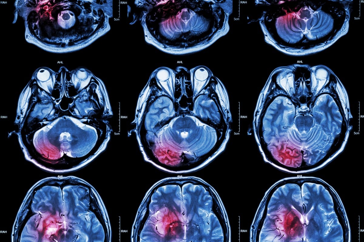
A 3-month-old, female Japanese Black calf that showed signs of neurological dysfunction soon after birth was twice examined by magnetic resonance imaging (MRI). Survey MR images showed changes in a hydrocephalus from mild to severe and the existence of a mass above the brain stem that could be distinguished from the surrounding cerebral parenchyma. Contrast MRI examinations using Gd-DO3A-butriol showed the mass to have a doughnut-like form. As the mass changed, the clinical signs aggravated. We diagnosed a brain stem abscess, which we confirmed pathologically. To our knowledge there are no other reports of the use of contrast MRI to examine cattle. (+info)In vivo proton MR spectroscopy of untreated and treated brain abscesses. (3/457)
MR spectroscopy was performed in three patients with brain abscesses. In two patients, MR spectroscopy revealed end-products of bacterial breakdown (acetate, succinate, amino acids, lactate) in the abscess cysts. In one of these, the spectrum was reversed to a single lactate peak after treatment. In the third patient, MR spectroscopy was performed only after treatment and showed a single nonspecific lactate peak. MR spectroscopy is a potential tool for noninvasive diagnosis of brain abscess and might be useful for evaluating changes after treatment. (+info)Use of diffusion-weighted MR imaging in differential diagnosis between intracerebral necrotic tumors and cerebral abscesses. (4/457)
The differential diagnosis between intracerebral necrotic tumors and cerebral abscesses is frequently impossible with conventional MR imaging. We report two cases of cerebral abscesses that showed high signal on diffusion-weighted echo planar imaging and a strongly reduced apparent diffusion coefficient. This appearance was not present in our cases of necrotic/cystic gliomas (eight cases) and necrotic metastases (two cases). We believe that diffusion-weighted MR imaging may be a diagnostic clue in cases of cerebral "ring-enhancing" masses. (+info)Neuroimaging findings of the development and resolution of solitary brainstem abscess: characteristics of neuroimagings in the early stage of brainstem abscess and importance of surgical management for brainstem abscess--case report. (5/457)
A 64-year-old female presented with a solitary brainstem abscess. Magnetic resonance imaging and computed tomography demonstrated the development and resolution of the brainstem abscess with an unusual and fluctuating clinical course over several months. Serial neuroimaging examinations are required to detect a brainstem abscess in the early stage to establish the optimum treatment. (+info)Cerebral abscesses produced by bacterial implantation and septic embolisation in primates. (6/457)
The degree of brain abscess encapsulation is positively related to surgical mortality and methods to enhance capsule wall formation, therefore, have therapeutic relevance. Two primate models are described which may be useful in the investigation of encapsulation of traumatic and metastatic brain abscesses. Direct intracerebral inoculation induces abscesses displaying more prominent inflammatory responses and encapsulation than does septic embolisation, despite similar abscess age and size. Cerebral ischaemia surrounding metastatic suppurative foci may retard capsule wall formation. (+info)In vivo antibacterial activity of FK041, a new orally active cephalosporin. (7/457)
The therapeutic activities of orally administered FK041 were evaluated in mouse models of systemic and local infections with a variety of bacteria and were compared with those of cefdinir (CFDN) and cefditoren pivoxil (CDTR-PI). FK041 exhibited potent therapeutic activity against lethal systemic infections induced by intraperitoneally inoculated Staphylococcus aureus, Escherichia coli, and Klebsiella pneumoniae with 50% effective doses (ED50) in the range of 0.20 to 0.36 mg/kg and was more active than CFDN and CDTR-PI. This result correlated well with its in vitro activity. The therapeutic effects of FK041 and reference drugs on murine local infections were evaluated in an in vivo pharmacokinetic model simulating human plasma concentrations for oral administration of 50 mg, 100 mg, and 200 mg. Against murine subcutaneous abscess induced by S. aureus, FK041 was as effective as CFDN and significantly more effective than CDTR-PI in reducing the number of recoverable viable bacteria in the skin at the infection sites. The efficacy of FK041 against murine pneumonia with H. influenzae was comparable to that of CDTR-PI and was superior to that of CFDN in reducing viable bacteria activity in the lungs. These results strongly suggest that FK041 has potential for clinical use against various bacterial infections. (+info)Pyogenic brain abscess managed by repeated elective aspiration. (8/457)
37 cases of capsular stage of brain abscess based upon CT scan staging were treated by repeated elective aspiration through a burr hole and intracavitary application of antibiotics on alternate days, till two consecutive negative aspirations were obtained. A combination of furosemide and antibiotics in multiple doses were also given. The mortality rate was 2.7% and the morbidity rate 8.3%. Corticosteroids were not used in the management of brain abscess. Thus, repeated elective aspiration was found to be an effective mode of surgical management of brain abscess. (+info)A brain abscess is a localized collection of pus in the brain that is caused by an infection. It can develop as a result of a bacterial, fungal, or parasitic infection that spreads to the brain from another part of the body or from an infection that starts in the brain itself (such as from a head injury or surgery).
The symptoms of a brain abscess may include headache, fever, confusion, seizures, weakness or numbness on one side of the body, and changes in vision, speech, or behavior. Treatment typically involves antibiotics to treat the infection, as well as surgical drainage of the abscess to relieve pressure on the brain.
It is a serious medical condition that requires prompt diagnosis and treatment to prevent potentially life-threatening complications such as brain herniation or permanent neurological damage.
An abscess is a localized collection of pus caused by an infection. It is typically characterized by inflammation, redness, warmth, pain, and swelling in the affected area. Abscesses can form in various parts of the body, including the skin, teeth, lungs, brain, and abdominal organs. They are usually treated with antibiotics to eliminate the infection and may require drainage if they are large or located in a critical area. If left untreated, an abscess can lead to serious complications such as sepsis or organ failure.
A liver abscess is a localized collection of pus within the liver tissue caused by an infection. It can result from various sources such as bacterial or amebic infections that spread through the bloodstream, bile ducts, or directly from nearby organs. The abscess may cause symptoms like fever, pain in the upper right abdomen, nausea, vomiting, and weight loss. If left untreated, a liver abscess can lead to serious complications, including sepsis and organ failure. Diagnosis typically involves imaging tests like ultrasound or CT scan, followed by drainage of the pus and antibiotic treatment.
An abdominal abscess is a localized collection of pus in the abdominal cavity, caused by an infection. It can occur as a result of complications from surgery, trauma, or inflammatory conditions such as appendicitis or diverticulitis. Symptoms may include abdominal pain, fever, and tenderness at the site of the abscess. Abdominal abscesses can be serious and require medical treatment, which may include antibiotics, drainage of the abscess, or surgery.
A lung abscess is a localized collection of pus in the lung parenchyma caused by an infectious process, often due to bacterial infection. It's characterized by necrosis and liquefaction of pulmonary tissue, resulting in a cavity filled with purulent material. The condition can develop as a complication of community-acquired or nosocomial pneumonia, aspiration of oral secretions containing anaerobic bacteria, septic embolism, or contiguous spread from a nearby infected site.
Symptoms may include cough with foul-smelling sputum, chest pain, fever, weight loss, and fatigue. Diagnosis typically involves imaging techniques such as chest X-ray or CT scan, along with microbiological examination of the sputum to identify the causative organism(s). Treatment often includes antibiotic therapy tailored to the identified pathogen(s), as well as supportive care such as bronchoscopy, drainage, or surgery in severe cases.
Nocardia infections are caused by Nocardia species, a type of gram-positive, aerobic, filamentous bacteria that can be found in soil, dust, and decaying vegetation. These infections primarily affect the lungs (pulmonary nocardiosis) when the bacteria are inhaled but can also spread to other parts of the body, causing disseminated nocardiosis. People with weakened immune systems, such as those with HIV/AIDS, organ transplants, or long-term steroid use, are at a higher risk of developing Nocardia infections. Symptoms vary depending on the site of infection and may include cough, chest pain, shortness of breath, skin abscesses, brain abscesses, or joint inflammation. Diagnosis typically involves microbiological culture and identification of the bacteria from clinical samples, while treatment usually consists of long-term antibiotic therapy, often involving multiple drugs.
An epidural abscess is a localized collection of pus (abscess) in the epidural space, which is the potential space between the dura mater (the outermost membrane covering the brain and spinal cord) and the vertebral column. The infection typically occurs as a result of bacterial invasion into this space and can cause compression of the spinal cord or nerves, leading to serious neurological deficits if not promptly diagnosed and treated.
Epidural abscesses can occur in any part of the spine but are most commonly found in the lumbar region. They may develop as a complication of a nearby infection, such as a skin or soft tissue infection, or as a result of hematogenous spread (spread through the bloodstream) from a distant site of infection. Risk factors for developing an epidural abscess include diabetes, intravenous drug use, spinal surgery, and spinal instrumentation.
Symptoms of an epidural abscess may include back pain, fever, neck stiffness, weakness or numbness in the limbs, and bladder or bowel dysfunction. Diagnosis typically involves imaging studies such as MRI or CT scans, along with laboratory tests to identify the causative organism. Treatment usually consists of surgical drainage of the abscess and administration of antibiotics to eliminate the infection. In some cases, corticosteroids may be used to reduce inflammation and prevent further neurological damage.
A psoas abscess is a localized collection of pus (infectious material) in the iliopsoas muscle compartment, which consists of the psoas major and iliacus muscles. These muscles are located in the lower back and pelvis, responsible for flexing the hip joint.
Psoas abscesses can be classified as primary or secondary:
1. Primary psoas abscess: This type is caused by hematogenous spread (dissemination through the blood) of a bacterial infection from a distant site, often involving the gastrointestinal tract, genitourinary system, or skin. It is less common and typically seen in individuals with compromised immune systems.
2. Secondary psoas abscess: This type is caused by direct extension of an infection from a nearby anatomical structure, such as the spine, vertebral column, or retroperitoneal space (the area behind the peritoneum, the lining of the abdominal cavity). Common causes include spinal osteomyelitis (spinal bone infection), discitis (infection of the intervertebral disc), or a perforated viscus (a hole in an organ like the bowel).
Symptoms of a psoas abscess may include lower back pain, hip pain, fever, chills, and difficulty walking. Diagnosis typically involves imaging studies such as CT scans or MRIs, which can confirm the presence and extent of the abscess. Treatment usually consists of antibiotic therapy and drainage of the abscess, often through a percutaneous (through the skin) approach guided by imaging. In some cases, surgical intervention may be necessary for adequate drainage and management.
Amebic liver abscess is a medical condition characterized by the presence of a pus-filled cavity (abscess) in the liver caused by the infection of the amoeba Entamoeba histolytica. This parasite typically enters the body through contaminated food or water and makes its way to the liver, where it can cause tissue damage and abscess formation. The abscess is usually solitary and contains necrotic debris and inflammatory cells, primarily composed of neutrophils. Symptoms may include fever, right upper quadrant pain, and tender hepatomegaly (enlarged liver). If left untreated, amebic liver abscess can lead to serious complications such as perforation of the liver, bacterial superinfection, or spread of the infection to other organs.
Suppuration is the process of forming or discharging pus. It is a condition that results from infection, tissue death (necrosis), or injury, where white blood cells (leukocytes) accumulate to combat the infection and subsequently die, forming pus. The pus consists of dead leukocytes, dead tissue, debris, and microbes (bacteria, fungi, or protozoa). Suppuration can occur in various body parts such as the lungs (empyema), brain (abscess), or skin (carbuncle, furuncle). Treatment typically involves draining the pus and administering appropriate antibiotics to eliminate the infection.
Citrobacter koseri (formerly known as Citrobacter diversus) is a gram-negative, facultatively anaerobic, motile, and encapsulated bacterium that belongs to the family Enterobacteriaceae. It is commonly found in soil, water, and the gastrointestinal tracts of humans and animals.
Citrobacter koseri can cause a range of infections in humans, including urinary tract infections, pneumonia, sepsis, and meningitis, particularly in immunocompromised individuals or neonates. It is also known to cause nosocomial infections, which are acquired in healthcare settings.
Citrobacter koseri is resistant to many antibiotics, including ampicillin and cephalosporins, making it difficult to treat infections caused by this bacterium. Therefore, accurate identification and appropriate antimicrobial therapy are essential for the successful management of Citrobacter koseri infections.
Central nervous system (CNS) fungal infections refer to invasive fungal diseases that affect the brain and/or spinal cord. These types of infections are relatively uncommon but can be serious and potentially life-threatening, especially in individuals with weakened immune systems due to conditions such as HIV/AIDS, cancer, or organ transplantation.
There are several types of fungi that can cause CNS infections, including:
1. Candida species: These are yeast-like fungi that can cause a range of infections, from superficial to systemic. When they invade the CNS, they can cause meningitis or brain abscesses.
2. Aspergillus species: These are mold-like fungi that can cause invasive aspergillosis, which can affect various organs, including the brain.
3. Cryptococcus neoformans: This is a yeast-like fungus that primarily affects people with weakened immune systems. It can cause meningitis or brain abscesses.
4. Coccidioides species: These are mold-like fungi that can cause coccidioidomycosis, also known as Valley Fever. While most infections are limited to the lungs, some people may develop disseminated disease, which can affect the CNS.
5. Histoplasma capsulatum: This is a mold-like fungus that causes histoplasmosis, which primarily affects the lungs but can disseminate and involve the CNS.
Symptoms of CNS fungal infections may include headache, fever, altered mental status, seizures, stiff neck, and focal neurologic deficits. Diagnosis typically involves a combination of clinical evaluation, imaging studies (such as MRI or CT), and laboratory tests (such as cerebrospinal fluid analysis or fungal cultures). Treatment usually involves long-term antifungal therapy, often with a combination of drugs, and may also include surgical intervention in some cases.
A tuberculoma is a specific type of granulomatous lesion that occurs in the brain due to infection with the Mycobacterium tuberculosis bacterium. This condition is relatively rare in developed countries but is still common in developing nations where tuberculosis (TB) is prevalent.
Intracranial tuberculomas are formed when M. tuberculosis bacteria spread through the bloodstream from a primary focus, usually in the lungs, and lodge in the brain tissue. The bacteria then multiply within the brain, leading to an inflammatory response characterized by the formation of granulomas. These granulomas consist of central caseous necrosis (cheese-like material) surrounded by a layer of epithelioid histiocytes, lymphocytes, and multinucleated giant cells.
Tuberculomas can vary in size from a few millimeters to several centimeters in diameter. They may be solitary or multiple and are often found near the surface of the brain, particularly in the cerebral cortex or meninges (the protective membranes surrounding the brain). The presence of intracranial tuberculomas can lead to various neurological symptoms, such as headaches, seizures, focal deficits, and cognitive impairment.
Diagnosis of intracranial tuberculomas typically involves a combination of imaging techniques (such as CT or MRI scans) and laboratory tests (such as cerebrospinal fluid analysis and PCR for M. tuberculosis). Treatment usually consists of a prolonged course of anti-tuberculous medications, which can help to reduce the size of the lesions and alleviate symptoms. In some cases, surgical intervention may be necessary to remove or decompress large or symptomatic tuberculomas.
A pyogenic liver abscess is a localized collection of pus within the liver parenchyma caused by an infectious process. It's typically characterized by the presence of a purulent material, which can be composed of white blood cells (neutrophils), necrotic debris, and microorganisms. The infection usually spreads to the liver through the hepatic blood vessels from a primary focus of infection elsewhere in the body, such as the gastrointestinal tract, lungs, or dental sources.
The most common causative organisms are Escherichia coli, Klebsiella pneumoniae, and Streptococcus species; however, anaerobes and fungi can also be responsible in certain populations. The clinical presentation of pyogenic liver abscess may include fever, chills, right upper quadrant abdominal pain, nausea, vomiting, and signs of systemic infection. Diagnosis is usually confirmed with imaging techniques such as ultrasound or CT scan, followed by aspiration and culture of the pus for identification of the causative organism(s) and antibiogram-guided antimicrobial therapy. Drainage of the abscess, either percutaneously or surgically, might be required in specific cases to ensure resolution and prevent recurrence.
Brain neoplasms, also known as brain tumors, are abnormal growths of cells within the brain. These growths can be benign (non-cancerous) or malignant (cancerous). Benign brain tumors typically grow slowly and do not spread to other parts of the body. However, they can still cause serious problems if they press on sensitive areas of the brain. Malignant brain tumors, on the other hand, are cancerous and can grow quickly, invading surrounding brain tissue and spreading to other parts of the brain or spinal cord.
Brain neoplasms can arise from various types of cells within the brain, including glial cells (which provide support and insulation for nerve cells), neurons (nerve cells that transmit signals in the brain), and meninges (the membranes that cover the brain and spinal cord). They can also result from the spread of cancer cells from other parts of the body, known as metastatic brain tumors.
Symptoms of brain neoplasms may vary depending on their size, location, and growth rate. Common symptoms include headaches, seizures, weakness or paralysis in the limbs, difficulty with balance and coordination, changes in speech or vision, confusion, memory loss, and changes in behavior or personality.
Treatment for brain neoplasms depends on several factors, including the type, size, location, and grade of the tumor, as well as the patient's age and overall health. Treatment options may include surgery, radiation therapy, chemotherapy, targeted therapy, or a combination of these approaches. Regular follow-up care is essential to monitor for recurrence and manage any long-term effects of treatment.
A focal infection is a focus or source of infection that can spread and cause harm to other parts of the body. A "focal infection, dental" refers to an infection that originates in the teeth or surrounding tissues of the mouth and then spreads to other parts of the body. This can occur when bacteria or other pathogens from a dental infection enter the bloodstream and travel to distant sites, where they can cause inflammation, tissue damage, and illness.
Dental focal infections can be caused by various conditions, such as tooth decay, periodontal disease, abscesses, or other oral infections. The bacteria involved in dental infections are often part of the normal oral flora but can become pathogenic under certain circumstances, such as when they gain access to deeper tissues or the bloodstream due to trauma, surgery, or poor oral hygiene.
If left untreated, dental focal infections can lead to serious health complications, including heart disease, brain abscesses, and other systemic infections. It is essential to maintain good oral hygiene and seek professional dental care to prevent and treat dental infections, reducing the risk of developing focal infections and related health issues.
Drainage, in medical terms, refers to the removal of excess fluid or accumulated collections of fluids from various body parts or spaces. This is typically accomplished through the use of medical devices such as catheters, tubes, or drains. The purpose of drainage can be to prevent the buildup of fluids that may cause discomfort, infection, or other complications, or to treat existing collections of fluid such as abscesses, hematomas, or pleural effusions. Drainage may also be used as a diagnostic tool to analyze the type and composition of the fluid being removed.
Central Nervous System (CNS) Tuberculosis is a specific form of tuberculosis (TB) that refers to the infection and inflammation caused by Mycobacterium tuberculosis in the brain or spinal cord. The two most common forms of CNS tuberculosis are tuberculous meningitis and tuberculomas.
1. Tuberculous Meningitis (TBM): This is the most frequent form of CNS TB, characterized by the inflammation of the membranes surrounding the brain and spinal cord (meninges). The infection can lead to the formation of caseous lesions (granulomas), which may obstruct cerebrospinal fluid (CSF) flow and result in increased intracranial pressure. Symptoms often include headache, fever, altered mental status, neck stiffness, vomiting, and focal neurological deficits.
2. Tuberculomas: These are localized granulomatous lesions formed by the immune response to M. tuberculosis in the brain parenchyma. They can cause various neurological symptoms depending on their size and location, such as seizures, focal deficits, or increased intracranial pressure.
CNS TB is a severe manifestation of tuberculosis that requires prompt diagnosis and treatment to prevent long-term neurological damage or even death. Diagnosis typically involves imaging studies (CT or MRI scans) and analysis of cerebrospinal fluid obtained through lumbar puncture. Treatment usually consists of a prolonged course of multiple antituberculous drugs, along with corticosteroids to manage inflammation and prevent complications.
In medical terms, suction refers to the process of creating and maintaining a partial vacuum in order to remove fluids or gases from a body cavity or wound. This is typically accomplished using specialized medical equipment such as a suction machine, which uses a pump to create the vacuum, and a variety of different suction tips or catheters that can be inserted into the area being treated.
Suction is used in a wide range of medical procedures and treatments, including wound care, surgical procedures, respiratory therapy, and diagnostic tests. It can help to remove excess fluids such as blood or pus from a wound, clear secretions from the airways during mechanical ventilation, or provide a means of visualizing internal structures during endoscopic procedures.
It is important to use proper technique when performing suctioning, as excessive or improperly applied suction can cause tissue damage or bleeding. Medical professionals are trained in the safe and effective use of suction equipment and techniques to minimize risks and ensure optimal patient outcomes.
Brain chemistry refers to the chemical processes that occur within the brain, particularly those involving neurotransmitters, neuromodulators, and neuropeptides. These chemicals are responsible for transmitting signals between neurons (nerve cells) in the brain, allowing for various cognitive, emotional, and physical functions.
Neurotransmitters are chemical messengers that transmit signals across the synapse (the tiny gap between two neurons). Examples of neurotransmitters include dopamine, serotonin, norepinephrine, GABA (gamma-aminobutyric acid), and glutamate. Each neurotransmitter has a specific role in brain function, such as regulating mood, motivation, attention, memory, and movement.
Neuromodulators are chemicals that modify the effects of neurotransmitters on neurons. They can enhance or inhibit the transmission of signals between neurons, thereby modulating brain activity. Examples of neuromodulators include acetylcholine, histamine, and substance P.
Neuropeptides are small protein-like molecules that act as neurotransmitters or neuromodulators. They play a role in various physiological functions, such as pain perception, stress response, and reward processing. Examples of neuropeptides include endorphins, enkephalins, and oxytocin.
Abnormalities in brain chemistry can lead to various neurological and psychiatric conditions, such as depression, anxiety disorders, schizophrenia, Parkinson's disease, and Alzheimer's disease. Understanding brain chemistry is crucial for developing effective treatments for these conditions.
Streptococcus intermedius is a type of Gram-positive coccus bacterium that is part of the Streptococcus anginosus group, also known as the Streptococcus milleri group. These bacteria are normal inhabitants of the mouth, upper respiratory tract, and gastrointestinal tract in humans. However, they can cause opportunistic infections in various parts of the body, such as the brain, lungs, liver, and heart valves, particularly in individuals with compromised immune systems.
S. intermedius infections can range from mild to severe and include abscesses, endocarditis, meningitis, and sepsis. Proper identification of this bacterium is essential for appropriate antibiotic therapy and management of associated infections.
Medical Definition:
Magnetic Resonance Imaging (MRI) is a non-invasive diagnostic imaging technique that uses a strong magnetic field and radio waves to create detailed cross-sectional or three-dimensional images of the internal structures of the body. The patient lies within a large, cylindrical magnet, and the scanner detects changes in the direction of the magnetic field caused by protons in the body. These changes are then converted into detailed images that help medical professionals to diagnose and monitor various medical conditions, such as tumors, injuries, or diseases affecting the brain, spinal cord, heart, blood vessels, joints, and other internal organs. MRI does not use radiation like computed tomography (CT) scans.
Bacillaceae is a family of Gram-positive bacteria that includes the genus Bacillus, which are known for their ability to form endospores. Some species of Bacillus can cause infections in humans, although this is relatively rare.
Infections caused by Bacillus species are typically associated with contaminated food or water, soil, or dust. The most common Bacillus species that causes infections in humans is Bacillus cereus, which can cause foodborne illness characterized by nausea, vomiting, and diarrhea. Other Bacillus species, such as Bacillus anthracis, can cause more serious infections such as anthrax, which can affect the skin, lungs, or gastrointestinal system.
In general, Bacillaceae infections can be treated with antibiotics, although the specific antibiotic used may depend on the species of bacteria causing the infection. Prevention measures include proper food handling and preparation, as well as avoiding contact with contaminated soil or water.
Fusobacterium infections are diseases or conditions caused by the bacterial genus Fusobacterium, which are gram-negative, anaerobic bacilli. These bacteria are commonly found as normal flora in the oral cavity, gastrointestinal tract, and female genital tract. However, under certain circumstances, they can cause infections, particularly in individuals with weakened immune systems or underlying medical conditions.
Fusobacterium infections can manifest in various forms, including:
1. Oral infections: Fusobacterium nucleatum is the most common species associated with oral infections, such as periodontitis, abscesses, and Ludwig's angina.
2. Respiratory tract infections: Fusobacterium necrophorum can cause lung abscesses, empyema, and bronchitis.
3. Bloodstream infections (bacteremia): Fusobacterium species can enter the bloodstream through various routes, such as dental procedures or invasive medical procedures, leading to bacteremia. This condition can be particularly dangerous for individuals with compromised immune systems or underlying medical conditions.
4. Intra-abdominal infections: Fusobacterium species can cause intra-abdominal abscesses, peritonitis, and appendicitis.
5. Skin and soft tissue infections: Fusobacterium species can cause cellulitis, myositis, and necrotizing fasciitis.
6. Bone and joint infections: Fusobacterium species can cause osteomyelitis and septic arthritis.
7. Central nervous system infections: Fusobacterium species can cause meningitis and brain abscesses, although these are rare.
Fusobacterium infections can be challenging to treat due to their anaerobic nature and resistance to certain antibiotics. Therefore, it is essential to seek medical attention if you suspect a Fusobacterium infection. Treatment typically involves the use of appropriate antibiotics, such as metronidazole or clindamycin, and sometimes surgical intervention may be necessary.
A periapical abscess is a localized infection that occurs at the tip of the tooth's root, specifically in the periapical tissue. This tissue surrounds the end of the tooth's root and helps anchor the tooth to the jawbone. The infection is usually caused by bacteria that enter the pulp chamber of the tooth as a result of dental caries (tooth decay), periodontal disease, or trauma that damages the tooth's protective enamel layer.
The infection leads to pus accumulation in the periapical tissue, forming an abscess. The symptoms of a periapical abscess may include:
1. Pain and tenderness in the affected tooth, which can be throbbing or continuous
2. Swelling in the gums surrounding the tooth
3. Sensitivity to hot, cold, or pressure on the tooth
4. Fever, general malaise, or difficulty swallowing (in severe cases)
5. A foul taste in the mouth or bad breath
6. Tooth mobility or loosening
7. Formation of a draining sinus tract (a small opening in the gums that allows pus to drain out)
Periapical abscesses require dental treatment, which typically involves removing the infected pulp tissue through root canal therapy and cleaning, shaping, and sealing the root canals. In some cases, antibiotics may be prescribed to help control the infection, but they do not replace the necessary dental treatment. If left untreated, a periapical abscess can lead to severe complications, such as the spread of infection to other parts of the body or tooth loss.
A retropharyngeal abscess is a deep neck infection involving the potential space between the buccopharyngeal fascia and the alar fascia, primarily located in the retropharyngeal space. This space extends from the base of the skull to the mediastinum and contains loose connective tissue, fat, and lymph nodes. The infection usually originates from an upper respiratory tract infection or a penetrating injury to the posterior pharyngeal wall.
The abscess can cause swelling and compression of surrounding structures, leading to potentially serious complications such as airway obstruction, mediastinitis, or sepsis if left untreated. Symptoms may include neck pain, difficulty swallowing, fever, drooling, and decreased appetite. Diagnosis is typically made through a combination of clinical examination, imaging studies (such as CT or MRI scans), and laboratory tests. Treatment usually involves surgical drainage of the abscess and antibiotic therapy to manage the infection.
A craniotomy is a surgical procedure where a bone flap is temporarily removed from the skull to access the brain. This procedure is typically performed to treat various neurological conditions, such as brain tumors, aneurysms, arteriovenous malformations, or traumatic brain injuries. After the underlying brain condition is addressed, the bone flap is usually replaced and secured back in place with plates and screws. The purpose of a craniotomy is to provide access to the brain for diagnostic or therapeutic interventions while minimizing potential damage to surrounding tissues.
A brain injury is defined as damage to the brain that occurs following an external force or trauma, such as a blow to the head, a fall, or a motor vehicle accident. Brain injuries can also result from internal conditions, such as lack of oxygen or a stroke. There are two main types of brain injuries: traumatic and acquired.
Traumatic brain injury (TBI) is caused by an external force that results in the brain moving within the skull or the skull being fractured. Mild TBIs may result in temporary symptoms such as headaches, confusion, and memory loss, while severe TBIs can cause long-term complications, including physical, cognitive, and emotional impairments.
Acquired brain injury (ABI) is any injury to the brain that occurs after birth and is not hereditary, congenital, or degenerative. ABIs are often caused by medical conditions such as strokes, tumors, anoxia (lack of oxygen), or infections.
Both TBIs and ABIs can range from mild to severe and may result in a variety of physical, cognitive, and emotional symptoms that can impact a person's ability to perform daily activities and function independently. Treatment for brain injuries typically involves a multidisciplinary approach, including medical management, rehabilitation, and supportive care.
X-ray computed tomography (CT or CAT scan) is a medical imaging method that uses computer-processed combinations of many X-ray images taken from different angles to produce cross-sectional (tomographic) images (virtual "slices") of the body. These cross-sectional images can then be used to display detailed internal views of organs, bones, and soft tissues in the body.
The term "computed tomography" is used instead of "CT scan" or "CAT scan" because the machines take a series of X-ray measurements from different angles around the body and then use a computer to process these data to create detailed images of internal structures within the body.
CT scanning is a noninvasive, painless medical test that helps physicians diagnose and treat medical conditions. CT imaging provides detailed information about many types of tissue including lung, bone, soft tissue and blood vessels. CT examinations can be performed on every part of the body for a variety of reasons including diagnosis, surgical planning, and monitoring of therapeutic responses.
In computed tomography (CT), an X-ray source and detector rotate around the patient, measuring the X-ray attenuation at many different angles. A computer uses this data to construct a cross-sectional image by the process of reconstruction. This technique is called "tomography". The term "computed" refers to the use of a computer to reconstruct the images.
CT has become an important tool in medical imaging and diagnosis, allowing radiologists and other physicians to view detailed internal images of the body. It can help identify many different medical conditions including cancer, heart disease, lung nodules, liver tumors, and internal injuries from trauma. CT is also commonly used for guiding biopsies and other minimally invasive procedures.
In summary, X-ray computed tomography (CT or CAT scan) is a medical imaging technique that uses computer-processed combinations of many X-ray images taken from different angles to produce cross-sectional images of the body. It provides detailed internal views of organs, bones, and soft tissues in the body, allowing physicians to diagnose and treat medical conditions.
The brain is the central organ of the nervous system, responsible for receiving and processing sensory information, regulating vital functions, and controlling behavior, movement, and cognition. It is divided into several distinct regions, each with specific functions:
1. Cerebrum: The largest part of the brain, responsible for higher cognitive functions such as thinking, learning, memory, language, and perception. It is divided into two hemispheres, each controlling the opposite side of the body.
2. Cerebellum: Located at the back of the brain, it is responsible for coordinating muscle movements, maintaining balance, and fine-tuning motor skills.
3. Brainstem: Connects the cerebrum and cerebellum to the spinal cord, controlling vital functions such as breathing, heart rate, and blood pressure. It also serves as a relay center for sensory information and motor commands between the brain and the rest of the body.
4. Diencephalon: A region that includes the thalamus (a major sensory relay station) and hypothalamus (regulates hormones, temperature, hunger, thirst, and sleep).
5. Limbic system: A group of structures involved in emotional processing, memory formation, and motivation, including the hippocampus, amygdala, and cingulate gyrus.
The brain is composed of billions of interconnected neurons that communicate through electrical and chemical signals. It is protected by the skull and surrounded by three layers of membranes called meninges, as well as cerebrospinal fluid that provides cushioning and nutrients.
A Peritonsillar Abscess (also known as a Quinsy) is a localized collection of pus in the peritonsillar space, which is the potential space between the tonsillar capsule and the pharyngeal constrictor muscle. It is a serious complication of tonsillitis or pharyngitis, often caused by bacterial infection. The abscess can cause severe pain, difficulty swallowing, fever, and swelling of the neck and face. If left untreated, it can lead to more severe complications such as airway obstruction or the spread of infection. Treatment typically involves drainage of the abscess, antibiotics, and supportive care.
Frontal sinusitis is a type of sinus infection that specifically involves the frontal sinuses, which are located in the forehead region above the eyes. The condition is characterized by inflammation and infection of the mucous membrane lining the frontal sinuses, leading to symptoms such as headaches, facial pain or pressure, nasal congestion, and thick nasal discharge.
Frontal sinusitis can be caused by viral, bacterial, or fungal infections, as well as structural issues like nasal polyps or deviated septum that obstruct the sinus drainage pathways. Treatment options for frontal sinitis may include antibiotics, nasal decongestants, corticosteroids, saline nasal irrigation, and in some cases, endoscopic sinus surgery to alleviate obstructions and improve sinus drainage.
Hereditary Hemorrhagic Telangiectasia (HHT) is a rare genetic disorder that affects the blood vessels. It is also known as Osler-Weber-Rendu syndrome. This condition is characterized by the formation of abnormal blood vessels called telangiectases, which are small red spots or tiny bulges that can be found in the skin, mucous membranes (like those inside the nose, mouth, and GI tract), and sometimes in vital organs like the lungs and brain.
These telangiectases have a tendency to bleed easily, leading to potentially serious complications such as anemia due to chronic blood loss, and in some cases, strokes or brain abscesses if the telangiectases in the brain rupture. HHT is typically inherited in an autosomal dominant pattern, meaning that a child has a 50% chance of inheriting the gene from an affected parent. There are several genes associated with HHT, the most common being ACVRL1, ENG, and SMAD4.
Streptococcus milleri Group (SMG) is not a single species, but a group of closely related streptococcal species that are often difficult to distinguish from each other using conventional laboratory methods. The group includes Streptococcus anginosus, Streptococcus intermedius, and Streptococcus constellatus. These bacteria are part of the normal flora in the human mouth, upper respiratory tract, and gastrointestinal system. However, they can cause a variety of infectious diseases, particularly in immunocompromised individuals or when they invade deep tissues or sterile sites. Infections caused by SMG can range from mild to severe, including abscesses, endocarditis, and sepsis. Due to the complexity of identifying these organisms to the species level, they are often reported together as the Streptococcus milleri Group.
"Gemella" is a genus of gram-positive, facultatively anaerobic, beta-hemolytic bacteria that are part of the normal flora in the human mouth, upper respiratory tract, and gastrointestinal tract. There are two species within this genus: Gemella haemolysans and Gemella morphilorufinata (previously known as Neisseria morphilorufinata). These bacteria can occasionally cause infections such as endocarditis, bacteremia, and meningitis, particularly in individuals with underlying medical conditions or compromised immune systems. The name "Gemella" is derived from the Latin word for "twin," reflecting the similarity of these organisms to each other.
Nocardia is a genus of aerobic, gram-positive, filamentous bacteria that can be found in soil, water, and decaying vegetation. It is known to cause various infectious diseases in humans and animals, known as nocardiosis. The infection often enters the body through inhalation, skin wounds, or surgical procedures. Nocardia species are opportunistic pathogens, meaning they mainly cause disease in individuals with weakened immune systems, such as those with HIV/AIDS, organ transplants, or cancer. The infection can affect various organs, including the lungs, brain, skin, and eyes, leading to symptoms like cough, fever, chest pain, weight loss, and skin abscesses. Proper diagnosis and treatment with antibiotics are crucial for managing nocardiosis.
Anti-bacterial agents, also known as antibiotics, are a type of medication used to treat infections caused by bacteria. These agents work by either killing the bacteria or inhibiting their growth and reproduction. There are several different classes of anti-bacterial agents, including penicillins, cephalosporins, fluoroquinolones, macrolides, and tetracyclines, among others. Each class of antibiotic has a specific mechanism of action and is used to treat certain types of bacterial infections. It's important to note that anti-bacterial agents are not effective against viral infections, such as the common cold or flu. Misuse and overuse of antibiotics can lead to antibiotic resistance, which is a significant global health concern.
Brain mapping is a broad term that refers to the techniques used to understand the structure and function of the brain. It involves creating maps of the various cognitive, emotional, and behavioral processes in the brain by correlating these processes with physical locations or activities within the nervous system. Brain mapping can be accomplished through a variety of methods, including functional magnetic resonance imaging (fMRI), positron emission tomography (PET) scans, electroencephalography (EEG), and others. These techniques allow researchers to observe which areas of the brain are active during different tasks or thoughts, helping to shed light on how the brain processes information and contributes to our experiences and behaviors. Brain mapping is an important area of research in neuroscience, with potential applications in the diagnosis and treatment of neurological and psychiatric disorders.
Empyema is a medical condition characterized by the accumulation of pus in a body cavity, most commonly in the pleural space surrounding the lungs. It is usually caused by a bacterial infection that spreads from the lung tissue to the pleural space. The buildup of pus can cause chest pain, cough, fever, and difficulty breathing. Empyema can be a complication of pneumonia or other respiratory infections, and it may require treatment with antibiotics, drainage of the pus, and sometimes surgery.
Neuroaspergillosis is a rare and serious invasive fungal infection caused by the Aspergillus species, which primarily affects the central nervous system (CNS), including the brain and spinal cord. This condition is often seen in individuals with weakened immune systems due to underlying medical conditions such as hematological malignancies, solid organ transplantation, or advanced HIV infection.
The infection can occur through various routes, including direct extension from the paranasal sinuses, hematogenous dissemination, or direct inoculation during neurosurgical procedures. Neuroaspergillosis may present with a wide range of symptoms, such as headache, altered mental status, seizures, focal neurologic deficits, and signs of increased intracranial pressure.
Diagnosis typically involves imaging studies (MRI or CT scans), cerebrospinal fluid analysis, and sometimes tissue biopsy to detect the presence of Aspergillus hyphae or DNA. Treatment usually consists of a combination of antifungal medications, such as voriconazole or isavuconazole, and surgical debridement when possible. The prognosis for neuroaspergillosis is generally poor due to the difficulty in treating CNS infections and the underlying immunocompromised state of affected individuals.
A fatal outcome is a term used in medical context to describe a situation where a disease, injury, or illness results in the death of an individual. It is the most severe and unfortunate possible outcome of any medical condition, and is often used as a measure of the severity and prognosis of various diseases and injuries. In clinical trials and research, fatal outcome may be used as an endpoint to evaluate the effectiveness and safety of different treatments or interventions.
Staphylococcal infections are a type of infection caused by Staphylococcus bacteria, which are commonly found on the skin and nose of healthy people. However, if they enter the body through a cut, scratch, or other wound, they can cause an infection.
There are several types of Staphylococcus bacteria, but the most common one that causes infections is Staphylococcus aureus. These infections can range from minor skin infections such as pimples, boils, and impetigo to serious conditions such as pneumonia, bloodstream infections, and toxic shock syndrome.
Symptoms of staphylococcal infections depend on the type and severity of the infection. Treatment typically involves antibiotics, either topical or oral, depending on the severity and location of the infection. In some cases, hospitalization may be necessary for more severe infections. It is important to note that some strains of Staphylococcus aureus have developed resistance to certain antibiotics, making them more difficult to treat.
The mastoid is a term used in anatomy and refers to the bony prominence located at the base of the skull, posterior to the ear. More specifically, it's part of the temporal bone, one of the bones that forms the side and base of the skull. The mastoid process provides attachment for various muscles involved in chewing and moving the head.
In a medical context, "mastoid" can also refer to conditions or procedures related to this area. For example, mastoiditis is an infection of the mastoid process, while a mastoidectomy is a surgical procedure that involves removing part or all of the mastoid process.
An encyclopedia is a comprehensive reference work containing articles on various topics, usually arranged in alphabetical order. In the context of medicine, a medical encyclopedia is a collection of articles that provide information about a wide range of medical topics, including diseases and conditions, treatments, tests, procedures, and anatomy and physiology. Medical encyclopedias may be published in print or electronic formats and are often used as a starting point for researching medical topics. They can provide reliable and accurate information on medical subjects, making them useful resources for healthcare professionals, students, and patients alike. Some well-known examples of medical encyclopedias include the Merck Manual and the Stedman's Medical Dictionary.
Paranasal sinuses are air-filled cavities in the skull that surround the nasal cavity. There are four pairs of paranasal sinuses, including the maxillary, frontal, ethmoid, and sphenoid sinuses. These sinuses help to warm, humidify, and filter the air we breathe. They also contribute to our voice resonance and provide a slight cushioning effect for the skull. The openings of the paranasal sinuses lead directly into the nasal cavity, allowing mucus produced in the sinuses to drain into the nose. Infections or inflammation of the paranasal sinuses can result in conditions such as sinusitis.
 Brain abscess
Amoebic brain abscess
Abscess
Vladimir Cerrón
Curvularia pallescens
Eugene Nicholas Myers
Encephalitis
Enterococcus avium
Methanobrevibacter oralis
Staphylococcus massiliensis
Magnetic resonance imaging of the brain
Rhinocladiella mackenziei
Palinopsia
Blood-brain barrier
Meningitis
William Cavendish-Scott-Bentinck, Marquess of Titchfield
Chaetomium
Cladophialophora bantiana
Citrobacter koseri
Anaerobic infection
Mycoplasma salivarium
Mycoplasmataceae
Tsukamurella tyrosinosolvens
Barry Young (musician)
Kocuria varians
Robert Galbraith Heath
Clostridium tertium
Nocardia farcinica
Elizabeth Hayes
Ali Niyaf
Brain abscess - Wikipedia
 Brain Abscess: Background, Pathophysiology, Epidemiology
Pediatric Streptococcus-Associated Brain Abscesses and Empyemas
 Brain Abscess
 Anaerobic Brain Abscess following Chronic Suppurative Otitis Media in a Child from Uganda
Mycotic Brain Abscess Caused by Opportunistic Reptile Pathogen - Volume 11, Number 2-February 2005 - Emerging Infectious...
 Man who died of brain abscess should have seen GP face-to-face, says NHS | ITV News Granada
 Nocardia puris brain abscess in an immunocompromised woman | Microbiology Society
 Meningitis, sinus thrombosis and abscess of the brain : with appendices on lumbar puncture and its uses and diseases of the...
 Listeria monocytogenes brain abscess: A case presentation | Eurorad
 ICD-9 Code 013.34 -Tuberculous abscess of brain tubercle bacilli not found (in sputum) by microscopy but found by bacterial...
 Intracranial Epidural Abscess and Subdural Empyema - Brain, Spinal Cord, and Nerve Disorders - Merck Manuals Consumer Version
 Brain Abscess
Brain Abscess and Risk of Cancer | Neurology
 A retrospective study on the aetiology, management, and outcome of brain abscess in an 11-year, single-centre study from China ...
 Brain abscess - Neurosurgery
 Brain Abscess Treatment - HealthFinder
 Brain abscess draining • GoreCenter
Brain Abscess in Emergency Medicine: Background, Pathophysiology, Epidemiology
Brain Abscess - Neurologic Disorders - MSD Manual Professional Edition
 Amoebic brain abscess | Patient Innovation
brain abscess - Emergency Medicine Ireland
 An immunosuppressed patient with systemic vasculitis suffering from cerebral abscesses due to Nocardia farcinica identified by...
Brain Abscess - brain and spine specialist
 Brain abscess | Guide to Diagnostic Tests
 Abscess / Deranged - Abscess / Deranged - Encyclopaedia Metallum: The Metal Archives
 Brain abscess | Anatomia Collection: anatomical plates 1522-1867
Brain Abscess | HKU E-learning Platform in Clinical Neurosciences
 Brain Abscess | University of Edinburgh Archive and Manuscript Collections
Meningitis17
- Organisms that are most frequently associated with brain abscess in patients with AIDS are poliovirus, Toxoplasma gondii, and Cryptococcus neoformans, though in infection with the latter organism, symptoms of meningitis generally predominate. (wikipedia.org)
- Intracranial abscesses can originate from infection of contiguous structures (eg, otitis media , dental infection, mastoiditis, sinusitis) secondary to hematogenous spread from a remote site (especially in patients with cyanotic congenital heart disease), after skull trauma or surgery, and, rarely, following meningitis . (medscape.com)
- CNS manifestations of listerosis depend on the affected intracranial compartment and include meningitis, encephalitis, rhombencephalitis, meningoencephalitis and less commonly cerebral abscess. (eurorad.org)
- Acute Bacterial Meningitis Acute bacterial meningitis is rapidly developing inflammation of the layers of tissue that cover the brain and spinal cord (meninges) and of the fluid-filled space between the meninges (subarachnoid. (merckmanuals.com)
- Because meningitis is now uncommon in children, epidural abscesses and subdural empyemas are also uncommon in children. (merckmanuals.com)
- Meningitis or a brain abscess may develop. (merckmanuals.com)
- Meningitis rarely results in brain abscess by direct extension, particularly in adults, and therefore in most cases the finding of brain abscess should not prompt a search for meningeal infection via lumbar puncture. (medscape.com)
- Here we report a case of brain abscess caused by Salmonella enterica serovar Enteritidis mimicking post-surgical meningitis in a patient with glioblastoma multiforme. (biomedcentral.com)
- In this case report, a brain abscess was initially diagnosed as Salmonella post-surgical meningitis before the imaging diagnosis of the brain abscess. (biomedcentral.com)
- The diagnosis of brain abscess should be considered in all cases of non-typhoidal Salmonella meningitis after surgery for brain tumor. (biomedcentral.com)
- Combining Ceftriaxone with Doxycycline and Daptomycin Reduces Mortality, Neuroinflammation, Brain Damage, and Hearing Loss in Infant Rat Pneumococcal Meningitis. (escmid.org)
- Both encephalitis and meningitis are severe illnesses caused due to viral or bacterial infection and result in the inflammation of the brain. (differencebetween.net)
- Meningeal infections, which affect the protective layers (cellular tissues) that encircle the brain and spinal cord, frequently result in meningitis. (differencebetween.net)
- The infection of the meninges (three layers of membranes that cover and protect your brain and spinal cord) is known as meningitis. (differencebetween.net)
- Meningitis is an inflammation of the meninges (which protects the nervous system), the protective layer surrounding the brain and spinal cord. (differencebetween.net)
- Encephalitis is an infection of the entire brain tissue, whereas meningitis is an inflammation of the (protective membranes of the brain and the spiral cord) meninges. (differencebetween.net)
- What is the difference between meningitis encephalitis and brain abscess? (differencebetween.net)
Infection26
- Brain abscess (or cerebral abscess) is an abscess within the brain tissue caused by inflammation and collection of infected material coming from local (ear infection, dental abscess, infection of paranasal sinuses, infection of the mastoid air cells of the temporal bone, epidural abscess) or remote (lung, heart, kidney etc.) infectious sources. (wikipedia.org)
- These symptoms are caused by a combination of increased intracranial pressure due to a space-occupying lesion (headache, vomiting, confusion, coma), infection (fever, fatigue etc.) and focal neurologic brain tissue damage (hemiparesis, aphasia etc. (wikipedia.org)
- The case of a Ugandan child with a polymicrobial brain abscess including infection with Tissierella praeacuta / Clostridium hastiforme requiring repeated drainage and eventual surgical excision is reported. (hindawi.com)
- Invasive infection is very rare in humans, and most were observed in immunocompromised patients, manifesting as osteomyelitis ( 3 , 4 ) or diffuse vascular brain invasion ( 5 ). (cdc.gov)
- An abscess may form in the brain when bacteria from an infection elsewhere in the head or in the bloodstream or from a wound enter the brain. (merckmanuals.com)
- Brain abscess is caused due to bacterial infection in the brain. (yashodahospitals.com)
- The infection in the brain may be due to infections in other parts of the body viz. (yashodahospitals.com)
- However, brain abscess may occur due to infection of the skull, through the blood stream and head injury. (yashodahospitals.com)
- Brain abscess es are suppurative infection s of the brain parenchyma surrounded by a vascularized capsule . (neurosurgery.directory)
- Initial parenchymal infection is known as cerebritis, which may progress into a cerebral abscess. (neurosurgery.directory)
- Brain abscess is a focal intracranial infection that may present as a life-threatening emergency. (medscape.com)
- improvements in the detection and treatment of ear, sinus, and orofacial infections has decreased the incidence of brain abscess as a consequence of direct spread of infection. (medscape.com)
- The pathogenesis of an invading organism that has inoculated the brain parenchyma is variable and dependent on the initial site of infection, host factors, and geographic location. (medscape.com)
- Brain abscess may be caused by the contiguous spread of pathogens from a primary focus of infection outside of the CNS that extends into the brain. (medscape.com)
- An abscess on the brain is a collection of pus that forms due to an infection or trauma. (brainandspinespecialist.com)
- A bacterial or fungal infection in the brain is the most common cause of a brain abscess. (brainandspinespecialist.com)
- Invasive Salmonella infections have been reported due to their potential to cause focal suppurative complications in urinary tract infection, osteoarticular infection and liver abscess [ 1 ]. (biomedcentral.com)
- A tooth abscess is a pocket of pus caused by a bacterial infection. (adam.com)
- If you have a severe infection, your tooth may need to be removed, or you may need surgery to drain the abscess. (adam.com)
- A brain abscess is a collection of pus, immune cells, and other material in the brain, caused by a bacterial or fungal infection. (adam.com)
- Brain biopsy showed Entamoeba histolytica infection. (aku.edu)
- A brain abscess is a focal infection characterized by a collection of pus in the brain parenchyma. (bvsalud.org)
- We describe a case of brain abscess secondary to a pulmonary infection in an elderly, diabetic, Afghani man with an extensive history of chewing tobacco use. (kabbesh.com)
- While this immune response can protect the brain by isolating the infection, it can also do more harm than good. (geometry.net)
- Oral pathogens from an odontogenic infection may enter the brain via a hematologic or lymphatic route, or by direct extension through the fascial planes. (bvsalud.org)
- The treatment of abscesses of the central nervous system (CNS) is based on three main points: clinical treatment with antibiotics, neurosurgical treatment of the abscess using a method appropriate to the case, and treatment of the primary focus of infection. (bvsalud.org)
Inflammation8
- Brain abscess is caused by intracranial inflammation with subsequent abscess formation. (medscape.com)
- [ 5 , 6 ] The process begins with direct inoculation of microorganisms into the brain parenchyma, resulting in focal inflammation in the 1-3 days following, which is referred to as early cerebritis. (medscape.com)
- An abscess forms when an area of cerebral inflammation becomes necrotic and encapsulated by glial cells and fibroblasts. (msdmanuals.com)
- Inflammation and swelling occur when bacteria, fungi, or parasites infect a portion of the brain. (brainandspinespecialist.com)
- The brain swells in response to the inflammation, and the mass may put pressure on delicate brain tissue. (geometry.net)
- On the other side, encephalitis occurs due to the swelling (inflammation) of the entire brain. (differencebetween.net)
- Brain inflammation known as encephalitis can be brought on by a virus, bacteria, medicine, or immune system problem. (differencebetween.net)
- Encephalitis is a rare but serious condition that causes inflammation of the brain itself. (differencebetween.net)
Cerebral abscesses2
- Cerebral abscesses result from pathogens growing within the brain parenchyma. (neurosurgery.directory)
- Multiple bilateral cerebral abscesses with hemorrhage. (geometry.net)
Epidural abscess2
- An intracranial epidural abscess is a pocket of pus that develops between the skull and the top layer of tissues (dura mater) covering the brain. (merckmanuals.com)
- To diagnose an epidural abscess or a subdural empyema, doctors use magnetic resonance imaging (MRI) done after gadolinium is injected intravenously. (merckmanuals.com)
Infections17
- Intracranial abscesses are uncommon, serious, life-threatening infections. (medscape.com)
- Direct extension usually causes a single brain abscess and may occur from necrotic areas of osteomyelitis in the posterior wall of the frontal sinus, the sphenoid and ethmoid sinuses, mandibular dental infections, as well as from subacute and chronic otitis media and mastoiditis. (medscape.com)
- The frequency of brain abscesses resulting from ear infections has declined in developed countries. (medscape.com)
- [ 1 ] Pediatric bacterial brain abscesses, epidural empyemas, and subdural empyemas, rare complications of respiratory infections and sinusitis, are often caused by Streptococcus species but might also be polymicrobial or caused by other genera, such as Staphylococcus . (medscape.com)
- Discussions with clinicians in multiple states raised concerns (intracranial abscess and granuloma) or G06.2 (extradural and about a possible increase in pediatric intracranial infections, subdural abscess, unspecified) during the study period were particularly those caused by Streptococcus bacteria, during the included. (cdc.gov)
- Brain abscesses are rare and account for 1-10% of CNS listerial infections (4,5). (eurorad.org)
- Majorly, the heart and lung infections are considered to cause brain abscess. (yashodahospitals.com)
- The brain is protected from all infections. (yashodahospitals.com)
- Brain abscess occurs as the result of a complication of variety of infections, trauma, or surgery and carries significant morbidity and mortality. (medscape.com)
- [ 12 ] Dental infections can lead to brain abscess via either contiguous or hematogenous routes. (medscape.com)
- Severe invasive infections such as brain abscess in a child should prompt an immune evaluation. (johnshopkins.edu)
- But poor dental hygiene and tooth abscess can increase your risk for lung infections caused by these bacteria. (medlineplus.gov)
- In the postgraduate education course "acute bacterial CNS infections of the brain", the first focus will be on the diagnostic work-up of patients with suspected CNS infections. (escmid.org)
- Nocardia are non-motile, Gram-positive, partially acid-fast bacilli, most commonly responsible for lung, brain, and skin infections in immunocompromised hosts including those with acquired immunodeficiency syndrome (AIDS), cancer, lupus, and diabetes. (kabbesh.com)
- Ear and sinus infections may also spread directly to the brain because of their close proximity. (geometry.net)
- Diagnosis: Cerebral abscess Discussion: Between (30% and 60%) of pyogenic abscesses are mixed infections, with aerobic isolates outnumbering anaerobic isolates approximately 2 to 1. (geometry.net)
- A circumscribed collection of purulent exudate in the brain, due to bacterial and other infections. (bvsalud.org)
Complications3
- Brain abscess may result in severe complications viz. (yashodahospitals.com)
- Untreated abscesses may get worse and can lead to life-threatening complications. (adam.com)
- Complications like brain abscess always remain a danger. (ispub.com)
Aspiration9
- A computer-guided needle-aspiration of the brain lesions yielded yellow-brown, creamy fluid in which abundant septated fungal hyphae were detected microscopically ( Figure C). Cytologic investigation was consistent with a necrotic abscess. (cdc.gov)
- To determine the demographics, management, and the variables that affect the outcome in subjects with brain abscesses treated at a single centre over an 11-year period, we retrospectively analysed data in 60 patients with brain abscesses surgically treated with stereotactically guided aspiration or open craniotomy excision in Shanghai Changzheng Hospital between January 2001 and December 2011. (biomedcentral.com)
- Stereotactically guided aspiration is an effective treatment for brain abscess with an overall favourable outcome. (biomedcentral.com)
- Two primary treatments are used to manage brain abscesses: stereotactically guided aspiration and open craniotomy excision. (biomedcentral.com)
- Despite the advent of modern neurosurgical techniques, including stereotactic brain biopsy and aspiration, better culturing techniques to identify the infectious agent, new antibiotics, and modern non-invasive neuroimaging procedures, brain abscess still poses a public health challenge, especially in developing countries. (biomedcentral.com)
- To reduce operative and anesthetic risk in these patients, abscesses should be managed by less invasive aspiration methods guided by computed tomography. (neurosurgery.directory)
- A CT-guided stereotactic aspiration of brain abscess was performed in a series of 15 patients, of which 12 patients had single abscess formation while 3 patients had multiple brain abscess. (tmu.edu.tw)
- Thus stereotactic aspiration appears to be a safe and effective method for the treatment of brain abscess. (tmu.edu.tw)
- Methods: Fifty-one patients with brain abscesses who underwent navigation-assisted abscess aspiration with antibiotic treatment were included in this study. (korea.ac.kr)
Polymicrobial2
- Many brain abscesses are polymicrobial. (wikipedia.org)
- We report here three cases of patients with otogenic brain abscesses of polymicrobial origin that had in common the isolation of Actinomyces europaeus, which has not been previously described in this location . (bvsalud.org)
Case of brain abscess1
- Here, we report a case of brain abscess caused by S. enterica subspecies (subsp. (biomedcentral.com)
Etiology of brain abscess1
- Hematogenous seeding of the brain from an extracranial source is the second most common etiology of brain abscess, accounting for approximately 25% of cases. (medscape.com)
Incidence of brain abscess1
- One trend is that there appears to be a trend toward a decreasing incidence of brain abscess. (neurosurgery.directory)
Treatment of brain abscess1
- Dr. Nitin Jagdhane is one of the best Neurosurgeon and Spine specialists for an accurate diagnosis and treatment of brain abscess in Mumbai, India. (brainandspinespecialist.com)
Computed Tomography3
- Brain Computed Tomography (CT) and Magnetic Resonance Imaging (MRI) were performed. (eurorad.org)
- Abscesses larger than 2 cm in diameter, in deep-located or parieto-occipital regions, should be aspirated immediately and repeatedly, mainly using computed tomography-guided methods to decrease intracranial pressure and avoid IVROBA. (neurosurgery.directory)
- Brain computed tomography revealed a large cerebral abscess located at the operative site. (biomedcentral.com)
Include brain abscess1
- They include brain abscess and subdural or extradural empyema and are classified according to the anatomical location or the etiologic agent. (medscape.com)
Collection of pus1
- A brain abscess is an intracerebral collection of pus. (msdmanuals.com)
Term brain abscess1
- The term brain abscess is used in this article to represent all types of intracranial abscesses. (medscape.com)
Surgical4
- Mortality due to brain abscess was not directly related to surgery nor surgical technique. (biomedcentral.com)
- What are the operations/ surgical treatments available for Brain abscess? (brainandspinespecialist.com)
- Surgical drainage of the abscess was performed and microbial cultures of surgical deep samples were positive for the same S. enterica Enteritidis isolate. (biomedcentral.com)
- Surgical brain abscess drainage followed by prolonged antibiotic treatment remains a major therapeutic option. (biomedcentral.com)
Drain the abscess1
- When abscess build-up is severe, surgery is performed to drain the abscess. (yashodahospitals.com)
Fungi1
- Brain abscesses commonly occur when bacteria or fungi infect part of the brain. (geometry.net)
DIAGNOSIS11
- All inpatient encounters from patients aged ≤18 years with a primary or secondary discharge diagnosis of International Classification of Diseases, Tenth Revision, Clinical Modification code G06.0 (intracranial abscess and granuloma) or G06.2 (extradural and subdural abscess, unspecified) during the study period were included. (medscape.com)
- In CDC's national call for cases, a case was defined as the diagnosis of brain abscess, epidural empyema, or subdural empyema in a person aged ≤18 years without a previous neurosurgical procedure or history of head trauma, hospitalized on or after June 1, 2021, irrespective of etiology. (medscape.com)
- In May 2022, CDC learned of three children in California encounters from patients aged 18 years with a primary or hospitalized concurrently for brain abscess, epidural empyema, secondary discharge diagnosis of International Classification or subdural empyema caused by Streptococcus intermedius . (cdc.gov)
- Pediatric bacterial brain abscesses, epidural empy- codes U07.1 or B97.29 on the discharge diagnosis list. (cdc.gov)
- On June 9, CDC asked clini- diagnosis of brain abscess, epidural empyema, or subdural cians and health departments to report possible cases of these empyema in a person aged 18 years without a previous neu- conditions and to submit clinical specimens for laboratory rosurgical procedure or history of head trauma, hospitalized testing. (cdc.gov)
- see Brain abscess diagnosis . (neurosurgery.directory)
- Significant advances in the diagnosis and management of bacterial brain abscess over the past several decades have improved the expected outcome of a disease once regarded as invariably fatal. (neurosurgery.directory)
- Diagnosis Intracranial tumors may involve the brain or other structures (eg, cranial nerves, meninges). (msdmanuals.com)
- Diagnosis Ischemic stroke is sudden neurologic deficits that result from focal cerebral ischemia associated with permanent brain infarction (eg, positive results on diffusion-weighted MRI). (msdmanuals.com)
- How is the diagnosis of Brain Abscess done? (brainandspinespecialist.com)
- Click on Images for Enlarged View Diagnosis cerebral abscess. (geometry.net)
Prognosis3
- We analysed the prognostic factors and strategies of treatment to look for trends in occurrence, prognosis, predisposing risk factors, and infectious agent in brain abscesses. (biomedcentral.com)
- Multicenter prospective studies and randomized clinical trials are needed to further advance treatment and prognosis in brain abscess patients. (neurosurgery.directory)
- Prognosis has improved over time, likely due to improved brain imaging techniques, minimally invasive neurosurgical procedures, and protocoled antibiotic treatment 5) . (neurosurgery.directory)
Cavity3
- The video shows an surgery in which pus is suctioned from the brain cavity. (gorecenter.com)
- After the operation, brain CT scans revealed immediate shrinkage of the abscess cavity and gradual disappearance of the enhanced ring. (tmu.edu.tw)
- Right fronto-parietal craniotomy was performed by the neurosurgeon and an encapsulated abscess cavity was drained and completely removed. (ispub.com)
Tooth abscess2
Multiple abscesses2
- If there are multiple abscesses, it may be too dangerous to remove them all. (brainandspinespecialist.com)
- Mortality was significantly associated with increasing age, multiple abscesses, immunosuppression and the presence of an underlying cardiac anomaly. (ox.ac.uk)
Parenchyma2
- Pathogens may originate from adjacent bone, teeth, sinus mucosa, internal auditory canal, or cochlear structures and travel into the intracranial vault via venous drainage or valveless emissary veins, thus inoculating the brain parenchyma. (medscape.com)
- The inflammatory reaction causes encephalitis if they manage to enter the brain parenchyma itself. (differencebetween.net)
Pocket of pus2
- A subdural empyema is a pocket of pus that develops between the dura mater and the middle layer of the tissues (arachnoid mater) covering the brain. (merckmanuals.com)
- Abscess of the Brain A brain abscess is a pocket of pus in the brain. (merckmanuals.com)
Mortality3
- 90% of brain abscesses in immunocompromised transplant patients with an associated mortality rate of 97% ( 10 ), despite aggressive surgery and antifungal therapy ( 9 ). (cdc.gov)
- BACKGROUND: Brain abscess is an uncommon condition, but carries high mortality. (ox.ac.uk)
- Long-term mortality and epilepsy in patients after brain abscess: A nationwide population-based matched cohort study. (escmid.org)
Antibiotic treatment1
- There has been a gradual improvement in the outcome of patients with brain abscess over the past 50 years, which might be driven by improved brain imaging techniques, minimally invasive neurosurgical procedures, and protocoled antibiotic treatment. (neurosurgery.directory)
20223
- In May 2022, CDC learned of three children in California hospitalized concurrently for brain abscess, epidural empyema, or subdural empyema caused by Streptococcus intermedius . (medscape.com)
- Available Streptococcus specimens isolated from a brain abscess, pandemic in March 2020, increased during summer 2021 to epidural empyema, subdural empyema, blood, or cerebrospinal a peak in March 2022, and then declined to baseline levels. (cdc.gov)
- He was first diagnosed with lung cancer in August 2020, but only publicly shared the news in November 2022 after the cancer spread to his brain. (yahoo.com)
Subdural empyema1
- Available Streptococcus specimens isolated from a brain abscess, epidural empyema, subdural empyema, blood, or cerebrospinal fluid were collected for antimicrobial susceptibility testing and whole-genome sequencing at CDC's Streptococcus reference laboratory to identify microbiological features shared among cases. (medscape.com)
Epidemiology2
- In a article, Chen review the literature to find out how the epidemiology of this disease has changed through the years and re-visit the basic pathological process of abscess evolution and highlight the new research in the biochemical pathways that initiate and regulate this process 1) . (neurosurgery.directory)
- There have been several trends in the epidemiology of brain abscess over recent decades. (neurosurgery.directory)
Congenital heart d2
- Brain abscess is usually associated with congenital heart disease in young children. (wikipedia.org)
- Other possible causes for brain abscess are congenital heart disease, pulmonary arteriovenous fistula, dental abscess or tooth decay, pneumonia, endocarditis, peritonitis and cystitis. (yashodahospitals.com)
Empyema2
- Through collaboration with the Children's Hospital Association (CHA), CDC analyzed nationally representative pediatric hospitalizations for brain abscess and empyema. (medscape.com)
- The call for Association (CHA), CDC analyzed nationally representative cases was shared with health departments and two provider list- pediatric hospitalizations for brain abscess and empyema. (cdc.gov)
Streptococcus2
- Microbial aetiology of brain abscess in a UK cohort: Prominent role of Streptococcus intermedius. (ox.ac.uk)
- Mixed brain abscess due to Streptococcus and Tuberculosis in a pregnant woman with favorable evolution. (globalsciencelibrary.com)
Sinusitis3
- However, abscesses complicating sinusitis has not decreased in frequency. (medscape.com)
- Magnetic resonance imaging (MRI) of the brain disclosed 2 masses, 3.3 and 4.8 cm in diameter, respectively ( Figure A), and signs of chronic sinusitis. (cdc.gov)
- Although less common, brain abscess has been described as a complication of frontal, ethmoidal, or sphenoidal sinusitis. (medscape.com)
Cerebellar1
- Brain MRI showed two lesions located on the left cerebellar peduncle (Fig 2A) and on the left frontal lobe (Fig 2B). (eurorad.org)
Frontal3
- Brain CT revealed a nonspecific hypodense lesion of the left frontal lobe (Fig 1). (eurorad.org)
- Most of the abscesses were solitary frontal or parietal lesions. (ox.ac.uk)
- Brain magnetic resonance imaging showed a mass in the left frontal lobe, while abdominal computerized tomography showed a left renal malignancy. (aku.edu)
Patients13
- female patients and patients greater than 40 years of age were most likely to suffer a brain abscess. (biomedcentral.com)
- Additional studies will continue to reveal patients trends that may improve treatment for brain abscess. (biomedcentral.com)
- This report is based on our experience with 82 brain abscesses surgically managed in 60 patients over an 11-year period at one single hospital. (biomedcentral.com)
- Sixty patients with a total of 82 brain abscesses verified by postoperative pathology were treated surgically between January 2001 and December 2011 at the department of Neurosurgery in Shanghai Changzheng Hospital. (biomedcentral.com)
- Patients were excluded from the study if, 1) there was evidence of neurological symptoms unrelated to brain abscess, 2) there was evidence showing the patient had not undergone a drainage procedure or intraoperative pus sampling, or 3) the patient was lost to follow-up during the study period. (biomedcentral.com)
- particularly in HIV-infected patients) can cause abscesses. (msdmanuals.com)
- We also review cases of Salmonella brain abscess in patients with cerebral tumors. (biomedcentral.com)
- is a cause of brain abscesses in patients with underlying pulmonary structural disease like bronchiectasis. (archivosdeneurociencias.org)
- METHODS: We undertook a retrospective, observational study of patients with brain abscess, based on hospital coding, in a UK tertiary referral teaching hospital. (ox.ac.uk)
- Objective: The purpose of this study was to describe the clinical characteristics, treatment outcomes, and prognostic factors in patients with brain abscesses treated in a single institute during a recent 10-year period. (korea.ac.kr)
- Conclusion: We found that the level of consciousness at admission was associated with treatment outcomes in patients with brain abscesses. (korea.ac.kr)
- In neutropenic patients, brain abscesses may be caused by candida or aspergillus. (geometry.net)
- A high index of suspicion is essential in susceptible patients presenting with pulmonary infiltrate, cerebral abscess or deep soft-tissue abscess and usually requires an active diagnostic workup and early administration of appropriate therapy. (who.int)
Bacteria3
- At least 3 different pathways have been suggested for CNS invasion: 1) through the blood-brain and blood-choroid barriers, within infected phagocytes, 2) direct invasion of the capillary endothelium by the circulating extracellular bacteria, 3) retrograde migration into the CNS, by travelling through the axons of cranial nerves and especially along trigerminal nerve (7). (eurorad.org)
- Brain abscess may rupture inside the skull due to gasses produced by the bacteria. (yashodahospitals.com)
- CNS abscess A brain abscess is a mass of immune cells, pus, and other material that can occur when the brain is infected by bacteria or fungus. (geometry.net)
Antibiotics1
- If a doctor detects a brain abscess, they will usually prescribe broad-spectrum antibiotics right away because an abscess can be fatal. (brainandspinespecialist.com)
Neurosurgery1
- Brain abscess can occur months after neurosurgery. (medscape.com)
Organism1
- It is also important to note that the immune status of the patient can influence the organism causing the abscess. (geometry.net)
Nocardia2
- was isolated from the brain abscess and identified as Nocardia abscessus complex by 16S rRNA sequencing. (archivosdeneurociencias.org)
- Browne W D, Lieberson R E, Kabbesh M J (November 08, 2021) Nocardia cyriacigeorgica Brain and Lung Abscesses in 77-Year-Old Man With Diabetes. (kabbesh.com)
Commonly1
- These abscesses are more commonly multiple and multiloculated and are frequently found in the distribution of the middle cerebral artery. (medscape.com)
Prevalence2
- Meanwhile, the population prevalence of chronic immune suppression and immunocompromise has grown, and with it there has been a rise in opportunistic and fungal brain abscess. (medscape.com)
- The prevalence of brain abscesses in young children has decreased as a result of vaccination campaigns. (brainandspinespecialist.com)
Tumor2
- Salmonella brain abscess associated with brain tumor is rare. (biomedcentral.com)
- Brain magnetic resonance imaging (MRI) revealed a single 40×35 mm tumor in her right mesial temporal region and a mass effect compression of her right lateral ventricle with transtentorial herniation (Fig. 1 ). (biomedcentral.com)
Amoebic2
Tissue2
- Within the skull, the brain is covered by three layers of tissue called the meninges. (merckmanuals.com)
- When an abscess expands, it puts more pressure on the brain tissue around it. (brainandspinespecialist.com)
Membrane2
- A brain abscess is a focal area of necrosis starting in an area of cerebritis surrounded by a membrane . (neurosurgery.directory)
- A wall or membrane forms around the abscess as the cells amass. (brainandspinespecialist.com)
Isolates1
- And if a disease isolates itself, it is referred to as a brain abscess. (differencebetween.net)
Pathology1
- cerebral abscess Looks at the common causes,bacterial aetiology, pathology, clinical presentation and treatment. (geometry.net)
Skull fracture1
- Trauma that causes an open skull fracture allows organisms to seed directly in the brain. (medscape.com)
Empyemas3
- CDC will continue to work with investigation partners to monitor ongoing trends in pediatric brain abscesses and empyemas. (medscape.com)
- Two data sources were analyzed: 1) pediatric hospitalizations for brain abscesses, epidural empyemas, and subdural empyemas reported to CHA's Pediatric Health Information System (PHIS) and 2) cases reported to CDC in response to a national call for cases. (medscape.com)
- 6 ). The agar diffusion gradient method (Etest, bioMérieux) brain abscesses and empyemas. (cdc.gov)