Brain Diseases
Brain
Brain Diseases, Metabolic, Inborn
Encephalitis
Magnetic Resonance Imaging
Mucopolysaccharidosis I
Leukoencephalopathies
Brain Diseases, Metabolic
Brain Chemistry
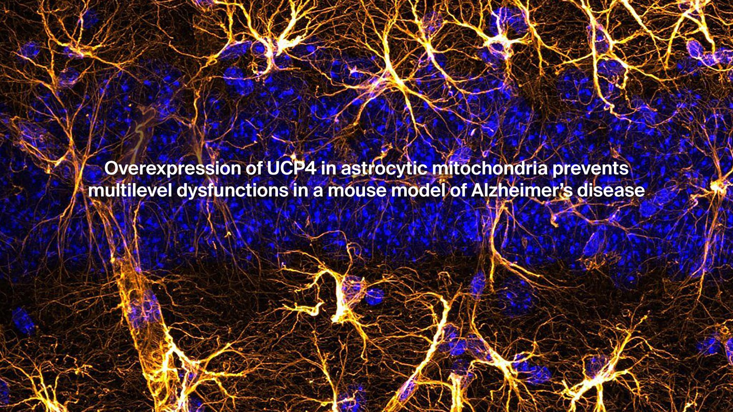
Alzheimer Disease
Blood-Brain Barrier
Neurons
Brain Injuries
Neural Stem Cells
Brain Neoplasms
Astrocytes
Schizophrenia
Brain Mapping
Disease Models, Animal
Microglia
Hippocampus
Image Processing, Computer-Assisted
Aging
Mental Disorders
Mice, Inbred C57BL
Rats, Sprague-Dawley
Magnetic Resonance Spectroscopy
Brain Edema
Cells, Cultured
Immunohistochemistry
Biological Markers
Brain Stem
Brain Ischemia
Brain Abscess
Hypoxia, Brain
Cerebral Cortex
Brain Damage, Chronic
MedlinePlus
Thinking
National Library of Medicine (U.S.)
Human complex sound analysis. (1/3110)
The analysis of complex sound features is important for the perception of environmental sounds, speech and music, and may be abnormal in disorders such as specific language impairment in children, and in common adult lesions including stroke and multiple sclerosis. This work addresses the problem of how the human auditory system detects features in complex sound, and uses those features to perceive the auditory world. The work has been carried out using two independent means of testing the same hypotheses; detailed psychophysical studies of neurological patients with central lesions, and functional imaging using positron emission tomography and functional magnetic resonance imaging of normal subjects. The psychophysical and imaging studies have both examined which brain areas are concerned with the analysis of auditory space, and which are concerned with the analysis of timing information in the auditory system. This differs from many previous human auditory studies, which have concentrated on the analysis of sound frequency. The combined lesion and functional imaging approach has demonstrated analysis of the spatial property of sound movement within the right parietal lobe. The timing work has confirmed that the primary auditory cortex is active as a function of the time structure of sound, and therefore not only concerned with frequency representation of sounds. (+info)Computerised tomography and intellectual impairment in the elderly. (2/3110)
Sixty-six elderly subjects (mean age 77 years) whose mental state was assessed clinically and by simple psychometric tests have been studied by computerised tomography. The mean maximum ventricular area in the 17 mentally normal subjects was above the upper limit of normal for younger subjects, and there was a broad relationship between increasing ventricular dilatation and increasing intellectual impairment. No such clear relationship was demonstrable for measures of cortical atrophy. (+info)Computerised axial tomography in patients with severe migraine: a preliminary report. (3/3110)
Patients suffering from severe migraine, usually for many years, have been examined by the EMI scanner between attacks. Judged by criteria validated originally by comparison with pneumoencephalography, about half of the patients showed evidence of cerebral atrophy. Perhaps of more significance than generalised atrophy was the frequency of areas of focal atrophy and of evidence of infarction. (+info)Activated human T cells, B cells, and monocytes produce brain-derived neurotrophic factor in vitro and in inflammatory brain lesions: a neuroprotective role of inflammation? (4/3110)
Brain-derived neurotrophic factor (BDNF) has potent effects on neuronal survival and plasticity during development and after injury. In the nervous system, neurons are considered the major cellular source of BDNF. We demonstrate here that in addition, activated human T cells, B cells, and monocytes secrete bioactive BDNF in vitro. Notably, in T helper (Th)1- and Th2-type CD4(+) T cell lines specific for myelin autoantigens such as myelin basic protein or myelin oligodendrocyte glycoprotein, BDNF production is increased upon antigen stimulation. The BDNF secreted by immune cells is bioactive, as it supports neuronal survival in vitro. Using anti-BDNF monoclonal antibody and polyclonal antiserum, BDNF immunoreactivity is demonstrable in inflammatory infiltrates in the brain of patients with acute disseminated encephalitis and multiple sclerosis. The results raise the possibility that in the nervous system, inflammatory infiltrates have a neuroprotective effect, which may limit the success of nonselective immunotherapies. (+info)Ma1, a novel neuron- and testis-specific protein, is recognized by the serum of patients with paraneoplastic neurological disorders. (5/3110)
The identification of antineuronal antibodies has facilitated the diagnosis of paraneoplastic neurological disorders and the early detection of the associated tumours. It has also led to the cloning of possibly important neuron-specific proteins. In this study we wanted to identify novel antineuronal antibodies in the sera of patients with paraneoplastic neurological disorders and to clone the corresponding antigens. Serological studies of 1705 sera from patients with suspected paraneoplastic neurological disorders resulted in the identification of four patients with antibodies that reacted with 37 and 40 kDa neuronal proteins (anti-Ma antibodies). Three patients had brainstem and cerebellar dysfunction, and one had dysphagia and motor weakness. Autopsy of two patients showed loss of Purkinje cells, Bergmann gliosis and deep cerebellar white matter inflammatory infiltrates. Extensive neuronal degeneration, gliosis and infiltrates mainly composed of CD8+ T cells were also found in the brainstem of one patient. In normal human and rat tissues, the anti-Ma antibodies reacted exclusively with neurons and with testicular germ cells; the reaction was mainly with subnuclear elements (including the nucleoli) and to a lesser degree the cytoplasm. Anti-Ma antibodies also reacted with the cancers (breast, colon and parotid) available from three anti-Ma patients, but not with 66 other tumours of varying histological types. Preincubation of tissues with any of the anti-Ma sera abrogated the reactivity of the other anti-Ma immunoglobulins. Probing of a human complementary DNA library with anti-Ma serum resulted in the cloning of a gene that encodes a novel 37 kDa protein (Mal). Recombinant Mal was specifically recognized by the four anti-Ma sera but not by 337 control sera, including those from 52 normal individuals, 179 cancer patients without paraneoplastic neurological symptoms, 96 patients with paraneoplastic syndromes and 10 patients with non-cancer-related neurological disorders. The expression of Mal mRNA is highly restricted to the brain and testis. Subsequent analysis suggested that Mal is likely to be a phosphoprotein. Our study demonstrates that some patients with paraneoplastic neurological disorders develop antibodies against Mal, a new member of an expanding family of 'brain/testis' proteins. (+info)Two similar cases of encephalopathy, possibly a reversible posterior leukoencephalopathy syndrome: serial findings of magnetic resonance imaging, SPECT and angiography. (6/3110)
Two young women who had encephalopathy that resembled reversible posterior leukoencephalopathy syndrome are presented. The brain magnetic resonance imaging (MRI) of these patients exhibited similar T2-high signal lesions, mostly in the white matter of the posterior hemispheres. Xe-SPECT during the patients' symptomatic period showed hypoperfusion in the corresponding areas, and angiography demonstrated irregular narrowing of the posterior cerebral artery. Clinical manifestations subsided soon after treatment, and the abnormal radiological findings also were almost completely resolved. Thus, we concluded that transient hypoperfusion followed by ischemia and cytotoxic edema might have had a pivotal role in these cases. (+info)Mitochondrial encephalomyopathies: the enigma of genotype versus phenotype. (7/3110)
Over the past decade a large body of evidence has accumulated implicating defects of human mitochondrial DNA in the pathogenesis of a group of disorders known collectively as the mitochondrial encephalomyopathies. Although impaired oxidative phosphorylation is likely to represent the final common pathway leading to cellular dysfunction in these diseases, fundamental issues still remain elusive. Perhaps the most challenging of these is to understand the mechanisms which underlie the complex relationship between genotype and phenotype. Here we examine this relationship and discuss some of the factors which are likely to be involved. (+info)Changes in the diffusion of water and intracellular metabolites after excitotoxic injury and global ischemia in neonatal rat brain. (8/3110)
The reduction of the apparent diffusion coefficient (ADC) of brain tissue water in acute cerebral ischemia, as measured by diffusion-weighted magnetic resonance imaging, is generally associated with the development of cytotoxic edema. However, the underlying mechanism is still unknown. Our aim was to elucidate diffusion changes in the intracellular environment in cytotoxic edematous tissue. The ADC of intracellular metabolites was measured by use of diffusion-weighted 1H-magnetic resonance spectroscopy after (1) unilateral N-methyl-D-aspartate (NMDA) injection and (2) cardiac arrest-induced global ischemia in neonatal rat brain. The distinct water ADC drop early after global ischemia was accompanied by a significant reduction of the ADC of all measured metabolites (P < 0.01, n = 8). In the first hours after excitotoxic injury, the ADC of water and the metabolites taurine and N-acetylaspartate dropped significantly (P < 0.05, n = 8). At 24 and 72 hours after NMDA injection brain metabolite levels were diminished and metabolite ADC approached contralateral values. Administration of the NMDA-antagonist MK-801 1.5 hours after NMDA injection completely normalized the water ADC but not the metabolite ADC after 1 to 2 hours (n = 8). No damage was detected 72 hours later and, water and metabolite ADC had normal values (n = 8). The contribution of brain temperature changes (calculated from the chemical shift between the water and N-acetylaspartate signals) and tissue deoxygenation to ischemia-induced intracellular ADC changes was minor. These data lend support to previous suggestions that the ischemia-induced brain water ADC drop may partly be caused by reduced diffusional displacement of intracellular water, possibly involving early alterations in intracellular tortuosity, cytoplasmic streaming, or intracellular molecular interactions. (+info)Brain diseases, also known as neurological disorders, refer to a wide range of conditions that affect the brain and nervous system. These diseases can be caused by various factors such as genetics, infections, injuries, degeneration, or structural abnormalities. They can affect different parts of the brain, leading to a variety of symptoms and complications.
Some examples of brain diseases include:
1. Alzheimer's disease - a progressive degenerative disorder that affects memory and cognitive function.
2. Parkinson's disease - a movement disorder characterized by tremors, stiffness, and difficulty with coordination and balance.
3. Multiple sclerosis - a chronic autoimmune disease that affects the nervous system and can cause a range of symptoms such as vision loss, muscle weakness, and cognitive impairment.
4. Epilepsy - a neurological disorder characterized by recurrent seizures.
5. Brain tumors - abnormal growths in the brain that can be benign or malignant.
6. Stroke - a sudden interruption of blood flow to the brain, which can cause paralysis, speech difficulties, and other neurological symptoms.
7. Meningitis - an infection of the membranes surrounding the brain and spinal cord.
8. Encephalitis - an inflammation of the brain that can be caused by viruses, bacteria, or autoimmune disorders.
9. Huntington's disease - a genetic disorder that affects muscle coordination, cognitive function, and mental health.
10. Migraine - a neurological condition characterized by severe headaches, often accompanied by nausea, vomiting, and sensitivity to light and sound.
Brain diseases can range from mild to severe and may be treatable or incurable. They can affect people of all ages and backgrounds, and early diagnosis and treatment are essential for improving outcomes and quality of life.
The brain is the central organ of the nervous system, responsible for receiving and processing sensory information, regulating vital functions, and controlling behavior, movement, and cognition. It is divided into several distinct regions, each with specific functions:
1. Cerebrum: The largest part of the brain, responsible for higher cognitive functions such as thinking, learning, memory, language, and perception. It is divided into two hemispheres, each controlling the opposite side of the body.
2. Cerebellum: Located at the back of the brain, it is responsible for coordinating muscle movements, maintaining balance, and fine-tuning motor skills.
3. Brainstem: Connects the cerebrum and cerebellum to the spinal cord, controlling vital functions such as breathing, heart rate, and blood pressure. It also serves as a relay center for sensory information and motor commands between the brain and the rest of the body.
4. Diencephalon: A region that includes the thalamus (a major sensory relay station) and hypothalamus (regulates hormones, temperature, hunger, thirst, and sleep).
5. Limbic system: A group of structures involved in emotional processing, memory formation, and motivation, including the hippocampus, amygdala, and cingulate gyrus.
The brain is composed of billions of interconnected neurons that communicate through electrical and chemical signals. It is protected by the skull and surrounded by three layers of membranes called meninges, as well as cerebrospinal fluid that provides cushioning and nutrients.
Metabolic brain diseases are a group of disorders caused by genetic defects that affect the body's metabolism and result in abnormal accumulation of harmful substances in the brain. These conditions are present at birth (inborn) or develop during infancy or early childhood. Examples of metabolic brain diseases that are present at birth include:
1. Phenylketonuria (PKU): A disorder caused by a deficiency of the enzyme phenylalanine hydroxylase, which leads to an accumulation of phenylalanine in the brain and can cause intellectual disability, seizures, and behavioral problems if left untreated.
2. Maple syrup urine disease (MSUD): A disorder caused by a deficiency of the enzyme branched-chain ketoacid dehydrogenase, which leads to an accumulation of branched-chain amino acids in the body and can cause intellectual disability, seizures, and metabolic crisis if left untreated.
3. Urea cycle disorders: A group of disorders caused by defects in enzymes that help remove ammonia from the body. Accumulation of ammonia in the blood can lead to brain damage, coma, or death if not treated promptly.
4. Organic acidemias: A group of disorders caused by defects in enzymes that help break down certain amino acids and other organic compounds. These conditions can cause metabolic acidosis, seizures, and developmental delays if left untreated.
Early diagnosis and treatment of these conditions are crucial to prevent irreversible brain damage and other complications. Treatment typically involves dietary restrictions, supplements, and medications to manage the underlying metabolic imbalance. In some cases, enzyme replacement therapy or liver transplantation may be necessary.
Encephalitis is defined as inflammation of the brain parenchyma, which is often caused by viral infections but can also be due to bacterial, fungal, or parasitic infections, autoimmune disorders, or exposure to toxins. The infection or inflammation can cause various symptoms such as headache, fever, confusion, seizures, and altered consciousness, ranging from mild symptoms to severe cases that can lead to brain damage, long-term disabilities, or even death.
The diagnosis of encephalitis typically involves a combination of clinical evaluation, imaging studies (such as MRI or CT scans), and laboratory tests (such as cerebrospinal fluid analysis). Treatment may include antiviral medications, corticosteroids, immunoglobulins, and supportive care to manage symptoms and prevent complications.
Medical Definition:
Magnetic Resonance Imaging (MRI) is a non-invasive diagnostic imaging technique that uses a strong magnetic field and radio waves to create detailed cross-sectional or three-dimensional images of the internal structures of the body. The patient lies within a large, cylindrical magnet, and the scanner detects changes in the direction of the magnetic field caused by protons in the body. These changes are then converted into detailed images that help medical professionals to diagnose and monitor various medical conditions, such as tumors, injuries, or diseases affecting the brain, spinal cord, heart, blood vessels, joints, and other internal organs. MRI does not use radiation like computed tomography (CT) scans.
Mucopolysaccharidosis I (MPS I) is a rare genetic disorder caused by the deficiency of an enzyme called alpha-L-iduronidase. This enzyme is responsible for breaking down complex sugars called glycosaminoglycans (GAGs), also known as mucopolysaccharides, in the body.
When the enzyme is deficient, GAGs accumulate in various tissues and organs, leading to a range of symptoms that can affect different parts of the body, including the skeletal system, heart, respiratory system, eyes, and central nervous system. There are three subtypes of MPS I: Hurler syndrome (the most severe form), Hurler-Scheie syndrome (an intermediate form), and Scheie syndrome (the least severe form).
The symptoms and severity of MPS I can vary widely depending on the specific subtype, with Hurler syndrome typically causing more significant health problems and a shorter life expectancy than the other two forms. Treatment options for MPS I include enzyme replacement therapy, bone marrow transplantation, and various supportive therapies to manage symptoms and improve quality of life.
Leukoencephalopathies are a group of medical conditions that primarily affect the white matter of the brain, which consists mainly of nerve fibers covered by myelin sheaths. These conditions are characterized by abnormalities in the structure and function of the white matter, leading to various neurological symptoms such as cognitive decline, motor impairment, seizures, and behavioral changes.
The term "leukoencephalopathy" is derived from two Greek words: "leukos," meaning white, and "enkephalos," meaning brain. The suffix "-pathy" refers to a disease or suffering. Therefore, leukoencephalopathies refer specifically to diseases that affect the white matter of the brain.
There are various types of leukoencephalopathies, including genetic, metabolic, infectious, toxic, and immune-mediated forms. Some examples include multiple sclerosis, adrenoleukodystrophy, Alexander disease, Canavan disease, and Marchiafava-Bignami disease. The diagnosis of leukoencephalopathies typically involves a combination of clinical evaluation, imaging studies such as MRI, and sometimes genetic or laboratory testing to identify the underlying cause. Treatment depends on the specific type and severity of the condition and may include medications, dietary modifications, physical therapy, or supportive care.
Metabolic brain diseases refer to a group of conditions that are caused by disruptions in the body's metabolic processes, which affect the brain. These disorders can be inherited or acquired and can result from problems with the way the body produces, breaks down, or uses energy and nutrients.
Examples of metabolic brain diseases include:
1. Mitochondrial encephalomyopathies: These are a group of genetic disorders that affect the mitochondria, which are the energy-producing structures in cells. When the mitochondria don't function properly, it can lead to muscle weakness, neurological problems, and developmental delays.
2. Leukodystrophies: These are a group of genetic disorders that affect the white matter of the brain, which is made up of nerve fibers covered in myelin, a fatty substance that insulates the fibers and helps them transmit signals. When the myelin breaks down or is not produced properly, it can lead to cognitive decline, motor problems, and other neurological symptoms.
3. Lysosomal storage disorders: These are genetic disorders that affect the lysosomes, which are structures in cells that break down waste products and recycle cellular materials. When the lysosomes don't function properly, it can lead to the accumulation of waste products in cells, including brain cells, causing damage and neurological symptoms.
4. Maple syrup urine disease: This is a genetic disorder that affects the way the body breaks down certain amino acids, leading to a buildup of toxic levels of these substances in the blood and urine. If left untreated, it can cause brain damage, developmental delays, and other neurological problems.
5. Homocystinuria: This is a genetic disorder that affects the way the body processes an amino acid called methionine, leading to a buildup of homocysteine in the blood. High levels of homocysteine can cause damage to the blood vessels and lead to neurological problems, including seizures, developmental delays, and cognitive decline.
Treatment for metabolic brain diseases may involve dietary changes, supplements, medications, or other therapies aimed at managing symptoms and preventing further damage to the brain. In some cases, a stem cell transplant may be recommended as a treatment option.
Brain chemistry refers to the chemical processes that occur within the brain, particularly those involving neurotransmitters, neuromodulators, and neuropeptides. These chemicals are responsible for transmitting signals between neurons (nerve cells) in the brain, allowing for various cognitive, emotional, and physical functions.
Neurotransmitters are chemical messengers that transmit signals across the synapse (the tiny gap between two neurons). Examples of neurotransmitters include dopamine, serotonin, norepinephrine, GABA (gamma-aminobutyric acid), and glutamate. Each neurotransmitter has a specific role in brain function, such as regulating mood, motivation, attention, memory, and movement.
Neuromodulators are chemicals that modify the effects of neurotransmitters on neurons. They can enhance or inhibit the transmission of signals between neurons, thereby modulating brain activity. Examples of neuromodulators include acetylcholine, histamine, and substance P.
Neuropeptides are small protein-like molecules that act as neurotransmitters or neuromodulators. They play a role in various physiological functions, such as pain perception, stress response, and reward processing. Examples of neuropeptides include endorphins, enkephalins, and oxytocin.
Abnormalities in brain chemistry can lead to various neurological and psychiatric conditions, such as depression, anxiety disorders, schizophrenia, Parkinson's disease, and Alzheimer's disease. Understanding brain chemistry is crucial for developing effective treatments for these conditions.
Alzheimer's disease is a progressive disorder that causes brain cells to waste away (degenerate) and die. It's the most common cause of dementia — a continuous decline in thinking, behavioral and social skills that disrupts a person's ability to function independently.
The early signs of the disease include forgetting recent events or conversations. As the disease progresses, a person with Alzheimer's disease will develop severe memory impairment and lose the ability to carry out everyday tasks.
Currently, there's no cure for Alzheimer's disease. However, treatments can temporarily slow the worsening of dementia symptoms and improve quality of life.
The Blood-Brain Barrier (BBB) is a highly specialized, selective interface between the central nervous system (CNS) and the circulating blood. It is formed by unique endothelial cells that line the brain's capillaries, along with tight junctions, astrocytic foot processes, and pericytes, which together restrict the passage of substances from the bloodstream into the CNS. This barrier serves to protect the brain from harmful agents and maintain a stable environment for proper neural function. However, it also poses a challenge in delivering therapeutics to the CNS, as most large and hydrophilic molecules cannot cross the BBB.
Neurons, also known as nerve cells or neurocytes, are specialized cells that constitute the basic unit of the nervous system. They are responsible for receiving, processing, and transmitting information and signals within the body. Neurons have three main parts: the dendrites, the cell body (soma), and the axon. The dendrites receive signals from other neurons or sensory receptors, while the axon transmits these signals to other neurons, muscles, or glands. The junction between two neurons is called a synapse, where neurotransmitters are released to transmit the signal across the gap (synaptic cleft) to the next neuron. Neurons vary in size, shape, and structure depending on their function and location within the nervous system.
A brain injury is defined as damage to the brain that occurs following an external force or trauma, such as a blow to the head, a fall, or a motor vehicle accident. Brain injuries can also result from internal conditions, such as lack of oxygen or a stroke. There are two main types of brain injuries: traumatic and acquired.
Traumatic brain injury (TBI) is caused by an external force that results in the brain moving within the skull or the skull being fractured. Mild TBIs may result in temporary symptoms such as headaches, confusion, and memory loss, while severe TBIs can cause long-term complications, including physical, cognitive, and emotional impairments.
Acquired brain injury (ABI) is any injury to the brain that occurs after birth and is not hereditary, congenital, or degenerative. ABIs are often caused by medical conditions such as strokes, tumors, anoxia (lack of oxygen), or infections.
Both TBIs and ABIs can range from mild to severe and may result in a variety of physical, cognitive, and emotional symptoms that can impact a person's ability to perform daily activities and function independently. Treatment for brain injuries typically involves a multidisciplinary approach, including medical management, rehabilitation, and supportive care.
Neural stem cells (NSCs) are a type of undifferentiated cells found in the central nervous system, including the brain and spinal cord. They have the ability to self-renew and generate the main types of cells found in the nervous system, such as neurons, astrocytes, and oligodendrocytes. NSCs are capable of dividing symmetrically to increase their own population or asymmetrically to produce one stem cell and one differentiated cell. They play a crucial role in the development and maintenance of the nervous system, and have the potential to be used in regenerative medicine and therapies for neurological disorders and injuries.
Brain neoplasms, also known as brain tumors, are abnormal growths of cells within the brain. These growths can be benign (non-cancerous) or malignant (cancerous). Benign brain tumors typically grow slowly and do not spread to other parts of the body. However, they can still cause serious problems if they press on sensitive areas of the brain. Malignant brain tumors, on the other hand, are cancerous and can grow quickly, invading surrounding brain tissue and spreading to other parts of the brain or spinal cord.
Brain neoplasms can arise from various types of cells within the brain, including glial cells (which provide support and insulation for nerve cells), neurons (nerve cells that transmit signals in the brain), and meninges (the membranes that cover the brain and spinal cord). They can also result from the spread of cancer cells from other parts of the body, known as metastatic brain tumors.
Symptoms of brain neoplasms may vary depending on their size, location, and growth rate. Common symptoms include headaches, seizures, weakness or paralysis in the limbs, difficulty with balance and coordination, changes in speech or vision, confusion, memory loss, and changes in behavior or personality.
Treatment for brain neoplasms depends on several factors, including the type, size, location, and grade of the tumor, as well as the patient's age and overall health. Treatment options may include surgery, radiation therapy, chemotherapy, targeted therapy, or a combination of these approaches. Regular follow-up care is essential to monitor for recurrence and manage any long-term effects of treatment.
Astrocytes are a type of star-shaped glial cell found in the central nervous system (CNS), including the brain and spinal cord. They play crucial roles in supporting and maintaining the health and function of neurons, which are the primary cells responsible for transmitting information in the CNS.
Some of the essential functions of astrocytes include:
1. Supporting neuronal structure and function: Astrocytes provide structural support to neurons by ensheathing them and maintaining the integrity of the blood-brain barrier, which helps regulate the entry and exit of substances into the CNS.
2. Regulating neurotransmitter levels: Astrocytes help control the levels of neurotransmitters in the synaptic cleft (the space between two neurons) by taking up excess neurotransmitters and breaking them down, thus preventing excessive or prolonged activation of neuronal receptors.
3. Providing nutrients to neurons: Astrocytes help supply energy metabolites, such as lactate, to neurons, which are essential for their survival and function.
4. Modulating synaptic activity: Through the release of various signaling molecules, astrocytes can modulate synaptic strength and plasticity, contributing to learning and memory processes.
5. Participating in immune responses: Astrocytes can respond to CNS injuries or infections by releasing pro-inflammatory cytokines and chemokines, which help recruit immune cells to the site of injury or infection.
6. Promoting neuronal survival and repair: In response to injury or disease, astrocytes can become reactive and undergo morphological changes that aid in forming a glial scar, which helps contain damage and promote tissue repair. Additionally, they release growth factors and other molecules that support the survival and regeneration of injured neurons.
Dysfunction or damage to astrocytes has been implicated in several neurological disorders, including Alzheimer's disease, Parkinson's disease, amyotrophic lateral sclerosis (ALS), and multiple sclerosis (MS).
Schizophrenia is a severe mental disorder characterized by disturbances in thought, perception, emotion, and behavior. It often includes hallucinations (usually hearing voices), delusions, paranoia, and disorganized speech and behavior. The onset of symptoms typically occurs in late adolescence or early adulthood. Schizophrenia is a complex, chronic condition that requires ongoing treatment and management. It significantly impairs social and occupational functioning, and it's often associated with reduced life expectancy due to comorbid medical conditions. The exact causes of schizophrenia are not fully understood, but research suggests that genetic, environmental, and neurodevelopmental factors play a role in its development.
Brain mapping is a broad term that refers to the techniques used to understand the structure and function of the brain. It involves creating maps of the various cognitive, emotional, and behavioral processes in the brain by correlating these processes with physical locations or activities within the nervous system. Brain mapping can be accomplished through a variety of methods, including functional magnetic resonance imaging (fMRI), positron emission tomography (PET) scans, electroencephalography (EEG), and others. These techniques allow researchers to observe which areas of the brain are active during different tasks or thoughts, helping to shed light on how the brain processes information and contributes to our experiences and behaviors. Brain mapping is an important area of research in neuroscience, with potential applications in the diagnosis and treatment of neurological and psychiatric disorders.
Animal disease models are specialized animals, typically rodents such as mice or rats, that have been genetically engineered or exposed to certain conditions to develop symptoms and physiological changes similar to those seen in human diseases. These models are used in medical research to study the pathophysiology of diseases, identify potential therapeutic targets, test drug efficacy and safety, and understand disease mechanisms.
The genetic modifications can include knockout or knock-in mutations, transgenic expression of specific genes, or RNA interference techniques. The animals may also be exposed to environmental factors such as chemicals, radiation, or infectious agents to induce the disease state.
Examples of animal disease models include:
1. Mouse models of cancer: Genetically engineered mice that develop various types of tumors, allowing researchers to study cancer initiation, progression, and metastasis.
2. Alzheimer's disease models: Transgenic mice expressing mutant human genes associated with Alzheimer's disease, which exhibit amyloid plaque formation and cognitive decline.
3. Diabetes models: Obese and diabetic mouse strains like the NOD (non-obese diabetic) or db/db mice, used to study the development of type 1 and type 2 diabetes, respectively.
4. Cardiovascular disease models: Atherosclerosis-prone mice, such as ApoE-deficient or LDLR-deficient mice, that develop plaque buildup in their arteries when fed a high-fat diet.
5. Inflammatory bowel disease models: Mice with genetic mutations affecting intestinal barrier function and immune response, such as IL-10 knockout or SAMP1/YitFc mice, which develop colitis.
Animal disease models are essential tools in preclinical research, but it is important to recognize their limitations. Differences between species can affect the translatability of results from animal studies to human patients. Therefore, researchers must carefully consider the choice of model and interpret findings cautiously when applying them to human diseases.
Microglia are a type of specialized immune cell found in the brain and spinal cord. They are part of the glial family, which provide support and protection to the neurons in the central nervous system (CNS). Microglia account for about 10-15% of all cells found in the CNS.
The primary role of microglia is to constantly survey their environment and eliminate any potentially harmful agents, such as pathogens, dead cells, or protein aggregates. They do this through a process called phagocytosis, where they engulf and digest foreign particles or cellular debris. In addition to their phagocytic function, microglia also release various cytokines, chemokines, and growth factors that help regulate the immune response in the CNS, promote neuronal survival, and contribute to synaptic plasticity.
Microglia can exist in different activation states depending on the nature of the stimuli they encounter. In a resting state, microglia have a small cell body with numerous branches that are constantly monitoring their surroundings. When activated by an injury, infection, or neurodegenerative process, microglia change their morphology and phenotype, retracting their processes and adopting an amoeboid shape to migrate towards the site of damage or inflammation. Based on the type of activation, microglia can release both pro-inflammatory and anti-inflammatory factors that contribute to either neuroprotection or neurotoxicity.
Dysregulation of microglial function has been implicated in several neurological disorders, including Alzheimer's disease, Parkinson's disease, multiple sclerosis, and Amyotrophic Lateral Sclerosis (ALS). Therefore, understanding the role of microglia in health and disease is crucial for developing novel therapeutic strategies to treat these conditions.
The hippocampus is a complex, curved formation in the brain that resembles a seahorse (hence its name, from the Greek word "hippos" meaning horse and "kampos" meaning sea monster). It's part of the limbic system and plays crucial roles in the formation of memories, particularly long-term ones.
This region is involved in spatial navigation and cognitive maps, allowing us to recognize locations and remember how to get to them. Additionally, it's one of the first areas affected by Alzheimer's disease, which often results in memory loss as an early symptom.
Anatomically, it consists of two main parts: the Ammon's horn (or cornu ammonis) and the dentate gyrus. These structures are made up of distinct types of neurons that contribute to different aspects of learning and memory.
Computer-assisted image processing is a medical term that refers to the use of computer systems and specialized software to improve, analyze, and interpret medical images obtained through various imaging techniques such as X-ray, CT (computed tomography), MRI (magnetic resonance imaging), ultrasound, and others.
The process typically involves several steps, including image acquisition, enhancement, segmentation, restoration, and analysis. Image processing algorithms can be used to enhance the quality of medical images by adjusting contrast, brightness, and sharpness, as well as removing noise and artifacts that may interfere with accurate diagnosis. Segmentation techniques can be used to isolate specific regions or structures of interest within an image, allowing for more detailed analysis.
Computer-assisted image processing has numerous applications in medical imaging, including detection and characterization of lesions, tumors, and other abnormalities; assessment of organ function and morphology; and guidance of interventional procedures such as biopsies and surgeries. By automating and standardizing image analysis tasks, computer-assisted image processing can help to improve diagnostic accuracy, efficiency, and consistency, while reducing the potential for human error.
Aging is a complex, progressive and inevitable process of bodily changes over time, characterized by the accumulation of cellular damage and degenerative changes that eventually lead to increased vulnerability to disease and death. It involves various biological, genetic, environmental, and lifestyle factors that contribute to the decline in physical and mental functions. The medical field studies aging through the discipline of gerontology, which aims to understand the underlying mechanisms of aging and develop interventions to promote healthy aging and extend the human healthspan.
A mental disorder is a syndrome characterized by clinically significant disturbance in an individual's cognition, emotion regulation, or behavior. It's associated with distress and/or impaired functioning in social, occupational, or other important areas of life, often leading to a decrease in quality of life. These disorders are typically persistent and can be severe and disabling. They may be related to factors such as genetics, early childhood experiences, or trauma. Examples include depression, anxiety disorders, bipolar disorder, schizophrenia, and personality disorders. It's important to note that a diagnosis should be made by a qualified mental health professional.
C57BL/6 (C57 Black 6) is an inbred strain of laboratory mouse that is widely used in biomedical research. The term "inbred" refers to a strain of animals where matings have been carried out between siblings or other closely related individuals for many generations, resulting in a population that is highly homozygous at most genetic loci.
The C57BL/6 strain was established in 1920 by crossing a female mouse from the dilute brown (DBA) strain with a male mouse from the black strain. The resulting offspring were then interbred for many generations to create the inbred C57BL/6 strain.
C57BL/6 mice are known for their robust health, longevity, and ease of handling, making them a popular choice for researchers. They have been used in a wide range of biomedical research areas, including studies of cancer, immunology, neuroscience, cardiovascular disease, and metabolism.
One of the most notable features of the C57BL/6 strain is its sensitivity to certain genetic modifications, such as the introduction of mutations that lead to obesity or impaired glucose tolerance. This has made it a valuable tool for studying the genetic basis of complex diseases and traits.
Overall, the C57BL/6 inbred mouse strain is an important model organism in biomedical research, providing a valuable resource for understanding the genetic and molecular mechanisms underlying human health and disease.
Sprague-Dawley rats are a strain of albino laboratory rats that are widely used in scientific research. They were first developed by researchers H.H. Sprague and R.C. Dawley in the early 20th century, and have since become one of the most commonly used rat strains in biomedical research due to their relatively large size, ease of handling, and consistent genetic background.
Sprague-Dawley rats are outbred, which means that they are genetically diverse and do not suffer from the same limitations as inbred strains, which can have reduced fertility and increased susceptibility to certain diseases. They are also characterized by their docile nature and low levels of aggression, making them easier to handle and study than some other rat strains.
These rats are used in a wide variety of research areas, including toxicology, pharmacology, nutrition, cancer, and behavioral studies. Because they are genetically diverse, Sprague-Dawley rats can be used to model a range of human diseases and conditions, making them an important tool in the development of new drugs and therapies.
Magnetic Resonance Spectroscopy (MRS) is a non-invasive diagnostic technique that provides information about the biochemical composition of tissues, including their metabolic state. It is often used in conjunction with Magnetic Resonance Imaging (MRI) to analyze various metabolites within body tissues, such as the brain, heart, liver, and muscles.
During MRS, a strong magnetic field, radio waves, and a computer are used to produce detailed images and data about the concentration of specific metabolites in the targeted tissue or organ. This technique can help detect abnormalities related to energy metabolism, neurotransmitter levels, pH balance, and other biochemical processes, which can be useful for diagnosing and monitoring various medical conditions, including cancer, neurological disorders, and metabolic diseases.
There are different types of MRS, such as Proton (^1^H) MRS, Phosphorus-31 (^31^P) MRS, and Carbon-13 (^13^C) MRS, each focusing on specific elements or metabolites within the body. The choice of MRS technique depends on the clinical question being addressed and the type of information needed for diagnosis or monitoring purposes.
Brain edema is a medical condition characterized by the abnormal accumulation of fluid in the brain, leading to an increase in intracranial pressure. This can result from various causes, such as traumatic brain injury, stroke, infection, brain tumors, or inflammation. The swelling of the brain can compress vital structures, impair blood flow, and cause neurological symptoms, which may range from mild headaches to severe cognitive impairment, seizures, coma, or even death if not treated promptly and effectively.
"Cells, cultured" is a medical term that refers to cells that have been removed from an organism and grown in controlled laboratory conditions outside of the body. This process is called cell culture and it allows scientists to study cells in a more controlled and accessible environment than they would have inside the body. Cultured cells can be derived from a variety of sources, including tissues, organs, or fluids from humans, animals, or cell lines that have been previously established in the laboratory.
Cell culture involves several steps, including isolation of the cells from the tissue, purification and characterization of the cells, and maintenance of the cells in appropriate growth conditions. The cells are typically grown in specialized media that contain nutrients, growth factors, and other components necessary for their survival and proliferation. Cultured cells can be used for a variety of purposes, including basic research, drug development and testing, and production of biological products such as vaccines and gene therapies.
It is important to note that cultured cells may behave differently than they do in the body, and results obtained from cell culture studies may not always translate directly to human physiology or disease. Therefore, it is essential to validate findings from cell culture experiments using additional models and ultimately in clinical trials involving human subjects.
Immunohistochemistry (IHC) is a technique used in pathology and laboratory medicine to identify specific proteins or antigens in tissue sections. It combines the principles of immunology and histology to detect the presence and location of these target molecules within cells and tissues. This technique utilizes antibodies that are specific to the protein or antigen of interest, which are then tagged with a detection system such as a chromogen or fluorophore. The stained tissue sections can be examined under a microscope, allowing for the visualization and analysis of the distribution and expression patterns of the target molecule in the context of the tissue architecture. Immunohistochemistry is widely used in diagnostic pathology to help identify various diseases, including cancer, infectious diseases, and immune-mediated disorders.
A biological marker, often referred to as a biomarker, is a measurable indicator that reflects the presence or severity of a disease state, or a response to a therapeutic intervention. Biomarkers can be found in various materials such as blood, tissues, or bodily fluids, and they can take many forms, including molecular, histologic, radiographic, or physiological measurements.
In the context of medical research and clinical practice, biomarkers are used for a variety of purposes, such as:
1. Diagnosis: Biomarkers can help diagnose a disease by indicating the presence or absence of a particular condition. For example, prostate-specific antigen (PSA) is a biomarker used to detect prostate cancer.
2. Monitoring: Biomarkers can be used to monitor the progression or regression of a disease over time. For instance, hemoglobin A1c (HbA1c) levels are monitored in diabetes patients to assess long-term blood glucose control.
3. Predicting: Biomarkers can help predict the likelihood of developing a particular disease or the risk of a negative outcome. For example, the presence of certain genetic mutations can indicate an increased risk for breast cancer.
4. Response to treatment: Biomarkers can be used to evaluate the effectiveness of a specific treatment by measuring changes in the biomarker levels before and after the intervention. This is particularly useful in personalized medicine, where treatments are tailored to individual patients based on their unique biomarker profiles.
It's important to note that for a biomarker to be considered clinically valid and useful, it must undergo rigorous validation through well-designed studies, including demonstrating sensitivity, specificity, reproducibility, and clinical relevance.
The brainstem is the lower part of the brain that connects to the spinal cord. It consists of the midbrain, pons, and medulla oblongata. The brainstem controls many vital functions such as heart rate, breathing, and blood pressure. It also serves as a relay center for sensory and motor information between the cerebral cortex and the rest of the body. Additionally, several cranial nerves originate from the brainstem, including those that control eye movements, facial movements, and hearing.
Brain ischemia is the medical term used to describe a reduction or interruption of blood flow to the brain, leading to a lack of oxygen and glucose delivery to brain tissue. This can result in brain damage or death of brain cells, known as infarction. Brain ischemia can be caused by various conditions such as thrombosis (blood clot formation), embolism (obstruction of a blood vessel by a foreign material), or hypoperfusion (reduced blood flow). The severity and duration of the ischemia determine the extent of brain damage. Symptoms can range from mild, such as transient ischemic attacks (TIAs or "mini-strokes"), to severe, including paralysis, speech difficulties, loss of consciousness, and even death. Immediate medical attention is required for proper diagnosis and treatment to prevent further damage and potential long-term complications.
A brain abscess is a localized collection of pus in the brain that is caused by an infection. It can develop as a result of a bacterial, fungal, or parasitic infection that spreads to the brain from another part of the body or from an infection that starts in the brain itself (such as from a head injury or surgery).
The symptoms of a brain abscess may include headache, fever, confusion, seizures, weakness or numbness on one side of the body, and changes in vision, speech, or behavior. Treatment typically involves antibiotics to treat the infection, as well as surgical drainage of the abscess to relieve pressure on the brain.
It is a serious medical condition that requires prompt diagnosis and treatment to prevent potentially life-threatening complications such as brain herniation or permanent neurological damage.
Brain hypoxia is a medical condition characterized by a reduced supply of oxygen to the brain. The brain requires a continuous supply of oxygen to function properly, and even a brief period of hypoxia can cause significant damage to brain cells.
Hypoxia can result from various conditions, such as cardiac arrest, respiratory failure, carbon monoxide poisoning, or high altitude exposure. When the brain is deprived of oxygen, it can lead to a range of symptoms, including confusion, disorientation, seizures, loss of consciousness, and ultimately, brain death.
Brain hypoxia is a medical emergency that requires immediate treatment to prevent long-term neurological damage or death. Treatment typically involves addressing the underlying cause of hypoxia, such as administering oxygen therapy, resuscitating the heart, or treating respiratory failure. In some cases, more invasive treatments, such as therapeutic hypothermia or mechanical ventilation, may be necessary to prevent further brain damage.
The cerebral cortex is the outermost layer of the brain, characterized by its intricate folded structure and wrinkled appearance. It is a region of great importance as it plays a key role in higher cognitive functions such as perception, consciousness, thought, memory, language, and attention. The cerebral cortex is divided into two hemispheres, each containing four lobes: the frontal, parietal, temporal, and occipital lobes. These areas are responsible for different functions, with some regions specializing in sensory processing while others are involved in motor control or associative functions. The cerebral cortex is composed of gray matter, which contains neuronal cell bodies, and is covered by a layer of white matter that consists mainly of myelinated nerve fibers.
Chronic brain damage is a condition characterized by long-term, persistent injury to the brain that results in cognitive, physical, and behavioral impairments. It can be caused by various factors such as trauma, hypoxia (lack of oxygen), infection, toxic exposure, or degenerative diseases. The effects of chronic brain damage may not be immediately apparent and can worsen over time, leading to significant disability and reduced quality of life.
The symptoms of chronic brain damage can vary widely depending on the severity and location of the injury. They may include:
* Cognitive impairments such as memory loss, difficulty concentrating, trouble with problem-solving and decision-making, and decreased learning ability
* Motor impairments such as weakness, tremors, poor coordination, and balance problems
* Sensory impairments such as hearing or vision loss, numbness, tingling, or altered sense of touch
* Speech and language difficulties such as aphasia (problems with understanding or producing speech) or dysarthria (slurred or slow speech)
* Behavioral changes such as irritability, mood swings, depression, anxiety, and personality changes
Chronic brain damage can be diagnosed through a combination of medical history, physical examination, neurological evaluation, and imaging studies such as MRI or CT scans. Treatment typically focuses on managing symptoms and maximizing function through rehabilitation therapies such as occupational therapy, speech therapy, and physical therapy. In some cases, medication or surgery may be necessary to address specific symptoms or underlying causes of the brain damage.
MedlinePlus is not a medical term, but rather a consumer health website that provides high-quality, accurate, and reliable health information, written in easy-to-understand language. It is produced by the U.S. National Library of Medicine, the world's largest medical library, and is widely recognized as a trusted source of health information.
MedlinePlus offers information on various health topics, including conditions, diseases, tests, treatments, and wellness. It also provides access to drug information, medical dictionary, and encyclopedia, as well as links to clinical trials, medical news, and patient organizations. The website is available in both English and Spanish and can be accessed for free.
Speech is the vocalized form of communication using sounds and words to express thoughts, ideas, and feelings. It involves the articulation of sounds through the movement of muscles in the mouth, tongue, and throat, which are controlled by nerves. Speech also requires respiratory support, phonation (vocal cord vibration), and prosody (rhythm, stress, and intonation).
Speech is a complex process that develops over time in children, typically beginning with cooing and babbling sounds in infancy and progressing to the use of words and sentences by around 18-24 months. Speech disorders can affect any aspect of this process, including articulation, fluency, voice, and language.
In a medical context, speech is often evaluated and treated by speech-language pathologists who specialize in diagnosing and managing communication disorders.
I'm sorry for any confusion, but "Gross Domestic Product" (GDP) is an economic term, not a medical one. GDP is the total monetary or market value of all the finished goods and services produced within a country's borders in a specific time period. It serves as a comprehensive measure of a nation’s overall economic activity.
If you have any medical questions or terms you would like defined, I would be happy to help!
I'm sorry for any confusion, but "thinking" is not a term that has a specific medical definition. It is a cognitive process, which is a general term used to describe various mental activities related to perception, reasoning, memory, attention, language use, learning, and problem-solving. These processes are studied across many fields, including psychology, neuroscience, philosophy, and linguistics.
If you're looking for medical definitions of cognitive processes or conditions that affect cognition, I'd be happy to help! Please provide more details.
 Metabolic Brain Disease
 Brain Diseases | MedlinePlus
 Healthy Brain Initiative | Alzheimer's Disease and Healthy Aging | CDC
 'Brain Aging' May Link Heart Disease, Dementia...
 Brain Protein Linked To Alzheimer's Disease | ScienceDaily
 Biomarkers in Brain Disease | Lund University Publications
 Treatment of brain disease in the mucopolysaccharidoses
 Scott Baio Wife Diagnosed With Brain Disease
 Fatal brain disease linked to COVID jabs
 PET provides improved knowledge of diseases of the brain | Uppsala universitet
 Officials Investigating Brain-Wasting Disease Cases in Idaho | Fox News
 Alzheimer 's Disease : A Degenerative Brain Disorder | Bartleby
 Brain Connectivity Reveals Preclinical Alzheimer's Disease | ALZFORUM
 A Deeply Personal Race Against A Fatal Brain Disease : Short Wave : NPR
 Midlife Brain Changes Seen With Asymptomatic Cardiac Disease | MedPage Today
 Iron Redox Chemistry and Implications in the Parkinson's Disease Brain
 Home - Brain Development and Disease - IIT
 3 brain diseases linked by toxic form of same neural protein - ScienceBlog.com
 Brain cholesterol associated with increased risk of Alzheimer's disease | University of Cambridge
 Doctors Find Way to Halt Deadly Child Brain Disease
 Brain Damage: Hypoxic Ischemic Insult | GreenMedInfo | Disease
 Brain oscillations and Parkinson disease
 Scientists grow human brain cells in rats to study diseases
 Mechanism and Therapy of Brain Edema after Intracerebral Hemorrhage | Cerebrovascular Diseases | Karger Publishers
 Brain Health: Diet, Exercise, and Disease Prevention | The New York Academy of Sciences