Breech Presentation
Labor Presentation
Delivery, Obstetric
Fetal Distress
Pelvimetry
Pregnancy
Cesarean Section, Repeat
Parity
Pregnancy Outcome
Birth Weight
Gestational Age
Should a preterm breech go for vaginal delivery or caesarean section. (1/92)
This study correlates the mode of breech delivery to the immediate neonatal outcome in preterm breeches. We had 9816 deliveries in the period between 1st January 1994 to 31st August 1996. The incidence of breech deliveries was 3.95% and the incidence of preterm breech deliveries was 1.9%. Totally 112 (69%) patients delivered vaginally and 50 (31%) underwent caesarean section. Between 30-36.6 weeks gestation the incidence of birth asphyxia was higher in the vaginal group. In this group the take home baby rate after vaginal delivery was 81% as compared to 86% in caesarean group. Head entrapment, cord prolapse, respiratory distress syndrome and intraventricular haemorrhage were the various complications seen with vaginal breech delivery. (+info)Birth trauma to muscles in babies born by breech delivery and its possible fatal consequences. (2/92)
Dissection and histological examination was made of the muscles of 86 babies who died after breech delivery, and of 38 babies who died after vertex presentation. A control group of 50 surviving breech-delivered babies was examined clinically and the results compared. It was concluded that the most common type of birth trauma to a baby born by breech delivery is injury to muscles and soft tissues of the back and lower extremities, which is often extensive. In some severly injured babies histological examination of organs revels signs of crush syndrome and disseminated intravascular coagulation. It is suggested that the extensive muscle trauma forms the background of these fatal conditions. (+info)A decision analytical cost analysis of offering ECV in a UK district general hospital. (3/92)
OBJECTIVE: To determine the care pathways and implications of offering mothers the choice of external cephalic version (ECV) at term for singleton babies who present with an uncomplicated breech pregnancy versus assisted breech delivery or elective caesarean. DESIGN: A prospective observational audit to construct a decision analysis of uncomplicated full term breech presentations. SETTING: The North Staffordshire NHS Trust. SUBJECTS: All women (n = 176) who presented at full term with a breech baby without complications during July 1995 and June 1997. MAIN OUTCOME MEASURES: The study determined to compare the outcome in terms of the costs and cost consequences for the care pathways that resulted from whether a women chose to accept the offer of ECV or not. All the associated events were then mapped for the two possible pathways. The costs were considered only within the hospital setting, from the perspective of the health care provider up to the point of delivery. RESULTS: The additional costs for ECV, assisted breech delivery and elective caesarean over and above a normal birth were 186.70 pounds sterling, 425.36 pounds sterling and 1,955.22 pounds sterling respectively. The total expected cost of the respective care pathways for "ECV accepted" and "ECV not accepted" (including the probability of adverse events) were 1,452 pounds sterling and 1,828 pounds sterling respectively, that is the cost of delivery through the ECV care pathways is less costly than the non ECV delivery care pathway. CONCLUSIONS: Implementing an ECV service may yield cost savings in secondary care over and above the traditional delivery methods for breech birth of assisted delivery or caesarean section. The scale of these expected cost savings are in the range of 248 pounds sterling to 376 pounds sterling per patient. This converts to a total expected cost saving of between 43,616 pounds sterling and 44,544 pounds sterling for the patient cohort considered in this study. (+info)Role of pelvimetry in active management of labour. (4/92)
All cases referred for pelvimetry in 1970-1 and all breech presentations referred for pelvimetry in 1972-4 were reviewed. Indications for pelvimetry fell into four main categories: high head in the antenatal clinic (47-8%); high head in labour (13-9%); breech presentation (20-9%); and previous caesarean section (14-8%). In the first two categories pelvimetry rarely if ever influenced management, and it should not be performed routinely. In breech presentation and cases of caesarean section pelvimetry seemed to be of value, but in the latter group it should be performed puerperally to avoid the known radiation hazard to the fetus. A fairly close correlation between obstetric conjugate and pelvic capacity was shown, which suggested that a 3400-g baby might pass through a pelvis of obstetric conjugate of 10 cm as a cephalic trial of labour, but would need an obstetric conjugate of 11-7 cm for safe vaginal breech delivery. (+info)Moxibustion in breech version--a descriptive review. (5/92)
The management of breech presentation at term remains controversial. It appears logical that maternal and perinatal outcomes would be improved if breech presentation could be avoided. External cephalic version is considered a safe procedure if cases are selected appropriately and anaesthesia avoided. Moxibustion is a traditional Chinese method of treatment, which utilizes the heat generated by burning herbal preparations containing the plant Artemisia vulgaris to stimulate the acupuncture points. It is used for breech version with a reported success rate of 84.6% after 34 weeks gestation. Moxibustion technique is cheap, safe, simple, self-administered, non-invasive, painless and generally well tolerated. Although many studies give encouraging results regarding the use of moxibustion in inducing cephalic version of breech presentation, a definitive conclusion cannot be made as most involve small sample sizes and are not randomised. Moxibustion could be an extra option offered to women with breech presentation along with vaginal delivery, caesarean section and external cephalic version. This article discusses the possible role of moxibustion in correction of breech presentation in the hope that, some interest will be stimulated in what is a very interesting area for future research. (+info)Obstetric outcome among women with unexplained infertility after IVF: a matched case-control study. (6/92)
BACKGROUND: Infertility itself and also assisted reproductive treatment increase the incidence of some obstetric complications. Women with unexplained infertility are reported to be at an increased risk of intrauterine growth restriction during pregnancy, but not for other perinatal complications. METHODS: A matched case-control study was performed on care during pregnancy and delivery, obstetric complications and infant perinatal outcomes of 107 women with unexplained infertility, with 118 clinical pregnancies after IVF or ICSI treatment. These resulted in 90 deliveries; of these, 69 were singleton, 20 twin and one triplet. Two control groups were chosen from the Finnish Medical Birth Register, one group for spontaneous pregnancies (including 445 women and 545 children), matched according to maternal age, parity, year of birth, mother's residence and number of children at birth, and the other group for all pregnancies after IVF, ICSI or frozen embryo transfer treatment (FET) during the study period (including 2377 women and 2853 children). RESULTS: Among singletons, no difference was found in the mean birthweight, and the incidence of low birthweight (<2500 g) was comparable with that of the control groups. No differences were found in gestational duration, major congenital malformations or perinatal mortality among the groups studied. Among singletons in the study group, there were more term breech presentations (10.1%) compared with both spontaneously conceiving women and all IVF women (P < 0.01). The rate of pregnancy-induced hypertension was significantly lower among singletons in the study group (P < 0.05) compared with other IVF singletons. The multiple pregnancy rate was 23.3% in the study group. The obstetric outcome of the IVF twins was similar to both control groups. CONCLUSIONS: The overall obstetric outcome among couples with unexplained infertility treated with IVF was good, with similar outcome compared with spontaneous pregnancies and IVF pregnancies generally. (+info)Erich Bracht (1882-1969) of Berlin and his "breech" manoeuvre. (7/92)
Erich Bracht, a German gynaecologist, described in 1935 the manoeuvre named after him for delivering the frank breech with minimal interference. In spite of the reported success of his method, it received little attention in the United Kingdom or North America. (+info)Introducing routine external cephalic version for the management of the malpresenting fetus near term. (8/92)
BACKGROUND: The aim of this study was to assess the efficacy and safety of external cephalic version (ECV) when its use was introduced in the routine management of breech presentation and transverse lie after 36 weeks by obstetricians with limited prior experience with the procedure. The influence of various factors on the outcome of ECV was also studied. METHODS: Retrospective study of 44 consecutive cases of ECV which were analysed with respect to outcome, parity, type of breech, placental site and birth weight. RESULTS: ECV was successful in 45% of women, 80% of women with successful ECV delivered vaginally while 10% underwent spontaneous reversion to a non-cephalic presentation. In contrast, only 15% of women with failed ECV delivered vaginally. Parity, type of breech presentation and placental location did not significantly affect the outcome of ECV although there was a trend towards better success rate of ECV with multiparity, flexed breech presentation, transverse lie and posteriorly-located placentae. The mean birth weight of fetuses of women with successful ECV was significantly heavier than those of women who failed ECV (p < 0.001). No significant fetal or maternal morbidity occurred as a result of ECV in this study. CONCLUSION: ECV is a safe and effective procedure that is useful in the management of breech presentation and transverse lie near term. The lack of prior experience with the procedure does not appear to influence the success rate or morbidity. (+info)Breech presentation is a term used in obstetrics to describe a situation where the fetus's buttocks or feet are positioned to come out first during childbirth, instead of the head. There are several types of breech presentations, including:
1. Frank breech: The fetus's hips are flexed and its knees are extended, so that the buttocks are the leading part of the body.
2. Complete breech: The fetus's hips and knees are flexed, and both thighs and legs are close to its chest, so that the buttocks are the leading part of the body.
3. Footling breech: One or both feet are presenting first, with the heels down.
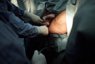
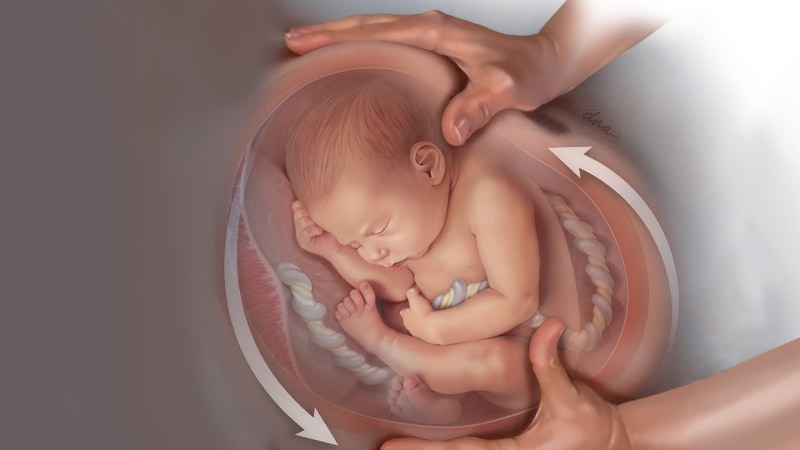
Breech presentation occurs in about 3-4% of all pregnancies at term. While some breech babies can be safely delivered vaginally, most obstetricians recommend a cesarean delivery for breech presentation due to the increased risk of complications such as cord prolapse, head entrapment, and fetal distress. However, there are some techniques that may be used to attempt a vaginal breech delivery in certain situations, such as external cephalic version (ECV), which is a procedure where a healthcare provider manually turns the fetus from a breech position to a head-down position while it is still in the uterus.
Fetal version is a medical term used to describe the position or presentation of the fetus in the uterus during pregnancy. It refers to the way the fetus is facing or lying in relation to the mother's pelvis.
There are several different types of fetal versions, including:
* Cephalic version: This is the most common and preferred position for birth. The fetus's head is downward, facing the mother's cervix.
* Breech version: In this position, the fetus's buttocks or feet are pointed downward toward the mother's cervix. There are several different types of breech versions, including frank breech (where the baby's legs are straight up in front of its body), complete breech (where the baby's legs are folded at the knees), and footling breech (where one or both of the baby's feet are coming out below the buttocks).
* Transverse version: This is a less common position where the fetus is lying sideways across the mother's uterus.
Fetal version can be assessed through physical examination, ultrasound, or both. In some cases, healthcare providers may attempt to manually turn the fetus into a different position using a procedure called external cephalic version (ECV). This is typically done in the third trimester of pregnancy and is used to reduce the risk of breech delivery and improve outcomes for both the mother and baby.
A Cesarean section, often referred to as a C-section, is a surgical procedure used to deliver a baby. It involves making an incision through the mother's abdomen and uterus to remove the baby. This procedure may be necessary when a vaginal delivery would put the mother or the baby at risk.
There are several reasons why a C-section might be recommended, including:
* The baby is in a breech position (feet first) or a transverse position (sideways) and cannot be turned to a normal head-down position.
* The baby is too large to safely pass through the mother's birth canal.
* The mother has a medical condition, such as heart disease or high blood pressure, that could make vaginal delivery risky.
* The mother has an infection, such as HIV or herpes, that could be passed to the baby during a vaginal delivery.
* The labor is not progressing and there are concerns about the health of the mother or the baby.
C-sections are generally safe for both the mother and the baby, but like any surgery, they do carry some risks. These can include infection, bleeding, blood clots, and injury to nearby organs. In addition, women who have a C-section are more likely to experience complications in future pregnancies, such as placenta previa or uterine rupture.
If you have questions about whether a C-section is necessary for your delivery, it's important to discuss your options with your healthcare provider.
'Labor presentation' is a term used in obstetrics to describe the part of the fetus that enters the mother's pelvis first during labor. This positioning determines the route the baby will take through the birth canal. The most common and uncomplicated presentation is vertex or cephalic presentation, where the baby's head is the presenting part. Other possible presentations include breech (buttocks or feet first), face, brow, and shoulder presentations, which can potentially lead to complications during delivery if not managed appropriately.
"Delivery, Obstetric" is a medical term that refers to the process of giving birth to a baby. It involves the passage of the fetus through the mother's vagina or via Caesarean section (C-section), which is a surgical procedure.
The obstetric delivery process typically includes three stages:
1. The first stage begins with the onset of labor and ends when the cervix is fully dilated.
2. The second stage starts with full dilation of the cervix and ends with the birth of the baby.
3. The third stage involves the delivery of the placenta, which is the organ that provides oxygen and nutrients to the developing fetus during pregnancy.
Obstetric delivery requires careful monitoring and management by healthcare professionals to ensure the safety and well-being of both the mother and the baby. Various interventions and techniques may be used during the delivery process to facilitate a safe and successful outcome, including the use of medications, assisted delivery with forceps or vacuum extraction, and C-section.
Fetal distress is a term used to describe situations where a fetus is experiencing problems during labor or delivery that are causing significant physiological changes. These changes may include an abnormal heart rate, decreased oxygen levels, or the presence of meconium (the baby's first stool) in the amniotic fluid. Fetal distress can be caused by a variety of factors, such as problems with the umbilical cord, placental abruption, maternal high blood pressure, or prolonged labor. It is important to monitor fetal well-being during labor and delivery to detect and address any signs of fetal distress promptly. Treatment may include changing the mother's position, administering oxygen, giving intravenous fluids, or performing an emergency cesarean section.
A postmature infant is a newborn who is delivered at or after 42 weeks (294 days) of gestation. These infants are also known as "post-term" or "post-dates." At this stage, the placenta may not function optimally, leading to potential issues such as decreased fetal movement, meconium staining of amniotic fluid, and low birth weight. Postmature infants may require close monitoring and evaluation after delivery to ensure their well-being.
Pelvimetry is a medical measurement and evaluation of the size and shape of the pelvis, which can be performed in several ways:
1. Clinical pelvimetry: This involves physical examination to assess the dimensions of the pelvis by palpation and measurement of the distance between bony landmarks.
2. Radiological pelvimetry: This uses X-ray or CT imaging to obtain more accurate measurements of the pelvic diameters, including the anteroposterior, transverse, and oblique dimensions.
3. Magnetic resonance imaging (MRI) pelvimetry: This method is considered the most accurate for assessing the size and shape of the pelvis, as it provides detailed images without radiation exposure.
Pelvimetry is often used in obstetrics to evaluate whether a woman's pelvis can accommodate a fetus during childbirth (known as "obstetric pelvimetry"). It helps healthcare providers determine if a vaginal delivery is possible or if a cesarean section may be necessary. However, the use of pelvimetry in modern obstetrics has become less common due to its limited predictive value and the increasing focus on individualized birth management.
Pregnancy is a physiological state or condition where a fertilized egg (zygote) successfully implants and grows in the uterus of a woman, leading to the development of an embryo and finally a fetus. This process typically spans approximately 40 weeks, divided into three trimesters, and culminates in childbirth. Throughout this period, numerous hormonal and physical changes occur to support the growing offspring, including uterine enlargement, breast development, and various maternal adaptations to ensure the fetus's optimal growth and well-being.
Dystocia is a medical term used to describe difficult or abnormal labor or delivery in animals, including humans. It refers to a situation where the natural process of childbirth is hindered or obstructed, making it difficult for the fetus to pass through the birth canal. This condition can be caused by various factors such as the size and position of the fetus, maternal pelvic size or shape, hormonal imbalances, or other medical conditions that affect the mother's ability to give birth.
Dystocia can lead to serious complications for both the mother and the fetus if not treated promptly and appropriately. Prolonged labor can result in fetal distress, hypoxia (lack of oxygen), or even death. In addition, maternal injuries such as uterine rupture, cervical trauma, or infection can occur during a difficult delivery.
The treatment for dystocia depends on the underlying cause and severity of the condition. In some cases, manual assistance or manipulation of the fetus may be sufficient to facilitate delivery. However, in more severe cases, medical intervention such as cesarean section (C-section) may be necessary to ensure the safety of both the mother and the fetus.
It is important for pregnant individuals to receive regular prenatal care from a qualified healthcare provider to monitor their pregnancy and identify any potential risk factors for dystocia or other complications. Prompt medical attention should be sought if any signs of difficult labor or delivery are observed.
A "repeat cesarean section" is a medical term that refers to the delivery of a fetus through surgical incision in the abdominal and uterine walls, which has been performed previously. It is also known as a "classical repeat cesarean delivery." This procedure may be recommended when vaginal birth poses potential risks to the mother or the baby, such as in cases of placenta previa, previous classical uterine incision, or multiple pregnancies. The decision for a repeat cesarean section is typically made after considering various factors, including the patient's medical history, current pregnancy status, and personal preferences.
In medical terms, parity refers to the number of times a woman has given birth to a viable fetus, usually defined as a pregnancy that reaches at least 20 weeks' gestation. It is often used in obstetrics and gynecology to describe a woman's childbearing history and to assess potential risks associated with childbirth.
Parity is typically categorized as follows:
* Nulliparous: A woman who has never given birth to a viable fetus.
* Primiparous: A woman who has given birth to one viable fetus.
* Multiparous: A woman who has given birth to more than one viable fetus.
In some cases, parity may also consider the number of pregnancies that resulted in stillbirths or miscarriages, although this is not always the case. It's important to note that parity does not necessarily reflect the total number of pregnancies a woman has had, only those that resulted in viable births.
Pregnancy outcome refers to the final result or status of a pregnancy, including both the health of the mother and the newborn baby. It can be categorized into various types such as:
1. Live birth: The delivery of one or more babies who show signs of life after separation from their mother.
2. Stillbirth: The delivery of a baby who has died in the womb after 20 weeks of pregnancy.
3. Miscarriage: The spontaneous loss of a pregnancy before the 20th week.
4. Abortion: The intentional termination of a pregnancy before the fetus can survive outside the uterus.
5. Ectopic pregnancy: A pregnancy that develops outside the uterus, usually in the fallopian tube, which is not viable and requires medical attention.
6. Preterm birth: The delivery of a baby before 37 weeks of gestation, which can lead to various health issues for the newborn.
7. Full-term birth: The delivery of a baby between 37 and 42 weeks of gestation.
8. Post-term pregnancy: The delivery of a baby after 42 weeks of gestation, which may increase the risk of complications for both mother and baby.
The pregnancy outcome is influenced by various factors such as maternal age, health status, lifestyle habits, genetic factors, and access to quality prenatal care.
A newborn infant is a baby who is within the first 28 days of life. This period is also referred to as the neonatal period. Newborns require specialized care and attention due to their immature bodily systems and increased vulnerability to various health issues. They are closely monitored for signs of well-being, growth, and development during this critical time.
Birth weight refers to the first weight of a newborn infant, usually taken immediately after birth. It is a critical vital sign that indicates the baby's health status and is used as a predictor for various short-term and long-term health outcomes.
Typically, a full-term newborn's weight ranges from 5.5 to 8.8 pounds (2.5 to 4 kg), although normal birth weights can vary significantly based on factors such as gestational age, genetics, maternal health, and nutrition. Low birth weight is defined as less than 5.5 pounds (2.5 kg), while high birth weight is greater than 8.8 pounds (4 kg).
Low birth weight babies are at a higher risk for various medical complications, including respiratory distress syndrome, jaundice, infections, and developmental delays. High birth weight babies may face challenges with delivery, increased risk of obesity, and potential metabolic issues later in life. Regular prenatal care is essential to monitor fetal growth and ensure a healthy pregnancy and optimal birth weight for the baby.
Gestational age is the length of time that has passed since the first day of the last menstrual period (LMP) in pregnant women. It is the standard unit used to estimate the age of a pregnancy and is typically expressed in weeks. This measure is used because the exact date of conception is often not known, but the start of the last menstrual period is usually easier to recall.
It's important to note that since ovulation typically occurs around two weeks after the start of the LMP, gestational age is approximately two weeks longer than fetal age, which is the actual time elapsed since conception. Medical professionals use both gestational and fetal age to track the development and growth of the fetus during pregnancy.
Prenatal ultrasonography, also known as obstetric ultrasound, is a medical diagnostic procedure that uses high-frequency sound waves to create images of the developing fetus, placenta, and amniotic fluid inside the uterus. It is a non-invasive and painless test that is widely used during pregnancy to monitor the growth and development of the fetus, detect any potential abnormalities or complications, and determine the due date.
During the procedure, a transducer (a small handheld device) is placed on the mother's abdomen and moved around to capture images from different angles. The sound waves travel through the mother's body and bounce back off the fetus, producing echoes that are then converted into electrical signals and displayed as images on a screen.
Prenatal ultrasonography can be performed at various stages of pregnancy, including early pregnancy to confirm the pregnancy and detect the number of fetuses, mid-pregnancy to assess the growth and development of the fetus, and late pregnancy to evaluate the position of the fetus and determine if it is head down or breech. It can also be used to guide invasive procedures such as amniocentesis or chorionic villus sampling.
Overall, prenatal ultrasonography is a valuable tool in modern obstetrics that helps ensure the health and well-being of both the mother and the developing fetus.
 Breech - series-Other presentations: MedlinePlus Medical Encyclopedia
 Model Predicts ECV Success in Breech Presentation
 Browsing by Subject "Breech Presentation"
 Breech presentation - Ontology Browser - Rat Genome Database
 A population-based case-control study of risk factors for breech presentation - PubMed
 The breech effect: is previous caesarean section for breech presentation an independent favourable factor in vaginal birth...
 Maternal outcomes of term breech presentation delivery: impact of successful external cephalic version in a nationwide sample...
 Breech Baby C-Section Delivery | Frank Breech Baby Presentation
 Chiropractic for Breech Presentation
 BL67- Breeched Baby Presentation
 Everything You Need to Know About Breech Presentation - Neelu Prajapat
 Breech birth - Wikipedia
 Subjects: Breech Presentation - Digital Collections - National Library of Medicine Search Results
 RESEARCH WEDNESDAY: Resolution of Breech Presentation in Pregnant Patient | Wellness Revolution
 Presentation from Youth Exchange Student | The Yellow Breeches Rotary Club
 Fetal Presentation, Position, and Lie (Including Breech Presentation) - Gynecology and Obstetrics - MSD Manual Professional...
 IJERPH | Free Full-Text | Reproductive and Obstetric Outcomes after UAE, HIFU, and TFA of Uterine Fibroids: Systematic Review...
 Prevention of Pertussis, Tetanus, and Diphtheria Among
Pregnant and Postpartum Women and their Infants
Prevention of Pertussis, Tetanus, and Diphtheria Among
Pregnant and Postpartum Women and their Infants The impact of assisted reproductive technology and ovulation induction on breech presentation: A whole of population-based...
 Labor & Delivery: Episiotomy | Healthline
 Risk factors and outcomes of abnormal bleeding after external cephalic version
 Hip dysplasia