Fiber Optic Technology
Bronchial Diseases
Bronchi
Tracheal Diseases
Bronchoalveolar Lavage
Hemoptysis
Tracheal Stenosis
Tracheal Neoplasms
Tracheomalacia
Carcinoma, Bronchogenic
Respiratory Aspiration
Bronchoalveolar Lavage Fluid
Biopsy
Pulmonary Atelectasis
Solitary Pulmonary Nodule
Tomography, X-Ray Computed
Trachea
Conscious Sedation
Sputum
Laryngismus
Radiography, Thoracic
Hydrocodone
Electromagnetic Phenomena
Pulmonary Medicine
Biopsy, Needle
Tracheobronchomalacia
Cough
Lung
Anthracosis
Tracheostomy
Multiple Pulmonary Nodules
Bronchitis
Deep Sedation
Respiratory Insufficiency
Pneumonia, Pneumocystis
Tracheitis
Intubation, Intratracheal
Bronchial Fistula
Mediastinoscopy
Gastric Lavage
Prospective Studies
Lung Diseases, Fungal
Anesthesia, Intratracheal
Argon Plasma Coagulation
Tomography Scanners, X-Ray Computed
Plasma Cell Granuloma, Pulmonary
Laryngostenosis
Optical Fibers
Midazolam
Risk factors for lower airway bacterial colonization in chronic bronchitis. (1/1686)
The aim of this study was to determine the prevalence and risk factors for lower airway bacterial colonization (LABC) in stable chronic bronchitis (CB). Forty-one outpatients with CB were enrolled in the study (age 63.8+/-9.1 yrs (mean+/-SD); forced expiratory volume in one second (FEV1)/forced vital capacity (FVC) 62.8+/-11.2; current/former smokers 24/17). All patients had normal chest radiographs and an indication for performing fibreoptic bronchoscopy (pulmonary nodule, remote haemoptysis). The protected specimen brush (PSB) was used for bacterial sampling, and concentrations > or = 1,000 colony-forming units (cfu) x mL(-1) were considered positive for LABC. The repeatability of the procedure in CB was assessed in a random subsample of 18 subjects. A 72.2% quantitative agreement was found in the repeatability assessment of the PSB technique. Positive PSB cultures, obtained in 9 out of 41 (22%) patients, mainly yielded Haemophilus influenzae. The logistic regression model, used to determine which variables were related to colonization, showed that LABC was associated with current smoking (odds ratio (OR) 9.83, confidence interval (CI) 1.16-83.20) and low FVC (OR 0.73, CI 0.65-0.81). Age and FEV1 were not related to LABC. It was concluded that the prevalence of LABC in stable CB is high (22%), and current smoking is an important risk factor. (+info)Predisposing factors to bacterial colonization in chronic obstructive pulmonary disease. (2/1686)
The aim of this prospective observational study was to determine those factors influencing bacterial colonization in patients with stable chronic obstructive pulmonary disease (COPD). Eighty-eight outpatients with stable COPD and 20 patients with normal spirometry and chest radiography (controls) had a fibreoptic bronchoscopy performed with topical aerosol anaesthesia. Bacterial colonization was determined using the protected specimen brush (PSB) with a cut-off > or = 10(3) colony-forming units (CFU x mL(-1)). The influence of age, degree of airflow obstruction, smoking habit, pack-yrs of smoking, and chest radiographic findings on bacterial colonization were assessed by univariate and multivariate analysis. Significant bacterial growth was found in 40% of patients and in none of the controls. Haemophilus influenzae, Streptococcus viridans, S. pneumoniae and Moraxella catarrhalis were the most frequent pathogens. After adjustment for other variables, severe airflow limitation (odds ratio (OR) 5.11, 95% confidence interval (CI) 1.45-17.9) and current smoking (OR 3.17, 95% CI 2.5-8) remained associated with positive bacterial cultures. When only potentially pathogenic micro-organisms were considered, significant bacterial growth was found in 30.7% of patients, with severe airflow obstruction (OR 9.28, 95% CI 2.19-39.3) being the only variable independently associated with positive bacterial cultures. Our results show that stable chronic obstructive pulmonary disease patients have a high prevalence of bacterial colonization of distal airways which is mainly related to the degree of airflow obstruction and cigarette smoking. (+info)Broncholithiasis: rare but still present. (3/1686)
Broncholithiasis is a rare but distinct and potentially dangerous pulmonary problem that still needs to be considered in the differential diagnosis of some patients with bronchial obstruction. Broncholiths originate from calcified material in peribronchial lymph nodes eroding into the tracheobronchial tree. The clinical and chest X-ray signs are usually non-specific, but the diagnosis can nowadays be made based on clinical suspicion, CT-scan and fibre-optic bronchoscopy findings, so that a malignant cause of airway obstruction can be ruled out. The removal of broncholiths during fibre-optic bronchoscopy is seldom possible and rather dangerous. They can be removed safely by rigid bronchoscopy with the aid of Nd-YAG laser photocoagulation. Thoracotomy is indicated in complicated cases with fistula formation or severe bleeding. (+info)Vascularity in asthmatic airways: relation to inhaled steroid dose. (4/1686)
BACKGROUND: There is an increase in vascularity in the asthmatic airway. Although inhaled corticosteroids (ICS) are an effective anti-inflammatory treatment in asthma, there are few data on any effects on structural changes. METHODS: Endobronchial biopsy specimens from seven asthmatic subjects not receiving ICS and 15 receiving 200-1500 microg/day beclomethasone dipropionate (BDP) were immunohistochemically stained with an anti-collagen type IV antibody to outline the endothelial basement membrane of the vessels. These were compared with biopsy tissue from 11 non-asthmatic controls (four atopic and seven non-atopic). RESULTS: There was a significant increase in the density of vessels (number of vessels/mm2 of lamina propria) in the asthmatic subjects not on ICS compared with non-asthmatic controls (mean 485 (interquartile range (IQR) 390-597) versus 329 (IQR 248-376) vessels/mm2, p<0.05; 95% CI for the difference 48 to 286). There was no significant difference between asthmatic subjects on ICS and those not on ICS or control subjects in the number of vessels/mm2 (mean 421 (IQR 281-534)). However, patients who received >/=800 microg/day BDP tended to have a reduced number of vessels/mm2 compared with patients not on ICS and those receiving +info)Aspirated foreign bodies in the tracheobronchial tree: report of 250 cases. (5/1686)
During the last 14 years, 250 patients with aspirated foreign bodies in the tracheobronchial tree were admitted to Kuwait Chest Diseases Hospital. Ninety-six per cent of the cases were under 10 years of age and 38% gave a clear history of foreign body inhalation. The rest were diagnosed either clinically, from the chest radiograph findings or because of unexplained pulmonary symptoms. In 247 cases, bronchoscopy under general anaesthesia was successful in removing the foreign bodies. In only three cases was bronchotomy needed. Seventy per cent of the foreign bodies were melon seeds. Asphyxia and cardiac arrest occurred in four cases during bronchoscopy but the patients were successfully resuscitated. In 10 cases a tracheostomy was done before bronchoscopy and the removal of the foreign body, while in five it was needed after bronchoscopy. Fifteen patients developed late complications such as recurrent pneumonia or atelectasis of the lung. Early diagnosis and adequate treatment are essential to prevent pulmonary and cardiac complications and to avoid radical lung surgery. (+info)Diagnostic value of endoscopic ultrasonography-guided fine-needle aspiration cytology of mediastinal masses in patients with intrapulmonary lesions and nondiagnostic bronchoscopy. (6/1686)
Several procedures are available for the cytopathological diagnosis of mediastinal lesions. The purpose of this study was to evaluate the diagnostic value of endoscopic ultrasonography (EUS)-guided fine-needle aspiration (FNA) in patients with mediastinal mass lesions/lymph node enlargement. All patients had intrapulmonary lesions on chest X ray and/or CT scan, and inconclusive findings by endobronchial forceps biopsy and/or brush cytology. EUS-guided FNA was performed in 16 patients using a modified oblique forward-viewing gastroscope with an electronic multielement curved linear ultrasound transducer. After the region of interest was localized, a 22-gauge Vilmann-Hancke needle was introduced via the 2-mm biopsy channel. The cytological diagnosis of EUS-guided FNA was conclusive for cancer in 9 patients and in the other 7 patients the aspirated samples revealed a benign lesion. In 10 patients the final diagnosis was cancer, thus EUS-guided FNA was diagnostic for malignancy in all but 1 of the lesions (sensitivity 90.0%). In 1 patient epitheloid cell granuloma was detected by cytological examination of the FNA. Following tuberculostatic treatment the lesions disappeared completely on CT scan and EUS. The overall accuracy in this study amounted to 93.7%. From this and other studies discussed, it is assumed that the procedure is an accurate and safe technique to examine nodular lesions suggestive of metastatic lymph node involvement. (+info)Forced expiratory wheezes in a patient with dynamic expiratory narrowing of central airways and an oscillating pattern of the flow-volume curve. (7/1686)
Forced expiratory wheezes (FEW) are common and the pathogenesis of this phenomenon might involve fluttering of the airways, but this theory has not been confirmed in patients. We report a case of a patient with FEW and a normal FEV1 that showed a bronchoscopically confirmed collapse of the trachea and main stem bronchi during forced expiration. Superimposed to the flow-volume curve was an oscillating pattern with a frequency that corresponded well with the wheeze generated during forced expiration. The oscillating pattern in the flow-volume curve and the collapse of the major airways supports the theory of wheezes generated by fluttering airways during forced expiration. Although FEW may be found also in healthy subjects, flow limitation is essential for the generation of FEW. The inclusion of a forced expiratory maneuver in the clinical examination might therefore be helpful in guiding the diagnosis towards airways obstruction. (+info)A man with a prosthetic ear and multiple pulmonary nodules. (8/1686)
Basal cell carcinoma is generally regarded as a relatively indolent tumor easily controlled with local therapy. When neglected or inadequately treated this tumor can become locally aggressive and in rare circumstances metastasize. This report documents a case of basal cell carcinoma metastatic to the lung that resulted in rapidly progressive respiratory failure and death. (+info)Bronchoscopy is a medical procedure that involves the examination of the inside of the airways and lungs with a flexible or rigid tube called a bronchoscope. This procedure allows healthcare professionals to directly visualize the airways, take tissue samples for biopsy, and remove foreign objects or secretions. Bronchoscopy can be used to diagnose and manage various respiratory conditions such as lung infections, inflammation, cancer, and bleeding. It is usually performed under local or general anesthesia to minimize discomfort and risks associated with the procedure.
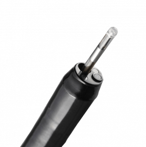
A bronchoscope is a medical device that is used to examine the airways and lungs. It is a long, thin, flexible tube that is equipped with a light and a camera at its tip. The bronchoscope is inserted through the nose or mouth and down the throat, allowing the doctor to visualize the trachea, bronchi, and smaller branches of the airway system.
Bronchoscopes can be used for diagnostic purposes, such as to take tissue samples (biopsies) or to investigate the cause of symptoms like coughing up blood or difficulty breathing. They can also be used for therapeutic purposes, such as to remove foreign objects from the airways or to place stents to keep them open.
There are several types of bronchoscopes, including flexible bronchoscopes and rigid bronchoscopes. Flexible bronchoscopes are more commonly used because they are less invasive and can be used to examine smaller airways. Rigid bronchoscopes, on the other hand, are larger and stiffer, and are typically used for more complex procedures or in emergency situations.
It is important to note that the use of bronchoscopes requires specialized training and should only be performed by healthcare professionals with the appropriate expertise.
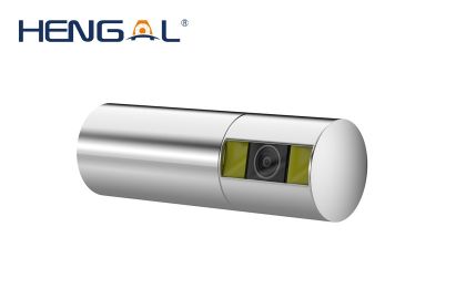
Fiber optic technology in the medical context refers to the use of thin, flexible strands of glass or plastic fibers that are designed to transmit light and images along their length. These fibers are used to create bundles, known as fiber optic cables, which can be used for various medical applications such as:
1. Illumination: Fiber optics can be used to deliver light to hard-to-reach areas during surgical procedures or diagnostic examinations.
2. Imaging: Fiber optics can transmit images from inside the body, enabling doctors to visualize internal structures and tissues. This is commonly used in medical imaging techniques such as endoscopy, colonoscopy, and laparoscopy.
3. Sensing: Fiber optic sensors can be used to measure various physiological parameters such as temperature, pressure, and strain within the body. These sensors can provide real-time data during surgical procedures or for monitoring patients' health status.
Fiber optic technology offers several advantages over traditional medical imaging techniques, including high resolution, flexibility, small diameter, and the ability to bend around corners without significant loss of image quality. Additionally, fiber optics are non-magnetic and can be used in MRI environments without causing interference.
Bronchial diseases refer to medical conditions that affect the bronchi, which are the large airways that lead into the lungs. These diseases can cause inflammation, narrowing, or obstruction of the bronchi, leading to symptoms such as coughing, wheezing, chest tightness, and difficulty breathing.
Some common bronchial diseases include:
1. Asthma: A chronic inflammatory disease of the airways that causes recurring episodes of wheezing, breathlessness, chest tightness, and coughing.
2. Chronic Bronchitis: A long-term inflammation of the bronchi that leads to a persistent cough and excessive mucus production.
3. Bronchiectasis: A condition in which the bronchi become damaged and widened, leading to chronic infection and inflammation.
4. Bronchitis: An inflammation of the bronchi that can cause coughing, wheezing, and chest tightness.
5. Emphysema: A lung condition that causes shortness of breath due to damage to the air sacs in the lungs. While not strictly a bronchial disease, it is often associated with chronic bronchitis and COPD (Chronic Obstructive Pulmonary Disease).
Treatment for bronchial diseases may include medications such as bronchodilators, corticosteroids, or antibiotics, as well as lifestyle changes such as quitting smoking and avoiding irritants. In severe cases, oxygen therapy or surgery may be necessary.
Bronchial neoplasms refer to abnormal growths or tumors in the bronchi, which are the large airways that lead into the lungs. These neoplasms can be benign (non-cancerous) or malignant (cancerous). Malignant bronchial neoplasms are often referred to as lung cancer and can be further classified into small cell lung cancer and non-small cell lung cancer, depending on the type of cells involved.
Benign bronchial neoplasms are less common than malignant ones and may include growths such as papillomas, hamartomas, or chondromas. While benign neoplasms are not cancerous, they can still cause symptoms and complications if they grow large enough to obstruct the airways or if they become infected.
Treatment for bronchial neoplasms depends on several factors, including the type, size, location, and stage of the tumor, as well as the patient's overall health and medical history. Treatment options may include surgery, radiation therapy, chemotherapy, or a combination of these approaches.
"Bronchi" are a pair of airways in the respiratory system that branch off from the trachea (windpipe) and lead to the lungs. They are responsible for delivering oxygen-rich air to the lungs and removing carbon dioxide during exhalation. The right bronchus is slightly larger and more vertical than the left, and they further divide into smaller branches called bronchioles within the lungs. Any abnormalities or diseases affecting the bronchi can impact lung function and overall respiratory health.
Tracheal diseases refer to a group of medical conditions that affect the trachea, also known as the windpipe. The trachea is a tube-like structure made up of rings of cartilage and smooth muscle, which extends from the larynx (voice box) to the bronchi (airways leading to the lungs). Its primary function is to allow the passage of air to and from the lungs.
Tracheal diseases can be categorized into several types, including:
1. Tracheitis: Inflammation of the trachea, often caused by viral or bacterial infections.
2. Tracheal stenosis: Narrowing of the trachea due to scarring, inflammation, or compression from nearby structures such as tumors or goiters.
3. Tracheomalacia: Weakening and collapse of the tracheal walls, often seen in newborns and young children but can also occur in adults due to factors like chronic cough, aging, or connective tissue disorders.
4. Tracheoesophageal fistula: An abnormal connection between the trachea and the esophagus, which can lead to respiratory complications and difficulty swallowing.
5. Tracheal tumors: Benign or malignant growths that develop within the trachea, obstructing airflow and potentially leading to more severe respiratory issues.
6. Tracheobronchial injury: Damage to the trachea and bronchi, often caused by trauma such as blunt force or penetrating injuries.
7. Congenital tracheal abnormalities: Structural defects present at birth, including complete tracheal rings, which can cause narrowing or collapse of the airway.
Symptoms of tracheal diseases may include cough, wheezing, shortness of breath, chest pain, and difficulty swallowing. Treatment options depend on the specific condition and its severity but may involve medications, surgery, or other interventions to alleviate symptoms and improve respiratory function.
Bronchoalveolar lavage (BAL) is a medical procedure in which a small amount of fluid is introduced into a segment of the lung and then gently suctioned back out. The fluid contains cells and other materials that can be analyzed to help diagnose various lung conditions, such as inflammation, infection, or cancer.
The procedure is typically performed during bronchoscopy, which involves inserting a thin, flexible tube with a light and camera on the end through the nose or mouth and into the lungs. Once the bronchoscope is in place, a small catheter is passed through the bronchoscope and into the desired lung segment. The fluid is then introduced and suctioned back out, and the sample is sent to a laboratory for analysis.
BAL can be helpful in diagnosing various conditions such as pneumonia, interstitial lung diseases, alveolar proteinosis, and some types of cancer. It can also be used to monitor the effectiveness of treatment for certain lung conditions. However, like any medical procedure, it carries some risks, including bleeding, infection, and respiratory distress. Therefore, it is important that the procedure is performed by a qualified healthcare professional in a controlled setting.
Bronchography is a medical imaging technique that involves the injection of a contrast material into the airways (bronchi) of the lungs, followed by X-ray imaging to produce detailed pictures of the bronchial tree. This diagnostic procedure was commonly used in the past to identify abnormalities such as narrowing, blockages, or inflammation in the airways, but it has largely been replaced by newer, less invasive techniques like computed tomography (CT) scans and bronchoscopy.
The process of bronchography involves the following steps:
1. The patient is sedated or given a local anesthetic to minimize discomfort during the procedure.
2. A radiopaque contrast material is introduced into the bronchi through a catheter that is inserted into the trachea, either via a nostril or through a small incision in the neck.
3. Once the contrast material has been distributed throughout the bronchial tree, X-ray images are taken from various angles to capture detailed views of the airways.
4. The images are then analyzed by a radiologist to identify any abnormalities or irregularities in the structure and function of the bronchi.
Although bronchography is considered a relatively safe procedure, it does carry some risks, including allergic reactions to the contrast material, infection, and bleeding. Additionally, the use of ionizing radiation during X-ray imaging should be carefully weighed against the potential benefits of the procedure.
"Foreign bodies" refer to any object or substance that is not normally present in a particular location within the body. These can range from relatively harmless items such as splinters or pieces of food in the skin or gastrointestinal tract, to more serious objects like bullets or sharp instruments that can cause significant damage and infection.
Foreign bodies can enter the body through various routes, including ingestion, inhalation, injection, or penetrating trauma. The location of the foreign body will determine the potential for harm and the necessary treatment. Some foreign bodies may pass through the body without causing harm, while others may require medical intervention such as removal or surgical extraction.
It is important to seek medical attention if a foreign body is suspected, as untreated foreign bodies can lead to complications such as infection, inflammation, and tissue damage.
Hemoptysis is the medical term for coughing up blood that originates from the lungs or lower respiratory tract. It can range in severity from streaks of blood mixed with mucus to large amounts of pure blood. Hemoptysis may be a sign of various underlying conditions, such as bronchitis, pneumonia, tuberculosis, cancer, or blood disorders. Immediate medical attention is required when hemoptysis occurs, especially if it's in significant quantities, to determine the cause and provide appropriate treatment.
Airway obstruction is a medical condition that occurs when the normal flow of air into and out of the lungs is partially or completely blocked. This blockage can be caused by a variety of factors, including swelling of the tissues in the airway, the presence of foreign objects or substances, or abnormal growths such as tumors.
When the airway becomes obstructed, it can make it difficult for a person to breathe normally. They may experience symptoms such as shortness of breath, wheezing, coughing, and chest tightness. In severe cases, airway obstruction can lead to respiratory failure and other life-threatening complications.
There are several types of airway obstruction, including:
1. Upper airway obstruction: This occurs when the blockage is located in the upper part of the airway, such as the nose, throat, or voice box.
2. Lower airway obstruction: This occurs when the blockage is located in the lower part of the airway, such as the trachea or bronchi.
3. Partial airway obstruction: This occurs when the airway is partially blocked, allowing some air to flow in and out of the lungs.
4. Complete airway obstruction: This occurs when the airway is completely blocked, preventing any air from flowing into or out of the lungs.
Treatment for airway obstruction depends on the underlying cause of the condition. In some cases, removing the obstruction may be as simple as clearing the airway of foreign objects or mucus. In other cases, more invasive treatments such as surgery may be necessary.
Tracheal stenosis is a medical condition characterized by the abnormal narrowing of the trachea (windpipe), which can lead to difficulty breathing. This narrowing can be caused by various factors such as inflammation, scarring, or the growth of abnormal tissue in the airway. Symptoms may include wheezing, coughing, shortness of breath, and chest discomfort, particularly during physical activity. Treatment options for tracheal stenosis depend on the severity and underlying cause of the condition and may include medications, bronchodilators, corticosteroids, or surgical interventions such as laser surgery, stent placement, or tracheal reconstruction.
Tracheal neoplasms refer to abnormal growths or tumors in the trachea, which is the windpipe that carries air from the nose and throat to the lungs. These growths can be benign (non-cancerous) or malignant (cancerous). Malignant tracheal neoplasms are relatively rare and can be primary (originating in the trachea) or secondary (spreading from another part of the body, such as lung cancer). Primary tracheal cancers can be squamous cell carcinoma, adenoid cystic carcinoma, mucoepidermoid carcinoma, or sarcomas. Symptoms may include cough, difficulty breathing, wheezing, or chest pain. Treatment options depend on the type, size, and location of the neoplasm and can include surgery, radiation therapy, chemotherapy, or a combination of these approaches.
Tracheomalacia is a medical condition that refers to the softening and weakening of the tracheal walls, leading to its collapse or narrowing. This can cause symptoms such as shortness of breath, wheezing, coughing, and difficulty breathing, especially during exertion or when lying down.
In newborns and infants, tracheomalacia is often present at birth (congenital) and may improve on its own as the child grows and the trachea becomes stronger. However, in some cases, it may persist into adulthood and require medical treatment, such as bronchodilators, oxygen therapy, or even surgery to support the tracheal walls.
Tracheomalacia can also occur as a result of damage to the trachea from long-term intubation, trauma, infection, or other medical conditions that weaken the tracheal muscles and cartilage.
Carcinoma, bronchogenic is a medical term that refers to a type of lung cancer that originates in the bronchi, which are the branching tubes that carry air into the lungs. It is the most common form of lung cancer and can be further classified into different types based on the specific cell type involved, such as squamous cell carcinoma, adenocarcinoma, or large cell carcinoma.
Bronchogenic carcinomas are often associated with smoking and exposure to environmental pollutants, although they can also occur in non-smokers. Symptoms may include coughing, chest pain, shortness of breath, wheezing, hoarseness, or unexplained weight loss. Treatment options depend on the stage and location of the cancer, as well as the patient's overall health and may include surgery, radiation therapy, chemotherapy, targeted therapy, or a combination of these approaches.
Lung diseases refer to a broad category of disorders that affect the lungs and other structures within the respiratory system. These diseases can impair lung function, leading to symptoms such as coughing, shortness of breath, chest pain, and wheezing. They can be categorized into several types based on the underlying cause and nature of the disease process. Some common examples include:
1. Obstructive lung diseases: These are characterized by narrowing or blockage of the airways, making it difficult to breathe out. Examples include chronic obstructive pulmonary disease (COPD), asthma, bronchiectasis, and cystic fibrosis.
2. Restrictive lung diseases: These involve stiffening or scarring of the lungs, which reduces their ability to expand and take in air. Examples include idiopathic pulmonary fibrosis, sarcoidosis, and asbestosis.
3. Infectious lung diseases: These are caused by bacteria, viruses, fungi, or parasites that infect the lungs. Examples include pneumonia, tuberculosis, and influenza.
4. Vascular lung diseases: These affect the blood vessels in the lungs, impairing oxygen exchange. Examples include pulmonary embolism, pulmonary hypertension, and chronic thromboembolic pulmonary hypertension (CTEPH).
5. Neoplastic lung diseases: These involve abnormal growth of cells within the lungs, leading to cancer. Examples include small cell lung cancer, non-small cell lung cancer, and mesothelioma.
6. Other lung diseases: These include interstitial lung diseases, pleural effusions, and rare disorders such as pulmonary alveolar proteinosis and lymphangioleiomyomatosis (LAM).
It is important to note that this list is not exhaustive, and there are many other conditions that can affect the lungs. Proper diagnosis and treatment of lung diseases require consultation with a healthcare professional, such as a pulmonologist or respiratory therapist.
Respiratory aspiration is defined as the entry of foreign materials (such as food, liquids, or vomit) into the lower respiratory tract during swallowing, which includes the trachea and lungs. This can lead to respiratory complications such as pneumonia, bronchitis, or lung abscesses. Aspiration can occur in individuals with impaired swallowing function due to various conditions like neurological disorders, stroke, or anesthesia.
Bronchoalveolar lavage (BAL) fluid is a type of clinical specimen obtained through a procedure called bronchoalveolar lavage. This procedure involves inserting a bronchoscope into the lungs and instilling a small amount of saline solution into a specific area of the lung, then gently aspirating the fluid back out. The fluid that is recovered is called bronchoalveolar lavage fluid.
BAL fluid contains cells and other substances that are present in the lower respiratory tract, including the alveoli (the tiny air sacs where gas exchange occurs). By analyzing BAL fluid, doctors can diagnose various lung conditions, such as pneumonia, interstitial lung disease, and lung cancer. They can also monitor the effectiveness of treatments for these conditions by comparing the composition of BAL fluid before and after treatment.
BAL fluid is typically analyzed for its cellular content, including the number and type of white blood cells present, as well as for the presence of bacteria, viruses, or other microorganisms. The fluid may also be tested for various proteins, enzymes, and other biomarkers that can provide additional information about lung health and disease.
Lung neoplasms refer to abnormal growths or tumors in the lung tissue. These tumors can be benign (non-cancerous) or malignant (cancerous). Malignant lung neoplasms are further classified into two main types: small cell lung carcinoma and non-small cell lung carcinoma. Lung neoplasms can cause symptoms such as cough, chest pain, shortness of breath, and weight loss. They are often caused by smoking or exposure to secondhand smoke, but can also occur due to genetic factors, radiation exposure, and other environmental carcinogens. Early detection and treatment of lung neoplasms is crucial for improving outcomes and survival rates.
A biopsy is a medical procedure in which a small sample of tissue is taken from the body to be examined under a microscope for the presence of disease. This can help doctors diagnose and monitor various medical conditions, such as cancer, infections, or autoimmune disorders. The type of biopsy performed will depend on the location and nature of the suspected condition. Some common types of biopsies include:
1. Incisional biopsy: In this procedure, a surgeon removes a piece of tissue from an abnormal area using a scalpel or other surgical instrument. This type of biopsy is often used when the lesion is too large to be removed entirely during the initial biopsy.
2. Excisional biopsy: An excisional biopsy involves removing the entire abnormal area, along with a margin of healthy tissue surrounding it. This technique is typically employed for smaller lesions or when cancer is suspected.
3. Needle biopsy: A needle biopsy uses a thin, hollow needle to extract cells or fluid from the body. There are two main types of needle biopsies: fine-needle aspiration (FNA) and core needle biopsy. FNA extracts loose cells, while a core needle biopsy removes a small piece of tissue.
4. Punch biopsy: In a punch biopsy, a round, sharp tool is used to remove a small cylindrical sample of skin tissue. This type of biopsy is often used for evaluating rashes or other skin abnormalities.
5. Shave biopsy: During a shave biopsy, a thin slice of tissue is removed from the surface of the skin using a sharp razor-like instrument. This technique is typically used for superficial lesions or growths on the skin.
After the biopsy sample has been collected, it is sent to a laboratory where a pathologist will examine the tissue under a microscope and provide a diagnosis based on their findings. The results of the biopsy can help guide further treatment decisions and determine the best course of action for managing the patient's condition.
Pulmonary atelectasis is a medical condition characterized by the collapse or closure of the alveoli (tiny air sacs) in the lungs, leading to reduced or absent gas exchange in the affected area. This results in decreased lung volume and can cause hypoxemia (low oxygen levels in the blood). Atelectasis can be caused by various factors such as obstruction of the airways, surfactant deficiency, pneumothorax, or compression from outside the lung. It can also occur after surgical procedures, particularly when the patient is lying in one position for a long time. Symptoms may include shortness of breath, cough, and chest discomfort, but sometimes it may not cause any symptoms, especially if only a small area of the lung is affected. Treatment depends on the underlying cause and may include bronchodilators, chest physiotherapy, or even surgery in severe cases.
A Solitary Pulmonary Nodule (SPN) is a single, round or oval-shaped lung shadow that measures up to 3 cm in diameter on a chest radiograph. It is also known as a "coin lesion" due to its appearance. SPNs are usually discovered incidentally during routine chest X-rays or CT scans. They can be benign or malignant, and their nature is determined through further diagnostic tests such as PET scans, biopsies, or follow-up imaging studies.
X-ray computed tomography (CT or CAT scan) is a medical imaging method that uses computer-processed combinations of many X-ray images taken from different angles to produce cross-sectional (tomographic) images (virtual "slices") of the body. These cross-sectional images can then be used to display detailed internal views of organs, bones, and soft tissues in the body.
The term "computed tomography" is used instead of "CT scan" or "CAT scan" because the machines take a series of X-ray measurements from different angles around the body and then use a computer to process these data to create detailed images of internal structures within the body.
CT scanning is a noninvasive, painless medical test that helps physicians diagnose and treat medical conditions. CT imaging provides detailed information about many types of tissue including lung, bone, soft tissue and blood vessels. CT examinations can be performed on every part of the body for a variety of reasons including diagnosis, surgical planning, and monitoring of therapeutic responses.
In computed tomography (CT), an X-ray source and detector rotate around the patient, measuring the X-ray attenuation at many different angles. A computer uses this data to construct a cross-sectional image by the process of reconstruction. This technique is called "tomography". The term "computed" refers to the use of a computer to reconstruct the images.
CT has become an important tool in medical imaging and diagnosis, allowing radiologists and other physicians to view detailed internal images of the body. It can help identify many different medical conditions including cancer, heart disease, lung nodules, liver tumors, and internal injuries from trauma. CT is also commonly used for guiding biopsies and other minimally invasive procedures.
In summary, X-ray computed tomography (CT or CAT scan) is a medical imaging technique that uses computer-processed combinations of many X-ray images taken from different angles to produce cross-sectional images of the body. It provides detailed internal views of organs, bones, and soft tissues in the body, allowing physicians to diagnose and treat medical conditions.
The trachea, also known as the windpipe, is a tube-like structure in the respiratory system that connects the larynx (voice box) to the bronchi (the two branches leading to each lung). It is composed of several incomplete rings of cartilage and smooth muscle, which provide support and flexibility. The trachea plays a crucial role in directing incoming air to the lungs during inspiration and outgoing air to the larynx during expiration.
Conscious sedation, also known as procedural sedation and analgesia, is a minimally depressed level of consciousness that retains the patient's ability to maintain airway spontaneously and respond appropriately to physical stimulation and verbal commands. It is typically achieved through the administration of sedative and/or analgesic medications and is commonly used in medical procedures that do not require general anesthesia. The goal of conscious sedation is to provide a comfortable and anxiety-free experience for the patient while ensuring their safety throughout the procedure.
Sputum is defined as a mixture of saliva and phlegm that is expelled from the respiratory tract during coughing, sneezing or deep breathing. It can be clear, mucoid, or purulent (containing pus) depending on the underlying cause of the respiratory issue. Examination of sputum can help diagnose various respiratory conditions such as infections, inflammation, or other lung diseases.
Laryngospasm, often mistakenly referred to as "laryngismus," is a medical condition characterized by an involuntary and sustained closure of the vocal cords (the structures that form the larynx or voice box). This spasm can occur in response to various stimuli, such as irritation, aspiration, or emotional distress, leading to difficulty breathing, coughing, and stridor (a high-pitched sound during inspiration).
The term "laryngismus" is not a widely accepted medical term; however, it may be used informally to refer to any condition affecting the larynx. The correct term for a prolonged or chronic issue with the larynx would be "laryngeal dyskinesia."
Thoracic radiography is a type of diagnostic imaging that involves using X-rays to produce images of the chest, including the lungs, heart, bronchi, great vessels, and the bones of the spine and chest wall. It is a commonly used tool in the diagnosis and management of various respiratory, cardiovascular, and thoracic disorders such as pneumonia, lung cancer, heart failure, and rib fractures.
During the procedure, the patient is positioned between an X-ray machine and a cassette containing a film or digital detector. The X-ray beam is directed at the chest, and the resulting image is captured on the film or detector. The images produced can help identify any abnormalities in the structure or function of the organs within the chest.
Thoracic radiography may be performed as a routine screening test for certain conditions, such as lung cancer, or it may be ordered when a patient presents with symptoms suggestive of a respiratory or cardiovascular disorder. It is a safe and non-invasive procedure that can provide valuable information to help guide clinical decision making and improve patient outcomes.
Hydrocodone is an opioid medication used to treat severe pain. It works by changing how the brain and nervous system respond to pain. Medically, it's defined as a semisynthetic opioid analgesic, synthesized from codeine, one of the natural opiates found in the resin of the poppy seed pod.
Hydrocodone is available only in combination with other drugs, such as acetaminophen or ibuprofen, which are added to enhance its pain-relieving effects and/or to prevent abuse and overdose. Common brand names include Vicodin, Lortab, and Norco.
Like all opioids, hydrocodone carries a risk of addiction and dependence, and it should be used only under the supervision of a healthcare provider. It's also important to note that misuse or abuse of hydrocodone can lead to overdose and death.
Electromagnetic phenomena refer to the interactions and effects that occur due to the combination of electrically charged particles and magnetic fields. These phenomena are described by the principles of electromagnetism, a branch of physics that deals with the fundamental forces between charged particles and their interaction with electromagnetic fields.
Electromagnetic phenomena can be observed in various forms, including:
1. Electric fields: The force that exists between charged particles at rest or in motion. Positive charges create an electric field that points away from them, while negative charges create an electric field that points towards them.
2. Magnetic fields: The force that exists around moving charges or current-carrying wires. Magnets and moving charges produce magnetic fields that exert forces on other moving charges or current-carrying wires.
3. Electromagnetic waves: Self-propagating disturbances in electric and magnetic fields, which can travel through space at the speed of light. Examples include visible light, radio waves, microwaves, and X-rays.
4. Electromagnetic induction: The process by which a changing magnetic field generates an electromotive force (EMF) in a conductor, leading to the flow of electric current.
5. Faraday's law of induction: A fundamental principle that relates the rate of change of magnetic flux through a closed loop to the induced EMF in the loop.
6. Lenz's law: A consequence of conservation of energy, which states that the direction of an induced current is such that it opposes the change in magnetic flux causing it.
7. Electromagnetic radiation: The emission and absorption of electromagnetic waves by charged particles undergoing acceleration or deceleration.
8. Maxwell's equations: A set of four fundamental equations that describe how electric and magnetic fields interact, giving rise to electromagnetic phenomena.
In a medical context, electromagnetic phenomena can be harnessed for various diagnostic and therapeutic applications, such as magnetic resonance imaging (MRI), electrocardiography (ECG), electromyography (EMG), and transcranial magnetic stimulation (TMS).
Pulmonary medicine is a medical specialty that deals with the diagnosis, treatment, and prevention of diseases and conditions affecting the respiratory system, including the lungs, trachea, bronchi, bronchioles, and alveoli. Pulmonologists are specialists who treat a wide range of respiratory disorders such as chronic obstructive pulmonary disease (COPD), asthma, bronchitis, pneumonia, lung cancer, sleep-disordered breathing, tuberculosis, and interstitial lung diseases. They use various diagnostic techniques including chest X-rays, CT scans, pulmonary function tests, bronchoscopy, and sleep studies to evaluate and manage respiratory disorders. Pulmonologists also provide care for patients who require long-term mechanical ventilation or oxygen therapy.
Pulmonary tuberculosis (TB) is an infectious disease caused by the bacterium Mycobacterium tuberculosis. It primarily affects the lungs and can spread to other parts of the body through the bloodstream or lymphatic system. The infection typically enters the body when a person inhales droplets containing the bacteria, which are released into the air when an infected person coughs, sneezes, or talks.
The symptoms of pulmonary TB can vary but often include:
* Persistent cough that lasts for more than three weeks and may produce phlegm or blood-tinged sputum
* Chest pain or discomfort, particularly when breathing deeply or coughing
* Fatigue and weakness
* Unexplained weight loss
* Fever and night sweats
* Loss of appetite
Pulmonary TB can cause serious complications if left untreated, including damage to the lungs, respiratory failure, and spread of the infection to other parts of the body. Treatment typically involves a course of antibiotics that can last several months, and it is essential for patients to complete the full treatment regimen to ensure that the infection is fully eradicated.
Preventive measures include vaccination with the Bacillus Calmette-Guérin (BCG) vaccine, which can provide some protection against severe forms of TB in children, and measures to prevent the spread of the disease, such as covering the mouth and nose when coughing or sneezing, wearing a mask in public places, and avoiding close contact with people who have active TB.
A needle biopsy is a medical procedure in which a thin, hollow needle is used to remove a small sample of tissue from a suspicious or abnormal area of the body. The tissue sample is then examined under a microscope to check for cancer cells or other abnormalities. Needle biopsies are often used to diagnose lumps or masses that can be felt through the skin, but they can also be guided by imaging techniques such as ultrasound, CT scan, or MRI to reach areas that cannot be felt. There are several types of needle biopsy procedures, including fine-needle aspiration (FNA) and core needle biopsy. FNA uses a thin needle and gentle suction to remove fluid and cells from the area, while core needle biopsy uses a larger needle to remove a small piece of tissue. The type of needle biopsy used depends on the location and size of the abnormal area, as well as the reason for the procedure.
Tracheobronchomalacia is a medical condition that refers to the abnormal softening and weakness of the tracheal and bronchial walls, leading to their collapse or narrowing during breathing, particularly during expiration. This collapse can cause symptoms such as wheezing, shortness of breath, coughing, and recurrent respiratory infections. The condition can be congenital or acquired, with common causes including aging, chronic obstructive pulmonary disease (COPD), and long-term intubation. In severe cases, tracheobronchomalacia may require surgical intervention to stabilize the airway and improve breathing.
A cough is a reflex action that helps to clear the airways of irritants, foreign particles, or excess mucus or phlegm. It is characterized by a sudden, forceful expulsion of air from the lungs through the mouth and nose. A cough can be acute (short-term) or chronic (long-term), and it can be accompanied by other symptoms such as chest pain, shortness of breath, or fever. Coughing can be caused by various factors, including respiratory infections, allergies, asthma, environmental pollutants, gastroesophageal reflux disease (GERD), and chronic lung diseases such as chronic obstructive pulmonary disease (COPD) and bronchitis. In some cases, a cough may be a symptom of a more serious underlying condition, such as heart failure or lung cancer.
A lung is a pair of spongy, elastic organs in the chest that work together to enable breathing. They are responsible for taking in oxygen and expelling carbon dioxide through the process of respiration. The left lung has two lobes, while the right lung has three lobes. The lungs are protected by the ribcage and are covered by a double-layered membrane called the pleura. The trachea divides into two bronchi, which further divide into smaller bronchioles, leading to millions of tiny air sacs called alveoli, where the exchange of gases occurs.
Anthracosis is a medical condition characterized by the accumulation of carbon particles, primarily from air pollution or coal dust, in the tissues of the lungs. This results in the formation of black deposits, known as anthracotic pigment, on the surfaces of the lung's air sacs (alveoli) and lymph nodes.
Repeated and prolonged exposure to these pollutants can cause inflammation and fibrosis in the lungs, potentially leading to respiratory symptoms such as coughing, wheezing, and shortness of breath. In severe cases, anthracosis may contribute to the development of chronic obstructive pulmonary disease (COPD) or restrictive lung disease.
It is important to note that while anthracosis is often associated with occupational exposure in coal miners and industrial workers, it can also occur in individuals living in urban areas with high levels of air pollution. Smokers are also at an increased risk due to the inhalation of tar and other carbon-based particles present in tobacco smoke.
Inhalation burns, also known as respiratory or pulmonary burns, refer to damage to the airways and lungs caused by inhaling hot gases, smoke, steam, or toxic fumes. This type of injury can occur during a fire or other thermal incidents and can result in significant morbidity and mortality.
Inhalation burns are classified into three categories based on the location and severity of the injury:
1. Upper airway burns: These involve the nose, throat, and voice box (larynx) and are usually caused by inhaling hot gases or steam. Symptoms may include singed nasal hairs, soot in the nose or mouth, coughing, wheezing, and difficulty speaking or swallowing.
2. Lower airway burns: These involve the trachea, bronchi, and bronchioles and are usually caused by inhaling smoke or toxic fumes. Symptoms may include coughing, chest pain, shortness of breath, and wheezing.
3. Systemic burns: These occur when toxic substances are absorbed into the bloodstream and can affect multiple organs. Symptoms may include nausea, vomiting, confusion, and organ failure.
Inhalation burns can lead to complications such as pneumonia, respiratory failure, and acute respiratory distress syndrome (ARDS). Treatment typically involves providing oxygen therapy, removing secretions from the airways, and administering bronchodilators and corticosteroids to reduce inflammation. Severe cases may require intubation and mechanical ventilation.
Prevention of inhalation burns includes avoiding smoke-filled areas during a fire, staying close to the ground where the air is cooler and cleaner, and using appropriate respiratory protection devices when exposed to toxic fumes or gases.
A tracheostomy is a surgically created opening through the neck into the trachea (windpipe). It is performed to provide an airway in cases where the upper airway is obstructed or access to the lower airway is required, such as in prolonged intubation, severe trauma, or chronic lung diseases. The procedure involves making an incision in the front of the neck and creating a direct opening into the trachea, through which a tracheostomy tube is inserted to maintain the patency of the airway. This allows for direct ventilation of the lungs, suctioning of secretions, and prevention of complications associated with upper airway obstruction.
Medical Definition: Multiple pulmonary nodules refer to multiple small rounded or irregularly shaped masses in the lungs, usually measuring less than 3 cm in diameter. These nodules can be caused by various conditions such as benign tumors, infections, inflammation, or malignancies like lung cancer. The presence of multiple pulmonary nodules often requires further evaluation with imaging studies and sometimes biopsy to determine the underlying cause and appropriate treatment.
Bronchitis is a medical condition characterized by inflammation of the bronchi, which are the large airways that lead to the lungs. This inflammation can cause a variety of symptoms, including coughing, wheezing, chest tightness, and shortness of breath. Bronchitis can be either acute or chronic.
Acute bronchitis is usually caused by a viral infection, such as a cold or the flu, and typically lasts for a few days to a week. Symptoms may include a productive cough (coughing up mucus or phlegm), chest discomfort, and fatigue. Acute bronchitis often resolves on its own without specific medical treatment, although rest, hydration, and over-the-counter medications to manage symptoms may be helpful.
Chronic bronchitis, on the other hand, is a long-term condition that is characterized by a persistent cough with mucus production that lasts for at least three months out of the year for two consecutive years. Chronic bronchitis is typically caused by exposure to irritants such as cigarette smoke, air pollution, or occupational dusts and chemicals. It is often associated with chronic obstructive pulmonary disease (COPD), which includes both chronic bronchitis and emphysema.
Treatment for chronic bronchitis may include medications to help open the airways, such as bronchodilators and corticosteroids, as well as lifestyle changes such as smoking cessation and avoiding irritants. In severe cases, oxygen therapy or lung transplantation may be necessary.
Deep sedation, also known as general anesthesia, is a drug-induced depression of consciousness during which patients cannot be easily aroused but respond purposefully following repeated or painful stimulation. It is characterized by the loss of protective reflexes such as cough and gag, and the ability to ventilate spontaneously may be impaired. Patients may require assistance in maintaining a patent airway, and positive pressure ventilation may be required.
Deep sedation/general anesthesia is typically used for surgical procedures or other medical interventions that require patients to be completely unaware and immobile, and it is administered by trained anesthesia professionals who monitor and manage the patient's vital signs and level of consciousness throughout the procedure.
Respiratory insufficiency is a condition characterized by the inability of the respiratory system to maintain adequate gas exchange, resulting in an inadequate supply of oxygen and/or removal of carbon dioxide from the body. This can occur due to various causes, such as lung diseases (e.g., chronic obstructive pulmonary disease, pneumonia), neuromuscular disorders (e.g., muscular dystrophy, spinal cord injury), or other medical conditions that affect breathing mechanics and/or gas exchange.
Respiratory insufficiency can manifest as hypoxemia (low oxygen levels in the blood) and/or hypercapnia (high carbon dioxide levels in the blood). Symptoms of respiratory insufficiency may include shortness of breath, rapid breathing, fatigue, confusion, and in severe cases, loss of consciousness or even death. Treatment depends on the underlying cause and severity of the condition and may include oxygen therapy, mechanical ventilation, medications, and/or other supportive measures.
"Pneumonia, Pneumocystis" is more commonly referred to as "Pneumocystis pneumonia (PCP)." It is a type of pneumonia caused by the microorganism Pneumocystis jirovecii. This organism was previously classified as a protozoan but is now considered a fungus.
PCP is an opportunistic infection, which means that it mainly affects people with weakened immune systems, such as those with HIV/AIDS, cancer, transplant recipients, or people taking immunosuppressive medications. The symptoms of PCP can include cough, shortness of breath, fever, and difficulty exercising. It is a serious infection that requires prompt medical treatment, typically with antibiotics.
It's important to note that PCP is not the same as pneumococcal pneumonia, which is caused by the bacterium Streptococcus pneumoniae. While both conditions are types of pneumonia, they are caused by different organisms and require different treatments.
Tracheitis is a medical condition that involves inflammation of the trachea, or windpipe. It can cause symptoms such as cough, sore throat, difficulty swallowing, and fever. Tracheitis can be caused by viral or bacterial infections, and it may also occur as a complication of other respiratory conditions. In some cases, tracheitis may require medical treatment, including antibiotics for bacterial infections or corticosteroids to reduce inflammation. It is important to seek medical attention if you experience symptoms of tracheitis, especially if they are severe or persistent.
Respiratory system abnormalities refer to any conditions or structures that do not function properly or are outside the normal range in the respiratory system. The respiratory system is responsible for taking in oxygen and expelling carbon dioxide through the process of breathing. It includes the nose, throat (pharynx), voice box (larynx), windpipe (trachea), bronchi, bronchioles, alveoli, and muscles and nerves that support breathing.
Respiratory system abnormalities can be congenital or acquired. Congenital abnormalities are present at birth and may include conditions such as cystic fibrosis, pulmonary hypoplasia, and congenital diaphragmatic hernia. Acquired abnormalities can develop at any time throughout a person's life due to various factors such as infections, injuries, environmental exposures, or aging. Examples of acquired respiratory system abnormalities include chronic obstructive pulmonary disease (COPD), asthma, pneumonia, lung cancer, and sleep apnea.
Respiratory system abnormalities can cause a range of symptoms, including coughing, wheezing, shortness of breath, chest pain, and fatigue. Treatment for respiratory system abnormalities depends on the specific condition and severity and may include medications, breathing treatments, surgery, or lifestyle changes.
Intubation, intratracheal is a medical procedure in which a flexible plastic or rubber tube called an endotracheal tube (ETT) is inserted through the mouth or nose, passing through the vocal cords and into the trachea (windpipe). This procedure is performed to establish and maintain a patent airway, allowing for the delivery of oxygen and the removal of carbon dioxide during mechanical ventilation in various clinical scenarios, such as:
1. Respiratory failure or arrest
2. Procedural sedation
3. Surgery under general anesthesia
4. Neuromuscular disorders
5. Ingestion of toxic substances
6. Head and neck trauma
7. Critical illness or injury affecting the airway
The process of intubation is typically performed by trained medical professionals, such as anesthesiologists, emergency medicine physicians, or critical care specialists, using direct laryngoscopy or video laryngoscopy to visualize the vocal cords and guide the ETT into the correct position. Once placed, the ETT is secured to prevent dislodgement, and the patient's respiratory status is continuously monitored to ensure proper ventilation and oxygenation.
A bronchial fistula is an abnormal connection or passage between the bronchial tree (the airways in the lungs) and the surrounding tissues, such as the pleural space (the space between the lungs and the chest wall), blood vessels, or other organs. This condition can result from various causes, including lung injury, infection, surgery, or certain diseases such as cancer or tuberculosis.
Bronchial fistulas can lead to symptoms like coughing, wheezing, shortness of breath, and chest pain. They may also cause air leaks, pneumothorax (collapsed lung), or chronic infections. Treatment for bronchial fistulas depends on the underlying cause and severity of the condition but often involves surgical repair or closure of the abnormal connection.
Mediastinoscopy is a surgical procedure in which a tubular instrument called mediastinoscope is inserted through a small incision made at the base of the neck, typically in the suprasternal notch. This procedure allows the medical professional to examine the mediastinum, which is the area within the chest between the lungs, containing the heart, trachea, esophagus, and other vital structures. The examination can help identify any abnormalities, such as tumors or inflammation, and in some cases, biopsies of suspicious tissues may be taken for further analysis. Mediastinoscopy is typically performed under general anesthesia in a hospital setting.
Gastric lavage, also known as stomach pumping, is a medical procedure where the stomach's contents are emptied using a tube that is inserted through the mouth or nose and into the stomach. The tube is then connected to suction, which helps remove the stomach contents. This procedure is often used in emergency situations to treat poisonings or overdoses by removing the toxic substance before it gets absorbed into the bloodstream. It can also be used to empty the stomach before certain surgeries or procedures.
Prospective studies, also known as longitudinal studies, are a type of cohort study in which data is collected forward in time, following a group of individuals who share a common characteristic or exposure over a period of time. The researchers clearly define the study population and exposure of interest at the beginning of the study and follow up with the participants to determine the outcomes that develop over time. This type of study design allows for the investigation of causal relationships between exposures and outcomes, as well as the identification of risk factors and the estimation of disease incidence rates. Prospective studies are particularly useful in epidemiology and medical research when studying diseases with long latency periods or rare outcomes.
Fungal lung diseases, also known as fungal pneumonia or mycoses, refer to a group of respiratory disorders caused by the infection of fungi in the lungs. These fungi are commonly found in the environment, such as soil, decaying organic matter, and contaminated materials. People can develop lung diseases from fungi after inhaling spores or particles that contain fungi.
There are several types of fungal lung diseases, including:
1. Aspergillosis: This is caused by the Aspergillus fungus and can affect people with weakened immune systems. It can cause allergic reactions, lung infections, or invasive aspergillosis, which can spread to other organs.
2. Cryptococcosis: This is caused by the Cryptococcus fungus and is usually found in soil contaminated with bird droppings. It can cause pneumonia, meningitis, or skin lesions.
3. Histoplasmosis: This is caused by the Histoplasma capsulatum fungus and is commonly found in the Ohio and Mississippi River valleys. It can cause flu-like symptoms, lung infections, or disseminated histoplasmosis, which can spread to other organs.
4. Blastomycosis: This is caused by the Blastomyces dermatitidis fungus and is commonly found in the southeastern and south-central United States. It can cause pneumonia, skin lesions, or disseminated blastomycosis, which can spread to other organs.
5. Coccidioidomycosis: This is caused by the Coccidioides immitis fungus and is commonly found in the southwestern United States. It can cause flu-like symptoms, lung infections, or disseminated coccidioidomycosis, which can spread to other organs.
Fungal lung diseases can range from mild to severe, depending on the type of fungus and the person's immune system. Treatment may include antifungal medications, surgery, or supportive care. Prevention measures include avoiding exposure to contaminated soil or dust, wearing protective masks in high-risk areas, and promptly seeking medical attention if symptoms develop.
Intratracheal anesthesia refers to the administration of anesthetic agents directly into the trachea. This type of anesthesia is typically used in specific medical procedures, such as bronchoscopy or airway surgery, where it is necessary to achieve adequate anesthesia and analgesia of the airways while avoiding systemic effects.
Intratracheal anesthesia is usually delivered through a specialized device called a laryngoscope, which is used to visualize the vocal cords and introduce a narrow tube (endotracheal tube) into the trachea. Once the endotracheal tube is in place, anesthetic gases or liquids can be administered directly into the airways, providing rapid onset of action and minimal systemic absorption.
It's important to note that intratracheal anesthesia should only be performed by trained medical professionals, as there are potential risks associated with this procedure, including damage to the airway, respiratory compromise, and other complications.
Argon Plasma Coagulation (APC) is a medical procedure that uses ionized argon gas to deliver electrical current and heat to tissue, resulting in coagulation. It is commonly used in the treatment of gastrointestinal bleeding, as well as for cutting and coagulating during surgical procedures. The argon plasma is created by passing argon gas through a high-voltage electrical field, which ionizes the gas and creates a highly precise and controllable plasma beam. This beam can be directed at the tissue to achieve hemostasis (stopping bleeding) or to cut tissue with minimal thermal damage to surrounding structures. The procedure is often performed under endoscopic guidance.
X-ray computed tomography (CT) scanner is a medical imaging device that uses computer-processed combinations of many X-ray images taken from different angles to produce cross-sectional (tomographic) images (virtual "slices") of the body. These cross-sections can then be manipulated, through either additional computer processing or interactive viewing, to show various bodily structures and functions in 2D or 3D.
In contrast to conventional X-ray imaging, CT scanning provides detailed images of many types of tissue including lung, bone, soft tissue and blood vessels. CT is often used when rapid, detailed images are needed such as in trauma situations or for the detection and diagnosis of stroke, cancer, appendicitis, pulmonary embolism, and musculoskeletal disorders.
CT scanning is associated with some risks, particularly from exposure to ionizing radiation, which can lead to cancer and other diseases. However, the benefits of CT scanning, in particular its ability to detect life-threatening conditions early and accurately, generally outweigh the risks. As a result, it has become an important tool in modern medicine.
Pulmonary plasma cell granuloma is a benign lung lesion characterized by the accumulation of plasma cells and the formation of granulomas. It is also known as inflammatory pseudotumor or plasma cell histiocytoma. The etiology of pulmonary plasma cell granuloma remains unclear, but it is thought to be related to a chronic inflammatory response or an abnormal immune reaction.
The lesion typically consists of a mass or nodule in the lung tissue, which may be discovered incidentally on chest imaging. Symptoms, if present, may include cough, chest pain, and shortness of breath. The diagnosis is usually made by histopathological examination of a biopsy specimen, which shows a mixture of plasma cells, lymphocytes, and histiocytes, with the formation of granulomas.
Treatment is generally not necessary unless the lesion is causing symptoms or growing in size. In such cases, surgical resection may be recommended. The prognosis is excellent, with a low risk of recurrence after surgical removal.
Laryngostenosis is a medical term that refers to a condition where the larynx (or voice box) becomes narrowed. This can occur due to various reasons such as scarring, swelling, or growths in the laryngeal area. The narrowing can cause difficulty with breathing, swallowing, and speaking. In severe cases, it may require medical intervention, such as surgery, to correct the problem.
Medical Definition of Optical Fibers:
Optical fibers are thin, transparent strands of glass or plastic fiber that are designed to transmit light along their length. In the medical field, optical fibers are used in various applications such as illumination, imaging, and data transmission. For instance, they are used in flexible endoscopes to provide illumination and visualization inside the body during diagnostic or surgical procedures. They are also used in optical communication systems for transmitting information in the form of light signals within medical devices or between medical facilities. The use of optical fibers allows for minimally invasive procedures, improved image quality, and increased data transmission rates.
Midazolam is a medication from the class of drugs known as benzodiazepines. It works by enhancing the effect of a neurotransmitter called gamma-aminobutyric acid (GABA), which has a calming effect on the brain and nervous system. Midazolam is often used for its sedative, hypnotic, anxiolytic, anticonvulsant, and muscle relaxant properties.
Medically, midazolam is used for various purposes, including:
1. Preoperative medication (sedation before surgery)
2. Procedural sedation (for minor surgical or diagnostic procedures)
3. Treatment of seizures (status epilepticus)
4. Sedation in critically ill patients
5. As an adjunct to anesthesia during surgeries
6. Treatment of alcohol withdrawal symptoms
7. To induce amnesia for certain medical or dental procedures
Midazolam is available in various forms, such as tablets, intravenous (IV) solutions, and intranasal sprays. It has a rapid onset of action and a short duration, making it suitable for brief, intermittent procedures. However, midazolam can cause side effects like drowsiness, confusion, respiratory depression, and memory impairment. Therefore, its use should be carefully monitored by healthcare professionals.
Fluorescence is not a medical term per se, but it is widely used in the medical field, particularly in diagnostic tests, medical devices, and research. Fluorescence is a physical phenomenon where a substance absorbs light at a specific wavelength and then emits light at a longer wavelength. This process, often referred to as fluorescing, results in the emission of visible light that can be detected and measured.
In medical terms, fluorescence is used in various applications such as:
1. In-vivo imaging: Fluorescent dyes or probes are introduced into the body to highlight specific structures, cells, or molecules during imaging procedures. This technique can help doctors detect and diagnose diseases such as cancer, inflammation, or infection.
2. Microscopy: Fluorescence microscopy is a powerful tool for visualizing biological samples at the cellular and molecular level. By labeling specific proteins, nucleic acids, or other molecules with fluorescent dyes, researchers can observe their distribution, interactions, and dynamics within cells and tissues.
3. Surgical guidance: Fluorescence-guided surgery is a technique where surgeons use fluorescent markers to identify critical structures such as blood vessels, nerves, or tumors during surgical procedures. This helps ensure precise and safe surgical interventions.
4. Diagnostic tests: Fluorescence-based assays are used in various diagnostic tests to detect and quantify specific biomarkers or analytes. These assays can be performed using techniques such as enzyme-linked immunosorbent assay (ELISA), polymerase chain reaction (PCR), or flow cytometry.
In summary, fluorescence is a physical process where a substance absorbs and emits light at different wavelengths. In the medical field, this phenomenon is harnessed for various applications such as in-vivo imaging, microscopy, surgical guidance, and diagnostic tests.
 Bronchoscopy
 Bronchoscopy
 Bronchoscopy with Transbronchial Biopsy
 Diagnostic Bronchoscopy - AARC
 Bronchoscopy - Health Encyclopedia - University of Rochester Medical Center
 Bronchoscopy: What It Is, How It's Done, Results
 Bronchoscopy | American Lung Association
 Bronchoscopy and Bronchoalveolar Lavage (BAL): MedlinePlus Medical Test
 Single-institution study evaluating the utility of surveillance bronchoscopy after lung transplantation
 Flexible bronchoscopy | Great Ormond Street Hospital
 Bronchoscopy. Diagnostic test. Clínica Universidad de Navarra
 A novel clip-on device for electromagnetic tracking in endobronchial ultrasound bronchoscopy - SINTEF
 Improving standards in flexible bronchoscopy for lung cancer | European Respiratory Society
 Complications of bronchoscopy: A concise synopsis : International Journal of Critical Illness and Injury Science
 Robotic-assisted bronchoscopy FAQ - Mayo Clinic Health System
 Bronchoscopy in San Leandro, CA | VCA Bay Area Veterinary Specialists & Emergency Hospital
 Yale First in State to Offer 'Shape Sensing' Robotic Bronchoscopy | Internal Medicine
 Bronchoscopy done today
 Bronchoscopy - Fibo Health
 Monarch Robotic-assisted Bronchoscopy | Genesis Healthcare System
 Bronchoscopy: Before Your Procedure
 Bronchoscopy. - Macmillan Online Community
 Robotic Bronchoscopy | The University of Kansas Cancer Center
 Freeman Reaches a Major Milestone with MONARCH® Robot-Assisted Bronchoscopy | Freeman Health
 Boston Scientific Corporation (US), Olympus Corporation (Japan) and KARL STORZ (Germany) are Leading Players in the...
 Midazolam in flexible bronchoscopy premedication: effects on patient-related and
procedure-related outcomes.
Midazolam in flexible bronchoscopy premedication: effects on patient-related and
procedure-related outcomes.