S100 Calcium Binding Protein G
Calcium-Binding Proteins
Calbindins
Calbindin 1
Calbindin 2
Parvalbumins
S100 Proteins
EF Hand Motifs
Calcium
Calmodulin
Recoverin
Calgranulin B
Calsequestrin
Calgranulin A
Salamandra
Fluorescamine
Molecular Sequence Data
Calcium Signaling
Amino Acid Sequence
Collodion
Binding Sites
Protein Binding
Calcium Radioisotopes
Entamoeba histolytica
Calcium Channels
Base Sequence
Sarcoplasmic Reticulum
Protein Conformation
Calcium, Dietary
Rabbits
Protein Structure, Tertiary
Poly(A)-Binding Proteins
Sequence Homology, Amino Acid
Calcium Isotopes
RNA, Messenger
Troponin C
Models, Molecular
Calcitriol
S100 Calcium Binding Protein beta Subunit
Cattle
Carrier Proteins
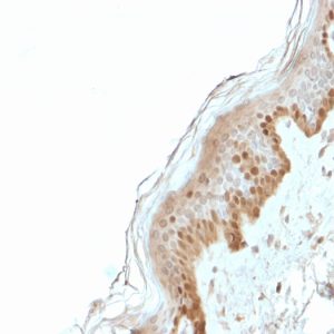
Immunohistochemistry
Cloning, Molecular
Chickens
Calcium Chloride
Nerve Tissue Proteins
Calcium Carbonate
Gene Expression Regulation
Neurons
Sequence Homology, Nucleic Acid
Terbium
Rats, Sprague-Dawley
Troponin
Calcium Phosphates
Magnesium
Tacrolimus Binding Proteins
Poly(A)-Binding Protein I
Calcium Channels, L-Type
Mutation
Hippocalcin
Insulin-Like Growth Factor Binding Proteins
Cells, Cultured
DNA-Binding Proteins
Calcium Oxalate
Chelating Agents
Ytterbium
Hydrogen-Ion Concentration
Lanthanum
Calcium Gluconate
Circular Dichroism
RNA-Binding Proteins
Cations, Divalent
Microfilament Proteins
Fatty Acid-Binding Proteins
Protein Structure, Secondary
Electrophoresis, Polyacrylamide Gel
Magnetic Resonance Spectroscopy
Recombinant Fusion Proteins
Cytosol
Dialysis
Naphthalenesulfonates
Poly(A)-Binding Protein II
Sequence Alignment
Models, Biological
Mutagenesis, Site-Directed
Marfan Syndrome
Metals, Rare Earth
Calcium Channels, N-Type
Peptide Fragments
Signal Transduction
Edetic Acid
Myocardium
Muscle Proteins
Insulin-Like Growth Factor Binding Protein 3
Ions
Transcription Factors
Dose-Response Relationship, Drug
Neuronal Calcium-Sensor Proteins
Periplasmic Binding Proteins
Phosphorylation
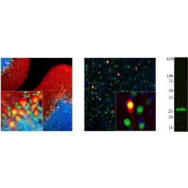
Blotting, Western
DNA, Complementary
Peptides
Calcium-Transporting ATPases
Structure-Activity Relationship
Trifluoperazine
Thermodynamics
Amino Acid Motifs
Ryanodine Receptor Calcium Release Channel
Transfection
Adenosine Triphosphatases
Aequorin
Membrane Proteins
Escherichia coli
Adenosine Triphosphate
1-Carboxyglutamic Acid
Fluorescent Dyes
DNA
Binding, Competitive
Neurocalcin
Calcium Channel Agonists
Tacrolimus Binding Protein 1A
Calreticulin
Membrane Potentials
Crystallography
Latent TGF-beta Binding Proteins
Crystallography, X-Ray
Calorimetry
Mollusca
Annexins
Nuclear Proteins
Apoproteins
Endoplasmic Reticulum
Insulin-Like Growth Factor Binding Protein 2
Taurodeoxycholic Acid
Thoracica
Cell Membrane
Cyclic AMP Response Element-Binding Protein
DNA Primers
Biological Transport
Calcimycin
TATA-Box Binding Protein
Alkanesulfonates
Gene Expression
Potassium Chloride
Muscle Contraction
Insulin-Like Growth Factor Binding Protein 1
Cytoplasm
Promoter Regions, Genetic
Buffers
Extracellular Matrix Proteins
Transcription, Genetic
Sarcolemma
Xanthenes
Consensus Sequence
Nuclear Magnetic Resonance, Biomolecular
Fibrinogens, Abnormal
Retinol-Binding Proteins
Microscopy, Fluorescence
Calcium Hydroxide
Reverse Transcriptase Polymerase Chain Reaction
Enzyme Activation
Muscle, Skeletal
CREB-Binding Protein
Actins
Phosphorus
Parathyroid Hormone
Microscopy, Electron
Protein Isoforms
Nifedipine
Temperature
Calcium Sulfate
Patch-Clamp Techniques
Tryptophan
Trypsin
Vitamin D-Binding Protein
CCAAT-Enhancer-Binding Proteins
Osmolar Concentration
Microsomes
HeLa Cells
Mathematics
Fura-2
Sodium
Polypyrimidine Tract-Binding Protein
Fluorescence Polarization
Ligands
Myosins
Dogs
Thapsigargin
Endocytosis: EH domains lend a hand. (1/7886)
A number of proteins that have been implicated in endocytosis feature a conserved protein-interaction module known as an EH domain. The three-dimensional structure of an EH domain has recently been solved, and is likely to presage significant advances in understanding molecular mechanisms of endocytosis. (+info)Binding of the G domains of laminin alpha1 and alpha2 chains and perlecan to heparin, sulfatides, alpha-dystroglycan and several extracellular matrix proteins. (2/7886)
The C-terminal G domain of the mouse laminin alpha2 chain consists of five lamin-type G domain (LG) modules (alpha2LG1 to alpha2LG5) and was obtained as several recombinant fragments, corresponding to either individual modules or the tandem arrays alpha2LG1-3 and alpha2LG4-5. These fragments were compared with similar modules from the laminin alpha1 chain and from the C-terminal region of perlecan (PGV) in several binding studies. Major heparin-binding sites were located on the two tandem fragments and the individual alpha2LG1, alpha2LG3 and alpha2LG5 modules. The binding epitope on alpha2LG5 could be localized to a cluster of lysines by site-directed mutagenesis. In the alpha1 chain, however, strong heparin binding was found on alpha1LG4 and not on alpha1LG5. Binding to sulfatides correlated to heparin binding in most but not all cases. Fragments alpha2LG1-3 and alpha2LG4-5 also bound to fibulin-1, fibulin-2 and nidogen-2 with Kd = 13-150 nM. Both tandem fragments, but not the individual modules, bound strongly to alpha-dystroglycan and this interaction was abolished by EDTA but not by high concentrations of heparin and NaCl. The binding of perlecan fragment PGV to alpha-dystroglycan was even stronger and was also not sensitive to heparin. This demonstrated similar binding repertoires for the LG modules of three basement membrane proteins involved in cell-matrix interactions and supramolecular assembly. (+info)Identification of a novel family of targets of PYK2 related to Drosophila retinal degeneration B (rdgB) protein. (3/7886)
The protein tyrosine kinase PYK2 has been implicated in signaling pathways activated by G-protein-coupled receptors, intracellular calcium, and stress signals. Here we describe the molecular cloning and characterization of a novel family of PYK2-binding proteins designated Nirs (PYK2 N-terminal domain-interacting receptors). The three Nir proteins (Nir1, Nir2, and Nir3) bind to the amino-terminal domain of PYK2 via a conserved sequence motif located in the carboxy terminus. The primary structures of Nirs reveal six putative transmembrane domains, a region homologous to phosphatidylinositol (PI) transfer protein, and an acidic domain. The Nir proteins are the human homologues of the Drosophila retinal degeneration B protein (rdgB), a protein implicated in the visual transduction pathway in flies. We demonstrate that Nirs are calcium-binding proteins that exhibit PI transfer activity in vivo. Activation of PYK2 by agents that elevate intracellular calcium or by phorbol ester induce tyrosine phosphorylation of Nirs. Moreover, PYK2 and Nirs exhibit similar expression patterns in several regions of the brain and retina. In addition, PYK2-Nir complexes are detected in lysates prepared from cultured cells or from brain tissues. Finally, the Nir1-encoding gene is located at human chromosome 17p13.1, in proximity to a locus responsible for several human retinal diseases. We propose that the Nir and rdgB proteins represent a new family of evolutionarily conserved PYK2-binding proteins that play a role in the control of calcium and phosphoinositide metabolism downstream of G-protein-coupled receptors. (+info)Phospholamban is present in endothelial cells and modulates endothelium-dependent relaxation. Evidence from phospholamban gene-ablated mice. (4/7886)
Vascular endothelial cells regulate vascular smooth muscle tone through Ca2+-dependent production and release of vasoactive molecules. Phospholamban (PLB) is a 24- to 27-kDa phosphoprotein that modulates activity of the sarco(endo)plasmic reticulum Ca2+ ATPase (SERCA). Expression of PLB is reportedly limited to cardiac, slow-twitch skeletal and smooth muscle in which PLB is an important regulator of [Ca2+]i and contractility in these muscles. In the present study, we report the existence of PLB in the vascular endothelium, a nonmuscle tissue, and provide functional data on PLB regulation of vascular contractility through its actions in the endothelium. Endothelium-dependent relaxation to acetylcholine was attenuated in aorta of PLB-deficient (PLB-KO) mice compared with wild-type (WT) controls. This effect was not due to actions of nitric oxide on the smooth muscle, because sodium nitroprusside-mediated relaxation in either denuded or endothelium-intact aortas was unaffected by PLB ablation. Relative to denuded vessels, relaxation to forskolin was enhanced in WT endothelium-intact aortas. The endothelium-dependent component of this relaxation was attenuated in PLB-KO aortas. To investigate whether these changes were due to PLB, WT mouse aorta endothelial cells were isolated. Both reverse transcriptase-polymerase chain reaction and Western blot analyses revealed the presence of PLB in endothelial cells, which were shown to be >98% pure by diI-acetylated LDL uptake and nuclear counterstaining. These data indicate that PLB is present and modulates vascular function as a result of its actions in endothelial cells. The presence of PLB in endothelial cells opens new fields for investigation of Ca2+ regulatory pathways in nonmuscle cells and for modulation of endothelial-vascular interactions. (+info)A novel interaction mechanism accounting for different acylphosphatase effects on cardiac and fast twitch skeletal muscle sarcoplasmic reticulum calcium pumps. (5/7886)
In cardiac and skeletal muscle Ca2+ translocation from cytoplasm into sarcoplasmic reticulum (SR) is accomplished by different Ca2+-ATPases whose functioning involves the formation and decomposition of an acylphosphorylated phosphoenzyme intermediate (EP). In this study we found that acylphosphatase, an enzyme well represented in muscular tissues and which actively hydrolyzes EP, had different effects on heart (SERCA2a) and fast twitch skeletal muscle SR Ca2+-ATPase (SERCA1). With physiological acylphosphatase concentrations SERCA2a exhibited a parallel increase in the rates of both ATP hydrolysis and Ca2+ transport; in contrast, SERCA1 appeared to be uncoupled since the stimulation of ATP hydrolysis matched an inhibition of Ca2+ pump. These different effects probably depend on phospholamban, which is associated with SERCA2a but not SERCA1. Consistent with this view, the present study suggests that acylphosphatase-induced stimulation of SERCA2a, in addition to an enhanced EP hydrolysis, may be due to a displacement of phospholamban, thus to a removal of its inhibitory effect. (+info)Oligosaccharide modification in the early secretory pathway directs the selection of a misfolded glycoprotein for degradation by the proteasome. (6/7886)
The role of conformation-based quality control in the early secretory pathway is to eliminate misfolded polypeptides and unassembled multimeric protein complexes from the endoplasmic reticulum, ensuring the deployment of only functional molecules to distal sites. The intracellular fate of terminally misfolded human alpha1-antitrypsin was examined in hepatoma cells to identify the functional role of asparagine-linked oligosaccharide modification in the selection of glycoproteins for degradation by the cytosolic proteasome. Proteasomal degradation required physical interaction with the molecular chaperone calnexin. Altered sedimentation of intracellular complexes following treatment with the specific proteasome inhibitor lactacystin, and in combination with mannosidase inhibition, revealed that the removal of mannose from attached oligosaccharides abrogates the release of misfolded alpha1-antitrypsin from calnexin prior to proteasomal degradation. Intracellular turnover was arrested with kifunensine, implicating the participation of endoplasmic reticulum mannosidase I in the disposal process. Accelerated degradation occurred in a mannosidase-independent manner and was arrested by lactacystin, in response to the posttranslational inhibition of glucosidase II, demonstrating that the attenuated removal of glucose from attached oligosaccharides functions as the underlying rate-limiting step in the proteasome-mediated pathway. A model is proposed in which the removal of mannose from multiple attached oligosaccharides directs calnexin in the selection of misfolded alpha1-antitrypsin for degradation by the proteasome. (+info)Complete exon-intron organization of the mouse fibulin-1 gene and its comparison with the human fibulin-1 gene. (7/7886)
Fibulin-1 is a 90 kDa calcium-binding protein present in the extracellular matrix and in the blood. Two major variants, C and D, differ in their C-termini as well as the ability to bind the basement membrane protein nidogen. Here we characterized genomic clones encoding the mouse fibulin-1 gene, which contains 18 exons spanning at least 75 kb of DNA. The two variants are generated by alternative splicing of exons in the 3' end. By searching the database we identified most of the exons encoding the human fibulin-1 gene and showed that its exon-intron organization is similar to that of the mouse gene. (+info)Function of WW domains as phosphoserine- or phosphothreonine-binding modules. (8/7886)
Protein-interacting modules help determine the specificity of signal transduction events, and protein phosphorylation can modulate the assembly of such modules into specific signaling complexes. Although phosphotyrosine-binding modules have been well-characterized, phosphoserine- or phosphothreonine-binding modules have not been described. WW domains are small protein modules found in various proteins that participate in cell signaling or regulation. WW domains of the essential mitotic prolyl isomerase Pin1 and the ubiquitin ligase Nedd4 bound to phosphoproteins, including physiological substrates of enzymes, in a phosphorylation-dependent manner. The Pin1 WW domain functioned as a phosphoserine- or phosphothreonine-binding module, with properties similar to those of SRC homology 2 domains. Phosphoserine- or phosphothreonine-binding activity was required for Pin1 to interact with its substrates in vitro and to perform its essential function in vivo. (+info)S100 calcium binding protein G, also known as calgranulin A or S100A8, is a member of the S100 family of proteins. These proteins are characterized by their ability to bind calcium ions and play a role in intracellular signaling and regulation of various cellular processes.
S100 calcium binding protein G forms a heterodimer with S100 calcium binding protein B (S100A9) and is involved in the inflammatory response, immune function, and tumor growth and progression. The S100A8/A9 heterocomplex has been shown to play a role in neutrophil activation and recruitment, as well as the regulation of cytokine production and cell proliferation.
Elevated levels of S100 calcium binding protein G have been found in various inflammatory conditions, such as rheumatoid arthritis, Crohn's disease, and psoriasis, as well as in several types of cancer, including breast, lung, and colon cancer. Therefore, it has been suggested that S100 calcium binding protein G may be a useful biomarker for the diagnosis and prognosis of these conditions.
Calcium-binding proteins (CaBPs) are a diverse group of proteins that have the ability to bind calcium ions (Ca^2+^) with high affinity and specificity. They play crucial roles in various cellular processes, including signal transduction, muscle contraction, neurotransmitter release, and protection against oxidative stress.
The binding of calcium ions to these proteins induces conformational changes that can either activate or inhibit their functions. Some well-known CaBPs include calmodulin, troponin C, S100 proteins, and parvalbumins. These proteins are essential for maintaining calcium homeostasis within cells and for mediating the effects of calcium as a second messenger in various cellular signaling pathways.
Calbindins are a family of calcium-binding proteins that are widely distributed in various tissues, including the gastrointestinal tract, brain, and kidney. They play important roles in regulating intracellular calcium levels and modulating calcium-dependent signaling pathways. Calbindin D28k, one of the major isoforms, is particularly abundant in the central nervous system and has been implicated in neuroprotection, neuronal plasticity, and regulation of neurotransmitter release. Deficiencies or alterations in calbindins have been associated with various pathological conditions, including neurological disorders and cancer.
Calbindin 1 is a calcium-binding protein that belongs to the family of EF-hand proteins. It is also known as calbindin D-28k, due to its molecular weight of approximately 28 kilodaltons. This protein is widely distributed in various tissues and organisms but is particularly abundant in the nervous system, where it plays crucial roles in calcium homeostasis, neuroprotection, and signal transduction.
In neurons, calbindin 1 is primarily located in the cytoplasm and dendrites, with lower concentrations found in the axons and nerve terminals. It helps regulate intracellular calcium levels by binding to calcium ions (Ca2+) with high affinity and capacity, thereby preventing rapid fluctuations in Ca2+ concentration that could trigger cellular damage or dysfunction.
Calbindin 1 has been implicated in several neuronal processes, including neurotransmitter release, synaptic plasticity, and neuronal excitability. Additionally, it is believed to provide neuroprotection against various insults, such as oxidative stress, glutamate excitotoxicity, and calcium overload, which are associated with neurological disorders like Alzheimer's disease, Parkinson's disease, and epilepsy.
In summary, calbindin 1 is a calcium-binding protein that plays essential roles in maintaining calcium homeostasis, neuroprotection, and neuronal signaling within the nervous system.
Calbindin 2 is a calcium-binding protein that belongs to the calbindin family and is found in various tissues, including the brain and intestines. It has a molecular weight of approximately 28 kDa and plays a crucial role in regulating intracellular calcium levels, neurotransmitter release, and protecting neurons from excitotoxicity. Calbindin 2 is also known as calbindin D-28k or calbindin-D9k, depending on the species and its molecular weight. It has multiple isoforms generated by alternative splicing and is involved in various physiological processes, including muscle contraction, hormone secretion, and cell proliferation. In the nervous system, calbindin 2 is expressed in specific populations of neurons and glial cells, where it functions as a neuroprotective agent and modulates synaptic plasticity.
Parvalbumins are a group of calcium-binding proteins that are primarily found in muscle and nerve tissues. They belong to the EF-hand superfamily, which is characterized by a specific structure containing helix-loop-helix motifs that bind calcium ions. Parvalbumins have a high affinity for calcium and play an essential role in regulating intracellular calcium concentrations during muscle contraction and nerve impulse transmission.
In muscle tissue, parvalbumins are found in fast-twitch fibers and help to facilitate rapid relaxation after muscle contraction by binding calcium ions and removing them from the cytoplasm. In nerve tissue, parvalbumins are expressed in inhibitory interneurons and modulate neuronal excitability by regulating intracellular calcium concentrations during synaptic transmission.
Parvalbumins have also been identified as potential allergens in certain foods, such as fish and shellfish, and may cause allergic reactions in sensitive individuals.
S100 proteins are a family of calcium-binding proteins that are involved in the regulation of various cellular processes, including cell growth and differentiation, intracellular signaling, and inflammation. They are found in high concentrations in certain types of cells, such as nerve cells (neurons), glial cells (supporting cells in the nervous system), and skin cells (keratinocytes).
The S100 protein family consists of more than 20 members, which are divided into several subfamilies based on their structural similarities. Some of the well-known members of this family include S100A1, S100B, S100 calcium-binding protein A8 (S100A8), and S100 calcium-binding protein A9 (S100A9).
Abnormal expression or regulation of S100 proteins has been implicated in various pathological conditions, such as neurodegenerative diseases, cancer, and inflammatory disorders. For example, increased levels of S100B have been found in the brains of patients with Alzheimer's disease, while overexpression of S100A8 and S100A9 has been associated with the development and progression of certain types of cancer.
Therefore, understanding the functions and regulation of S100 proteins is important for developing new diagnostic and therapeutic strategies for various diseases.
"EF hand motifs" are structural domains found in proteins that bind calcium ions. The name "EF hand" comes from the initials of the parvalbumin protein, where these structures were first identified, and the shape of the domain, which resembles the capital letters 'E' and 'F' lying on their sides when viewed in a certain orientation.
Each EF hand motif is composed of a helix-loop-helix structure, with the calcium-binding site located in the loop region. When calcium binds to the EF hand, it causes a conformational change in the protein, which can then activate or inhibit various cellular processes.
EF hand motifs are found in many different types of proteins, including calmodulin, troponin C, and S100 proteins. They play important roles in calcium signaling pathways, muscle contraction, and other physiological processes.
Calcium is an essential mineral that is vital for various physiological processes in the human body. The medical definition of calcium is as follows:
Calcium (Ca2+) is a crucial cation and the most abundant mineral in the human body, with approximately 99% of it found in bones and teeth. It plays a vital role in maintaining structural integrity, nerve impulse transmission, muscle contraction, hormonal secretion, blood coagulation, and enzyme activation.
Calcium homeostasis is tightly regulated through the interplay of several hormones, including parathyroid hormone (PTH), calcitonin, and vitamin D. Dietary calcium intake, absorption, and excretion are also critical factors in maintaining optimal calcium levels in the body.
Hypocalcemia refers to low serum calcium levels, while hypercalcemia indicates high serum calcium levels. Both conditions can have detrimental effects on various organ systems and require medical intervention to correct.
Calmodulin is a small, ubiquitous calcium-binding protein that plays a critical role in various intracellular signaling pathways. It functions as a calcium sensor, binding to and regulating the activity of numerous target proteins upon calcium ion (Ca^2+^) binding. Calmodulin is expressed in all eukaryotic cells and participates in many cellular processes, including muscle contraction, neurotransmitter release, gene expression, metabolism, and cell cycle progression.
The protein contains four EF-hand motifs that can bind Ca^2+^ ions. Upon calcium binding, conformational changes occur in the calmodulin structure, exposing hydrophobic surfaces that facilitate its interaction with target proteins. Calmodulin's targets include enzymes (such as protein kinases and phosphatases), ion channels, transporters, and cytoskeletal components. By modulating the activity of these proteins, calmodulin helps regulate essential cellular functions in response to changes in intracellular Ca^2+^ concentrations.
Calmodulin's molecular weight is approximately 17 kDa, and it consists of a single polypeptide chain with 148-150 amino acid residues. The protein can be found in both the cytoplasm and the nucleus of cells. In addition to its role as a calcium sensor, calmodulin has been implicated in various pathological conditions, including cancer, neurodegenerative diseases, and cardiovascular disorders.
Recoverin is a protein found in the retina of the eye that plays a role in protecting photoreceptor cells from light-induced damage. It is a member of the neuronal calcium sensor family and functions as a calmodulin-binding protein, which means it can bind to calcium ions and regulate various cellular processes.
Recoverin is particularly important for the regulation of visual transduction, the process by which light is converted into electrical signals in the eye. When exposed to light, photoreceptor cells release calcium ions, which then bind to recoverin and cause it to change shape. This shape change allows recoverin to inhibit a key enzyme involved in the visual transduction cascade, helping to prevent excessive signaling and protect the photoreceptor cells from damage.
Mutations in the gene that encodes recoverin have been associated with certain inherited eye diseases, such as congenital stationary night blindness and retinitis pigmentosa. These mutations can disrupt the normal function of recoverin and lead to progressive vision loss.
Calgranulin B is also known as S100 calcium-binding protein B or S100A9. It is a calcium-binding protein that plays a role in inflammation and immune response. Calgranulin B can be found in granulocytes, monocytes, and some epithelial cells. It forms heterocomplexes with calgranulin A (S100A8) and these complexes are involved in the regulation of innate immunity and inflammation. They can act as damage-associated molecular patterns (DAMPs) and contribute to the pathogenesis of various inflammatory diseases, such as rheumatoid arthritis, inflammatory bowel disease, and certain types of cancer.
Calsequestrin is a protein found primarily in the sarcoplasmic reticulum of muscle cells, including both cardiac and skeletal muscles. It plays a crucial role in muscle function by binding calcium ions (Ca²+) and regulating calcium release during muscle contraction and relaxation cycles.
There are two main types of calsequestrin:
1. Calsequestrin 1 (CSQ1): This form is predominantly found in the sarcoplasmic reticulum of fast-twitch skeletal muscle fibers, which have a higher contraction speed and fatigability. CSQ1 has a high capacity for calcium binding but a lower affinity compared to calsequestrin 2.
2. Calsequestrin 2 (CSQ2): This form is primarily found in the sarcoplasmic reticulum of cardiac and slow-twitch skeletal muscle fibers, which have a lower contraction speed and fatigability. CSQ2 has a lower capacity for calcium binding but a higher affinity compared to calsequestrin 1.
Calsequestrin's ability to bind large amounts of calcium ions helps maintain low cytoplasmic calcium concentrations during muscle relaxation, while also serving as a reservoir for rapid calcium release during muscle contraction. Dysregulation of calsequestrin function has been implicated in several muscle disorders, including certain forms of cardiomyopathy and neuromuscular diseases.
Calgranulin A is also known as S100A8 or MRP-14. It is a calcium-binding protein that belongs to the S100 family of proteins. Calgranulin A is primarily found in the cytoplasm of neutrophils, a type of white blood cell involved in inflammation and immune response.
Calgranulin A can be released from neutrophils during inflammation and has been implicated in various biological processes, including regulation of innate immunity, inflammation, and cancer progression. It can also interact with other proteins to form heterodimers or multimers, such as calprotectin (S100A8/S100A9), which has been associated with several pathological conditions, including autoimmune diseases, infections, and cancer.
In medical research, Calgranulin A is often used as a biomarker for various inflammatory conditions, such as rheumatoid arthritis, inflammatory bowel disease, and chronic obstructive pulmonary disease (COPD). Elevated levels of Calgranulin A in body fluids, such as blood or sputum, may indicate the presence of an ongoing inflammatory response.
"Salamandra" is a term that refers to a genus of amphibians commonly known as fire salamanders. However, in a medical context, it is specifically used to refer to a homeopathic remedy made from the secretions of this animal. The homeopathic preparation is believed to stimulate the body's natural healing abilities and is used to treat various conditions such as skin disorders, respiratory issues, and psychological problems. Please note that the effectiveness and safety of homeopathic remedies are not widely accepted by the mainstream medical community and more research is needed to confirm their benefits.
Fluorescamine is not a medical term itself, but it is a chemical compound that is often used in laboratory settings for various biological and medical assays. Here is the general definition:
Fluorescamine (4-phenylspiro[furan-2(3H),1'-phthalan]-3,3'-dione) is a fluorogenic compound that reacts with primary amines, including the side chains of lysine residues in proteins, to produce highly fluorescent products. This reaction is commonly used for the detection and quantification of proteins or peptides in solution. The intensity of the fluorescence is proportional to the amount of protein or amine-containing compound present in the sample. Fluorescamine itself is non-fluorescent, but upon reacting with a primary amine, it forms a fluorescent isoindole derivative that can be easily detected and measured using various analytical techniques such as fluorometry or fluorescence microscopy.
Molecular sequence data refers to the specific arrangement of molecules, most commonly nucleotides in DNA or RNA, or amino acids in proteins, that make up a biological macromolecule. This data is generated through laboratory techniques such as sequencing, and provides information about the exact order of the constituent molecules. This data is crucial in various fields of biology, including genetics, evolution, and molecular biology, allowing for comparisons between different organisms, identification of genetic variations, and studies of gene function and regulation.
Calcium signaling is the process by which cells regulate various functions through changes in intracellular calcium ion concentrations. Calcium ions (Ca^2+^) are crucial second messengers that play a critical role in many cellular processes, including muscle contraction, neurotransmitter release, gene expression, and programmed cell death (apoptosis).
Intracellular calcium levels are tightly regulated by a complex network of channels, pumps, and exchangers located on the plasma membrane and intracellular organelles such as the endoplasmic reticulum (ER) and mitochondria. These proteins control the influx, efflux, and storage of calcium ions within the cell.
Calcium signaling is initiated when an external signal, such as a hormone or neurotransmitter, binds to a specific receptor on the plasma membrane. This interaction triggers the opening of ion channels, allowing extracellular Ca^2+^ to flow into the cytoplasm. In some cases, this influx of calcium ions is sufficient to activate downstream targets directly. However, in most instances, the increase in intracellular Ca^2+^ serves as a trigger for the release of additional calcium from internal stores, such as the ER.
The release of calcium from the ER is mediated by ryanodine receptors (RyRs) and inositol trisphosphate receptors (IP3Rs), which are activated by specific second messengers generated in response to the initial external signal. The activation of these channels leads to a rapid increase in cytoplasmic Ca^2+^, creating a transient intracellular calcium signal known as a "calcium spark" or "calcium puff."
These localized increases in calcium concentration can then propagate throughout the cell as waves of elevated calcium, allowing for the spatial and temporal coordination of various cellular responses. The duration and amplitude of these calcium signals are finely tuned by the interplay between calcium-binding proteins, pumps, and exchangers, ensuring that appropriate responses are elicited in a controlled manner.
Dysregulation of intracellular calcium signaling has been implicated in numerous pathological conditions, including neurodegenerative diseases, cardiovascular disorders, and cancer. Therefore, understanding the molecular mechanisms governing calcium homeostasis and signaling is crucial for the development of novel therapeutic strategies targeting these diseases.
An amino acid sequence is the specific order of amino acids in a protein or peptide molecule, formed by the linking of the amino group (-NH2) of one amino acid to the carboxyl group (-COOH) of another amino acid through a peptide bond. The sequence is determined by the genetic code and is unique to each type of protein or peptide. It plays a crucial role in determining the three-dimensional structure and function of proteins.
Collodion is a clear, colorless, viscous solution that is used in medicine and photography. Medically, collodion is often used as a temporary protective dressing for wounds, burns, or skin abrasions. When applied to the skin, it dries to form a flexible, waterproof film that helps to prevent infection and promote healing. Collodion is typically made from a mixture of nitrocellulose, alcohol, and ether.
In photography, collodion was historically used as a medium for wet plate photography, which was popular in the mid-19th century. The photographer would coat a glass plate with a thin layer of collodion, then sensitize it with silver salts before exposing and developing the image while the collodion was still wet. This process required the photographer to carry a portable darkroom and develop the plates immediately after exposure. Despite its challenges, the wet plate collodion process was able to produce highly detailed images, making it a popular technique for portrait photography during its time.
In the context of medical and biological sciences, a "binding site" refers to a specific location on a protein, molecule, or cell where another molecule can attach or bind. This binding interaction can lead to various functional changes in the original protein or molecule. The other molecule that binds to the binding site is often referred to as a ligand, which can be a small molecule, ion, or even another protein.
The binding between a ligand and its target binding site can be specific and selective, meaning that only certain ligands can bind to particular binding sites with high affinity. This specificity plays a crucial role in various biological processes, such as signal transduction, enzyme catalysis, or drug action.
In the case of drug development, understanding the location and properties of binding sites on target proteins is essential for designing drugs that can selectively bind to these sites and modulate protein function. This knowledge can help create more effective and safer therapeutic options for various diseases.
Protein binding, in the context of medical and biological sciences, refers to the interaction between a protein and another molecule (known as the ligand) that results in a stable complex. This process is often reversible and can be influenced by various factors such as pH, temperature, and concentration of the involved molecules.
In clinical chemistry, protein binding is particularly important when it comes to drugs, as many of them bind to proteins (especially albumin) in the bloodstream. The degree of protein binding can affect a drug's distribution, metabolism, and excretion, which in turn influence its therapeutic effectiveness and potential side effects.
Protein-bound drugs may be less available for interaction with their target tissues, as only the unbound or "free" fraction of the drug is active. Therefore, understanding protein binding can help optimize dosing regimens and minimize adverse reactions.
Calcium radioisotopes are radioactive isotopes of the element calcium. An isotope is a variant of an element that has the same number of protons in its atoms but a different number of neutrons, resulting in different mass numbers. Calcium has several radioisotopes, including calcium-41, calcium-45, calcium-47, and calcium-49.
These radioisotopes are used in various medical applications, such as in diagnostic imaging and research. For example, calcium-45 is commonly used in bone scans to help diagnose conditions like fractures, tumors, or infections. When administered to the patient, the calcium-45 is taken up by the bones, and a special camera can detect the gamma rays emitted by the radioisotope, providing images of the skeleton.
Similarly, calcium-47 is used in research to study calcium metabolism and bone physiology. The short half-life and low energy of the radiation emitted by these radioisotopes make them relatively safe for medical use, with minimal risk of harm to patients. However, as with any medical procedure involving radiation, appropriate precautions must be taken to ensure safety and minimize exposure.
'Entamoeba histolytica' is a species of microscopic, single-celled protozoan parasites that can cause a range of human health problems, primarily in the form of intestinal and extra-intestinal infections. The medical definition of 'Entamoeba histolytica' is as follows:
Entamoeba histolytica: A species of pathogenic protozoan parasites belonging to the family Entamoebidae, order Amoebida, and phylum Sarcomastigophora. These microorganisms are typically found in the form of cysts or trophozoites and can infect humans through the ingestion of contaminated food, water, or feces.
Once inside the human body, 'Entamoeba histolytica' parasites can colonize the large intestine, where they may cause a range of symptoms, from mild diarrhea to severe dysentery, depending on the individual's immune response and the location of the infection. In some cases, these parasites can also invade other organs, such as the liver, lungs, or brain, leading to more serious health complications.
The life cycle of 'Entamoeba histolytica' involves two main stages: the cyst stage and the trophozoite stage. The cysts are the infective form, which can be transmitted from person to person through fecal-oral contact or by ingesting contaminated food or water. Once inside the human body, these cysts excyst in the small intestine, releasing the motile and feeding trophozoites.
The trophozoites then migrate to the large intestine, where they can multiply by binary fission and cause tissue damage through their ability to phagocytize host cells and release cytotoxic substances. Some of these trophozoites may transform back into cysts, which are excreted in feces and can then infect other individuals.
Diagnosis of 'Entamoeba histolytica' infection typically involves the examination of stool samples for the presence of cysts or trophozoites, as well as serological tests to detect antibodies against the parasite. Treatment usually involves the use of antiparasitic drugs such as metronidazole or tinidazole, which can kill the trophozoites and help to control the infection. However, it is important to note that these drugs do not affect the cysts, so proper sanitation and hygiene measures are crucial to prevent the spread of the parasite.
Calcium channels are specialized proteins that span the membrane of cells and allow calcium ions (Ca²+) to flow in and out of the cell. They are crucial for many physiological processes, including muscle contraction, neurotransmitter release, hormone secretion, and gene expression.
There are several types of calcium channels, classified based on their biophysical and pharmacological properties. The most well-known are:
1. Voltage-gated calcium channels (VGCCs): These channels are activated by changes in the membrane potential. They are further divided into several subtypes, including L-type, P/Q-type, N-type, R-type, and T-type. VGCCs play a critical role in excitation-contraction coupling in muscle cells and neurotransmitter release in neurons.
2. Receptor-operated calcium channels (ROCCs): These channels are activated by the binding of an extracellular ligand, such as a hormone or neurotransmitter, to a specific receptor on the cell surface. ROCCs are involved in various physiological processes, including smooth muscle contraction and platelet activation.
3. Store-operated calcium channels (SOCCs): These channels are activated by the depletion of intracellular calcium stores, such as those found in the endoplasmic reticulum. SOCCs play a critical role in maintaining calcium homeostasis and signaling within cells.
Dysregulation of calcium channel function has been implicated in various diseases, including hypertension, arrhythmias, migraine, epilepsy, and neurodegenerative disorders. Therefore, calcium channels are an important target for drug development and therapy.
I apologize, but I am not able to provide a medical definition for "Egtazic Acid" because it is not a term that is recognized in the field of medicine or pharmacology. It is possible that you may have meant "Egтарic Acid," which also does not have a specific medical meaning, or "Ethylene Glycol Tetraacetic Acid (EGTA)," which is a chemical compound used in research and medicine for its ability to bind calcium ions. If you have any other questions, I would be happy to try to help answer them.
A base sequence in the context of molecular biology refers to the specific order of nucleotides in a DNA or RNA molecule. In DNA, these nucleotides are adenine (A), guanine (G), cytosine (C), and thymine (T). In RNA, uracil (U) takes the place of thymine. The base sequence contains genetic information that is transcribed into RNA and ultimately translated into proteins. It is the exact order of these bases that determines the genetic code and thus the function of the DNA or RNA molecule.
The sarcoplasmic reticulum (SR) is a specialized type of smooth endoplasmic reticulum found in muscle cells, particularly in striated muscles such as skeletal and cardiac muscles. It is a complex network of tubules that surrounds the myofibrils, the contractile elements of the muscle fiber.
The primary function of the sarcoplasmic reticulum is to store calcium ions (Ca2+) and regulate their release during muscle contraction and uptake during muscle relaxation. The SR contains a high concentration of calcium-binding proteins, such as calsequestrin, which help to maintain this storage.
The release of calcium ions from the sarcoplasmic reticulum is triggered by an action potential that travels along the muscle fiber's sarcolemma and into the muscle fiber's interior (the sarcoplasm). This action potential causes the voltage-gated calcium channels in the SR membrane, known as ryanodine receptors, to open, releasing Ca2+ ions into the sarcoplasm.
The increased concentration of Ca2+ ions in the sarcoplasm triggers muscle contraction by binding to troponin, a protein associated with actin filaments, causing a conformational change that exposes the active sites on actin for myosin heads to bind and generate force.
After muscle contraction, the calcium ions must be actively transported back into the sarcoplasmic reticulum by Ca2+ ATPase pumps, also known as sarco(endo)plasmic reticulum calcium ATPases (SERCAs). This process helps to lower the concentration of Ca2+ in the sarcoplasm and allows the muscle fiber to relax.
Overall, the sarcoplasmic reticulum plays a crucial role in excitation-contraction coupling, the process by which action potentials trigger muscle contraction.
Protein conformation refers to the specific three-dimensional shape that a protein molecule assumes due to the spatial arrangement of its constituent amino acid residues and their associated chemical groups. This complex structure is determined by several factors, including covalent bonds (disulfide bridges), hydrogen bonds, van der Waals forces, and ionic bonds, which help stabilize the protein's unique conformation.
Protein conformations can be broadly classified into two categories: primary, secondary, tertiary, and quaternary structures. The primary structure represents the linear sequence of amino acids in a polypeptide chain. The secondary structure arises from local interactions between adjacent amino acid residues, leading to the formation of recurring motifs such as α-helices and β-sheets. Tertiary structure refers to the overall three-dimensional folding pattern of a single polypeptide chain, while quaternary structure describes the spatial arrangement of multiple folded polypeptide chains (subunits) that interact to form a functional protein complex.
Understanding protein conformation is crucial for elucidating protein function, as the specific three-dimensional shape of a protein directly influences its ability to interact with other molecules, such as ligands, nucleic acids, or other proteins. Any alterations in protein conformation due to genetic mutations, environmental factors, or chemical modifications can lead to loss of function, misfolding, aggregation, and disease states like neurodegenerative disorders and cancer.
Dietary calcium is a type of calcium that is obtained through food sources. Calcium is an essential mineral that is necessary for many bodily functions, including bone formation and maintenance, muscle contraction, nerve impulse transmission, and blood clotting.
The recommended daily intake of dietary calcium varies depending on age, sex, and other factors. For example, the recommended daily intake for adults aged 19-50 is 1000 mg, while women over 50 and men over 70 require 1200 mg per day.
Good dietary sources of calcium include dairy products such as milk, cheese, and yogurt; leafy green vegetables like broccoli and kale; fortified cereals and juices; and certain types of fish, such as salmon and sardines. It is important to note that some foods can inhibit the absorption of calcium, including oxalates found in spinach and rhubarb, and phytates found in whole grains and legumes.
If a person is unable to get enough calcium through their diet, they may need to take calcium supplements. However, it is important to talk to a healthcare provider before starting any new supplement regimen, as excessive intake of calcium can lead to negative health effects.
I believe there may be some confusion in your question. "Rabbits" is a common name used to refer to the Lagomorpha species, particularly members of the family Leporidae. They are small mammals known for their long ears, strong legs, and quick reproduction.
However, if you're referring to "rabbits" in a medical context, there is a term called "rabbit syndrome," which is a rare movement disorder characterized by repetitive, involuntary movements of the fingers, resembling those of a rabbit chewing. It is also known as "finger-chewing chorea." This condition is usually associated with certain medications, particularly antipsychotics, and typically resolves when the medication is stopped or adjusted.
Tertiary protein structure refers to the three-dimensional arrangement of all the elements (polypeptide chains) of a single protein molecule. It is the highest level of structural organization and results from interactions between various side chains (R groups) of the amino acids that make up the protein. These interactions, which include hydrogen bonds, ionic bonds, van der Waals forces, and disulfide bridges, give the protein its unique shape and stability, which in turn determines its function. The tertiary structure of a protein can be stabilized by various factors such as temperature, pH, and the presence of certain ions. Any changes in these factors can lead to denaturation, where the protein loses its tertiary structure and thus its function.
A muscle is a soft tissue in our body that contracts to produce force and motion. It is composed mainly of specialized cells called muscle fibers, which are bound together by connective tissue. There are three types of muscles: skeletal (voluntary), smooth (involuntary), and cardiac. Skeletal muscles attach to bones and help in movement, while smooth muscles are found within the walls of organs and blood vessels, helping with functions like digestion and circulation. Cardiac muscle is the specific type that makes up the heart, allowing it to pump blood throughout the body.
Fluorescence spectrometry is a type of analytical technique used to investigate the fluorescent properties of a sample. It involves the measurement of the intensity of light emitted by a substance when it absorbs light at a specific wavelength and then re-emits it at a longer wavelength. This process, known as fluorescence, occurs because the absorbed energy excites electrons in the molecules of the substance to higher energy states, and when these electrons return to their ground state, they release the excess energy as light.
Fluorescence spectrometry typically measures the emission spectrum of a sample, which is a plot of the intensity of emitted light versus the wavelength of emission. This technique can be used to identify and quantify the presence of specific fluorescent molecules in a sample, as well as to study their photophysical properties.
Fluorescence spectrometry has many applications in fields such as biochemistry, environmental science, and materials science. For example, it can be used to detect and measure the concentration of pollutants in water samples, to analyze the composition of complex biological mixtures, or to study the properties of fluorescent nanomaterials.
Sequence homology, amino acid, refers to the similarity in the order of amino acids in a protein or a portion of a protein between two or more species. This similarity can be used to infer evolutionary relationships and functional similarities between proteins. The higher the degree of sequence homology, the more likely it is that the proteins are related and have similar functions. Sequence homology can be determined through various methods such as pairwise alignment or multiple sequence alignment, which compare the sequences and calculate a score based on the number and type of matching amino acids.
Calcium isotopes refer to variants of the chemical element calcium (ca) that have different numbers of neutrons in their atomic nuclei, and therefore differ in their atomic masses while having the same number of protons. The most common and stable calcium isotope is Calcium-40, which contains 20 protons and 20 neutrons. However, calcium has several other isotopes, including Calcium-42, Calcium-43, Calcium-44, and Calcium-46 to -52, each with different numbers of neutrons. Some of these isotopes are radioactive and decay over time. The relative abundances of calcium isotopes can vary in different environments and can provide information about geological and biological processes.
Messenger RNA (mRNA) is a type of RNA (ribonucleic acid) that carries genetic information copied from DNA in the form of a series of three-base code "words," each of which specifies a particular amino acid. This information is used by the cell's machinery to construct proteins, a process known as translation. After being transcribed from DNA, mRNA travels out of the nucleus to the ribosomes in the cytoplasm where protein synthesis occurs. Once the protein has been synthesized, the mRNA may be degraded and recycled. Post-transcriptional modifications can also occur to mRNA, such as alternative splicing and addition of a 5' cap and a poly(A) tail, which can affect its stability, localization, and translation efficiency.
Troponin C is a subunit of the troponin complex, which is a protein complex that plays a crucial role in muscle contraction. In the heart, the troponin complex is found in the myofibrils of cardiac muscle cells (cardiomyocytes). It is composed of three subunits: troponin C, troponin T, and troponin I.
Troponin C has the ability to bind calcium ions (Ca²+), which is essential for muscle contraction. When Ca²+ binds to troponin C, it causes a conformational change that leads to the exposure of binding sites on troponin I for another protein called actin. This interaction allows for the cross-bridge formation between actin and myosin, generating the force needed for muscle contraction.
In clinical settings, cardiac troponins (including troponin T and troponin I) are commonly measured in blood tests to diagnose and monitor heart damage, particularly in conditions like myocardial infarction (heart attack). However, Troponin C is not typically used as a biomarker for heart injury because it is less specific to the heart than troponin T and troponin I. Increased levels of Troponin C in the blood can be found in various conditions involving muscle damage or disease, making it less useful for diagnosing heart-specific issues.
Molecular models are three-dimensional representations of molecular structures that are used in the field of molecular biology and chemistry to visualize and understand the spatial arrangement of atoms and bonds within a molecule. These models can be physical or computer-generated and allow researchers to study the shape, size, and behavior of molecules, which is crucial for understanding their function and interactions with other molecules.
Physical molecular models are often made up of balls (representing atoms) connected by rods or sticks (representing bonds). These models can be constructed manually using materials such as plastic or wooden balls and rods, or they can be created using 3D printing technology.
Computer-generated molecular models, on the other hand, are created using specialized software that allows researchers to visualize and manipulate molecular structures in three dimensions. These models can be used to simulate molecular interactions, predict molecular behavior, and design new drugs or chemicals with specific properties. Overall, molecular models play a critical role in advancing our understanding of molecular structures and their functions.
Calcitriol is the active form of vitamin D, also known as 1,25-dihydroxyvitamin D. It is a steroid hormone that plays a crucial role in regulating calcium and phosphate levels in the body to maintain healthy bones. Calcitriol is produced in the kidneys from its precursor, calcidiol (25-hydroxyvitamin D), which is derived from dietary sources or synthesized in the skin upon exposure to sunlight.
Calcitriol promotes calcium absorption in the intestines, helps regulate calcium and phosphate levels in the kidneys, and stimulates bone cells (osteoblasts) to form new bone tissue while inhibiting the activity of osteoclasts, which resorb bone. This hormone is essential for normal bone mineralization and growth, as well as for preventing hypocalcemia (low calcium levels).
In addition to its role in bone health, calcitriol has various other physiological functions, including modulating immune responses, cell proliferation, differentiation, and apoptosis. Calcitriol deficiency or resistance can lead to conditions such as rickets in children and osteomalacia or osteoporosis in adults.
The S100 calcium binding protein beta subunit, also known as S100B, is a member of the S100 family of proteins. These proteins are characterized by their ability to bind calcium ions and play a role in intracellular signaling pathways. The S100B protein is made up of two subunits, alpha and beta, which form a homodimer. It is primarily expressed in astrocytes, a type of glial cell found in the central nervous system.
S100B has been shown to have both intracellular and extracellular functions. Inside cells, it regulates various processes such as the dynamics of cytoskeleton, calcium homeostasis and cell proliferation. Extracellularly, S100B acts as a damage-associated molecular pattern (DAMP) molecule, released from damaged or stressed cells, where it can interact with receptors on other cells to induce inflammatory responses, neuronal death and contribute to the pathogenesis of several neurological disorders.
Elevated levels of S100B in cerebrospinal fluid (CSF) or blood are associated with various central nervous system injuries such as traumatic brain injury, spinal cord injury, stroke, neurodegenerative diseases and some types of cancer. Therefore, it is considered a biomarker for these conditions.
Molecular weight, also known as molecular mass, is the mass of a molecule. It is expressed in units of atomic mass units (amu) or daltons (Da). Molecular weight is calculated by adding up the atomic weights of each atom in a molecule. It is a useful property in chemistry and biology, as it can be used to determine the concentration of a substance in a solution, or to calculate the amount of a substance that will react with another in a chemical reaction.
"Cattle" is a term used in the agricultural and veterinary fields to refer to domesticated animals of the genus *Bos*, primarily *Bos taurus* (European cattle) and *Bos indicus* (Zebu). These animals are often raised for meat, milk, leather, and labor. They are also known as bovines or cows (for females), bulls (intact males), and steers/bullocks (castrated males). However, in a strict medical definition, "cattle" does not apply to humans or other animals.
Carrier proteins, also known as transport proteins, are a type of protein that facilitates the movement of molecules across cell membranes. They are responsible for the selective and active transport of ions, sugars, amino acids, and other molecules from one side of the membrane to the other, against their concentration gradient. This process requires energy, usually in the form of ATP (adenosine triphosphate).
Carrier proteins have a specific binding site for the molecule they transport, and undergo conformational changes upon binding, which allows them to move the molecule across the membrane. Once the molecule has been transported, the carrier protein returns to its original conformation, ready to bind and transport another molecule.
Carrier proteins play a crucial role in maintaining the balance of ions and other molecules inside and outside of cells, and are essential for many physiological processes, including nerve impulse transmission, muscle contraction, and nutrient uptake.
Immunohistochemistry (IHC) is a technique used in pathology and laboratory medicine to identify specific proteins or antigens in tissue sections. It combines the principles of immunology and histology to detect the presence and location of these target molecules within cells and tissues. This technique utilizes antibodies that are specific to the protein or antigen of interest, which are then tagged with a detection system such as a chromogen or fluorophore. The stained tissue sections can be examined under a microscope, allowing for the visualization and analysis of the distribution and expression patterns of the target molecule in the context of the tissue architecture. Immunohistochemistry is widely used in diagnostic pathology to help identify various diseases, including cancer, infectious diseases, and immune-mediated disorders.
Molecular cloning is a laboratory technique used to create multiple copies of a specific DNA sequence. This process involves several steps:
1. Isolation: The first step in molecular cloning is to isolate the DNA sequence of interest from the rest of the genomic DNA. This can be done using various methods such as PCR (polymerase chain reaction), restriction enzymes, or hybridization.
2. Vector construction: Once the DNA sequence of interest has been isolated, it must be inserted into a vector, which is a small circular DNA molecule that can replicate independently in a host cell. Common vectors used in molecular cloning include plasmids and phages.
3. Transformation: The constructed vector is then introduced into a host cell, usually a bacterial or yeast cell, through a process called transformation. This can be done using various methods such as electroporation or chemical transformation.
4. Selection: After transformation, the host cells are grown in selective media that allow only those cells containing the vector to grow. This ensures that the DNA sequence of interest has been successfully cloned into the vector.
5. Amplification: Once the host cells have been selected, they can be grown in large quantities to amplify the number of copies of the cloned DNA sequence.
Molecular cloning is a powerful tool in molecular biology and has numerous applications, including the production of recombinant proteins, gene therapy, functional analysis of genes, and genetic engineering.
"Chickens" is a common term used to refer to the domesticated bird, Gallus gallus domesticus, which is widely raised for its eggs and meat. However, in medical terms, "chickens" is not a standard term with a specific definition. If you have any specific medical concern or question related to chickens, such as food safety or allergies, please provide more details so I can give a more accurate answer.
Calcium chloride is an inorganic compound with the chemical formula CaCl2. It is a white, odorless, and tasteless solid that is highly soluble in water. Calcium chloride is commonly used as a de-icing agent, a desiccant (drying agent), and a food additive to enhance texture and flavor.
In medical terms, calcium chloride can be used as a medication to treat hypocalcemia (low levels of calcium in the blood) or hyperkalemia (high levels of potassium in the blood). It is administered intravenously and works by increasing the concentration of calcium ions in the blood, which helps to regulate various physiological processes such as muscle contraction, nerve impulse transmission, and blood clotting.
However, it is important to note that calcium chloride can have adverse effects if not used properly or in excessive amounts. It can cause tissue irritation, cardiac arrhythmias, and other serious complications. Therefore, its use should be monitored carefully by healthcare professionals.
Nerve tissue proteins are specialized proteins found in the nervous system that provide structural and functional support to nerve cells, also known as neurons. These proteins include:
1. Neurofilaments: These are type IV intermediate filaments that provide structural support to neurons and help maintain their shape and size. They are composed of three subunits - NFL (light), NFM (medium), and NFH (heavy).
2. Neuronal Cytoskeletal Proteins: These include tubulins, actins, and spectrins that provide structural support to the neuronal cytoskeleton and help maintain its integrity.
3. Neurotransmitter Receptors: These are specialized proteins located on the postsynaptic membrane of neurons that bind neurotransmitters released by presynaptic neurons, triggering a response in the target cell.
4. Ion Channels: These are transmembrane proteins that regulate the flow of ions across the neuronal membrane and play a crucial role in generating and transmitting electrical signals in neurons.
5. Signaling Proteins: These include enzymes, receptors, and adaptor proteins that mediate intracellular signaling pathways involved in neuronal development, differentiation, survival, and death.
6. Adhesion Proteins: These are cell surface proteins that mediate cell-cell and cell-matrix interactions, playing a crucial role in the formation and maintenance of neural circuits.
7. Extracellular Matrix Proteins: These include proteoglycans, laminins, and collagens that provide structural support to nerve tissue and regulate neuronal migration, differentiation, and survival.
Calcium carbonate is a chemical compound with the formula CaCO3. It is a common substance found in rocks and in the shells of many marine animals. As a mineral, it is known as calcite or aragonite.
In the medical field, calcium carbonate is often used as a dietary supplement to prevent or treat calcium deficiency. It is also commonly used as an antacid to neutralize stomach acid and relieve symptoms of heartburn, acid reflux, and indigestion.
Calcium carbonate works by reacting with hydrochloric acid in the stomach to form water, carbon dioxide, and calcium chloride. This reaction helps to raise the pH level in the stomach and neutralize excess acid.
It is important to note that excessive use of calcium carbonate can lead to hypercalcemia, a condition characterized by high levels of calcium in the blood, which can cause symptoms such as nausea, vomiting, constipation, confusion, and muscle weakness. Therefore, it is recommended to consult with a healthcare provider before starting any new supplement regimen.
In the context of medicine and pharmacology, "kinetics" refers to the study of how a drug moves throughout the body, including its absorption, distribution, metabolism, and excretion (often abbreviated as ADME). This field is called "pharmacokinetics."
1. Absorption: This is the process of a drug moving from its site of administration into the bloodstream. Factors such as the route of administration (e.g., oral, intravenous, etc.), formulation, and individual physiological differences can affect absorption.
2. Distribution: Once a drug is in the bloodstream, it gets distributed throughout the body to various tissues and organs. This process is influenced by factors like blood flow, protein binding, and lipid solubility of the drug.
3. Metabolism: Drugs are often chemically modified in the body, typically in the liver, through processes known as metabolism. These changes can lead to the formation of active or inactive metabolites, which may then be further distributed, excreted, or undergo additional metabolic transformations.
4. Excretion: This is the process by which drugs and their metabolites are eliminated from the body, primarily through the kidneys (urine) and the liver (bile).
Understanding the kinetics of a drug is crucial for determining its optimal dosing regimen, potential interactions with other medications or foods, and any necessary adjustments for special populations like pediatric or geriatric patients, or those with impaired renal or hepatic function.
A cell line is a culture of cells that are grown in a laboratory for use in research. These cells are usually taken from a single cell or group of cells, and they are able to divide and grow continuously in the lab. Cell lines can come from many different sources, including animals, plants, and humans. They are often used in scientific research to study cellular processes, disease mechanisms, and to test new drugs or treatments. Some common types of human cell lines include HeLa cells (which come from a cancer patient named Henrietta Lacks), HEK293 cells (which come from embryonic kidney cells), and HUVEC cells (which come from umbilical vein endothelial cells). It is important to note that cell lines are not the same as primary cells, which are cells that are taken directly from a living organism and have not been grown in the lab.
'Gene expression regulation' refers to the processes that control whether, when, and where a particular gene is expressed, meaning the production of a specific protein or functional RNA encoded by that gene. This complex mechanism can be influenced by various factors such as transcription factors, chromatin remodeling, DNA methylation, non-coding RNAs, and post-transcriptional modifications, among others. Proper regulation of gene expression is crucial for normal cellular function, development, and maintaining homeostasis in living organisms. Dysregulation of gene expression can lead to various diseases, including cancer and genetic disorders.
Neurons, also known as nerve cells or neurocytes, are specialized cells that constitute the basic unit of the nervous system. They are responsible for receiving, processing, and transmitting information and signals within the body. Neurons have three main parts: the dendrites, the cell body (soma), and the axon. The dendrites receive signals from other neurons or sensory receptors, while the axon transmits these signals to other neurons, muscles, or glands. The junction between two neurons is called a synapse, where neurotransmitters are released to transmit the signal across the gap (synaptic cleft) to the next neuron. Neurons vary in size, shape, and structure depending on their function and location within the nervous system.
Sequence homology in nucleic acids refers to the similarity or identity between the nucleotide sequences of two or more DNA or RNA molecules. It is often used as a measure of biological relationship between genes, organisms, or populations. High sequence homology suggests a recent common ancestry or functional constraint, while low sequence homology may indicate a more distant relationship or different functions.
Nucleic acid sequence homology can be determined by various methods such as pairwise alignment, multiple sequence alignment, and statistical analysis. The degree of homology is typically expressed as a percentage of identical or similar nucleotides in a given window of comparison.
It's important to note that the interpretation of sequence homology depends on the biological context and the evolutionary distance between the sequences compared. Therefore, functional and experimental validation is often necessary to confirm the significance of sequence homology.
Terbium is not a medical term, but a chemical element. It is a rare earth element with the symbol Tb and atomic number 65. It is soft, silvery-white, and has a metallic shine. Terbium is not used in medicine to treat or diagnose diseases directly. However, it does have some applications in medical technology such as in doping materials for magnetic resonance imaging (MRI) machines and in the creation of high-intensity gas discharge lamps that are used in medical lighting.
Sprague-Dawley rats are a strain of albino laboratory rats that are widely used in scientific research. They were first developed by researchers H.H. Sprague and R.C. Dawley in the early 20th century, and have since become one of the most commonly used rat strains in biomedical research due to their relatively large size, ease of handling, and consistent genetic background.
Sprague-Dawley rats are outbred, which means that they are genetically diverse and do not suffer from the same limitations as inbred strains, which can have reduced fertility and increased susceptibility to certain diseases. They are also characterized by their docile nature and low levels of aggression, making them easier to handle and study than some other rat strains.
These rats are used in a wide variety of research areas, including toxicology, pharmacology, nutrition, cancer, and behavioral studies. Because they are genetically diverse, Sprague-Dawley rats can be used to model a range of human diseases and conditions, making them an important tool in the development of new drugs and therapies.
Troponin is a protein complex found in cardiac and skeletal muscle cells that plays a critical role in muscle contraction. It consists of three subunits: troponin C, which binds calcium ions; troponin I, which inhibits the interaction between actin and myosin in the absence of calcium; and troponin T, which binds to tropomyosin and helps anchor the complex to the muscle filament.
In clinical medicine, "troponin" usually refers to cardiac-specific isoforms of these proteins (cTnI and cTnT) that are released into the bloodstream following damage to the heart muscle, such as occurs in myocardial infarction (heart attack). Measurement of troponin levels in the blood is a sensitive and specific biomarker for the diagnosis of acute myocardial infarction.
Calcium phosphates are a group of minerals that are important components of bones and teeth. They are also found in some foods and are used in dietary supplements and medical applications. Chemically, calcium phosphates are salts of calcium and phosphoric acid, and they exist in various forms, including hydroxyapatite, which is the primary mineral component of bone tissue. Other forms of calcium phosphates include monocalcium phosphate, dicalcium phosphate, and tricalcium phosphate, which are used as food additives and dietary supplements. Calcium phosphates are important for maintaining strong bones and teeth, and they also play a role in various physiological processes, such as nerve impulse transmission and muscle contraction.
Magnesium is an essential mineral that plays a crucial role in various biological processes in the human body. It is the fourth most abundant cation in the body and is involved in over 300 enzymatic reactions, including protein synthesis, muscle and nerve function, blood glucose control, and blood pressure regulation. Magnesium also contributes to the structural development of bones and teeth.
In medical terms, magnesium deficiency can lead to several health issues, such as muscle cramps, weakness, heart arrhythmias, and seizures. On the other hand, excessive magnesium levels can cause symptoms like diarrhea, nausea, and muscle weakness. Magnesium supplements or magnesium-rich foods are often recommended to maintain optimal magnesium levels in the body.
Some common dietary sources of magnesium include leafy green vegetables, nuts, seeds, legumes, whole grains, and dairy products. Magnesium is also available in various forms as a dietary supplement, including magnesium oxide, magnesium citrate, magnesium chloride, and magnesium glycinate.
Tacrolimus binding proteins, also known as FK506 binding proteins (FKBPs), are a group of intracellular proteins that bind to the immunosuppressive drug tacrolimus (also known as FK506) and play a crucial role in its mechanism of action. Tacrolimus is primarily used in organ transplantation to prevent rejection of the transplanted organ.
FKBPs are a family of peptidyl-prolyl cis-trans isomerases (PPIases) that catalyze the conversion of proline residues from their cis to trans conformations in proteins, thereby regulating protein folding and function. FKBP12, a member of this family, has a high affinity for tacrolimus and forms a complex with it upon entry into the cell.
The formation of the tacrolimus-FKBP12 complex inhibits calcineurin, a serine/threonine phosphatase that plays a critical role in T-cell activation. Calcineurin inhibition prevents the dephosphorylation and nuclear translocation of the transcription factor NFAT (nuclear factor of activated T-cells), thereby blocking the expression of genes involved in T-cell activation, proliferation, and cytokine production.
In summary, tacrolimus binding proteins are intracellular proteins that bind to tacrolimus and inhibit calcineurin, leading to the suppression of T-cell activation and immune response, which is essential in organ transplantation and other immunological disorders.
Recombinant proteins are artificially created proteins produced through the use of recombinant DNA technology. This process involves combining DNA molecules from different sources to create a new set of genes that encode for a specific protein. The resulting recombinant protein can then be expressed, purified, and used for various applications in research, medicine, and industry.
Recombinant proteins are widely used in biomedical research to study protein function, structure, and interactions. They are also used in the development of diagnostic tests, vaccines, and therapeutic drugs. For example, recombinant insulin is a common treatment for diabetes, while recombinant human growth hormone is used to treat growth disorders.
The production of recombinant proteins typically involves the use of host cells, such as bacteria, yeast, or mammalian cells, which are engineered to express the desired protein. The host cells are transformed with a plasmid vector containing the gene of interest, along with regulatory elements that control its expression. Once the host cells are cultured and the protein is expressed, it can be purified using various chromatography techniques.
Overall, recombinant proteins have revolutionized many areas of biology and medicine, enabling researchers to study and manipulate proteins in ways that were previously impossible.
Calcium channels, L-type, are a type of voltage-gated calcium channel that are widely expressed in many excitable cells, including cardiac and skeletal muscle cells, as well as certain neurons. These channels play a crucial role in the regulation of various cellular functions, such as excitation-contraction coupling, hormone secretion, and gene expression.
L-type calcium channels are composed of five subunits: alpha-1, alpha-2, beta, gamma, and delta. The alpha-1 subunit is the pore-forming subunit that contains the voltage sensor and the selectivity filter for calcium ions. It has four repeated domains (I-IV), each containing six transmembrane segments (S1-S6). The S4 segment in each domain functions as a voltage sensor, moving outward upon membrane depolarization to open the channel and allow calcium ions to flow into the cell.
L-type calcium channels are activated by membrane depolarization and have a relatively slow activation and inactivation time course. They are also modulated by various intracellular signaling molecules, such as protein kinases and G proteins. L-type calcium channel blockers, such as nifedipine and verapamil, are commonly used in the treatment of hypertension, angina, and certain cardiac arrhythmias.
A mutation is a permanent change in the DNA sequence of an organism's genome. Mutations can occur spontaneously or be caused by environmental factors such as exposure to radiation, chemicals, or viruses. They may have various effects on the organism, ranging from benign to harmful, depending on where they occur and whether they alter the function of essential proteins. In some cases, mutations can increase an individual's susceptibility to certain diseases or disorders, while in others, they may confer a survival advantage. Mutations are the driving force behind evolution, as they introduce new genetic variability into populations, which can then be acted upon by natural selection.
Hippocalcin is a type of neuronal calcium sensor protein, which is primarily expressed in the hippocampus region of the brain. It belongs to the family of EF-hand calcium-binding proteins and plays a crucial role in regulating intracellular calcium signaling pathways that are involved in various cellular processes such as neurotransmitter release, gene expression, and synaptic plasticity. Hippocalcin has been implicated in several neurological disorders, including epilepsy and Alzheimer's disease.
Insulin-like growth factor binding proteins (IGFBPs) are a family of proteins that bind to and regulate the biological activity of insulin-like growth factors (IGFs), specifically IGF-1 and IGF-2. There are six distinct IGFBPs (IGFBP-1 to IGFBP-6) in humans, each with unique structural features, expression patterns, and functions.
The primary function of IGFBPs is to modulate the interaction between IGFs and their cell surface receptors, thereby controlling IGF-mediated intracellular signaling pathways involved in cell growth, differentiation, and survival. IGFBPs can either enhance or inhibit IGF actions depending on the specific context, such as cell type, subcellular localization, and presence of other binding partners.
In addition to their role in IGF regulation, some IGFBPs have IGF-independent functions, including direct interaction with cell surface receptors, modulation of extracellular matrix composition, and participation in cell migration and apoptosis. Dysregulation of IGFBP expression and function has been implicated in various pathological conditions, such as cancer, diabetes, and cardiovascular diseases.
"Cells, cultured" is a medical term that refers to cells that have been removed from an organism and grown in controlled laboratory conditions outside of the body. This process is called cell culture and it allows scientists to study cells in a more controlled and accessible environment than they would have inside the body. Cultured cells can be derived from a variety of sources, including tissues, organs, or fluids from humans, animals, or cell lines that have been previously established in the laboratory.
Cell culture involves several steps, including isolation of the cells from the tissue, purification and characterization of the cells, and maintenance of the cells in appropriate growth conditions. The cells are typically grown in specialized media that contain nutrients, growth factors, and other components necessary for their survival and proliferation. Cultured cells can be used for a variety of purposes, including basic research, drug development and testing, and production of biological products such as vaccines and gene therapies.
It is important to note that cultured cells may behave differently than they do in the body, and results obtained from cell culture studies may not always translate directly to human physiology or disease. Therefore, it is essential to validate findings from cell culture experiments using additional models and ultimately in clinical trials involving human subjects.
DNA-binding proteins are a type of protein that have the ability to bind to DNA (deoxyribonucleic acid), the genetic material of organisms. These proteins play crucial roles in various biological processes, such as regulation of gene expression, DNA replication, repair and recombination.
The binding of DNA-binding proteins to specific DNA sequences is mediated by non-covalent interactions, including electrostatic, hydrogen bonding, and van der Waals forces. The specificity of binding is determined by the recognition of particular nucleotide sequences or structural features of the DNA molecule.
DNA-binding proteins can be classified into several categories based on their structure and function, such as transcription factors, histones, and restriction enzymes. Transcription factors are a major class of DNA-binding proteins that regulate gene expression by binding to specific DNA sequences in the promoter region of genes and recruiting other proteins to modulate transcription. Histones are DNA-binding proteins that package DNA into nucleosomes, the basic unit of chromatin structure. Restriction enzymes are DNA-binding proteins that recognize and cleave specific DNA sequences, and are widely used in molecular biology research and biotechnology applications.
Calcium oxalate is a chemical compound with the formula CaC2O4. It is the most common type of stone found in kidneys, also known as kidney stones. Calcium oxalate forms when there is too much calcium or oxalate in the urine. This can occur due to various reasons such as dietary habits, dehydration, medical conditions like hyperparathyroidism, or genetic factors.
Calcium oxalate stones are hard and crystalline and can cause severe pain during urination or while passing through the urinary tract. They may also lead to other symptoms like blood in the urine, nausea, vomiting, or fever. Prevention strategies for calcium oxalate stones include staying hydrated, following a balanced diet, and taking prescribed medications to control the levels of calcium and oxalate in the body.
Chelating agents are substances that can bind and form stable complexes with certain metal ions, preventing them from participating in chemical reactions. In medicine, chelating agents are used to remove toxic or excessive amounts of metal ions from the body. For example, ethylenediaminetetraacetic acid (EDTA) is a commonly used chelating agent that can bind with heavy metals such as lead and mercury, helping to eliminate them from the body and reduce their toxic effects. Other chelating agents include dimercaprol (BAL), penicillamine, and deferoxamine. These agents are used to treat metal poisoning, including lead poisoning, iron overload, and copper toxicity.
I'm sorry for any confusion, but "Ytterbium" is not a medical term. It is a chemical element with the symbol "Yb" and atomic number 70. Ytterbium is a rare earth metal that is silvery white, soft, malleable, and has many uses in scientific research, particularly in the field of laser technology.
If you have any medical questions or terms you would like me to define, please let me know!
Hydrogen-ion concentration, also known as pH, is a measure of the acidity or basicity of a solution. It is defined as the negative logarithm (to the base 10) of the hydrogen ion activity in a solution. The standard unit of measurement is the pH unit. A pH of 7 is neutral, less than 7 is acidic, and greater than 7 is basic.
In medical terms, hydrogen-ion concentration is important for maintaining homeostasis within the body. For example, in the stomach, a high hydrogen-ion concentration (low pH) is necessary for the digestion of food. However, in other parts of the body such as blood, a high hydrogen-ion concentration can be harmful and lead to acidosis. Conversely, a low hydrogen-ion concentration (high pH) in the blood can lead to alkalosis. Both acidosis and alkalosis can have serious consequences on various organ systems if not corrected.
Lanthanum is not a medical term itself, but it is a chemical element with the symbol "La" and atomic number 57. It is a soft, ductile, silvery-white metal that belongs to the lanthanide series in the periodic table.
However, in medical contexts, lanthanum may be mentioned as a component of certain medications or medical devices. For example, lanthanum carbonate (trade name Fosrenol) is a medication used to treat hyperphosphatemia (elevated levels of phosphate in the blood) in patients with chronic kidney disease. Lanthanum carbonate works by binding to phosphate in the gastrointestinal tract, preventing its absorption into the bloodstream.
It is important to note that lanthanum compounds are not biologically active and do not have any specific medical effects on their own. Any medical uses of lanthanum are related to its physical or chemical properties, rather than its biological activity.
Calcium gluconate is a medical compound that is used primarily as a medication to treat conditions related to low calcium levels in the body (hypocalcemia) or to prevent calcium deficiency. It is also used as an antidote for treating poisoning from certain chemicals, such as beta-blockers and fluoride.
Calcium gluconate is a form of calcium salt, which is combined with gluconic acid, a natural organic acid found in various fruits and honey. This compound has a high concentration of calcium, making it an effective supplement for increasing calcium levels in the body.
In medical settings, calcium gluconate can be administered orally as a tablet or liquid solution, or it can be given intravenously (directly into a vein) by a healthcare professional. The intravenous route is typically used in emergency situations to quickly raise calcium levels and treat symptoms of hypocalcemia, such as muscle cramps, spasms, or seizures.
It's important to note that while calcium gluconate can be beneficial for treating low calcium levels, it should only be used under the guidance of a healthcare provider, as improper use or overdose can lead to serious side effects, including kidney damage and heart problems.
Circular dichroism (CD) is a technique used in physics and chemistry to study the structure of molecules, particularly large biological molecules such as proteins and nucleic acids. It measures the difference in absorption of left-handed and right-handed circularly polarized light by a sample. This difference in absorption can provide information about the three-dimensional structure of the molecule, including its chirality or "handedness."
In more technical terms, CD is a form of spectroscopy that measures the differential absorption of left and right circularly polarized light as a function of wavelength. The CD signal is measured in units of millidegrees (mdeg) and can be positive or negative, depending on the type of chromophore and its orientation within the molecule.
CD spectra can provide valuable information about the secondary and tertiary structure of proteins, as well as the conformation of nucleic acids. For example, alpha-helical proteins typically exhibit a strong positive band near 190 nm and two negative bands at around 208 nm and 222 nm, while beta-sheet proteins show a strong positive band near 195 nm and two negative bands at around 217 nm and 175 nm.
CD spectroscopy is a powerful tool for studying the structural changes that occur in biological molecules under different conditions, such as temperature, pH, or the presence of ligands or other molecules. It can also be used to monitor the folding and unfolding of proteins, as well as the binding of drugs or other small molecules to their targets.
RNA-binding proteins (RBPs) are a class of proteins that selectively interact with RNA molecules to form ribonucleoprotein complexes. These proteins play crucial roles in the post-transcriptional regulation of gene expression, including pre-mRNA processing, mRNA stability, transport, localization, and translation. RBPs recognize specific RNA sequences or structures through their modular RNA-binding domains, which can be highly degenerate and allow for the recognition of a wide range of RNA targets. The interaction between RBPs and RNA is often dynamic and can be regulated by various post-translational modifications of the proteins or by environmental stimuli, allowing for fine-tuning of gene expression in response to changing cellular needs. Dysregulation of RBP function has been implicated in various human diseases, including neurological disorders and cancer.
Divalent cations are ions that carry a positive charge of +2. They are called divalent because they have two positive charges. Common examples of divalent cations include calcium (Ca²+), magnesium (Mg²+), and iron (Fe²+). These ions play important roles in various biological processes, such as muscle contraction, nerve impulse transmission, and bone metabolism. They can also interact with certain drugs and affect their absorption, distribution, and elimination in the body.
Microfilament proteins are a type of structural protein that form part of the cytoskeleton in eukaryotic cells. They are made up of actin monomers, which polymerize to form long, thin filaments. These filaments are involved in various cellular processes such as muscle contraction, cell division, and cell motility. Microfilament proteins also interact with other cytoskeletal components like intermediate filaments and microtubules to maintain the overall shape and integrity of the cell. Additionally, they play a crucial role in the formation of cell-cell junctions and cell-matrix adhesions, which are essential for tissue structure and function.
Fatty acid-binding proteins (FABPs) are a group of small intracellular proteins that play a crucial role in the transport and metabolism of fatty acids within cells. They are responsible for binding long-chain fatty acids, which are hydrophobic molecules, and facilitating their movement across the cell while protecting the cells from lipotoxicity.
FABPs are expressed in various tissues, including the heart, liver, muscle, and brain, with different isoforms found in specific organs. These proteins have a high affinity for long-chain fatty acids and can regulate their intracellular concentration by controlling the uptake, storage, and metabolism of these molecules.
FABPs also play a role in modulating cell signaling pathways that are involved in various physiological processes such as inflammation, differentiation, and apoptosis. Dysregulation of FABP expression and function has been implicated in several diseases, including diabetes, obesity, cancer, and neurodegenerative disorders.
In summary, fatty acid-binding proteins are essential intracellular proteins that facilitate the transport and metabolism of long-chain fatty acids while regulating cell signaling pathways.
Secondary protein structure refers to the local spatial arrangement of amino acid chains in a protein, typically described as regular repeating patterns held together by hydrogen bonds. The two most common types of secondary structures are the alpha-helix (α-helix) and the beta-pleated sheet (β-sheet). In an α-helix, the polypeptide chain twists around itself in a helical shape, with each backbone atom forming a hydrogen bond with the fourth amino acid residue along the chain. This forms a rigid rod-like structure that is resistant to bending or twisting forces. In β-sheets, adjacent segments of the polypeptide chain run parallel or antiparallel to each other and are connected by hydrogen bonds, forming a pleated sheet-like arrangement. These secondary structures provide the foundation for the formation of tertiary and quaternary protein structures, which determine the overall three-dimensional shape and function of the protein.
Electrophoresis, polyacrylamide gel (EPG) is a laboratory technique used to separate and analyze complex mixtures of proteins or nucleic acids (DNA or RNA) based on their size and electrical charge. This technique utilizes a matrix made of cross-linked polyacrylamide, a type of gel, which provides a stable and uniform environment for the separation of molecules.
In this process:
1. The polyacrylamide gel is prepared by mixing acrylamide monomers with a cross-linking agent (bis-acrylamide) and a catalyst (ammonium persulfate) in the presence of a buffer solution.
2. The gel is then poured into a mold and allowed to polymerize, forming a solid matrix with uniform pore sizes that depend on the concentration of acrylamide used. Higher concentrations result in smaller pores, providing better resolution for separating smaller molecules.
3. Once the gel has set, it is placed in an electrophoresis apparatus containing a buffer solution. Samples containing the mixture of proteins or nucleic acids are loaded into wells on the top of the gel.
4. An electric field is applied across the gel, causing the negatively charged molecules to migrate towards the positive electrode (anode) while positively charged molecules move toward the negative electrode (cathode). The rate of migration depends on the size, charge, and shape of the molecules.
5. Smaller molecules move faster through the gel matrix and will migrate farther from the origin compared to larger molecules, resulting in separation based on size. Proteins and nucleic acids can be selectively stained after electrophoresis to visualize the separated bands.
EPG is widely used in various research fields, including molecular biology, genetics, proteomics, and forensic science, for applications such as protein characterization, DNA fragment analysis, cloning, mutation detection, and quality control of nucleic acid or protein samples.
Magnetic Resonance Spectroscopy (MRS) is a non-invasive diagnostic technique that provides information about the biochemical composition of tissues, including their metabolic state. It is often used in conjunction with Magnetic Resonance Imaging (MRI) to analyze various metabolites within body tissues, such as the brain, heart, liver, and muscles.
During MRS, a strong magnetic field, radio waves, and a computer are used to produce detailed images and data about the concentration of specific metabolites in the targeted tissue or organ. This technique can help detect abnormalities related to energy metabolism, neurotransmitter levels, pH balance, and other biochemical processes, which can be useful for diagnosing and monitoring various medical conditions, including cancer, neurological disorders, and metabolic diseases.
There are different types of MRS, such as Proton (^1^H) MRS, Phosphorus-31 (^31^P) MRS, and Carbon-13 (^13^C) MRS, each focusing on specific elements or metabolites within the body. The choice of MRS technique depends on the clinical question being addressed and the type of information needed for diagnosis or monitoring purposes.
Recombinant fusion proteins are artificially created biomolecules that combine the functional domains or properties of two or more different proteins into a single protein entity. They are generated through recombinant DNA technology, where the genes encoding the desired protein domains are linked together and expressed as a single, chimeric gene in a host organism, such as bacteria, yeast, or mammalian cells.
The resulting fusion protein retains the functional properties of its individual constituent proteins, allowing for novel applications in research, diagnostics, and therapeutics. For instance, recombinant fusion proteins can be designed to enhance protein stability, solubility, or immunogenicity, making them valuable tools for studying protein-protein interactions, developing targeted therapies, or generating vaccines against infectious diseases or cancer.
Examples of recombinant fusion proteins include:
1. Etaglunatide (ABT-523): A soluble Fc fusion protein that combines the heavy chain fragment crystallizable region (Fc) of an immunoglobulin with the extracellular domain of the human interleukin-6 receptor (IL-6R). This fusion protein functions as a decoy receptor, neutralizing IL-6 and its downstream signaling pathways in rheumatoid arthritis.
2. Etanercept (Enbrel): A soluble TNF receptor p75 Fc fusion protein that binds to tumor necrosis factor-alpha (TNF-α) and inhibits its proinflammatory activity, making it a valuable therapeutic option for treating autoimmune diseases like rheumatoid arthritis, ankylosing spondylitis, and psoriasis.
3. Abatacept (Orencia): A fusion protein consisting of the extracellular domain of cytotoxic T-lymphocyte antigen 4 (CTLA-4) linked to the Fc region of an immunoglobulin, which downregulates T-cell activation and proliferation in autoimmune diseases like rheumatoid arthritis.
4. Belimumab (Benlysta): A monoclonal antibody that targets B-lymphocyte stimulator (BLyS) protein, preventing its interaction with the B-cell surface receptor and inhibiting B-cell activation in systemic lupus erythematosus (SLE).
5. Romiplostim (Nplate): A fusion protein consisting of a thrombopoietin receptor agonist peptide linked to an immunoglobulin Fc region, which stimulates platelet production in patients with chronic immune thrombocytopenia (ITP).
6. Darbepoetin alfa (Aranesp): A hyperglycosylated erythropoiesis-stimulating protein that functions as a longer-acting form of recombinant human erythropoietin, used to treat anemia in patients with chronic kidney disease or cancer.
7. Palivizumab (Synagis): A monoclonal antibody directed against the F protein of respiratory syncytial virus (RSV), which prevents RSV infection and is administered prophylactically to high-risk infants during the RSV season.
8. Ranibizumab (Lucentis): A recombinant humanized monoclonal antibody fragment that binds and inhibits vascular endothelial growth factor A (VEGF-A), used in the treatment of age-related macular degeneration, diabetic retinopathy, and other ocular disorders.
9. Cetuximab (Erbitux): A chimeric monoclonal antibody that binds to epidermal growth factor receptor (EGFR), used in the treatment of colorectal cancer and head and neck squamous cell carcinoma.
10. Adalimumab (Humira): A fully humanized monoclonal antibody that targets tumor necrosis factor-alpha (TNF-α), used in the treatment of various inflammatory diseases, including rheumatoid arthritis, psoriasis, and Crohn's disease.
11. Bevacizumab (Avastin): A recombinant humanized monoclonal antibody that binds to VEGF-A, used in the treatment of various cancers, including colorectal, lung, breast, and kidney cancer.
12. Trastuzumab (Herceptin): A humanized monoclonal antibody that targets HER2/neu receptor, used in the treatment of breast cancer.
13. Rituximab (Rituxan): A chimeric monoclonal antibody that binds to CD20 antigen on B cells, used in the treatment of non-Hodgkin's lymphoma and rheumatoid arthritis.
14. Palivizumab (Synagis): A humanized monoclonal antibody that binds to the F protein of respiratory syncytial virus, used in the prevention of respiratory syncytial virus infection in high-risk infants.
15. Infliximab (Remicade): A chimeric monoclonal antibody that targets TNF-α, used in the treatment of various inflammatory diseases, including Crohn's disease, ulcerative colitis, rheumatoid arthritis, and ankylosing spondylitis.
16. Natalizumab (Tysabri): A humanized monoclonal antibody that binds to α4β1 integrin, used in the treatment of multiple sclerosis and Crohn's disease.
17. Adalimumab (Humira): A fully human monoclonal antibody that targets TNF-α, used in the treatment of various inflammatory diseases, including rheumatoid arthritis, psoriatic arthritis, ankylosing spondylitis, Crohn's disease, and ulcerative colitis.
18. Golimumab (Simponi): A fully human monoclonal antibody that targets TNF-α, used in the treatment of rheumatoid arthritis, psoriatic arthritis, ankylosing spondylitis, and ulcerative colitis.
19. Certolizumab pegol (Cimzia): A PEGylated Fab' fragment of a humanized monoclonal antibody that targets TNF-α, used in the treatment of rheumatoid arthritis, psoriatic arthritis, ankylosing spondylitis, and Crohn's disease.
20. Ustekinumab (Stelara): A fully human monoclonal antibody that targets IL-12 and IL-23, used in the treatment of psoriasis, psoriatic arthritis, and Crohn's disease.
21. Secukinumab (Cosentyx): A fully human monoclonal antibody that targets IL-17A, used in the treatment of psoriasis, psoriatic arthritis, and ankylosing spondylitis.
22. Ixekizumab (Taltz): A fully human monoclonal antibody that targets IL-17A, used in the treatment of psoriasis and psoriatic arthritis.
23. Brodalumab (Siliq): A fully human monoclonal antibody that targets IL-17 receptor A, used in the treatment of psoriasis.
24. Sarilumab (Kevzara): A fully human monoclonal antibody that targets the IL-6 receptor, used in the treatment of rheumatoid arthritis.
25. Tocilizumab (Actemra): A humanized monoclonal antibody that targets the IL-6 receptor, used in the treatment of rheumatoid arthritis, systemic juvenile idiopathic arthritis, polyarticular juvenile idiopathic arthritis, giant cell arteritis, and chimeric antigen receptor T-cell-induced cytokine release syndrome.
26. Siltuximab (Sylvant): A chimeric monoclonal antibody that targets IL-6, used in the treatment of multicentric Castleman disease.
27. Satralizumab (Enspryng): A humanized monoclonal antibody that targets IL-6 receptor alpha, used in the treatment of neuromyelitis optica spectrum disorder.
28. Sirukumab (Plivensia): A human monoclonal antibody that targets IL-6, used in the treatment
Cytosol refers to the liquid portion of the cytoplasm found within a eukaryotic cell, excluding the organelles and structures suspended in it. It is the site of various metabolic activities and contains a variety of ions, small molecules, and enzymes. The cytosol is where many biochemical reactions take place, including glycolysis, protein synthesis, and the regulation of cellular pH. It is also where some organelles, such as ribosomes and vesicles, are located. In contrast to the cytosol, the term "cytoplasm" refers to the entire contents of a cell, including both the cytosol and the organelles suspended within it.
Dialysis is a medical treatment that is used to remove waste and excess fluid from the blood when the kidneys are no longer able to perform these functions effectively. This life-sustaining procedure uses a specialized machine, called a dialyzer or artificial kidney, to filter the blood outside of the body and return clean, chemically balanced blood back into the body.
There are two main types of dialysis: hemodialysis and peritoneal dialysis.
1. Hemodialysis: In this method, a patient's blood is passed through an external filter (dialyzer) that removes waste products, toxins, and excess fluids. The cleaned blood is then returned to the body with the help of a specialized machine. Hemodialysis typically requires access to a large vein, often created by a surgical procedure called an arteriovenous (AV) fistula or graft. Hemodialysis sessions usually last for about 3-5 hours and are performed three times a week in a clinical setting, such as a dialysis center or hospital.
2. Peritoneal Dialysis: This method uses the lining of the patient's own abdomen (peritoneum) as a natural filter to clean the blood. A sterile dialysate solution is introduced into the peritoneal cavity via a permanently implanted catheter. The solution absorbs waste products and excess fluids from the blood vessels lining the peritoneum through a process called diffusion. After a dwell time, usually several hours, the used dialysate is drained out and replaced with fresh dialysate. This process is known as an exchange and is typically repeated multiple times throughout the day or night, depending on the specific type of peritoneal dialysis (continuous ambulatory peritoneal dialysis or automated peritoneal dialysis).
Both methods have their advantages and disadvantages, and the choice between them depends on various factors, such as a patient's overall health, lifestyle, and personal preferences. Dialysis is a life-saving treatment for people with end-stage kidney disease or severe kidney dysfunction, allowing them to maintain their quality of life and extend their lifespan until a kidney transplant becomes available or their kidney function improves.
Naphthalenesulfonates are a group of chemical compounds that consist of a naphthalene ring, which is a bicyclic aromatic hydrocarbon, substituted with one or more sulfonate groups. Sulfonates are salts or esters of sulfuric acid. Naphthalenesulfonates are commonly used as detergents, dyes, and research chemicals.
In the medical field, naphthalenesulfonates may be used in diagnostic tests to detect certain enzyme activities or metabolic disorders. For example, 1-naphthyl sulfate is a substrate for the enzyme arylsulfatase A, which is deficient in individuals with the genetic disorder metachromatic leukodystrophy. By measuring the activity of this enzyme using 1-naphthyl sulfate as a substrate, doctors can diagnose or monitor the progression of this disease.
It's worth noting that some naphthalenesulfonates have been found to have potential health hazards and environmental concerns. For instance, sodium naphthalenesulfonate has been classified as a possible human carcinogen by the International Agency for Research on Cancer (IARC). Therefore, their use should be handled with caution and in accordance with established safety protocols.
In genetics, sequence alignment is the process of arranging two or more DNA, RNA, or protein sequences to identify regions of similarity or homology between them. This is often done using computational methods to compare the nucleotide or amino acid sequences and identify matching patterns, which can provide insight into evolutionary relationships, functional domains, or potential genetic disorders. The alignment process typically involves adjusting gaps and mismatches in the sequences to maximize the similarity between them, resulting in an aligned sequence that can be visually represented and analyzed.
In the field of medicine, "time factors" refer to the duration of symptoms or time elapsed since the onset of a medical condition, which can have significant implications for diagnosis and treatment. Understanding time factors is crucial in determining the progression of a disease, evaluating the effectiveness of treatments, and making critical decisions regarding patient care.
For example, in stroke management, "time is brain," meaning that rapid intervention within a specific time frame (usually within 4.5 hours) is essential to administering tissue plasminogen activator (tPA), a clot-busting drug that can minimize brain damage and improve patient outcomes. Similarly, in trauma care, the "golden hour" concept emphasizes the importance of providing definitive care within the first 60 minutes after injury to increase survival rates and reduce morbidity.
Time factors also play a role in monitoring the progression of chronic conditions like diabetes or heart disease, where regular follow-ups and assessments help determine appropriate treatment adjustments and prevent complications. In infectious diseases, time factors are crucial for initiating antibiotic therapy and identifying potential outbreaks to control their spread.
Overall, "time factors" encompass the significance of recognizing and acting promptly in various medical scenarios to optimize patient outcomes and provide effective care.
Biological models, also known as physiological models or organismal models, are simplified representations of biological systems, processes, or mechanisms that are used to understand and explain the underlying principles and relationships. These models can be theoretical (conceptual or mathematical) or physical (such as anatomical models, cell cultures, or animal models). They are widely used in biomedical research to study various phenomena, including disease pathophysiology, drug action, and therapeutic interventions.
Examples of biological models include:
1. Mathematical models: These use mathematical equations and formulas to describe complex biological systems or processes, such as population dynamics, metabolic pathways, or gene regulation networks. They can help predict the behavior of these systems under different conditions and test hypotheses about their underlying mechanisms.
2. Cell cultures: These are collections of cells grown in a controlled environment, typically in a laboratory dish or flask. They can be used to study cellular processes, such as signal transduction, gene expression, or metabolism, and to test the effects of drugs or other treatments on these processes.
3. Animal models: These are living organisms, usually vertebrates like mice, rats, or non-human primates, that are used to study various aspects of human biology and disease. They can provide valuable insights into the pathophysiology of diseases, the mechanisms of drug action, and the safety and efficacy of new therapies.
4. Anatomical models: These are physical representations of biological structures or systems, such as plastic models of organs or tissues, that can be used for educational purposes or to plan surgical procedures. They can also serve as a basis for developing more sophisticated models, such as computer simulations or 3D-printed replicas.
Overall, biological models play a crucial role in advancing our understanding of biology and medicine, helping to identify new targets for therapeutic intervention, develop novel drugs and treatments, and improve human health.
Site-directed mutagenesis is a molecular biology technique used to introduce specific and targeted changes to a specific DNA sequence. This process involves creating a new variant of a gene or a specific region of interest within a DNA molecule by introducing a planned, deliberate change, or mutation, at a predetermined site within the DNA sequence.
The methodology typically involves the use of molecular tools such as PCR (polymerase chain reaction), restriction enzymes, and/or ligases to introduce the desired mutation(s) into a plasmid or other vector containing the target DNA sequence. The resulting modified DNA molecule can then be used to transform host cells, allowing for the production of large quantities of the mutated gene or protein for further study.
Site-directed mutagenesis is a valuable tool in basic research, drug discovery, and biotechnology applications where specific changes to a DNA sequence are required to understand gene function, investigate protein structure/function relationships, or engineer novel biological properties into existing genes or proteins.
Marfan syndrome is a genetic disorder that affects the body's connective tissue. Connective tissue helps to strengthen and support various structures in the body, including the skin, ligaments, blood vessels, and heart. In Marfan syndrome, the body produces an abnormal amount of a protein called fibrillin-1, which is a key component of connective tissue. This leads to problems with the formation and function of connective tissue throughout the body.
The most serious complications of Marfan syndrome typically involve the heart and blood vessels. The aorta, which is the large artery that carries blood away from the heart, can become weakened and stretched, leading to an increased risk of aortic dissection or rupture. Other common features of Marfan syndrome include long, thin fingers and toes; tall stature; a curved spine; and eye problems such as nearsightedness and lens dislocation.
Marfan syndrome is usually inherited in an autosomal dominant pattern, which means that a child has a 50% chance of inheriting the gene mutation from a parent who has the condition. However, about 25% of cases are the result of a new mutation and occur in people with no family history of the disorder. There is no cure for Marfan syndrome, but treatment can help to manage the symptoms and reduce the risk of complications.
Rare earth metals, also known as rare earth elements, are a group of 17 metallic elements found in the periodic table. They include:
1. Lanthanum (La)
2. Cerium (Ce)
3. Praseodymium (Pr)
4. Neodymium (Nd)
5. Promethium (Pm)
6. Samarium (Sm)
7. Europium (Eu)
8. Gadolinium (Gd)
9. Terbium (Tb)
10. Dysprosium (Dy)
11. Holmium (Ho)
12. Erbium (Er)
13. Thulium (Tm)
14. Ytterbium (Yb)
15. Lutetium (Lu)
1
Calcium compounds are chemical substances that contain calcium ions (Ca2+) bonded to various anions. Calcium is an essential mineral for human health, and calcium compounds have numerous biological and industrial applications. Here are some examples of calcium compounds with their medical definitions:
1. Calcium carbonate (CaCO3): A common mineral found in rocks and sediments, calcium carbonate is also a major component of shells, pearls, and bones. It is used as a dietary supplement to prevent or treat calcium deficiency and as an antacid to neutralize stomach acid.
2. Calcium citrate (C6H8CaO7): A calcium salt of citric acid, calcium citrate is often used as a dietary supplement to prevent or treat calcium deficiency. It is more soluble in water and gastric juice than calcium carbonate, making it easier to absorb, especially for people with low stomach acid.
3. Calcium gluconate (C12H22CaO14): A calcium salt of gluconic acid, calcium gluconate is used as a medication to treat or prevent hypocalcemia (low blood calcium levels) and hyperkalemia (high blood potassium levels). It can be given intravenously, orally, or topically.
4. Calcium chloride (CaCl2): A white, deliquescent salt, calcium chloride is used as a de-icing agent, a food additive, and a desiccant. In medical settings, it can be used to treat hypocalcemia or hyperkalemia, or as an antidote for magnesium overdose.
5. Calcium lactate (C6H10CaO6): A calcium salt of lactic acid, calcium lactate is used as a dietary supplement to prevent or treat calcium deficiency. It is less commonly used than calcium carbonate or calcium citrate but may be better tolerated by some people.
6. Calcium phosphate (Ca3(PO4)2): A mineral found in rocks and bones, calcium phosphate is used as a dietary supplement to prevent or treat calcium deficiency. It can also be used as a food additive or a pharmaceutical excipient.
7. Calcium sulfate (CaSO4): A white, insoluble powder, calcium sulfate is used as a desiccant, a plaster, and a fertilizer. In medical settings, it can be used to treat hypocalcemia or as an antidote for magnesium overdose.
8. Calcium hydroxide (Ca(OH)2): A white, alkaline powder, calcium hydroxide is used as a disinfectant, a flocculant, and a building material. In medical settings, it can be used to treat hyperkalemia or as an antidote for aluminum overdose.
9. Calcium acetate (Ca(C2H3O2)2): A white, crystalline powder, calcium acetate is used as a food additive and a medication. It can be used to treat hyperphosphatemia (high blood phosphate levels) in patients with kidney disease.
10. Calcium carbonate (CaCO3): A white, chalky powder, calcium carbonate is used as a dietary supplement, a food additive, and a pharmaceutical excipient. It can also be used as a building material and a mineral supplement.
Calcium channels, N-type ( Cav2.2) are voltage-gated calcium channels found in excitable cells such as neurons and cardiac myocytes. They play a crucial role in regulating various cellular functions, including neurotransmitter release, gene expression, and cell excitability.
N-type calcium channels are composed of five subunits: an alpha1 (Cav2.2) subunit that forms the ion-conducting pore, and four auxiliary subunits (alpha2delta, beta, and gamma) that modulate channel function and stability. The alpha1 subunit contains the voltage sensor and the selectivity filter for calcium ions.
N-type calcium channels are activated by depolarization of the cell membrane and mediate a rapid influx of calcium ions into the cytoplasm. This calcium influx triggers neurotransmitter release from presynaptic terminals, regulates gene expression in the nucleus, and contributes to the electrical excitability of neurons.
N-type calcium channels are also targets for various drugs and toxins that modulate their activity. For example, the peptide toxin from cone snail venom, known as ω-conotoxin MVIIA (Ziconotide), specifically binds to N-type calcium channels and inhibits their activity, making it a potent analgesic for treating chronic pain.
A peptide fragment is a short chain of amino acids that is derived from a larger peptide or protein through various biological or chemical processes. These fragments can result from the natural breakdown of proteins in the body during regular physiological processes, such as digestion, or they can be produced experimentally in a laboratory setting for research or therapeutic purposes.
Peptide fragments are often used in research to map the structure and function of larger peptides and proteins, as well as to study their interactions with other molecules. In some cases, peptide fragments may also have biological activity of their own and can be developed into drugs or diagnostic tools. For example, certain peptide fragments derived from hormones or neurotransmitters may bind to receptors in the body and mimic or block the effects of the full-length molecule.
Signal transduction is the process by which a cell converts an extracellular signal, such as a hormone or neurotransmitter, into an intracellular response. This involves a series of molecular events that transmit the signal from the cell surface to the interior of the cell, ultimately resulting in changes in gene expression, protein activity, or metabolism.
The process typically begins with the binding of the extracellular signal to a receptor located on the cell membrane. This binding event activates the receptor, which then triggers a cascade of intracellular signaling molecules, such as second messengers, protein kinases, and ion channels. These molecules amplify and propagate the signal, ultimately leading to the activation or inhibition of specific cellular responses.
Signal transduction pathways are highly regulated and can be modulated by various factors, including other signaling molecules, post-translational modifications, and feedback mechanisms. Dysregulation of these pathways has been implicated in a variety of diseases, including cancer, diabetes, and neurological disorders.
Edetic acid, also known as ethylenediaminetetraacetic acid (EDTA), is not a medical term per se, but a chemical compound with various applications in medicine. EDTA is a synthetic amino acid that acts as a chelating agent, which means it can bind to metallic ions and form stable complexes.
In medicine, EDTA is primarily used in the treatment of heavy metal poisoning, such as lead or mercury toxicity. It works by binding to the toxic metal ions in the body, forming a stable compound that can be excreted through urine. This helps reduce the levels of harmful metals in the body and alleviate their toxic effects.
EDTA is also used in some diagnostic tests, such as the determination of calcium levels in blood. Additionally, it has been explored as a potential therapy for conditions like atherosclerosis and Alzheimer's disease, although its efficacy in these areas remains controversial and unproven.
It is important to note that EDTA should only be administered under medical supervision due to its potential side effects and the need for careful monitoring of its use.
The myocardium is the middle layer of the heart wall, composed of specialized cardiac muscle cells that are responsible for pumping blood throughout the body. It forms the thickest part of the heart wall and is divided into two sections: the left ventricle, which pumps oxygenated blood to the rest of the body, and the right ventricle, which pumps deoxygenated blood to the lungs.
The myocardium contains several types of cells, including cardiac muscle fibers, connective tissue, nerves, and blood vessels. The muscle fibers are arranged in a highly organized pattern that allows them to contract in a coordinated manner, generating the force necessary to pump blood through the heart and circulatory system.
Damage to the myocardium can occur due to various factors such as ischemia (reduced blood flow), infection, inflammation, or genetic disorders. This damage can lead to several cardiac conditions, including heart failure, arrhythmias, and cardiomyopathy.
Muscle proteins are a type of protein that are found in muscle tissue and are responsible for providing structure, strength, and functionality to muscles. The two major types of muscle proteins are:
1. Contractile proteins: These include actin and myosin, which are responsible for the contraction and relaxation of muscles. They work together to cause muscle movement by sliding along each other and shortening the muscle fibers.
2. Structural proteins: These include titin, nebulin, and desmin, which provide structural support and stability to muscle fibers. Titin is the largest protein in the human body and acts as a molecular spring that helps maintain the integrity of the sarcomere (the basic unit of muscle contraction). Nebulin helps regulate the length of the sarcomere, while desmin forms a network of filaments that connects adjacent muscle fibers together.
Overall, muscle proteins play a critical role in maintaining muscle health and function, and their dysregulation can lead to various muscle-related disorders such as muscular dystrophy, myopathies, and sarcopenia.
Insulin-like Growth Factor Binding Protein 3 (IGFBP-3) is a protein that binds to and regulates the bioavailability and activity of Insulin-like Growth Factors (IGFs), specifically IGF-1 and IGF-2. It plays a crucial role in the growth, development, and homeostasis of various tissues and organs by modulating IGF signaling. IGFBP-3 is the most abundant IGF binding protein in circulation and has a longer half-life than IGFs, allowing it to act as a reservoir and transport protein for IGFs. Additionally, IGFBP-3 has been found to have IGF-independent functions, including roles in cell growth, differentiation, apoptosis, and tumor suppression.
An ion is an atom or molecule that has gained or lost one or more electrons, resulting in a net electric charge. Cations are positively charged ions, which have lost electrons, while anions are negatively charged ions, which have gained electrons. Ions can play a significant role in various physiological processes within the human body, including enzyme function, nerve impulse transmission, and maintenance of acid-base balance. They also contribute to the formation of salts and buffer systems that help regulate fluid composition and pH levels in different bodily fluids.
Transcription factors are proteins that play a crucial role in regulating gene expression by controlling the transcription of DNA to messenger RNA (mRNA). They function by binding to specific DNA sequences, known as response elements, located in the promoter region or enhancer regions of target genes. This binding can either activate or repress the initiation of transcription, depending on the properties and interactions of the particular transcription factor. Transcription factors often act as part of a complex network of regulatory proteins that determine the precise spatiotemporal patterns of gene expression during development, differentiation, and homeostasis in an organism.
A dose-response relationship in the context of drugs refers to the changes in the effects or symptoms that occur as the dose of a drug is increased or decreased. Generally, as the dose of a drug is increased, the severity or intensity of its effects also increases. Conversely, as the dose is decreased, the effects of the drug become less severe or may disappear altogether.
The dose-response relationship is an important concept in pharmacology and toxicology because it helps to establish the safe and effective dosage range for a drug. By understanding how changes in the dose of a drug affect its therapeutic and adverse effects, healthcare providers can optimize treatment plans for their patients while minimizing the risk of harm.
The dose-response relationship is typically depicted as a curve that shows the relationship between the dose of a drug and its effect. The shape of the curve may vary depending on the drug and the specific effect being measured. Some drugs may have a steep dose-response curve, meaning that small changes in the dose can result in large differences in the effect. Other drugs may have a more gradual dose-response curve, where larger changes in the dose are needed to produce significant effects.
In addition to helping establish safe and effective dosages, the dose-response relationship is also used to evaluate the potential therapeutic benefits and risks of new drugs during clinical trials. By systematically testing different doses of a drug in controlled studies, researchers can identify the optimal dosage range for the drug and assess its safety and efficacy.
Neuronal Calcium-Sensor Proteins (NCSPs) are a family of proteins that are primarily expressed in neurons and play crucial roles in calcium signaling pathways. They are characterized by their ability to bind calcium ions (Ca2+) with high affinity and specificity, which induces conformational changes leading to the activation of various downstream effectors.
The NCSP family includes several subfamilies:
1. Calmodulin (CaM): This is the most well-known and extensively studied member of the NCSP family. CaM contains four EF-hand motifs that bind calcium ions, causing a conformational change that enables it to interact with and regulate various target proteins, including protein kinases, phosphatases, ion channels, and transcription factors.
2. Calmodulin-like proteins (CMLs): These proteins share structural similarities with calmodulin but have distinct expression patterns and functions. They are involved in diverse cellular processes such as gene regulation, neurotransmitter release, and synaptic plasticity.
3. Visinin-like proteins (VILIPs) and Neuronal Calcium Sensor 1 (NCS-1): These proteins contain a unique EF-hand motif called the "visinin-like domain" and are primarily expressed in neurons. They play essential roles in synaptic plasticity, neurotransmitter release, and neural development.
4. Guanylate cyclase-activating proteins (GCAPs): These proteins regulate retinal guanylyl cyclases, which are critical for maintaining the light sensitivity of photoreceptor cells in the eye. GCAPs modulate the activity of these enzymes in response to changes in intracellular calcium concentrations.
In summary, Neuronal Calcium-Sensor Proteins are a group of proteins that bind calcium ions and regulate various cellular processes in neurons, including neurotransmitter release, synaptic plasticity, gene regulation, and neural development. Dysregulation of these proteins has been implicated in several neurological disorders, such as Alzheimer's disease, Parkinson's disease, and epilepsy.
Periplasmic binding proteins (PBPs) are a type of water-soluble protein found in the periplasmic space of gram-negative bacteria. They play a crucial role in the bacterial uptake of specific nutrients, such as amino acids, sugars, and ions, through a process known as active transport.
PBPs function by specifically binding to their target substrates in the extracellular environment and then shuttling them across the inner membrane into the cytoplasm. This is achieved through a complex series of interactions with other proteins, including transmembrane permeases and ATP-binding cassette (ABC) transporters.
The binding of PBPs to their substrates typically results in a conformational change that allows for the transport of the substrate across the inner membrane. Once inside the cytoplasm, the substrate can be used for various metabolic processes, such as energy production or biosynthesis.
PBPs are often used as targets for the development of new antibiotics, as they play a critical role in bacterial survival and virulence. Inhibiting their function can disrupt essential physiological processes and lead to bacterial death.
Phosphorylation is the process of adding a phosphate group (a molecule consisting of one phosphorus atom and four oxygen atoms) to a protein or other organic molecule, which is usually done by enzymes called kinases. This post-translational modification can change the function, localization, or activity of the target molecule, playing a crucial role in various cellular processes such as signal transduction, metabolism, and regulation of gene expression. Phosphorylation is reversible, and the removal of the phosphate group is facilitated by enzymes called phosphatases.
Western blotting is a laboratory technique used in molecular biology to detect and quantify specific proteins in a mixture of many different proteins. This technique is commonly used to confirm the expression of a protein of interest, determine its size, and investigate its post-translational modifications. The name "Western" blotting distinguishes this technique from Southern blotting (for DNA) and Northern blotting (for RNA).
The Western blotting procedure involves several steps:
1. Protein extraction: The sample containing the proteins of interest is first extracted, often by breaking open cells or tissues and using a buffer to extract the proteins.
2. Separation of proteins by electrophoresis: The extracted proteins are then separated based on their size by loading them onto a polyacrylamide gel and running an electric current through the gel (a process called sodium dodecyl sulfate-polyacrylamide gel electrophoresis or SDS-PAGE). This separates the proteins according to their molecular weight, with smaller proteins migrating faster than larger ones.
3. Transfer of proteins to a membrane: After separation, the proteins are transferred from the gel onto a nitrocellulose or polyvinylidene fluoride (PVDF) membrane using an electric current in a process called blotting. This creates a replica of the protein pattern on the gel but now immobilized on the membrane for further analysis.
4. Blocking: The membrane is then blocked with a blocking agent, such as non-fat dry milk or bovine serum albumin (BSA), to prevent non-specific binding of antibodies in subsequent steps.
5. Primary antibody incubation: A primary antibody that specifically recognizes the protein of interest is added and allowed to bind to its target protein on the membrane. This step may be performed at room temperature or 4°C overnight, depending on the antibody's properties.
6. Washing: The membrane is washed with a buffer to remove unbound primary antibodies.
7. Secondary antibody incubation: A secondary antibody that recognizes the primary antibody (often coupled to an enzyme or fluorophore) is added and allowed to bind to the primary antibody. This step may involve using a horseradish peroxidase (HRP)-conjugated or alkaline phosphatase (AP)-conjugated secondary antibody, depending on the detection method used later.
8. Washing: The membrane is washed again to remove unbound secondary antibodies.
9. Detection: A detection reagent is added to visualize the protein of interest by detecting the signal generated from the enzyme-conjugated or fluorophore-conjugated secondary antibody. This can be done using chemiluminescent, colorimetric, or fluorescent methods.
10. Analysis: The resulting image is analyzed to determine the presence and quantity of the protein of interest in the sample.
Western blotting is a powerful technique for identifying and quantifying specific proteins within complex mixtures. It can be used to study protein expression, post-translational modifications, protein-protein interactions, and more. However, it requires careful optimization and validation to ensure accurate and reproducible results.
Complementary DNA (cDNA) is a type of DNA that is synthesized from a single-stranded RNA molecule through the process of reverse transcription. In this process, the enzyme reverse transcriptase uses an RNA molecule as a template to synthesize a complementary DNA strand. The resulting cDNA is therefore complementary to the original RNA molecule and is a copy of its coding sequence, but it does not contain non-coding regions such as introns that are present in genomic DNA.
Complementary DNA is often used in molecular biology research to study gene expression, protein function, and other genetic phenomena. For example, cDNA can be used to create cDNA libraries, which are collections of cloned cDNA fragments that represent the expressed genes in a particular cell type or tissue. These libraries can then be screened for specific genes or gene products of interest. Additionally, cDNA can be used to produce recombinant proteins in heterologous expression systems, allowing researchers to study the structure and function of proteins that may be difficult to express or purify from their native sources.
Peptides are short chains of amino acid residues linked by covalent bonds, known as peptide bonds. They are formed when two or more amino acids are joined together through a condensation reaction, which results in the elimination of a water molecule and the formation of an amide bond between the carboxyl group of one amino acid and the amino group of another.
Peptides can vary in length from two to about fifty amino acids, and they are often classified based on their size. For example, dipeptides contain two amino acids, tripeptides contain three, and so on. Oligopeptides typically contain up to ten amino acids, while polypeptides can contain dozens or even hundreds of amino acids.
Peptides play many important roles in the body, including serving as hormones, neurotransmitters, enzymes, and antibiotics. They are also used in medical research and therapeutic applications, such as drug delivery and tissue engineering.
Calcium-transporting ATPases, also known as calcium pumps, are a type of enzyme that use the energy from ATP (adenosine triphosphate) hydrolysis to transport calcium ions across membranes against their concentration gradient. This process helps maintain low intracellular calcium concentrations and is essential for various cellular functions, including muscle contraction, neurotransmitter release, and gene expression.
There are two main types of calcium-transporting ATPases: the sarcoplasmic/endoplasmic reticulum Ca^2+^-ATPase (SERCA) and the plasma membrane Ca^2+^-ATPase (PMCA). SERCA is found in the sarcoplasmic reticulum of muscle cells and endoplasmic reticulum of other cell types, where it pumps calcium ions into these organelles to initiate muscle relaxation or signal transduction. PMCA, on the other hand, is located in the plasma membrane and extrudes calcium ions from the cell to maintain low cytosolic calcium concentrations.
Calcium-transporting ATPases play a crucial role in maintaining calcium homeostasis in cells and are important targets for drug development in various diseases, including heart failure, hypertension, and neurological disorders.
A Structure-Activity Relationship (SAR) in the context of medicinal chemistry and pharmacology refers to the relationship between the chemical structure of a drug or molecule and its biological activity or effect on a target protein, cell, or organism. SAR studies aim to identify patterns and correlations between structural features of a compound and its ability to interact with a specific biological target, leading to a desired therapeutic response or undesired side effects.
By analyzing the SAR, researchers can optimize the chemical structure of lead compounds to enhance their potency, selectivity, safety, and pharmacokinetic properties, ultimately guiding the design and development of novel drugs with improved efficacy and reduced toxicity.
Trifluoperazine is an antipsychotic medication that belongs to the class of drugs called phenothiazines. It works by blocking the action of dopamine, a neurotransmitter in the brain, and helps to reduce symptoms of schizophrenia such as hallucinations, delusions, paranoia, and disordered thought. Trifluoperazine may also be used to manage anxiety or agitation in certain medical conditions. It is available in the form of tablets for oral administration. As with any medication, trifluoperazine should be taken under the supervision of a healthcare provider due to potential side effects and risks associated with its use.
I'm sorry for any confusion, but "thermodynamics" is not a term that has a specific medical definition. It is a branch of physics that deals with the relationships between heat and other forms of energy. However, the principles of thermodynamics can be applied to biological systems, including those in the human body, such as in the study of metabolism or muscle function. But in a medical context, "thermodynamics" would not be a term used independently as a diagnosis, treatment, or any medical condition.
Amino acid motifs are recurring patterns or sequences of amino acids in a protein molecule. These motifs can be identified through various sequence analysis techniques and often have functional or structural significance. They can be as short as two amino acids in length, but typically contain at least three to five residues.
Some common examples of amino acid motifs include:
1. Active site motifs: These are specific sequences of amino acids that form the active site of an enzyme and participate in catalyzing chemical reactions. For example, the catalytic triad in serine proteases consists of three residues (serine, histidine, and aspartate) that work together to hydrolyze peptide bonds.
2. Signal peptide motifs: These are sequences of amino acids that target proteins for secretion or localization to specific organelles within the cell. For example, a typical signal peptide consists of a positively charged n-region, a hydrophobic h-region, and a polar c-region that directs the protein to the endoplasmic reticulum membrane for translocation.
3. Zinc finger motifs: These are structural domains that contain conserved sequences of amino acids that bind zinc ions and play important roles in DNA recognition and regulation of gene expression.
4. Transmembrane motifs: These are sequences of hydrophobic amino acids that span the lipid bilayer of cell membranes and anchor transmembrane proteins in place.
5. Phosphorylation sites: These are specific serine, threonine, or tyrosine residues that can be phosphorylated by protein kinases to regulate protein function.
Understanding amino acid motifs is important for predicting protein structure and function, as well as for identifying potential drug targets in disease-associated proteins.
The Ryanodine Receptor (RyR) is a calcium release channel located on the sarcoplasmic reticulum (SR), a type of endoplasmic reticulum found in muscle cells. It plays a crucial role in excitation-contraction coupling, which is the process by which electrical signals are converted into mechanical responses in muscle fibers.
In more detail, when an action potential reaches the muscle fiber's surface membrane, it triggers the opening of voltage-gated L-type calcium channels (Dihydropyridine Receptors or DHPRs) in the sarcolemma (the cell membrane of muscle fibers). This influx of calcium ions into the cytoplasm causes a conformational change in the RyR, leading to its own opening and the release of stored calcium from the SR into the cytoplasm. The increased cytoplasmic calcium concentration then initiates muscle contraction through interaction with contractile proteins like actin and myosin.
There are three isoforms of RyR: RyR1, RyR2, and RyR3. RyR1 is primarily found in skeletal muscle, while RyR2 is predominantly expressed in cardiac muscle. Both RyR1 and RyR2 are large homotetrameric proteins with a molecular weight of approximately 2.2 million Daltons. They contain multiple domains including an ion channel pore, regulatory domains, and a foot structure that interacts with DHPRs. RyR3 is more widely distributed, being found in various tissues such as the brain, smooth muscle, and some types of neurons.
Dysfunction of these channels has been implicated in several diseases including malignant hyperthermia, central core disease, catecholaminergic polymorphic ventricular tachycardia (CPVT), and certain forms of heart failure.
Transfection is a term used in molecular biology that refers to the process of deliberately introducing foreign genetic material (DNA, RNA or artificial gene constructs) into cells. This is typically done using chemical or physical methods, such as lipofection or electroporation. Transfection is widely used in research and medical settings for various purposes, including studying gene function, producing proteins, developing gene therapies, and creating genetically modified organisms. It's important to note that transfection is different from transduction, which is the process of introducing genetic material into cells using viruses as vectors.
Adenosine triphosphatases (ATPases) are a group of enzymes that catalyze the conversion of adenosine triphosphate (ATP) into adenosine diphosphate (ADP) and inorganic phosphate. This reaction releases energy, which is used to drive various cellular processes such as muscle contraction, transport of ions across membranes, and synthesis of proteins and nucleic acids.
ATPases are classified into several types based on their structure, function, and mechanism of action. Some examples include:
1. P-type ATPases: These ATPases form a phosphorylated intermediate during the reaction cycle and are involved in the transport of ions across membranes, such as the sodium-potassium pump and calcium pumps.
2. F-type ATPases: These ATPases are found in mitochondria, chloroplasts, and bacteria, and are responsible for generating a proton gradient across the membrane, which is used to synthesize ATP.
3. V-type ATPases: These ATPases are found in vacuolar membranes and endomembranes, and are involved in acidification of intracellular compartments.
4. A-type ATPases: These ATPases are found in the plasma membrane and are involved in various functions such as cell signaling and ion transport.
Overall, ATPases play a crucial role in maintaining the energy balance of cells and regulating various physiological processes.
Aequorin is a bioluminescent protein found in certain jellyfish species, such as Aequorea victoria. It emits light when it undergoes a conformational change in the presence of calcium ions (Ca^2+^). This property makes aequorin a valuable tool in studying intracellular calcium levels and dynamics in various biological systems, including cells and model organisms.
The reaction that leads to light emission involves the binding of Ca^2+^ ions to aequorin, which then triggers the oxidation of coelenterazine, a chromophore molecule, to produce coelenteramide along with the release of energy in the form of blue light (approximately 469 nm). The intensity of the light emitted is directly proportional to the concentration of Ca^2+^ ions, allowing researchers to monitor and measure calcium levels in real-time.
Aequorin has been widely used in various research fields, such as neuroscience, cardiology, and cell biology, to investigate calcium signaling pathways and their roles in numerous physiological processes and diseases. Additionally, aequorin-based biosensors have been developed to study calcium dynamics in vivo, providing valuable insights into the complex interplay between calcium homeostasis and cellular functions.
Membrane proteins are a type of protein that are embedded in the lipid bilayer of biological membranes, such as the plasma membrane of cells or the inner membrane of mitochondria. These proteins play crucial roles in various cellular processes, including:
1. Cell-cell recognition and signaling
2. Transport of molecules across the membrane (selective permeability)
3. Enzymatic reactions at the membrane surface
4. Energy transduction and conversion
5. Mechanosensation and signal transduction
Membrane proteins can be classified into two main categories: integral membrane proteins, which are permanently associated with the lipid bilayer, and peripheral membrane proteins, which are temporarily or loosely attached to the membrane surface. Integral membrane proteins can further be divided into three subcategories based on their topology:
1. Transmembrane proteins, which span the entire width of the lipid bilayer with one or more alpha-helices or beta-barrels.
2. Lipid-anchored proteins, which are covalently attached to lipids in the membrane via a glycosylphosphatidylinositol (GPI) anchor or other lipid modifications.
3. Monotopic proteins, which are partially embedded in the membrane and have one or more domains exposed to either side of the bilayer.
Membrane proteins are essential for maintaining cellular homeostasis and are targets for various therapeutic interventions, including drug development and gene therapy. However, their structural complexity and hydrophobicity make them challenging to study using traditional biochemical methods, requiring specialized techniques such as X-ray crystallography, nuclear magnetic resonance (NMR) spectroscopy, and single-particle cryo-electron microscopy (cryo-EM).
'Escherichia coli' (E. coli) is a type of gram-negative, facultatively anaerobic, rod-shaped bacterium that commonly inhabits the intestinal tract of humans and warm-blooded animals. It is a member of the family Enterobacteriaceae and one of the most well-studied prokaryotic model organisms in molecular biology.
While most E. coli strains are harmless and even beneficial to their hosts, some serotypes can cause various forms of gastrointestinal and extraintestinal illnesses in humans and animals. These pathogenic strains possess virulence factors that enable them to colonize and damage host tissues, leading to diseases such as diarrhea, urinary tract infections, pneumonia, and sepsis.
E. coli is a versatile organism with remarkable genetic diversity, which allows it to adapt to various environmental niches. It can be found in water, soil, food, and various man-made environments, making it an essential indicator of fecal contamination and a common cause of foodborne illnesses. The study of E. coli has contributed significantly to our understanding of fundamental biological processes, including DNA replication, gene regulation, and protein synthesis.
Adenosine Triphosphate (ATP) is a high-energy molecule that stores and transports energy within cells. It is the main source of energy for most cellular processes, including muscle contraction, nerve impulse transmission, and protein synthesis. ATP is composed of a base (adenine), a sugar (ribose), and three phosphate groups. The bonds between these phosphate groups contain a significant amount of energy, which can be released when the bond between the second and third phosphate group is broken, resulting in the formation of adenosine diphosphate (ADP) and inorganic phosphate. This process is known as hydrolysis and can be catalyzed by various enzymes to drive a wide range of cellular functions. ATP can also be regenerated from ADP through various metabolic pathways, such as oxidative phosphorylation or substrate-level phosphorylation, allowing for the continuous supply of energy to cells.
1-Carboxyglutamic acid, also known as γ-carboxyglutamic acid, is a post-translational modification found on certain blood clotting factors and other calcium-binding proteins. It is formed by the carboxylation of glutamic acid residues in these proteins, which enhances their ability to bind to calcium ions. This modification is essential for the proper functioning of many physiological processes, including blood coagulation, bone metabolism, and wound healing.
Fluorescent dyes are substances that emit light upon excitation by absorbing light of a shorter wavelength. In a medical context, these dyes are often used in various diagnostic tests and procedures to highlight or mark certain structures or substances within the body. For example, fluorescent dyes may be used in imaging techniques such as fluorescence microscopy or fluorescence angiography to help visualize cells, tissues, or blood vessels. These dyes can also be used in flow cytometry to identify and sort specific types of cells. The choice of fluorescent dye depends on the specific application and the desired properties, such as excitation and emission spectra, quantum yield, and photostability.
Deoxyribonucleic acid (DNA) is the genetic material present in the cells of organisms where it is responsible for the storage and transmission of hereditary information. DNA is a long molecule that consists of two strands coiled together to form a double helix. Each strand is made up of a series of four nucleotide bases - adenine (A), guanine (G), cytosine (C), and thymine (T) - that are linked together by phosphate and sugar groups. The sequence of these bases along the length of the molecule encodes genetic information, with A always pairing with T and C always pairing with G. This base-pairing allows for the replication and transcription of DNA, which are essential processes in the functioning and reproduction of all living organisms.
"Competitive binding" is a term used in pharmacology and biochemistry to describe the behavior of two or more molecules (ligands) competing for the same binding site on a target protein or receptor. In this context, "binding" refers to the physical interaction between a ligand and its target.
When a ligand binds to a receptor, it can alter the receptor's function, either activating or inhibiting it. If multiple ligands compete for the same binding site, they will compete to bind to the receptor. The ability of each ligand to bind to the receptor is influenced by its affinity for the receptor, which is a measure of how strongly and specifically the ligand binds to the receptor.
In competitive binding, if one ligand is present in high concentrations, it can prevent other ligands with lower affinity from binding to the receptor. This is because the higher-affinity ligand will have a greater probability of occupying the binding site and blocking access to the other ligands. The competition between ligands can be described mathematically using equations such as the Langmuir isotherm, which describes the relationship between the concentration of ligand and the fraction of receptors that are occupied by the ligand.
Competitive binding is an important concept in drug development, as it can be used to predict how different drugs will interact with their targets and how they may affect each other's activity. By understanding the competitive binding properties of a drug, researchers can optimize its dosage and delivery to maximize its therapeutic effect while minimizing unwanted side effects.
Neurocalcin, also known as Neurocalcin delta or NCALD, is a protein that belongs to the family of neuronal calcium sensor proteins. It is primarily expressed in the nervous system and plays a role in regulating intracellular calcium levels, which are critical for various cellular functions such as neurotransmitter release, gene expression, and cell survival. Neurocalcin has been found to interact with other proteins involved in neuronal signaling pathways and may be associated with certain neurological disorders, including Alzheimer's disease and epilepsy. However, further research is needed to fully understand its functions and clinical significance.
Calcium channel agonists are substances that increase the activity or function of calcium channels. Calcium channels are specialized proteins in cell membranes that regulate the flow of calcium ions into and out of cells. They play a crucial role in various physiological processes, including muscle contraction, hormone secretion, and nerve impulse transmission.
Calcium channel agonists can enhance the opening of these channels, leading to an increased influx of calcium ions into the cells. This can result in various pharmacological effects, depending on the type of cell and tissue involved. For example, calcium channel agonists may be used to treat conditions such as hypotension (low blood pressure) or heart block by increasing cardiac contractility and heart rate. However, these agents should be used with caution due to their potential to cause adverse effects, including increased heart rate, hypertension, and arrhythmias.
Examples of calcium channel agonists include drugs such as Bay K 8644, FPL 64176, and A23187. It's important to note that some substances can act as both calcium channel agonists and antagonists, depending on the dose, concentration, or duration of exposure.
Tacrolimus Binding Protein 1A, also known as FKBP12 or FK506 binding protein 12, is a intracellular protein that binds to the immunosuppressive drug tacrolimus (FK506) and forms a complex. This complex inhibits the calcium-dependent serine/threonine phosphatase calcineurin, which plays a crucial role in T-cell activation. By inhibiting calcineurin, tacrolimus suppresses the immune response, particularly the activation of T-lymphocytes, and is used to prevent rejection in organ transplantation. FKBP12 is a member of the immunophilin family and has peptidyl-prolyl cis-trans isomerase activity.
Calreticulin is a multifunctional protein found in the endoplasmic reticulum (ER) of eukaryotic cells. Its primary function is as a calcium-binding chaperone, helping to ensure proper folding and quality control of newly synthesized glycoproteins in the ER. Calreticulin also plays roles in ER-to-Golgi transport, regulation of ER calcium homeostasis, and acts as a sensor for ER stress. Additionally, it has been implicated in various cellular processes such as adhesion, migration, phagocytosis, and immune response. Defects in calreticulin have been linked to several diseases, including neurodegenerative disorders and cancer.
Membrane potential is the electrical potential difference across a cell membrane, typically for excitable cells such as nerve and muscle cells. It is the difference in electric charge between the inside and outside of a cell, created by the selective permeability of the cell membrane to different ions. The resting membrane potential of a typical animal cell is around -70 mV, with the interior being negative relative to the exterior. This potential is generated and maintained by the active transport of ions across the membrane, primarily through the action of the sodium-potassium pump. Membrane potentials play a crucial role in many physiological processes, including the transmission of nerve impulses and the contraction of muscle cells.
Crystallography is a branch of science that deals with the geometric properties, internal arrangement, and formation of crystals. It involves the study of the arrangement of atoms, molecules, or ions in a crystal lattice and the physical properties that result from this arrangement. Crystallographers use techniques such as X-ray diffraction to determine the structure of crystals at the atomic level. This information is important for understanding the properties of various materials and can be used in fields such as materials science, chemistry, and biology.
Latent Transforming Growth Factor-beta (TGF-β) binding proteins (LTBPs) are a family of extracellular matrix proteins that play a crucial role in the regulation and localization of TGF-β, a cytokine involved in various cellular processes such as cell growth, differentiation, and apoptosis. LTBPs bind to and help to stabilize the latent form of TGF-β, which is an inactive form of the cytokine. This binding keeps TGF-β in its inactive state until it is needed for use.
There are four members in the LTBP family (LTBP-1, -2, -3, and -4) that share structural similarities with fibrillin, a major component of microfibrils in the extracellular matrix. LTBPs can undergo proteolytic processing, releasing the latent TGF-β complex from the extracellular matrix, allowing for its activation and subsequent interaction with its receptors on the cell surface.
Abnormalities in LTBP function or expression have been implicated in various diseases, including fibrosis, cancer, and Marfan syndrome. Therefore, understanding the role of LTBPs in TGF-β regulation is essential for developing therapeutic strategies to target these conditions.
X-ray crystallography is a technique used in structural biology to determine the three-dimensional arrangement of atoms in a crystal lattice. In this method, a beam of X-rays is directed at a crystal and diffracts, or spreads out, into a pattern of spots called reflections. The intensity and angle of each reflection are measured and used to create an electron density map, which reveals the position and type of atoms in the crystal. This information can be used to determine the molecular structure of a compound, including its shape, size, and chemical bonds. X-ray crystallography is a powerful tool for understanding the structure and function of biological macromolecules such as proteins and nucleic acids.
Calorimetry is the measurement and study of heat transfer, typically using a device called a calorimeter. In the context of medicine and physiology, calorimetry can be used to measure heat production or dissipation in the body, which can provide insight into various bodily functions and metabolic processes.
There are different types of calorimeters used for medical research and clinical applications, including direct and indirect calorimeters. Direct calorimetry measures the heat produced directly by the body, while indirect calorimetry estimates heat production based on oxygen consumption and carbon dioxide production rates. Indirect calorimetry is more commonly used in clinical settings to assess energy expenditure and metabolic rate in patients with various medical conditions or during specific treatments, such as critical illness, surgery, or weight management programs.
In summary, calorimetry in a medical context refers to the measurement of heat exchange within the body or between the body and its environment, which can offer valuable information for understanding metabolic processes and developing personalized treatment plans.
Mollusca is not a medical term per se, but a major group of invertebrate animals that includes snails, clams, octopuses, and squids. However, medically, some mollusks can be relevant as they can act as vectors for various diseases, such as schistosomiasis (transmitted by freshwater snails) and fascioliasis (transmitted by aquatic snails). Therefore, a medical definition might describe Mollusca as a phylum of mostly marine invertebrates that can sometimes play a role in the transmission of certain infectious diseases.
Annexins are a family of calcium-dependent phospholipid-binding proteins that are found in various organisms, including humans. They are involved in several cellular processes, such as membrane organization, signal transduction, and regulation of ion channels. Some annexins also have roles in inflammation, blood coagulation, and apoptosis (programmed cell death).
Annexins have a conserved structure, consisting of a core domain that binds to calcium ions and a variable number of domains that bind to phospholipids. This allows annexins to interact with membranes in a calcium-dependent manner, which is important for their functions.
There are several different annexin proteins, each with its own specific functions and expression patterns. For example, annexin A1 is involved in the regulation of inflammation and has been studied as a potential target for anti-inflammatory therapies. Annexin A2 is involved in the regulation of coagulation and has been studied as a potential target for anticoagulant therapies. Other annexins have roles in cell division, differentiation, and survival.
Overall, annexins are important regulators of various cellular processes and have potential as targets for therapeutic intervention in a variety of diseases.
Nuclear proteins are a category of proteins that are primarily found in the nucleus of a eukaryotic cell. They play crucial roles in various nuclear functions, such as DNA replication, transcription, repair, and RNA processing. This group includes structural proteins like lamins, which form the nuclear lamina, and regulatory proteins, such as histones and transcription factors, that are involved in gene expression. Nuclear localization signals (NLS) often help target these proteins to the nucleus by interacting with importin proteins during active transport across the nuclear membrane.
Apoproteins are the protein components of lipoprotein complexes, which are responsible for transporting fat molecules, such as cholesterol and triglycerides, throughout the body. Apoproteins play a crucial role in the metabolism of lipids by acting as recognition signals that allow lipoproteins to interact with specific receptors on cell surfaces.
There are several different types of apoproteins, each with distinct functions. For example, apolipoprotein A-1 (apoA-1) is the major protein component of high-density lipoproteins (HDL), which are responsible for transporting excess cholesterol from tissues to the liver for excretion. Apolipoprotein B (apoB) is a large apoprotein found in low-density lipoproteins (LDL), very low-density lipoproteins (VLDL), and lipoprotein(a). ApoB plays a critical role in the assembly and secretion of VLDL from the liver, and it also mediates the uptake of LDL by cells.
Abnormalities in apoprotein levels or function can contribute to the development of various diseases, including cardiovascular disease, diabetes, and Alzheimer's disease. Therefore, measuring apoprotein levels in the blood can provide valuable information for diagnosing and monitoring these conditions.
The endoplasmic reticulum (ER) is a network of interconnected tubules and sacs that are present in the cytoplasm of eukaryotic cells. It is a continuous membranous organelle that plays a crucial role in the synthesis, folding, modification, and transport of proteins and lipids.
The ER has two main types: rough endoplasmic reticulum (RER) and smooth endoplasmic reticulum (SER). RER is covered with ribosomes, which give it a rough appearance, and is responsible for protein synthesis. On the other hand, SER lacks ribosomes and is involved in lipid synthesis, drug detoxification, calcium homeostasis, and steroid hormone production.
In summary, the endoplasmic reticulum is a vital organelle that functions in various cellular processes, including protein and lipid metabolism, calcium regulation, and detoxification.
Insulin-like Growth Factor Binding Protein 2 (IGFBP-2) is a protein that belongs to the insulin-like growth factor binding protein family. These proteins play a crucial role in regulating the bioavailability and activity of insulin-like growth factors (IGFs), particularly IGF-I and IGF-II, which are important for cell growth, differentiation, and survival.
IGFBP-2 has a high affinity for both IGF-I and IGF-II and functions to modulate their interaction with the IGF receptors. By binding to IGFs, IGFBP-2 can either prolong or shorten their half-life, influence their distribution, and control their access to cell surface receptors. This regulation is essential for maintaining proper growth and development, as well as for preventing uncontrolled cell proliferation and cancer progression.
In addition to its IGF-binding function, IGFBP-2 has also been shown to have IGF-independent effects on various cellular processes, including inflammation, apoptosis, and angiogenesis. These properties make IGFBP-2 a potential biomarker for several diseases, such as cancer, diabetes, and neurodegenerative disorders.
Taurodeoxycholic acid (TDCA) is a bile acid, which is a type of organic compound that is produced in the liver and essential for the digestion and absorption of fats. It is a conjugated bile acid, meaning it is formed from the combination of a deoxycholic acid with a taurine molecule.
TDCA helps to emulsify dietary fats, making them easier to absorb in the small intestine. It also plays a role in the elimination of cholesterol from the body by promoting its conversion into bile acids and excretion through the digestive system.
Abnormal levels of TDCA and other bile acids have been associated with various medical conditions, including liver disease, gallstones, and intestinal disorders. Therefore, measuring the levels of TDCA in blood or other bodily fluids can provide valuable diagnostic information for these conditions.
"Thoracica" is not a term that has a widely accepted medical definition. However, in the field of anatomy and zoology, "Thoracica" is used to refer to a superorder of small, marine animals known as barnacles, which attach themselves permanently to rocks, whales, and other surfaces. The thoracican barnacles have a unique body structure, with their heads enclosed in a shell and their legs extended through an operculum (a trapdoor-like structure) to filter food from the water.
If you meant to ask about a different medical or scientific term, please let me know and I will be happy to help.
A cell membrane, also known as the plasma membrane, is a thin semi-permeable phospholipid bilayer that surrounds all cells in animals, plants, and microorganisms. It functions as a barrier to control the movement of substances in and out of the cell, allowing necessary molecules such as nutrients, oxygen, and signaling molecules to enter while keeping out harmful substances and waste products. The cell membrane is composed mainly of phospholipids, which have hydrophilic (water-loving) heads and hydrophobic (water-fearing) tails. This unique structure allows the membrane to be flexible and fluid, yet selectively permeable. Additionally, various proteins are embedded in the membrane that serve as channels, pumps, receptors, and enzymes, contributing to the cell's overall functionality and communication with its environment.
CREB (Cyclic AMP Response Element-Binding Protein) is a transcription factor that plays a crucial role in regulating gene expression in response to various cellular signals. CREB binds to the cAMP response element (CRE) sequence in the promoter region of target genes and regulates their transcription.
When activated, CREB undergoes phosphorylation at a specific serine residue (Ser-133), which leads to its binding to the coactivator protein CBP/p300 and recruitment of additional transcriptional machinery to the promoter region. This results in the activation of target gene transcription.
CREB is involved in various cellular processes, including metabolism, differentiation, survival, and memory formation. Dysregulation of CREB has been implicated in several diseases, such as cancer, neurodegenerative disorders, and mood disorders.
Protein folding is the process by which a protein molecule naturally folds into its three-dimensional structure, following the synthesis of its amino acid chain. This complex process is determined by the sequence and properties of the amino acids, as well as various environmental factors such as temperature, pH, and the presence of molecular chaperones. The final folded conformation of a protein is crucial for its proper function, as it enables the formation of specific interactions between different parts of the molecule, which in turn define its biological activity. Protein misfolding can lead to various diseases, including neurodegenerative disorders such as Alzheimer's and Parkinson's disease.
DNA primers are short single-stranded DNA molecules that serve as a starting point for DNA synthesis. They are typically used in laboratory techniques such as the polymerase chain reaction (PCR) and DNA sequencing. The primer binds to a complementary sequence on the DNA template through base pairing, providing a free 3'-hydroxyl group for the DNA polymerase enzyme to add nucleotides and synthesize a new strand of DNA. This allows for specific and targeted amplification or analysis of a particular region of interest within a larger DNA molecule.
Biological transport refers to the movement of molecules, ions, or solutes across biological membranes or through cells in living organisms. This process is essential for maintaining homeostasis, regulating cellular functions, and enabling communication between cells. There are two main types of biological transport: passive transport and active transport.
Passive transport does not require the input of energy and includes:
1. Diffusion: The random movement of molecules from an area of high concentration to an area of low concentration until equilibrium is reached.
2. Osmosis: The diffusion of solvent molecules (usually water) across a semi-permeable membrane from an area of lower solute concentration to an area of higher solute concentration.
3. Facilitated diffusion: The assisted passage of polar or charged substances through protein channels or carriers in the cell membrane, which increases the rate of diffusion without consuming energy.
Active transport requires the input of energy (in the form of ATP) and includes:
1. Primary active transport: The direct use of ATP to move molecules against their concentration gradient, often driven by specific transport proteins called pumps.
2. Secondary active transport: The coupling of the movement of one substance down its electrochemical gradient with the uphill transport of another substance, mediated by a shared transport protein. This process is also known as co-transport or counter-transport.
Calcimycin is a ionophore compound that is produced by the bacterium Streptomyces chartreusensis. It is also known as Calcineurin A inhibitor because it can bind to and inhibit the activity of calcineurin, a protein phosphatase. In medical research, calcimycin is often used to study calcium signaling in cells.
It has been also used in laboratory studies for its antiproliferative and pro-apoptotic effects on certain types of cancer cells. However, it is not approved for use as a drug in humans.
The TATA-box binding protein (TBP) is a general transcription factor that plays a crucial role in the initiation of transcription of protein-coding genes in archaea and eukaryotes. It is named after its ability to bind to the TATA box, a conserved DNA sequence found in the promoter regions of many genes.
TBP is a key component of the transcription preinitiation complex (PIC), which also includes other general transcription factors and RNA polymerase II in eukaryotes. The TBP protein has a unique structure, characterized by a saddle-shaped DNA-binding domain that allows it to recognize and bind to the TATA box in a sequence-specific manner.
By binding to the TATA box, TBP helps to position the RNA polymerase II complex at the start site of transcription, allowing for the initiation of RNA synthesis. TBP also plays a role in regulating gene expression by interacting with various coactivators and corepressors that modulate its activity.
Mutations in the TBP gene have been associated with several human diseases, including some forms of cancer and neurodevelopmental disorders.
Alkanesulfonates are organic compounds that consist of a hydrocarbon chain, typically consisting of alkane molecules, which is bonded to a sulfonate group. The sulfonate group (-SO3-) consists of a sulfur atom bonded to three oxygen atoms, with one of the oxygen atoms carrying a negative charge.
Alkanesulfonates are commonly used as detergents and surfactants due to their ability to reduce surface tension and improve the wetting, emulsifying, and dispersing properties of liquids. They are also used in various industrial applications, such as in the production of paper, textiles, and leather.
In medical terms, alkanesulfonates may be used as topical antimicrobial agents or as ingredients in personal care products. However, some alkanesulfonates have been found to have potential health and environmental hazards, such as irritation of the skin and eyes, respiratory effects, and potential toxicity to aquatic life. Therefore, their use is subject to regulatory oversight and safety assessments.
Gene expression is the process by which the information encoded in a gene is used to synthesize a functional gene product, such as a protein or RNA molecule. This process involves several steps: transcription, RNA processing, and translation. During transcription, the genetic information in DNA is copied into a complementary RNA molecule, known as messenger RNA (mRNA). The mRNA then undergoes RNA processing, which includes adding a cap and tail to the mRNA and splicing out non-coding regions called introns. The resulting mature mRNA is then translated into a protein on ribosomes in the cytoplasm through the process of translation.
The regulation of gene expression is a complex and highly controlled process that allows cells to respond to changes in their environment, such as growth factors, hormones, and stress signals. This regulation can occur at various stages of gene expression, including transcriptional activation or repression, RNA processing, mRNA stability, and translation. Dysregulation of gene expression has been implicated in many diseases, including cancer, genetic disorders, and neurological conditions.
Potassium chloride is an essential electrolyte that is often used in medical settings as a medication. It's a white, crystalline salt that is highly soluble in water and has a salty taste. In the body, potassium chloride plays a crucial role in maintaining fluid and electrolyte balance, nerve function, and muscle contraction.
Medically, potassium chloride is commonly used to treat or prevent low potassium levels (hypokalemia) in the blood. Hypokalemia can occur due to various reasons such as certain medications, kidney diseases, vomiting, diarrhea, or excessive sweating. Potassium chloride is available in various forms, including tablets, capsules, and liquids, and it's usually taken by mouth.
It's important to note that potassium chloride should be used with caution and under the supervision of a healthcare provider, as high levels of potassium (hyperkalemia) can be harmful and even life-threatening. Hyperkalemia can cause symptoms such as muscle weakness, irregular heartbeat, and cardiac arrest.
Lactalbumin is a protein found in milk, specifically in the whey fraction. It is a globular protein with a molecular weight of around 14,000 daltons and consists of 123 amino acids. Lactalbumin is denatured and coagulates under heat, which makes it useful in cooking and baking as a stabilizer and emulsifier.
In addition to its use as a food ingredient, lactalbumin has also been studied for its potential health benefits. It contains all essential amino acids and is easily digestible, making it a high-quality source of protein. Some research suggests that lactalbumin may have immune-enhancing properties and could potentially be used in the treatment of certain medical conditions. However, more research is needed to confirm these potential benefits.
Muscle contraction is the physiological process in which muscle fibers shorten and generate force, leading to movement or stability of a body part. This process involves the sliding filament theory where thick and thin filaments within the sarcomeres (the functional units of muscles) slide past each other, facilitated by the interaction between myosin heads and actin filaments. The energy required for this action is provided by the hydrolysis of adenosine triphosphate (ATP). Muscle contractions can be voluntary or involuntary, and they play a crucial role in various bodily functions such as locomotion, circulation, respiration, and posture maintenance.
Bacterial proteins are a type of protein that are produced by bacteria as part of their structural or functional components. These proteins can be involved in various cellular processes, such as metabolism, DNA replication, transcription, and translation. They can also play a role in bacterial pathogenesis, helping the bacteria to evade the host's immune system, acquire nutrients, and multiply within the host.
Bacterial proteins can be classified into different categories based on their function, such as:
1. Enzymes: Proteins that catalyze chemical reactions in the bacterial cell.
2. Structural proteins: Proteins that provide structural support and maintain the shape of the bacterial cell.
3. Signaling proteins: Proteins that help bacteria to communicate with each other and coordinate their behavior.
4. Transport proteins: Proteins that facilitate the movement of molecules across the bacterial cell membrane.
5. Toxins: Proteins that are produced by pathogenic bacteria to damage host cells and promote infection.
6. Surface proteins: Proteins that are located on the surface of the bacterial cell and interact with the environment or host cells.
Understanding the structure and function of bacterial proteins is important for developing new antibiotics, vaccines, and other therapeutic strategies to combat bacterial infections.
Insulin-like growth factor binding protein 1 (IGFBP-1) is a protein that belongs to the insulin-like growth factor binding protein family. These proteins play a crucial role in regulating the biological actions of insulin-like growth factors (IGFs), specifically IGF-I and IGF-II, by controlling their availability and activity in the body.
IGFBP-1 is primarily produced by the liver and secreted into the bloodstream. It has a high affinity for IGF-I and, to a lesser extent, IGF-II, forming complexes that can either prolong or shorten the half-life of these growth factors in circulation, depending on various physiological conditions.
In addition to its role as an IGF carrier protein, IGFBP-1 also exhibits IGF-independent functions, such as interacting with cell surface receptors and extracellular matrix components, which contribute to the regulation of cell growth, differentiation, and survival. The expression and secretion of IGFBP-1 are influenced by several factors, including hormonal status, nutritional state, and metabolic conditions, making it a valuable biomarker for various physiological and pathological processes.
Cytoplasm is the material within a eukaryotic cell (a cell with a true nucleus) that lies between the nuclear membrane and the cell membrane. It is composed of an aqueous solution called cytosol, in which various organelles such as mitochondria, ribosomes, endoplasmic reticulum, Golgi apparatus, lysosomes, and vacuoles are suspended. Cytoplasm also contains a variety of dissolved nutrients, metabolites, ions, and enzymes that are involved in various cellular processes such as metabolism, signaling, and transport. It is where most of the cell's metabolic activities take place, and it plays a crucial role in maintaining the structure and function of the cell.
Promoter regions in genetics refer to specific DNA sequences located near the transcription start site of a gene. They serve as binding sites for RNA polymerase and various transcription factors that regulate the initiation of gene transcription. These regulatory elements help control the rate of transcription and, therefore, the level of gene expression. Promoter regions can be composed of different types of sequences, such as the TATA box and CAAT box, and their organization and composition can vary between different genes and species.
A buffer in the context of physiology and medicine refers to a substance or system that helps to maintain stable or neutral conditions, particularly in relation to pH levels, within the body or biological fluids.
Buffers are weak acids or bases that can react with strong acids or bases to minimize changes in the pH level. They do this by taking up excess hydrogen ions (H+) when acidity increases or releasing hydrogen ions when alkalinity increases, thereby maintaining a relatively constant pH.
In the human body, some of the key buffer systems include:
1. Bicarbonate buffer system: This is the major buffer in blood and extracellular fluids. It consists of bicarbonate ions (HCO3-) and carbonic acid (H2CO3). When there is an increase in acidity, the bicarbonate ion accepts a hydrogen ion to form carbonic acid, which then dissociates into water and carbon dioxide. The carbon dioxide can be exhaled, helping to remove excess acid from the body.
2. Phosphate buffer system: This is primarily found within cells. It consists of dihydrogen phosphate (H2PO4-) and monohydrogen phosphate (HPO42-) ions. When there is an increase in alkalinity, the dihydrogen phosphate ion donates a hydrogen ion to form monohydrogen phosphate, helping to neutralize the excess base.
3. Protein buffer system: Proteins, particularly histidine-rich proteins, can also act as buffers due to the presence of ionizable groups on their surfaces. These groups can bind or release hydrogen ions in response to changes in pH, thus maintaining a stable environment within cells and organelles.
Maintaining appropriate pH levels is crucial for various biological processes, including enzyme function, cell membrane stability, and overall homeostasis. Buffers play a vital role in preserving these balanced conditions despite internal or external challenges that might disrupt them.
Extracellular matrix (ECM) proteins are a group of structural and functional molecules that provide support, organization, and regulation to the cells in tissues and organs. The ECM is composed of a complex network of proteins, glycoproteins, and carbohydrates that are secreted by the cells and deposited outside of them.
ECM proteins can be classified into several categories based on their structure and function, including:
1. Collagens: These are the most abundant ECM proteins and provide strength and stability to tissues. They form fibrils that can withstand high tensile forces.
2. Proteoglycans: These are complex molecules made up of a core protein and one or more glycosaminoglycan (GAG) chains. The GAG chains attract water, making proteoglycans important for maintaining tissue hydration and resilience.
3. Elastin: This is an elastic protein that allows tissues to stretch and recoil, such as in the lungs and blood vessels.
4. Fibronectins: These are large glycoproteins that bind to cells and ECM components, providing adhesion, migration, and signaling functions.
5. Laminins: These are large proteins found in basement membranes, which provide structural support for epithelial and endothelial cells.
6. Tenascins: These are large glycoproteins that modulate cell adhesion and migration, and regulate ECM assembly and remodeling.
Together, these ECM proteins create a microenvironment that influences cell behavior, differentiation, and function. Dysregulation of ECM proteins has been implicated in various diseases, including fibrosis, cancer, and degenerative disorders.
Genetic transcription is the process by which the information in a strand of DNA is used to create a complementary RNA molecule. This process is the first step in gene expression, where the genetic code in DNA is converted into a form that can be used to produce proteins or functional RNAs.
During transcription, an enzyme called RNA polymerase binds to the DNA template strand and reads the sequence of nucleotide bases. As it moves along the template, it adds complementary RNA nucleotides to the growing RNA chain, creating a single-stranded RNA molecule that is complementary to the DNA template strand. Once transcription is complete, the RNA molecule may undergo further processing before it can be translated into protein or perform its functional role in the cell.
Transcription can be either "constitutive" or "regulated." Constitutive transcription occurs at a relatively constant rate and produces essential proteins that are required for basic cellular functions. Regulated transcription, on the other hand, is subject to control by various intracellular and extracellular signals, allowing cells to respond to changing environmental conditions or developmental cues.
Sarcolemma is the medical term for the cell membrane that surrounds a muscle fiber or a skeletal muscle cell. It is responsible for providing protection and structure to the muscle fiber, as well as regulating the movement of ions and other molecules in and out of the cell. The sarcolemma plays a crucial role in the excitation-contraction coupling process that allows muscles to contract and relax.
The sarcolemma is composed of two main layers: the outer plasma membrane, which is similar to the cell membranes of other cells, and the inner basal lamina, which provides structural support and helps to anchor the muscle fiber to surrounding tissues. The sarcolemma also contains various ion channels, receptors, and transporters that are involved in regulating muscle function and communication with other cells.
Damage to the sarcolemma can lead to a variety of muscle disorders, including muscular dystrophy and myasthenia gravis.
Xanthenes are a class of organic compounds that contain a xanthene core, which is a tricyclic compound made up of two benzene rings fused to a central pyran ring. They have the basic structure:
While xanthenes themselves do not have significant medical applications, many of their derivatives are widely used in medicine and research. For example, fluorescein and eosin are xanthene dyes that are commonly used as diagnostic tools in ophthalmology and as stains in histology. Additionally, some xanthene derivatives have been explored for their potential therapeutic benefits, such as anti-inflammatory, antimicrobial, and anticancer activities. However, it is important to note that individual medical definitions would depend on the specific xanthene derivative in question.
A consensus sequence in genetics refers to the most common nucleotide (DNA or RNA) or amino acid at each position in a multiple sequence alignment. It is derived by comparing and analyzing several sequences of the same gene or protein from different individuals or organisms. The consensus sequence provides a general pattern or motif that is shared among these sequences and can be useful in identifying functional regions, conserved domains, or evolutionary relationships. However, it's important to note that not every sequence will exactly match the consensus sequence, as variations can occur naturally due to mutations or genetic differences among individuals.
Nuclear Magnetic Resonance (NMR) Biomolecular is a research technique that uses magnetic fields and radio waves to study the structure and dynamics of biological molecules, such as proteins and nucleic acids. This technique measures the magnetic properties of atomic nuclei within these molecules, specifically their spin, which can be influenced by the application of an external magnetic field.
When a sample is placed in a strong magnetic field, the nuclei absorb and emit electromagnetic radiation at specific frequencies, known as resonance frequencies, which are determined by the molecular structure and environment of the nuclei. By analyzing these resonance frequencies and their interactions, researchers can obtain detailed information about the three-dimensional structure, dynamics, and interactions of biomolecules.
NMR spectroscopy is a non-destructive technique that allows for the study of biological molecules in solution, which makes it an important tool for understanding the function and behavior of these molecules in their natural environment. Additionally, NMR can be used to study the effects of drugs, ligands, and other small molecules on biomolecular structure and dynamics, making it a valuable tool in drug discovery and development.
Abnormal fibrinogen refers to any variation in the structure, function, or concentration of fibrinogen proteins outside of their normal physiological range. Fibrinogen is a soluble glycoprotein complex produced by the liver that plays a crucial role in blood coagulation. It is composed of three pairs of nonidentical polypeptide chains (Aα, Bβ, and γ) and is converted into fibrin by thrombin during the coagulation cascade.
Abnormalities in fibrinogen can be quantitative or qualitative and may result from genetic mutations, acquired conditions, or medications. Examples of abnormal fibrinogens include:
1. Hypofibrinogenemia: A decrease in the concentration of fibrinogen below the normal range (200-400 mg/dL). This can be caused by genetic defects, liver disease, or consumption during disseminated intravascular coagulation (DIC).
2. Afibrinogenemia: A rare autosomal recessive disorder characterized by the complete absence of fibrinogen due to mutations in the genes encoding its subunits. This condition results in a severe bleeding diathesis.
3. Dysfibrinogenemia: A qualitative defect in fibrinogen structure or function caused by genetic mutations affecting the assembly, configuration, or stability of the fibrinogen complex. These abnormalities can lead to impaired clot formation, increased fibrinolysis, or both, resulting in a bleeding diathesis or thrombotic tendency.
4. Dysproteinemias: Abnormal fibrinogens may also be observed in various dysproteinemias, such as dysglobulinemias and paraproteinemias, where monoclonal immunoglobulins produced by plasma cell dyscrasias can interfere with fibrinogen function.
5. Medication-induced abnormalities: Certain medications, like fibrinolytic agents (e.g., tissue plasminogen activator), can lower fibrinogen levels or impair its function by promoting premature fibrin degradation.
In summary, various genetic and acquired conditions can lead to the production of abnormal fibrinogens with altered structure, stability, or function. These defects may result in bleeding diatheses, thrombotic tendencies, or both, depending on the specific nature of the abnormality.
Retinol-binding proteins (RBPs) are specialized transport proteins that bind and carry retinol (vitamin A alcohol) in the bloodstream. The most well-known and studied RBP is serum retinol-binding protein 4 (RBP4), which is primarily produced in the liver and circulates in the bloodstream.
RBP4 plays a crucial role in delivering retinol to target tissues, where it gets converted into active forms of vitamin A, such as retinal and retinoic acid, which are essential for various physiological functions, including vision, immune response, cell growth, and differentiation. RBP4 binds to retinol in a 1:1 molar ratio, forming a complex that is stable and soluble in the bloodstream.
Additionally, RBP4 has been identified as an adipokine, a protein hormone produced by adipose tissue, and has been associated with insulin resistance, metabolic syndrome, and type 2 diabetes. However, the precise mechanisms through which RBP4 contributes to these conditions are not yet fully understood.
Fluorescence microscopy is a type of microscopy that uses fluorescent dyes or proteins to highlight and visualize specific components within a sample. In this technique, the sample is illuminated with high-energy light, typically ultraviolet (UV) or blue light, which excites the fluorescent molecules causing them to emit lower-energy, longer-wavelength light, usually visible light in the form of various colors. This emitted light is then collected by the microscope and detected to produce an image.
Fluorescence microscopy has several advantages over traditional brightfield microscopy, including the ability to visualize specific structures or molecules within a complex sample, increased sensitivity, and the potential for quantitative analysis. It is widely used in various fields of biology and medicine, such as cell biology, neuroscience, and pathology, to study the structure, function, and interactions of cells and proteins.
There are several types of fluorescence microscopy techniques, including widefield fluorescence microscopy, confocal microscopy, two-photon microscopy, and total internal reflection fluorescence (TIRF) microscopy, each with its own strengths and limitations. These techniques can provide valuable insights into the behavior of cells and proteins in health and disease.
Calcium hydroxide is an inorganic compound with the chemical formula Ca(OH)2. It is also known as slaked lime or hydrated lime. Calcium hydroxide is a white, odorless, tasteless, and alkaline powder that dissolves in water to form a caustic solution.
Medically, calcium hydroxide is used as an antacid to neutralize stomach acid and relieve symptoms of heartburn, indigestion, and upset stomach. It is also used as a topical agent to treat skin conditions such as poison ivy rash, sunburn, and minor burns. When applied to the skin, calcium hydroxide helps to reduce inflammation, neutralize irritants, and promote healing.
In dental applications, calcium hydroxide is used as a filling material for root canals and as a paste to treat tooth sensitivity. It has the ability to stimulate the formation of new dentin, which is the hard tissue that makes up the bulk of the tooth.
It's important to note that calcium hydroxide should be used with caution, as it can cause irritation and burns if it comes into contact with the eyes or mucous membranes. It should also be stored in a cool, dry place away from heat and open flames.
Reverse Transcriptase Polymerase Chain Reaction (RT-PCR) is a laboratory technique used in molecular biology to amplify and detect specific DNA sequences. This technique is particularly useful for the detection and quantification of RNA viruses, as well as for the analysis of gene expression.
The process involves two main steps: reverse transcription and polymerase chain reaction (PCR). In the first step, reverse transcriptase enzyme is used to convert RNA into complementary DNA (cDNA) by reading the template provided by the RNA molecule. This cDNA then serves as a template for the PCR amplification step.
In the second step, the PCR reaction uses two primers that flank the target DNA sequence and a thermostable polymerase enzyme to repeatedly copy the targeted cDNA sequence. The reaction mixture is heated and cooled in cycles, allowing the primers to anneal to the template, and the polymerase to extend the new strand. This results in exponential amplification of the target DNA sequence, making it possible to detect even small amounts of RNA or cDNA.
RT-PCR is a sensitive and specific technique that has many applications in medical research and diagnostics, including the detection of viruses such as HIV, hepatitis C virus, and SARS-CoV-2 (the virus that causes COVID-19). It can also be used to study gene expression, identify genetic mutations, and diagnose genetic disorders.
Enzyme activation refers to the process by which an enzyme becomes biologically active and capable of carrying out its specific chemical or biological reaction. This is often achieved through various post-translational modifications, such as proteolytic cleavage, phosphorylation, or addition of cofactors or prosthetic groups to the enzyme molecule. These modifications can change the conformation or structure of the enzyme, exposing or creating a binding site for the substrate and allowing the enzymatic reaction to occur.
For example, in the case of proteolytic cleavage, an inactive precursor enzyme, known as a zymogen, is cleaved into its active form by a specific protease. This is seen in enzymes such as trypsin and chymotrypsin, which are initially produced in the pancreas as inactive precursors called trypsinogen and chymotrypsinogen, respectively. Once they reach the small intestine, they are activated by enteropeptidase, a protease that cleaves a specific peptide bond, releasing the active enzyme.
Phosphorylation is another common mechanism of enzyme activation, where a phosphate group is added to a specific serine, threonine, or tyrosine residue on the enzyme by a protein kinase. This modification can alter the conformation of the enzyme and create a binding site for the substrate, allowing the enzymatic reaction to occur.
Enzyme activation is a crucial process in many biological pathways, as it allows for precise control over when and where specific reactions take place. It also provides a mechanism for regulating enzyme activity in response to various signals and stimuli, such as hormones, neurotransmitters, or changes in the intracellular environment.
Skeletal muscle, also known as striated or voluntary muscle, is a type of muscle that is attached to bones by tendons or aponeuroses and functions to produce movements and support the posture of the body. It is composed of long, multinucleated fibers that are arranged in parallel bundles and are characterized by alternating light and dark bands, giving them a striped appearance under a microscope. Skeletal muscle is under voluntary control, meaning that it is consciously activated through signals from the nervous system. It is responsible for activities such as walking, running, jumping, and lifting objects.
CREB-binding protein (CBP) is a transcription coactivator that plays a crucial role in regulating gene expression. It is called a "coactivator" because it works together with other proteins, such as transcription factors, to enhance the process of gene transcription. CBP is so named because it can bind to the cAMP response element-binding (CREB) protein, which is a transcription factor that regulates the expression of various genes in response to different signals within cells.
CBP has intrinsic histone acetyltransferase (HAT) activity, which means it can add acetyl groups to histone proteins around which DNA is wound. This modification loosens the chromatin structure, making it more accessible for transcription factors and other proteins involved in gene expression. As a result, CBP acts as a global regulator of gene expression, influencing various cellular processes such as development, differentiation, and homeostasis.
Mutations in the CBP gene have been associated with several human diseases, including Rubinstein-Taybi syndrome, a rare genetic disorder characterized by growth retardation, mental deficiency, and distinct facial features. Additionally, CBP has been implicated in cancer, as its dysregulation can lead to uncontrolled cell growth and malignant transformation.
Actin is a type of protein that forms part of the contractile apparatus in muscle cells, and is also found in various other cell types. It is a globular protein that polymerizes to form long filaments, which are important for many cellular processes such as cell division, cell motility, and the maintenance of cell shape. In muscle cells, actin filaments interact with another type of protein called myosin to enable muscle contraction. Actins can be further divided into different subtypes, including alpha-actin, beta-actin, and gamma-actin, which have distinct functions and expression patterns in the body.
Phosphorus is an essential mineral that is required by every cell in the body for normal functioning. It is a key component of several important biomolecules, including adenosine triphosphate (ATP), which is the primary source of energy for cells, and deoxyribonucleic acid (DNA) and ribonucleic acid (RNA), which are the genetic materials in cells.
Phosphorus is also a major constituent of bones and teeth, where it combines with calcium to provide strength and structure. In addition, phosphorus plays a critical role in various metabolic processes, including energy production, nerve impulse transmission, and pH regulation.
The medical definition of phosphorus refers to the chemical element with the atomic number 15 and the symbol P. It is a highly reactive non-metal that exists in several forms, including white phosphorus, red phosphorus, and black phosphorus. In the body, phosphorus is primarily found in the form of organic compounds, such as phospholipids, phosphoproteins, and nucleic acids.
Abnormal levels of phosphorus in the body can lead to various health problems. For example, high levels of phosphorus (hyperphosphatemia) can occur in patients with kidney disease or those who consume large amounts of phosphorus-rich foods, and can contribute to the development of calcification of soft tissues and cardiovascular disease. On the other hand, low levels of phosphorus (hypophosphatemia) can occur in patients with malnutrition, vitamin D deficiency, or alcoholism, and can lead to muscle weakness, bone pain, and an increased risk of infection.
Parathyroid hormone (PTH) is a polypeptide hormone that plays a crucial role in the regulation of calcium and phosphate levels in the body. It is produced and secreted by the parathyroid glands, which are four small endocrine glands located on the back surface of the thyroid gland.
The primary function of PTH is to maintain normal calcium levels in the blood by increasing calcium absorption from the gut, mobilizing calcium from bones, and decreasing calcium excretion by the kidneys. PTH also increases phosphate excretion by the kidneys, which helps to lower serum phosphate levels.
In addition to its role in calcium and phosphate homeostasis, PTH has been shown to have anabolic effects on bone tissue, stimulating bone formation and preventing bone loss. However, chronic elevations in PTH levels can lead to excessive bone resorption and osteoporosis.
Overall, Parathyroid Hormone is a critical hormone that helps maintain mineral homeostasis and supports healthy bone metabolism.
Electron microscopy (EM) is a type of microscopy that uses a beam of electrons to create an image of the sample being examined, resulting in much higher magnification and resolution than light microscopy. There are several types of electron microscopy, including transmission electron microscopy (TEM), scanning electron microscopy (SEM), and reflection electron microscopy (REM).
In TEM, a beam of electrons is transmitted through a thin slice of the sample, and the electrons that pass through the sample are focused to form an image. This technique can provide detailed information about the internal structure of cells, viruses, and other biological specimens, as well as the composition and structure of materials at the atomic level.
In SEM, a beam of electrons is scanned across the surface of the sample, and the electrons that are scattered back from the surface are detected to create an image. This technique can provide information about the topography and composition of surfaces, as well as the structure of materials at the microscopic level.
REM is a variation of SEM in which the beam of electrons is reflected off the surface of the sample, rather than scattered back from it. This technique can provide information about the surface chemistry and composition of materials.
Electron microscopy has a wide range of applications in biology, medicine, and materials science, including the study of cellular structure and function, disease diagnosis, and the development of new materials and technologies.
Protein isoforms are different forms or variants of a protein that are produced from a single gene through the process of alternative splicing, where different exons (or parts of exons) are included in the mature mRNA molecule. This results in the production of multiple, slightly different proteins that share a common core structure but have distinct sequences and functions. Protein isoforms can also arise from genetic variations such as single nucleotide polymorphisms or mutations that alter the protein-coding sequence of a gene. These differences in protein sequence can affect the stability, localization, activity, or interaction partners of the protein isoform, leading to functional diversity and specialization within cells and organisms.
Nifedipine is an antihypertensive and calcium channel blocker medication. It works by relaxing the muscles of the blood vessels, which helps to lower blood pressure and improve the supply of oxygen and nutrients to the heart. Nifedipine is used to treat high blood pressure (hypertension), angina (chest pain), and certain types of heart rhythm disorders.
In medical terms, nifedipine can be defined as: "A dihydropyridine calcium channel blocker that is used in the treatment of hypertension, angina pectoris, and Raynaud's phenomenon. It works by inhibiting the influx of calcium ions into vascular smooth muscle and cardiac muscle, which results in relaxation of the vascular smooth muscle and decreased workload on the heart."
Temperature, in a medical context, is a measure of the degree of hotness or coldness of a body or environment. It is usually measured using a thermometer and reported in degrees Celsius (°C), degrees Fahrenheit (°F), or kelvin (K). In the human body, normal core temperature ranges from about 36.5-37.5°C (97.7-99.5°F) when measured rectally, and can vary slightly depending on factors such as time of day, physical activity, and menstrual cycle. Elevated body temperature is a common sign of infection or inflammation, while abnormally low body temperature can indicate hypothermia or other medical conditions.
Calcium sulfate is an inorganic compound with the chemical formula CaSO4. It is a white, odorless, and tasteless solid that is insoluble in alcohol but soluble in water. Calcium sulfate is commonly found in nature as the mineral gypsum, which is used in various industrial applications such as plaster, wallboard, and cement.
In the medical field, calcium sulfate may be used as a component of some pharmaceutical products or as a surgical material. For example, it can be used as a bone void filler to promote healing after bone fractures or surgeries. Calcium sulfate is also used in some dental materials and medical devices.
It's important to note that while calcium sulfate has various industrial and medical uses, it should not be taken as a dietary supplement or medication without the guidance of a healthcare professional.
Patch-clamp techniques are a group of electrophysiological methods used to study ion channels and other electrical properties of cells. These techniques were developed by Erwin Neher and Bert Sakmann, who were awarded the Nobel Prize in Physiology or Medicine in 1991 for their work. The basic principle of patch-clamp techniques involves creating a high resistance seal between a glass micropipette and the cell membrane, allowing for the measurement of current flowing through individual ion channels or groups of channels.
There are several different configurations of patch-clamp techniques, including:
1. Cell-attached configuration: In this configuration, the micropipette is attached to the outer surface of the cell membrane, and the current flowing across a single ion channel can be measured. This configuration allows for the study of the properties of individual channels in their native environment.
2. Whole-cell configuration: Here, the micropipette breaks through the cell membrane, creating a low resistance electrical connection between the pipette and the inside of the cell. This configuration allows for the measurement of the total current flowing across all ion channels in the cell membrane.
3. Inside-out configuration: In this configuration, the micropipette is pulled away from the cell after establishing a seal, resulting in the exposure of the inner surface of the cell membrane to the solution in the pipette. This configuration allows for the study of the properties of ion channels in isolation from other cellular components.
4. Outside-out configuration: Here, the micropipette is pulled away from the cell after establishing a seal, resulting in the exposure of the outer surface of the cell membrane to the solution in the pipette. This configuration allows for the study of the properties of ion channels in their native environment, but with the ability to control the composition of the extracellular solution.
Patch-clamp techniques have been instrumental in advancing our understanding of ion channel function and have contributed to numerous breakthroughs in neuroscience, pharmacology, and physiology.
Tryptophan is an essential amino acid, meaning it cannot be synthesized by the human body and must be obtained through dietary sources. Its chemical formula is C11H12N2O2. Tryptophan plays a crucial role in various biological processes as it serves as a precursor to several important molecules, including serotonin, melatonin, and niacin (vitamin B3). Serotonin is a neurotransmitter involved in mood regulation, appetite control, and sleep-wake cycles, while melatonin is a hormone that regulates sleep-wake patterns. Niacin is essential for energy production and DNA repair.
Foods rich in tryptophan include turkey, chicken, fish, eggs, cheese, milk, nuts, seeds, and whole grains. In some cases, tryptophan supplementation may be recommended to help manage conditions related to serotonin imbalances, such as depression or insomnia, but this should only be done under the guidance of a healthcare professional due to potential side effects and interactions with other medications.
Trypsin is a proteolytic enzyme, specifically a serine protease, that is secreted by the pancreas as an inactive precursor, trypsinogen. Trypsinogen is converted into its active form, trypsin, in the small intestine by enterokinase, which is produced by the intestinal mucosa.
Trypsin plays a crucial role in digestion by cleaving proteins into smaller peptides at specific arginine and lysine residues. This enzyme helps to break down dietary proteins into amino acids, allowing for their absorption and utilization by the body. Additionally, trypsin can activate other zymogenic pancreatic enzymes, such as chymotrypsinogen and procarboxypeptidases, thereby contributing to overall protein digestion.
Vitamin D-Binding Protein (DBP), also known as Group-specific Component (Gc-globulin), is a protein that binds and transports vitamin D and its metabolites in the bloodstream. It plays a crucial role in maintaining the homeostasis of vitamin D by regulating the amount of free, active vitamin D available to cells. DBP also has other functions, including acting as an actin scavenger to prevent the formation of harmful actin aggregates in circulation and participating in immune responses.
CCAAT-Enhancer-Binding Proteins (C/EBPs) are a family of transcription factors that play crucial roles in the regulation of various biological processes, including cell growth, development, and differentiation. They bind to specific DNA sequences called CCAAT boxes, which are found in the promoter or enhancer regions of many genes.
The C/EBP family consists of several members, including C/EBPα, C/EBPβ, C/EBPγ, C/EBPδ, and C/EBPε. These proteins share a highly conserved basic region-leucine zipper (bZIP) domain, which is responsible for their DNA-binding and dimerization activities.
C/EBPs can form homodimers or heterodimers with other bZIP proteins, allowing them to regulate gene expression in a combinatorial manner. They are involved in the regulation of various physiological processes, such as inflammation, immune response, metabolism, and cell cycle control. Dysregulation of C/EBP function has been implicated in several diseases, including cancer, diabetes, and inflammatory disorders.
Osmolar concentration is a measure of the total number of solute particles (such as ions or molecules) dissolved in a solution per liter of solvent (usually water), which affects the osmotic pressure. It is expressed in units of osmoles per liter (osmol/L). Osmolarity and osmolality are related concepts, with osmolarity referring to the number of osmoles per unit volume of solution, typically measured in liters, while osmolality refers to the number of osmoles per kilogram of solvent. In clinical contexts, osmolar concentration is often used to describe the solute concentration of bodily fluids such as blood or urine.
Microsomes are subcellular membranous vesicles that are obtained as a byproduct during the preparation of cellular homogenates. They are not naturally occurring structures within the cell, but rather formed due to fragmentation of the endoplasmic reticulum (ER) during laboratory procedures. Microsomes are widely used in various research and scientific studies, particularly in the fields of biochemistry and pharmacology.
Microsomes are rich in enzymes, including the cytochrome P450 system, which is involved in the metabolism of drugs, toxins, and other xenobiotics. These enzymes play a crucial role in detoxifying foreign substances and eliminating them from the body. As such, microsomes serve as an essential tool for studying drug metabolism, toxicity, and interactions, allowing researchers to better understand and predict the effects of various compounds on living organisms.
HeLa cells are a type of immortalized cell line used in scientific research. They are derived from a cancer that developed in the cervical tissue of Henrietta Lacks, an African-American woman, in 1951. After her death, cells taken from her tumor were found to be capable of continuous division and growth in a laboratory setting, making them an invaluable resource for medical research.
HeLa cells have been used in a wide range of scientific studies, including research on cancer, viruses, genetics, and drug development. They were the first human cell line to be successfully cloned and are able to grow rapidly in culture, doubling their population every 20-24 hours. This has made them an essential tool for many areas of biomedical research.
It is important to note that while HeLa cells have been instrumental in numerous scientific breakthroughs, the story of their origin raises ethical questions about informed consent and the use of human tissue in research.
I'm sorry for any confusion, but there seems to be a misunderstanding. Mathematics is not a medical term; it is a branch of science dedicated to the study of numbers, shapes, and structures. However, mathematics does have many applications in medicine, such as in modeling disease spread, analyzing medical images, or designing clinical trials. If you have any questions related to mathematics in a medical context, I'd be happy to help clarify those for you!
Fura-2 is not a medical term per se, but a chemical compound used in scientific research, particularly in the field of physiology and cell biology. Fura-2 is a calcium indicator dye that is commonly used to measure intracellular calcium concentrations in living cells. It works by binding to calcium ions (Ca²+) in the cytoplasm of cells, which causes a change in its fluorescence emission spectrum.
When excited with ultraviolet light at specific wavelengths, Fura-2 exhibits different fluorescence intensities depending on the concentration of calcium ions it has bound to. By measuring these changes in fluorescence intensity, researchers can quantify intracellular calcium levels and study how they change in response to various stimuli or experimental conditions.
While Fura-2 is not a medical term itself, understanding its function and use is essential for researchers working in the fields of physiology, pharmacology, neuroscience, and other biomedical disciplines.
Sodium is an essential mineral and electrolyte that is necessary for human health. In a medical context, sodium is often discussed in terms of its concentration in the blood, as measured by serum sodium levels. The normal range for serum sodium is typically between 135 and 145 milliequivalents per liter (mEq/L).
Sodium plays a number of important roles in the body, including:
* Regulating fluid balance: Sodium helps to regulate the amount of water in and around your cells, which is important for maintaining normal blood pressure and preventing dehydration.
* Facilitating nerve impulse transmission: Sodium is involved in the generation and transmission of electrical signals in the nervous system, which is necessary for proper muscle function and coordination.
* Assisting with muscle contraction: Sodium helps to regulate muscle contractions by interacting with other minerals such as calcium and potassium.
Low sodium levels (hyponatremia) can cause symptoms such as confusion, seizures, and coma, while high sodium levels (hypernatremia) can lead to symptoms such as weakness, muscle cramps, and seizures. Both conditions require medical treatment to correct.
Polypyrimidine Tract-Binding Protein (PTB) is a protein that binds to specific sequences of RNA molecules, including polypyrimidine tracts, which are stretches of uracil and cytosine nucleotides. PTB plays a crucial role in post-transcriptional regulation of gene expression by affecting alternative splicing, polyadenylation, stability, and translation of target RNAs. It has been implicated in various cellular processes, such as neuronal development, differentiation, and oncogenesis. Mutations in the PTB gene have been associated with several human diseases, including neurological disorders and cancer.
Fluorescence Polarization (FP) is not a medical term per se, but a technique used in medical research and diagnostics. Here's a general definition:
Fluorescence Polarization is a biophysical technique used to measure the rotational movement of molecules in solution after they have been excited by polarized light. When a fluorophore (a fluorescent molecule) absorbs light, its electrons become excited and then return to their ground state, releasing energy in the form of light. This emitted light often has different properties than the incident light, one of which can be its polarization. If the fluorophore is large or bound to a large structure, it may not rotate significantly during the time between absorption and emission, resulting in emitted light that maintains the same polarization as the excitation light. Conversely, if the fluorophore is small or unbound, it will rotate rapidly during this period, and the emitted light will be depolarized. By measuring the degree of polarization of the emitted light, researchers can gain information about the size, shape, and mobility of the fluorophore and the molecules to which it is attached. This technique is widely used in various fields including life sciences, biochemistry, and diagnostics.
A ligand, in the context of biochemistry and medicine, is a molecule that binds to a specific site on a protein or a larger biomolecule, such as an enzyme or a receptor. This binding interaction can modify the function or activity of the target protein, either activating it or inhibiting it. Ligands can be small molecules, like hormones or neurotransmitters, or larger structures, like antibodies. The study of ligand-protein interactions is crucial for understanding cellular processes and developing drugs, as many therapeutic compounds function by binding to specific targets within the body.
Myosins are a large family of motor proteins that play a crucial role in various cellular processes, including muscle contraction and intracellular transport. They consist of heavy chains, which contain the motor domain responsible for generating force and motion, and light chains, which regulate the activity of the myosin. Based on their structural and functional differences, myosins are classified into over 35 classes, with classes II, V, and VI being the most well-studied.
Class II myosins, also known as conventional myosins, are responsible for muscle contraction in skeletal, cardiac, and smooth muscles. They form filaments called thick filaments, which interact with actin filaments to generate force and movement during muscle contraction.
Class V myosins, also known as unconventional myosins, are involved in intracellular transport and organelle positioning. They have a long tail that can bind to various cargoes, such as vesicles, mitochondria, and nuclei, and a motor domain that moves along actin filaments to transport the cargoes to their destinations.
Class VI myosins are also unconventional myosins involved in intracellular transport and organelle positioning. They have two heads connected by a coiled-coil tail, which can bind to various cargoes. Class VI myosins move along actin filaments in a unique hand-over-hand motion, allowing them to transport their cargoes efficiently.
Overall, myosins are essential for many cellular functions and have been implicated in various diseases, including cardiovascular diseases, neurological disorders, and cancer.
I believe there might be a misunderstanding in your question. "Dogs" is not a medical term or condition. It is the common name for a domesticated carnivore of the family Canidae, specifically the genus Canis, which includes wolves, foxes, and other extant and extinct species of mammals. Dogs are often kept as pets and companions, and they have been bred in a wide variety of forms and sizes for different purposes, such as hunting, herding, guarding, assisting police and military forces, and providing companionship and emotional support.
If you meant to ask about a specific medical condition or term related to dogs, please provide more context so I can give you an accurate answer.
Thapsigargin is not a medical term per se, but it is a chemical compound that has been studied in the field of medicine and biology. Thapsigargin is a substance that is derived from the plant Thapsia garganica, also known as the "deadly carrot." It is a powerful inhibitor of the sarcoendoplasmic reticulum calcium ATPase (SERCA) pump, which is responsible for maintaining calcium homeostasis within cells.
Thapsigargin has been studied for its potential use in cancer therapy due to its ability to induce cell death in certain types of cancer cells. However, its use as a therapeutic agent is still being investigated and is not yet approved for medical use. It should be noted that thapsigargin can also have toxic effects on normal cells, so its therapeutic use must be carefully studied and optimized to minimize harm to healthy tissues.
Insulin-like Growth Factor Binding Protein 4 (IGFBP-4) is a protein that belongs to the family of Insulin-like Growth Factor Binding Proteins (IGFBPs). These proteins play a crucial role in regulating the biological actions of Insulin-like Growth Factors (IGFs), particularly IGF-1 and IGF-2, by binding to them and controlling their availability to receptors.
IGFBP-4 is primarily produced by various cell types, including those found in the liver, skeletal muscle, and placenta. It has a high affinity for IGFs, reducing their bioavailability and modulating their interaction with cell surface receptors. This binding protein can also exert IGF-independent effects on cellular processes such as proliferation, differentiation, apoptosis, and migration.
In addition to its role in regulating IGF activity, IGFBP-4 has been implicated in several physiological and pathophysiological processes, including embryonic development, bone metabolism, cancer progression, and cardiovascular diseases. Its expression levels and post-translational modifications can serve as biomarkers for various conditions and disease states.
Macromolecular substances, also known as macromolecules, are large, complex molecules made up of repeating subunits called monomers. These substances are formed through polymerization, a process in which many small molecules combine to form a larger one. Macromolecular substances can be naturally occurring, such as proteins, DNA, and carbohydrates, or synthetic, such as plastics and synthetic fibers.
In the context of medicine, macromolecular substances are often used in the development of drugs and medical devices. For example, some drugs are designed to bind to specific macromolecules in the body, such as proteins or DNA, in order to alter their function and produce a therapeutic effect. Additionally, macromolecular substances may be used in the creation of medical implants, such as artificial joints and heart valves, due to their strength and durability.
It is important for healthcare professionals to have an understanding of macromolecular substances and how they function in the body, as this knowledge can inform the development and use of medical treatments.
X-ray diffraction (XRD) is not strictly a medical definition, but it is a technique commonly used in the field of medical research and diagnostics. XRD is a form of analytical spectroscopy that uses the phenomenon of X-ray diffraction to investigate the crystallographic structure of materials. When a beam of X-rays strikes a crystal, it is scattered in specific directions and with specific intensities that are determined by the arrangement of atoms within the crystal. By measuring these diffraction patterns, researchers can determine the crystal structures of various materials, including biological macromolecules such as proteins and viruses.
In the medical field, XRD is often used to study the structure of drugs and drug candidates, as well as to analyze the composition and structure of tissues and other biological samples. For example, XRD can be used to investigate the crystal structures of calcium phosphate minerals in bone tissue, which can provide insights into the mechanisms of bone formation and disease. Additionally, XRD is sometimes used in the development of new medical imaging techniques, such as phase-contrast X-ray imaging, which has the potential to improve the resolution and contrast of traditional X-ray images.
Calcium citrate is a dietary supplement and medication that contains calcium in the form of calcium citrate malate. It is used to prevent and treat calcium deficiency, and as a dietary supplement for people who do not get enough calcium from their diets. Calcium citrate is also used to treat conditions caused by low levels of calcium in the blood, such as osteoporosis and certain types of muscle cramps. It works by increasing the amount of calcium in the body, which is necessary for many important functions, including bone formation and maintenance, muscle contraction, and nerve function. Calcium citrate is available in tablet and powder form, and it can be taken with or without food.
Myofibrils are the basic contractile units of muscle fibers, composed of highly organized arrays of thick and thin filaments. They are responsible for generating the force necessary for muscle contraction. The thick filaments are primarily made up of the protein myosin, while the thin filaments are mainly composed of actin. Myofibrils are surrounded by a membrane called the sarcolemma and are organized into repeating sections called sarcomeres, which are the functional units of muscle contraction.
The cell nucleus is a membrane-bound organelle found in the eukaryotic cells (cells with a true nucleus). It contains most of the cell's genetic material, organized as DNA molecules in complex with proteins, RNA molecules, and histones to form chromosomes.
The primary function of the cell nucleus is to regulate and control the activities of the cell, including growth, metabolism, protein synthesis, and reproduction. It also plays a crucial role in the process of mitosis (cell division) by separating and protecting the genetic material during this process. The nuclear membrane, or nuclear envelope, surrounding the nucleus is composed of two lipid bilayers with numerous pores that allow for the selective transport of molecules between the nucleoplasm (nucleus interior) and the cytoplasm (cell exterior).
The cell nucleus is a vital structure in eukaryotic cells, and its dysfunction can lead to various diseases, including cancer and genetic disorders.
'Tumor cells, cultured' refers to the process of removing cancerous cells from a tumor and growing them in controlled laboratory conditions. This is typically done by isolating the tumor cells from a patient's tissue sample, then placing them in a nutrient-rich environment that promotes their growth and multiplication.
The resulting cultured tumor cells can be used for various research purposes, including the study of cancer biology, drug development, and toxicity testing. They provide a valuable tool for researchers to better understand the behavior and characteristics of cancer cells outside of the human body, which can lead to the development of more effective cancer treatments.
It is important to note that cultured tumor cells may not always behave exactly the same way as they do in the human body, so findings from cell culture studies must be validated through further research, such as animal models or clinical trials.
COS cells are a type of cell line that are commonly used in molecular biology and genetic research. The name "COS" is an acronym for "CV-1 in Origin," as these cells were originally derived from the African green monkey kidney cell line CV-1. COS cells have been modified through genetic engineering to express high levels of a protein called SV40 large T antigen, which allows them to efficiently take up and replicate exogenous DNA.
There are several different types of COS cells that are commonly used in research, including COS-1, COS-3, and COS-7 cells. These cells are widely used for the production of recombinant proteins, as well as for studies of gene expression, protein localization, and signal transduction.
It is important to note that while COS cells have been a valuable tool in scientific research, they are not without their limitations. For example, because they are derived from monkey kidney cells, there may be differences in the way that human genes are expressed or regulated in these cells compared to human cells. Additionally, because COS cells express SV40 large T antigen, they may have altered cell cycle regulation and other phenotypic changes that could affect experimental results. Therefore, it is important to carefully consider the choice of cell line when designing experiments and interpreting results.
A precipitin test is a type of immunodiagnostic test used to detect and measure the presence of specific antibodies or antigens in a patient's serum. The test is based on the principle of antigen-antibody interaction, where the addition of an antigen to a solution containing its corresponding antibody results in the formation of an insoluble immune complex known as a precipitin.
In this test, a small amount of the patient's serum is added to a solution containing a known antigen or antibody. If the patient has antibodies or antigens that correspond to the added reagent, they will bind and form a visible precipitate. The size and density of the precipitate can be used to quantify the amount of antibody or antigen present in the sample.
Precipitin tests are commonly used in the diagnosis of various infectious diseases, autoimmune disorders, and allergies. They can also be used in forensic science to identify biological samples. However, they have largely been replaced by more modern immunological techniques such as enzyme-linked immunosorbent assays (ELISAs) and radioimmunoassays (RIAs).
Glycoproteins are complex proteins that contain oligosaccharide chains (glycans) covalently attached to their polypeptide backbone. These glycans are linked to the protein through asparagine residues (N-linked) or serine/threonine residues (O-linked). Glycoproteins play crucial roles in various biological processes, including cell recognition, cell-cell interactions, cell adhesion, and signal transduction. They are widely distributed in nature and can be found on the outer surface of cell membranes, in extracellular fluids, and as components of the extracellular matrix. The structure and composition of glycoproteins can vary significantly depending on their function and location within an organism.
"Wistar rats" are a strain of albino rats that are widely used in laboratory research. They were developed at the Wistar Institute in Philadelphia, USA, and were first introduced in 1906. Wistar rats are outbred, which means that they are genetically diverse and do not have a fixed set of genetic characteristics like inbred strains.
Wistar rats are commonly used as animal models in biomedical research because of their size, ease of handling, and relatively low cost. They are used in a wide range of research areas, including toxicology, pharmacology, nutrition, cancer, cardiovascular disease, and behavioral studies. Wistar rats are also used in safety testing of drugs, medical devices, and other products.
Wistar rats are typically larger than many other rat strains, with males weighing between 500-700 grams and females weighing between 250-350 grams. They have a lifespan of approximately 2-3 years. Wistar rats are also known for their docile and friendly nature, making them easy to handle and work with in the laboratory setting.
Insulin-like Growth Factor Binding Protein 5 (IGFBP-5) is a protein that belongs to the insulin-like growth factor binding protein family. These proteins play a crucial role in regulating the biological actions of insulin-like growth factors (IGFs), particularly IGF-I and IGF-II, by controlling their availability and activity in the body.
IGFBP-5 has a high affinity for IGF-I and IGF-II and can inhibit or modulate their interactions with cell surface receptors. It is primarily produced by various cell types, including hepatocytes, fibroblasts, and osteoblasts, in response to growth hormone stimulation.
In addition to its role in regulating IGF activity, IGFBP-5 has been implicated in several other biological processes, such as:
1. Cell proliferation and differentiation: IGFBP-5 can either promote or inhibit cell growth depending on the context and cell type. It may also contribute to the regulation of cell differentiation, particularly in tissues like bone and cartilage.
2. Apoptosis (programmed cell death): IGFBP-5 has been shown to induce apoptosis under certain conditions, suggesting its potential role in tissue homeostasis and disease processes.
3. Extracellular matrix remodeling: IGFBP-5 can bind to various extracellular matrix components, such as collagens and proteoglycans, and participate in the regulation of matrix turnover and organization.
4. Cell adhesion and migration: IGFBP-5 may influence cell adhesion and migration through interactions with integrins and other cell surface receptors.
Dysregulation of IGFBP-5 expression and activity has been linked to several pathological conditions, including cancer, fibrosis, and bone diseases.
Amino acids are organic compounds that serve as the building blocks of proteins. They consist of a central carbon atom, also known as the alpha carbon, which is bonded to an amino group (-NH2), a carboxyl group (-COOH), a hydrogen atom (H), and a variable side chain (R group). The R group can be composed of various combinations of atoms such as hydrogen, oxygen, sulfur, nitrogen, and carbon, which determine the unique properties of each amino acid.
There are 20 standard amino acids that are encoded by the genetic code and incorporated into proteins during translation. These include:
1. Alanine (Ala)
2. Arginine (Arg)
3. Asparagine (Asn)
4. Aspartic acid (Asp)
5. Cysteine (Cys)
6. Glutamine (Gln)
7. Glutamic acid (Glu)
8. Glycine (Gly)
9. Histidine (His)
10. Isoleucine (Ile)
11. Leucine (Leu)
12. Lysine (Lys)
13. Methionine (Met)
14. Phenylalanine (Phe)
15. Proline (Pro)
16. Serine (Ser)
17. Threonine (Thr)
18. Tryptophan (Trp)
19. Tyrosine (Tyr)
20. Valine (Val)
Additionally, there are several non-standard or modified amino acids that can be incorporated into proteins through post-translational modifications, such as hydroxylation, methylation, and phosphorylation. These modifications expand the functional diversity of proteins and play crucial roles in various cellular processes.
Amino acids are essential for numerous biological functions, including protein synthesis, enzyme catalysis, neurotransmitter production, energy metabolism, and immune response regulation. Some amino acids can be synthesized by the human body (non-essential), while others must be obtained through dietary sources (essential).
Synaptotagmins are a family of calcium-binding proteins that are primarily located in the presynaptic terminals of neurons. They play a crucial role in the regulation of synaptic vesicle exocytosis, which is the process by which neurotransmitters are released into the synaptic cleft. Synaptotagmins function as calcium sensors for synaptic vesicle fusion, and they are involved in the rapid synchronization of neurotransmitter release in response to action potentials. There are several isoforms of synaptotagmin, each with distinct biochemical and functional properties, that contribute to the diversity and specificity of synaptic transmission.
Intracellular membranes refer to the membrane structures that exist within a eukaryotic cell (excluding bacteria and archaea, which are prokaryotic and do not have intracellular membranes). These membranes compartmentalize the cell, creating distinct organelles or functional regions with specific roles in various cellular processes.
Major types of intracellular membranes include:
1. Nuclear membrane (nuclear envelope): A double-membraned structure that surrounds and protects the genetic material within the nucleus. It consists of an outer and inner membrane, perforated by nuclear pores that regulate the transport of molecules between the nucleus and cytoplasm.
2. Endoplasmic reticulum (ER): An extensive network of interconnected tubules and sacs that serve as a major site for protein folding, modification, and lipid synthesis. The ER has two types: rough ER (with ribosomes on its surface) and smooth ER (without ribosomes).
3. Golgi apparatus/Golgi complex: A series of stacked membrane-bound compartments that process, sort, and modify proteins and lipids before they are transported to their final destinations within the cell or secreted out of the cell.
4. Lysosomes: Membrane-bound organelles containing hydrolytic enzymes for breaking down various biomolecules (proteins, carbohydrates, lipids, and nucleic acids) in the process called autophagy or from outside the cell via endocytosis.
5. Peroxisomes: Single-membrane organelles involved in various metabolic processes, such as fatty acid oxidation and detoxification of harmful substances like hydrogen peroxide.
6. Vacuoles: Membrane-bound compartments that store and transport various molecules, including nutrients, waste products, and enzymes. Plant cells have a large central vacuole for maintaining turgor pressure and storing metabolites.
7. Mitochondria: Double-membraned organelles responsible for generating energy (ATP) through oxidative phosphorylation and other metabolic processes, such as the citric acid cycle and fatty acid synthesis.
8. Chloroplasts: Double-membraned organelles found in plant cells that convert light energy into chemical energy during photosynthesis, producing oxygen and organic compounds (glucose) from carbon dioxide and water.
9. Endoplasmic reticulum (ER): A network of interconnected membrane-bound tubules involved in protein folding, modification, and transport; it is divided into two types: rough ER (with ribosomes on the surface) and smooth ER (without ribosomes).
10. Nucleus: Double-membraned organelle containing genetic material (DNA) and associated proteins involved in replication, transcription, RNA processing, and DNA repair. The nuclear membrane separates the nucleoplasm from the cytoplasm and contains nuclear pores for transporting molecules between the two compartments.
Protein interaction domains and motifs refer to specific regions or sequences within proteins that are involved in mediating interactions between two or more proteins. These elements can be classified into two main categories: domains and motifs.
Domains are structurally conserved regions of a protein that can fold independently and perform specific functions, such as binding to other molecules like DNA, RNA, or other proteins. They typically range from 25 to 500 amino acids in length and can be found in multiple copies within a single protein or shared among different proteins.
Motifs, on the other hand, are shorter sequences of 3-10 amino acids that mediate more localized interactions with other molecules. Unlike domains, motifs may not have well-defined structures and can be found in various contexts within a protein.
Together, these protein interaction domains and motifs play crucial roles in many biological processes, including signal transduction, gene regulation, enzyme function, and protein complex formation. Understanding the specificity and dynamics of these interactions is essential for elucidating cellular functions and developing therapeutic strategies.
Cadmium is a toxic heavy metal that is a byproduct of the mining and smelting of zinc, lead, and copper. It has no taste or smell and can be found in small amounts in air, water, and soil. Cadmium can also be found in some foods, such as kidneys, liver, and shellfish.
Exposure to cadmium can cause a range of health effects, including kidney damage, lung disease, fragile bones, and cancer. Cadmium is classified as a known human carcinogen by the International Agency for Research on Cancer (IARC) and the National Toxicology Program (NTP).
Occupational exposure to cadmium can occur in industries that produce or use cadmium, such as battery manufacturing, metal plating, and pigment production. Workers in these industries may be exposed to cadmium through inhalation of cadmium-containing dusts or fumes, or through skin contact with cadmium-containing materials.
The general population can also be exposed to cadmium through the environment, such as by eating contaminated food or breathing secondhand smoke. Smoking is a major source of cadmium exposure for smokers and those exposed to secondhand smoke.
Prevention measures include reducing occupational exposure to cadmium, controlling emissions from industrial sources, and reducing the use of cadmium in consumer products. Regular monitoring of air, water, and soil for cadmium levels can also help identify potential sources of exposure and prevent health effects.
Zinc is an essential mineral that is vital for the functioning of over 300 enzymes and involved in various biological processes in the human body, including protein synthesis, DNA synthesis, immune function, wound healing, and cell division. It is a component of many proteins and participates in the maintenance of structural integrity and functionality of proteins. Zinc also plays a crucial role in maintaining the sense of taste and smell.
The recommended daily intake of zinc varies depending on age, sex, and life stage. Good dietary sources of zinc include red meat, poultry, seafood, beans, nuts, dairy products, and fortified cereals. Zinc deficiency can lead to various health problems, including impaired immune function, growth retardation, and developmental delays in children. On the other hand, excessive intake of zinc can also have adverse effects on health, such as nausea, vomiting, and impaired immune function.
Epidermal Growth Factor (EGF) is a small polypeptide that plays a significant role in various biological processes, including cell growth, proliferation, differentiation, and survival. It primarily binds to the Epidermal Growth Factor Receptor (EGFR) on the surface of target cells, leading to the activation of intracellular signaling pathways that regulate these functions.
EGF is naturally produced in various tissues, such as the skin, and is involved in wound healing, tissue regeneration, and maintaining the integrity of epithelial tissues. In addition to its physiological roles, EGF has been implicated in several pathological conditions, including cancer, where it can contribute to tumor growth and progression by promoting cell proliferation and survival.
As a result, EGF and its signaling pathways have become targets for therapeutic interventions in various diseases, particularly cancer. Inhibitors of EGFR or downstream signaling components are used in the treatment of several types of malignancies, such as non-small cell lung cancer, colorectal cancer, and head and neck cancer.
Taurochenodeoxycholic acid (TCDCA) is a bile acid that is conjugated with the amino acid taurine. Bile acids are synthesized from cholesterol in the liver and released into the small intestine to aid in the digestion and absorption of fats and fat-soluble vitamins. TCDCA, along with other bile acids, is reabsorbed in the terminal ileum and transported back to the liver through the enterohepatic circulation. It plays a role in maintaining cholesterol homeostasis and has been studied for its potential therapeutic effects in various medical conditions, including gallstones, cholestatic liver diseases, and neurological disorders.
Fluorescence is not a medical term per se, but it is widely used in the medical field, particularly in diagnostic tests, medical devices, and research. Fluorescence is a physical phenomenon where a substance absorbs light at a specific wavelength and then emits light at a longer wavelength. This process, often referred to as fluorescing, results in the emission of visible light that can be detected and measured.
In medical terms, fluorescence is used in various applications such as:
1. In-vivo imaging: Fluorescent dyes or probes are introduced into the body to highlight specific structures, cells, or molecules during imaging procedures. This technique can help doctors detect and diagnose diseases such as cancer, inflammation, or infection.
2. Microscopy: Fluorescence microscopy is a powerful tool for visualizing biological samples at the cellular and molecular level. By labeling specific proteins, nucleic acids, or other molecules with fluorescent dyes, researchers can observe their distribution, interactions, and dynamics within cells and tissues.
3. Surgical guidance: Fluorescence-guided surgery is a technique where surgeons use fluorescent markers to identify critical structures such as blood vessels, nerves, or tumors during surgical procedures. This helps ensure precise and safe surgical interventions.
4. Diagnostic tests: Fluorescence-based assays are used in various diagnostic tests to detect and quantify specific biomarkers or analytes. These assays can be performed using techniques such as enzyme-linked immunosorbent assay (ELISA), polymerase chain reaction (PCR), or flow cytometry.
In summary, fluorescence is a physical process where a substance absorbs and emits light at different wavelengths. In the medical field, this phenomenon is harnessed for various applications such as in-vivo imaging, microscopy, surgical guidance, and diagnostic tests.
Sterol Regulatory Element Binding Protein 1 (SREBP-1) is a transcription factor that plays a crucial role in the regulation of lipid metabolism, primarily cholesterol and fatty acid biosynthesis. It binds to specific DNA sequences called sterol regulatory elements (SREs), which are present in the promoter regions of genes involved in lipid synthesis.
SREBP-1 exists in two isoforms, SREBP-1a and SREBP-1c, encoded by a single gene through alternative splicing. SREBP-1a is a stronger transcriptional activator than SREBP-1c and can activate both cholesterol and fatty acid synthesis genes. In contrast, SREBP-1c primarily regulates fatty acid synthesis genes.
Under normal conditions, SREBP-1 is found in the endoplasmic reticulum (ER) membrane as an inactive precursor bound to another protein called SREBP cleavage-activating protein (SCAP). When cells detect low levels of cholesterol or fatty acids, SCAP escorts SREBP-1 to the Golgi apparatus, where it undergoes proteolytic processing to release the active transcription factor. The active SREBP-1 then translocates to the nucleus and binds to SREs, promoting the expression of genes involved in lipid synthesis.
Overall, SREBP-1 is a critical regulator of lipid homeostasis, and its dysregulation has been implicated in various diseases, including obesity, insulin resistance, nonalcoholic fatty liver disease (NAFLD), and atherosclerosis.
The brain is the central organ of the nervous system, responsible for receiving and processing sensory information, regulating vital functions, and controlling behavior, movement, and cognition. It is divided into several distinct regions, each with specific functions:
1. Cerebrum: The largest part of the brain, responsible for higher cognitive functions such as thinking, learning, memory, language, and perception. It is divided into two hemispheres, each controlling the opposite side of the body.
2. Cerebellum: Located at the back of the brain, it is responsible for coordinating muscle movements, maintaining balance, and fine-tuning motor skills.
3. Brainstem: Connects the cerebrum and cerebellum to the spinal cord, controlling vital functions such as breathing, heart rate, and blood pressure. It also serves as a relay center for sensory information and motor commands between the brain and the rest of the body.
4. Diencephalon: A region that includes the thalamus (a major sensory relay station) and hypothalamus (regulates hormones, temperature, hunger, thirst, and sleep).
5. Limbic system: A group of structures involved in emotional processing, memory formation, and motivation, including the hippocampus, amygdala, and cingulate gyrus.
The brain is composed of billions of interconnected neurons that communicate through electrical and chemical signals. It is protected by the skull and surrounded by three layers of membranes called meninges, as well as cerebrospinal fluid that provides cushioning and nutrients.
Tropomyosin is a protein that plays a crucial role in muscle contraction. It is a long, thin filamentous protein that runs along the length of actin filaments in muscle cells, forming part of the troponin-tropomyosin complex. This complex regulates the interaction between actin and myosin, which are the other two key proteins involved in muscle contraction.
In a relaxed muscle, tropomyosin blocks the myosin-binding sites on actin, preventing muscle contraction from occurring. When a signal is received to contract, calcium ions are released into the muscle cell, which binds to troponin and causes a conformational change that moves tropomyosin out of the way, exposing the myosin-binding sites on actin. This allows myosin to bind to actin and generate force, leading to muscle contraction.
Tropomyosin is composed of two alpha-helical chains that wind around each other in a coiled-coil structure. There are several isoforms of tropomyosin found in different types of muscle cells, including skeletal, cardiac, and smooth muscle. Mutations in the genes encoding tropomyosin have been associated with various inherited muscle disorders, such as hypertrophic cardiomyopathy and distal arthrogryposis.
"Plant proteins" refer to the proteins that are derived from plant sources. These can include proteins from legumes such as beans, lentils, and peas, as well as proteins from grains like wheat, rice, and corn. Other sources of plant proteins include nuts, seeds, and vegetables.
Plant proteins are made up of individual amino acids, which are the building blocks of protein. While animal-based proteins typically contain all of the essential amino acids that the body needs to function properly, many plant-based proteins may be lacking in one or more of these essential amino acids. However, by consuming a variety of plant-based foods throughout the day, it is possible to get all of the essential amino acids that the body needs from plant sources alone.
Plant proteins are often lower in calories and saturated fat than animal proteins, making them a popular choice for those following a vegetarian or vegan diet, as well as those looking to maintain a healthy weight or reduce their risk of chronic diseases such as heart disease and cancer. Additionally, plant proteins have been shown to have a number of health benefits, including improving gut health, reducing inflammation, and supporting muscle growth and repair.
Northern blotting is a laboratory technique used in molecular biology to detect and analyze specific RNA molecules (such as mRNA) in a mixture of total RNA extracted from cells or tissues. This technique is called "Northern" blotting because it is analogous to the Southern blotting method, which is used for DNA detection.
The Northern blotting procedure involves several steps:
1. Electrophoresis: The total RNA mixture is first separated based on size by running it through an agarose gel using electrical current. This separates the RNA molecules according to their length, with smaller RNA fragments migrating faster than larger ones.
2. Transfer: After electrophoresis, the RNA bands are denatured (made single-stranded) and transferred from the gel onto a nitrocellulose or nylon membrane using a technique called capillary transfer or vacuum blotting. This step ensures that the order and relative positions of the RNA fragments are preserved on the membrane, similar to how they appear in the gel.
3. Cross-linking: The RNA is then chemically cross-linked to the membrane using UV light or heat treatment, which helps to immobilize the RNA onto the membrane and prevent it from washing off during subsequent steps.
4. Prehybridization: Before adding the labeled probe, the membrane is prehybridized in a solution containing blocking agents (such as salmon sperm DNA or yeast tRNA) to minimize non-specific binding of the probe to the membrane.
5. Hybridization: A labeled nucleic acid probe, specific to the RNA of interest, is added to the prehybridization solution and allowed to hybridize (form base pairs) with its complementary RNA sequence on the membrane. The probe can be either a DNA or an RNA molecule, and it is typically labeled with a radioactive isotope (such as ³²P) or a non-radioactive label (such as digoxigenin).
6. Washing: After hybridization, the membrane is washed to remove unbound probe and reduce background noise. The washing conditions (temperature, salt concentration, and detergent concentration) are optimized based on the stringency required for specific hybridization.
7. Detection: The presence of the labeled probe is then detected using an appropriate method, depending on the type of label used. For radioactive probes, this typically involves exposing the membrane to X-ray film or a phosphorimager screen and analyzing the resulting image. For non-radioactive probes, detection can be performed using colorimetric, chemiluminescent, or fluorescent methods.
8. Data analysis: The intensity of the signal is quantified and compared to controls (such as housekeeping genes) to determine the relative expression level of the RNA of interest. This information can be used for various purposes, such as identifying differentially expressed genes in response to a specific treatment or comparing gene expression levels across different samples or conditions.
Inosine triphosphate (ITP) is not a medical condition, but rather a biochemical compound that plays a role in the body's energy metabolism and nucleic acid synthesis. It is an ester of inosine and triphosphoric acid. ITP can be produced from adenosine triphosphate (ATP) by the action of enzymes such as adenylate kinase or nucleoside diphosphate kinase, and it can also be degraded back to inosine monophosphate (IMP) by the enzyme ITP pyrophosphatase.
In certain disease states, such as some types of anemia, there may be an accumulation of ITP due to impaired breakdown. However, ITP is not typically used as a diagnostic or clinical marker in these conditions.
Dimerization is a process in which two molecules, usually proteins or similar structures, bind together to form a larger complex. This can occur through various mechanisms, such as the formation of disulfide bonds, hydrogen bonding, or other non-covalent interactions. Dimerization can play important roles in cell signaling, enzyme function, and the regulation of gene expression.
In the context of medical research and therapy, dimerization is often studied in relation to specific proteins that are involved in diseases such as cancer. For example, some drugs have been developed to target and inhibit the dimerization of certain proteins, with the goal of disrupting their function and slowing or stopping the progression of the disease.
C57BL/6 (C57 Black 6) is an inbred strain of laboratory mouse that is widely used in biomedical research. The term "inbred" refers to a strain of animals where matings have been carried out between siblings or other closely related individuals for many generations, resulting in a population that is highly homozygous at most genetic loci.
The C57BL/6 strain was established in 1920 by crossing a female mouse from the dilute brown (DBA) strain with a male mouse from the black strain. The resulting offspring were then interbred for many generations to create the inbred C57BL/6 strain.
C57BL/6 mice are known for their robust health, longevity, and ease of handling, making them a popular choice for researchers. They have been used in a wide range of biomedical research areas, including studies of cancer, immunology, neuroscience, cardiovascular disease, and metabolism.
One of the most notable features of the C57BL/6 strain is its sensitivity to certain genetic modifications, such as the introduction of mutations that lead to obesity or impaired glucose tolerance. This has made it a valuable tool for studying the genetic basis of complex diseases and traits.
Overall, the C57BL/6 inbred mouse strain is an important model organism in biomedical research, providing a valuable resource for understanding the genetic and molecular mechanisms underlying human health and disease.
Calcium pyrophosphate is a mineral compound made up of calcium and pyrophosphate ions. In the body, it can form crystals that deposit in joints, causing a type of arthritis known as calcium pyrophosphate deposition (CPPD) disease or pseudogout. CPPD disease is characterized by sudden attacks of joint pain and swelling, often in the knee or wrist. The condition is more common in older adults and can also occur in people with underlying medical conditions such as hyperparathyroidism, hemochromatosis, and hypophosphatasia. Calcium pyrophosphate crystals may also be found in the fluid around the heart (pericardial fluid) or in other tissues, but they do not always cause symptoms.
Troponin I is a protein that is found in the cardiac muscle cells (myocytes) of the heart. It is a component of the troponin complex, which also includes troponin C and troponin T, that regulates the calcium-mediated interaction between actin and myosin filaments during muscle contraction.
Troponin I is specific to the cardiac muscle tissue, making it a useful biomarker for detecting damage to the heart muscle. When there is injury or damage to the heart muscle cells, such as during a heart attack (myocardial infarction), troponin I is released into the bloodstream.
Measurement of cardiac troponin I levels in the blood is used in the diagnosis and management of acute coronary syndrome (ACS) and other conditions that cause damage to the heart muscle. Elevated levels of troponin I in the blood are indicative of myocardial injury, and the degree of elevation can help determine the severity of the injury.
Insulin-like growth factor I (IGF-I) is a hormone that plays a crucial role in growth and development. It is a small protein with structural and functional similarity to insulin, hence the name "insulin-like." IGF-I is primarily produced in the liver under the regulation of growth hormone (GH).
IGF-I binds to its specific receptor, the IGF-1 receptor, which is widely expressed throughout the body. This binding activates a signaling cascade that promotes cell proliferation, differentiation, and survival. In addition, IGF-I has anabolic effects on various tissues, including muscle, bone, and cartilage, contributing to their growth and maintenance.
IGF-I is essential for normal growth during childhood and adolescence, and it continues to play a role in maintaining tissue homeostasis throughout adulthood. Abnormal levels of IGF-I have been associated with various medical conditions, such as growth disorders, diabetes, and certain types of cancer.
Calcium metabolism disorders refer to a group of medical conditions that affect the body's ability to properly regulate the levels of calcium in the blood and tissues. Calcium is an essential mineral that plays a critical role in many bodily functions, including bone health, muscle contraction, nerve function, and blood clotting.
There are several types of calcium metabolism disorders, including:
1. Hypocalcemia: This is a condition characterized by low levels of calcium in the blood. It can be caused by various factors such as vitamin D deficiency, hypoparathyroidism, and certain medications. Symptoms may include muscle cramps, spasms, and tingling sensations in the fingers and toes.
2. Hypercalcemia: This is a condition characterized by high levels of calcium in the blood. It can be caused by various factors such as hyperparathyroidism, cancer, and certain medications. Symptoms may include fatigue, weakness, confusion, and kidney stones.
3. Osteoporosis: This is a condition characterized by weak and brittle bones due to low calcium levels in the bones. It can be caused by various factors such as aging, menopause, vitamin D deficiency, and certain medications. Symptoms may include bone fractures and loss of height.
4. Paget's disease: This is a condition characterized by abnormal bone growth and deformities due to disordered calcium metabolism. It can be caused by various factors such as genetics, age, and certain medications. Symptoms may include bone pain, fractures, and deformities.
Treatment for calcium metabolism disorders depends on the underlying cause of the condition. It may involve supplements, medication, dietary changes, or surgery. Proper diagnosis and management are essential to prevent complications such as kidney stones, bone fractures, and neurological damage.
Inositol 1,4,5-trisphosphate receptors (IP3Rs) are a type of calcium ion channel found in the endoplasmic reticulum (ER) membrane of many cell types. They play a crucial role in intracellular calcium signaling and are activated by the second messenger molecule, inositol 1,4,5-trisphosphate (IP3).
IP3 is produced by enzymatic cleavage of the membrane lipid phosphatidylinositol 4,5-bisphosphate (PIP2) in response to extracellular signals such as hormones and neurotransmitters. When IP3 binds to the IP3R, it triggers a conformational change that opens the channel, allowing calcium ions to flow from the ER into the cytosol. This increase in cytosolic calcium can then activate various cellular processes such as gene expression, protein synthesis, and cell survival or death pathways.
There are three isoforms of IP3Rs (IP3R1, IP3R2, and IP3R3) that differ in their tissue distribution, regulation, and sensitivity to IP3. Dysregulation of IP3R-mediated calcium signaling has been implicated in various pathological conditions, including neurological disorders, cardiovascular diseases, and cancer.
An amino acid substitution is a type of mutation in which one amino acid in a protein is replaced by another. This occurs when there is a change in the DNA sequence that codes for a particular amino acid in a protein. The genetic code is redundant, meaning that most amino acids are encoded by more than one codon (a sequence of three nucleotides). As a result, a single base pair change in the DNA sequence may not necessarily lead to an amino acid substitution. However, if a change does occur, it can have a variety of effects on the protein's structure and function, depending on the nature of the substituted amino acids. Some substitutions may be harmless, while others may alter the protein's activity or stability, leading to disease.
A plasmid is a small, circular, double-stranded DNA molecule that is separate from the chromosomal DNA of a bacterium or other organism. Plasmids are typically not essential for the survival of the organism, but they can confer beneficial traits such as antibiotic resistance or the ability to degrade certain types of pollutants.
Plasmids are capable of replicating independently of the chromosomal DNA and can be transferred between bacteria through a process called conjugation. They often contain genes that provide resistance to antibiotics, heavy metals, and other environmental stressors. Plasmids have also been engineered for use in molecular biology as cloning vectors, allowing scientists to replicate and manipulate specific DNA sequences.
Plasmids are important tools in genetic engineering and biotechnology because they can be easily manipulated and transferred between organisms. They have been used to produce vaccines, diagnostic tests, and genetically modified organisms (GMOs) for various applications, including agriculture, medicine, and industry.
Verapamil is a calcium channel blocker medication that is primarily used to treat hypertension (high blood pressure), angina (chest pain), and certain types of cardiac arrhythmias (irregular heart rhyats). It works by relaxing the smooth muscle cells in the walls of blood vessels, which causes them to dilate or widen, reducing the resistance to blood flow and thereby lowering blood pressure. Verapamil also slows down the conduction of electrical signals within the heart, which can help to regulate the heart rate and rhythm.
In addition to its cardiovascular effects, verapamil is sometimes used off-label for the treatment of other conditions such as migraine headaches, Raynaud's phenomenon, and certain types of tremors. It is available in various forms, including immediate-release tablets, extended-release capsules, and intravenous (IV) injection.
It is important to note that verapamil can interact with other medications, so it is essential to inform your healthcare provider about all the drugs you are taking before starting this medication. Additionally, verapamil should be used with caution in people with certain medical conditions, such as heart failure, liver disease, and low blood pressure.
Protein transport, in the context of cellular biology, refers to the process by which proteins are actively moved from one location to another within or between cells. This is a crucial mechanism for maintaining proper cell function and regulation.
Intracellular protein transport involves the movement of proteins within a single cell. Proteins can be transported across membranes (such as the nuclear envelope, endoplasmic reticulum, Golgi apparatus, or plasma membrane) via specialized transport systems like vesicles and transport channels.
Intercellular protein transport refers to the movement of proteins from one cell to another, often facilitated by exocytosis (release of proteins in vesicles) and endocytosis (uptake of extracellular substances via membrane-bound vesicles). This is essential for communication between cells, immune response, and other physiological processes.
It's important to note that any disruption in protein transport can lead to various diseases, including neurological disorders, cancer, and metabolic conditions.
A cation is a type of ion, which is a charged particle, that has a positive charge. In chemistry and biology, cations are formed when a neutral atom loses one or more electrons during chemical reactions. The removal of electrons results in the atom having more protons than electrons, giving it a net positive charge.
Cations are important in many biological processes, including nerve impulse transmission, muscle contraction, and enzyme function. For example, sodium (Na+), potassium (K+), calcium (Ca2+), and magnesium (Mg2+) are all essential cations that play critical roles in various physiological functions.
In medical contexts, cations can also be relevant in the diagnosis and treatment of various conditions. For instance, abnormal levels of certain cations, such as potassium or calcium, can indicate specific diseases or disorders. Additionally, medications used to treat various conditions may work by altering cation concentrations or activity within the body.
Protein denaturation is a process in which the native structure of a protein is altered, leading to loss of its biological activity. This can be caused by various factors such as changes in temperature, pH, or exposure to chemicals or radiation. The three-dimensional shape of a protein is crucial for its function, and denaturation causes the protein to lose this shape, resulting in impaired or complete loss of function. Denaturation is often irreversible and can lead to the aggregation of proteins, which can have negative effects on cellular function and can contribute to diseases such as Alzheimer's and Parkinson's.
Myocardial contraction refers to the rhythmic and forceful shortening of heart muscle cells (myocytes) in the myocardium, which is the muscular wall of the heart. This process is initiated by electrical signals generated by the sinoatrial node, causing a wave of depolarization that spreads throughout the heart.
During myocardial contraction, calcium ions flow into the myocytes, triggering the interaction between actin and myosin filaments, which are the contractile proteins in the muscle cells. This interaction causes the myofilaments to slide past each other, resulting in the shortening of the sarcomeres (the functional units of muscle contraction) and ultimately leading to the contraction of the heart muscle.
Myocardial contraction is essential for pumping blood throughout the body and maintaining adequate circulation to vital organs. Any impairment in myocardial contractility can lead to various cardiac disorders, such as heart failure, cardiomyopathy, and arrhythmias.
Cell surface receptors, also known as membrane receptors, are proteins located on the cell membrane that bind to specific molecules outside the cell, known as ligands. These receptors play a crucial role in signal transduction, which is the process of converting an extracellular signal into an intracellular response.
Cell surface receptors can be classified into several categories based on their structure and mechanism of action, including:
1. Ion channel receptors: These receptors contain a pore that opens to allow ions to flow across the cell membrane when they bind to their ligands. This ion flux can directly activate or inhibit various cellular processes.
2. G protein-coupled receptors (GPCRs): These receptors consist of seven transmembrane domains and are associated with heterotrimeric G proteins that modulate intracellular signaling pathways upon ligand binding.
3. Enzyme-linked receptors: These receptors possess an intrinsic enzymatic activity or are linked to an enzyme, which becomes activated when the receptor binds to its ligand. This activation can lead to the initiation of various signaling cascades within the cell.
4. Receptor tyrosine kinases (RTKs): These receptors contain intracellular tyrosine kinase domains that become activated upon ligand binding, leading to the phosphorylation and activation of downstream signaling molecules.
5. Integrins: These receptors are transmembrane proteins that mediate cell-cell or cell-matrix interactions by binding to extracellular matrix proteins or counter-receptors on adjacent cells. They play essential roles in cell adhesion, migration, and survival.
Cell surface receptors are involved in various physiological processes, including neurotransmission, hormone signaling, immune response, and cell growth and differentiation. Dysregulation of these receptors can contribute to the development of numerous diseases, such as cancer, diabetes, and neurological disorders.
Affinity chromatography is a type of chromatography technique used in biochemistry and molecular biology to separate and purify proteins based on their biological characteristics, such as their ability to bind specifically to certain ligands or molecules. This method utilizes a stationary phase that is coated with a specific ligand (e.g., an antibody, antigen, receptor, or enzyme) that selectively interacts with the target protein in a sample.
The process typically involves the following steps:
1. Preparation of the affinity chromatography column: The stationary phase, usually a solid matrix such as agarose beads or magnetic beads, is modified by covalently attaching the ligand to its surface.
2. Application of the sample: The protein mixture is applied to the top of the affinity chromatography column, allowing it to flow through the stationary phase under gravity or pressure.
3. Binding and washing: As the sample flows through the column, the target protein selectively binds to the ligand on the stationary phase, while other proteins and impurities pass through. The column is then washed with a suitable buffer to remove any unbound proteins and contaminants.
4. Elution of the bound protein: The target protein can be eluted from the column using various methods, such as changing the pH, ionic strength, or polarity of the buffer, or by introducing a competitive ligand that displaces the bound protein.
5. Collection and analysis: The eluted protein fraction is collected and analyzed for purity and identity, often through techniques like SDS-PAGE or mass spectrometry.
Affinity chromatography is a powerful tool in biochemistry and molecular biology due to its high selectivity and specificity, enabling the efficient isolation of target proteins from complex mixtures. However, it requires careful consideration of the binding affinity between the ligand and the protein, as well as optimization of the elution conditions to minimize potential damage or denaturation of the purified protein.
The actin cytoskeleton is a complex, dynamic network of filamentous (threadlike) proteins that provides structural support and shape to cells, allows for cell movement and division, and plays a role in intracellular transport. Actin filaments are composed of actin monomers that polymerize to form long, thin fibers. These filaments can be organized into different structures, such as stress fibers, which provide tension and support, or lamellipodia and filopodia, which are involved in cell motility. The actin cytoskeleton is constantly remodeling in response to various intracellular and extracellular signals, allowing for changes in cell shape and behavior.
A two-hybrid system technique is a type of genetic screening method used in molecular biology to identify protein-protein interactions within an organism, most commonly baker's yeast (Saccharomyces cerevisiae) or Escherichia coli. The name "two-hybrid" refers to the fact that two separate proteins are being examined for their ability to interact with each other.
The technique is based on the modular nature of transcription factors, which typically consist of two distinct domains: a DNA-binding domain (DBD) and an activation domain (AD). In a two-hybrid system, one protein of interest is fused to the DBD, while the second protein of interest is fused to the AD. If the two proteins interact, the DBD and AD are brought in close proximity, allowing for transcriptional activation of a reporter gene that is linked to a specific promoter sequence recognized by the DBD.
The main components of a two-hybrid system include:
1. Bait protein (fused to the DNA-binding domain)
2. Prey protein (fused to the activation domain)
3. Reporter gene (transcribed upon interaction between bait and prey proteins)
4. Promoter sequence (recognized by the DBD when brought in proximity due to interaction)
The two-hybrid system technique has several advantages, including:
1. Ability to screen large libraries of potential interacting partners
2. High sensitivity for detecting weak or transient interactions
3. Applicability to various organisms and protein types
4. Potential for high-throughput analysis
However, there are also limitations to the technique, such as false positives (interactions that do not occur in vivo) and false negatives (lack of detection of true interactions). Additionally, the fusion proteins may not always fold or localize correctly, leading to potential artifacts. Despite these limitations, two-hybrid system techniques remain a valuable tool for studying protein-protein interactions and have contributed significantly to our understanding of various cellular processes.
"Swine" is a common term used to refer to even-toed ungulates of the family Suidae, including domestic pigs and wild boars. However, in a medical context, "swine" often appears in the phrase "swine flu," which is a strain of influenza virus that typically infects pigs but can also cause illness in humans. The 2009 H1N1 pandemic was caused by a new strain of swine-origin influenza A virus, which was commonly referred to as "swine flu." It's important to note that this virus is not transmitted through eating cooked pork products; it spreads from person to person, mainly through respiratory droplets produced when an infected person coughs or sneezes.
Synaptotagmin I is a protein found in the presynaptic vesicles of neurons, specifically in the active zone where neurotransmitter release occurs. It is known as a calcium sensor and plays a critical role in synaptic transmission by triggering the fusion of synaptic vesicles with the plasma membrane upon an increase in intracellular calcium concentrations. This process facilitates the rapid release of neurotransmitters into the synaptic cleft, allowing for communication between neurons. Synaptotagmin I is also involved in the regulation of synaptic plasticity and has been implicated in various neurological disorders.
 Calcium-binding protein
Calcium-binding protein 1
Calcium-binding mitochondrial carrier protein Aralar1
Calcium-binding mitochondrial carrier protein SCaMC-1
Calponin
Α-Lactalbumin
Psoriasin
Nerve tissue protein
Calprotectin
S100A7A
MS100a7a15
S100A14
Calcineurin B homologous protein 1
Calmodulin
S100A16
S100A2
Filaggrin
FBLN2
S100A11
S100A7
CAB39L
S100A13
Renilla-luciferin 2-monooxygenase
S100A8
S100A9
S100A3
S100A6
S100A12
Calcium-binding EGF domain
Oncomodulin 2
Calcium-binding protein - Wikipedia
![CAB39 calcium binding protein 39 [Homo sapiens (human)] - Gene - NCBI](data:image/png;base64,iVBORw0KGgoAAAANSUhEUgAAABAAAAAQCAYAAAAf8/9hAAAB1ElEQVQ4jaWSPWgTcRjGf/ehuWhobO2JxGJRY3taTTRV2yoqSpW6iIWO4iAoUsRBioNDKUWKLU7i4KA4OfhVREQnETRia03k7IdiS0LaQYKJQg3mLtfc30GySNUDn/V5nx/vy/vAf0pqad3db2xquiBJku93s2Tb2eEHdw1rTcsxol23sObTjN7oIp9KVmaU9kMdTxcLAyiqGtA0bfms+XKQULSdQG2EmnUx0q9ughAA8p/CFW0IN3Sv0vUI5p2zIMpUrd5JeP/Jii//80ZJUlrb9lyV8qn3zI5dB8A4MoBWtcITAKBmZe3eRmPzccYf9uIUsyzx6zQd7fMMAIjFdgxpkuPy4clFANbu6qa6fouybXtznxeAoqoBn0/zz5kvBqVQ5DBasJ5gXaPnDQAWFpwCkiwLZekyAMp2wTPAsqy5d8nEZcIHThPQo7jlIua9854BibdvekqKX8PouARAOn6F+c8pT4Bc7svz6U8f77O1cwDVV439PcPU4yHw8AUhhDPyOn4OfWOMuuZfBZp41INTLACorhC2/Jc2zsxMX8vl8lMcPBUHFL5mnpEZGa748sS42esKYS8WLtl2NjE22s/6fScIhtr48W2S5O0zIFwvp3vST6Z+myCvkaonAAAAAElFTkSuQmCC) CAB39 calcium binding protein 39 [Homo sapiens (human)] - Gene - NCBI
CAB39 calcium binding protein 39 [Homo sapiens (human)] - Gene - NCBI
 Remembering Professor Emeritus Robert Wasserman, discoverer of calcium-binding protein | Cornell University College of...
 RCSB PDB - 2NXQ: Crystal structure of calcium binding protein 1 from Entamoeba histolytica: a novel arrangement of EF hand...
 Medical Science Monitor | Inhibition of Expression of the S100A8 Gene Encoding the S100 Calcium-Binding Protein A8 Promotes...
A mysterious family of calcium-binding proteins from parasitic worms<...
 Protein Folding and Calcium Binding Defects Account for Errors in Familial Hypercholesterolemia | Whitehead Institute
 The Allostery and Specificity of EF Hand Calcium-Binding Proteins
Calcium-Binding Proteins | Profiles RNS
Calcium-Binding Proteins | Profiles RNS
 ALG-2, a multifunctional calcium binding protein?
 The adaptor protein 14-3-3 binds to the calcium-sensing receptor and attenuates receptor-mediated Rho kinase signalling
 S100 Calcium Binding Protein A5 (S100A5) Monoclonal Antibody, Cat#CAU29400 - Biomatik
 A deep learning-based model for screening and staging pneumoconiosis | Scientific Reports
 DJBP: a novel DJ-1-binding protein, negatively regulates the androgen receptor by recruiting histone deacetylase complex, and...
 The Yeast Phase of Histoplasma Capsulatum Secretes: A Low Molecular Weight Calcium Binding Protein
 Arabidopsis thaliana calcium-dependent lipid-binding protein (AtCLB): a novel repressor of abiotic stress response
Structural, biochemical, and functional analyses of CED-9 recognition by the proapoptotic proteins EGL-1 and CED-4
 2016, Thesis, Calcium-Dependent Membrane-Binding and Localization of Copine Proteins in Dictyostelium. - Central Michigan...
 Peutz-Jeghers Syndrome: Overview, Genetics, Epidemiology
Retinal calcium-binding proteins. Their function and pathology - Postępy Higieny i Medycyny Doświadczalnej - Volume 51, Issue 1...
Subpopulations of cholinergic, GABAergic and glutamatergic neurons in the pedunculopontine nucleus contain calcium-binding...
Calcium regulation of chloroplast protein translocation is mediated by calmodulin binding to Tic32. - Nuffield Department of...
Comparative anatomical distribution of neuronal calcium-binding protein (NECAB) 1 and -2 in rodent and human spinal cord.
 Current Graduate Students: Chemistry & Biochemistry, Department of : Loyola University Chicago
Modulation of Drosophila Slowpoke Calcium-Dependent Potassium Channel Activity by Bound Protein Kinase A Catalytic Subunit<...
NEURL | Cancer Genetics Web
S100A10 | Cancer Genetics Web
OCM2 oncomodulin 2 [Homo sapiens (human)] - Gene - NCBI
 SCOPe 2.08: Structural Classification of Proteins - extended
S1007
- The aim of this study was to investigate the expression and silencing of the S100A8 gene, which encodes the S100 calcium-binding protein A8 (S100A8), and apoptosis and phosphorylation of protein kinase B (Akt) in tissue samples of endometrial carcinoma and HEC-1A endometrial adenocarcinoma cells in vitro . (medscimonit.com)
- EF-hand Ca2+-binding proteins (CBPs), such as calmodulin (CaM) or those belonging to the S100 protein family (S100s) undergo conformational changes upon increasing intracellular Ca2+, facilitating interactions with protein targets and inducing important biological responses. (umaryland.edu)
- This research seeks to elucidate both the mechanisms of allostery and the basis of ligand- and target- specificity for S100 proteins and CaM. (umaryland.edu)
- First, molecular fragments were used to differentiate between two highly similar S100 proteins, S100B and S100A1, with the goal of designing S100-specific inhibitors to block these elevated S100 proteins in various disease states. (umaryland.edu)
- This provided insight into the specificity of S100A1 versus S100B for small molecules and will enable improved S100 protein-based drug design efforts. (umaryland.edu)
- The protein encoded by this gene is a member of the S100 family of proteins containing 2 EF-hand calcium-binding motifs. (cancerindex.org)
- S100 proteins are localized in the cytoplasm and/or nucleus of a wide range of cells, and involved in the regulation of a number of cellular processes such as cell cycle progression and differentiation. (cancerindex.org)
Calmodulin9
- The most ubiquitous Ca2+-sensing protein, found in all eukaryotic organisms including yeasts, is calmodulin. (wikipedia.org)
- In both the SmTAL and FhCaBP families, the proteins also differ in their ability to bind calmodulin antagonists and related drugs. (brighton.ac.uk)
- Calcium regulation of chloroplast protein translocation is mediated by calmodulin binding to Tic32. (ox.ac.uk)
- Regulatory circuits include redox-control as well as calcium-regulation, with calmodulin being the likely mediator of the latter. (ox.ac.uk)
- Using affinity-chromatography on calmodulin-agarose, we could identify the inner envelope translocon component Tic32 as the predominant calmodulin-binding protein of this membrane. (ox.ac.uk)
- Calmodulin-binding assays corroborate the interaction for heterologously expressed as well as native Tic32. (ox.ac.uk)
- The interaction is calcium-dependent and is mediated by a calmodulin-binding domain between Leu-296 and Leu-314 close to the C-proximal end of the pea Tic32. (ox.ac.uk)
- In particular, binding of NADPH and calmodulin to Tic32 appear to be mutually exclusive. (ox.ac.uk)
- This gene is similar to the oncomodulin gene, a high-affinity calcium ion-binding protein that belongs to the superfamily of calmodulin proteins, also known as the EF-hand proteins. (nih.gov)
Signal transduction1
- With their role in signal transduction, calcium-binding proteins contribute to all aspects of the cell's functioning, from homeostasis to learning and memory. (wikipedia.org)
Apoptosis4
- DEL1 protects against chondrocyte apoptosis through integrin binding. (uchicago.edu)
- Several putative binding partners of ALG-2 have been identified hinting to functions of ALG-2 in apoptosis and possibly also in proliferation, endocytosis and transcriptional regulation during development. (imrpress.com)
- These data suggest a working mechanism for the release of CED-4 from the CED-4/CED-9 complex upon EGL-1 binding and provide a mechanistic framework for understanding apoptosis activation in C. elegans. (nih.gov)
- The various markers that enable assessment of the progression of preneoplastic lesions to spindle cell carcinoma include the p16 protein, which halts the cell cycle and induces apoptosis by pRb-mediated phosphorylation of cyclin-dependent kinase 4 (CDK4). (bvsalud.org)
Kinase3
- The biologic function of LKB1 includes the regulation of downstream kinases, including adenosine monophosphate-activated protein kinase (AMPK) and the related kinases (microtube affinity-regulating kinase [MARK] 1 through MARK4 and brain-specific kinase/synapses of the amphid-defective kinase [Brsk/SAD]), which are involved in cellular metabolic regulation-stress response and cellular polarity, the latter through tubulin stabilization, tight junction formation, and E-cadherin localization. (medscape.com)
- Drosophila Slowpoke (dSlo) calcium-dependent potassium channels bind directly to the catalytic subunit of cAMP-dependent protein kinase (PKAc). (elsevierpure.com)
- PKG: protein kinase G. (frontiersin.org)
Superfamily3
- Also, another member of the EF-hand superfamily is the S100B protein, which regulates p53. (wikipedia.org)
- Calcium is involved in most cellular processes through calcium-binding proteins, belonging to the EF-hand superfamily. (edu.pl)
- Parvalbumin is a glycoprotein that belongs to calcium binding family of EF-hand superfamily of proteins. (usda.gov)
Sequence3
- SCPP genes are recognized by exon structure rather than protein sequence. (wikipedia.org)
- Even though these proteins have very high sequence similarity, they bind to different target proteins in a Ca2+ dependent manner, leading to different functional pathways (Yadava et al. (rcsb.org)
- Parvalbumin has a highly conserved amino acid sequence and shows high percentage of identity among different fish species, making this protein the most common cause of cross reactivity in fish allergy. (usda.gov)
Membranes3
- The import of nuclear-encoded proteins into chloroplasts is tightly controlled on both sides of the envelope membranes. (ox.ac.uk)
- This protein is found in basement membranes, which are thin, sheet-like structures that support cells in many tissues and help anchor cells to one another during embryonic development. (medlineplus.gov)
- May act as a scaffolding protein within caveolar membranes. (aacrjournals.org)
Homeostasis3
- The cellular regulation of calcium is known as calcium homeostasis. (wikipedia.org)
- Hormonal control of calcium homeostasis. (medscape.com)
- An ionized calcium value within the reference range implies adequate calcium homeostasis, as this is a direct measure of calcium in its active form. (medscape.com)
Gene8
- The secretory calcium-binding phosphoprotein (SCPP) gene family consists of an ancient group of genes emerging around the same time as bony fish. (wikipedia.org)
- What does this gene/protein do? (cancerindex.org)
- Huntington's disease (HD) is a severe autosomal-dominant neurodegenerative disorder caused by a mutation within a gene, encoding huntingtin protein. (frontiersin.org)
- At the molecular level, HD occurs due to an increase in the number of CAG repeats in the first exon of the gene encoding the huntingtin protein. (frontiersin.org)
- The SMOC1 gene provides instructions for making a protein called secreted modular calcium-binding protein 1 (SMOC-1). (medlineplus.gov)
- SMOC1 gene mutations often result in a nonfunctional SMOC-1 protein. (medlineplus.gov)
- Mutations in the SPARC-related modular calcium-binding protein 1 gene, SMOC1, cause waardenburg anophthalmia syndrome. (medlineplus.gov)
- Silver J, Yalcindag C, Sela-Brown A, Kilav R, Naveh-Many T. Regulation of the parathyroid hormone gene by vitamin D, calcium and phosphate. (medscape.com)
Mutation1
- A novel PAX3 heterozygous mutation of c.372-373delGA (p.N125fs) was found that gave rise to a frameshift and truncation of the PAX3 protein. (medscape.com)
Regulate3
- One of the functions of calcium binding proteins is to regulate the amount of free (unbound) Ca2+ in the cytosol of the cell. (wikipedia.org)
- The SMOC-1 protein attaches (binds) to many different proteins and is thought to regulate molecules called growth factors that stimulate the growth and development of tissues throughout the body. (medlineplus.gov)
- Cells carefully regulate the levels of chaperone proteins. (elifesciences.org)
Extracellular3
- Calcium-binding proteins can be either intracellular and extracellular. (wikipedia.org)
- Extracellular calcium-binding proteins are classified into six groups. (wikipedia.org)
- Extracellular calcium sensing and signalling. (medscape.com)
S100B2
- S100B proteins are abundantly found in cancerous tumor cells causing them to be overexpressed, therefore making these proteins useful for classifying tumors. (wikipedia.org)
- S100A1 and S100B binding to ions was also compared and differences between ion binding sites within the two highly similar proteins were determined. (umaryland.edu)
Family of proteins1
- There is a family of proteins from parasitic worms which combine N-terminal EF-hand domains with C-terminal dynein light chain-like domains. (brighton.ac.uk)
Genes3
- P53 is known as a tumor suppressor protein and in this case acts as a transcriptional activator or repressor of numerous genes. (wikipedia.org)
- Thus, stress response studies during these growth stages reveal novel differentially regulated genes or proteins with important functions in plant stress adaptation. (intechopen.com)
- An atypical pulmonary fibrosis is associated with co-inheritance of mutations in the calcium binding protein genes S100A3 and S100A13 . (bvsalud.org)
Target proteins1
- The effect of CaM on multiple full-length target proteins was then discussed to further describe CaM allostery. (umaryland.edu)
Total serum calcium2
- Elevated total serum calcium levels are associated with idiopathic hypercalcemia, vitamin D intoxication, hyperparathyroidism, sarcoidosis, pneumocystic carinii pneumonia, and blue diaper syndrome. (cdc.gov)
- Ionized calcium is interpreted in conjunction with the total serum calcium measurement. (medscape.com)
Enzymes2
- Calcium is essential for bone mineralization, neuromuscular function, and secretion of hormones and enzymes. (medscape.com)
- The toxic effects of lead result from its interference with the physiologic actions of calcium, zinc, and iron, through the inhibition of certain enzymes, and through binding to ion channels and regulatory proteins. (cdc.gov)
Membrane2
Phosphate2
- Abnormalities of proximal tubular function included reduced reabsorption of beta-2-microglobulin, retinol binding protein, calcium and phosphate. (cdc.gov)
- These include lead's ability to inhibit or mimic the actions of calcium (which can affect calcium-dependent or related processes) and to interact with proteins (including those with sulfhydryl, amine, phosphate, and carboxyl groups) [ATSDR 2010]. (cdc.gov)
Regulator1
- They can act as transport proteins, regulator proteins, or activator proteins. (uchicago.edu)
Intracellular1
- Intracellular storage and release of Ca2+ from the sarcoplasmic reticulum is associated with the high-capacity, low-affinity calcium-binding protein calsequestrin. (wikipedia.org)
Neurons5
- For example, the neuron-specific calexcitin has been found to have an excitatory effect on neurons, and interacts with proteins that control the firing state of neurons, such as the voltage-dependent potassium channel. (wikipedia.org)
- Subpopulations of cholinergic, GABAergic and glutamatergic neurons in the pedunculopontine nucleus contain calcium-binding proteins and are heterogeneously distributed. (ox.ac.uk)
- We have shown that calcium signaling is dramatically disturbed in HD76 neurons, specifically demonstrating higher levels of store-operated and voltage-gated calcium uptakes. (frontiersin.org)
- However, comparing the HD76 neurons with the previously described low-repeat HD models, we have demonstrated that the severity of calcium signaling alterations does not depend on the length of the polyglutamine tract of the mutant huntingtin. (frontiersin.org)
- Here we have also observed greater expression of huntingtin and an activator of store-operated calcium channels STIM2 in HD76 neurons. (frontiersin.org)
Endoplasmic reticulum4
- The mechanisms responsible for the processing and quality control of the calcium-sensing receptor (CaSR) in the endoplasmic reticulum (ER) are largely unknown. (edu.au)
- Many proteins are made in a compartment within the cell called the endoplasmic reticulum and 'chaperone' proteins help them fold correctly. (elifesciences.org)
- This leads to an increase in the number of unfolded proteins in the endoplasmic reticulum and so the cell increases the levels of chaperone proteins to cope with this. (elifesciences.org)
- used biochemical techniques to study how an Hsp70 protein in the endoplasmic reticulum called BiP forms oligomers. (elifesciences.org)
Ions5
- Whereas SmTAL1 and SmTAL2 are able to bind calcium (and some other) ions, SmTAL3 appears to be unable to bind any divalent cations. (brighton.ac.uk)
- All of these bind to calcium ions. (brighton.ac.uk)
- However, FhCaBP4 dimerizes in the presence of calcium ions, FhCaBP3 dimerizes in the absence of calcium ions and FhCaBP2 dimerizes regardless of the prevailing calcium ion concentration. (brighton.ac.uk)
- Proteins to which calcium ions are bound. (uchicago.edu)
- Ionized calcium binds to negatively charged sites on protein molecules, competing with hydrogen ions for the same binding sites on albumin and other calcium-binding proteins. (medscape.com)
Phosphorylation2
- Activators of endogenous PKA that stimulate dSlo phosphorylation, but do not produce detectable PKAc binding to dSlo, do not modulate channel function. (elsevierpure.com)
- These results are consistent with the hypothesis that both binding of active PKAc to dSlo and phosphorylation of dSlo or some other protein are necessary for channel modulation. (elsevierpure.com)
CBPs2
- The Ca2+-tightening of these CBPs is described here by the "binding and functional folding" framework for detailing this physiologically relevant phenomenon. (umaryland.edu)
- To gain further insight into the molecular composition of these three populations in the rat, we investigated the pattern of expression of calcium binding proteins (CBPs) across distinct regions of the PPN and in relation to the presence of other neurochemical markers. (ox.ac.uk)
High affinity1
- Due to its ability to bind calcium with high affinity it was postulated to provide a link between the known effect of calcium in programmed cell death and the molecular death execution machinery. (imrpress.com)
Interactions1
- It also indicates that in addition to freely diffusing through the cytoplasm to attain a homogeneous distribution, calcium binding proteins can bind to cellular structures through interactions that are likely important for their functions. (wikipedia.org)
Encodes1
- STK11/LKB1 encodes a 433 amino acid ubiquitously expressed protein with a central catalytic domain and regulatory N- and C-terminal domains. (medscape.com)
Molecule2
- These were found to consist of adjacent protomers engaging the interdomain linker of one molecule in the substrate binding site of another, attenuating the chaperone function of oligomeric BiP. (elifesciences.org)
- The experiments show that oligomers form when the interdomain linker of one BiP molecule is bound to the region of an adjacent BiP molecule that is normally reserved for binding to unfolded proteins. (elifesciences.org)
Cellular processes1
- Calcium-binding proteins are proteins that participate in calcium cell signaling pathways by binding to Ca2+, the calcium ion that plays an important role in many cellular processes. (wikipedia.org)
Recombinant1
- Sample: Recombinant protein. (biomatik.com)
Cells10
- Calcium binding proteins also serve an important physiological role for cells. (wikipedia.org)
- This type of protein is also found in large quantities in malignant mesothelial cells, which can be easily differentiated from carcinomas. (wikipedia.org)
- Compartmentalization of calcium binding proteins such as calretinin and calbindin-28 kDa has been noted within cells, suggesting that these proteins perform distinct functions in localized calcium signaling. (wikipedia.org)
- Roy A, Mondal S, Kordower JH, Pahan K. Attenuation of microglial RANTES by NEMO-binding domain peptide inhibits the infiltration of CD8(+) T cells in the nigra of hemiparkinsonian monkey. (rush.edu)
- Here we demonstrate that the C-terminal half of EGL-1 is necessary and sufficient for binding to CED-9 and for killing cells. (nih.gov)
- The SMOC-1 protein also likely promotes the maturation (differentiation) of cells that build bones, called osteoblasts . (medlineplus.gov)
- Further experiments challenged cells with chemicals that caused the number of unfolded proteins in the cells to increase. (elifesciences.org)
- However, when the levels of unfolded proteins rise, cells can rapidly break up these oligomers to make active monomers that help to deal with the excess numbers of unfolded proteins. (elifesciences.org)
- Further work is needed to understand how changes in the number of unfolded proteins in cells leads to the formation and disassembly of BiP oligomers. (elifesciences.org)
- Cystatin A (Cys A), a cysteine protease inhibitor, is a precursor of proteins involves in keratinocyte keratinization, and is expressed during the late phase of differentiation of these cells. (bvsalud.org)
Serum calcium levels1
- Because calcium is not made in the body, a diet that contains calcium is essential to maintain normal serum calcium levels. (medscape.com)
Neuronal2
- Comparative anatomical distribution of neuronal calcium-binding protein (NECAB) 1 and -2 in rodent and human spinal cord. (scilifelab.se)
- Neuronal calcium-binding protein 1 and -2 (NECAB1/2) localize to multiple excitatory neuron populations in the mouse spinal cord. (scilifelab.se)
Mesh1
- Calcium-Binding Proteins" is a descriptor in the National Library of Medicine's controlled vocabulary thesaurus, MeSH (Medical Subject Headings) . (uchicago.edu)
Highly conserved2
- The protein appears to be highly conserved throughout these evolutionarily diverse strains and may be important in the virulence of the organism. (kzoo.edu)
- Several allergens have been identified and characterized from different fish species, parvalbumin, as a highly conserved protein, is the most common causative agent of fish-borne allergies. (usda.gov)
Intestinal2
- While at the college, Wasserman studied the mechanisms of mineral ion transport, focusing on the function of vitamin D on intestinal absorption of calcium and phosphorus. (cornell.edu)
- Parathyroid hormone raises serum calcium by increasing renal tubular calcium reabsorption, increasing intestinal calcium absorption, and increasing bone turnover. (medscape.com)
Function4
- The role of calcium-binding proteins in the control of transcription: structure to function" (PDF). (wikipedia.org)
- This review article discusses the current knowledge on the structure and potential function of this protein. (imrpress.com)
- This protein may function in exocytosis and endocytosis. (cancerindex.org)
- Calcium moderates nerve and muscle performance by regulating the action potential excitation threshold and facilitating normal cardiac function. (medscape.com)
Regulation5
- In addition, this explains why this protein can easily interact with p53 when transcriptional regulation takes place. (wikipedia.org)
- These results suggest that redox modulation and calcium regulation of chloroplast protein import convene at the Tic translocon and that both could be mediated by Tic32. (ox.ac.uk)
- These strategies involve rearrangements at the molecular level starting from transcription, regulation of mRNA processing, translation, and protein modification or its turnover. (intechopen.com)
- Conclusion: p63, p16, MIB, Cal A, Cys A are markedly expressed and p16 is strongly suppressed in oral cavity tumors, which suggests that the latter protein may play a role in negative regulation of cell cycle progression. (bvsalud.org)
- Another protein, calgranulin A (Cal A), is involved in the regulation of several cell processes, including the cell cycle and cell differentiation. (bvsalud.org)
Affinity1
- Another study revealed an allosteric mechanism in which a peptide termed BP2, derived from the STRA6 vitamin A transporter, increased the Ca2+-binding affinity of CaM upon binding. (umaryland.edu)
Tumor1
- The p63 protein, a homologue of p53, may be associated with tumor formation in the epithelial tissue, acting as an oncogene 11,12 . (bvsalud.org)
Distinct1
- Serum (plasma) calcium exists in 3 distinct forms. (medscape.com)
Renal1
- Yamamoto M, Kawanobe Y, Takahashi H, Shimazawa E, Kimura S, Ogata E. Vitamin D deficiency and renal calcium transport in the rat. (medscape.com)
Molecules1
- These proteins can be present in a cell as single molecules (monomers) or as a group of several chaperone molecules (oligomers). (elifesciences.org)
Functions2
- Many different calcium-binding proteins exist, with different cellular and tissue distribution and involvement in specific functions. (wikipedia.org)
- Functions of these proteins and their relations to some neurological diseases are reviewed. (edu.pl)
Regulatory1
- dSlo binds to free PKAc but not to the PKA holoenzyme that includes regulatory subunits and is inactive. (elsevierpure.com)
Biol1
- J Biol Chem 2004;279:12898-12908) The crystal structure of the Entamoeba histolytica calcium binding protein-1 (EhCaBP1) has been determined at 2.4 A resolution. (rcsb.org)
Characterization1
- Wasserman would make foundational discoveries in this line of study, including the identification and characterization of calcium-binding protein, known today as calbindin. (cornell.edu)
Concentration4
- Recently, it has been reported that phagosomes have a very low concentration of calcium (Pollack et al. (kzoo.edu)
- Diagnosis involves measurement of serum calcium with adjustment for serum albumin concentration. (msdmanuals.com)
- Overview of Disorders of Calcium Concentration Calcium (Ca) is required for the proper functioning of muscle contraction, nerve conduction, hormone release, and blood coagulation. (msdmanuals.com)
- In addition, proper calcium concentration is required for. (msdmanuals.com)