Capsule Opacification
Lens Capsule, Crystalline
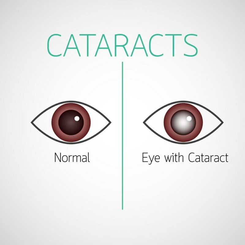
Cataract
Capsulorhexis
Lens Implantation, Intraocular
Posterior Capsule of the Lens
Phacoemulsification
Silicone Elastomers
Acrylic Resins
Lens, Crystalline
Polymethyl Methacrylate
Acrylates
Laser Therapy
Silicones
Corneal Opacity
Postoperative Complications
Visual Acuity
Diagnostic Techniques, Ophthalmological
Transforming Growth Factor beta2
Hydrophobic and Hydrophilic Interactions
Bacterial Capsules
Capsule Endoscopy
Lens Cortex, Crystalline
Epithelial Cells
Joint Capsule
Capsule Endoscopes
Crystallins
Follow-Up Studies
Photography
Prospective Studies
Internal Capsule
Fluorocarbons
Lens Diseases
Cornea
Sodium Selenite
Onchocerciasis, Ocular
Trichiasis
beta-Crystallins
Iopamidol
Maxillary Sinusitis
Maxillary Sinus
Tomography, X-Ray Computed
alpha-Crystallins
Perception of difficulties with vision-related activities of daily living among patients undergoing unilateral posterior capsulotomy. (1/20)
(+info)Aldose reductase-mediated induction of epithelium-to-mesenchymal transition (EMT) in lens. (2/20)
(+info)SiRNA targeting EGFR effectively prevents posterior capsular opacification after cataract surgery. (3/20)
PURPOSE: We investigated the effect of epidermal growth factor receptor (EGFR) siRNA on human lens epithelium (HLE) cells and the development of posterior capsular opacity (PCO). METHODS: We designed EGFR siRNA and used it to knockdown the expression of EGFR in HLE cells. Cell proliferation was examined by 3-(4,5-Dimethylthiazol-2-yl)-2,5-diphenyltetrazolium bromide (MTT), cell growth curve assay and cell cycle analysis. Next, we selected an adaptable concentration of recombinant epidermal growth factor (EGF) for stimulating the growth of HLE cells to further test the suppressive effect of siRNA. At last, we established the model of PCO in rats to further investigate whether knocking down EGFR would prevent the progression of PCO in vivo. RESULTS: The cell proliferation of EGFR siRNA group was apparently inhibited no matter in short or long term and cell cycle was arrested in G(1) phase. Over expression EGF cannot rescue the inhibition of EGFR siRNA on HLE cells and the proliferation activity in HLE cells greatly decreased when EGF-EGFR signal pathway blockaded. In vivo experiments, the extent of PCO of EGFR siRNA group is much lower than the control group. CONCLUSIONS: Our results demonstrate that EGFR siRNA can effectively inhibit the progression of PCO. Thus, siRNA targeting EGFR may provide a totally new way for preventing PCO or even cataract. (+info)Complications, adverse events, and additional intraocular surgery 1 year after cataract surgery in the infant Aphakia Treatment Study. (4/20)
(+info)The impact of capsulorhexis diameter, localization and shape on posterior capsule opacification. (5/20)
BACKGROUND: The aim of this study was to evaluate the impact of capsulorhexis diameter, localization and shape on posterior capsule opacification (PCO) development after cataract extraction with phacoemulsification. MATERIAL/METHODS: We retrospectively analyzed of 297 patients who underwent phacoemulsification and AcrySof SA60AT implantation. In a first group of 97 patients, 53 received small capsulorhexis (3.9 to 4.9 mm in diameter) and 44 patients received large capsulorhexis (5.0 to 5.9 mm in diameter). Another group of 99 patients was split into subgroups--66 patients whose capsulorhexis were centrally located and 33 patients whose capsulorhexis were paracentral. A third group of 101 patients was split into subgroups--a subgroup of 59 patients were classified as having a regularly rimmed capsulorhexis and a subgroup of 42 patients as having an irregularly rimmed capsulorhexis. At 6 months follow-up, PCO was classified as none, mild, moderate, or severe, depending on the number of quadrants involved. RESULTS: 86.79% of the patients with a small capsulorhexis had no or mild PCO (p<0.001), whereas, 68.18% of the patients with a large capsulorhexis experienced moderate or severe PCO; 89.4% of the patients with a central capsulorhexis had no or mild PCO (p<0.001), whereas, 75.75% of the patients with a paracentral capsulorhexis had moderate or severe PCO; 86.44% of the patients with a regularly rimmed anterior capsulorhexis had no or mild PCO (p<0.001); and 69.04% of the patients with an irregular capsulorhexis rim had moderate or severe PCO. CONCLUSIONS: A small capsulorhexis diameter, its central localization and regular shape result in less PCO following phacoemulsification. (+info)Zebularine suppresses TGF-beta-induced lens epithelial cell-myofibroblast transdifferentiation by inhibiting MeCP2. (6/20)
PURPOSE: Posterior capsular opacification (PCO) is a common long-term complication of modern cataract surgery. Remnant lens epithelial cells (LECs) undergo a myofibroblast transdifferentiation that is thought to be the initial step of PCO pathogenesis. The purpose of this study is to determine the effects of zebularine on transforming growth factor-beta (TGFbeta)-induced, LEC-myofibroblast transdifferentiation. METHODS: The expression levels of methyl CpG binding protein 2 (MeCP2) and alpha-smooth muscle actin (alpha-SMA) in human PCO membranes were evaluated by confocal microscopy. The role that MeCP2 played in TGFbeta2-induced alpha-SMA expression was analyzed by western blotting both before and after MeCP2 knockdown with MeCP2-specific siRNA. The effect of zebularine on MeCP2 expression was analyzed over time using a variety of dosages. The effect of zebularine on TGFbeta2-induced alpha-SMA expression was determined by western blot analysis. RESULTS: MeCP2 and alpha-SMA co-localized in human PCO membranes. When MeCP2 was depleted, TGFbeta2 could not induce alpha-SMA expression. Zebularine decreased MeCP2 expression in lens epithelial cells in a time- and dose-dependent pattern and reversed TGFbeta2-induced alpha-SMA expression. CONCLUSIONS: MeCP2 plays an important role in TGFbeta2-induced alpha-SMA expression in lens epithelial cells. Zebularine could reverse the TGFbeta2-induced alpha-SMA expression by inhibiting MeCP2 expression. Therefore, zebularine could potentially prevent PCO formation. (+info)A fully human in vitro capsular bag model to permit intraocular lens evaluation. (7/20)
(+info)Cytoskeletal drugs prevent posterior capsular opacification in human lens capsule in vitro. (8/20)
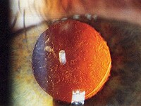
(+info)Capsule opacification, also known as posterior capsular opacification (PCO) or "after-cataract," is a condition that can occur after cataract surgery. During cataract surgery, the cloudy natural lens of the eye is removed and replaced with an artificial intraocular lens (IOL). However, over time, the remaining capsule that holds the IOL in place can become cloudy, leading to blurry or distorted vision. This clouding of the capsule is called capsule opacification. It is not a true reformation of the cataract but a separate condition that can occur after cataract surgery.
Capsule opacification can be treated with a simple laser procedure called YAG capsulotomy, which creates an opening in the cloudy capsule to restore clear vision. This procedure is typically quick, painless, and performed on an outpatient basis.
The crystalline lens of the eye is covered by a transparent, elastic capsule known as the lens capsule. This capsule is made up of collagen and forms the continuous outer layer of the lens. It is highly resistant to both physical and chemical insults, which allows it to protect the lens fibers within. The lens capsule is important for maintaining the shape and transparency of the lens, which are essential for proper focusing of light onto the retina.
A cataract is a clouding of the natural lens in the eye that affects vision. This clouding can cause vision to become blurry, faded, or dim, making it difficult to see clearly. Cataracts are a common age-related condition, but they can also be caused by injury, disease, or medication use. In most cases, cataracts develop gradually over time and can be treated with surgery to remove the cloudy lens and replace it with an artificial one.
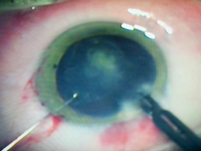
Capsulorhexis is a surgical procedure that is commonly performed during cataract surgery. It involves creating a circular opening in the front part of the lens capsule, which is a clear membrane that surrounds and holds the lens in place inside the eye. This opening allows the cloudy lens material (cataract) to be removed and replaced with an artificial intraocular lens (IOL).
The procedure is typically performed using a specialized instrument called a cystotome or a femtosecond laser, which creates a small tear in the capsule that can be carefully enlarged to the desired size. The capsulorhexis is crucial for the successful removal of the cataract and the proper placement of the IOL. If the capsulorhexis is not performed correctly, it can lead to complications such as posterior capsular opacification (PCO), which is a thickening and clouding of the back part of the lens capsule that can cause visual symptoms similar to those of a cataract.
Intraocular lenses (IOLs) are artificial lens implants that are placed inside the eye during ophthalmic surgery, such as cataract removal. These lenses are designed to replace the natural lens of the eye that has become clouded or damaged, thereby restoring vision impairment caused by cataracts or other conditions.
There are several types of intraocular lenses available, including monofocal, multifocal, toric, and accommodative lenses. Monofocal IOLs provide clear vision at a single fixed distance, while multifocal IOLs offer clear vision at multiple distances. Toric IOLs are designed to correct astigmatism, and accommodative IOLs can change shape and position within the eye to allow for a range of vision.
The selection of the appropriate type of intraocular lens depends on various factors, including the patient's individual visual needs, lifestyle, and ocular health. The implantation procedure is typically performed on an outpatient basis and involves minimal discomfort or recovery time. Overall, intraocular lenses have become a safe and effective treatment option for patients with vision impairment due to cataracts or other eye conditions.
Intraocular lens (IOL) implantation is a surgical procedure that involves placing a small artificial lens inside the eye to replace the natural lens that has been removed. This procedure is typically performed during cataract surgery, where the cloudy natural lens is removed and replaced with an IOL to restore clear vision.
During the procedure, a small incision is made in the eye, and the cloudy lens is broken up and removed using ultrasound waves or laser energy. Then, the folded IOL is inserted through the same incision and positioned in the correct place inside the eye. Once in place, the IOL unfolds and is secured into position.
There are several types of IOLs available, including monofocal, multifocal, toric, and accommodating lenses. Monofocal lenses provide clear vision at one distance, while multifocal lenses offer clear vision at multiple distances. Toric lenses correct astigmatism, and accommodating lenses can change shape to focus on objects at different distances.
Overall, intraocular lens implantation is a safe and effective procedure that can help restore clear vision in patients with cataracts or other eye conditions that require the removal of the natural lens.
The posterior capsule of the lens is a thin, transparent layer of tissue that lies behind the lens cortex in the eye. It surrounds and helps to maintain the shape of the lens, which is necessary for focusing light onto the retina. The posterior capsule is one of the five layers that make up the adult human lens, along with the anterior capsule, lens epithelium, lens cortex, and lens nucleus.
Damage or opacification of the posterior capsule can result in a clouding of vision known as a posterior capsular opacity (PCO) or "secondary cataract." This is a common complication following cataract surgery, where the cloudy lens has been removed but the posterior capsule remains. In such cases, a laser procedure called a YAG capsulotomy may be performed to create an opening in the posterior capsule and restore clear vision.
Phacoemulsification is a surgical procedure used in cataract removal. It involves using an ultrasonic device to emulsify (break up) the cloudy lens (cataract) into small pieces, which are then aspirated or sucked out through a small incision. This procedure allows for smaller incisions and faster recovery times compared to traditional cataract surgery methods. After the cataract is removed, an artificial intraocular lens (IOL) is typically implanted to replace the natural lens and restore vision.
Cataract extraction is a surgical procedure that involves removing the cloudy lens (cataract) from the eye. This procedure is typically performed to restore vision impairment caused by cataracts and improve overall quality of life. There are two primary methods for cataract extraction:
1. Phacoemulsification: This is the most common method used today. It involves making a small incision in the front part of the eye (cornea), inserting an ultrasonic probe to break up the cloudy lens into tiny pieces, and then removing those pieces with suction. After removing the cataract, an artificial intraocular lens (IOL) is inserted to replace the natural lens and help focus light onto the retina.
2. Extracapsular Cataract Extraction: In this method, a larger incision is made on the side of the cornea, allowing the surgeon to remove the cloudy lens in one piece without breaking it up. The back part of the lens capsule is left intact to support the IOL. This technique is less common and typically reserved for more advanced cataracts or when phacoemulsification cannot be performed.
Recovery from cataract extraction usually involves using eye drops to prevent infection and inflammation, as well as protecting the eye with a shield or glasses during sleep for a few weeks after surgery. Most people experience improved vision within a few days to a week following the procedure.
Silicone elastomers are a type of synthetic rubber made from silicone, which is a polymer composed primarily of silicon-oxygen bonds. They are known for their durability, flexibility, and resistance to heat, cold, and moisture. Silicone elastomers can be manufactured in various forms, including liquids, gels, and solids, and they are used in a wide range of medical applications such as:
1. Breast implants: Silicone elastomer shells filled with silicone gel are commonly used for breast augmentation and reconstruction.
2. Contact lenses: Some contact lenses are made from silicone elastomers due to their high oxygen permeability, which allows for better eye health.
3. Catheters: Silicone elastomer catheters are flexible and resistant to kinking, making them suitable for long-term use in various medical procedures.
4. Implantable drug delivery systems: Silicone elastomers can be used as a matrix for controlled release of drugs, allowing for sustained and targeted medication administration.
5. Medical adhesives: Silicone elastomer adhesives are biocompatible and can be used to attach medical devices to the skin or other tissues.
6. Sealants and coatings: Silicone elastomers can be used as sealants and coatings in medical devices to prevent leakage, improve durability, and reduce infection risk.
It is important to note that while silicone elastomers are generally considered safe for medical use, there have been concerns about the potential health risks associated with breast implants, such as capsular contracture, breast pain, and immune system reactions. However, these risks vary depending on the individual's health status and the specific type of silicone elastomer used.
Acrylic resins are a type of synthetic polymer made from methacrylate monomers. They are widely used in various industrial, commercial, and medical applications due to their unique properties such as transparency, durability, resistance to breakage, and ease of coloring or molding. In the medical field, acrylic resins are often used to make dental restorations like false teeth and fillings, medical devices like intraocular lenses, and surgical instruments. They can also be found in orthopedic implants, bone cement, and other medical-grade plastics. Acrylic resins are biocompatible, meaning they do not typically cause adverse reactions when in contact with living tissue. However, they may release small amounts of potentially toxic chemicals over time, so their long-term safety in certain applications is still a subject of ongoing research.
Pseudophakia is a medical term that refers to the condition where a person's natural lens in the eye has been replaced with an artificial one. This procedure is typically performed during cataract surgery, where the cloudy, natural lens is removed and replaced with a clear, artificial lens to improve vision. The prefix "pseudo" means false or fake, and "phakia" refers to the natural lens of the eye, hence the term "Pseudophakia" implies a false or artificial lens.
The crystalline lens is a biconvex transparent structure in the eye that helps to refract (bend) light rays and focus them onto the retina. It is located behind the iris and pupil and is suspended by small fibers called zonules that connect it to the ciliary body. The lens can change its shape to accommodate and focus on objects at different distances, a process known as accommodation. With age, the lens may become cloudy or opaque, leading to cataracts.
Polymethyl methacrylate (PMMA) is a type of synthetic resin that is widely used in the medical field due to its biocompatibility and versatility. It is a transparent, rigid, and lightweight material that can be easily molded into different shapes and forms. Here are some of the medical definitions of PMMA:
1. A biocompatible acrylic resin used in various medical applications such as bone cement, intraocular lenses, dental restorations, and drug delivery systems.
2. A type of synthetic material that is used as a bone cement to fix prosthetic joint replacements and vertebroplasty for the treatment of spinal fractures.
3. A transparent and shatter-resistant material used in the manufacture of medical devices such as intravenous (IV) fluid bags, dialyzer housings, and oxygenators.
4. A drug delivery system that can be used to administer drugs locally or systemically, such as intraocular sustained-release drug implants for the treatment of chronic eye diseases.
5. A component of dental restorations such as fillings, crowns, and bridges due to its excellent mechanical properties and esthetic qualities.
Overall, PMMA is a versatile and valuable material in the medical field, with numerous applications that take advantage of its unique properties.
A capsule is a type of solid pharmaceutical dosage form in which the drug is enclosed in a small shell or container, usually composed of gelatin or other suitable material. The shell serves to protect the drug from degradation, improve its stability and shelf life, and facilitate swallowing by making it easier to consume. Capsules come in various sizes and colors and can contain one or more drugs in powder, liquid, or solid form. They are typically administered orally but can also be used for other routes of administration, such as rectal or vaginal.
Acrylates are a group of chemical compounds that are derived from acrylic acid. They are commonly used in various industrial and commercial applications, including the production of plastics, resins, paints, and adhesives. In the medical field, acrylates are sometimes used in the formation of dental restorations, such as fillings and dentures, due to their strong bonding properties and durability.
However, it is important to note that some people may have allergic reactions or sensitivities to acrylates, which can cause skin irritation, allergic contact dermatitis, or other adverse effects. Therefore, medical professionals must use caution when working with these materials and ensure that patients are informed of any potential risks associated with their use.
Laser therapy, also known as phototherapy or laser photobiomodulation, is a medical treatment that uses low-intensity lasers or light-emitting diodes (LEDs) to stimulate healing, reduce pain, and decrease inflammation. It works by promoting the increase of cellular metabolism, blood flow, and tissue regeneration through the process of photobiomodulation.
The therapy can be used on patients suffering from a variety of acute and chronic conditions, including musculoskeletal injuries, arthritis, neuropathic pain, and wound healing complications. The wavelength and intensity of the laser light are precisely controlled to ensure a safe and effective treatment.
During the procedure, the laser or LED device is placed directly on the skin over the area of injury or discomfort. The non-ionizing light penetrates the tissue without causing heat or damage, interacting with chromophores in the cells to initiate a series of photochemical reactions. This results in increased ATP production, modulation of reactive oxygen species, and activation of transcription factors that lead to improved cellular function and reduced pain.
In summary, laser therapy is a non-invasive, drug-free treatment option for various medical conditions, providing patients with an alternative or complementary approach to traditional therapies.
Silicones are not a medical term, but they are commonly used in the medical field, particularly in medical devices and healthcare products. Silicones are synthetic polymers made up of repeating units of siloxane, which is a chain of alternating silicon and oxygen atoms. They can exist in various forms such as oils, gels, rubbers, and resins.
In the medical context, silicones are often used for their unique properties, including:
1. Biocompatibility - Silicones have a low risk of causing an adverse reaction when they come into contact with living tissue.
2. Inertness - They do not react chemically with other substances, making them suitable for use in medical devices that need to remain stable over time.
3. Temperature resistance - Silicones can maintain their flexibility and elasticity even under extreme temperature conditions.
4. Gas permeability - Some silicone materials allow gases like oxygen and water vapor to pass through, which is useful in applications where maintaining a moist environment is essential.
5. Durability - Silicones have excellent resistance to aging, weathering, and environmental factors, ensuring long-lasting performance.
Examples of medical applications for silicones include:
1. Breast implants
2. Contact lenses
3. Catheters
4. Artificial joints and tendons
5. Bandages and wound dressings
6. Drug delivery systems
7. Medical adhesives
8. Infant care products (nipples, pacifiers)
Corneal opacity refers to a condition in which the cornea, the clear front part of the eye, becomes cloudy or opaque. This can occur due to various reasons such as injury, infection, degenerative changes, or inherited disorders. As a result, light is not properly refracted and vision becomes blurred or distorted. In some cases, corneal opacity can lead to complete loss of vision in the affected eye. Treatment options depend on the underlying cause and may include medication, corneal transplantation, or other surgical procedures.
Postoperative complications refer to any unfavorable condition or event that occurs during the recovery period after a surgical procedure. These complications can vary in severity and may include, but are not limited to:
1. Infection: This can occur at the site of the incision or inside the body, such as pneumonia or urinary tract infection.
2. Bleeding: Excessive bleeding (hemorrhage) can lead to a drop in blood pressure and may require further surgical intervention.
3. Blood clots: These can form in the deep veins of the legs (deep vein thrombosis) and can potentially travel to the lungs (pulmonary embolism).
4. Wound dehiscence: This is when the surgical wound opens up, which can lead to infection and further complications.
5. Pulmonary issues: These include atelectasis (collapsed lung), pneumonia, or respiratory failure.
6. Cardiovascular problems: These include abnormal heart rhythms (arrhythmias), heart attack, or stroke.
7. Renal failure: This can occur due to various reasons such as dehydration, blood loss, or the use of certain medications.
8. Pain management issues: Inadequate pain control can lead to increased stress, anxiety, and decreased mobility.
9. Nausea and vomiting: These can be caused by anesthesia, opioid pain medication, or other factors.
10. Delirium: This is a state of confusion and disorientation that can occur in the elderly or those with certain medical conditions.
Prompt identification and management of these complications are crucial to ensure the best possible outcome for the patient.
Visual acuity is a measure of the sharpness or clarity of vision. It is usually tested by reading an eye chart from a specific distance, such as 20 feet (6 meters). The standard eye chart used for this purpose is called the Snellen chart, which contains rows of letters that decrease in size as you read down the chart.
Visual acuity is typically expressed as a fraction, with the numerator representing the testing distance and the denominator indicating the smallest line of type that can be read clearly. For example, if a person can read the line on the eye chart that corresponds to a visual acuity of 20/20, it means they have normal vision at 20 feet. If their visual acuity is 20/40, it means they must be as close as 20 feet to see what someone with normal vision can see at 40 feet.
It's important to note that visual acuity is just one aspect of overall vision and does not necessarily reflect other important factors such as peripheral vision, depth perception, color vision, or contrast sensitivity.
Diagnostic techniques in ophthalmology refer to the various methods and tests used by eye specialists (ophthalmologists) to examine, evaluate, and diagnose conditions related to the eyes and visual system. Here are some commonly used diagnostic techniques:
1. Visual Acuity Testing: This is a basic test to measure the sharpness of a person's vision. It typically involves reading letters or numbers from an eye chart at a specific distance.
2. Refraction Test: This test helps determine the correct lens prescription for glasses or contact lenses by measuring how light is bent as it passes through the cornea and lens.
3. Slit Lamp Examination: A slit lamp is a microscope that allows an ophthalmologist to examine the structures of the eye, including the cornea, iris, lens, and retina, in great detail.
4. Tonometry: This test measures the pressure inside the eye (intraocular pressure) to detect conditions like glaucoma. Common methods include applanation tonometry and non-contact tonometry.
5. Retinal Imaging: Several techniques are used to capture images of the retina, including fundus photography, fluorescein angiography, and optical coherence tomography (OCT). These tests help diagnose conditions like macular degeneration, diabetic retinopathy, and retinal detachments.
6. Color Vision Testing: This test evaluates a person's ability to distinguish between different colors, which can help detect color vision deficiencies or neurological disorders affecting the visual pathway.
7. Visual Field Testing: This test measures a person's peripheral (or side) vision and can help diagnose conditions like glaucoma, optic nerve damage, or brain injuries.
8. Pupillary Reactions Tests: These tests evaluate how the pupils respond to light and near objects, which can provide information about the condition of the eye's internal structures and the nervous system.
9. Ocular Motility Testing: This test assesses eye movements and alignment, helping diagnose conditions like strabismus (crossed eyes) or nystagmus (involuntary eye movement).
10. Corneal Topography: This non-invasive imaging technique maps the curvature of the cornea, which can help detect irregularities, assess the fit of contact lenses, and plan refractive surgery procedures.
Transforming Growth Factor beta2 (TGF-β2) is a type of cytokine, specifically a growth factor, that plays a role in cell growth, division, and apoptosis (programmed cell death). It belongs to the TGF-β family of proteins. TGF-β2 is involved in various biological processes such as embryonic development, tissue homeostasis, wound healing, and immune regulation. In particular, it has been implicated in the regulation of extracellular matrix production and fibrosis, making it an important factor in diseases that involve excessive scarring or fibrotic changes, such as glaucoma, Marfan syndrome, and systemic sclerosis.
Hydrophobic interactions: These are the interactions that occur between non-polar molecules or groups of atoms in an aqueous environment, leading to their association or aggregation. The term "hydrophobic" means "water-fearing" and describes the tendency of non-polar substances to repel water. When non-polar molecules or groups are placed in water, they tend to clump together to minimize contact with the polar water molecules. These interactions are primarily driven by the entropy increase of the system as a whole, rather than energy minimization. Hydrophobic interactions play crucial roles in various biological processes, such as protein folding, membrane formation, and molecular self-assembly.
Hydrophilic interactions: These are the interactions that occur between polar molecules or groups of atoms and water molecules. The term "hydrophilic" means "water-loving" and describes the attraction of polar substances to water. When polar molecules or groups are placed in water, they can form hydrogen bonds with the surrounding water molecules, which helps solvate them. Hydrophilic interactions contribute to the stability and functionality of various biological systems, such as protein structure, ion transport across membranes, and enzyme catalysis.
Bacterial capsules are slimy, gel-like layers that surround many types of bacteria. They are made up of polysaccharides, proteins, or lipopolysaccharides and are synthesized by the bacterial cell. These capsules play a crucial role in the virulence and pathogenicity of bacteria as they help the bacteria to evade the host's immune system and promote their survival and colonization within the host. The presence of a capsule can also contribute to the bacteria's resistance to desiccation, phagocytosis, and antibiotics.
The chemical composition and structure of bacterial capsules vary among different species of bacteria, which is one factor that contributes to their serological specificity and allows for their identification and classification using methods such as the Quellung reaction or immunofluorescence microscopy.
Prosthesis design is a specialized field in medical device technology that involves creating and developing artificial substitutes to replace a missing body part, such as a limb, tooth, eye, or internal organ. The design process typically includes several stages: assessment of the patient's needs, selection of appropriate materials, creation of a prototype, testing and refinement, and final fabrication and fitting of the prosthesis.
The goal of prosthesis design is to create a device that functions as closely as possible to the natural body part it replaces, while also being comfortable, durable, and aesthetically pleasing for the patient. The design process may involve collaboration between medical professionals, engineers, and designers, and may take into account factors such as the patient's age, lifestyle, occupation, and overall health.
Prosthesis design can be highly complex, particularly for advanced devices such as robotic limbs or implantable organs. These devices often require sophisticated sensors, actuators, and control systems to mimic the natural functions of the body part they replace. As a result, prosthesis design is an active area of research and development in the medical field, with ongoing efforts to improve the functionality, comfort, and affordability of these devices for patients.
Capsule endoscopy is a medical procedure that uses a small, pill-sized camera to capture images of the digestive tract. The capsule is swallowed and transmits images wirelessly as it moves through the gastrointestinal (GI) tract, allowing doctors to examine the lining of the small intestine, which can be difficult to reach with traditional endoscopes.
The procedure is commonly used to diagnose and monitor conditions such as Crohn's disease, celiac disease, obscure gastrointestinal bleeding, and tumors in the small intestine. The images captured by the capsule are transmitted to a recorder worn by the patient, and then reviewed and analyzed by a healthcare professional.
Capsule endoscopy is generally considered safe and non-invasive, with few risks or side effects. However, it may not be suitable for everyone, including patients with swallowing difficulties, pacemakers, or certain gastrointestinal obstructions. It's important to consult with a healthcare provider to determine if capsule endoscopy is the right diagnostic tool for a particular condition.
The crystalline lens in the eye is composed of three main parts: the capsule, the cortex, and the nucleus. The lens cortex is the outer layer of the lens, located between the capsule and the nucleus. It is made up of proteins and water, and its primary function is to help refract (bend) light rays as they pass through the eye, contributing to the focusing power of the eye.
The cortex is more flexible than the central nucleus, allowing it to change shape and adjust the focus of the eye for different distances. However, with age, the lens cortex can become less elastic, leading to presbyopia, a common age-related condition that affects the ability to focus on close objects. Additionally, changes in the lens cortex have been associated with cataracts, a clouding of the lens that can impair vision.
Epithelial cells are types of cells that cover the outer surfaces of the body, line the inner surfaces of organs and glands, and form the lining of blood vessels and body cavities. They provide a protective barrier against the external environment, regulate the movement of materials between the internal and external environments, and are involved in the sense of touch, temperature, and pain. Epithelial cells can be squamous (flat and thin), cuboidal (square-shaped and of equal height), or columnar (tall and narrow) in shape and are classified based on their location and function.
A joint capsule is the fibrous sac that encloses a synovial joint, which is a type of joint characterized by the presence of a cavity filled with synovial fluid. The joint capsule provides stability and strength to the joint, while also allowing for a range of motion. It consists of two layers: an outer fibrous layer and an inner synovial membrane. The fibrous layer is made up of dense connective tissue that helps to stabilize the joint, while the synovial membrane produces synovial fluid, which lubricates the joint and reduces friction during movement.
The lens nucleus, also known as the crystalline lens nucleus, is the central part of the crystalline lens in the eye. The crystalline lens is a biconvex structure located behind the iris and pupil, which helps to refract (bend) light rays and focus them onto the retina.
The lens nucleus is composed of densely packed lens fibers that have lost their nuclei and cytoplasm during differentiation. It is surrounded by the lens cortex, which consists of younger lens fiber cells that are still metabolically active. The lens nucleus is relatively avascular and receives its nutrients through diffusion from the aqueous humor in the anterior chamber of the eye.
The lens nucleus plays an important role in the accommodation process, which allows the eye to focus on objects at different distances. During accommodation, the ciliary muscles contract and release tension on the lens zonules, allowing the lens to become thicker and increase its curvature. This results in a decrease in the focal length of the lens and enables the eye to focus on nearby objects. The lens nucleus is more rigid than the cortex and helps maintain the shape of the lens during accommodation.
Changes in the lens nucleus are associated with several age-related eye conditions, including cataracts and presbyopia. Cataracts occur when the lens becomes cloudy or opaque, leading to a decrease in vision clarity. Presbyopia is a condition that affects the ability to focus on near objects and is caused by a hardening of the lens nucleus and a loss of elasticity in the lens fibers.
A capsule endoscope is a type of medical device used for minimally invasive examination of the digestive tract. It is a small, pill-sized capsule that contains a miniaturized camera, light source, and transmitter. The patient swallows the capsule, which then travels through the gastrointestinal (GI) tract while transmitting images to an external receiver worn by the patient.
The capsule endoscope typically captures approximately 50,000 to 60,000 color images during its journey through the digestive tract, providing detailed visualization of the mucosal lining of the small intestine, which can be difficult to reach with traditional endoscopes. The examination is called capsule endoscopy or wireless capsule enteroscopy.
Capsule endoscopes are mainly used for diagnosing various gastrointestinal conditions such as obscure gastrointestinal bleeding, inflammatory bowel disease (IBD), small bowel tumors, and celiac disease. The procedure is generally safe, non-invasive, and well-tolerated by patients, with minimal discomfort or preparation required compared to traditional endoscopies. However, it may not be suitable for all patients, particularly those with swallowing difficulties, known or suspected gastrointestinal obstructions, or certain implanted electronic devices that could interfere with the capsule's signal transmission.
Crystallins are the major proteins found in the lens of the eye in vertebrates. They make up about 90% of the protein content in the lens and are responsible for maintaining the transparency and refractive properties of the lens, which are essential for clear vision. There are two main types of crystallins, alpha (α) and beta/gamma (β/γ), which are further divided into several subtypes. These proteins are highly stable and have a long half-life, which allows them to remain in the lens for an extended period of time. Mutations in crystallin genes have been associated with various eye disorders, including cataracts and certain types of glaucoma.
Follow-up studies are a type of longitudinal research that involve repeated observations or measurements of the same variables over a period of time, in order to understand their long-term effects or outcomes. In medical context, follow-up studies are often used to evaluate the safety and efficacy of medical treatments, interventions, or procedures.
In a typical follow-up study, a group of individuals (called a cohort) who have received a particular treatment or intervention are identified and then followed over time through periodic assessments or data collection. The data collected may include information on clinical outcomes, adverse events, changes in symptoms or functional status, and other relevant measures.
The results of follow-up studies can provide important insights into the long-term benefits and risks of medical interventions, as well as help to identify factors that may influence treatment effectiveness or patient outcomes. However, it is important to note that follow-up studies can be subject to various biases and limitations, such as loss to follow-up, recall bias, and changes in clinical practice over time, which must be carefully considered when interpreting the results.
I'm sorry for any confusion, but "photography" is not a term typically used in medical definitions. Photography refers to the art, application, or process of creating images by recording light or other electromagnetic radiation, either electronically by means of an image sensor, or chemically by means of a light-sensitive material such as photographic film.
If you're looking for a medical term related to imaging, there are several terms that might be relevant, such as:
1. Radiography: This is a technique using X-rays to visualize the internal structures of the body.
2. Ultrasonography: Also known as ultrasound, this is a diagnostic imaging technique using high-frequency sound waves to create images of the inside of the body.
3. Computed Tomography (CT): A type of imaging that uses X-rays to create detailed cross-sectional images of the body.
4. Magnetic Resonance Imaging (MRI): A type of imaging that uses magnetic fields and radio waves to create detailed images of the organs and tissues within the body.
5. Nuclear Medicine: This is a branch of medical imaging that uses small amounts of radioactive material to diagnose and treat diseases.
If you have any questions related to medical definitions or topics, feel free to ask!
Prospective studies, also known as longitudinal studies, are a type of cohort study in which data is collected forward in time, following a group of individuals who share a common characteristic or exposure over a period of time. The researchers clearly define the study population and exposure of interest at the beginning of the study and follow up with the participants to determine the outcomes that develop over time. This type of study design allows for the investigation of causal relationships between exposures and outcomes, as well as the identification of risk factors and the estimation of disease incidence rates. Prospective studies are particularly useful in epidemiology and medical research when studying diseases with long latency periods or rare outcomes.
Contrast media are substances that are administered to a patient in order to improve the visibility of internal body structures or processes in medical imaging techniques such as X-rays, CT scans, MRI scans, and ultrasounds. These media can be introduced into the body through various routes, including oral, rectal, or intravenous administration.
Contrast media work by altering the appearance of bodily structures in imaging studies. For example, when a patient undergoes an X-ray examination, contrast media can be used to highlight specific organs, tissues, or blood vessels, making them more visible on the resulting images. In CT and MRI scans, contrast media can help to enhance the differences between normal and abnormal tissues, allowing for more accurate diagnosis and treatment planning.
There are several types of contrast media available, each with its own specific properties and uses. Some common examples include barium sulfate, which is used as a contrast medium in X-ray studies of the gastrointestinal tract, and iodinated contrast media, which are commonly used in CT scans to highlight blood vessels and other structures.
While contrast media are generally considered safe, they can sometimes cause adverse reactions, ranging from mild symptoms such as nausea or hives to more serious complications such as anaphylaxis or kidney damage. As a result, it is important for healthcare providers to carefully evaluate each patient's medical history and individual risk factors before administering contrast media.
The internal capsule is a critical structure in the brain that consists of a bundle of white matter fibers (nerve tracts) located deep within the cerebral hemispheres. It serves as a major pathway for the transmission of motor, sensory, and cognitive information between different regions of the brain. The internal capsule is divided into several segments, including the anterior limb, genu, posterior limb, and retrolentiform and sublentiform parts.
The fibers within the internal capsule can be categorized into three groups: corticopontine fibers, corticospinal and corticobulbar fibers, and thalamocortical fibers. Corticopontine fibers originate from the cerebral cortex and terminate in the pons. Corticospinal and corticobulbar fibers are responsible for motor functions, with corticospinal fibers controlling movements of the trunk and limbs, while corticobulbar fibers control movements of the face and head. Thalamocortical fibers carry sensory information from the thalamus to the cerebral cortex.
Damage to the internal capsule can result in various neurological deficits, depending on the specific location and extent of the injury. These may include motor impairments, sensory loss, cognitive dysfunction, or a combination of these symptoms.
I'm sorry for any confusion, but "Fluorocarbons" are not typically associated with medical definitions. Fluorocarbons are chemical compounds that contain carbon atoms bonded to fluorine atoms. They are often used in a variety of applications including refrigerants, fire extinguishing agents, and in the manufacturing of Teflon and other non-stick coatings.
If you have any medical terms or concepts you'd like me to define or explain, please let me know!
Eyelashes are defined in medical terms as the slender, hair-like growths that originate from the edges of the eyelids. They are made up of keratin and follicles, and their primary function is to protect the eyes from debris, sweat, and other irritants by acting as a physical barrier. Additionally, they play a role in enhancing the aesthetic appeal of the eyes and can also serve as a sensory organ, helping to detect potential threats near the eye area.
Lens diseases refer to conditions that affect the lens of the eye, which is a transparent structure located behind the iris and pupil. The main function of the lens is to focus light onto the retina, enabling clear vision. Here are some examples of lens diseases:
1. Cataract: A cataract is a clouding of the lens that affects vision. It is a common age-related condition, but can also be caused by injury, disease, or medication.
2. Presbyopia: This is not strictly a "disease," but rather an age-related change in the lens that causes difficulty focusing on close objects. It typically becomes noticeable in people over the age of 40.
3. Lens dislocation: This occurs when the lens slips out of its normal position, usually due to trauma or a genetic disorder. It can cause vision problems and may require surgical intervention.
4. Lens opacity: This refers to any clouding or opacification of the lens that is not severe enough to be considered a cataract. It can cause visual symptoms such as glare or blurred vision.
5. Anterior subcapsular cataract: This is a type of cataract that forms in the front part of the lens, often as a result of injury or inflammation. It can cause significant visual impairment.
6. Posterior subcapsular cataract: This is another type of cataract that forms at the back of the lens, often as a result of diabetes or certain medications. It can also cause significant visual impairment.
Overall, lens diseases can have a significant impact on vision and quality of life, and may require medical intervention to manage or treat.
The cornea is the clear, dome-shaped surface at the front of the eye. It plays a crucial role in focusing vision. The cornea protects the eye from harmful particles and microorganisms, and it also serves as a barrier against UV light. Its transparency allows light to pass through and get focused onto the retina. The cornea does not contain blood vessels, so it relies on tears and the fluid inside the eye (aqueous humor) for nutrition and oxygen. Any damage or disease that affects its clarity and shape can significantly impact vision and potentially lead to blindness if left untreated.
Sodium Selenite is not a medical term per se, but it is a chemical compound with the formula Na2SeO3. It is used in medicine as a dietary supplement and also in veterinary medicine. Medically, it is used to treat selenium deficiency, which is rare.
Selenium is an essential trace element for human health, playing a crucial role in various physiological processes, such as antioxidant defense systems, thyroid hormone metabolism, and DNA synthesis. Sodium Selenite serves as a source of selenium in these medical applications.
Please note that supplementation with sodium selenite should be under the supervision of a healthcare professional, as excessive selenium intake can lead to selenosis, a condition characterized by symptoms like nausea, vomiting, hair loss, and neurological damage.
Onchocerciasis, Ocular is a medical condition that specifically refers to the eye manifestations caused by the parasitic infection, Onchocerca volvulus. Also known as "river blindness," this disease is spread through the bite of infected blackflies.
Ocular onchocerciasis affects various parts of the eye, including the conjunctiva, cornea, iris, and retina. The infection can cause symptoms such as itching, burning, and redness of the eyes. Over time, it may lead to more serious complications like punctate keratitis (small, scattered opacities on the cornea), cataracts, glaucoma, and ultimately, blindness.
The infection is diagnosed through a skin snip or blood test, which can detect the presence of microfilariae (the larval stage of the parasite) or antibodies against the parasite. Treatment typically involves administering oral medications such as ivermectin, which kills the microfilariae and reduces the risk of eye damage. However, it does not kill the adult worms, so multiple doses are often required to control the infection. In some cases, surgery may be necessary to remove advanced ocular lesions.
Trichiasis is a medical condition where the eyelashes are abnormally positioned and grow inward, so that they rub against the cornea or the inner surface of the eyelid. This can cause irritation, discomfort, and potentially lead to corneal abrasions, scarring, or infection if left untreated. It is often caused by inflammation, injury, or an aging process that affects the eyelids. Treatment options include epilation (removal of the lashes), electrolysis, or surgery to reposition or remove the misdirected lashes and prevent recurrence.
Beta-crystallins are proteins that make up a significant portion of the lens in our eyes. They are part of the crystallin family, which also includes alpha- and gamma-crystallins. These proteins are essential for maintaining the transparency and refractive properties of the eye's lens, allowing us to focus light onto the retina.
Beta-crystallins are organized into two subgroups: beta-A and beta-B. Each subgroup is made up of several different proteins called isoforms, which vary slightly in their amino acid sequences. These isoforms are produced by alternative splicing of the beta-crystallin genes during gene expression.
Mutations in the genes that encode beta-crystallins have been associated with various eye disorders, including cataracts and certain inherited forms of blindness. Cataracts are characterized by the clouding or opacification of the lens, which can lead to vision loss if not treated surgically. Inherited forms of blindness such as congenital nuclear cataracts and retinal degeneration have also been linked to mutations in beta-crystallin genes.
Overall, beta-crystallins play a crucial role in maintaining the health and function of our eyes, and their dysregulation can contribute to various eye disorders.
Iopamidol is a non-ionic, low-osmolar contrast media (LOCM) used in diagnostic imaging procedures such as X-rays, CT scans, and angiography. It is a type of radiocontrast agent that contains iodine atoms, which absorb X-rays and make the internal structures of the body visible on X-ray images. Iopamidol has a low osmolarity, which means it has fewer particles per unit volume compared to high-osmolar contrast media (HOCM). This makes it safer and more comfortable for patients as it reduces the risk of adverse reactions such as pain, vasodilation, and kidney damage. Iopamidol is elimated from the body primarily through the kidneys and excreted in the urine.
Maxillary sinusitis is a medical condition characterized by inflammation or infection of the maxillary sinuses, which are air-filled cavities located in the upper part of the cheekbones. These sinuses are lined with mucous membranes that produce mucus to help filter and humidify the air we breathe.
When the maxillary sinuses become inflamed or infected, they can fill with fluid and pus, leading to symptoms such as:
* Pain or pressure in the cheeks, upper teeth, or behind the eyes
* Nasal congestion or stuffiness
* Runny nose or postnasal drip
* Reduced sense of smell or taste
* Headache or facial pain
* Fatigue or fever (in cases of bacterial infection)
Maxillary sinusitis can be caused by viruses, bacteria, or fungi, and may also result from allergies, structural abnormalities, or exposure to environmental irritants such as smoke or pollution. Treatment typically involves managing symptoms with over-the-counter remedies or prescription medications, such as decongestants, antihistamines, or antibiotics. In some cases, more invasive treatments such as sinus surgery may be necessary.
Keratitis is a medical condition that refers to inflammation of the cornea, which is the clear, dome-shaped surface at the front of the eye. The cornea plays an essential role in focusing vision, and any damage or infection can cause significant visual impairment. Keratitis can result from various causes, including bacterial, viral, fungal, or parasitic infections, as well as trauma, allergies, or underlying medical conditions such as dry eye syndrome. Symptoms of keratitis may include redness, pain, tearing, sensitivity to light, blurred vision, and a feeling of something foreign in the eye. Treatment for keratitis depends on the underlying cause but typically includes antibiotics, antivirals, or anti-fungal medications, as well as measures to alleviate symptoms and promote healing.
The maxillary sinuses, also known as the antrums of Highmore, are the largest of the four pairs of paranasal sinuses located in the maxilla bones. They are air-filled cavities that surround the nasolacrimal duct and are situated superior to the upper teeth and lateral to the nasal cavity. Each maxillary sinus is lined with a mucous membrane, which helps to warm, humidify, and filter the air we breathe. Inflammation or infection of the maxillary sinuses can result in conditions such as sinusitis, leading to symptoms like facial pain, headaches, and nasal congestion.
Cerebral veins are the blood vessels that carry deoxygenated blood from the brain to the dural venous sinuses, which are located between the layers of tissue covering the brain. The largest cerebral vein is the superior sagittal sinus, which runs along the top of the brain. Other major cerebral veins include the straight sinus, transverse sinus, sigmoid sinus, and cavernous sinus. These veins receive blood from smaller veins called venules that drain the surface and deep structures of the brain. The cerebral veins play an important role in maintaining normal circulation and pressure within the brain.
Corneal diseases are a group of disorders that affect the cornea, which is the clear, dome-shaped surface at the front of the eye. The cornea plays an important role in focusing vision, and any damage or disease can cause significant visual impairment or loss. Some common types of corneal diseases include:
1. Keratoconus: A progressive disorder in which the cornea thins and bulges outward into a cone shape, causing distorted vision.
2. Fuchs' dystrophy: A genetic disorder that affects the inner layer of the cornea called the endothelium, leading to swelling, cloudiness, and decreased vision.
3. Dry eye syndrome: A condition in which the eyes do not produce enough tears or the tears evaporate too quickly, causing discomfort, redness, and blurred vision.
4. Corneal ulcers: Open sores on the cornea that can be caused by infection, trauma, or other factors.
5. Herpes simplex keratitis: A viral infection of the cornea that can cause recurrent episodes of inflammation, scarring, and vision loss.
6. Corneal dystrophies: Inherited disorders that affect the structure and clarity of the cornea, leading to visual impairment or blindness.
7. Bullous keratopathy: A condition in which the endothelium fails to pump fluid out of the cornea, causing it to swell and form blisters.
8. Corneal trauma: Injury to the cornea caused by foreign objects, chemicals, or other factors that can lead to scarring, infection, and vision loss.
Treatment for corneal diseases varies depending on the specific condition and severity of the disease. Options may include eyedrops, medications, laser surgery, corneal transplantation, or other treatments.
X-ray computed tomography (CT or CAT scan) is a medical imaging method that uses computer-processed combinations of many X-ray images taken from different angles to produce cross-sectional (tomographic) images (virtual "slices") of the body. These cross-sectional images can then be used to display detailed internal views of organs, bones, and soft tissues in the body.
The term "computed tomography" is used instead of "CT scan" or "CAT scan" because the machines take a series of X-ray measurements from different angles around the body and then use a computer to process these data to create detailed images of internal structures within the body.
CT scanning is a noninvasive, painless medical test that helps physicians diagnose and treat medical conditions. CT imaging provides detailed information about many types of tissue including lung, bone, soft tissue and blood vessels. CT examinations can be performed on every part of the body for a variety of reasons including diagnosis, surgical planning, and monitoring of therapeutic responses.
In computed tomography (CT), an X-ray source and detector rotate around the patient, measuring the X-ray attenuation at many different angles. A computer uses this data to construct a cross-sectional image by the process of reconstruction. This technique is called "tomography". The term "computed" refers to the use of a computer to reconstruct the images.
CT has become an important tool in medical imaging and diagnosis, allowing radiologists and other physicians to view detailed internal images of the body. It can help identify many different medical conditions including cancer, heart disease, lung nodules, liver tumors, and internal injuries from trauma. CT is also commonly used for guiding biopsies and other minimally invasive procedures.
In summary, X-ray computed tomography (CT or CAT scan) is a medical imaging technique that uses computer-processed combinations of many X-ray images taken from different angles to produce cross-sectional images of the body. It provides detailed internal views of organs, bones, and soft tissues in the body, allowing physicians to diagnose and treat medical conditions.
Alpha-crystallins are small heat shock proteins found in the lens of the eye. They are composed of two subunits, alpha-A and alpha-B, which can form homo- or hetero-oligomers. Alpha-crystallins have chaperone-like activity, helping to prevent protein aggregation and maintain transparency of the lens. Additionally, they play a role in maintaining the structural integrity of the lens and protecting it from oxidative stress. Mutations in alpha-crystallin genes have been associated with certain forms of cataracts and other eye diseases.
 Intraocular lens
David J. Apple
Nd:YAG laser
Capsulotomy
Cataract surgery
Cataract
Capsule of lens
Phacoemulsification
Multifocal intraocular lens
Soemmering ring
Perlecan
Eye surgery
Snakebite
Transparent ceramics
 Posterior capsule opacification | RNIB
Are lens implant modifications the best way to prevent posterior capsule opacification? | British Journal of Ophthalmology
Diagnostic accuracy of code-free deep learning for detection and evaluation of posterior capsule opacification | BMJ Open...
 Posterior Capsule Opacification of the Optic Nerve Head
 Posterior Capsule Opacification of the Optic Nerve Head
 Posterior Capsule Opacification | Vagelos College of Physicians and Surgeons
Detection of proteoglycans in human posterior capsule opacification<...
 Metformin Reduced Collagen Deposition and Contractility, but Increased Collagen Degradation in in vitro Posterior Capsule...
Histopathologic study of anterior capsule opacification in pseudophakic eyes<...
 Efficacy and safety of heavy silicone oil Densiron 68® in the treatment of complicated retinal detachment with large inferior...
Intraocular lens - Wikipedia
 Pharmaceutics | Free Full-Text | Therapeutic Ophthalmic Lenses: A Review
 DailyMed - YUTIQ- fluocinolone acetonide implant
 Traumatic Cataract: Background, Pathophysiology, Epidemiology
 Frontiers | The application of drug loading and drug release characteristics of two-dimensional nanocarriers for targeted...
 Yag Laser Capsulotomy | Elland, West Yorkshire (Near Huddersfield & Halifax) | Spire Elland Hospital
 Omidria - Side Effects, Uses, Dosage, Overdose, Pregnancy, Alcohol | RxWiki
 Machine learning in Ophthalmology
 YAG Capsulotomy</span><i class="icon" aria-hidden=...
 Cataract Surgery: Risks, Recovery, Costs - American Academy of Ophthalmology
 Cataract surgery | Health information | Bupa UK
 Outcomes of cataract surgery in a rural and urban south Indi... : Indian Journal of Ophthalmology
Indian Journal of Ophthalmology
Indian Journal of Ophthalmology
 An In Vitro Study of Human Lens Epithelial Cell Adhesion to Intraocular Lenses with and without a Fibronectin Coating | IOVS |...
 Cataract: Symptoms, diagnosis, and treatment - Harvard Health
Mitosol (mitomycin ophthalmic) dosing, indications, interactions, adverse effects, and more
 FreeVis: Prof. Dr. Michael Knorz
Eye / Ophthalmology and Visual Sciences - Khoo Teck Puat Hospital
 Transforming growth factor-beta-induced epithelial-mesenchymal transition in the lens: a model for cataract formation
Capsular7
- This condition is called posterior capsular opacification (PCO). (spirehealthcare.com)
- Your doctor might call this a "posterior capsular opacification (or PCO). (aao.org)
- Posterior capsular opacification (PCO) is a well-recognized late complication of modern phacoemulsification cataract surgery and occurs in 25% to 50% of patients by 5 years after surgery. (arvojournals.org)
- The Volk1 Capsulotomy lens is for performing resection of the capsular bag using a laser, following an IOL implantation where opacification of the remaining capsule occurred. (visionmonday.com)
- Cataract formation can also result after lens touch with intraocular instruments, in response to the introduction of intraocular tamponading agents such as silicone oil and gas, and if crystallization on the anterior hyaloid or posterior capsule results in posterior capsular lens feathering and inflammation. (crstodayeurope.com)
- over the posterior pill that create capsular opacification. (offere.us)
- Posterior capsular opacification (PCO) is the most common complication of cataract surgery and is characterized by monocular diplopia, light scattering, and reductions in visual acuity and contrast sensitivity [ 1 , 2 ]. (ekjo.org)
Cataract surgery15
- Posterior capsule opacification (PCO) is a complication that can occur some time after cataract surgery. (rnib.org.uk)
- Your lens capsule is clear and should remain clear following your cataract surgery. (rnib.org.uk)
- PCO occurs because cells remaining after cataract surgery grow over the back (posterior) of the capsule causing it to thicken and become slightly opaque (cloudy). (rnib.org.uk)
- Posterior capsule opacification (PCO) is the most common complication in intraocular lens surgery, occurring months to years after uncomplicated cataract surgery, and decreasing visual acuity significantly. (bmj.com)
- The lens capsule is the thin, elastic-like bag that holds the intraocular lens (IOL) in position after cataract surgery. (tablemountaineyecare.com)
- The remaining portion of the capsule becomes clouded in about 25% of cataract surgery patients. (tablemountaineyecare.com)
- Advances in cataract surgery techniques and intraocular lens design have been shown to reduce the frequency of posterior capsule opacification. (columbia.edu)
- BACKGROUND: Posterior capsule opacification (PCO) often occurs after cataract surgery. (unair.ac.id)
- METHODS: HLEC were isolated from the anterior lens capsule of patients undergoing cataract surgery. (unair.ac.id)
- During cataract surgery the natural lens is removed from the capsule and replaced with a clear plastic lens. (spirehealthcare.com)
- A small proportion of patients will develop clouding of the capsule which covers the lens following cataract surgery. (nuffieldhealth.com)
- However, complications of cataract surgery can lead to posterior capsule opacification (PCO). (eyefaqs.com)
- Posterior capsule opacification (PCO) is a common complication that can occur after cataract surgery. (erikandersonmd.com)
- In this type of cataract surgery, the surgeon makes a larger incision at the edge of the cornea, removes the clouded lens in one piece, and leaves the back of the lens capsule intact. (medfin.in)
- Also called laser-assisted cataract surgery (LACS) or femtosecond laser-assisted cataract surgery (FLACS), this type of cataract surgery uses a femtosecond laser (an infrared laser that emits bursts of laser energy at a very fast rate) to make precise incisions in the cornea and lens capsule, as well as to soften the clouded cataract lens before it is removed. (medfin.in)
Posterior lens capsule2
- Opacification of the posterior lens capsule following extracapsular cataract extraction. (columbia.edu)
- The implant may migrate into the anterior chamber if the posterior lens capsule is not intact. (nih.gov)
Known as secondary cataract1
- That's why posterior capsule opacification is also known as secondary cataract. (tablemountaineyecare.com)
Wrinkling of the posterior capsule1
- Contributing factors: fibrotic changes associated with wrinkling of the posterior capsule and the formation of Elschnig's pearls (the migrating growth of residual epithelial cells towards the posterior capsule). (columbia.edu)
Cataracts3
- Cataracts caused by blunt trauma classically form stellate- or rosette-shaped posterior axial opacities that may be stable or progressive, whereas penetrating trauma with disruption of the lens capsule forms cortical changes that may remain focal if small or may progress rapidly to total cortical opacification. (medscape.com)
- Mr Nanavaty's areas of research interest include astigmatism, wavefront aberrations, ectatic corneal disorders, endothelial disease, lamellar corneal transplant surgeries, quality of vision after surgical interventions, cataracts surgical techniques, intraocular lenses and posterior capsule opacification. (nuffieldhealth.com)
- While these are often called cataracts, posterior capsule opacification is actually a different phenomenon that just has a similar impact on the patient's vision. (myvision.org)
Capsulotomy4
- A simple procedure called a YAG posterior capsulotomy is performed to restore vision lost from the clouded capsule. (tablemountaineyecare.com)
- If necessary, Neodynium:YAG laser posterior capsulotomy is usually performed to create a sufficient opening in the opacified posterior capsule. (columbia.edu)
- YAG capsulotomy is a laser surgery that is performed to create a small hole in the cloudy lens capsule. (nuffieldhealth.com)
- Nevertheless, very periodically a YAG capsulotomy requires to be bigger at a later day and also a lot more hardly ever YAG laser requires to be carried out to opacification before the lens dental implant. (offere.us)
Intraocular lens2
- Without intraocular lens implantation, the edge of the anterior capsule may adhere to the posterior capsule forming Soemmering's ring. (columbia.edu)
- The lens capsule (the membrane surrounding the lens) is left intact and an artificial intraocular lens implant (IOL) is placed there to help restore vision. (medfin.in)
Pseudophakic1
- Using best-corrected visual acuity for classification, the single most important cause for visual impairment was cystoid macular edema in the aphakic group and posterior capsule opacification in the pseudophakic group. (lww.com)
Anterior lens capsule1
- It is responsible for Vossius ring (imprinted iris pigment) sometimes found on the anterior lens capsule following blunt injury. (medscape.com)
Membrane2
- This new artificial lens is placed inside your lens capsule, the membrane that originally covered your natural lens. (rnib.org.uk)
- It happens when a membrane called the posterior capsule becomes cloudy. (aao.org)
Rupture1
- During the 1990s a flexible open loop ACIOL has been increasingly used in the western world for secondary implantation, or if planned extracapsular cataract extraction (ECCE) with PCIOL has not been possible due to capsule rupture. (cehjournal.org)
Lens implant2
- Are lens implant modifications the best way to prevent posterior capsule opacification? (bmj.com)
- Treats blurred vision caused by thickening of the capsule holding the lens implant. (spirehealthcare.com)
Thicken1
- Over time in some patients the capsule behind the lens can thicken. (spirehealthcare.com)
Cloudy2
- In order to do the treatment your consultant will dilate the pupils, apply local anaesthetic (eye drops) and use the YAG laser to create a small hole in the cloudy the lens capsule. (nuffieldhealth.com)
- In some people, the surgery causes posterior capsule opacification, in which the part of the eye behind the artificial lens becomes cloudy. (harvard.edu)
Artificial2
- This leaves some of the capsule behind to keep your artificial lens in place (like a cuff around the IOL) but removes enough in the middle to allow the light to pass directly through to the retina. (rnib.org.uk)
- The artificial IOL is added in place of the damaged lens into the lens capsule. (medfin.in)
Vision1
- It functions to eliminate obscuring of vision which can happen after cataract surgical procedure, if the posterior (back) of the lens capsule has come to be gloomy. (offere.us)
Patients1
- However, posterior capsule opacification has to be considered as a potential long-term problem after ECCE in patients who do not return for follow-up. (cehjournal.org)
Original1
- This is called posterior capsule opacification or PCO and feels similar to the original cataract. (nuffieldhealth.com)
Laser4
- This will help to keep your head and eye still, which is necessary, while the ophthalmologist uses the laser to remove part of the capsule. (rnib.org.uk)
- The ophthalmologist focuses the laser exactly onto the back of the lens capsule to cut away a small circle-shaped area to allow light to pass without obstruction. (rnib.org.uk)
- The YAG is a type of cold laser used to create a small opening in the center of the capsule, allowing a clear area for light to enter the eye. (tablemountaineyecare.com)
- YAG laser treatment opens the capsule allowing light to pass through to the back of the eye and help you see better again. (spirehealthcare.com)
Surgery1
- It used bursts of yttrium-aluminum-garnet (YAG) to remove cloudiness as a result of post-surgery opacification. (eyefaqs.com)
Thin1
- The lens of your eye is held in a thin clear lining called a capsule. (spirehealthcare.com)
Doctor1
- The doctor can diagnose posterior capsule opacification during a routine eye exam using a slit lamp microscope. (tablemountaineyecare.com)
Visual2
- Posterior capsule opacification is not routinely treated, unless when significant visual disturbances are noted. (columbia.edu)
- Whereas peripheral or mild PCO does not have a significant impact on visual acuity, PCO occurring within 3 mm of the central posterior capsule affects visual acuity significantly [ 4 ]. (ekjo.org)
Human1
- Using cuprolinic blue staining, we histochemically examined the ultrastructural localization of proteoglycans in the fibrous-type human posterior capsule opacification. (elsevierpure.com)
Forms1
- It is usually just needed when since the treatment eliminates the scaffold upon which the opacification forms. (offere.us)
Transparent1
- It might help to think of the posterior capsule as a transparent pocket. (aao.org)
Treatment1
- How Much Does Private Treatment For Posterior Pill Opacification Cost? (offere.us)