Cardiac Complexes, Premature
Myocytes, Cardiac
Cardiac Output
Arrhythmias, Cardiac
Death, Sudden, Cardiac
Cardiomegaly
Cardiac Tamponade
Cardiac Pacing, Artificial
Heart Diseases
Myocardium
Cardiac Catheterization
Heart Arrest
Heart Ventricles
Myoblasts, Cardiac
Heart Failure
Cardiomyopathies
Cardiac Imaging Techniques
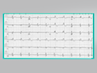
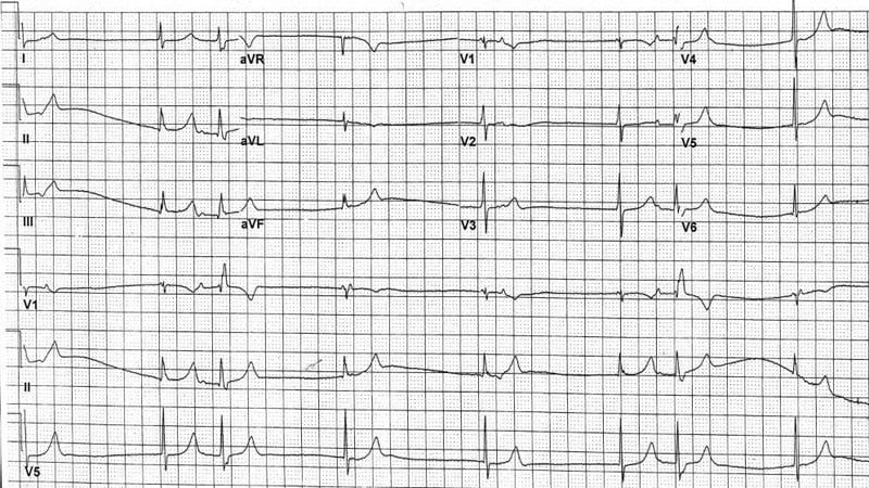
Electrocardiography
Cardiac Glycosides
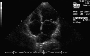
Echocardiography
Cardiac Output, Low
Out-of-Hospital Cardiac Arrest
Hemodynamics
Myocardial Infarction
Cardiac Volume
Ventricular Remodeling
Troponin I
Ventricular Function, Left
Heart Defects, Congenital
Stroke Volume
Cardiac Resynchronization Therapy
Troponin T
Cellular basis for the Brugada syndrome and other mechanisms of arrhythmogenesis associated with ST-segment elevation. (1/200)
BACKGROUND: The Brugada syndrome is characterized by marked ST-segment elevation in the right precordial ECG leads and is associated with a high incidence of sudden and unexpected arrhythmic death. Our study examines the cellular basis for this syndrome. METHODS AND RESULTS: Using arterially perfused wedges of canine right ventricle (RV), we simultaneously recorded transmembrane action potentials from 2 epicardial and 1 endocardial sites, together with unipolar electrograms and a transmural ECG. Loss of the action potential dome in epicardium but not endocardium after exposure to pinacidil (2 to 5 micromol/L), a K(+) channel opener, or the combination of a Na(+) channel blocker (flecainide, 7 micromol/L) and acetylcholine (ACh, 2 to 3 micromol/L) resulted in an abbreviation of epicardial response and a transmural dispersion of repolarization, which caused an ST-segment elevation in the ECG. ACh facilitated loss of the action potential dome, whereas isoproterenol (0.1 to 1 micromol/L) restored the epicardial dome, thus reducing or eliminating the ST-segment elevation. Heterogeneous loss of the dome caused a marked dispersion of repolarization within the epicardium and transmurally, thus giving rise to phase 2 reentrant extrasystole, which precipitated ventricular tachycardia (VT) and ventricular fibrillation (VF). Transient outward current (I(to)) block with 4-aminopyridine (1 to 2 mmol/L) or quinidine (5 micromol/L) restored the dome, normalized the ST segment, and prevented VT/VF. Conclusions-Depression or loss of the action potential dome in RV epicardium creates a transmural voltage gradient that may be responsible for the ST-segment elevation observed in the Brugada syndrome and other syndromes exhibiting similar ECG manifestations. Our results also demonstrate that extrasystolic activity due to phase 2 reentry can arise in the intact wall of the canine RV and serve as the trigger for VT/VF. Our data point to I(to) block (4-aminopyridine, quinidine) as an effective pharmacological treatment. (+info)Pulmonary vein morphology in patients with paroxysmal atrial fibrillation initiated by ectopic beats originating from the pulmonary veins: implications for catheter ablation. (2/200)
BACKGROUND: Successful ablation of ectopic beats originating from the pulmonary veins (PV) could eliminate paroxysmal atrial fibrillation (PAF). However, information about the structure of the PV in patients with PAF that is initiated by PV ectopic beats has not been reported. METHODS AND RESULTS: We studied the morphology of the PVs and measured their diameters in 3 groups of patients. Group I included 52 patients (aged 66+/-14 years; 44 men) with focal atrial fibrillation (AF) from the PVs. Group II included 8 patients (aged 50+/-10 years; 3 men) with focal AF from the superior vena cava or cristal terminalis. Group III included 23 control patients (aged 55+/-16 years; 17 men). Of the control patients, 11 had AV node and 12 had AV reentrant tachycardia. After an atrial transseptal procedure, selective PV angiography using a biplane system with a right anterior oblique view of 30 degrees, a left anterior oblique view of 60 degrees, and a cranial angle of 20 degrees was performed. The ostial and proximal portions of the right and left superior PVs (RSPV and LSPV) were significantly dilated in group I patients compared with those in groups II and III. Furthermore, the ostia of the RSPV and LSPV were significantly dilated in group II compared with group III patients. However, the mean diameters of the inferior PVs were similar between the 3 groups. Comparisons of the individual PV diameters among the 3 subgroups of group I (which was divided according to where the ectopic focus was located) showed nonselective dilatation of the PV. CONCLUSIONS: Nonspecific dilatation of the ostia and proximal portion of superior PVs were found in patients with PAF initiated by ectopic beats from the superior PVs. (+info)Predictive power of increased QT dispersion in ventricular extrasystoles and in sinus beats for risk stratification after myocardial infarction. (3/200)
BACKGROUND: QT dispersion, commonly measured in sinus beats (QTd-S), can also be calculated in premature ventricular beats (QTd-V). To date, no studies have addressed the relation between these 2 variables. METHODS AND RESULTS: In 148 patients with remote myocardial infarction and premature ventricular beats on a routine ECG, QT dispersion, defined as the difference between the maximum and the minimum QT interval across the 12-lead ECG, was calculated separately for the ventricular extrasystole and the preceding sinus beat. In the total group of patients, QTd-V was greater than QTd-S (83+/-33 versus 74+/-34 ms, respectively; P=0.001). During a follow-up period of 35+/-17 months, arrhythmic events (sustained ventricular tachycardia, ventricular fibrillation, or sudden death) were noted in 30 patients. A QTd-V of >/=100 ms was a stronger univariate marker of arrhythmic events than was a QTd-S of >/=100 ms, and multivariate analysis selected only prolonged QTd-V (hazard ratio 3.81, 95% CI 2.2 to 11.2) and low ejection fraction (hazard ratio 3.05, 95% CI 1.6 to 7.6) as independent predictors of arrhythmic events. CONCLUSIONS: The magnitude of QTd-V was greater than that of QTd-S in the total group of patients. Prolonged QTd-V is associated with a significantly increased risk for arrhythmic events in postinfarction patients, and the prognostic significance of QTd-V exceeds that of QTd-S. (+info)Experimental study of the effects of multi-site sequential ventricular pacing on the prophylaxis of ventricular fibrillation. (4/200)
Previous studies report a significant prophylactic effect on the occurrence of atrial fibrillation by simultaneous multi-site atrial pacing. We investigated the effects of multi-site sequential ventricular pacing (MSVP), which may be preferable to simultaneous multi-site pacing in terms of the prophylaxis of the occurrence of ventricular fibrillation (VF). Needle electrodes were inserted at ten different epicardial sites on both ventricles for MSVP in 12 adult beagle dogs. Four premature ventricular extrastimuli (PVE) were introduced to provoke VF reproducibly from a separate electrode in the left ventricle. The 4 PVE were applied to try to provoke VF during MSVP in a comparable fashion to the activation sequence during sinus rhythm. We compared the prophylactic effects of MSVP on the inducibility of VF by changing the number of stimulation sites to either 1, 3, 5, or 10 epicardial sites. We performed a total of 363 trials of induction and suppression of VF. The occurrence rates of VF by the 4 PVE for the various number of epicardial stimulation sites of MSVP, i.e., at 1, 3, 5, and 10 sites, were 0.8263, 0.4286, 0.4450, and 0.2857, respectively (p < 0.05). There was a significant prophylactic effect of MSVP on the inducibility of VF, and this effect became stronger as the number of MSVP sites was increased from 3 to 10. The hemodynamic state was relatively stable during MSVP. MSVP seems to be a promising method with which to reduce the occurrence of VF, and a larger number of stimulation sites would be more effective in terms of the prophylaxis of VF. (+info)Octreotide improved ventricular arrhythmia in an acromegalic patient. (5/200)
We saw a remarkable effect of octreotide, the long-acting somatostatin analogue, in reducing the number of ventricular premature complexes (VPCs) in a 59-year-old woman with acromegaly. Her basal GH and IGF-1 levels were up to 22.9 ng/ml and 934.9 ng/ml respectively. MRI revealed a 14 x 12 x 10 mm mass lesion in the pituitary gland. She had hypertension and echocardiography showed an increase in left ventricular wall thickness. Electric cardiography showed the presence of frequent VPCs and 24-h Holter monitoring revealed 24,277 beats of multifocal VPCs/24 h. She was treated with 300 microg/day of octreotide for four weeks before transsphenoidal surgery. After octreotide treatment, GH and IGF-1 were suppressed to 1.8 ng/ml and 145.3 ng/ml respectively, and the tumor size was remarkably reduced. Furthermore, the number of VPCs was also dramatically reduced to 2062 VPCs/24-h (8.5% of pretreatment) with 24-h Holter monitoring. This case shows that VPCs of acromegalic patients can be controlled by suppressing GH and IGF-1 with octreotide, and this agent is useful for reducing both tumor size and frequency of VPCs prior to surgery. (+info)Postextrasystolic contractile decay always contains exponential and alternans components in canine heart. (6/200)
In isolated, blood-perfused canine hearts, postextrasystolic potentiation (PESP) decays monotonically after a noncompensatory pause following a spontaneous extrasystole (ES). The monotonic PESP decay yields myocardial internal Ca(2+) recirculation fraction (RF). We have found that after a compensatory pause (CP), PESP decays in alternans, consisting of an exponential and a sinusoidal decay component. We have proposed that this exponential component also yields RF. In the present study, we examined the reliability of this alternative method by widely changing the ES coupling interval (ESI), CP, and heart rate in the canine excised, cross-circulated left ventricle. We found that all PESP decays consisted of the sum of an exponential and a sinusoidal decay component of variable magnitudes whether a CP existed or not. Their decay constants as well as the calculated RF were independent of the ESI and CP. This confirmed the utility of our alternative RF determination method regardless of the ESI, CP, and heart rate. Direct experimental evidence of Ca(2+) dynamics supportive of this alternative method, however, remains to be obtained. (+info)Cardiac arrhythmias and left ventricular hypertrophy in dipper and nondipper patients with essential hypertension. (7/200)
To evaluate the behavior of cardiac arrhythmias in dipper and nondipper hypertensive patients, 48-h ambulatory blood pressure monitoring, 24-h Holter electrocardiogram recording and echocardiographic studies were performed in 56 untreated outpatients with essential hypertension. These patients were divided into 2 groups according to the presence (dipper, n=33) or absence (nondipper, n=23) of reduction of both systolic and diastolic blood pressure during nighttime by an average of more than 10% of daytime blood pressure. Mean 48-h systolic and diastolic blood pressures did not differ between the 2 groups. Nondipper patients had a significantly larger left atrial dimension (31.9+/-3.8 vs 35.6+/-3.7 mm; p<0.01), left ventricular mass index (114+/-26 vs 136+/-36 g/m2; p<0.05), as well as a larger number of total supraventricular (16+/-19 vs 89+/-197 beats; p<0.05) and ventricular ectopic beats (7+/-14 vs 47+/-96 beats; p<0.05) during daytime as compared with dippers. In conclusion, nondipper hypertensive patients are likely to experience supraventricular and ventricular arrhythmias more frequently than dippers. A blunted nocturnal blood pressure fall may be involved in the appearance of cardiac arrhythmias in patients with essential hypertension. (+info)Periodicity of arrhythmias in healthy elderly men at the moderate altitude. (8/200)
The 24-hour periodicity of supraventricular (SVPB) and ventricular (VEB) extrasystoles in healthy elderly men (age 49-69 years) was studied at two altitudes during 24 h Holter ECG monitoring. At the low altitude (200 m, n = 26), SVPB were more frequent than VEB. The highest occurrence of SVPB was at 17:00 h, the lowest at 01:00 and 02:00 h (P<0.001). The highest occurrence of VEB was at 09:00 h, the lowest one at 04:00 h (P<0.001). At 1350 m (n=9) the incidence of both SVPB and VEB was approximately twofold higher compared to that at the low altitude (P<0.001). The highest occurrence of SVPB was at 13:00 h, the lowest at 06:00 h (P<0.001). VEB were the most frequent at 10:00 h and 13:00 h, while the lowest frequency was observed at 06:00 h (P<0.001). Our results indicate that the incidence of SVPB and VEB in healthy persons at the moderate altitude is twofold and its periodicity is shifted compared to the low altitude. The cause of increased occurrence of extrasystoles is probably due to beta-adrenergic activation of the heart at the higher altitude. (+info)Premature cardiac complexes, also known as premature heartbeats or premature ventricular contractions (PVCs), refer to extra or early heartbeats that originate in the lower chambers of the heart (the ventricles). These extra beats disrupt the normal rhythm and sequence of heartbeats, causing the heart to beat earlier than expected.
Premature cardiac complexes can occur in healthy individuals as well as those with heart disease. They are usually harmless and do not cause any symptoms, but in some cases, they may cause palpitations, skipped beats, or a fluttering sensation in the chest. In rare cases, frequent premature cardiac complexes can lead to more serious heart rhythm disorders or decreased heart function.
The diagnosis of premature cardiac complexes is usually made through an electrocardiogram (ECG) or Holter monitoring, which records the electrical activity of the heart over a period of time. Treatment is typically not necessary unless the premature complexes are frequent, symptomatic, or associated with underlying heart disease. In such cases, medications, cardioversion, or catheter ablation may be recommended.
Cardiac myocytes are the muscle cells that make up the heart muscle, also known as the myocardium. These specialized cells are responsible for contracting and relaxing in a coordinated manner to pump blood throughout the body. They differ from skeletal muscle cells in several ways, including their ability to generate their own electrical impulses, which allows the heart to function as an independent rhythmical pump. Cardiac myocytes contain sarcomeres, the contractile units of the muscle, and are connected to each other by intercalated discs that help coordinate contraction and ensure the synchronous beating of the heart.
Cardiac output is a measure of the amount of blood that is pumped by the heart in one minute. It is defined as the product of stroke volume (the amount of blood pumped by the left ventricle during each contraction) and heart rate (the number of contractions per minute). Normal cardiac output at rest for an average-sized adult is about 5 to 6 liters per minute. Cardiac output can be increased during exercise or other conditions that require more blood flow, such as during illness or injury. It can be measured noninvasively using techniques such as echocardiography or invasively through a catheter placed in the heart.
In medical terms, the heart is a muscular organ located in the thoracic cavity that functions as a pump to circulate blood throughout the body. It's responsible for delivering oxygen and nutrients to the tissues and removing carbon dioxide and other wastes. The human heart is divided into four chambers: two atria on the top and two ventricles on the bottom. The right side of the heart receives deoxygenated blood from the body and pumps it to the lungs, while the left side receives oxygenated blood from the lungs and pumps it out to the rest of the body. The heart's rhythmic contractions and relaxations are regulated by a complex electrical conduction system.
Cardiac arrhythmias are abnormal heart rhythms that result from disturbances in the electrical conduction system of the heart. The heart's normal rhythm is controlled by an electrical signal that originates in the sinoatrial (SA) node, located in the right atrium. This signal travels through the atrioventricular (AV) node and into the ventricles, causing them to contract and pump blood throughout the body.
An arrhythmia occurs when there is a disruption in this electrical pathway or when the heart's natural pacemaker produces an abnormal rhythm. This can cause the heart to beat too fast (tachycardia), too slow (bradycardia), or irregularly.
There are several types of cardiac arrhythmias, including:
1. Atrial fibrillation: A rapid and irregular heartbeat that starts in the atria (the upper chambers of the heart).
2. Atrial flutter: A rapid but regular heartbeat that starts in the atria.
3. Supraventricular tachycardia (SVT): A rapid heartbeat that starts above the ventricles, usually in the atria or AV node.
4. Ventricular tachycardia: A rapid and potentially life-threatening heart rhythm that originates in the ventricles.
5. Ventricular fibrillation: A chaotic and disorganized electrical activity in the ventricles, which can be fatal if not treated immediately.
6. Heart block: A delay or interruption in the conduction of electrical signals from the atria to the ventricles.
Cardiac arrhythmias can cause various symptoms, such as palpitations, dizziness, shortness of breath, chest pain, and fatigue. In some cases, they may not cause any symptoms and go unnoticed. However, if left untreated, certain types of arrhythmias can lead to serious complications, including stroke, heart failure, or even sudden cardiac death.
Treatment for cardiac arrhythmias depends on the type, severity, and underlying causes. Options may include lifestyle changes, medications, cardioversion (electrical shock therapy), catheter ablation, implantable devices such as pacemakers or defibrillators, and surgery. It is essential to consult a healthcare professional for proper evaluation and management of cardiac arrhythmias.
Cardiac surgical procedures are operations that are performed on the heart or great vessels (the aorta and vena cava) by cardiothoracic surgeons. These surgeries are often complex and require a high level of skill and expertise. Some common reasons for cardiac surgical procedures include:
1. Coronary artery bypass grafting (CABG): This is a surgery to improve blood flow to the heart in patients with coronary artery disease. During the procedure, a healthy blood vessel from another part of the body is used to create a detour around the blocked or narrowed portion of the coronary artery.
2. Valve repair or replacement: The heart has four valves that control blood flow through and out of the heart. If one or more of these valves become damaged or diseased, they may need to be repaired or replaced. This can be done using artificial valves or valves from animal or human donors.
3. Aneurysm repair: An aneurysm is a weakened area in the wall of an artery that can bulge out and potentially rupture. If an aneurysm occurs in the aorta, it may require surgical repair to prevent rupture.
4. Heart transplantation: In some cases, heart failure may be so severe that a heart transplant is necessary. This involves removing the diseased heart and replacing it with a healthy donor heart.
5. Arrhythmia surgery: Certain types of abnormal heart rhythms (arrhythmias) may require surgical treatment. One such procedure is called the Maze procedure, which involves creating a pattern of scar tissue in the heart to disrupt the abnormal electrical signals that cause the arrhythmia.
6. Congenital heart defect repair: Some people are born with structural problems in their hearts that require surgical correction. These may include holes between the chambers of the heart or abnormal blood vessels.
Cardiac surgical procedures carry risks, including bleeding, infection, stroke, and death. However, for many patients, these surgeries can significantly improve their quality of life and longevity.
Sudden cardiac death (SCD) is a sudden, unexpected natural death caused by the cessation of cardiac activity. It is often caused by cardiac arrhythmias, particularly ventricular fibrillation, and is often associated with underlying heart disease, although it can occur in people with no known heart condition. SCD is typically defined as a natural death due to cardiac causes that occurs within one hour of the onset of symptoms, or if the individual was last seen alive in a normal state of health, it can be defined as occurring within 24 hours.
It's important to note that sudden cardiac arrest (SCA) is different from SCD, although they are related. SCA refers to the sudden cessation of cardiac activity, which if not treated immediately can lead to SCD.
Cardiomegaly is a medical term that refers to an enlarged heart. It can be caused by various conditions such as high blood pressure, heart valve problems, cardiomyopathy, or fluid accumulation around the heart (pericardial effusion). Cardiomegaly can be detected through imaging tests like chest X-rays or echocardiograms. Depending on the underlying cause, treatment options may include medications, lifestyle changes, or in some cases, surgery. It is important to consult with a healthcare professional for proper diagnosis and treatment.
Cardiac tamponade is a serious medical condition that occurs when there is excessive fluid or blood accumulation in the pericardial sac, which surrounds the heart. This accumulation puts pressure on the heart, preventing it from filling properly and reducing its ability to pump blood effectively. As a result, cardiac output decreases, leading to symptoms such as low blood pressure, shortness of breath, chest pain, and a rapid pulse. If left untreated, cardiac tamponade can be life-threatening, requiring emergency medical intervention to drain the fluid and relieve the pressure on the heart.
Artificial cardiac pacing is a medical procedure that involves the use of an artificial device to regulate and stimulate the contraction of the heart muscle. This is often necessary when the heart's natural pacemaker, the sinoatrial node, is not functioning properly and the heart is beating too slowly or irregularly.
The artificial pacemaker consists of a small generator that produces electrical impulses and leads that are positioned in the heart to transmit the impulses. The generator is typically implanted just under the skin in the chest, while the leads are inserted into the heart through a vein.
There are different types of artificial cardiac pacing systems, including single-chamber pacemakers, which stimulate either the right atrium or right ventricle, and dual-chamber pacemakers, which stimulate both chambers of the heart. Some pacemakers also have additional features that allow them to respond to changes in the body's needs, such as during exercise or sleep.
Artificial cardiac pacing is a safe and effective treatment for many people with abnormal heart rhythms, and it can significantly improve their quality of life and longevity.
Heart disease is a broad term for a class of diseases that involve the heart or blood vessels. It's often used to refer to conditions that include:
1. Coronary artery disease (CAD): This is the most common type of heart disease. It occurs when the arteries that supply blood to the heart become hardened and narrowed due to the buildup of cholesterol and other substances, which can lead to chest pain (angina), shortness of breath, or a heart attack.
2. Heart failure: This condition occurs when the heart is unable to pump blood efficiently to meet the body's needs. It can be caused by various conditions, including coronary artery disease, high blood pressure, and cardiomyopathy.
3. Arrhythmias: These are abnormal heart rhythms, which can be too fast, too slow, or irregular. They can lead to symptoms such as palpitations, dizziness, and fainting.
4. Valvular heart disease: This involves damage to one or more of the heart's four valves, which control blood flow through the heart. Damage can be caused by various conditions, including infection, rheumatic fever, and aging.
5. Cardiomyopathy: This is a disease of the heart muscle that makes it harder for the heart to pump blood efficiently. It can be caused by various factors, including genetics, viral infections, and drug abuse.
6. Pericardial disease: This involves inflammation or other problems with the sac surrounding the heart (pericardium). It can cause chest pain and other symptoms.
7. Congenital heart defects: These are heart conditions that are present at birth, such as a hole in the heart or abnormal blood vessels. They can range from mild to severe and may require medical intervention.
8. Heart infections: The heart can become infected by bacteria, viruses, or parasites, leading to various symptoms and complications.
It's important to note that many factors can contribute to the development of heart disease, including genetics, lifestyle choices, and certain medical conditions. Regular check-ups and a healthy lifestyle can help reduce the risk of developing heart disease.
The myocardium is the middle layer of the heart wall, composed of specialized cardiac muscle cells that are responsible for pumping blood throughout the body. It forms the thickest part of the heart wall and is divided into two sections: the left ventricle, which pumps oxygenated blood to the rest of the body, and the right ventricle, which pumps deoxygenated blood to the lungs.
The myocardium contains several types of cells, including cardiac muscle fibers, connective tissue, nerves, and blood vessels. The muscle fibers are arranged in a highly organized pattern that allows them to contract in a coordinated manner, generating the force necessary to pump blood through the heart and circulatory system.
Damage to the myocardium can occur due to various factors such as ischemia (reduced blood flow), infection, inflammation, or genetic disorders. This damage can lead to several cardiac conditions, including heart failure, arrhythmias, and cardiomyopathy.
Cardiac catheterization is a medical procedure used to diagnose and treat cardiovascular conditions. In this procedure, a thin, flexible tube called a catheter is inserted into a blood vessel in the arm or leg and threaded up to the heart. The catheter can be used to perform various diagnostic tests, such as measuring the pressure inside the heart chambers and assessing the function of the heart valves.
Cardiac catheterization can also be used to treat certain cardiovascular conditions, such as narrowed or blocked arteries. In these cases, a balloon or stent may be inserted through the catheter to open up the blood vessel and improve blood flow. This procedure is known as angioplasty or percutaneous coronary intervention (PCI).
Cardiac catheterization is typically performed in a hospital cardiac catheterization laboratory by a team of healthcare professionals, including cardiologists, radiologists, and nurses. The procedure may be done under local anesthesia with sedation or general anesthesia, depending on the individual patient's needs and preferences.
Overall, cardiac catheterization is a valuable tool in the diagnosis and treatment of various heart conditions, and it can help improve symptoms, reduce complications, and prolong life for many patients.
Myocardial contraction refers to the rhythmic and forceful shortening of heart muscle cells (myocytes) in the myocardium, which is the muscular wall of the heart. This process is initiated by electrical signals generated by the sinoatrial node, causing a wave of depolarization that spreads throughout the heart.
During myocardial contraction, calcium ions flow into the myocytes, triggering the interaction between actin and myosin filaments, which are the contractile proteins in the muscle cells. This interaction causes the myofilaments to slide past each other, resulting in the shortening of the sarcomeres (the functional units of muscle contraction) and ultimately leading to the contraction of the heart muscle.
Myocardial contraction is essential for pumping blood throughout the body and maintaining adequate circulation to vital organs. Any impairment in myocardial contractility can lead to various cardiac disorders, such as heart failure, cardiomyopathy, and arrhythmias.
Cardiac arrest, also known as heart arrest, is a medical condition where the heart suddenly stops beating or functioning properly. This results in the cessation of blood flow to the rest of the body, including the brain, leading to loss of consciousness and pulse. Cardiac arrest is often caused by electrical disturbances in the heart that disrupt its normal rhythm, known as arrhythmias. If not treated immediately with cardiopulmonary resuscitation (CPR) and defibrillation, it can lead to death or permanent brain damage due to lack of oxygen supply. It's important to note that a heart attack is different from cardiac arrest; a heart attack occurs when blood flow to a part of the heart is blocked, often by a clot, causing damage to the heart muscle, but the heart continues to beat. However, a heart attack can sometimes trigger a cardiac arrest.
The heart ventricles are the two lower chambers of the heart that receive blood from the atria and pump it to the lungs or the rest of the body. The right ventricle pumps deoxygenated blood to the lungs, while the left ventricle pumps oxygenated blood to the rest of the body. Both ventricles have thick, muscular walls to generate the pressure necessary to pump blood through the circulatory system.
Myoblasts are immature cells that later develop into muscle cells (also known as myocytes). Cardiac myoblasts, therefore, are the immature cells that will specialize and develop into cardiac muscle cells. These cells play a crucial role in the growth, repair, and regeneration of heart muscles. In adults, however, the ability of these cells to regenerate damaged heart muscle tissue is limited. Recent research has focused on the potential use of cardiac myoblasts in cell-based therapies for various heart conditions, such as heart failure and myocardial infarction (heart attack).
Heart failure is a pathophysiological state in which the heart is unable to pump sufficient blood to meet the metabolic demands of the body or do so only at the expense of elevated filling pressures. It can be caused by various cardiac disorders, including coronary artery disease, hypertension, valvular heart disease, cardiomyopathy, and arrhythmias. Symptoms may include shortness of breath, fatigue, and fluid retention. Heart failure is often classified based on the ejection fraction (EF), which is the percentage of blood that is pumped out of the left ventricle during each contraction. A reduced EF (less than 40%) is indicative of heart failure with reduced ejection fraction (HFrEF), while a preserved EF (greater than or equal to 50%) is indicative of heart failure with preserved ejection fraction (HFpEF). There is also a category of heart failure with mid-range ejection fraction (HFmrEF) for those with an EF between 40-49%.
Cardiomyopathies are a group of diseases that affect the heart muscle, leading to mechanical and/or electrical dysfunction. The American Heart Association (AHA) defines cardiomyopathies as "a heterogeneous group of diseases of the myocardium associated with mechanical and/or electrical dysfunction that usually (but not always) exhibit inappropriate ventricular hypertrophy or dilatation and frequently lead to heart failure."
There are several types of cardiomyopathies, including:
1. Dilated cardiomyopathy (DCM): This is the most common type of cardiomyopathy, characterized by an enlarged left ventricle and impaired systolic function, leading to heart failure.
2. Hypertrophic cardiomyopathy (HCM): In this type, there is abnormal thickening of the heart muscle, particularly in the septum between the two ventricles, which can obstruct blood flow and increase the risk of arrhythmias.
3. Restrictive cardiomyopathy (RCM): This is a rare form of cardiomyopathy characterized by stiffness of the heart muscle, impaired relaxation, and diastolic dysfunction, leading to reduced filling of the ventricles and heart failure.
4. Arrhythmogenic right ventricular cardiomyopathy (ARVC): In this type, there is replacement of the normal heart muscle with fatty or fibrous tissue, primarily affecting the right ventricle, which can lead to arrhythmias and sudden cardiac death.
5. Unclassified cardiomyopathies: These are conditions that do not fit into any of the above categories but still significantly affect the heart muscle and function.
Cardiomyopathies can be caused by genetic factors, acquired conditions (e.g., infections, toxins, or autoimmune disorders), or a combination of both. The diagnosis typically involves a comprehensive evaluation, including medical history, physical examination, electrocardiogram (ECG), echocardiography, cardiac magnetic resonance imaging (MRI), and sometimes genetic testing. Treatment depends on the type and severity of the condition but may include medications, lifestyle modifications, implantable devices, or even heart transplantation in severe cases.
Cardiac myosins are a type of myosin protein that are specifically expressed in the cardiac muscle cells (or cardiomyocytes) of the heart. These proteins play a crucial role in the contraction and relaxation of heart muscles, which is essential for proper heart function and blood circulation.
Myosins are molecular motors that use chemical energy from ATP to generate force and movement. In the context of cardiac muscle cells, cardiac myosins interact with another protein called actin to form sarcomeres, which are the basic contractile units of muscle fibers. During contraction, the heads of cardiac myosin molecules bind to actin filaments and pull them together, causing the muscle fiber to shorten and generate force.
There are different isoforms of cardiac myosins that can vary in their structure and function. Mutations in the genes encoding these proteins have been linked to various forms of cardiomyopathy, which are diseases of the heart muscle that can lead to heart failure and other complications. Therefore, understanding the structure and function of cardiac myosins is an important area of research for developing therapies and treatments for heart disease.
Cardiac imaging techniques are diagnostic methods used to visualize and assess the structure and function of the heart. These techniques can be non-invasive or invasive, and they use various forms of energy such as sound waves, radiation, and magnetic fields to produce detailed images of the heart. Some common cardiac imaging techniques include:
1. Echocardiography: This technique uses ultrasound waves to create images of the heart's structure and function. It can provide information about the size and shape of the heart chambers, the thickness and movement of the heart walls, and the valves' function.
2. Cardiac Magnetic Resonance Imaging (MRI): This technique uses a strong magnetic field and radio waves to create detailed images of the heart's structure and function. It can provide information about the size and shape of the heart chambers, the thickness and movement of the heart walls, the valves' function, and the blood flow in the heart.
3. Computed Tomography (CT) Angiography: This technique uses X-rays to create detailed images of the heart's blood vessels. It can provide information about the presence and extent of blockages or narrowing in the coronary arteries.
4. Nuclear Cardiac Imaging: This technique uses small amounts of radioactive substances to produce images of the heart's blood flow. It can provide information about the size and function of the heart chambers, the presence of damaged heart muscle, and the extent of coronary artery disease.
5. Invasive Coronary Angiography: This technique involves inserting a catheter into a blood vessel in the arm or leg and guiding it to the heart's coronary arteries. A contrast dye is then injected through the catheter, and X-ray images are taken to visualize the blood flow in the coronary arteries. This technique can provide detailed information about the presence and extent of blockages or narrowing in the coronary arteries.
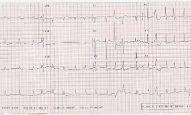
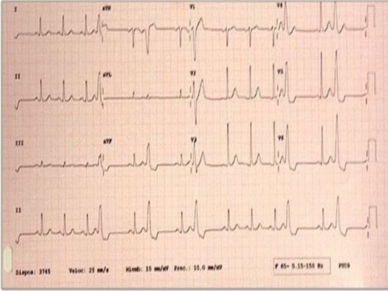
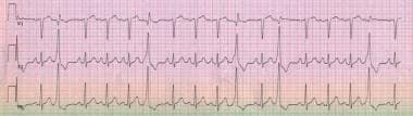
Electrocardiography (ECG or EKG) is a medical procedure that records the electrical activity of the heart. It provides a graphic representation of the electrical changes that occur during each heartbeat. The resulting tracing, called an electrocardiogram, can reveal information about the heart's rate and rhythm, as well as any damage to its cells or abnormalities in its conduction system.
During an ECG, small electrodes are placed on the skin of the chest, arms, and legs. These electrodes detect the electrical signals produced by the heart and transmit them to a machine that amplifies and records them. The procedure is non-invasive, painless, and quick, usually taking only a few minutes.
ECGs are commonly used to diagnose and monitor various heart conditions, including arrhythmias, coronary artery disease, heart attacks, and electrolyte imbalances. They can also be used to evaluate the effectiveness of certain medications or treatments.
Cardiac glycosides are a group of naturally occurring compounds that have a toxic effect on the heart. They are found in certain plants, including foxglove and lily of the valley, as well as in some toads and beetles. The most well-known cardiac glycoside is digoxin, which is derived from the foxglove plant and is used as a medication to treat heart failure and atrial arrhythmias.
Cardiac glycosides work by inhibiting the sodium-potassium pump in heart muscle cells, leading to an increase in intracellular calcium levels. This increases the force of heart contractions, which can be beneficial in treating heart failure. However, if the dose is too high, cardiac glycosides can also cause dangerous arrhythmias and even death.
It's important for healthcare professionals to carefully monitor patients taking cardiac glycosides, as the therapeutic and toxic doses are very close together. Additionally, certain medications and medical conditions can interact with cardiac glycosides and increase the risk of toxicity.
Heart rate is the number of heartbeats per unit of time, often expressed as beats per minute (bpm). It can vary significantly depending on factors such as age, physical fitness, emotions, and overall health status. A resting heart rate between 60-100 bpm is generally considered normal for adults, but athletes and individuals with high levels of physical fitness may have a resting heart rate below 60 bpm due to their enhanced cardiovascular efficiency. Monitoring heart rate can provide valuable insights into an individual's health status, exercise intensity, and response to various treatments or interventions.
Echocardiography is a medical procedure that uses sound waves to produce detailed images of the heart's structure, function, and motion. It is a non-invasive test that can help diagnose various heart conditions, such as valve problems, heart muscle damage, blood clots, and congenital heart defects.
During an echocardiogram, a transducer (a device that sends and receives sound waves) is placed on the chest or passed through the esophagus to obtain images of the heart. The sound waves produced by the transducer bounce off the heart structures and return to the transducer, which then converts them into electrical signals that are processed to create images of the heart.
There are several types of echocardiograms, including:
* Transthoracic echocardiography (TTE): This is the most common type of echocardiogram and involves placing the transducer on the chest.
* Transesophageal echocardiography (TEE): This type of echocardiogram involves passing a specialized transducer through the esophagus to obtain images of the heart from a closer proximity.
* Stress echocardiography: This type of echocardiogram is performed during exercise or medication-induced stress to assess how the heart functions under stress.
* Doppler echocardiography: This type of echocardiogram uses sound waves to measure blood flow and velocity in the heart and blood vessels.
Echocardiography is a valuable tool for diagnosing and managing various heart conditions, as it provides detailed information about the structure and function of the heart. It is generally safe, non-invasive, and painless, making it a popular choice for doctors and patients alike.
Cardiac output is a measure of the amount of blood that the heart pumps in one minute. It is calculated by multiplying the stroke volume (the amount of blood pumped by the left ventricle during each contraction) by the heart rate (the number of times the heart beats per minute). Low cardiac output refers to a condition in which the heart is not able to pump enough blood to meet the body's needs. This can occur due to various reasons such as heart failure, heart attack, or any other conditions that weaken the heart muscle. Symptoms of low cardiac output may include fatigue, shortness of breath, and decreased mental status. Treatment for low cardiac output depends on the underlying cause and may include medications, surgery, or medical devices to help support heart function.
Out-of-hospital cardiac arrest (OHCA) is a medical condition where the heart suddenly and unexpectedly stops functioning outside of a hospital setting, leading to the cessation of blood circulation and breathing. This results in immediate unconsciousness and can be caused by various factors such as electrical disturbances in the heart, severe trauma, or suffocation. It is a serious emergency that requires immediate cardiopulmonary resuscitation (CPR) and advanced life support measures to restore spontaneous circulation and improve survival outcomes.
In the field of medicine, "time factors" refer to the duration of symptoms or time elapsed since the onset of a medical condition, which can have significant implications for diagnosis and treatment. Understanding time factors is crucial in determining the progression of a disease, evaluating the effectiveness of treatments, and making critical decisions regarding patient care.
For example, in stroke management, "time is brain," meaning that rapid intervention within a specific time frame (usually within 4.5 hours) is essential to administering tissue plasminogen activator (tPA), a clot-busting drug that can minimize brain damage and improve patient outcomes. Similarly, in trauma care, the "golden hour" concept emphasizes the importance of providing definitive care within the first 60 minutes after injury to increase survival rates and reduce morbidity.
Time factors also play a role in monitoring the progression of chronic conditions like diabetes or heart disease, where regular follow-ups and assessments help determine appropriate treatment adjustments and prevent complications. In infectious diseases, time factors are crucial for initiating antibiotic therapy and identifying potential outbreaks to control their spread.
Overall, "time factors" encompass the significance of recognizing and acting promptly in various medical scenarios to optimize patient outcomes and provide effective care.
Hemodynamics is the study of how blood flows through the cardiovascular system, including the heart and the vascular network. It examines various factors that affect blood flow, such as blood volume, viscosity, vessel length and diameter, and pressure differences between different parts of the circulatory system. Hemodynamics also considers the impact of various physiological and pathological conditions on these variables, and how they in turn influence the function of vital organs and systems in the body. It is a critical area of study in fields such as cardiology, anesthesiology, and critical care medicine.
Myocardial infarction (MI), also known as a heart attack, is a medical condition characterized by the death of a segment of heart muscle (myocardium) due to the interruption of its blood supply. This interruption is most commonly caused by the blockage of a coronary artery by a blood clot formed on the top of an atherosclerotic plaque, which is a buildup of cholesterol and other substances in the inner lining of the artery.
The lack of oxygen and nutrients supply to the heart muscle tissue results in damage or death of the cardiac cells, causing the affected area to become necrotic. The extent and severity of the MI depend on the size of the affected area, the duration of the occlusion, and the presence of collateral circulation.
Symptoms of a myocardial infarction may include chest pain or discomfort, shortness of breath, nausea, lightheadedness, and sweating. Immediate medical attention is necessary to restore blood flow to the affected area and prevent further damage to the heart muscle. Treatment options for MI include medications, such as thrombolytics, antiplatelet agents, and pain relievers, as well as procedures such as percutaneous coronary intervention (PCI) or coronary artery bypass grafting (CABG).
Cardiac volume refers to the amount of blood contained within the heart chambers at any given point in time. It is a measure of the volume of blood that is being moved by the heart during each cardiac cycle, which includes both systole (contraction) and diastole (relaxation) phases.
There are several types of cardiac volumes that are commonly measured or estimated using medical imaging techniques such as echocardiography or cardiac magnetic resonance imaging (MRI). These include:
1. End-diastolic volume (EDV): This is the volume of blood in the heart chambers at the end of diastole, when the heart chambers are fully filled with blood.
2. End-systolic volume (ESV): This is the volume of blood in the heart chambers at the end of systole, when the heart chambers have contracted and ejected most of the blood.
3. Stroke volume (SV): This is the difference between the EDV and ESV, and represents the amount of blood that is pumped out of the heart with each beat.
4. Cardiac output (CO): This is the product of the stroke volume and heart rate, and represents the total amount of blood that is pumped by the heart in one minute.
Abnormalities in cardiac volumes can indicate various heart conditions such as heart failure, valvular heart disease, or cardiomyopathy.
Ventricular remodeling is a structural adaptation process of the heart in response to stress or injury, such as myocardial infarction (heart attack) or pressure overload. This process involves changes in size, shape, and function of the ventricles (the lower chambers of the heart).
In ventricular remodeling, the heart muscle may thicken, enlarge, or become more stiff, leading to alterations in the pumping ability of the heart. These changes can ultimately result in cardiac dysfunction, heart failure, and an increased risk of arrhythmias (irregular heart rhythms).
Ventricular remodeling is often classified into two types:
1. Concentric remodeling: This occurs when the ventricular wall thickens (hypertrophy) without a significant increase in chamber size, leading to a decrease in the cavity volume and an increase in the thickness of the ventricular wall.
2. Eccentric remodeling: This involves an increase in both the ventricular chamber size and wall thickness due to the addition of new muscle cells (hyperplasia) or enlargement of existing muscle cells (hypertrophy). As a result, the overall shape of the ventricle becomes more spherical and less elliptical.
Both types of remodeling can negatively impact heart function and contribute to the development of heart failure. Close monitoring and appropriate treatment are essential for managing ventricular remodeling and preventing further complications.
Heart transplantation is a surgical procedure where a diseased, damaged, or failing heart is removed and replaced with a healthy donor heart. This procedure is usually considered as a last resort for patients with end-stage heart failure or severe coronary artery disease who have not responded to other treatments. The donor heart typically comes from a brain-dead individual whose family has agreed to donate their loved one's organs for transplantation. Heart transplantation is a complex and highly specialized procedure that requires a multidisciplinary team of healthcare professionals, including cardiologists, cardiac surgeons, anesthesiologists, perfusionists, nurses, and other support staff. The success rates for heart transplantation have improved significantly over the past few decades, with many patients experiencing improved quality of life and increased survival rates. However, recipients of heart transplants require lifelong immunosuppressive therapy to prevent rejection of the donor heart, which can increase the risk of infections and other complications.
Troponin I is a protein that is found in the cardiac muscle cells (myocytes) of the heart. It is a component of the troponin complex, which also includes troponin C and troponin T, that regulates the calcium-mediated interaction between actin and myosin filaments during muscle contraction.
Troponin I is specific to the cardiac muscle tissue, making it a useful biomarker for detecting damage to the heart muscle. When there is injury or damage to the heart muscle cells, such as during a heart attack (myocardial infarction), troponin I is released into the bloodstream.
Measurement of cardiac troponin I levels in the blood is used in the diagnosis and management of acute coronary syndrome (ACS) and other conditions that cause damage to the heart muscle. Elevated levels of troponin I in the blood are indicative of myocardial injury, and the degree of elevation can help determine the severity of the injury.
Left ventricular function refers to the ability of the left ventricle (the heart's lower-left chamber) to contract and relax, thereby filling with and ejecting blood. The left ventricle is responsible for pumping oxygenated blood to the rest of the body. Its function is evaluated by measuring several parameters, including:
1. Ejection fraction (EF): This is the percentage of blood that is pumped out of the left ventricle with each heartbeat. A normal ejection fraction ranges from 55% to 70%.
2. Stroke volume (SV): The amount of blood pumped by the left ventricle in one contraction. A typical SV is about 70 mL/beat.
3. Cardiac output (CO): The total volume of blood that the left ventricle pumps per minute, calculated as the product of stroke volume and heart rate. Normal CO ranges from 4 to 8 L/minute.
Assessment of left ventricular function is crucial in diagnosing and monitoring various cardiovascular conditions such as heart failure, coronary artery disease, valvular heart diseases, and cardiomyopathies.
Congenital heart defects (CHDs) are structural abnormalities in the heart that are present at birth. They can affect any part of the heart's structure, including the walls of the heart, the valves inside the heart, and the major blood vessels that lead to and from the heart.
Congenital heart defects can range from mild to severe and can cause various symptoms depending on the type and severity of the defect. Some common symptoms of CHDs include cyanosis (a bluish tint to the skin, lips, and fingernails), shortness of breath, fatigue, poor feeding, and slow growth in infants and children.
There are many different types of congenital heart defects, including:
1. Septal defects: These are holes in the walls that separate the four chambers of the heart. The two most common septal defects are atrial septal defect (ASD) and ventricular septal defect (VSD).
2. Valve abnormalities: These include narrowed or leaky valves, which can affect blood flow through the heart.
3. Obstruction defects: These occur when blood flow is blocked or restricted due to narrowing or absence of a part of the heart's structure. Examples include pulmonary stenosis and coarctation of the aorta.
4. Cyanotic heart defects: These cause a lack of oxygen in the blood, leading to cyanosis. Examples include tetralogy of Fallot and transposition of the great arteries.
The causes of congenital heart defects are not fully understood, but genetic factors and environmental influences during pregnancy may play a role. Some CHDs can be detected before birth through prenatal testing, while others may not be diagnosed until after birth or later in childhood. Treatment for CHDs may include medication, surgery, or other interventions to improve blood flow and oxygenation of the body's tissues.
Stroke volume is a term used in cardiovascular physiology and medicine. It refers to the amount of blood that is pumped out of the left ventricle of the heart during each contraction (systole). Specifically, it is the difference between the volume of blood in the left ventricle at the end of diastole (when the ventricle is filled with blood) and the volume at the end of systole (when the ventricle has contracted and ejected its contents into the aorta).
Stroke volume is an important measure of heart function, as it reflects the ability of the heart to pump blood effectively to the rest of the body. A low stroke volume may indicate that the heart is not pumping efficiently, while a high stroke volume may suggest that the heart is working too hard. Stroke volume can be affected by various factors, including heart disease, high blood pressure, and physical fitness level.
The formula for calculating stroke volume is:
Stroke Volume = End-Diastolic Volume - End-Systolic Volume
Where end-diastolic volume (EDV) is the volume of blood in the left ventricle at the end of diastole, and end-systolic volume (ESV) is the volume of blood in the left ventricle at the end of systole.
Blood pressure is the force exerted by circulating blood on the walls of the blood vessels. It is measured in millimeters of mercury (mmHg) and is given as two figures:
1. Systolic pressure: This is the pressure when the heart pushes blood out into the arteries.
2. Diastolic pressure: This is the pressure when the heart rests between beats, allowing it to fill with blood.
Normal blood pressure for adults is typically around 120/80 mmHg, although this can vary slightly depending on age, sex, and other factors. High blood pressure (hypertension) is generally considered to be a reading of 130/80 mmHg or higher, while low blood pressure (hypotension) is usually defined as a reading below 90/60 mmHg. It's important to note that blood pressure can fluctuate throughout the day and may be affected by factors such as stress, physical activity, and medication use.
Cardiac Resynchronization Therapy (CRT) is a medical treatment for heart failure that involves the use of a specialized device, called a biventricular pacemaker or a cardiac resynchronization therapy device, to help coordinate the timing of contractions between the left and right ventricles of the heart.
In a healthy heart, the ventricles contract in a coordinated manner, with the left ventricle contracting slightly before the right ventricle. However, in some people with heart failure, the electrical signals that control the contraction of the heart become disrupted, causing the ventricles to contract at different times. This is known as ventricular dyssynchrony and can lead to reduced pumping efficiency and further worsening of heart failure symptoms.
CRT works by delivering small electrical impulses to both ventricles simultaneously or in a coordinated manner, which helps restore normal synchrony and improve the efficiency of the heart's pumping function. This can lead to improved symptoms, reduced hospitalizations, and increased survival rates in some people with heart failure.
CRT is typically recommended for people with moderate to severe heart failure who have evidence of ventricular dyssynchrony and a wide QRS complex on an electrocardiogram (ECG). The procedure involves the implantation of a small device under the skin, usually in the upper chest area, which is connected to leads that are placed in the heart through veins.
While CRT can be an effective treatment for some people with heart failure, it is not without risks and potential complications, such as infection, bleeding, or damage to blood vessels or nerves. Therefore, careful consideration should be given to the potential benefits and risks of CRT before deciding whether it is appropriate for a particular individual.
Heart function tests are a group of diagnostic exams that are used to evaluate the structure and functioning of the heart. These tests help doctors assess the pumping efficiency of the heart, the flow of blood through the heart, the presence of any heart damage, and the overall effectiveness of the heart in delivering oxygenated blood to the rest of the body.
Some common heart function tests include:
1. Echocardiogram (Echo): This test uses sound waves to create detailed images of the heart's structure and functioning. It can help detect any damage to the heart muscle, valves, or sac surrounding the heart.
2. Nuclear Stress Test: This test involves injecting a small amount of radioactive substance into the patient's bloodstream and taking images of the heart while it is at rest and during exercise. The test helps evaluate blood flow to the heart and detect any areas of reduced blood flow, which could indicate coronary artery disease.
3. Cardiac Magnetic Resonance Imaging (MRI): This test uses magnetic fields and radio waves to create detailed images of the heart's structure and function. It can help detect any damage to the heart muscle, valves, or other structures of the heart.
4. Electrocardiogram (ECG): This test measures the electrical activity of the heart and helps detect any abnormalities in the heart's rhythm or conduction system.
5. Exercise Stress Test: This test involves walking on a treadmill or riding a stationary bike while being monitored for changes in heart rate, blood pressure, and ECG readings. It helps evaluate exercise capacity and detect any signs of coronary artery disease.
6. Cardiac Catheterization: This is an invasive procedure that involves inserting a catheter into the heart to measure pressures and take samples of blood from different parts of the heart. It can help diagnose various heart conditions, including heart valve problems, congenital heart defects, and coronary artery disease.
Overall, heart function tests play an essential role in diagnosing and managing various heart conditions, helping doctors provide appropriate treatment and improve patient outcomes.
Troponin T is a subunit of the troponin complex, which is a protein complex that plays a crucial role in muscle contraction. In particular, Troponin T is responsible for binding the troponin complex to tropomyosin, another protein that helps regulate muscle contraction.
In the context of medical diagnostics, Troponin T is often measured as a biomarker for heart damage. When heart muscle cells are damaged or die, such as in a myocardial infarction (heart attack), troponin T is released into the bloodstream. Therefore, measuring the levels of Troponin T in the blood can help diagnose and assess the severity of heart damage.
It's important to note that Troponin T is specific to cardiac muscle cells, which makes it a more reliable biomarker for heart damage than other markers that may also be found in skeletal muscle cells. However, it's worth noting that Troponin T levels can also be elevated in conditions other than heart attacks, such as heart failure, myocarditis, and pulmonary embolism, so clinical context is important when interpreting test results.
 Subarachnoid Hemorrhage Clinical Presentation: History, Physical Examination, Clinical Grading Scales
 Deep learning for comprehensive ECG annotation
 Premature ventricular contraction - Wikipedia
 Notes from the Field: Online Weight Loss Supplements Labeled as Tejocote (Crataegus mexicana) Root, Substituted with Yellow...
 Cardiac complications in Duchenne and Becker muscular dystrophies
 SciELO - Arquivo Brasileiro de Medicina Veterinária e Zootecnia, Volume: 58, Issue: 1, Published: 2006
 DailyMed - DEXDOMITOR- dexmedetomidine hydrochloride injection, solution
 Disopyramide - Side Effects, Uses, Dosage, Overdose, Pregnancy, Alcohol | RxWiki
 Cardiac Arrhythmia and Conduction Disturbances on Patients with Obstructive Sleep Apnea: Clinical Implications and Technical...
 Frontiers | β-Adrenergic Receptor Stimulation and Alternans in the Border Zone of a Healed Infarct: An ex vivo Study and...
 UCHealth Heart and Vascular Clinic - Harmony Campus | Fort Collins
 Eagle's Eye View: Your Weekly CV Update from ACC.org (Week of Oct. 17) - American College of Cardiology
 Supine Frequent Ventricular Extrasystoles in a Pregnant Woman without Structural Heart Disease
 Premature Ventricular Complex Archives - ECG Made Simple
 About Us | Rocky Mountain Hospital for Children
 Practical use of the ECG in practice (Proceedings)
 Pediatric Feeding & Swallowing CEUs | OT, SLP, PT Training
 Eric Michael Crespo, MD, FACC, FHRS | Hartford HealthCare | CT
 ESC 365 - A cardiomyopathy with multiple causes of sudden cardiac death.
 Management of Anesthetic Complications - WSAVA 2003 Congress - VIN
 Department of Bio-Medical Engineering - Research output
- Tel Aviv University
Department of Bio-Medical Engineering - Research output
- Tel Aviv University