Carotid Body Tumor
Carotid Body
Vagus Nerve Injuries
Horner Syndrome
Aortic Bodies
Paraganglioma
Paraganglioma, Extra-Adrenal
Succinate Dehydrogenase
Carotid Arteries
Chemoreceptor Cells
Carotid Stenosis
Head and Neck Neoplasms
Familial carotid body tumors: a closer look. (1/77)
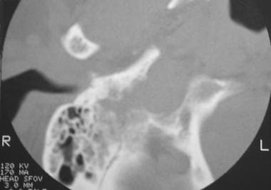
PURPOSE: A family spanning three generations with a history of familial carotid body tumors (CBTs) was studied, and previously proposed hypotheses of tumor characteristics and genetic mode of transmission were addressed. METHODS: Clinically occult lesions in adult subjects were detected by means of high-resolution computed tomography. RESULTS: A 60% incidence of bilaterality of CBTs associated with multiple paragangliomas was noted in the family studied. The genetic mode for CBTs in this family was not simple autosomal dominant transmission and appeared to be paternally directed with complete penetrance. CONCLUSION: In patients with familial CBTs, high-resolution computed tomography is recommended for early screening as a means of prompting diagnosis and definitive treatment, an approach that minimizes morbidity and facilitates surgical excision. (+info)Mutations in SDHD, a mitochondrial complex II gene, in hereditary paraganglioma. (2/77)
Hereditary paraganglioma (PGL) is characterized by the development of benign, vascularized tumors in the head and neck. The most common tumor site is the carotid body (CB), a chemoreceptive organ that senses oxygen levels in the blood. Analysis of families carrying the PGL1 gene, described here, revealed germ line mutations in the SDHD gene on chromosome 11q23. SDHD encodes a mitochondrial respiratory chain protein-the small subunit of cytochrome b in succinate-ubiquinone oxidoreductase (cybS). In contrast to expectations based on the inheritance pattern of PGL, the SDHD gene showed no evidence of imprinting. These findings indicate that mitochondria play an important role in the pathogenesis of certain tumors and that cybS plays a role in normal CB physiology. (+info)Bilateral carotid body paraganglioma: case report. (3/77)
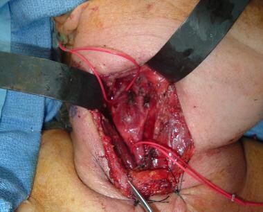
CONTEXT: Surgical treatment of carotid body paragangliomas is a challenge to the surgeon because of the large vascularization of the tumor, involvement of the carotid vessels and the close anatomical relationship with the cranial nerves. CASE REPORT: A 63-year-old patient was submitted to resection of two carotid body paraganglioma tumors found in the right-side and left-side carotid bodies at the bifurcation of the common carotid arteries. Two surgeries were performed at different times and neither of them presented any morbidity. Arteriography was fundamental for diagnosis of the small, asymptomatic tumor on the right side. DESIGN: Case Report (+info)Baroreflex failure syndrome after bilateral excision of carotid body tumors: an underestimated problem. (4/77)
Carotid body tumors (CBTs) are relatively rare paragangliomas that develop from neural crest cells at the bifurcation of the common carotid artery. They are generally slow growing and benign. Excision is currently considered the treatment of choice, although vascular and especially neural injuries are still relatively frequent in patients with large or bilaterally resected tumors. The baroreflex failure syndrome (BFS) has recently been identified as a severe, rarely recognized, and certainly underestimated complication after the bilateral excision of CBTs. The present report describes a case of a bilateral CBT followed by BFS and reviews the experiences reported in the literature. In light of the low incidence of malignancy of these tumors, their biologic behavior, their very high rate of cranial nerve palsy, and the occurrence of BFS in bilaterally resected paragangliomas, the current practice of bilaterally removing these tumors is questioned. (+info)Power Doppler scanning in the diagnosis of carotid body tumors. (5/77)
The aim of this work was to show contribution of power Doppler imaging in the diagnosis of the carotid body tumors. Six patients with a nontender mass beneath the mandibular angle were evaluated with gray scale and power Doppler sonography. Well-defined, solid, weakly hyperechoic masses were noted on gray scale sonography in the carotid bifurcation. Power Doppler sonography showed abundant flow, characterized as an intense blush, throughout the entire tumor in all patients. We believe that invasive and expensive diagnostic modalities are not necessary to evaluate carotid body tumors. Gray scale sonography and power Doppler imaging are sufficient for primary diagnosis of carotid body tumors. (+info)Malignant carotid body tumor: a case report. (6/77)
Carotid body tumors (CBTs) have an unpredictable history with no correlation between histology and clinical behavior. Of reported cases since 1891, local and distant metastases appear in approximately 10% of cases and remain the hallmark of malignancy. Currently, there are not enough data to support a single treatment regimen for malignant CBTs. The reported case demonstrates some unanswered issues with regard to malignant CBTs to include lymph node dissection, the need for carotid resection, and the role of radiation therapy. A 46-year-old pathologist underwent a resection of a Shamblin I CBT, to include jugular lymph node sampling, without complication. There was lymph node involvement, and tumor cells were found on the margins of the pathologic specimen. Subsequent carotid resection with reversed interposition saphenous vein graft and modified neck dissection were performed again without complication. Follow-up at 4 years has been uneventful. Diagnosis of CBTs with the use of magnetic resonance angiography, magnetic resonance imaging, color flow duplex scanning, and the role of arteriography are reviewed. The current treatment options are discussed with reference to primary lymph node sampling, carotid resection, and neck dissection in malignant cases. This case demonstrates that the unpredictable nature of CBTs and their malignant potential warrant aggressive initial local treatment to include jugular lymph node sampling and complete tumor resection. (+info)Long-term effects of carotid sinus denervation on arterial blood pressure in humans. (7/77)
BACKGROUND: After experimental carotid sinus denervation in animals, blood pressure (BP) level and variability increase markedly but normalize to preoperative levels within 10 to 14 days. We investigated the course of arterial BP level and variability after bilateral denervation of the carotid sinus baroreceptors in humans. METHODS AND RESULTS: We studied 4 women (age 41 to 63 years) who were referred for evaluation of arterial baroreflex function because of clinical suspicion of carotid sinus denervation attributable to bilateral carotid body tumor resection. The course of BP level and variability was assessed from repeated office and 24-hour ambulatory measurements (Spacelabs/Portapres) during 1 to 10 years of (retrospective) follow-up. Rapid cardiovascular reflex adjustments to active standing and Valsalva's maneuver were assessed. Office BP level increased from 132/86 mm Hg (range, 118 to 148/80 to 92 mm Hg) before bilateral surgery to 160/105 mm Hg (range, 143 to 194/90 to 116 mm Hg) 1 to 10 years after surgery. During continuous 24-hour noninvasive BP recording (Portapres), a marked BP variability was apparent in all 4 patients. Initial symptomatic hypotension on change to the upright posture and abnormal responses to Valsalva's maneuver were observed. CONCLUSIONS: Acute carotid sinus denervation, as a result of bilateral carotid body tumor resection, has a long-term effect on the level, variability, and rapid reflex control of arterial BP. Therefore, in contrast to earlier experimental observations, the compensatory ability of the baroreceptor areas outside the carotid sinus seems to be of limited importance in the regulation of BP in humans. (+info)Baroreflex control of muscle sympathetic nerve activity after carotid body tumor resection. (8/77)
Bilateral carotid body tumor resection causes a permanent attenuation of vagal baroreflex sensitivity. We retrospectively examined the effects of bilateral carotid body tumor resection on the baroreflex control of sympathetic nerve traffic. Muscle sympathetic nerve activity was recorded in 5 patients after bilateral carotid body tumor resection (1 man and 4 women, 51+/-11 years) and 6 healthy control subjects (2 men and 4 women, 50+/-7 years). Baroreflex sensitivity was calculated from changes in R-R interval and muscle sympathetic nerve activity in response to bolus injections of phenylephrine and nitroprusside. In addition, sympathetic responses to the Valsalva maneuver and cold pressor test were measured. The integrated neurogram of patients and control subjects contained a similar pattern of pulse synchronous burst of nerve activity. Baroreflex control of both heart rate and sympathetic nerve activity were attenuated in patients as compared with control subjects [heart rate baroreflex sensitivity: 3.68+/-0.93 versus 11.61+/-4.72 ms/mm Hg (phenylephrine, P=0.011) and 2.53+/-1.36 versus 5.82+/-1.94 ms/mm Hg (nitroprusside, P=0.05); sympathetic baroreflex sensitivity: 3.70+/-2.90 versus 7.53+/-4.12 activity/100 beats/mm Hg (phenylephrine, P=0.10) and 3.93+/-4.43 versus 15.27+/-10.03 activity/100 beats/mm Hg (nitroprusside, P=0.028)]. The Valsalva maneuver elicited normal reflex changes in muscle sympathetic nerve activity, whereas heart rate responses were blunted in the patients with bilateral carotid body tumor resection. Maximal sympathetic responses to the cold pressor test did not differ between the two groups. Denervation of carotid sinus baroreceptors as the result of bilateral carotid body tumor resection produces chronic impairment of baroreflex control of both heart rate and sympathetic nerve activity. During the Valsalva maneuver, loss of carotid baroreflex control of heart rate is less well compensated for by the extra carotid baroreceptors than the control of muscle sympathetic nerve activity. (+info)A carotid body tumor is a rare, usually noncancerous (benign) growth that develops in the carotid body, a small structure located near the bifurcation (fork) of the common carotid artery in the neck. The carotid body is part of the chemoreceptor system that helps regulate breathing and blood pressure by responding to changes in oxygen, carbon dioxide, and pH levels in the blood.
Carotid body tumors are also known as carotid body paragangliomas or chemodectomas. They typically grow slowly and may not cause any symptoms for many years. However, as they enlarge, they can cause a visible or palpable mass in the neck, along with symptoms such as difficulty swallowing, hoarseness, or voice changes. In some cases, carotid body tumors can compress nearby nerves or blood vessels, leading to more serious complications like stroke or nerve damage.
Treatment for carotid body tumors typically involves surgical removal of the growth, which may be performed using traditional open surgery or minimally invasive techniques such as endovascular surgery or robotic-assisted surgery. Radiation therapy and chemotherapy are generally not effective in treating these tumors. Regular follow-up care is important to monitor for recurrence or development of new tumors.
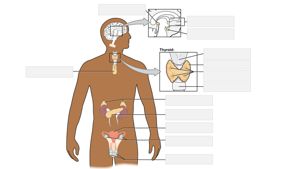
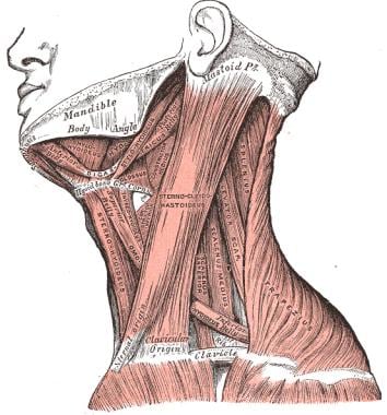
The carotid body is a small chemoreceptor organ located near the bifurcation of the common carotid artery into the internal and external carotid arteries. It plays a crucial role in the regulation of respiration, blood pressure, and pH balance by detecting changes in the chemical composition of the blood, particularly oxygen levels, carbon dioxide levels, and hydrogen ion concentration (pH).
The carotid body contains specialized nerve endings called glomus cells that are sensitive to changes in these chemical parameters. When there is a decrease in oxygen or an increase in carbon dioxide or hydrogen ions, the glomus cells release neurotransmitters such as acetylcholine and dopamine, which activate afferent nerve fibers leading to the brainstem's nucleus tractus solitarius. This information is then integrated with other physiological signals in the brainstem, resulting in appropriate adjustments in breathing rate, depth, and pattern, as well as changes in heart rate and blood vessel diameter to maintain homeostasis.
Dysfunction of the carotid body can lead to various disorders, such as hypertension, sleep apnea, and chronic lung disease. In some cases, overactivity of the carotid body may result in conditions like primary breathing pattern disorders or pseudohypoxia, where the body responds as if it is experiencing hypoxia despite normal oxygen levels.
Vagus nerve injuries refer to damages or traumas affecting the vagus nerve, which is the tenth cranial nerve (CN X) in the human body. This nerve plays a crucial role in the autonomic nervous system, regulating essential functions such as heart rate, respiratory rate, and digestion.
Vagus nerve injuries can occur due to various reasons, including trauma during surgical procedures, neck or head injuries, inflammation, compression, or tumors affecting the nerve. Symptoms of vagus nerve injuries may include:
1. Hoarseness or voice changes
2. Difficulty swallowing (dysphagia)
3. Pain in the throat or ear
4. Changes in heart rate and blood pressure
5. Nausea, vomiting, or abdominal pain
6. Shortness of breath or difficulty breathing
The severity and nature of symptoms can vary depending on the location and extent of the injury to the vagus nerve. Treatment for vagus nerve injuries typically involves addressing the underlying cause, such as surgical intervention, physical therapy, or medication to manage pain and inflammation. In some cases, recovery may be incomplete, leading to long-term complications or disabilities.
Hypoglossal nerve injuries refer to damages or impairments to the twelfth cranial nerve, also known as the hypoglossal nerve. This nerve is primarily responsible for controlling the movements of the tongue.
An injury to this nerve can result in various symptoms, depending on the severity and location of the damage. These may include:
1. Deviation of the tongue to one side when protruded (usually away from the side of the lesion)
2. Weakness or paralysis of the tongue muscles
3. Difficulty with speaking, swallowing, and articulation
4. Changes in taste and sensation on the back of the tongue (in some cases)
Hypoglossal nerve injuries can occur due to various reasons, such as trauma, surgical complications, tumors, or neurological disorders like stroke or multiple sclerosis. Treatment for hypoglossal nerve injuries typically focuses on managing symptoms and may involve speech and language therapy, exercises to strengthen the tongue muscles, and, in some cases, surgical intervention.
Horner syndrome, also known as Horner's syndrome or oculosympathetic palsy, is a neurological disorder characterized by the interruption of sympathetic nerve pathways that innervate the head and neck, leading to a constellation of signs affecting the eye and face on one side of the body.
The classic triad of symptoms includes:
1. Ptosis (drooping) of the upper eyelid: This is due to the weakness or paralysis of the levator palpebrae superioris muscle, which is responsible for elevating the eyelid.
2. Miosis (pupillary constriction): The affected pupil becomes smaller in size compared to the other side, and it may not react as robustly to light.
3. Anhydrosis (decreased sweating): There is reduced or absent sweating on the ipsilateral (same side) of the face, particularly around the forehead and upper eyelid.
Horner syndrome can be caused by various underlying conditions, such as brainstem stroke, tumors, trauma, or certain medical disorders affecting the sympathetic nervous system. The diagnosis typically involves a thorough clinical examination, pharmacological testing, and sometimes imaging studies to identify the underlying cause. Treatment is directed towards managing the underlying condition responsible for Horner syndrome.
Aortic bodies, also known as aortic arch chemoreceptors or simply as carotid and aortic bodies, are small clusters of nerve cells located near the bifurcation of the common carotid artery (carotid body) and in the wall of the aortic arch (aortic body). They are part of the peripheral chemoreceptor system that responds to changes in chemical composition of the blood, particularly to decreases in oxygen levels, increases in carbon dioxide levels, and changes in pH. These receptors send signals to the brainstem, which in turn regulates breathing rate and depth to maintain adequate gas exchange and acid-base balance in the body.
Paraganglioma is a rare type of tumor that develops in the nervous system, specifically in the paraganglia. Paraganglia are clusters of specialized nerve cells throughout the body that release hormones in response to stress or physical activity. Most paragangliomas are benign (noncancerous), but some can be malignant (cancerous) and may spread to other parts of the body.
Paragangliomas can occur in various locations, including the head and neck region (called "head and neck paragangliomas") or near the spine, abdomen, or chest (called "extra-adrenal paragangliomas"). When they develop in the adrenal glands, which are located on top of each kidney, they are called pheochromocytomas.
Paragangliomas can produce and release hormones such as epinephrine (adrenaline) and norepinephrine, leading to symptoms like high blood pressure, rapid heart rate, sweating, anxiety, and headaches. Treatment typically involves surgical removal of the tumor, along with medications to manage symptoms and control hormone levels before and after surgery.
Paraganglioma, extra-adrenal, is a type of rare tumor that develops in the nervous system's paraganglia, which are groups of specialized cells that are responsible for regulating blood pressure and other bodily functions. Unlike adrenal paragangliomas, which form in the adrenal glands located on top of the kidneys, extra-adrenal paragangliomas develop outside of the adrenal glands, in various locations along the sympathetic and parasympathetic nervous systems. These tumors can be functional or nonfunctional, meaning they may or may not produce hormones such as catecholamines (epinephrine, norepinephrine, and dopamine). Functional extra-adrenal paragangliomas can cause symptoms related to excessive hormone production, including hypertension, sweating, headaches, and rapid heartbeat. Treatment typically involves surgical removal of the tumor, along with preoperative preparation to manage potential hormonal imbalances.
Succinate dehydrogenase (SDH) is an enzyme complex that plays a crucial role in the process of cellular respiration, specifically in the citric acid cycle (also known as the Krebs cycle) and the electron transport chain. It is located in the inner mitochondrial membrane of eukaryotic cells.
SDH catalyzes the oxidation of succinate to fumarate, converting it into a molecule of fadaquate in the process. During this reaction, two electrons are transferred from succinate to the FAD cofactor within the SDH enzyme complex, reducing it to FADH2. These electrons are then passed on to ubiquinone (CoQ), which is a mobile electron carrier in the electron transport chain, leading to the generation of ATP, the main energy currency of the cell.
SDH is also known as mitochondrial complex II because it is the second complex in the electron transport chain. Mutations in the genes encoding SDH subunits or associated proteins have been linked to various human diseases, including hereditary paragangliomas, pheochromocytomas, gastrointestinal stromal tumors (GISTs), and some forms of neurodegenerative disorders.
The carotid arteries are a pair of vital blood vessels in the human body that supply oxygenated blood to the head and neck. Each person has two common carotid arteries, one on each side of the neck, which branch off from the aorta, the largest artery in the body.
The right common carotid artery originates from the brachiocephalic trunk, while the left common carotid artery arises directly from the aortic arch. As they ascend through the neck, they split into two main branches: the internal and external carotid arteries.
The internal carotid artery supplies oxygenated blood to the brain, eyes, and other structures within the skull, while the external carotid artery provides blood to the face, scalp, and various regions of the neck.
Maintaining healthy carotid arteries is crucial for overall cardiovascular health and preventing serious conditions like stroke, which can occur when the arteries become narrowed or blocked due to the buildup of plaque or fatty deposits (atherosclerosis). Regular check-ups with healthcare professionals may include monitoring carotid artery health through ultrasound or other imaging techniques.
Chemoreceptor cells are specialized sensory neurons that detect and respond to chemical changes in the internal or external environment. They play a crucial role in maintaining homeostasis within the body by converting chemical signals into electrical impulses, which are then transmitted to the central nervous system for further processing and response.
There are two main types of chemoreceptor cells:
1. Oxygen Chemoreceptors: These cells are located in the carotid bodies near the bifurcation of the common carotid artery and in the aortic bodies close to the aortic arch. They monitor the levels of oxygen, carbon dioxide, and pH in the blood and respond to decreases in oxygen concentration or increases in carbon dioxide and hydrogen ions (indicating acidity) by increasing their firing rate. This signals the brain to increase respiratory rate and depth, thereby restoring normal oxygen levels.
2. Taste Cells: These chemoreceptor cells are found within the taste buds of the tongue and other areas of the oral cavity. They detect specific tastes (salty, sour, sweet, bitter, and umami) by interacting with molecules from food. When a tastant binds to receptors on the surface of a taste cell, it triggers a series of intracellular signaling events that ultimately lead to the generation of an action potential. This information is then relayed to the brain, where it is interpreted as taste sensation.
In summary, chemoreceptor cells are essential for maintaining physiological balance by detecting and responding to chemical stimuli in the body. They play a critical role in regulating vital functions such as respiration and digestion.
Carotid stenosis is a medical condition that refers to the narrowing or constriction of the lumen (inner space) of the carotid artery. The carotid arteries are major blood vessels that supply oxygenated blood to the head and neck. Carotid stenosis usually results from the buildup of plaque, made up of fat, cholesterol, calcium, and other substances, on the inner walls of the artery. This process is called atherosclerosis.
As the plaque accumulates, it causes the artery to narrow, reducing blood flow to the brain. Severe carotid stenosis can increase the risk of stroke, as a clot or debris from the plaque can break off and travel to the brain, blocking a smaller blood vessel and causing tissue damage or death.
Carotid stenosis is typically diagnosed through imaging tests such as ultrasound, CT angiography, or MRI angiography. Treatment options may include lifestyle modifications (such as quitting smoking, controlling blood pressure, and managing cholesterol levels), medications to reduce the risk of clots, or surgical procedures like endarterectomy or stenting to remove or bypass the blockage.
Head and neck neoplasms refer to abnormal growths or tumors in the head and neck region, which can be benign (non-cancerous) or malignant (cancerous). These tumors can develop in various sites, including the oral cavity, nasopharynx, oropharynx, larynx, hypopharynx, paranasal sinuses, salivary glands, and thyroid gland.
Benign neoplasms are slow-growing and generally do not spread to other parts of the body. However, they can still cause problems if they grow large enough to press on surrounding tissues or structures. Malignant neoplasms, on the other hand, can invade nearby tissues and organs and may also metastasize (spread) to other parts of the body.
Head and neck neoplasms can have various symptoms depending on their location and size. Common symptoms include difficulty swallowing, speaking, or breathing; pain in the mouth, throat, or ears; persistent coughing or hoarseness; and swelling or lumps in the neck or face. Early detection and treatment of head and neck neoplasms are crucial for improving outcomes and reducing the risk of complications.
 Thomas Cecil Hunt
Carotid body
Paraganglioma
Kill Matilda
Glomus
Aortic body
Interventional neuroradiology
Thyroid cancer
Paraganglion
SDHAF2
Sphenoid wing meningioma
Horner's syndrome
List of MeSH codes (C04)
International Classification of Diseases for Oncology
Incidental imaging finding
Victor Horsley
Amorphosynthesis
Treponema denticola
Nosebleed
Cavernous sinus
Chromaffin cell
Edith E. Sproul
Anaplastic thyroid cancer
Glomus body
Vocal cord paresis
Laryngeal cancer
Neurosurgery
Ischemia
Sphenoid sinus
Heart nanotechnology
 Carotid Body Tumors: Practice Essentials, History of the Procedure, Epidemiology
 Carotid Body Tumor: Symptoms, Causes & Treatment
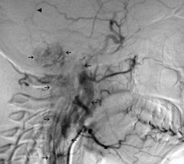
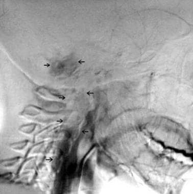
 Temporary balloon occlusion and ethanol injection for preoperative embolization of carotid-body tumor
 Carotid Body tumor - THANC Foundation
 Paraganglioma / Carotid Body Tumor Question | Mayo Clinic Connect
 Carotid Body Tumor Excision - Florida Surgical Clinic
Multi-institutional survey of carotid body tumors in Japan - Fingerprint - Keio University
 February 1968 - Volume 41 - Issue 2 : Plastic and Reconstructive Surgery
 View of A case of successfully resected Shamblin type III carotid body tumour without vascular reconstruction
 CAROTID BODY TUMORS OUR EXPERIENCE WITH 20 PATIENTS - Medical Journal of The Islamic Republic of Iran (MJIRI)
Thomas Cecil Hunt - Wikipedia
 Propuesta de clasificación tomografica preoperatoria para el tumor del cuerpo carotideo Translated title: Proposal for...
 Multicentric Cervical Paraganglioma. Rare Case of Five Locations in One Patient. Case Report and Literature Review
 Dr. Athos Patsalides, MD, Diagnostic Radiology Specialist - Great Neck, NY | Sharecare
 Essential Head and Neck Oncology and Surgery - Nova Science Publishers
Jon Quatromoni, MD | Cleveland Clinic
Glomus Jugulare Tumors Treatment & Management: Medical Therapy, Surgical Therapy, Preoperative, Intraoperative, and...
 Atlas of Oral and Maxillofacial Surgery - Elsevier E-Book on VitalSource, 2nd Edition - 9780323789653
Clinical case reports Thyroid/Others | 0035 | ECE2014 | 16th European Congress of Endocrinology | Endocrine Abstracts
 A rare case of malignant metastatic tumor diagnosed on fine-needle aspiration of cervical lymph node - CytoJournal
 Earsite.com
Servet Dogan - Articles - Scientific Research Publishing
What Is Your Diagnosis? in: Journal of the American Veterinary Medical Association Volume 254 Issue 4 ()
Chirurgia 2020 February;33(1) - Minerva Medica - Riviste
 Vascular Ultrasound Courses | Hands-On CME Training | GCUS
 Images | Radiopaedia.org
 WikiGenes - Pentetreotide - 2-[2-[[1-[[10-(4-aminobutyl)-16-benzyl-4...
 Karen Williams - AMEND
 Multimedia | Portal Regional da BVS
 Hence Verhagen - Research output
- Erasmus University Rotterdam
Hence Verhagen - Research output
- Erasmus University RotterdamParaganglioma10
- A carotid body tumor is also called a carotid body paraganglioma or a carotid body chemodectoma. (clevelandclinic.org)
- We report on the preoperative embolization of a carotid-body paraganglioma by temporary balloon occlusion and ethanol injection. (nih.gov)
- I was just diagnosed with paraganglioma and carotid body tumor. (mayoclinic.org)
- The diagnosis of carotid body tumor, also known as a chemodectoma or paraganglioma, is typically made with radiological studies. (mayoclinic.org)
- Objective: To report a case of a multiple paraganglioma with 5 concurrent locations and also describe a complication not found in the literature, spontaneous tumor bleeding. (scirp.org)
- Köhler, H.F., Carvalho, A.L., Nishinari, K. and Kowalski, L.P. (2010) Internal Carotid Artery Reconstruction after Paraganglioma Resection: Report of Six Cases and Analysis of Available Techniques. (scirp.org)
- On the basis of characteristic cytological features, a diagnosis of metastatic neuroendocrine tumor/paraganglioma was suggested. (cytojournal.com)
- She underwent surgery, and resection of the tumor with neck dissection was performed, which was reported as malignant carotid body paraganglioma on histopathology. (cytojournal.com)
- Ultimately, this caused a carotid body tumour, known as a paraganglioma. (amend.org.uk)
- Parasympathetic PGLs are most often nonsecreting, although about 30% are associated with elevated levels of the dopamine metabolite 3-methoxytyramine (3-MT). [ 5 ] Pheochromocytoma (PHEO) and sympathetic paraganglioma (SPGL) are catecholamine-secreting tumors. (medscape.com)
Paragangliomas11
- Carotid body tumors (CBTs) are rare neoplasms, although they represent about 50-60% of head and neck paragangliomas. (medscape.com)
- When choosing treatment, consider the following factors: the presence of other paragangliomas, the presence of bilateral carotid body tumors (CBTs), the age and the health of the patient, and the patient's preference. (medscape.com)
- [ 9 ] Carotid body tumors (CBTs) constitute about 50-60% of head and neck paragangliomas. (medscape.com)
- Compared with unembolized and polyvinyl-alcohol-embolized carotid-body paragangliomas, our technique resulted in no greater adverse effects on the tumor-vessel interface. (nih.gov)
- Ask your doctor if he or she regularly treats people with this condition, as most doctors rarely (if ever) encounter paragangliomas and are unfamiliar with the best approaches to diagnosing and treating this rare tumor. (mayoclinic.org)
- Under such conditions, it's important to seek a second opinion from a team that specializes in the care of people with rare neuroendocrine tumors such as paragangliomas. (mayoclinic.org)
- Case report: A female patient of 32 years old complaining of tinnitus and diagnosed with five paragangliomas (jugulo-timpanic, bilateral carotid body and bilateral brachiocephalic trunk) through imaging studies. (scirp.org)
- When such tumors arise outside of the adrenal gland, they are termed extra-adrenal pheochromocytomas, or paragangliomas. (medscape.com)
- Of extra-adrenal tumors, known as paragangliomas, 30% are malignant. (msdmanuals.com)
- Paragangliomas (PGLs) are rare neuroendocrine tumors that carry the highest degree of heritability among human neoplasms. (medscape.com)
- Head and neck paragangliomas (HNPGLs) emerge from the parasympathetic nervous systemand are usually benign, slow-growing tumors. (medscape.com)
Resection4
- In the United States, the earliest successful carotid body tumor resection was performed by Scudder in 1903. (medscape.com)
- Hayes Martin, in his textbook of head and neck tumors, recommended against resection of any tumor that is now considered a Shamblin type III (see Staging). (medscape.com)
- A large retrospective, multicenter, international study analyzed the long-term outcome in 132 patients with primary radiation treatment or radiation after partial resection of a glomus tumor. (medscape.com)
- Surgical resection of the tumor is the treatment of choice and usually cures the hypertension. (medscape.com)
Endarterectomy1
- Other significant medical history included palpitations, angina, and carotid endarterectomy. (medscape.com)
Sites include the carotid body1
- 1 Common tumour sites include the carotid body in the head and neck and adrenal and extra-adrenal paraganglia in the abdomen. (bmj.com)
Arteries9
- Computed tomography (CT) scanning of the head and neck is also helpful and typically reveals a hypervascular tumor located between the external and internal carotid arteries. (medscape.com)
- Carotid body tumors are growths in the blood vessels near your carotid arteries. (clevelandclinic.org)
- A carotid body tumor is a mass that grows in the blood vessels near the large arteries in either side of your neck (carotid arteries). (clevelandclinic.org)
- The carotid body is a group of cells located at the bifurcation of the internal and external carotid arteries. (floridasurgicalclinic.com)
- This requires a neck incision and possible excision of part of the carotid arteries. (floridasurgicalclinic.com)
- On color Doppler evaluation, the lesion was observed to lie between the internal and external carotid arteries. (cytojournal.com)
- Shamblin's classification is based on the relationship between tumor and carotid arteries, which has limitations. (pumch.cn)
- Including both the horizontal relationship to carotid arteries and vertical extension of the tumors, PUMCH (Peking union medical college hospital) classification can better predict surgical complications and guide the surgery and thus might help to improve the surgical outcomes of the lesions. (pumch.cn)
- The carotid arteries provide the main blood supply to your brain. (medlineplus.gov)
Treatment of carotid body2
Malignant8
- Some studies estimate that less than 10% of carotid body tumors are malignant (cancerous). (clevelandclinic.org)
- Infrequently, these tumors run in families and can be malignant. (floridasurgicalclinic.com)
- The average tumor size was 5.3 cm in diameter and was found to be malignant in four cases. (ac.ir)
- Among the features that suggest a malignant course are large tumor size and an abnormal DNA ploidy pattern (aneuploidy, tetraploidy). (medscape.com)
- In general, larger tumors are more likely to be malignant. (msdmanuals.com)
- Overview of Multiple Endocrine Neoplasias (MEN) The multiple endocrine neoplasia (MEN) syndromes comprise 4 genetically distinct familial diseases involving adenomatous hyperplasia and malignant tumors in several endocrine glands. (msdmanuals.com)
- Von Hippel-Lindau Disease (VHL) Von Hippel-Lindau disease is a rare hereditary neurocutaneous disorder characterized by benign and malignant tumors in multiple organs. (msdmanuals.com)
- The tumor is malignant in 10% of cases but may be cured completely by surgical removal. (medscape.com)
Bifurcation5
- [ 1 ] These tumors develop within the adventitia of the medial aspect of the carotid bifurcation. (medscape.com)
- CECT revealed, in addition, a lobulated, ill-marginated enhancing space-occupying mass in the right carotid bifurcation. (cytojournal.com)
- It was in close proximity to right common carotid artery beginning at the level of carotid bifurcation. (cytojournal.com)
- A 3-D reconstruction optimized for vasculature (red) shows the location of the highly vascular mass (arrows) lying medial to the right mandibular salivary gland (MSG) at the level of the common carotid artery bifurcation (asterisk). (avma.org)
- Carotid body tumor is a rare neoplasm located at the bifurcation of the carotid artery. (pumch.cn)
Vascular3
- If the tumor is large, your vascular surgeon can embolize the tumor to shrink it prior to surgery. (floridasurgicalclinic.com)
- Gulfcoast Ultrasound Institute is recognized as a worldwide leader in Vascular Ultrasound continuing medical education covering Carotid Ultrasound, Peripheral Vascular Sonography, and Transcranial Doppler Imaging. (gcus.com)
- We used summary statistics from a genome-wide association study on each protein biomarker (meta-analysis of EpiHealth, PIVUS, ULSAM, and IMPROVE [Carotid Intima-Media Thickness and IMT-Progression as Predictors of Vascular Events in a High-Risk European Population]) and publicly available data from Global Lipids Genetics Consortium to perform Mendelian randomization analyses to address possible causality of protein levels. (lu.se)
Neuroendocrine2
- Go to the Neuroendocrine Tumors (NETs) Support Group. (mayoclinic.org)
- [ 10 ] SDH- associated syndromes are characterized by the development of PGLs, with an additional risk for developing other tumor types [ e.g. , clear cell renal cancer (RCC), gastrointestinal stromal tumors (GISTs), and, more rarely, neuroendocrine tumors and pituitary adenomas]. (medscape.com)
Diagnosis3
- Various imaging studies can be used to confirm the diagnosis of carotid body tumor (CBT), starting with simple ultrasonography with color Doppler. (medscape.com)
- Various imaging studies can be used to confirm the diagnosis of carotid body tumor (CBT), starting with simple ultrasonography with color Doppler, which can assess the vascularity of the neck mass and can sometimes reveal a possibility of a carotid body tumor (CBT), although it is not the best imaging modality to detect these tumors. (medscape.com)
- Material and methods: All patients operated with a diagnosis of Carotid Body Tumor between 2005 and 2014 at the Obrero Hospital No. 1 of the National Health Fund in La Paz - Bolivia Results: 115 patients with an average age of 52 years (SD±11.725 and a mode of 57 years) were analyzed and operated on, of which 109 (94.80%) corresponded to the female gender with a ratio of 18: 1. (scienceopen.com)
Bilateral1
- The tumor was as equally frequent on the right as it was on the left, and was bilateral in four cases. (ac.ir)
Neck3
- Carotid body tumors are often painless, but your healthcare provider may want to remove the tumor because it can become large and affect the blood vessels in your neck or cause other symptoms. (clevelandclinic.org)
- A carotid body tumor is suspected if a pulsatile mass in the neck is observed. (earsite.com)
- Lateral projection image of whole-body FDG-PET shows the focal increased FDG activity in the lower mediastinum (blue arrow) and in the neck (red arrow). (medscape.com)
Benign4
- Are carotid body tumors benign? (clevelandclinic.org)
- Most carotid body tumors are benign (not cancer ). (clevelandclinic.org)
- However, most of the time these are benign, sporadic tumors. (floridasurgicalclinic.com)
- Regardless of the histologic appearance, the tumor is considered benign if it has not invaded the capsule and no metastases are found, although exceptions occur. (msdmanuals.com)
Postoperative complications1
- Abstract Objective: To propose a preoperative classification of patients with Carotid Body Tumor and relate them to postoperative complications. (scienceopen.com)
Complications3
- Are there complications of carotid body tumor treatment? (clevelandclinic.org)
- Many people who have treatment for a carotid body tumor don't have complications. (clevelandclinic.org)
- Fine needle aspiration biopsy (FNAB) is seldom requested for this purpose due to rare but dreadful reported complications such as hemorrhage and damage to the carotid artery. (mayoclinic.org)
Pituitary Tumor3
- Being diagnosed with a pituitary tumor or pituitary disorder probably set many questions racing through your mind. (barrowneuro.org)
- This experience means that our doctors and nurses have seen nearly every kind of pituitary tumor and disorder imaginable. (barrowneuro.org)
- Not only is Barrow a place to receive the most advanced treatment and care for your pituitary tumor or disorder, we are also a place where you can help contribute to the scientific understanding and therapeutic research surrounding acromegaly, adenomas, gigantism, and other disorders that implicate the pituitary gland. (barrowneuro.org)
Embolization4
- Healthcare providers often use surgery and embolization to treat carotid body tumors. (clevelandclinic.org)
- Transcatheter embolization to stop blood flow to the tumor, which you may have before surgery to help shrink the tumor. (clevelandclinic.org)
- This procedure is an effective and promising method of preoperative embolization of carotid-body tumors and warrants further experience and study. (nih.gov)
- In this article, we also review the literature on carotid-body tumor embolization and ethanol embolization. (nih.gov)
Pheochromocytoma5
- A pheochromocytoma (see the image below) is a rare, catecholamine-secreting tumor derived from chromaffin cells. (medscape.com)
- The term pheochromocytoma (in Greek, phios means dusky, chroma means color, and cytoma means tumor) refers to the color the tumor cells acquire when stained with chromium salts. (medscape.com)
- Pritchett, J.W.: Familial Concurrence of Carotid Body Tumor and Pheochromocytoma. (tophipsurgeons.com)
- A pheochromocytoma is a catecholamine-secreting tumor of chromaffin cells typically located in the adrenals. (msdmanuals.com)
- A pheochromocytoma (see the image below) is a rare, catecholamine-secreting tumor that may precipitate life-threatening hypertension. (medscape.com)
Aortic1
- The Enigma of Carotid and Aortic Body Tumors. (bvsalud.org)
Paraganglia1
- Germline loss of function mutations followed by somatic loss of non-mutant alleles in the tumours 2- 4 suggests a tumour suppressor role for mitochondrial complex II in the paraganglia. (bmj.com)
Rare tumor1
- In this report of 20 patients with 24 carotid body tumors which is the largest series reported so far from Iran, we have evaluated the various characteristics of this relatively rare tumor in our population and compared our results with that of the literature. (ac.ir)
Average tumor1
- The average tumor volume was 7.26 cm 3 . (medscape.com)
Surgically2
- When providers surgically remove large carotid body tumors, a hole may remain in your carotid artery . (clevelandclinic.org)
- The tumor can then be removed surgically. (floridasurgicalclinic.com)
Tinnitus1
- The study found long-term successful control of the tumor growthi and mprovement of tinnitus and overall neurological status, as well as cranial nerve function. (medscape.com)
Brain2
- Several conditions can cause a facial paralysis e.g. stroke, brain tumor and Lyme disease. (krishnaherbals.com)
- Brain tumor or other growth (mass). (medlineplus.gov)
Temporal Bone1
- Dall′Igna, C., Antunes, M.B. and Dall′Igna, D.P. (2005) Radiation Therapy for Glomus Tumors of the Temporal Bone. (scirp.org)
Surgery8
- Carotid body tumors (CBTs) are treated with either surgery or radiotherapy. (medscape.com)
- Descriptions of surgery for carotid body tumors have existed for over 100 years. (medscape.com)
- Surgery to remove the tumor. (clevelandclinic.org)
- Recovery time after surgery for a carotid body tumor is typically three to four weeks. (clevelandclinic.org)
- If you have surgery to remove a carotid body tumor, you typically don't need further treatment. (clevelandclinic.org)
- Surgery is the treatment of choice for glomus jugulare tumors. (medscape.com)
- However, radiation therapy, particularly stereotactic radiosurgery (eg, Gamma Knife surgery), has been shown to provide good tumor growth control with a low risk of treatment-related cranial nerve injury. (medscape.com)
- Of 22 patients with glomus jugulare tumors who underwent Gamma Knife surgery, neurologic status improved in 12 patients, 7 showed stable clinical condition, and 3 patients developed new moderate deficits. (medscape.com)
Familial1
- Familial pheochromocytomas and carotid body tumors may be due to mutations in genes encoding the enzyme succinate dehydrogenase or other signaling molecules. (msdmanuals.com)
Adrenal1
- Common locations for extra-adrenal pheochromocytomas include the organ of Zuckerkandl (close to the origin of the inferior mesenteric artery), bladder wall, heart, mediastinum, and carotid and glomus jugulare bodies. (medscape.com)
Surgical3
- Often, glomus jugulare tumors are diagnosed within the sixth or seventh decade of life and can be followed by imaging only and may not need surgical intervention. (medscape.com)
- Current surgical management of carotid body tumors[J]. J Vasc Surg, 2016, 64: 1703-1710. (pumch.cn)
- A Systematic Review and Meta-Analysis of the Presentation and Surgical Management of Patients With Carotid Body Tumours[J]. Eur J Vasc Endovasc Surg, 2019, 57: 477-486. (pumch.cn)
Classification1
- The PUMCH Classification of Carotid Body Tumor[J]. Medical Journal of Peking Union Medical College Hospital, 2021, 12(6): 825-828. (pumch.cn)
Vagal1
- Vagal and Carotid Body Tumors. (scirp.org)
Abdominal1
- Paroxysmal attacks may be provoked by palpation of the tumor, postural changes, abdominal compression or massage, induction of anesthesia, emotional trauma, unopposed beta-blockade (which paradoxically increases blood pressure by blocking beta-mediated vasodilation), or micturition (if the tumor is in the bladder). (msdmanuals.com)
Symptoms5
- With regard to laboratory studies, check urinary catecholamines in patients who have any symptoms of a functional carotid body tumor (CBT). (medscape.com)
- What are the symptoms of a carotid body tumor? (clevelandclinic.org)
- A carotid body tumor may not cause any symptoms, but as the mass grows, it may press on nearby nerves and blood vessels. (clevelandclinic.org)
- If your provider recommends observing the tumor (watchful waiting), let them know right away if you develop new symptoms. (clevelandclinic.org)
- Therefore routine examination is not justifiable when symptoms relative to tumor presence are not present. (scirp.org)
Cancerous1
- Note that examining tumor tissue under a microscope cannot with certainty determine whether a tumor is cancerous. (mayoclinic.org)
Pheochromocytomas1
- Although pheochromocytomas have classically been associated with 3 syndromes-von Hippel-Lindau (VHL) syndrome, multiple endocrine neoplasia type 2 (MEN 2), and neurofibromatosis type 1 (NF1)-there are now 10 genes that have been identified as sites of mutations leading to these tumors. (medscape.com)
Sporadic1
- The sporadic form is the most common type, representing approximately 85% of carotid body tumors (CBTs). (medscape.com)
Originates1
- The carotid body, which originates in the neural crest, is important in the body's acute adaptation to fluctuating concentrations of oxygen, carbon dioxide, and pH. (medscape.com)
Blood vessels1
- But carotid body tumor treatment can involve many blood vessels. (clevelandclinic.org)
Histologic1
- Histologic evaluation revealed that the tumor contained diffuse ethanol-induced microemboli. (nih.gov)
Catecholamines1
- Alpha-blockers and beta-blockers are useful for tumors secreting catecholamines. (medscape.com)