Catheters, Indwelling
Catheters
Catheterization
Catheterization, Central Venous
Central Venous Catheters
Cardiac Catheters
Catheterization, Peripheral
Urinary Catheters
Equipment Failure
Catheter-Related Infections
Foreign-Body Migration
Catheterization, Swan-Ganz
Cardiac Catheterization
Jugular Veins
Subclavian Vein
Silicone Elastomers
Atrial Fibrillation
Pulmonary Veins
Treatment Outcome
Peritoneal Dialysis
Vascular Access Devices
Peritonitis
Silicones
Bacteremia
Peritoneal Dialysis, Continuous Ambulatory
Atrial Flutter
Body Surface Potential Mapping
Prospective Studies
Magnetic Resonance Imaging, Interventional
Renal Dialysis
Radiography, Interventional
Anti-Infective Agents, Local
Staphylococcus epidermidis
Suction
Electrocardiography
Vena Cava, Superior
Retrospective Studies
Polyvinyl Chloride
Feasibility Studies
Heart Conduction System
Ventriculoperitoneal Shunt
Analgesia, Epidural
Tachycardia, Atrioventricular Nodal Reentry
Wolff-Parkinson-White Syndrome
Ultrasonography, Interventional
Follow-Up Studies
Surgery, Computer-Assisted
Chlorhexidine
Biofilms
Femoral Vein
Tachycardia, Ventricular
Tachycardia, Ectopic Atrial
Therapeutic Irrigation
Nerve Block
Urinary Tract Infections
Swine
Atrioventricular Node
Dogs
Equipment Reuse
Cerebrospinal Fluid Shunts
Burns, Electric
Thrombectomy
Kidney Failure, Chronic
Polyurethanes
Electrodes
Arteriovenous Shunt, Surgical
Cardiac Pacing, Artificial
Infusions, Parenteral
Anesthetics, Local
Extravasation of Diagnostic and Therapeutic Materials
Embolism, Air
Phlebitis
Injections, Spinal
Tricuspid Valve
Monitoring, Physiologic
Equipment Failure Analysis
Radial Artery
Sterilization
Hemodynamics
Parenteral Nutrition
Sepsis
Angioplasty, Balloon
Heparin
Parenteral Nutrition, Total
Electrocoagulation
Cardiac Tamponade
Fungemia
Urethra
Femoral Nerve
Infection
Infection Control
Tachycardia
Infusions, Intra-Arterial
Pulmonary Artery
Stents
Pressure
Urinary Bladder Calculi
Infusion Pumps, Implantable
Central Venous Pressure
Bundle of His
Hydrocephalus
Ventricular Premature Complexes
Infusion Pumps
Risk Factors
Analgesia, Obstetrical
Phlebography
Brachiocephalic Veins
Thermodilution
Transducers
Punctures
Povidone-Iodine
Pericardial Effusion
Coronary Sinus
Microscopy, Electron, Scanning
Minocycline
Silver
Pulmonary Veno-Occlusive Disease
Robotics
Arrhythmias, Cardiac
Brachial Plexus
Cardiac Output
Subarachnoid Space
Safety
Tomography, X-Ray Computed
Vena Cava, Inferior
Tachycardia, Sinoatrial Nodal Reentry
Pneumothorax
Intensive Care Units
Radio Waves
Pericardium
Monitoring, Intraoperative
Radiology, Interventional
Coronary Angiography
Candidiasis
Postoperative Care
Urinary Bladder, Neurogenic
Bacterial Adhesion
Embolization, Therapeutic
Upper Extremity Deep Vein Thrombosis
Constriction, Pathologic
Embolism
Heart Ventricles
Staphylococcus aureus
Sheep
Models, Animal
Omentum
Urinary Bladder Fistula
Infusions, Intravenous
Azygos Vein
Bacteria
Peritoneum
Reproducibility of Results
Thoracostomy
Colony Count, Microbial
Angioplasty, Balloon, Coronary
Equipment and Supplies
Candida
Echocardiography
The effects of previous abdominal operations and intraperitoneal adhesions on the outcome of peritoneal dialysis catheters. (1/350)
(+info)Comparative analysis of two-piece extended peritoneal dialysis catheters with remote exit-site locations and conventional abdominal catheters. (2/350)
(+info)A prospective randomized study comparing tenckhoff catheters inserted using the triple incision method with standard swan neck catheters. (3/350)
(+info)Comparison of the use of conventional, hydrophilic and gel-lubricated catheters with regard to urethral micro trauma, urinary system infection, and patient satisfaction in patients with spinal cord injury: a randomized controlled study. (4/350)
BACKGROUND: Management of the lower urinary tract is crucially important in patients with spinal cord injuries in order to prevent damage to the upper urinary tract and to preserve renal function. AIM: This study was designed to compare the use of standard polyvinyl chloride (PVC), hydrophilic-coated, and gel-lubricated non-hydrophilic catheters with regard to urethral micro trauma, urinary system infection, and patient satisfaction in patients with spinal cord injuries. Study design. Randomized, controlled study. SETTING: University hospital, inpatient clinic. POPULATION: Twenty-five male patients with spinal cord injuries. METHODS: The patients were asked to use 3 different types of catheters. The selection of catheter order was determined randomly, and all 3 catheters were used for 6 weeks consecutively. All patients were assessed at the beginning of treatment and at weeks 6, 12, and 18, in terms of urethral cytology, urinalysis, urine culture, and patient satisfaction (Visual Analog Scale, VAS). RESULTS: Ten patients completed the study. Regarding the urethral trauma evaluation, urethral cell counts were reduced with gel-lubricated non-hydrophilic catheter use (P<0.05), increased with PVC catheter use (P<0.05), and showed no change with hydrophilic-coated catheter use (P>0.05). The number of leucocytes in the urine sediment was significantly reduced after gel-lubricated catheter use (P<0.05). There was significantly less microhematuria with hydrophilic-coated and gel-lubricated non-hydrophilic catheter use compared with PVC catheter use (P<0.05). There were no significant differences among catheters with respect to symptomatic urinary tract infection and microbiological analysis of urine culture (P>0.05). The mean VAS was better with the gel-lubricated non-hydrophilic catheter than with the other two catheter types (P<0.05). CONCLUSION: The hydrophilic-coated catheter and especially the gel-lubricated non-hydrophilic catheter reduce trauma to the urethral surfaces and enable easy and comfortable catheterization. CLINICAL REHABILITATION IMPACT: The hydrophilic and gel-lubricated catheters represent an attractive alternative to standard PVC catheters for urological rehabilitation in patients with spinal cord injuries. (+info)Effect of imaging and catheter characteristics on clinical outcome for patients in the PRECISE study. (5/350)
(+info)In vivo efficacy of anidulafungin against mature Candida albicans biofilms in a novel rat model of catheter-associated Candidiasis. (6/350)
(+info)Prolonged catheter survival in patients with acute kidney injury on continuous renal replacement therapy using a less thrombogenic micropatterned polymer modification. (7/350)
(+info)Hepatic artery-targeting guidewire technique during transjugular intrahepatic portosystemic shunt. (8/350)
(+info)Indwelling catheters, also known as Foley catheters, are medical devices that are inserted into the bladder to drain urine. They have a small balloon at the tip that is inflated with water once the catheter is in the correct position in the bladder, allowing it to remain in place and continuously drain urine. Indwelling catheters are typically used for patients who are unable to empty their bladders on their own, such as those who are bedridden or have nerve damage that affects bladder function. They are also used during and after certain surgical procedures. Prolonged use of indwelling catheters can increase the risk of urinary tract infections and other complications.
A catheter is a flexible tube that can be inserted into the body to treat various medical conditions or to perform certain medical procedures. Catheters are used to drain fluids, deliver medications, or provide access to different parts of the body for diagnostic or therapeutic purposes. They come in various sizes and materials, depending on their intended use.
In a general sense, catheters can be classified into two main categories:
1. **External catheters:** These are applied to the outside of the body and are commonly used for urinary drainage. For example, a condom catheter is an external collection device that fits over the penis to drain urine into a bag. Similarly, a Texas or Foley catheter can be used in females, where a small tube is inserted into the urethra and inflated with a balloon to keep it in place.
2. **Internal catheters:** These are inserted into the body through various openings or surgical incisions. They have different applications based on their placement:
* **Urinary catheters:** Used for bladder drainage, similar to external catheters but inserted through the urethra.
* **Vascular catheters:** Inserted into veins or arteries to administer medication, fluids, or to perform diagnostic tests like angiography.
* **Cardiovascular catheters:** Used in procedures such as cardiac catheterization to diagnose and treat heart conditions.
* **Neurological catheters:** Placed in the cerebrospinal fluid spaces of the brain or spinal cord for diagnostic or therapeutic purposes, like draining excess fluid or delivering medication.
* **Gastrointestinal catheters:** Used to provide enteral nutrition, drain fluids, or perform procedures within the gastrointestinal tract.
Proper care and maintenance of catheters are crucial to prevent infection and other complications. Patients with indwelling catheters should follow their healthcare provider's instructions for cleaning, handling, and monitoring the catheter site.
Catheterization is a medical procedure in which a catheter (a flexible tube) is inserted into the body to treat various medical conditions or for diagnostic purposes. The specific definition can vary depending on the area of medicine and the particular procedure being discussed. Here are some common types of catheterization:
1. Urinary catheterization: This involves inserting a catheter through the urethra into the bladder to drain urine. It is often performed to manage urinary retention, monitor urine output in critically ill patients, or assist with surgical procedures.
2. Cardiac catheterization: A procedure where a catheter is inserted into a blood vessel, usually in the groin or arm, and guided to the heart. This allows for various diagnostic tests and treatments, such as measuring pressures within the heart chambers, assessing blood flow, or performing angioplasty and stenting of narrowed coronary arteries.
3. Central venous catheterization: A catheter is inserted into a large vein, typically in the neck, chest, or groin, to administer medications, fluids, or nutrition, or to monitor central venous pressure.
4. Peritoneal dialysis catheterization: A catheter is placed into the abdominal cavity for individuals undergoing peritoneal dialysis, a type of kidney replacement therapy.
5. Neurological catheterization: In some cases, a catheter may be inserted into the cerebrospinal fluid space (lumbar puncture) or the brain's ventricular system (ventriculostomy) to diagnose or treat various neurological conditions.
These are just a few examples of catheterization procedures in medicine. The specific definition and purpose will depend on the medical context and the particular organ or body system involved.
Central venous catheterization is a medical procedure in which a flexible tube called a catheter is inserted into a large vein in the body, usually in the neck (internal jugular vein), chest (subclavian vein), or groin (femoral vein). The catheter is threaded through the vein until it reaches a central location, such as the superior vena cava or the right atrium of the heart.
Central venous catheterization may be performed for several reasons, including:
1. To administer medications, fluids, or nutritional support directly into the bloodstream.
2. To monitor central venous pressure (CVP), which can help assess a patient's volume status and cardiac function.
3. To draw blood samples for laboratory tests.
4. To deliver chemotherapy drugs or other medications that may be harmful to peripheral veins.
5. To provide access for hemodialysis or other long-term therapies.
The procedure requires careful attention to sterile technique to minimize the risk of infection, and it is usually performed under local anesthesia with sedation or general anesthesia. Complications of central venous catheterization may include bleeding, infection, pneumothorax (collapsed lung), arterial puncture, and catheter-related bloodstream infections (CRBSI).
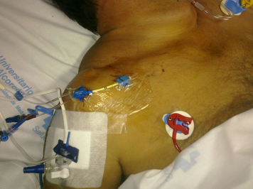
Central venous catheters (CVCs) are medical devices used to access the central venous system, typically placed in one of the large great veins such as the internal jugular, subclavian, or femoral vein. They can be used for a variety of purposes including administration of medications and fluids, monitoring central venous pressure, and obtaining blood samples. CVCs come in different types, such as non-tunneled, tunneled, and implantable ports, each with its own specific indications and uses. Proper placement and maintenance of CVCs are crucial to prevent complications such as infection, thrombosis, and catheter-related bloodstream infections.
A cardiac catheter is a thin, flexible tube that is inserted into the heart or adjacent blood vessels during a cardiac catheterization procedure. This procedure is typically performed to diagnose and treat various cardiovascular conditions such as heart disease, heart defects, or abnormal heart rhythms.
Cardiac catheters can be used for several purposes:
1. To measure the pressure and oxygen levels in different chambers of the heart and blood vessels.
2. To inject dye into the coronary arteries to visualize blockages or narrowing through angiography.
3. To perform interventions such as balloon angioplasty, stent placement, or valvuloplasty to open up blocked or narrowed blood vessels or repair damaged heart valves.
4. To collect samples of heart muscle tissue for biopsy, which can help diagnose conditions like cardiomyopathy or myocarditis.
There are various types of cardiac catheters, including:
1. Diagnostic catheters - used to measure pressure and oxygen levels in the heart and blood vessels.
2. Guiding catheters - used to guide other interventional devices like balloons or stents into place.
3. Angioplasty balloon catheters - used to inflate a balloon at the tip of the catheter, which helps open up blocked or narrowed blood vessels.
4. Thermodilution catheters - used to measure cardiac output and other hemodynamic parameters.
5. Microcatheters - smaller, more flexible catheters used for complex interventions or accessing difficult-to-reach areas of the heart and blood vessels.
Cardiac catheterization is a minimally invasive procedure that usually requires only local anesthesia and mild sedation. The recovery time is typically short, with most patients returning home within 24 hours after the procedure.
Peripheral catheterization is a medical procedure that involves the insertion of a thin, flexible tube (catheter) into a peripheral vein, which is a blood vessel located outside of the chest and abdomen. This type of catheterization is typically performed to administer medications, fluids, or nutritional support, or to monitor various physiological parameters such as central venous pressure.
Peripheral catheters are usually inserted into veins in the hands or arms, although they can also be placed in other peripheral veins. The procedure is typically performed using aseptic technique to minimize the risk of infection. Once the catheter is in place, it may be secured with a dressing or suture to prevent movement and dislodgement.
Peripheral catheterization is a relatively safe and common procedure that is routinely performed in hospitals, clinics, and other healthcare settings. However, like any medical procedure, it carries a small risk of complications such as infection, bleeding, or damage to the vein or surrounding tissues.
Urinary catheterization is a medical procedure in which a flexible tube (catheter) is inserted into the bladder through the urethra to drain urine. This may be done to manage urinary retention, monitor urine output, or obtain a urine sample for laboratory testing. It can be performed as a clean, intermittent catheterization, or with an indwelling catheter (also known as Foley catheter) that remains in place for a longer period of time. The procedure should be performed using sterile technique to reduce the risk of urinary tract infection.
A urinary catheter is a flexible tube that is inserted into the bladder to drain urine. It can be made of rubber, plastic, or latex and comes in various sizes and lengths. The catheter can be inserted through the urethra (the tube that carries urine out of the body from the bladder) and is called a Foley catheter or an indwelling catheter. A straight catheter, on the other hand, is inserted through the urethra and removed after it has drained the urine.
Urinary catheters are used in various medical situations, such as when a person is unable to empty their bladder due to surgery, anesthesia, medication, or conditions that affect bladder function. They may also be used for long-term management of urinary incontinence or to drain the bladder during certain medical procedures.
It's important to note that the use of urinary catheters carries a risk of complications, such as urinary tract infections, bladder spasms, and injury to the urethra or bladder. Therefore, they should only be used when necessary and under the guidance of a healthcare professional.
Equipment failure is a term used in the medical field to describe the malfunction or breakdown of medical equipment, devices, or systems that are essential for patient care. This can include simple devices like syringes and thermometers, as well as complex machines such as ventilators, infusion pumps, and imaging equipment.
Equipment failure can have serious consequences for patients, including delayed or inappropriate treatment, injury, or even death. It is therefore essential that medical equipment is properly maintained, tested, and repaired to ensure its safe and effective operation.
There are many potential causes of equipment failure, including:
* Wear and tear from frequent use
* Inadequate cleaning or disinfection
* Improper handling or storage
* Power supply issues
* Software glitches or bugs
* Mechanical failures or defects
* Human error or misuse
To prevent equipment failure, healthcare facilities should have established policies and procedures for the acquisition, maintenance, and disposal of medical equipment. Staff should be trained in the proper use and handling of equipment, and regular inspections and testing should be performed to identify and address any potential issues before they lead to failure.
Catheter-related infections are infections that occur due to the presence of a catheter, a flexible tube that is inserted into the body to perform various medical functions such as draining urine or administering medication. These infections can affect any part of the body where a catheter is inserted, including the bladder, bloodstream, heart, and lungs.
The most common type of catheter-related infection is a catheter-associated urinary tract infection (CAUTI), which occurs when bacteria enter the urinary tract through the catheter and cause an infection. Symptoms of CAUTI may include fever, chills, pain or burning during urination, and cloudy or foul-smelling urine.
Other types of catheter-related infections include catheter-associated bloodstream infections (CLABSI), which can occur when bacteria enter the bloodstream through the catheter, and catheter-related pulmonary infections, which can occur when secretions from the respiratory tract enter the lungs through a catheter.
Catheter-related infections are a significant concern in healthcare settings, as they can lead to serious complications such as sepsis, organ failure, and even death. Proper catheter insertion and maintenance techniques, as well as regular monitoring for signs of infection, can help prevent these types of infections.
Equipment design, in the medical context, refers to the process of creating and developing medical equipment and devices, such as surgical instruments, diagnostic machines, or assistive technologies. This process involves several stages, including:
1. Identifying user needs and requirements
2. Concept development and brainstorming
3. Prototyping and testing
4. Design for manufacturing and assembly
5. Safety and regulatory compliance
6. Verification and validation
7. Training and support
The goal of equipment design is to create safe, effective, and efficient medical devices that meet the needs of healthcare providers and patients while complying with relevant regulations and standards. The design process typically involves a multidisciplinary team of engineers, clinicians, designers, and researchers who work together to develop innovative solutions that improve patient care and outcomes.
Foreign-body migration is a medical condition that occurs when a foreign object, such as a surgical implant, tissue graft, or trauma-induced fragment, moves from its original position within the body to a different location. This displacement can cause various complications and symptoms depending on the type of foreign body, the location it migrated to, and the individual's specific physiological response.
Foreign-body migration may result from insufficient fixation or anchoring of the object during implantation, inadequate wound healing, infection, or an inflammatory reaction. Symptoms can include pain, swelling, redness, or infection at the new location, as well as potential damage to surrounding tissues and organs. Diagnosis typically involves imaging techniques like X-rays, CT scans, or MRIs to locate the foreign body, followed by a surgical procedure to remove it and address any resulting complications.
Swan-Ganz catheterization is a medical procedure in which a Swan-Ganz catheter, also known as a pulmonary artery catheter, is inserted into a patient's vein and guided through the heart to the pulmonary artery. The procedure is named after its inventors, Dr. Jeremy Swan and Dr. William Ganz.
The Swan-Ganz catheter is a thin, flexible tube that is equipped with sensors that measure various cardiac functions, such as blood pressure in the heart chambers and lungs, oxygen saturation of the blood, and cardiac output. This information helps doctors evaluate heart function, diagnose heart conditions, and monitor treatment effectiveness.
Swan-Ganz catheterization is typically performed in a hospital setting by trained medical professionals, such as cardiologists or critical care specialists. The procedure may be used to diagnose and manage various heart conditions, including heart failure, pulmonary hypertension, and shock. It may also be used during major surgeries or other medical procedures to monitor the patient's hemodynamic status.
Like any medical procedure, Swan-Ganz catheterization carries some risks, such as infection, bleeding, and damage to blood vessels or heart structures. However, these complications are relatively rare when the procedure is performed by experienced medical professionals.
Cardiac catheterization is a medical procedure used to diagnose and treat cardiovascular conditions. In this procedure, a thin, flexible tube called a catheter is inserted into a blood vessel in the arm or leg and threaded up to the heart. The catheter can be used to perform various diagnostic tests, such as measuring the pressure inside the heart chambers and assessing the function of the heart valves.
Cardiac catheterization can also be used to treat certain cardiovascular conditions, such as narrowed or blocked arteries. In these cases, a balloon or stent may be inserted through the catheter to open up the blood vessel and improve blood flow. This procedure is known as angioplasty or percutaneous coronary intervention (PCI).
Cardiac catheterization is typically performed in a hospital cardiac catheterization laboratory by a team of healthcare professionals, including cardiologists, radiologists, and nurses. The procedure may be done under local anesthesia with sedation or general anesthesia, depending on the individual patient's needs and preferences.
Overall, cardiac catheterization is a valuable tool in the diagnosis and treatment of various heart conditions, and it can help improve symptoms, reduce complications, and prolong life for many patients.
The jugular veins are a pair of large, superficial veins that carry blood from the head and neck to the heart. They are located in the neck and are easily visible when looking at the side of a person's neck. The external jugular vein runs along the surface of the muscles in the neck, while the internal jugular vein runs within the carotid sheath along with the carotid artery and the vagus nerve.
The jugular veins are important in clinical examinations because they can provide information about a person's cardiovascular function and intracranial pressure. For example, distention of the jugular veins may indicate heart failure or increased intracranial pressure, while decreased venous pulsations may suggest a low blood pressure or shock.
It is important to note that medical conditions such as deep vein thrombosis (DVT) can also affect the jugular veins and can lead to serious complications if not treated promptly.
The subclavian vein is a large venous structure that carries deoxygenated blood from the upper limb and part of the thorax back to the heart. It forms when the axillary vein passes through the narrow space between the first rib and the clavicle (collarbone), becoming the subclavian vein.
On the left side, the subclavian vein joins with the internal jugular vein to form the brachiocephalic vein, while on the right side, the subclavian vein directly merges with the internal jugular vein to create the brachiocephalic vein. These brachiocephalic veins then unite to form the superior vena cava, which drains blood into the right atrium of the heart.
The subclavian vein is an essential structure for venous access in various medical procedures and interventions, such as placing central venous catheters or performing blood tests.
Fluoroscopy is a type of medical imaging that uses X-rays to obtain real-time moving images of the internal structures of the body. A continuous X-ray beam is passed through the body part being examined, and the resulting fluoroscopic images are transmitted to a monitor, allowing the medical professional to view the structure and movement of the internal organs and bones in real time.
Fluoroscopy is often used to guide minimally invasive procedures such as catheterization, stent placement, or joint injections. It can also be used to diagnose and monitor a variety of medical conditions, including gastrointestinal disorders, musculoskeletal injuries, and cardiovascular diseases.
It is important to note that fluoroscopy involves exposure to ionizing radiation, and the risks associated with this exposure should be carefully weighed against the benefits of the procedure. Medical professionals are trained to use the lowest possible dose of radiation necessary to obtain the desired diagnostic information.
Silicone elastomers are a type of synthetic rubber made from silicone, which is a polymer composed primarily of silicon-oxygen bonds. They are known for their durability, flexibility, and resistance to heat, cold, and moisture. Silicone elastomers can be manufactured in various forms, including liquids, gels, and solids, and they are used in a wide range of medical applications such as:
1. Breast implants: Silicone elastomer shells filled with silicone gel are commonly used for breast augmentation and reconstruction.
2. Contact lenses: Some contact lenses are made from silicone elastomers due to their high oxygen permeability, which allows for better eye health.
3. Catheters: Silicone elastomer catheters are flexible and resistant to kinking, making them suitable for long-term use in various medical procedures.
4. Implantable drug delivery systems: Silicone elastomers can be used as a matrix for controlled release of drugs, allowing for sustained and targeted medication administration.
5. Medical adhesives: Silicone elastomer adhesives are biocompatible and can be used to attach medical devices to the skin or other tissues.
6. Sealants and coatings: Silicone elastomers can be used as sealants and coatings in medical devices to prevent leakage, improve durability, and reduce infection risk.
It is important to note that while silicone elastomers are generally considered safe for medical use, there have been concerns about the potential health risks associated with breast implants, such as capsular contracture, breast pain, and immune system reactions. However, these risks vary depending on the individual's health status and the specific type of silicone elastomer used.
Atrial fibrillation (A-tre-al fi-bru-la'shun) is a type of abnormal heart rhythm characterized by rapid and irregular beating of the atria, the upper chambers of the heart. In this condition, the electrical signals that coordinate heartbeats don't function properly, causing the atria to quiver instead of contracting effectively. As a result, blood may not be pumped efficiently into the ventricles, which can lead to blood clots, stroke, and other complications. Atrial fibrillation is a common type of arrhythmia and can cause symptoms such as palpitations, shortness of breath, fatigue, and dizziness. It can be caused by various factors, including heart disease, high blood pressure, age, and genetics. Treatment options include medications, electrical cardioversion, and surgical procedures to restore normal heart rhythm.
Pulmonary veins are blood vessels that carry oxygenated blood from the lungs to the left atrium of the heart. There are four pulmonary veins in total, two from each lung, and they are the only veins in the body that carry oxygen-rich blood. The oxygenated blood from the pulmonary veins is then pumped by the left ventricle to the rest of the body through the aorta. Any blockage or damage to the pulmonary veins can lead to various cardiopulmonary conditions, such as pulmonary hypertension and congestive heart failure.
Treatment outcome is a term used to describe the result or effect of medical treatment on a patient's health status. It can be measured in various ways, such as through symptoms improvement, disease remission, reduced disability, improved quality of life, or survival rates. The treatment outcome helps healthcare providers evaluate the effectiveness of a particular treatment plan and make informed decisions about future care. It is also used in clinical research to compare the efficacy of different treatments and improve patient care.
Peritoneal dialysis is a type of renal replacement therapy used to treat patients with severe kidney dysfunction or end-stage renal disease. It is a process that utilizes the peritoneum, a membranous sac lining the abdominal cavity, as a natural semipermeable membrane for filtering waste products, excess fluids, and electrolytes from the bloodstream.
In peritoneal dialysis, a sterile dialysate solution is infused into the peritoneal cavity via a permanently implanted catheter. The dialysate contains various substances such as glucose or other osmotic agents, electrolytes, and buffer solutions that facilitate the diffusion of waste products and fluids from the blood vessels surrounding the peritoneum into the dialysate.
There are two primary types of peritoneal dialysis: continuous ambulatory peritoneal dialysis (CAPD) and automated peritoneal dialysis (APD). CAPD is performed manually, several times a day, while APD is carried out using a cycler machine overnight.
Peritoneal dialysis offers certain advantages over hemodialysis, such as better preservation of residual renal function, fewer dietary restrictions, and greater flexibility in scheduling treatments. However, it also has potential complications, including peritonitis (inflammation of the peritoneum), catheter-related infections, fluid imbalances, and membrane failure over time.
Vascular access devices (VADs) are medical devices that are used to gain access to a patient's vascular system for the purpose of administering treatments, monitoring vital signs, or obtaining diagnostic samples. These devices can be categorized into short-term and long-term based on their intended duration of use.
Short-term VADs include peripheral intravenous catheters (PIVs), midline catheters, and peripherally inserted central catheters (PICCs). PIVs are thin, flexible tubes that are inserted into a vein in the arm or hand for short-term use. Midlines are similar to PIVs but are longer and can be used for up to 4 weeks. PICCs are inserted into a vein in the upper arm and threaded through to the larger veins near the heart, allowing for long-term access.
Long-term VADs include tunneled central venous catheters (CVCs), non-tunneled CVCs, and implanted ports. Tunneled CVCs are inserted into a large vein in the neck or chest and then threaded under the skin to an exit site, reducing the risk of infection. Non-tunneled CVCs are similar but do not have a tunnel, making them more prone to infection. Implanted ports are small devices that are surgically implanted under the skin, usually in the chest or arm, and connected to a catheter that is inserted into a large vein.
VADs can be used for various medical treatments such as chemotherapy, antibiotic therapy, parenteral nutrition, dialysis, and blood transfusions. Proper care and maintenance of VADs are essential to prevent complications such as infection, thrombosis, and catheter-related bloodstream infections (CRBSI).
Peritonitis is a medical condition characterized by inflammation of the peritoneum, which is the serous membrane that lines the inner wall of the abdominal cavity and covers the abdominal organs. The peritoneum has an important role in protecting the abdominal organs and providing a smooth surface for them to move against each other.
Peritonitis can occur as a result of bacterial or fungal infection, chemical irritation, or trauma to the abdomen. The most common cause of peritonitis is a rupture or perforation of an organ in the abdominal cavity, such as the appendix, stomach, or intestines, which allows bacteria from the gut to enter the peritoneal cavity.
Symptoms of peritonitis may include abdominal pain and tenderness, fever, nausea and vomiting, loss of appetite, and decreased bowel movements. In severe cases, peritonitis can lead to sepsis, a life-threatening condition characterized by widespread inflammation throughout the body.
Treatment for peritonitis typically involves antibiotics to treat the infection, as well as surgical intervention to repair any damage to the abdominal organs and remove any infected fluid or tissue from the peritoneal cavity. In some cases, a temporary or permanent drain may be placed in the abdomen to help remove excess fluid and promote healing.
Silicones are not a medical term, but they are commonly used in the medical field, particularly in medical devices and healthcare products. Silicones are synthetic polymers made up of repeating units of siloxane, which is a chain of alternating silicon and oxygen atoms. They can exist in various forms such as oils, gels, rubbers, and resins.
In the medical context, silicones are often used for their unique properties, including:
1. Biocompatibility - Silicones have a low risk of causing an adverse reaction when they come into contact with living tissue.
2. Inertness - They do not react chemically with other substances, making them suitable for use in medical devices that need to remain stable over time.
3. Temperature resistance - Silicones can maintain their flexibility and elasticity even under extreme temperature conditions.
4. Gas permeability - Some silicone materials allow gases like oxygen and water vapor to pass through, which is useful in applications where maintaining a moist environment is essential.
5. Durability - Silicones have excellent resistance to aging, weathering, and environmental factors, ensuring long-lasting performance.
Examples of medical applications for silicones include:
1. Breast implants
2. Contact lenses
3. Catheters
4. Artificial joints and tendons
5. Bandages and wound dressings
6. Drug delivery systems
7. Medical adhesives
8. Infant care products (nipples, pacifiers)
Bacteremia is the presence of bacteria in the bloodstream. It is a medical condition that occurs when bacteria from another source, such as an infection in another part of the body, enter the bloodstream. Bacteremia can cause symptoms such as fever, chills, and rapid heart rate, and it can lead to serious complications such as sepsis if not treated promptly with antibiotics.
Bacteremia is often a result of an infection elsewhere in the body that allows bacteria to enter the bloodstream. This can happen through various routes, such as during medical procedures, intravenous (IV) drug use, or from infected wounds or devices that come into contact with the bloodstream. In some cases, bacteremia may also occur without any obvious source of infection.
It is important to note that not all bacteria in the bloodstream cause harm, and some people may have bacteria in their blood without showing any symptoms. However, if bacteria in the bloodstream multiply and cause an immune response, it can lead to bacteremia and potentially serious complications.
Peritoneal dialysis, continuous ambulatory (CAPD), is a type of renal replacement therapy used to treat patients with end-stage kidney disease. It is a form of peritoneal dialysis that is performed continuously, without the need for machines or hospitalization. CAPD uses the patient's own peritoneum, a thin membrane that lines the abdominal cavity, as a natural filter to remove waste products and excess fluids from the bloodstream.
In CAPD, a sterile dialysis solution is introduced into the peritoneal cavity through a permanent catheter implanted in the patient's abdomen. The solution remains in the peritoneal cavity for a dwell time of several hours, during which diffusion occurs across the peritoneal membrane, allowing waste products and excess fluids to move from the bloodstream into the dialysis solution.
After the dwell time, the used dialysis solution is drained from the peritoneal cavity and discarded, and a fresh batch of dialysis solution is introduced. This process is typically repeated four to five times a day, with each exchange taking about 30 minutes to complete. Patients can perform CAPD exchanges while going about their daily activities, making it a convenient and flexible treatment option for many patients with end-stage kidney disease.
Overall, CAPD is a highly effective form of dialysis that offers several advantages over other types of renal replacement therapy, including improved quality of life, better preservation of residual kidney function, and lower costs. However, it does require careful attention to sterile technique and regular monitoring to ensure proper functioning of the peritoneal membrane and adequate clearance of waste products and fluids.
Atrial flutter is a type of abnormal heart rhythm or arrhythmia that originates in the atria - the upper chambers of the heart. In atrial flutter, the atria beat too quickly, usually between 250 and 350 beats per minute, which is much faster than the normal resting rate of 60 to 100 beats per minute.
This rapid beating causes the atria to quiver or "flutter" instead of contracting effectively. As a result, blood may not be pumped efficiently into the ventricles - the lower chambers of the heart - which can lead to reduced cardiac output and symptoms such as palpitations, shortness of breath, fatigue, dizziness, or chest discomfort.
Atrial flutter is often caused by underlying heart conditions, such as coronary artery disease, hypertension, valvular heart disease, or congenital heart defects. It can also be a complication of cardiac surgery or other medical procedures. In some cases, atrial flutter may occur without any apparent underlying cause, which is known as lone atrial flutter.
Treatment for atrial flutter typically involves medications to control the heart rate and rhythm, electrical cardioversion to restore a normal heart rhythm, or catheter ablation to destroy the abnormal electrical pathways in the heart that are causing the arrhythmia. In some cases, surgical intervention may be necessary to treat atrial flutter.
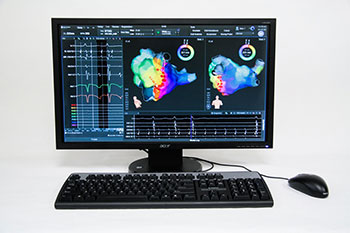
Body Surface Potential Mapping (BSPM) is a non-invasive medical technique used to record and analyze the electrical activity of the heart from the surface of the body. It involves placing multiple electrodes on the skin of the chest, back, and limbs to measure the potential differences between these points during each heartbeat. This information is then used to create a detailed, visual representation of the electrical activation pattern of the heart, which can help in the diagnosis and evaluation of various cardiac disorders such as arrhythmias, myocardial infarction, and ventricular hypertrophy.
The BSPM technique provides high-resolution spatial and temporal information about the cardiac electrical activity, making it a valuable tool for both clinical and research purposes. It can help identify the origin and spread of abnormal electrical signals in the heart, which is crucial for determining appropriate treatment strategies. Overall, Body Surface Potential Mapping is an important diagnostic modality that offers unique insights into the electrical functioning of the heart.
Prospective studies, also known as longitudinal studies, are a type of cohort study in which data is collected forward in time, following a group of individuals who share a common characteristic or exposure over a period of time. The researchers clearly define the study population and exposure of interest at the beginning of the study and follow up with the participants to determine the outcomes that develop over time. This type of study design allows for the investigation of causal relationships between exposures and outcomes, as well as the identification of risk factors and the estimation of disease incidence rates. Prospective studies are particularly useful in epidemiology and medical research when studying diseases with long latency periods or rare outcomes.
The heart atria are the upper chambers of the heart that receive blood from the veins and deliver it to the lower chambers, or ventricles. There are two atria in the heart: the right atrium receives oxygen-poor blood from the body and pumps it into the right ventricle, which then sends it to the lungs to be oxygenated; and the left atrium receives oxygen-rich blood from the lungs and pumps it into the left ventricle, which then sends it out to the rest of the body. The atria contract before the ventricles during each heartbeat, helping to fill the ventricles with blood and prepare them for contraction.
In the field of medicine, "time factors" refer to the duration of symptoms or time elapsed since the onset of a medical condition, which can have significant implications for diagnosis and treatment. Understanding time factors is crucial in determining the progression of a disease, evaluating the effectiveness of treatments, and making critical decisions regarding patient care.
For example, in stroke management, "time is brain," meaning that rapid intervention within a specific time frame (usually within 4.5 hours) is essential to administering tissue plasminogen activator (tPA), a clot-busting drug that can minimize brain damage and improve patient outcomes. Similarly, in trauma care, the "golden hour" concept emphasizes the importance of providing definitive care within the first 60 minutes after injury to increase survival rates and reduce morbidity.
Time factors also play a role in monitoring the progression of chronic conditions like diabetes or heart disease, where regular follow-ups and assessments help determine appropriate treatment adjustments and prevent complications. In infectious diseases, time factors are crucial for initiating antibiotic therapy and identifying potential outbreaks to control their spread.
Overall, "time factors" encompass the significance of recognizing and acting promptly in various medical scenarios to optimize patient outcomes and provide effective care.
Interventional Magnetic Resonance Imaging (MRI) is a medical imaging technique that combines the diagnostic capabilities of MRI with minimally invasive image-guided procedures. It uses a strong magnetic field, radio waves, and computer software to produce detailed images of the body's internal structures and soft tissues.
In interventional MRI, the technology is used in real-time to guide the placement of needles, catheters, or other medical instruments for diagnostic or therapeutic purposes. This can include biopsies, tumor ablations, or targeted drug deliveries. The primary advantage of interventional MRI over traditional interventional radiology techniques is its ability to provide high-resolution imaging without the use of radiation, making it a safer option for certain patients. However, it requires specialized equipment and trained personnel to perform these procedures.
Renal dialysis is a medical procedure that is used to artificially remove waste products, toxins, and excess fluids from the blood when the kidneys are no longer able to perform these functions effectively. This process is also known as hemodialysis.
During renal dialysis, the patient's blood is circulated through a special machine called a dialyzer or an artificial kidney, which contains a semi-permeable membrane that filters out waste products and excess fluids from the blood. The cleaned blood is then returned to the patient's body.
Renal dialysis is typically recommended for patients with advanced kidney disease or kidney failure, such as those with end-stage renal disease (ESRD). It is a life-sustaining treatment that helps to maintain the balance of fluids and electrolytes in the body, prevent the buildup of waste products and toxins, and control blood pressure.
There are two main types of renal dialysis: hemodialysis and peritoneal dialysis. Hemodialysis is the most common type and involves using a dialyzer to filter the blood outside the body. Peritoneal dialysis, on the other hand, involves placing a catheter in the abdomen and using the lining of the abdomen (peritoneum) as a natural filter to remove waste products and excess fluids from the body.
Overall, renal dialysis is an essential treatment option for patients with kidney failure, helping them to maintain their quality of life and prolong their survival.
Interventional radiography is a subspecialty of radiology that uses imaging guidance (such as X-ray fluoroscopy, ultrasound, CT, or MRI) to perform minimally invasive diagnostic and therapeutic procedures. These procedures typically involve the insertion of needles, catheters, or other small instruments through the skin or a natural body opening, allowing for targeted treatment with reduced risk, trauma, and recovery time compared to traditional open surgeries.
Examples of interventional radiography procedures include:
1. Angiography: Imaging of blood vessels to diagnose and treat conditions like blockages, narrowing, or aneurysms.
2. Biopsy: The removal of tissue samples for diagnostic purposes.
3. Drainage: The removal of fluid accumulations (e.g., abscesses, cysts) or the placement of catheters to drain fluids continuously.
4. Embolization: The blocking of blood vessels to control bleeding, tumor growth, or reduce the size of an aneurysm.
5. Stenting and angioplasty: The widening of narrowed or blocked vessels using stents (small mesh tubes) or balloon catheters.
6. Radiofrequency ablation: The use of heat to destroy tumors or abnormal tissues.
7. Cryoablation: The use of extreme cold to destroy tumors or abnormal tissues.
Interventional radiologists are medical doctors who have completed specialized training in both diagnostic imaging and interventional procedures, allowing them to provide comprehensive care for patients requiring image-guided treatments.
Anti-infective agents, local, are medications that are applied directly to a specific area of the body to prevent or treat infections caused by bacteria, fungi, viruses, or parasites. These agents include topical antibiotics, antifungals, antivirals, and anti-parasitic drugs. They work by killing or inhibiting the growth of the infectious organisms, thereby preventing their spread and reducing the risk of infection. Local anti-infective agents are often used to treat skin infections, eye infections, and other localized infections, and can be administered as creams, ointments, gels, solutions, or drops.
Silver Sulfadiazine is a topical antimicrobial cream, primarily used for the prevention and treatment of burn wounds' infections. It has broad-spectrum activity against various bacteria, including gram-positive and gram-negative organisms, as well as some fungi. The cream creates a physical barrier that helps minimize bacterial growth and contains silver, which has antimicrobial properties. Silver Sulfadiazine is often used in combination with other burn wound care treatments to optimize healing and reduce the risk of complications such as sepsis.
The medical definition of Silver Sulfadiazine can be stated as:
A topical antimicrobial agent, chemically described as silver(I) 1-(4-amino-2-sulfonylphenyl)-2-(N-pyrimidin-2-ylsulfamoyl)ethanone dihydrate. It is primarily used for the prevention and treatment of infections associated with burn wounds due to its broad-spectrum antibacterial and antifungal properties. The compound is available as a white cream, which forms a protective layer on the wound, releasing silver ions that inhibit bacterial growth and promote healing.
Staphylococcus epidermidis is a type of coagulase-negative staphylococcal bacterium that is commonly found on the human skin and mucous membranes. It is a part of the normal flora and usually does not cause infection in healthy individuals. However, it can cause serious infections in people with weakened immune systems or when it enters the body through medical devices such as catheters or artificial joints. Infections caused by S. epidermidis are often difficult to treat due to its ability to form biofilms.
Medical Definition: Staphylococcus epidermidis is a gram-positive, catalase-positive, coagulase-negative coccus that commonly inhabits the skin and mucous membranes. It is a leading cause of nosocomial infections associated with indwelling medical devices and is known for its ability to form biofilms. S. epidermidis infections can cause a range of clinical manifestations, including bacteremia, endocarditis, urinary tract infections, and device-related infections.
In medical terms, suction refers to the process of creating and maintaining a partial vacuum in order to remove fluids or gases from a body cavity or wound. This is typically accomplished using specialized medical equipment such as a suction machine, which uses a pump to create the vacuum, and a variety of different suction tips or catheters that can be inserted into the area being treated.
Suction is used in a wide range of medical procedures and treatments, including wound care, surgical procedures, respiratory therapy, and diagnostic tests. It can help to remove excess fluids such as blood or pus from a wound, clear secretions from the airways during mechanical ventilation, or provide a means of visualizing internal structures during endoscopic procedures.
It is important to use proper technique when performing suctioning, as excessive or improperly applied suction can cause tissue damage or bleeding. Medical professionals are trained in the safe and effective use of suction equipment and techniques to minimize risks and ensure optimal patient outcomes.
Electrocardiography (ECG or EKG) is a medical procedure that records the electrical activity of the heart. It provides a graphic representation of the electrical changes that occur during each heartbeat. The resulting tracing, called an electrocardiogram, can reveal information about the heart's rate and rhythm, as well as any damage to its cells or abnormalities in its conduction system.
During an ECG, small electrodes are placed on the skin of the chest, arms, and legs. These electrodes detect the electrical signals produced by the heart and transmit them to a machine that amplifies and records them. The procedure is non-invasive, painless, and quick, usually taking only a few minutes.
ECGs are commonly used to diagnose and monitor various heart conditions, including arrhythmias, coronary artery disease, heart attacks, and electrolyte imbalances. They can also be used to evaluate the effectiveness of certain medications or treatments.
The superior vena cava is a large vein that carries deoxygenated blood from the upper half of the body to the right atrium of the heart. It is formed by the union of the left and right brachiocephalic veins (also known as the internal jugular and subclavian veins) near the base of the neck. The superior vena cava runs posteriorly to the sternum and enters the upper right portion of the right atrium, just posterior to the opening of the inferior vena cava. It plays a crucial role in the circulatory system by allowing blood returning from the head, neck, upper limbs, and thorax to bypass the liver before entering the heart.
Retrospective studies, also known as retrospective research or looking back studies, are a type of observational study that examines data from the past to draw conclusions about possible causal relationships between risk factors and outcomes. In these studies, researchers analyze existing records, medical charts, or previously collected data to test a hypothesis or answer a specific research question.
Retrospective studies can be useful for generating hypotheses and identifying trends, but they have limitations compared to prospective studies, which follow participants forward in time from exposure to outcome. Retrospective studies are subject to biases such as recall bias, selection bias, and information bias, which can affect the validity of the results. Therefore, retrospective studies should be interpreted with caution and used primarily to generate hypotheses for further testing in prospective studies.
I'm sorry for any confusion, but "Polyvinyl Chloride" (PVC) is not a medical term. It is a type of synthetic plastic polymer material. PVC is commonly used in various industrial, commercial, and consumer products, such as pipes, cable insulation, clothing, and inflatable items.
If you have any medical concerns or questions, please provide them, and I would be happy to help you find a relevant answer.
Drainage, in medical terms, refers to the removal of excess fluid or accumulated collections of fluids from various body parts or spaces. This is typically accomplished through the use of medical devices such as catheters, tubes, or drains. The purpose of drainage can be to prevent the buildup of fluids that may cause discomfort, infection, or other complications, or to treat existing collections of fluid such as abscesses, hematomas, or pleural effusions. Drainage may also be used as a diagnostic tool to analyze the type and composition of the fluid being removed.
A feasibility study is a preliminary investigation or analysis conducted to determine the viability of a proposed project, program, or product. In the medical field, feasibility studies are often conducted before implementing new treatments, procedures, equipment, or facilities. These studies help to assess the practicality and effectiveness of the proposed intervention, as well as its potential benefits and risks.
Feasibility studies in healthcare typically involve several steps:
1. Problem identification: Clearly define the problem that the proposed project, program, or product aims to address.
2. Objectives setting: Establish specific, measurable, achievable, relevant, and time-bound (SMART) objectives for the study.
3. Literature review: Conduct a thorough review of existing research and best practices related to the proposed intervention.
4. Methodology development: Design a methodology for data collection and analysis that will help answer the research questions and achieve the study's objectives.
5. Resource assessment: Evaluate the availability and adequacy of resources, including personnel, time, and finances, required to carry out the proposed intervention.
6. Risk assessment: Identify potential risks and challenges associated with the implementation of the proposed intervention and develop strategies to mitigate them.
7. Cost-benefit analysis: Estimate the costs and benefits of the proposed intervention, including direct and indirect costs, as well as short-term and long-term benefits.
8. Stakeholder engagement: Engage relevant stakeholders, such as patients, healthcare providers, administrators, and policymakers, to gather their input and support for the proposed intervention.
9. Decision-making: Based on the findings of the feasibility study, make an informed decision about whether or not to proceed with the proposed project, program, or product.
Feasibility studies are essential in healthcare as they help ensure that resources are allocated efficiently and effectively, and that interventions are evidence-based, safe, and beneficial for patients.
The heart conduction system is a group of specialized cardiac muscle cells that generate and conduct electrical impulses to coordinate the contraction of the heart chambers. The main components of the heart conduction system include:
1. Sinoatrial (SA) node: Also known as the sinus node, it is located in the right atrium near the entrance of the superior vena cava and functions as the primary pacemaker of the heart. It sets the heart rate by generating electrical impulses at regular intervals.
2. Atrioventricular (AV) node: Located in the interatrial septum, near the opening of the coronary sinus, it serves as a relay station for electrical signals between the atria and ventricles. The AV node delays the transmission of impulses to allow the atria to contract before the ventricles.
3. Bundle of His: A bundle of specialized cardiac muscle fibers that conducts electrical impulses from the AV node to the ventricles. It divides into two main branches, the right and left bundle branches, which further divide into smaller Purkinje fibers.
4. Right and left bundle branches: These are extensions of the Bundle of His that transmit electrical impulses to the respective right and left ventricular myocardium. They consist of specialized conducting tissue with large diameters and minimal resistance, allowing for rapid conduction of electrical signals.
5. Purkinje fibers: Fine, branching fibers that arise from the bundle branches and spread throughout the ventricular myocardium. They are responsible for transmitting electrical impulses to the working cardiac muscle cells, triggering coordinated ventricular contraction.
In summary, the heart conduction system is a complex network of specialized muscle cells responsible for generating and conducting electrical signals that coordinate the contraction of the atria and ventricles, ensuring efficient blood flow throughout the body.
A Ventriculoperitoneal (VP) shunt is a surgical procedure that involves the insertion of a long, flexible tube (shunt) into the cerebral ventricles of the brain to drain excess cerebrospinal fluid (CSF). The other end of the shunt is directed into the peritoneal cavity, where the CSF can be absorbed.
The VP shunt is typically used to treat hydrocephalus, a condition characterized by an abnormal accumulation of CSF within the ventricles of the brain, which can cause increased intracranial pressure and damage to the brain. By diverting the excess CSF from the ventricles into the peritoneal cavity, the VP shunt helps to relieve the symptoms of hydrocephalus and prevent further neurological damage.
The shunt system consists of several components, including a ventricular catheter that is placed in the ventricle, a one-way valve that regulates the flow of CSF, and a distal catheter that is directed into the peritoneal cavity. The valve helps to prevent backflow of CSF into the brain and ensures that the fluid flows in only one direction, from the ventricles to the peritoneal cavity.
VP shunts are generally safe and effective, but they can be associated with complications such as infection, obstruction, or malfunction of the shunt system. Regular follow-up with a healthcare provider is necessary to monitor the function of the shunt and ensure that any potential issues are addressed promptly.
Epidural analgesia is a type of regional anesthesia used to manage pain, most commonly during childbirth and after surgery. The term "epidural" refers to the location of the injection, which is in the epidural space of the spinal column.
In this procedure, a small amount of local anesthetic or narcotic medication is injected into the epidural space using a thin catheter. This medication blocks nerve impulses from the lower body, reducing or eliminating pain sensations without causing complete loss of feeling or muscle movement.
Epidural analgesia can be used for both short-term and long-term pain management. It is often preferred in situations where patients require prolonged pain relief, such as during labor and delivery or after major surgery. The medication can be administered continuously or intermittently, depending on the patient's needs and the type of procedure being performed.
While epidural analgesia is generally safe and effective, it can have side effects, including low blood pressure, headache, and difficulty urinating. In rare cases, it may also cause nerve damage or infection. Patients should discuss the risks and benefits of this procedure with their healthcare provider before deciding whether to undergo epidural analgesia.
Bacterial infections are caused by the invasion and multiplication of bacteria in or on tissues of the body. These infections can range from mild, like a common cold, to severe, such as pneumonia, meningitis, or sepsis. The symptoms of a bacterial infection depend on the type of bacteria invading the body and the area of the body that is affected.
Bacteria are single-celled microorganisms that can live in many different environments, including in the human body. While some bacteria are beneficial to humans and help with digestion or protect against harmful pathogens, others can cause illness and disease. When bacteria invade the body, they can release toxins and other harmful substances that damage tissues and trigger an immune response.
Bacterial infections can be treated with antibiotics, which work by killing or inhibiting the growth of bacteria. However, it is important to note that misuse or overuse of antibiotics can lead to antibiotic resistance, making treatment more difficult. It is also essential to complete the full course of antibiotics as prescribed, even if symptoms improve, to ensure that all bacteria are eliminated and reduce the risk of recurrence or development of antibiotic resistance.
Atrioventricular (AV) nodal reentrant tachycardia (AVNRT) is a type of supraventricular tachycardia (SVT), which is a rapid heart rhythm originating at or above the atrioventricular node. In AVNRT, an abnormal electrical circuit in or near the AV node creates a reentry pathway that allows for rapid heart rates, typically greater than 150-250 beats per minute.
In normal conduction, the electrical impulse travels from the atria to the ventricles through the AV node and then continues down the bundle branches to the Purkinje fibers, resulting in a coordinated contraction of the heart. In AVNRT, an extra electrical pathway exists that allows for the reentry of the electrical impulse back into the atria, creating a rapid and abnormal circuit.
AVNRT is classified based on the direction of the reentry circuit:
1. Typical or common AVNRT: The most common form, accounting for 90% of cases. In this type, the reentry circuit involves an "anterior" and a "posterior" loop in or near the AV node. The anterior loop has slower conduction velocity than the posterior loop, creating a "short" reentry circuit that is responsible for the rapid heart rate.
2. Atypical AVNRT: Less common, accounting for 10% of cases. In this type, the reentry circuit involves an "outer" and an "inner" loop around the AV node. The outer loop has slower conduction velocity than the inner loop, creating a "long" reentry circuit that is responsible for the rapid heart rate.
AVNRT can present with symptoms such as palpitations, dizziness, lightheadedness, shortness of breath, chest discomfort, or syncope (fainting). Treatment options include observation, vagal maneuvers, medications, and catheter ablation. Catheter ablation is a curative treatment that involves the destruction of the abnormal electrical pathway using radiofrequency energy or cryotherapy.
Wolff-Parkinson-White (WPW) Syndrome is a heart condition characterized by the presence of an accessory pathway or abnormal electrical connection between the atria (the upper chambers of the heart) and ventricles (the lower chambers of the heart). This accessory pathway allows electrical impulses to bypass the normal conduction system, leading to a shorter PR interval and a "delta wave" on the electrocardiogram (ECG), which is the hallmark of WPW Syndrome.
Individuals with WPW Syndrome may experience no symptoms or may have palpitations, rapid heartbeat (tachycardia), or episodes of atrial fibrillation. In some cases, WPW Syndrome can lead to more serious heart rhythm disturbances and may require treatment, such as medication, catheter ablation, or in rare cases, surgery.
It is important to note that not all individuals with WPW Syndrome will experience symptoms or complications, and many people with this condition can lead normal, active lives with appropriate monitoring and management.
Interventional ultrasonography is a medical procedure that involves the use of real-time ultrasound imaging to guide minimally invasive diagnostic and therapeutic interventions. This technique combines the advantages of ultrasound, such as its non-ionizing nature (no radiation exposure), relatively low cost, and portability, with the ability to perform precise and targeted procedures.
In interventional ultrasonography, a specialized physician called an interventional radiologist or an interventional sonographer uses high-frequency sound waves to create detailed images of internal organs and tissues. These images help guide the placement of needles, catheters, or other instruments used during the procedure. Common interventions include biopsies (tissue sampling), fluid drainage, tumor ablation, and targeted drug delivery.
The real-time visualization provided by ultrasonography allows for increased accuracy and safety during these procedures, minimizing complications and reducing recovery time compared to traditional surgical approaches. Additionally, interventional ultrasonography can be performed on an outpatient basis, further contributing to its appeal as a less invasive alternative in many clinical scenarios.
Follow-up studies are a type of longitudinal research that involve repeated observations or measurements of the same variables over a period of time, in order to understand their long-term effects or outcomes. In medical context, follow-up studies are often used to evaluate the safety and efficacy of medical treatments, interventions, or procedures.
In a typical follow-up study, a group of individuals (called a cohort) who have received a particular treatment or intervention are identified and then followed over time through periodic assessments or data collection. The data collected may include information on clinical outcomes, adverse events, changes in symptoms or functional status, and other relevant measures.
The results of follow-up studies can provide important insights into the long-term benefits and risks of medical interventions, as well as help to identify factors that may influence treatment effectiveness or patient outcomes. However, it is important to note that follow-up studies can be subject to various biases and limitations, such as loss to follow-up, recall bias, and changes in clinical practice over time, which must be carefully considered when interpreting the results.
Computer-assisted surgery (CAS) refers to the use of computer systems and technologies to assist and enhance surgical procedures. These systems can include a variety of tools such as imaging software, robotic systems, and navigation devices that help surgeons plan, guide, and perform surgeries with greater precision and accuracy.
In CAS, preoperative images such as CT scans or MRI images are used to create a three-dimensional model of the surgical site. This model can be used to plan the surgery, identify potential challenges, and determine the optimal approach. During the surgery, the surgeon can use the computer system to navigate and guide instruments with real-time feedback, allowing for more precise movements and reduced risk of complications.
Robotic systems can also be used in CAS to perform minimally invasive procedures with smaller incisions and faster recovery times. The surgeon controls the robotic arms from a console, allowing for greater range of motion and accuracy than traditional hand-held instruments.
Overall, computer-assisted surgery provides a number of benefits over traditional surgical techniques, including improved precision, reduced risk of complications, and faster recovery times for patients.
Chlorhexidine is an antimicrobial agent used for its broad-spectrum germicidal properties. It is effective against bacteria, viruses, and fungi. It is commonly used as a surgical scrub, hand sanitizer, and healthcare disinfectant. Chlorhexidine is available in various forms, including solutions, gels, and sprays. It works by disrupting the microbial cell membrane, leading to the death of the organism. It is also used in mouthwashes and skin cleansers for its antimicrobial effects.
The epidural space is the potential space located outside the dura mater, which is the outermost of the three membranes covering the brain and spinal cord (the meninges). This space runs the entire length of the spinal canal and contains fatty tissue, blood vessels, and nerve roots. It is often used as a route for administering anesthesia during childbirth or surgery, as well as for pain management in certain medical conditions. The injection of medications into this space is called an epidural block.
Biofilms are defined as complex communities of microorganisms, such as bacteria and fungi, that adhere to surfaces and are enclosed in a matrix made up of extracellular polymeric substances (EPS). The EPS matrix is composed of polysaccharides, proteins, DNA, and other molecules that provide structural support and protection to the microorganisms within.
Biofilms can form on both living and non-living surfaces, including medical devices, implants, and biological tissues. They are resistant to antibiotics, disinfectants, and host immune responses, making them difficult to eradicate and a significant cause of persistent infections. Biofilms have been implicated in a wide range of medical conditions, including chronic wounds, urinary tract infections, middle ear infections, and device-related infections.
The formation of biofilms typically involves several stages, including initial attachment, microcolony formation, maturation, and dispersion. Understanding the mechanisms underlying biofilm formation and development is crucial for developing effective strategies to prevent and treat biofilm-associated infections.
The femoral vein is the large vein that runs through the thigh and carries oxygen-depleted blood from the lower limbs back to the heart. It is located in the femoral triangle, along with the femoral artery and nerve. The femoral vein begins at the knee as the popliteal vein, which then joins with the deep vein of the thigh to form the femoral vein. As it moves up the leg, it is joined by several other veins, including the great saphenous vein, before it becomes the external iliac vein at the inguinal ligament in the groin.
"Foreign bodies" refer to any object or substance that is not normally present in a particular location within the body. These can range from relatively harmless items such as splinters or pieces of food in the skin or gastrointestinal tract, to more serious objects like bullets or sharp instruments that can cause significant damage and infection.
Foreign bodies can enter the body through various routes, including ingestion, inhalation, injection, or penetrating trauma. The location of the foreign body will determine the potential for harm and the necessary treatment. Some foreign bodies may pass through the body without causing harm, while others may require medical intervention such as removal or surgical extraction.
It is important to seek medical attention if a foreign body is suspected, as untreated foreign bodies can lead to complications such as infection, inflammation, and tissue damage.
Ventricular Tachycardia (VT) is a rapid heart rhythm that originates from the ventricles, the lower chambers of the heart. It is defined as three or more consecutive ventricular beats at a rate of 120 beats per minute or greater in a resting adult. This abnormal heart rhythm can cause the heart to pump less effectively, leading to inadequate blood flow to the body and potentially life-threatening conditions such as hypotension, shock, or cardiac arrest.
VT can be classified into three types based on its duration, hemodynamic stability, and response to treatment:
1. Non-sustained VT (NSVT): It lasts for less than 30 seconds and is usually well tolerated without causing significant symptoms or hemodynamic instability.
2. Sustained VT (SVT): It lasts for more than 30 seconds, causes symptoms such as palpitations, dizziness, shortness of breath, or chest pain, and may lead to hemodynamic instability.
3. Pulseless VT: It is a type of sustained VT that does not produce a pulse, blood pressure, or adequate cardiac output, requiring immediate electrical cardioversion or defibrillation to restore a normal heart rhythm.
VT can occur in people with various underlying heart conditions such as coronary artery disease, cardiomyopathy, valvular heart disease, congenital heart defects, and electrolyte imbalances. It can also be triggered by certain medications, substance abuse, or electrical abnormalities in the heart. Prompt diagnosis and treatment of VT are crucial to prevent complications and improve outcomes.
Tachycardia is a heart rate that is faster than normal when resting. In adults, a normal resting heart rate is typically between 60 and 100 beats per minute (bpm). Tachycardia is generally considered to be a heart rate of more than 100 bpm.
Ectopic atrial tachycardia (EAT) is a type of supraventricular tachycardia (SVT), which means that the abnormal rapid heartbeats originate in the atria, the upper chambers of the heart. EAT is caused by an ectopic focus, or an abnormal electrical focus outside of the sinoatrial node (the heart's natural pacemaker). This ectopic focus can be located in one of the pulmonary veins or in other atrial tissue.
EAT may present with symptoms such as palpitations, lightheadedness, shortness of breath, chest discomfort, or syncope (fainting). In some cases, EAT may not cause any symptoms and can be an incidental finding on an electrocardiogram (ECG) or Holter monitor.
The diagnosis of EAT is typically made based on the ECG findings, which show a regular narrow QRS complex tachycardia with P waves that are inverted in the inferior leads and often dissociated from the QRS complexes. Treatment options for EAT include observation, pharmacologic therapy, cardioversion, or catheter ablation.
Therapeutic irrigation, also known as lavage, is a medical procedure that involves the introduction of fluids or other agents into a body cavity or natural passageway for therapeutic purposes. This technique is used to cleanse, flush out, or introduce medication into various parts of the body, such as the bladder, lungs, stomach, or colon.
The fluid used in therapeutic irrigation can be sterile saline solution, distilled water, or a medicated solution, depending on the specific purpose of the procedure. The flow and pressure of the fluid are carefully controlled to ensure that it reaches the desired area without causing damage to surrounding tissues.
Therapeutic irrigation is used to treat a variety of medical conditions, including infections, inflammation, obstructions, and toxic exposures. It can also be used as a diagnostic tool to help identify abnormalities or lesions within body cavities.
Overall, therapeutic irrigation is a valuable technique in modern medicine that allows healthcare providers to deliver targeted treatment directly to specific areas of the body, improving patient outcomes and quality of life.
A nerve block is a medical procedure in which an anesthetic or neurolytic agent is injected near a specific nerve or bundle of nerves to block the transmission of pain signals from that area to the brain. This technique can be used for both diagnostic and therapeutic purposes, such as identifying the source of pain, providing temporary or prolonged relief, or facilitating surgical procedures in the affected region.
The injection typically contains a local anesthetic like lidocaine or bupivacaine, which numbs the nerve, preventing it from transmitting pain signals. In some cases, steroids may also be added to reduce inflammation and provide longer-lasting relief. Depending on the type of nerve block and its intended use, the injection might be administered close to the spine (neuraxial blocks), at peripheral nerves (peripheral nerve blocks), or around the sympathetic nervous system (sympathetic nerve blocks).
While nerve blocks are generally safe, they can have side effects such as infection, bleeding, nerve damage, or in rare cases, systemic toxicity from the anesthetic agent. It is essential to consult with a qualified medical professional before undergoing this procedure to ensure proper evaluation, technique, and post-procedure care.
Urinary Tract Infections (UTIs) are defined as the presence of pathogenic microorganisms, typically bacteria, in any part of the urinary system, which includes the kidneys, ureters, bladder, and urethra, resulting in infection and inflammation. The majority of UTIs are caused by Escherichia coli (E. coli) bacteria, but other organisms such as Klebsiella, Proteus, Staphylococcus saprophyticus, and Enterococcus can also cause UTIs.
UTIs can be classified into two types based on the location of the infection:
1. Lower UTI or bladder infection (cystitis): This type of UTI affects the bladder and urethra. Symptoms may include a frequent and urgent need to urinate, pain or burning during urination, cloudy or strong-smelling urine, and discomfort in the lower abdomen or back.
2. Upper UTI or kidney infection (pyelonephritis): This type of UTI affects the kidneys and can be more severe than a bladder infection. Symptoms may include fever, chills, nausea, vomiting, and pain in the flanks or back.
UTIs are more common in women than men due to their shorter urethra, which makes it easier for bacteria to reach the bladder. Other risk factors for UTIs include sexual activity, use of diaphragms or spermicides, urinary catheterization, diabetes, and weakened immune systems.
UTIs are typically diagnosed through a urinalysis and urine culture to identify the causative organism and determine the appropriate antibiotic treatment. In some cases, imaging studies such as ultrasound or CT scan may be necessary to evaluate for any underlying abnormalities in the urinary tract.
"Swine" is a common term used to refer to even-toed ungulates of the family Suidae, including domestic pigs and wild boars. However, in a medical context, "swine" often appears in the phrase "swine flu," which is a strain of influenza virus that typically infects pigs but can also cause illness in humans. The 2009 H1N1 pandemic was caused by a new strain of swine-origin influenza A virus, which was commonly referred to as "swine flu." It's important to note that this virus is not transmitted through eating cooked pork products; it spreads from person to person, mainly through respiratory droplets produced when an infected person coughs or sneezes.
The atrioventricular (AV) node is a critical part of the electrical conduction system of the heart. It is a small cluster of specialized cardiac muscle cells located in the lower interatrial septum, near the opening of the coronary sinus. The AV node receives electrical impulses from the sinoatrial node (the heart's natural pacemaker) via the internodal pathways and delays their transmission for a brief period before transmitting them to the bundle of His and then to the ventricles. This delay allows the atria to contract and empty their contents into the ventricles before the ventricles themselves contract, ensuring efficient pumping of blood throughout the body.
The AV node plays an essential role in maintaining a normal heart rhythm, as it can also function as a backup pacemaker if the sinoatrial node fails to generate impulses. However, certain heart conditions or medications can affect the AV node's function and lead to abnormal heart rhythms, such as atrioventricular block or atrial tachycardia.
A cystostomy is a surgical procedure that creates an opening through the wall of the bladder to allow urine to drain out. This opening, or stoma, is usually connected to a external collection device, such as a bag or a tube. The purpose of a cystostomy is to provide a stable and reliable way for urine to leave the body when a person is unable to urinate naturally due to injury, illness, or other medical conditions that affect bladder function.
There are several types of cystostomies, including temporary and permanent procedures. A temporary cystostomy may be performed as a short-term solution while a patient recovers from surgery or an injury, or when a person is unable to urinate temporarily due to an obstruction in the urinary tract. In these cases, the cystostomy can be closed once the underlying issue has been resolved.
A permanent cystostomy may be recommended for individuals who have irreversible bladder damage or dysfunction, such as those with spinal cord injuries, neurological disorders, or certain types of cancer. In these cases, a cystostomy can help improve quality of life by allowing for regular and reliable urinary drainage, reducing the risk of complications like urinary tract infections and kidney damage.
It's important to note that a cystostomy is a significant surgical procedure that carries risks and potential complications, such as bleeding, infection, and injury to surrounding tissues. As with any surgery, it's essential to discuss the benefits and risks of a cystostomy with a healthcare provider to determine whether it's the right option for an individual's specific medical needs.
I believe there might be a misunderstanding in your question. "Dogs" is not a medical term or condition. It is the common name for a domesticated carnivore of the family Canidae, specifically the genus Canis, which includes wolves, foxes, and other extant and extinct species of mammals. Dogs are often kept as pets and companions, and they have been bred in a wide variety of forms and sizes for different purposes, such as hunting, herding, guarding, assisting police and military forces, and providing companionship and emotional support.
If you meant to ask about a specific medical condition or term related to dogs, please provide more context so I can give you an accurate answer.
I cannot provide a medical definition for "equipment reuse" as it is not a term that is commonly used in the medical field. However, I can give you some general information about equipment reuse in healthcare settings.
Equipment reuse refers to the practice of using previously owned or disposable medical equipment and supplies multiple times after they have been sterilized or disinfected according to established protocols. This practice is often adopted by healthcare facilities as a way to reduce costs, conserve resources, and minimize waste.
Examples of medical equipment that may be reused include anesthesia breathing circuits, blood pressure cuffs, stethoscopes, and electronic thermometers. It's important to note that any reprocessed or reused medical equipment must undergo strict cleaning, disinfection, and sterilization procedures to ensure the safety of patients and healthcare workers.
Reusing medical equipment can have benefits such as reducing costs and waste, but it also carries risks if not done properly. Proper training and adherence to established protocols are crucial to ensuring that reused equipment is safe for use.
Thrombosis is the formation of a blood clot (thrombus) inside a blood vessel, obstructing the flow of blood through the circulatory system. When a clot forms in an artery, it can cut off the supply of oxygen and nutrients to the tissues served by that artery, leading to damage or tissue death. If a thrombus forms in the heart, it can cause a heart attack. If a thrombus breaks off and travels through the bloodstream, it can lodge in a smaller vessel, causing blockage and potentially leading to damage in the organ that the vessel supplies. This is known as an embolism.
Thrombosis can occur due to various factors such as injury to the blood vessel wall, abnormalities in blood flow, or changes in the composition of the blood. Certain medical conditions, medications, and lifestyle factors can increase the risk of thrombosis. Treatment typically involves anticoagulant or thrombolytic therapy to dissolve or prevent further growth of the clot, as well as addressing any underlying causes.
Recurrence, in a medical context, refers to the return of symptoms or signs of a disease after a period of improvement or remission. It indicates that the condition has not been fully eradicated and may require further treatment. Recurrence is often used to describe situations where a disease such as cancer comes back after initial treatment, but it can also apply to other medical conditions. The likelihood of recurrence varies depending on the type of disease and individual patient factors.
Cerebrospinal fluid (CSF) shunts are medical devices used to divert the flow of excess CSF from the brain and spinal cord to another part of the body, usually the abdominal cavity. The shunt consists of a catheter, a valve, and a reservoir.
The catheter is inserted into one of the ventricles in the brain or the subarachnoid space surrounding the spinal cord to drain the excess CSF. The valve regulates the flow of CSF to prevent over-drainage, which can cause complications such as low CSF pressure and brain sagging. The reservoir is a small chamber that allows for easy access to the shunt system for monitoring and adjusting the pressure settings.
CSF shunts are typically used to treat conditions associated with increased production or impaired absorption of CSF, such as hydrocephalus, communicating hydrocephalus, normal pressure hydrocephalus, and pseudotumor cerebri. By reducing the buildup of CSF in the brain, shunts can help alleviate symptoms such as headaches, nausea, vomiting, vision problems, and cognitive impairment.
It is important to note that while CSF shunts are effective in managing these conditions, they also carry risks of complications such as infection, obstruction, malfunction, and over-drainage. Regular monitoring and follow-up care are necessary to ensure proper functioning and minimize the risk of complications.
Electric burns are injuries to the skin and underlying tissues caused by exposure to electrical current. The damage can be both internal and external, and it depends on the voltage, amperage, type of current (alternating or direct), duration of exposure, and the pathway the current takes through the body.
Electric burns can cause extensive tissue damage, including deep burns, nerve damage, muscle damage, and fractures. They may also result in cardiac arrest, irregular heart rhythms, and respiratory failure. In some cases, electric burns may not appear severe on the surface of the skin, but they can still cause significant internal injuries.
Treatment for electric burns typically involves wound care, pain management, and monitoring for complications such as infection or organ damage. In severe cases, surgery may be necessary to remove damaged tissue and repair injured muscles, nerves, and blood vessels.
A thrombectomy is a medical procedure that involves the removal of a blood clot (thrombus) from a blood vessel. This is typically performed to restore blood flow in cases where the clot is causing significant blockage, which can lead to serious complications such as tissue damage or organ dysfunction.
During a thrombectomy, a surgeon makes an incision and accesses the affected blood vessel, often with the help of imaging guidance. Specialized tools are then used to extract the clot, after which the blood vessel is usually repaired. Thrombectomies can be performed on various blood vessels throughout the body, including those in the brain, heart, lungs, and limbs.
This procedure may be recommended for patients with deep vein thrombosis (DVT), pulmonary embolism (PE), or certain types of stroke, depending on the specific circumstances and the patient's overall health. It is generally considered when anticoagulation therapy or clot-dissolving medications are not sufficient or appropriate to treat the blood clot.
Chronic kidney failure, also known as chronic kidney disease (CKD) stage 5 or end-stage renal disease (ESRD), is a permanent loss of kidney function that occurs gradually over a period of months to years. It is defined as a glomerular filtration rate (GFR) of less than 15 ml/min, which means the kidneys are filtering waste and excess fluids at less than 15% of their normal capacity.
CKD can be caused by various underlying conditions such as diabetes, hypertension, glomerulonephritis, polycystic kidney disease, and recurrent kidney infections. Over time, the damage to the kidneys can lead to a buildup of waste products and fluids in the body, which can cause a range of symptoms including fatigue, weakness, shortness of breath, nausea, vomiting, and confusion.
Treatment for chronic kidney failure typically involves managing the underlying condition, making lifestyle changes such as following a healthy diet, and receiving supportive care such as dialysis or a kidney transplant to replace lost kidney function.
I'm sorry for any confusion, but "Polyurethanes" are not a medical term. They are a type of polymer that is used in a variety of industrial and consumer products, including foam cushions, insulation, and packaging materials. Polyurethanes are created through a chemical reaction between diisocyanates and polyols. While they have many applications in the medical field, such as in the production of medical devices and equipment, they are not a medical term themselves.
An electrode is a medical device that can conduct electrical currents and is used to transmit or receive electrical signals, often in the context of medical procedures or treatments. In a medical setting, electrodes may be used for a variety of purposes, such as:
1. Recording electrical activity in the body: Electrodes can be attached to the skin or inserted into body tissues to measure electrical signals produced by the heart, brain, muscles, or nerves. This information can be used to diagnose medical conditions, monitor the effectiveness of treatments, or guide medical procedures.
2. Stimulating nerve or muscle activity: Electrodes can be used to deliver electrical impulses to nerves or muscles, which can help to restore function or alleviate symptoms in people with certain medical conditions. For example, electrodes may be used to stimulate the nerves that control bladder function in people with spinal cord injuries, or to stimulate muscles in people with muscle weakness or paralysis.
3. Administering treatments: Electrodes can also be used to deliver therapeutic treatments, such as transcranial magnetic stimulation (TMS) for depression or deep brain stimulation (DBS) for movement disorders like Parkinson's disease. In these procedures, electrodes are implanted in specific areas of the brain and connected to a device that generates electrical impulses, which can help to regulate abnormal brain activity and improve symptoms.
Overall, electrodes play an important role in many medical procedures and treatments, allowing healthcare professionals to diagnose and treat a wide range of conditions that affect the body's electrical systems.
Equipment safety in a medical context refers to the measures taken to ensure that medical equipment is free from potential harm or risks to patients, healthcare providers, and others who may come into contact with the equipment. This includes:
1. Designing and manufacturing the equipment to meet safety standards and regulations.
2. Properly maintaining and inspecting the equipment to ensure it remains safe over time.
3. Providing proper training for healthcare providers on how to use the equipment safely.
4. Implementing safeguards, such as alarms and warnings, to alert users of potential hazards.
5. Conducting regular risk assessments to identify and address any potential safety concerns.
6. Reporting and investigating any incidents or accidents involving the equipment to determine their cause and prevent future occurrences.
Cross infection, also known as cross-contamination, is the transmission of infectious agents or diseases between patients in a healthcare setting. This can occur through various means such as contaminated equipment, surfaces, hands of healthcare workers, or the air. It is an important concern in medical settings and measures are taken to prevent its occurrence, including proper hand hygiene, use of personal protective equipment (PPE), environmental cleaning and disinfection, and safe injection practices.
An arteriovenous shunt is a surgically created connection between an artery and a vein. This procedure is typically performed to reroute blood flow or to provide vascular access for various medical treatments. In a surgical setting, the creation of an arteriovenous shunt involves connecting an artery directly to a vein, bypassing the capillary network in between.
There are different types of arteriovenous shunts used for specific medical purposes:
1. Arteriovenous Fistula (AVF): This is a surgical connection created between an artery and a vein, usually in the arm or leg. The procedure involves dissecting both the artery and vein, then suturing them directly together. Over time, the increased blood flow to the vein causes it to dilate and thicken, making it suitable for repeated needle punctures during hemodialysis treatments for patients with kidney failure.
2. Arteriovenous Graft (AVG): An arteriovenous graft is a synthetic tube used to connect an artery and a vein when a direct AVF cannot be created due to insufficient vessel size or poor quality. The graft can be made of various materials, such as polytetrafluoroethylene (PTFE) or Dacron. Grafts are more prone to infection and clotting compared to native AVFs but remain an essential option for patients requiring hemodialysis access.
3. Central Venous Catheter (CVC): A central venous catheter is a flexible tube inserted into a large vein, often in the neck or groin, and advanced towards the heart. CVCs can be used as temporary arteriovenous shunts for patients who require immediate hemodialysis access but do not have time to wait for an AVF or AVG to mature. However, they are associated with higher risks of infection and thrombosis compared to native AVFs and AVGs.
In summary, a surgical arteriovenous shunt is a connection between an artery and a vein established through a medical procedure. The primary purpose of these shunts is to provide vascular access for hemodialysis in patients with end-stage renal disease or to serve as temporary access when native AVFs or AVGs are not feasible.
Artificial cardiac pacing is a medical procedure that involves the use of an artificial device to regulate and stimulate the contraction of the heart muscle. This is often necessary when the heart's natural pacemaker, the sinoatrial node, is not functioning properly and the heart is beating too slowly or irregularly.
The artificial pacemaker consists of a small generator that produces electrical impulses and leads that are positioned in the heart to transmit the impulses. The generator is typically implanted just under the skin in the chest, while the leads are inserted into the heart through a vein.
There are different types of artificial cardiac pacing systems, including single-chamber pacemakers, which stimulate either the right atrium or right ventricle, and dual-chamber pacemakers, which stimulate both chambers of the heart. Some pacemakers also have additional features that allow them to respond to changes in the body's needs, such as during exercise or sleep.
Artificial cardiac pacing is a safe and effective treatment for many people with abnormal heart rhythms, and it can significantly improve their quality of life and longevity.
Parenteral infusions refer to the administration of fluids or medications directly into a patient's vein or subcutaneous tissue using a needle or catheter. This route bypasses the gastrointestinal tract and allows for rapid absorption and onset of action. Parenteral infusions can be used to correct fluid and electrolyte imbalances, administer medications that cannot be given orally, provide nutritional support, and deliver blood products. Common types of parenteral infusions include intravenous (IV) drips, IV push, and subcutaneous infusions. It is important that parenteral infusions are administered using aseptic technique to reduce the risk of infection.
Local anesthetics are a type of medication that is used to block the sensation of pain in a specific area of the body. They work by temporarily numbing the nerves in that area, preventing them from transmitting pain signals to the brain. Local anesthetics can be administered through various routes, including topical application (such as creams or gels), injection (such as into the skin or tissues), or regional nerve blocks (such as epidural or spinal anesthesia).
Some common examples of local anesthetics include lidocaine, prilocaine, bupivacaine, and ropivacaine. These medications can be used for a variety of medical procedures, ranging from minor surgeries (such as dental work or skin biopsies) to more major surgeries (such as joint replacements or hernia repairs).
Local anesthetics are generally considered safe when used appropriately, but they can have side effects and potential complications. These may include allergic reactions, toxicity (if too much is administered), and nerve damage (if the medication is injected into a nerve). It's important to follow your healthcare provider's instructions carefully when using local anesthetics, and to report any unusual symptoms or side effects promptly.
Extravasation of diagnostic and therapeutic materials refers to the unintended leakage or escape of these substances from the intended vasculature into the surrounding tissues. This can occur during the administration of various medical treatments, such as chemotherapy, contrast agents for imaging studies, or other injectable medications.
The extravasation can result in a range of complications, depending on the type and volume of the material that has leaked, as well as the location and sensitivity of the surrounding tissues. Possible consequences include local tissue damage, inflammation, pain, and potential long-term effects such as fibrosis or necrosis.
Prompt recognition and management of extravasation are essential to minimize these complications. Treatment may involve local cooling or heating, the use of hyaluronidase or other agents to facilitate dispersion of the extravasated material, or surgical intervention in severe cases.
A pressure transducer is a device that converts a mechanical force or pressure exerted upon it into an electrical signal which can be measured and standardized. In medical terms, pressure transducers are often used to measure various bodily pressures such as blood pressure, intracranial pressure, or intraocular pressure. These transducers typically consist of a diaphragm that is deflected by the pressure being measured, which then generates an electrical signal proportional to the amount of deflection. This signal can be processed and displayed in various ways, such as on a monitor or within an electronic medical record system.
An air embolism is a medical condition that occurs when one or more air bubbles enter the bloodstream and block or obstruct blood vessels. This can lead to various symptoms depending on the severity and location of the obstruction, including shortness of breath, chest pain, confusion, stroke, or even death.
Air embolisms can occur in a variety of ways, such as during certain medical procedures (e.g., when air is accidentally introduced into a vein or artery), trauma to the lungs or blood vessels, scuba diving, or mountain climbing. Treatment typically involves administering oxygen and supportive care, as well as removing the source of the air bubbles if possible. In severe cases, hyperbaric oxygen therapy may be used to help reduce the size of the air bubbles and improve outcomes.
In the context of medicine, "needles" are thin, sharp, and typically hollow instruments used in various medical procedures to introduce or remove fluids from the body, administer medications, or perform diagnostic tests. They consist of a small-gauge metal tube with a sharp point on one end and a hub on the other, where a syringe is attached.
There are different types of needles, including:
1. Hypodermic needles: These are used for injections, such as intramuscular (IM), subcutaneous (SC), or intravenous (IV) injections, to deliver medications directly into the body. They come in various sizes and lengths depending on the type of injection and the patient's age and weight.
2. Blood collection needles: These are used for drawing blood samples for diagnostic tests. They have a special vacuum-assisted design that allows them to easily penetrate veins and collect the required amount of blood.
3. Surgical needles: These are used in surgeries for suturing (stitching) wounds or tissues together. They are typically curved and made from stainless steel, with a triangular or reverse cutting point to facilitate easy penetration through tissues.
4. Acupuncture needles: These are thin, solid needles used in traditional Chinese medicine for acupuncture therapy. They are inserted into specific points on the body to stimulate energy flow and promote healing.
It is essential to follow proper infection control procedures when handling and disposing of needles to prevent the spread of bloodborne pathogens and infectious diseases.
Staphylococcal infections are a type of infection caused by Staphylococcus bacteria, which are commonly found on the skin and nose of healthy people. However, if they enter the body through a cut, scratch, or other wound, they can cause an infection.
There are several types of Staphylococcus bacteria, but the most common one that causes infections is Staphylococcus aureus. These infections can range from minor skin infections such as pimples, boils, and impetigo to serious conditions such as pneumonia, bloodstream infections, and toxic shock syndrome.
Symptoms of staphylococcal infections depend on the type and severity of the infection. Treatment typically involves antibiotics, either topical or oral, depending on the severity and location of the infection. In some cases, hospitalization may be necessary for more severe infections. It is important to note that some strains of Staphylococcus aureus have developed resistance to certain antibiotics, making them more difficult to treat.
Cryosurgery is a medical procedure that uses extreme cold, such as liquid nitrogen or argon gas, to destroy abnormal or unwanted tissue. The intense cold causes the water inside the cells to freeze and form ice crystals, which can rupture the cell membrane and cause the cells to die. Cryosurgery is often used to treat a variety of conditions including skin growths such as warts and tumors, precancerous lesions, and some types of cancer. The procedure is typically performed in a doctor's office or outpatient setting and may require local anesthesia.
Anti-bacterial agents, also known as antibiotics, are a type of medication used to treat infections caused by bacteria. These agents work by either killing the bacteria or inhibiting their growth and reproduction. There are several different classes of anti-bacterial agents, including penicillins, cephalosporins, fluoroquinolones, macrolides, and tetracyclines, among others. Each class of antibiotic has a specific mechanism of action and is used to treat certain types of bacterial infections. It's important to note that anti-bacterial agents are not effective against viral infections, such as the common cold or flu. Misuse and overuse of antibiotics can lead to antibiotic resistance, which is a significant global health concern.
Phlebitis is a medical term that refers to the inflammation of a vein, usually occurring in the legs. The inflammation can be caused by blood clots (thrombophlebitis) or other conditions that cause irritation and swelling in the vein's lining. Symptoms may include redness, warmth, pain, and swelling in the affected area. In some cases, phlebitis may lead to serious complications such as deep vein thrombosis (DVT) or pulmonary embolism (PE), so it is essential to seek medical attention if you suspect you have this condition.
Catheter obstruction is a medical condition that occurs when a catheter, a flexible tube used for draining or injecting fluids into body cavities or passages, becomes blocked and cannot function properly. The blockage can be caused by various factors such as the accumulation of debris, blood clots, or crystals, depending on the type of catheter and its location in the body.
For instance, an indwelling urinary catheter may become obstructed due to the formation of encrustations, which are mineral deposits that build up on the catheter surface over time. Similarly, a central venous catheter can get obstructed by a blood clot or a fibrin sheath, a layer of tissue that forms around the catheter and can eventually block the flow of fluids.
Catheter obstruction can lead to various complications, including infection, pain, and damage to surrounding tissues. Therefore, it is essential to identify and address the issue promptly by flushing the catheter, changing its position, or replacing it entirely, depending on the severity and cause of the obstruction.
Spinal injections, also known as epidural injections or intrathecal injections, are medical procedures involving the injection of medications directly into the spinal canal. The medication is usually delivered into the space surrounding the spinal cord (the epidural space) or into the cerebrospinal fluid that surrounds and protects the spinal cord (the subarachnoid space).
The medications used in spinal injections can include local anesthetics, steroids, opioids, or a combination of these. The purpose of spinal injections is to provide diagnostic information, therapeutic relief, or both. They are commonly used to treat various conditions affecting the spine, such as radicular pain (pain that radiates down the arms or legs), disc herniation, spinal stenosis, and degenerative disc disease.
Spinal injections can be administered using different techniques, including fluoroscopy-guided injections, computed tomography (CT) scan-guided injections, or with the help of a nerve stimulator. These techniques ensure accurate placement of the medication and minimize the risk of complications.
It is essential to consult a healthcare professional for specific information regarding spinal injections and their potential benefits and risks.
The tricuspid valve is the heart valve that separates the right atrium and the right ventricle in the human heart. It is called "tricuspid" because it has three leaflets or cusps, which are also referred to as flaps or segments. These cusps are named anterior, posterior, and septal. The tricuspid valve's function is to prevent the backflow of blood from the ventricle into the atrium during systole, ensuring unidirectional flow of blood through the heart.
Physiological monitoring is the continuous or intermittent observation and measurement of various body functions or parameters in a patient, with the aim of evaluating their health status, identifying any abnormalities or changes, and guiding clinical decision-making and treatment. This may involve the use of specialized medical equipment, such as cardiac monitors, pulse oximeters, blood pressure monitors, and capnographs, among others. The data collected through physiological monitoring can help healthcare professionals assess the effectiveness of treatments, detect complications early, and make timely adjustments to patient care plans.
Equipment Failure Analysis is a process of identifying the cause of failure in medical equipment or devices. This involves a systematic examination and evaluation of the equipment, its components, and operational history to determine why it failed. The analysis may include physical inspection, chemical testing, and review of maintenance records, as well as assessment of design, manufacturing, and usage factors that may have contributed to the failure.
The goal of Equipment Failure Analysis is to identify the root cause of the failure, so that corrective actions can be taken to prevent similar failures in the future. This is important in medical settings to ensure patient safety and maintain the reliability and effectiveness of medical equipment.
The radial artery is a key blood vessel in the human body, specifically a part of the peripheral arterial system. Originating from the brachial artery in the upper arm, the radial artery travels down the arm and crosses over the wrist, where it can be palpated easily. It then continues into the hand, dividing into several branches to supply blood to the hand's tissues and digits.
The radial artery is often used for taking pulse readings due to its easy accessibility at the wrist. Additionally, in medical procedures such as coronary angiography or bypass surgery, the radial artery can be utilized as a site for catheter insertion. This allows healthcare professionals to examine the heart's blood vessels and assess cardiovascular health.
Sterilization, in a medical context, refers to the process of eliminating or removing all forms of microbial life, including fungi, bacteria, viruses, spores, and any other biological agents from a surface, object, or environment. This is typically achieved through various methods such as heat (using autoclaves), chemical processes, irradiation, or filtration.
In addition, sterilization can also refer to the surgical procedure that renders individuals unable to reproduce. This is often referred to as "permanent contraception" and can be performed through various methods such as vasectomy for men and tubal ligation for women. It's important to note that these procedures are typically permanent and not easily reversible.
Hemodynamics is the study of how blood flows through the cardiovascular system, including the heart and the vascular network. It examines various factors that affect blood flow, such as blood volume, viscosity, vessel length and diameter, and pressure differences between different parts of the circulatory system. Hemodynamics also considers the impact of various physiological and pathological conditions on these variables, and how they in turn influence the function of vital organs and systems in the body. It is a critical area of study in fields such as cardiology, anesthesiology, and critical care medicine.
Anticoagulants are a class of medications that work to prevent the formation of blood clots in the body. They do this by inhibiting the coagulation cascade, which is a series of chemical reactions that lead to the formation of a clot. Anticoagulants can be given orally, intravenously, or subcutaneously, depending on the specific drug and the individual patient's needs.
There are several different types of anticoagulants, including:
1. Heparin: This is a naturally occurring anticoagulant that is often used in hospitalized patients who require immediate anticoagulation. It works by activating an enzyme called antithrombin III, which inhibits the formation of clots.
2. Low molecular weight heparin (LMWH): LMWH is a form of heparin that has been broken down into smaller molecules. It has a longer half-life than standard heparin and can be given once or twice daily by subcutaneous injection.
3. Direct oral anticoagulants (DOACs): These are newer oral anticoagulants that work by directly inhibiting specific clotting factors in the coagulation cascade. Examples include apixaban, rivaroxaban, and dabigatran.
4. Vitamin K antagonists: These are older oral anticoagulants that work by inhibiting the action of vitamin K, which is necessary for the formation of clotting factors. Warfarin is an example of a vitamin K antagonist.
Anticoagulants are used to prevent and treat a variety of conditions, including deep vein thrombosis (DVT), pulmonary embolism (PE), atrial fibrillation, and prosthetic heart valve thrombosis. It is important to note that anticoagulants can increase the risk of bleeding, so they must be used with caution and regular monitoring of blood clotting times may be required.
Parenteral nutrition (PN) is a medical term used to describe the delivery of nutrients directly into a patient's bloodstream through a vein, bypassing the gastrointestinal tract. It is a specialized medical treatment that is typically used when a patient cannot receive adequate nutrition through enteral feeding, which involves the ingestion and digestion of food through the mouth or a feeding tube.
PN can be used to provide essential nutrients such as carbohydrates, proteins, fats, vitamins, minerals, and electrolytes to patients who have conditions that prevent them from absorbing nutrients through their gut, such as severe gastrointestinal tract disorders, malabsorption syndromes, or short bowel syndrome.
PN is administered through a catheter that is inserted into a vein, typically in the chest or arm. The nutrient solution is prepared under sterile conditions and delivered through an infusion pump to ensure accurate and controlled delivery of the solution.
While PN can be a life-saving intervention for some patients, it also carries risks such as infection, inflammation, and organ damage. Therefore, it should only be prescribed and administered by healthcare professionals with specialized training in this area.
The endocardium is the innermost layer of tissue that lines the chambers of the heart and the valves between them. It is a thin, smooth membrane that is in contact with the blood within the heart. This layer helps to maintain the heart's internal environment, facilitates the smooth movement of blood through the heart, and provides a protective barrier against infection and other harmful substances. The endocardium is composed of simple squamous epithelial cells called endothelial cells, which are supported by a thin layer of connective tissue.
Sepsis is a life-threatening condition that arises when the body's response to an infection injures its own tissues and organs. It is characterized by a whole-body inflammatory state (systemic inflammation) that can lead to blood clotting issues, tissue damage, and multiple organ failure.
Sepsis happens when an infection you already have triggers a chain reaction throughout your body. Infections that lead to sepsis most often start in the lungs, urinary tract, skin, or gastrointestinal tract.
Sepsis is a medical emergency. If you suspect sepsis, seek immediate medical attention. Early recognition and treatment of sepsis are crucial to improve outcomes. Treatment usually involves antibiotics, intravenous fluids, and may require oxygen, medication to raise blood pressure, and corticosteroids. In severe cases, surgery may be required to clear the infection.
Angioplasty, balloon refers to a medical procedure used to widen narrowed or obstructed blood vessels, particularly the coronary arteries that supply blood to the heart muscle. This procedure is typically performed using a catheter-based technique, where a thin, flexible tube called a catheter is inserted into an artery, usually through the groin or wrist, and guided to the site of the narrowing or obstruction in the coronary artery.
Once the catheter reaches the affected area, a small balloon attached to the tip of the catheter is inflated, which compresses the plaque against the artery wall and stretches the artery, thereby restoring blood flow. The balloon is then deflated and removed, along with the catheter.
Balloon angioplasty is often combined with the placement of a stent, a small metal mesh tube that helps to keep the artery open and prevent it from narrowing again. This procedure is known as percutaneous coronary intervention (PCI) or coronary angioplasty and stenting.
Overall, balloon angioplasty is a relatively safe and effective treatment for coronary artery disease, although complications such as bleeding, infection, or re-narrowing of the artery can occur in some cases.
Heparin is defined as a highly sulfated glycosaminoglycan (a type of polysaccharide) that is widely present in many tissues, but is most commonly derived from the mucosal tissues of mammalian lungs or intestinal mucosa. It is an anticoagulant that acts as an inhibitor of several enzymes involved in the blood coagulation cascade, primarily by activating antithrombin III which then neutralizes thrombin and other clotting factors.
Heparin is used medically to prevent and treat thromboembolic disorders such as deep vein thrombosis, pulmonary embolism, and certain types of heart attacks. It can also be used during hemodialysis, cardiac bypass surgery, and other medical procedures to prevent the formation of blood clots.
It's important to note that while heparin is a powerful anticoagulant, it does not have any fibrinolytic activity, meaning it cannot dissolve existing blood clots. Instead, it prevents new clots from forming and stops existing clots from growing larger.
Total Parenteral Nutrition (TPN) is a medical term used to describe a specialized nutritional support system that is delivered through a vein (intravenously). It provides all the necessary nutrients that a patient needs, such as carbohydrates, proteins, fats, vitamins, and minerals. TPN is typically used when a patient cannot eat or digest food through their gastrointestinal tract for various reasons, such as severe malabsorption, intestinal obstruction, or inflammatory bowel disease. The term "total" indicates that the nutritional support is complete and meets all of the patient's nutritional needs.
Electrocoagulation is a medical procedure that uses heat generated from an electrical current to cause coagulation (clotting) of tissue. This procedure is often used to treat a variety of medical conditions, such as:
* Gastrointestinal bleeding: Electrocoagulation can be used to control bleeding in the stomach or intestines by applying an electrical current to the affected blood vessels, causing them to shrink and clot.
* Skin lesions: Electrocoagulation can be used to remove benign or malignant skin lesions, such as warts, moles, or skin tags, by applying an electrical current to the growth, which causes it to dehydrate and eventually fall off.
* Vascular malformations: Electrocoagulation can be used to treat vascular malformations (abnormal blood vessels) by applying an electrical current to the affected area, causing the abnormal vessels to shrink and clot.
The procedure is typically performed using a specialized device that delivers an electrical current through a needle or probe. The intensity and duration of the electrical current can be adjusted to achieve the desired effect. Electrocoagulation may be used alone or in combination with other treatments, such as surgery or medication.
It's important to note that electrocoagulation is not without risks, including burns, infection, and scarring. It should only be performed by a qualified medical professional who has experience with the procedure.
Cardiac tamponade is a serious medical condition that occurs when there is excessive fluid or blood accumulation in the pericardial sac, which surrounds the heart. This accumulation puts pressure on the heart, preventing it from filling properly and reducing its ability to pump blood effectively. As a result, cardiac output decreases, leading to symptoms such as low blood pressure, shortness of breath, chest pain, and a rapid pulse. If left untreated, cardiac tamponade can be life-threatening, requiring emergency medical intervention to drain the fluid and relieve the pressure on the heart.
Fungemia is the presence of fungi (fungal organisms) in the blood. It's a type of bloodstream infection, which can be serious and life-threatening, particularly for people with weakened immune systems. The fungi that cause fungemia often enter the bloodstream through medical devices like catheters or from a fungal infection somewhere else in the body.
Fungemia is often associated with conditions like candidemia (caused by Candida species) and aspergillemia (caused by Aspergillus species). Symptoms can vary widely but often include fever, chills, and other signs of infection. It's important to diagnose and treat fungemia promptly to prevent serious complications like sepsis.
The urethra is the tube that carries urine from the bladder out of the body. In males, it also serves as the conduit for semen during ejaculation. The male urethra is longer than the female urethra and is divided into sections: the prostatic, membranous, and spongy (or penile) urethra. The female urethra extends from the bladder to the external urethral orifice, which is located just above the vaginal opening.
The femoral nerve is a major nerve in the thigh region of the human body. It originates from the lumbar plexus, specifically from the ventral rami (anterior divisions) of the second, third, and fourth lumbar nerves (L2-L4). The femoral nerve provides motor and sensory innervation to various muscles and areas in the lower limb.
Motor Innervation:
The femoral nerve is responsible for providing motor innervation to several muscles in the anterior compartment of the thigh, including:
1. Iliacus muscle
2. Psoas major muscle
3. Quadriceps femoris muscle (consisting of four heads: rectus femoris, vastus lateralis, vastus medialis, and vastus intermedius)
These muscles are involved in hip flexion, knee extension, and stabilization of the hip joint.
Sensory Innervation:
The sensory distribution of the femoral nerve includes:
1. Anterior and medial aspects of the thigh
2. Skin over the anterior aspect of the knee and lower leg (via the saphenous nerve, a branch of the femoral nerve)
The saphenous nerve provides sensation to the skin on the inner side of the leg and foot, as well as the medial malleolus (the bony bump on the inside of the ankle).
In summary, the femoral nerve is a crucial component of the lumbar plexus that controls motor functions in the anterior thigh muscles and provides sensory innervation to the anterior and medial aspects of the thigh and lower leg.
Infection is defined medically as the invasion and multiplication of pathogenic microorganisms such as bacteria, viruses, fungi, or parasites within the body, which can lead to tissue damage, illness, and disease. This process often triggers an immune response from the host's body in an attempt to eliminate the infectious agents and restore homeostasis. Infections can be transmitted through various routes, including airborne particles, direct contact with contaminated surfaces or bodily fluids, sexual contact, or vector-borne transmission. The severity of an infection may range from mild and self-limiting to severe and life-threatening, depending on factors such as the type and quantity of pathogen, the host's immune status, and any underlying health conditions.
'Infection Control' is a set of practices, procedures, and protocols designed to prevent the spread of infectious agents in healthcare settings. It includes measures to minimize the risk of transmission of pathogens from both recognized and unrecognized sources, such as patients, healthcare workers, visitors, and the environment.
Infection control strategies may include:
* Hand hygiene (handwashing and use of alcohol-based hand sanitizers)
* Use of personal protective equipment (PPE), such as gloves, masks, gowns, and eye protection
* Respiratory etiquette, including covering the mouth and nose when coughing or sneezing
* Environmental cleaning and disinfection
* Isolation precautions for patients with known or suspected infectious diseases
* Immunization of healthcare workers
* Safe injection practices
* Surveillance and reporting of infections and outbreaks
The goal of infection control is to protect patients, healthcare workers, and visitors from acquiring and transmitting infections.
Tachycardia is a medical term that refers to an abnormally rapid heart rate, often defined as a heart rate greater than 100 beats per minute in adults. It can occur in either the atria (upper chambers) or ventricles (lower chambers) of the heart. Different types of tachycardia include supraventricular tachycardia (SVT), atrial fibrillation, atrial flutter, and ventricular tachycardia.
Tachycardia can cause various symptoms such as palpitations, shortness of breath, dizziness, lightheadedness, chest discomfort, or syncope (fainting). In some cases, tachycardia may not cause any symptoms and may only be detected during a routine physical examination or medical test.
The underlying causes of tachycardia can vary widely, including heart disease, electrolyte imbalances, medications, illicit drug use, alcohol abuse, smoking, stress, anxiety, and other medical conditions. In some cases, the cause may be unknown. Treatment for tachycardia depends on the underlying cause, type, severity, and duration of the arrhythmia.
Balloon occlusion is a medical procedure that involves the use of a small, deflated balloon at the end of a catheter, which can be inserted into a blood vessel or other tubular structure in the body. Once the balloon is in position, it is inflated with a fluid or gas to create a blockage or obstruction in the vessel. This can be used for various medical purposes, such as:
1. Controlling bleeding: By inflating the balloon in a blood vessel, doctors can temporarily stop the flow of blood to a specific area, allowing them to treat injuries or abnormalities that are causing excessive bleeding.
2. Vessel narrowing or blockage assessment: Balloon occlusion can be used to assess the severity of narrowing or blockages in blood vessels. By inflating the balloon and measuring the pressure differences upstream and downstream, doctors can determine the extent of the obstruction and plan appropriate treatment.
3. Embolization therapy: In some cases, balloon occlusion is used to deliver embolic agents (such as coils, particles, or glue) that block off blood flow to specific areas. This can be useful in treating conditions like tumors, arteriovenous malformations, or aneurysms.
4. Temporary vessel occlusion during surgery: During certain surgical procedures, it may be necessary to temporarily stop the flow of blood to a specific area. Balloon occlusion can be used to achieve this quickly and safely.
5. Assisting in the placement of stents or other devices: Balloon occlusion can help position and deploy stents or other medical devices by providing temporary support or blocking off blood flow during the procedure.
It is important to note that balloon occlusion procedures carry potential risks, such as vessel injury, infection, or embolism (the blockage of a blood vessel by a clot or foreign material). These risks should be carefully weighed against the benefits when considering this type of treatment.
Postoperative pain is defined as the pain or discomfort experienced by patients following a surgical procedure. It can vary in intensity and duration depending on the type of surgery performed, individual pain tolerance, and other factors. The pain may be caused by tissue trauma, inflammation, or nerve damage resulting from the surgical intervention. Proper assessment and management of postoperative pain is essential to promote recovery, prevent complications, and improve patient satisfaction.
Intra-arterial infusion is a medical procedure in which a liquid medication or fluid is delivered directly into an artery. This technique is used to deliver drugs directly to a specific organ or region of the body, bypassing the usual systemic circulation and allowing for higher concentrations of the drug to reach the target area. It is often used in cancer treatment to deliver chemotherapeutic agents directly to tumors, as well as in other conditions such as severe infections or inflammation.
Intra-arterial infusions are typically administered through a catheter that is inserted into an artery, usually under the guidance of imaging techniques such as fluoroscopy, CT, or MRI. The procedure requires careful monitoring and precise control to ensure proper placement of the catheter and accurate delivery of the medication.
It's important to note that intra-arterial infusions are different from intra venous (IV) infusions, where medications are delivered into a vein instead of an artery. The choice between intra-arterial and intra-venous infusion depends on various factors such as the type of medication being used, the location of the target area, and the patient's overall medical condition.
Infectious skin diseases are conditions characterized by an infection or infestation of the skin caused by various microorganisms such as bacteria, viruses, fungi, or parasites. These organisms invade the skin, causing inflammation, redness, itching, pain, and other symptoms. Examples of infectious skin diseases include:
1. Bacterial infections: Cellulitis, impetigo, folliculitis, and MRSA (methicillin-resistant Staphylococcus aureus) infections are examples of bacterial skin infections.
2. Viral infections: Herpes simplex virus (HSV), varicella-zoster virus (VZV), human papillomavirus (HPV), and molluscum contagiosum are common viruses that can cause skin infections.
3. Fungal infections: Tinea pedis (athlete's foot), tinea corporis (ringworm), candidiasis (yeast infection), and pityriasis versicolor are examples of fungal skin infections.
4. Parasitic infestations: Scabies, lice, and bed bugs are examples of parasites that can cause infectious skin diseases.
Treatment for infectious skin diseases depends on the underlying cause and may include topical or oral antibiotics, antiviral medications, antifungal treatments, or insecticides to eliminate parasitic infestations. Proper hygiene, wound care, and avoiding contact with infected individuals can help prevent the spread of infectious skin diseases.
The pulmonary artery is a large blood vessel that carries deoxygenated blood from the right ventricle of the heart to the lungs for oxygenation. It divides into two main branches, the right and left pulmonary arteries, which further divide into smaller vessels called arterioles, and then into a vast network of capillaries in the lungs where gas exchange occurs. The thin walls of these capillaries allow oxygen to diffuse into the blood and carbon dioxide to diffuse out, making the blood oxygen-rich before it is pumped back to the left side of the heart through the pulmonary veins. This process is crucial for maintaining proper oxygenation of the body's tissues and organs.
A stent is a small mesh tube that's used to treat narrow or weak arteries. Arteries are blood vessels that carry blood away from your heart to other parts of your body. A stent is placed in an artery as part of a procedure called angioplasty. Angioplasty restores blood flow through narrowed or blocked arteries by inflating a tiny balloon inside the blocked artery to widen it.
The stent is then inserted into the widened artery to keep it open. The stent is usually made of metal, but some are coated with medication that is slowly and continuously released to help prevent the formation of scar tissue in the artery. This can reduce the chance of the artery narrowing again.
Stents are also used in other parts of the body, such as the neck (carotid artery) and kidneys (renal artery), to help maintain blood flow and prevent blockages. They can also be used in the urinary system to treat conditions like ureteropelvic junction obstruction or narrowing of the urethra.
In medical terms, pressure is defined as the force applied per unit area on an object or body surface. It is often measured in millimeters of mercury (mmHg) in clinical settings. For example, blood pressure is the force exerted by circulating blood on the walls of the arteries and is recorded as two numbers: systolic pressure (when the heart beats and pushes blood out) and diastolic pressure (when the heart rests between beats).
Pressure can also refer to the pressure exerted on a wound or incision to help control bleeding, or the pressure inside the skull or spinal canal. High or low pressure in different body systems can indicate various medical conditions and require appropriate treatment.
Urinary bladder calculi, also known as bladder stones, refer to the formation of solid mineral deposits within the urinary bladder. These calculi develop when urine becomes concentrated, allowing minerals to crystallize and stick together, forming a stone. Bladder stones can vary in size, ranging from tiny sand-like particles to larger ones that can occupy a significant portion of the bladder's volume.
Bladder stones typically form as a result of underlying urinary tract issues, such as bladder infection, enlarged prostate, nerve damage, or urinary retention. Symptoms may include lower abdominal pain, difficulty urinating, frequent urination, blood in the urine, and sudden, strong urges to urinate. If left untreated, bladder stones can lead to complications like urinary tract infections and kidney damage. Treatment usually involves surgical removal of the stones or using other minimally invasive procedures to break them up and remove the fragments.
Blood pressure is the force exerted by circulating blood on the walls of the blood vessels. It is measured in millimeters of mercury (mmHg) and is given as two figures:
1. Systolic pressure: This is the pressure when the heart pushes blood out into the arteries.
2. Diastolic pressure: This is the pressure when the heart rests between beats, allowing it to fill with blood.
Normal blood pressure for adults is typically around 120/80 mmHg, although this can vary slightly depending on age, sex, and other factors. High blood pressure (hypertension) is generally considered to be a reading of 130/80 mmHg or higher, while low blood pressure (hypotension) is usually defined as a reading below 90/60 mmHg. It's important to note that blood pressure can fluctuate throughout the day and may be affected by factors such as stress, physical activity, and medication use.
An implantable infusion pump is a small, programmable medical device that is surgically placed under the skin to deliver precise amounts of medication directly into the body over an extended period. These pumps are often used for long-term therapies, such as managing chronic pain, delivering chemotherapy drugs, or administering hormones for conditions like diabetes or growth hormone deficiency.
The implantable infusion pump consists of a reservoir to hold the medication and a mechanism to control the rate and timing of its delivery. The device can be refilled periodically through a small incision in the skin. Implantable infusion pumps are designed to provide consistent, controlled dosing with minimal side effects and improved quality of life compared to traditional methods like injections or oral medications.
It is important to note that implantable infusion pumps should only be used under the guidance and care of a healthcare professional, as they require careful programming and monitoring to ensure safe and effective use.
Central venous pressure (CVP) is the blood pressure measured in the large veins that enter the right atrium of the heart. It reflects the amount of blood returning to the heart and the ability of the heart to pump it effectively. CVP is used as an indicator of a person's intravascular volume status, cardiac function, and overall hemodynamic performance. The measurement is taken using a central venous catheter placed in a large vein such as the internal jugular or subclavian vein. Normal CVP values range from 0 to 8 mmHg (millimeters of mercury) in adults when measured at the level of the right atrium.
The femoral artery is the major blood vessel that supplies oxygenated blood to the lower extremity of the human body. It is a continuation of the external iliac artery and becomes the popliteal artery as it passes through the adductor hiatus in the adductor magnus muscle of the thigh.
The femoral artery is located in the femoral triangle, which is bound by the sartorius muscle anteriorly, the adductor longus muscle medially, and the biceps femoris muscle posteriorly. It can be easily palpated in the groin region, making it a common site for taking blood samples, measuring blood pressure, and performing surgical procedures such as femoral artery catheterization and bypass grafting.
The femoral artery gives off several branches that supply blood to the lower limb, including the deep femoral artery, the superficial femoral artery, and the profunda femoris artery. These branches provide blood to the muscles, bones, skin, and other tissues of the leg, ankle, and foot.
Angiography is a medical procedure in which an x-ray image is taken to visualize the internal structure of blood vessels, arteries, or veins. This is done by injecting a radiopaque contrast agent (dye) into the blood vessel using a thin, flexible catheter. The dye makes the blood vessels visible on an x-ray image, allowing doctors to diagnose and treat various medical conditions such as blockages, narrowing, or malformations of the blood vessels.
There are several types of angiography, including:
* Cardiac angiography (also called coronary angiography) - used to examine the blood vessels of the heart
* Cerebral angiography - used to examine the blood vessels of the brain
* Peripheral angiography - used to examine the blood vessels in the limbs or other parts of the body.
Angiography is typically performed by a radiologist, cardiologist, or vascular surgeon in a hospital setting. It can help diagnose conditions such as coronary artery disease, aneurysms, and peripheral arterial disease, among others.
The Bundle of His is a bundle of specialized cardiac muscle fibers that conduct electrical impulses to the Purkinje fibers, which then stimulate contraction of the ventricles in the heart. It is named after Wilhelm His, Jr., who first described it in 1893.
The Bundle of His is a part of the electrical conduction system of the heart that helps coordinate the contraction of the atria and ventricles to ensure efficient pumping of blood. The bundle originates from the atrioventricular node, which receives electrical impulses from the sinoatrial node (the heart's natural pacemaker) and transmits them through the Bundle of His to the Purkinje fibers.
The Bundle of His is divided into two main branches, known as the right and left bundle branches, which further divide into smaller fascicles that spread throughout the ventricular myocardium. This ensures a coordinated contraction of the ventricles, allowing for efficient pumping of blood to the rest of the body.
Hydrocephalus is a medical condition characterized by an abnormal accumulation of cerebrospinal fluid (CSF) within the brain, leading to an increase in intracranial pressure and potentially causing damage to the brain tissues. This excessive buildup of CSF can result from either overproduction or impaired absorption of the fluid, which typically causes the ventricles (fluid-filled spaces) inside the brain to expand and put pressure on surrounding brain structures.
The condition can be congenital, present at birth due to genetic factors or abnormalities during fetal development, or acquired later in life as a result of injuries, infections, tumors, or other disorders affecting the brain's ability to regulate CSF flow and absorption. Symptoms may vary depending on age, severity, and duration but often include headaches, vomiting, balance problems, vision issues, cognitive impairment, and changes in behavior or personality.
Treatment for hydrocephalus typically involves surgically implanting a shunt system that diverts the excess CSF from the brain to another part of the body where it can be absorbed, such as the abdominal cavity. In some cases, endoscopic third ventriculostomy (ETV) might be an alternative treatment option, creating a new pathway for CSF flow within the brain. Regular follow-ups with neurosurgeons and other healthcare professionals are essential to monitor the condition and make any necessary adjustments to the treatment plan.
Ventricular Premature Complexes (VPCs), also known as Ventricular Extrasystoles or Premature Ventricular Contractions (PVCs), are extra heartbeats that originate in the ventricles, the lower chambers of the heart. These premature beats disrupt the normal sequence of electrical impulses in the heart and cause the ventricles to contract earlier than they should.
VPCs can result in a noticeable "skipped" or "extra" beat sensation, often followed by a stronger beat as the heart returns to its regular rhythm. They may occur occasionally in healthy individuals with no underlying heart condition, but frequent VPCs could indicate an underlying issue such as heart disease, electrolyte imbalance, or digitalis toxicity. In some cases, VPCs can be harmless and require no treatment; however, if they are frequent or associated with structural heart problems, further evaluation and management may be necessary to prevent potential complications like reduced cardiac output or heart failure.
An infusion pump is a medical device used to deliver fluids, such as medications, nutrients, or supplements, into a patient's body in a controlled and precise manner. These pumps can be programmed to deliver specific amounts of fluid over set periods, allowing for accurate and consistent administration. They are often used in hospitals, clinics, and home care settings to administer various types of therapies, including pain management, chemotherapy, antibiotic treatment, and parenteral nutrition.
Infusion pumps come in different sizes and configurations, with some being portable and battery-operated for use outside of a medical facility. They typically consist of a reservoir for the fluid, a pumping mechanism to move the fluid through tubing and into the patient's body, and a control system that allows healthcare professionals to program the desired flow rate and volume. Some advanced infusion pumps also include safety features such as alarms to alert healthcare providers if there are any issues with the pump's operation or if the patient's condition changes unexpectedly.
Medical Definition:
"Risk factors" are any attribute, characteristic or exposure of an individual that increases the likelihood of developing a disease or injury. They can be divided into modifiable and non-modifiable risk factors. Modifiable risk factors are those that can be changed through lifestyle choices or medical treatment, while non-modifiable risk factors are inherent traits such as age, gender, or genetic predisposition. Examples of modifiable risk factors include smoking, alcohol consumption, physical inactivity, and unhealthy diet, while non-modifiable risk factors include age, sex, and family history. It is important to note that having a risk factor does not guarantee that a person will develop the disease, but rather indicates an increased susceptibility.
Obstetrical analgesia refers to the use of medications or techniques to relieve pain during childbirth. The goal of obstetrical analgesia is to provide comfort and relaxation for the mother during labor and delivery while minimizing risks to both the mother and the baby. There are several methods of obstetrical analgesia, including:
1. Systemic opioids: These medications, such as morphine or fentanyl, can be given intravenously to help reduce the pain of contractions. However, they can cause side effects such as drowsiness, nausea, and respiratory depression in the mother and may also affect the baby's breathing and alertness at birth.
2. Regional anesthesia: This involves numbing a specific area of the body using local anesthetics. The two most common types of regional anesthesia used during childbirth are epidural and spinal anesthesia.
a. Epidural anesthesia: A catheter is inserted into the lower back, near the spinal cord, to deliver a continuous infusion of local anesthetic and sometimes opioids. This numbs the lower half of the body, reducing the pain of contractions and allowing for a more comfortable delivery. Epidural anesthesia can also be used for cesarean sections.
b. Spinal anesthesia: A single injection of local anesthetic is given into the spinal fluid, numbing the lower half of the body. This type of anesthesia is often used for cesarean sections and can also be used for vaginal deliveries in some cases.
3. Nitrous oxide: Also known as laughing gas, this colorless, odorless gas can be inhaled through a mask to help reduce anxiety and provide some pain relief during labor. It is not commonly used in the United States but is more popular in other countries.
When choosing an obstetrical analgesia method, it's essential to consider the potential benefits and risks for both the mother and the baby. Factors such as the mother's health, the progression of labor, and personal preferences should all be taken into account when making this decision. It is crucial to discuss these options with a healthcare provider to determine the most appropriate choice for each individual situation.
Phlebography is a medical imaging technique used to visualize and assess the veins, particularly in the legs. It involves the injection of a contrast agent into the veins, followed by X-ray imaging to capture the flow of the contrast material through the veins. This allows doctors to identify any abnormalities such as blood clots, blockages, or malformations in the venous system.
There are different types of phlebography, including ascending phlebography (where the contrast agent is injected into a foot vein and travels up the leg) and descending phlebography (where the contrast agent is injected into a vein in the groin or neck and travels down the leg).
Phlebography is an invasive procedure that requires careful preparation and monitoring, and it is typically performed by radiologists or vascular specialists. It has largely been replaced by non-invasive imaging techniques such as ultrasound and CT angiography in many clinical settings.
Urinary retention is a medical condition in which the bladder cannot empty completely or at all, resulting in the accumulation of urine in the bladder. This can lead to discomfort, pain, and difficulty in passing urine. Urinary retention can be acute (sudden onset) or chronic (long-term). Acute urinary retention is a medical emergency that requires immediate attention, while chronic urinary retention may be managed with medications or surgery. The causes of urinary retention include nerve damage, bladder muscle weakness, prostate gland enlargement, and side effects of certain medications.
The brachiocephalic veins, also known as the innominate veins, are large veins in the human body. They are formed by the union of the subclavian vein and the internal jugular vein on each side of the body. The resulting vein then carries blood from the upper limbs, head, and neck to the superior vena cava, which is the large vein that returns blood to the heart.
Here's a more detailed medical definition:
The brachiocephalic veins are paired venous structures that result from the union of the subclavian vein and the internal jugular vein on each side of the body. These veins are located in the superior mediastinum, near the base of the neck, and are typically about 2 to 3 centimeters in length. The brachiocephalic veins receive blood from several sources, including the upper extremities, head, neck, and thoracic wall. They then transport this blood to the superior vena cava, which is a large vein that returns blood to the right atrium of the heart.
It's worth noting that the brachiocephalic veins are subject to various pathological conditions, including thrombosis (blood clots), stenosis (narrowing), and compression by nearby structures such as the first rib or the scalene muscles. These conditions can lead to a variety of symptoms, including swelling, pain, and difficulty breathing.
Thermodilution is a method used to measure various hemodynamic parameters, such as cardiac output and intracardiac pressures. It is based on the principle that the change in temperature of a fluid can be used to determine its flow rate.
In thermodilution, a known amount of cold or room-temperature saline solution is injected into the right atrium of the heart, while a thermistor-tipped catheter placed in the pulmonary artery measures the change in blood temperature as the cool fluid mixes with the surrounding blood. The degree and duration of the temperature change are then used to calculate the cardiac output, which is the volume of blood pumped by the heart per minute.
Thermodilution is a widely used and well-established technique for measuring cardiac output and other hemodynamic parameters in clinical settings. However, it does have some limitations, such as the potential for errors due to variations in injection technique or patient factors, and the need for invasive catheterization.
A transducer is a device that converts one form of energy into another. In the context of medicine and biology, transducers often refer to devices that convert a physiological parameter (such as blood pressure, temperature, or sound waves) into an electrical signal that can be measured and analyzed. Examples of medical transducers include:
1. Blood pressure transducer: Converts the mechanical force exerted by blood on the walls of an artery into an electrical signal.
2. Temperature transducer: Converts temperature changes into electrical signals.
3. ECG transducer (electrocardiogram): Converts the electrical activity of the heart into a visual representation called an electrocardiogram.
4. Ultrasound transducer: Uses sound waves to create images of internal organs and structures.
5. Piezoelectric transducer: Generates an electric charge when subjected to pressure or vibration, used in various medical devices such as hearing aids, accelerometers, and pressure sensors.
A puncture, in medical terms, refers to a small hole or wound that is caused by a sharp object penetrating the skin or other body tissues. This can result in damage to underlying structures such as blood vessels, nerves, or organs, and may lead to complications such as bleeding, infection, or inflammation.
Punctures can occur accidentally, such as from stepping on a nail or getting pricked by a needle, or they can be inflicted intentionally, such as during medical procedures like injections or blood draws. In some cases, puncture wounds may require medical attention to clean and close the wound, prevent infection, and promote healing.
Povidone-Iodine is a broad-spectrum antimicrobial agent, which is a complex of iodine with polyvinylpyrrolidone (PVP). This complex allows for sustained release of iodine, providing persistent antimicrobial activity. It has been widely used in various clinical settings, including as a surgical scrub, wound disinfection, and skin preparation before invasive procedures. Povidone-Iodine is effective against bacteria, viruses, fungi, and spores. The mechanism of action involves the release of iodine ions, which oxidize cellular components and disrupt microbial membranes, leading to cell death.
Pericardial effusion is an abnormal accumulation of fluid in the pericardial space, which is the potential space between the two layers of the pericardium - the fibrous and serous layers. The pericardium is a sac that surrounds the heart to provide protection and lubrication for the heart's movement during each heartbeat. Normally, there is only a small amount of fluid (5-15 mL) in this space to ensure smooth motion of the heart. However, when an excessive amount of fluid accumulates, it can cause increased pressure on the heart, leading to various complications such as decreased cardiac output and even cardiac tamponade, a life-threatening condition that requires immediate medical attention.
Pericardial effusion may result from several causes, including infections (viral, bacterial, or fungal), inflammatory conditions (such as rheumatoid arthritis, lupus, or cancer), trauma, heart surgery, kidney failure, or iatrogenic causes. The symptoms of pericardial effusion can vary depending on the rate and amount of fluid accumulation. Slowly developing effusions may not cause any symptoms, while rapid accumulations can lead to chest pain, shortness of breath, cough, palpitations, or even hypotension (low blood pressure). Diagnosis is usually confirmed through imaging techniques such as echocardiography, CT scan, or MRI. Treatment depends on the underlying cause and severity of the effusion, ranging from close monitoring to drainage procedures or medications to address the root cause.
The coronary sinus is a large vein that receives blood from the heart's muscle tissue. It is located on the posterior side of the heart and is a part of the cardiovascular system. The coronary sinus collects oxygen-depleted blood from the myocardium (the heart muscle) and drains it into the right atrium, where it will then be pumped to the lungs for oxygenation.
The coronary sinus is an essential structure in medical procedures such as cardiac catheterization and electrophysiological studies. It is also a common site for the implantation of pacemakers and other cardiac devices.
A bandage is a medical dressing or covering applied to a wound, injury, or sore with the intention of promoting healing or preventing infection. Bandages can be made of a variety of materials such as gauze, cotton, elastic, or adhesive tape and come in different sizes and shapes to accommodate various body parts. They can also have additional features like fasteners, non-slip surfaces, or transparent windows for monitoring the condition of the wound.
Bandages serve several purposes, including:
1. Absorbing drainage or exudate from the wound
2. Protecting the wound from external contaminants and bacteria
3. Securing other medical devices such as catheters or splints in place
4. Reducing swelling or promoting immobilization of the affected area
5. Providing compression to control bleeding or prevent fluid accumulation
6. Relieving pain by reducing pressure on sensitive nerves or structures.
Proper application and care of bandages are essential for effective wound healing and prevention of complications such as infection or delayed recovery.
I'm sorry for any confusion, but "magnetics" is not a term that is commonly used in medical definitions. It is a term more frequently used in physics and engineering to refer to things related to magnets or magnetic fields. If you have any questions about medical terminology or concepts, I would be happy to try to help with those!
Scanning electron microscopy (SEM) is a type of electron microscopy that uses a focused beam of electrons to scan the surface of a sample and produce a high-resolution image. In SEM, a beam of electrons is scanned across the surface of a specimen, and secondary electrons are emitted from the sample due to interactions between the electrons and the atoms in the sample. These secondary electrons are then detected by a detector and used to create an image of the sample's surface topography. SEM can provide detailed images of the surface of a wide range of materials, including metals, polymers, ceramics, and biological samples. It is commonly used in materials science, biology, and electronics for the examination and analysis of surfaces at the micro- and nanoscale.
Paroxysmal Tachycardia is a type of arrhythmia (abnormal heart rhythm) characterized by rapid and abrupt onset and offset of episodes of tachycardia, which are faster than normal heart rates. The term "paroxysmal" refers to the sudden and recurring nature of these episodes.
Paroxysmal Tachycardia can occur in various parts of the heart, including the atria (small upper chambers) or ventricles (larger lower chambers). The two most common types are Atrial Paroxysmal Tachycardia (APT) and Ventricular Paroxysmal Tachycardia (VPT).
APT is more common and typically results in a rapid heart rate of 100-250 beats per minute. It usually begins and ends suddenly, lasting for seconds to hours. APT can cause symptoms such as palpitations, lightheadedness, shortness of breath, chest discomfort, or anxiety.
VPT is less common but more serious because it involves the ventricles, which are responsible for pumping blood to the rest of the body. VPT can lead to decreased cardiac output and potentially life-threatening conditions such as syncope (fainting) or even cardiac arrest.
Treatment options for Paroxysmal Tachycardia depend on the underlying cause, severity, and frequency of symptoms. These may include lifestyle modifications, medications, cardioversion (electrical shock to restore normal rhythm), catheter ablation (destroying problematic heart tissue), or implantable devices such as pacemakers or defibrillators.
Minocycline is an antibiotic medication that belongs to the tetracycline class. Medically, it is defined as a semisynthetic derivative of tetracycline and has a broader spectrum of activity compared to other tetracyclines. It is bacteriostatic, meaning it inhibits bacterial growth rather than killing them outright.
Minocycline is commonly used to treat various infections caused by susceptible bacteria, including acne, respiratory infections, urinary tract infections, skin and soft tissue infections, and sexually transmitted diseases. Additionally, it has been found to have anti-inflammatory properties and is being investigated for its potential use in treating neurological disorders such as multiple sclerosis and Alzheimer's disease.
As with all antibiotics, minocycline should be taken under the guidance of a healthcare professional, and its usage should be based on the results of bacterial culture and sensitivity testing to ensure its effectiveness against the specific bacteria causing the infection.
I'm sorry for any confusion, but "Silver" is not a medical term. It is a chemical element with the symbol Ag and atomic number 47. Silver is a soft, white, lustrous transition metal that occurs in its pure, free form (native silver) as an alloy with gold and other metals, and in minerals such as argentite and chlorargyrite.
In the medical field, silver compounds have been used for their antimicrobial properties. For example, silver sulfadiazine is a common topical cream used to prevent or treat wound infections. Colloidal silver, a suspension of silver particles in a liquid, has also been promoted as a dietary supplement and alternative treatment for various conditions, but its effectiveness and safety are not well-established.
Pulmonary Veno-Occlusive Disease (PVOD) is a rare form of pulmonary hypertension, characterized by the obstruction or blockage of the pulmonary veins. This obstruction can lead to increased pressure in the pulmonary circulation, ultimately causing right heart failure.
The medical definition of Pulmonary Veno-Occlusive Disease is: "A progressive and often fatal condition in which there is a selective occlusion or obliteration of the pulmonary venules and small veins, resulting in pulmonary hypertension, right ventricular failure, and death."
The obstruction of the pulmonary veins can be caused by various factors, including inflammation, fibrosis, or thrombosis. Symptoms of PVOD may include shortness of breath, fatigue, coughing up blood, and signs of right heart failure such as peripheral edema and ascites.
Diagnosis of PVOD can be challenging due to its rarity and nonspecific symptoms. Imaging studies, such as chest X-ray or CT scan, may show signs of pulmonary congestion and enlarged central pulmonary veins. A definitive diagnosis usually requires a lung biopsy.
Treatment options for PVOD are limited, and there is no cure for the disease. Currently, lung transplantation remains the only potentially curative treatment option for patients with PVOD.
Antisepsis is the process of preventing or limiting the growth and reproduction of microorganisms (such as bacteria, fungi, and viruses) that can cause infection or disease. This is typically achieved through the use of antiseptic agents, which are substances that inhibit the growth of microorganisms when applied to living tissue or non-living material like surfaces.
Antiseptics work by either killing the microorganisms outright (bactericidal) or preventing them from reproducing and growing (bacteriostatic). They can be applied topically, in the form of creams, ointments, gels, sprays, or washes, to prevent infection in wounds, cuts, burns, or other types of skin damage. Antiseptics are also used in medical devices and equipment to maintain sterility and prevent cross-contamination during procedures.
Examples of antiseptic agents include alcohol, chlorhexidine, hydrogen peroxide, iodine, and povidone-iodine. The choice of antiseptic depends on the type of microorganism being targeted, the location and severity of the infection, and any potential adverse effects or interactions with other medications or medical conditions.
It's important to note that antisepsis is different from sterilization, which involves the complete destruction of all living organisms, including spores, using methods such as heat, radiation, or chemicals. Sterilization is typically used for surgical instruments and other medical equipment that come into direct contact with sterile tissues or bodily fluids during procedures.
Robotics, in the medical context, refers to the branch of technology that deals with the design, construction, operation, and application of robots in medical fields. These machines are capable of performing a variety of tasks that can aid or replicate human actions, often with high precision and accuracy. They can be used for various medical applications such as surgery, rehabilitation, prosthetics, patient care, and diagnostics. Surgical robotics, for example, allows surgeons to perform complex procedures with increased dexterity, control, and reduced fatigue, while minimizing invasiveness and improving patient outcomes.
Cardiac arrhythmias are abnormal heart rhythms that result from disturbances in the electrical conduction system of the heart. The heart's normal rhythm is controlled by an electrical signal that originates in the sinoatrial (SA) node, located in the right atrium. This signal travels through the atrioventricular (AV) node and into the ventricles, causing them to contract and pump blood throughout the body.
An arrhythmia occurs when there is a disruption in this electrical pathway or when the heart's natural pacemaker produces an abnormal rhythm. This can cause the heart to beat too fast (tachycardia), too slow (bradycardia), or irregularly.
There are several types of cardiac arrhythmias, including:
1. Atrial fibrillation: A rapid and irregular heartbeat that starts in the atria (the upper chambers of the heart).
2. Atrial flutter: A rapid but regular heartbeat that starts in the atria.
3. Supraventricular tachycardia (SVT): A rapid heartbeat that starts above the ventricles, usually in the atria or AV node.
4. Ventricular tachycardia: A rapid and potentially life-threatening heart rhythm that originates in the ventricles.
5. Ventricular fibrillation: A chaotic and disorganized electrical activity in the ventricles, which can be fatal if not treated immediately.
6. Heart block: A delay or interruption in the conduction of electrical signals from the atria to the ventricles.
Cardiac arrhythmias can cause various symptoms, such as palpitations, dizziness, shortness of breath, chest pain, and fatigue. In some cases, they may not cause any symptoms and go unnoticed. However, if left untreated, certain types of arrhythmias can lead to serious complications, including stroke, heart failure, or even sudden cardiac death.
Treatment for cardiac arrhythmias depends on the type, severity, and underlying causes. Options may include lifestyle changes, medications, cardioversion (electrical shock therapy), catheter ablation, implantable devices such as pacemakers or defibrillators, and surgery. It is essential to consult a healthcare professional for proper evaluation and management of cardiac arrhythmias.
The brachial plexus is a network of nerves that originates from the spinal cord in the neck region and supplies motor and sensory innervation to the upper limb. It is formed by the ventral rami (branches) of the lower four cervical nerves (C5-C8) and the first thoracic nerve (T1). In some cases, contributions from C4 and T2 may also be included.
The brachial plexus nerves exit the intervertebral foramen, pass through the neck, and travel down the upper chest before branching out to form major peripheral nerves of the upper limb. These include the axillary, radial, musculocutaneous, median, and ulnar nerves, which further innervate specific muscles and sensory areas in the arm, forearm, and hand.
Damage to the brachial plexus can result in various neurological deficits, such as weakness or paralysis of the upper limb, numbness, or loss of sensation in the affected area, depending on the severity and location of the injury.
Cardiac output is a measure of the amount of blood that is pumped by the heart in one minute. It is defined as the product of stroke volume (the amount of blood pumped by the left ventricle during each contraction) and heart rate (the number of contractions per minute). Normal cardiac output at rest for an average-sized adult is about 5 to 6 liters per minute. Cardiac output can be increased during exercise or other conditions that require more blood flow, such as during illness or injury. It can be measured noninvasively using techniques such as echocardiography or invasively through a catheter placed in the heart.
The subarachnoid space is the area between the arachnoid mater and pia mater, which are two of the three membranes covering the brain and spinal cord (the third one being the dura mater). This space is filled with cerebrospinal fluid (CSF), which provides protection and cushioning to the central nervous system. The subarachnoid space also contains blood vessels that supply the brain and spinal cord with oxygen and nutrients. It's important to note that subarachnoid hemorrhage, a type of stroke, can occur when there is bleeding into this space.
In the context of healthcare, "safety" refers to the freedom from harm or injury that is intentionally designed into a process, system, or environment. It involves the prevention of adverse events or injuries, as well as the reduction of risk and the mitigation of harm when accidents do occur. Safety in healthcare aims to protect patients, healthcare workers, and other stakeholders from potential harm associated with medical care, treatments, or procedures. This is achieved through evidence-based practices, guidelines, protocols, training, and continuous quality improvement efforts.
Coronary vessels refer to the network of blood vessels that supply oxygenated blood and nutrients to the heart muscle, also known as the myocardium. The two main coronary arteries are the left main coronary artery and the right coronary artery.
The left main coronary artery branches off into the left anterior descending artery (LAD) and the left circumflex artery (LCx). The LAD supplies blood to the front of the heart, while the LCx supplies blood to the side and back of the heart.
The right coronary artery supplies blood to the right lower part of the heart, including the right atrium and ventricle, as well as the back of the heart.
Coronary vessel disease (CVD) occurs when these vessels become narrowed or blocked due to the buildup of plaque, leading to reduced blood flow to the heart muscle. This can result in chest pain, shortness of breath, or a heart attack.
X-ray computed tomography (CT or CAT scan) is a medical imaging method that uses computer-processed combinations of many X-ray images taken from different angles to produce cross-sectional (tomographic) images (virtual "slices") of the body. These cross-sectional images can then be used to display detailed internal views of organs, bones, and soft tissues in the body.
The term "computed tomography" is used instead of "CT scan" or "CAT scan" because the machines take a series of X-ray measurements from different angles around the body and then use a computer to process these data to create detailed images of internal structures within the body.
CT scanning is a noninvasive, painless medical test that helps physicians diagnose and treat medical conditions. CT imaging provides detailed information about many types of tissue including lung, bone, soft tissue and blood vessels. CT examinations can be performed on every part of the body for a variety of reasons including diagnosis, surgical planning, and monitoring of therapeutic responses.
In computed tomography (CT), an X-ray source and detector rotate around the patient, measuring the X-ray attenuation at many different angles. A computer uses this data to construct a cross-sectional image by the process of reconstruction. This technique is called "tomography". The term "computed" refers to the use of a computer to reconstruct the images.
CT has become an important tool in medical imaging and diagnosis, allowing radiologists and other physicians to view detailed internal images of the body. It can help identify many different medical conditions including cancer, heart disease, lung nodules, liver tumors, and internal injuries from trauma. CT is also commonly used for guiding biopsies and other minimally invasive procedures.
In summary, X-ray computed tomography (CT or CAT scan) is a medical imaging technique that uses computer-processed combinations of many X-ray images taken from different angles to produce cross-sectional images of the body. It provides detailed internal views of organs, bones, and soft tissues in the body, allowing physicians to diagnose and treat medical conditions.
The inferior vena cava (IVC) is the largest vein in the human body that carries deoxygenated blood from the lower extremities, pelvis, and abdomen to the right atrium of the heart. It is formed by the union of the left and right common iliac veins at the level of the fifth lumbar vertebra. The inferior vena cava is a retroperitoneal structure, meaning it lies behind the peritoneum, the lining that covers the abdominal cavity. It ascends through the posterior abdominal wall and passes through the central tendon of the diaphragm to enter the thoracic cavity.
The inferior vena cava is composed of three parts:
1. The infrarenal portion, which lies below the renal veins
2. The renal portion, which receives blood from the renal veins
3. The suprahepatic portion, which lies above the liver and receives blood from the hepatic veins before draining into the right atrium of the heart.
The inferior vena cava plays a crucial role in maintaining venous return to the heart and contributing to cardiovascular function.
Tachycardia is a heart rate that is faster than normal. In sinoatrial nodal reentry tachycardia (SANRT), the abnormally fast heart rhythm originates in the sinoatrial node, which is the natural pacemaker of the heart. This type of tachycardia occurs due to a reentry circuit within the sinoatrial node, where an electrical impulse travels in a circular pattern and repeatedly stimulates the node to fire off abnormal rapid heartbeats. SANRT is typically characterized by a heart rate of over 100 beats per minute, palpitations, lightheadedness, or occasionally chest discomfort. It is usually a benign condition but can cause symptoms that affect quality of life. In some cases, treatment may be required to prevent recurrences and manage symptoms.
Pneumothorax is a medical condition that refers to the presence of air in the pleural space, which is the potential space between the lungs and the chest wall. This collection of air can result in a partial or complete collapse of the lung. The symptoms of pneumothorax may include sudden chest pain, shortness of breath, cough, and rapid heartbeat.
The two main types of pneumothorax are spontaneous pneumothorax, which occurs without any apparent cause or underlying lung disease, and secondary pneumothorax, which is caused by an underlying lung condition such as chronic obstructive pulmonary disease (COPD), asthma, or lung cancer.
Treatment for pneumothorax may include observation, oxygen therapy, needle aspiration, or chest tube insertion to remove the excess air from the pleural space and allow the lung to re-expand. In severe cases, surgery may be required to prevent recurrence.
An Intensive Care Unit (ICU) is a specialized hospital department that provides continuous monitoring and advanced life support for critically ill patients. The ICU is equipped with sophisticated technology and staffed by highly trained healthcare professionals, including intensivists, nurses, respiratory therapists, and other specialists.
Patients in the ICU may require mechanical ventilation, invasive monitoring, vasoactive medications, and other advanced interventions due to conditions such as severe infections, trauma, cardiac arrest, respiratory failure, or post-surgical complications. The goal of the ICU is to stabilize patients' condition, prevent further complications, and support organ function while the underlying illness is treated.
ICUs may be organized into different units based on the type of care provided, such as medical, surgical, cardiac, neurological, or pediatric ICUs. The length of stay in the ICU can vary widely depending on the patient's condition and response to treatment.
Radio waves are not a medical term, but rather a type of electromagnetic radiation with frequencies ranging from about 30 kilohertz (kHz) to 300 gigahertz (GHz). They have longer wavelengths and lower frequencies than other types of electromagnetic radiation such as microwaves, infrared light, visible light, ultraviolet light, X-rays, and gamma rays.
In the medical field, radio waves are used in various diagnostic and therapeutic applications, including:
* Diagnostic imaging: Magnetic resonance imaging (MRI) uses radio waves in combination with a strong magnetic field to generate detailed images of internal organs and tissues.
* Radiation therapy: High-energy radio waves are used to destroy cancer cells or shrink tumors in radiation therapy.
* Cardiac ablation: Radiofrequency ablation is a medical procedure that uses radio waves to destroy small areas of heart tissue that cause abnormal heart rhythms.
It's important to note that while radio waves have many medical applications, they are not themselves a medical term or condition.
Heart rate is the number of heartbeats per unit of time, often expressed as beats per minute (bpm). It can vary significantly depending on factors such as age, physical fitness, emotions, and overall health status. A resting heart rate between 60-100 bpm is generally considered normal for adults, but athletes and individuals with high levels of physical fitness may have a resting heart rate below 60 bpm due to their enhanced cardiovascular efficiency. Monitoring heart rate can provide valuable insights into an individual's health status, exercise intensity, and response to various treatments or interventions.
The pericardium is the double-walled sac that surrounds the heart. It has an outer fibrous layer and an inner serous layer, which further divides into two parts: the parietal layer lining the fibrous pericardium and the visceral layer (epicardium) closely adhering to the heart surface.
The space between these two layers is filled with a small amount of lubricating serous fluid, allowing for smooth movement of the heart within the pericardial cavity. The pericardium provides protection, support, and helps maintain the heart's normal position within the chest while reducing friction during heart contractions.
Intraoperative monitoring (IOM) is the practice of using specialized techniques to monitor physiological functions or neural structures in real-time during surgical procedures. The primary goal of IOM is to provide continuous information about the patient's status and the effects of surgery on neurological function, allowing surgeons to make informed decisions and minimize potential risks.
IOM can involve various methods such as:
1. Electrophysiological monitoring: This includes techniques like somatosensory evoked potentials (SSEP), motor evoked potentials (MEP), and electroencephalography (EEG) to assess the integrity of neural pathways and brain function during surgery.
2. Neuromonitoring: Direct electrical stimulation of nerves or spinal cord structures can help identify critical neuroanatomical structures, evaluate their functional status, and guide surgical interventions.
3. Hemodynamic monitoring: Measuring blood pressure, heart rate, cardiac output, and oxygen saturation helps assess the patient's overall physiological status during surgery.
4. Imaging modalities: Intraoperative imaging techniques like ultrasound, computed tomography (CT), or magnetic resonance imaging (MRI) can provide real-time visualization of anatomical structures and surgical progress.
The specific IOM methods employed depend on the type of surgery, patient characteristics, and potential risks involved. Intraoperative monitoring is particularly crucial in procedures where there is a risk of neurological injury, such as spinal cord or brain surgeries, vascular interventions, or tumor resections near critical neural structures.
Interventional radiology (IR) is a subspecialty of radiology that uses minimally invasive image-guided procedures to diagnose and treat various medical conditions. The main goal of interventional radiology is to offer patients less invasive options for treatment, which can result in smaller incisions, reduced recovery time, and fewer complications compared to traditional open surgeries.
Interventional radiologists use a variety of imaging techniques, such as X-rays, fluoroscopy, computed tomography (CT), magnetic resonance imaging (MRI), and ultrasound, to guide catheters, wires, needles, and other small instruments through the body to target specific areas. These targeted interventions can be used for both diagnostic and therapeutic purposes, including:
1. Biopsies: Obtaining tissue samples from organs or tumors to determine a diagnosis.
2. Drainage procedures: Removing fluid from abscesses, cysts, or blocked areas to alleviate symptoms and promote healing.
3. Stent placements: Opening narrowed or obstructed blood vessels, bile ducts, or airways using small mesh tubes called stents.
4. Embolization: Blocking abnormal blood vessels or reducing blood flow to tumors, aneurysms, or other problematic areas.
5. Tumor ablation: Destroying tumors using heat (radiofrequency ablation, microwave ablation), cold (cryoablation), or other energy sources.
6. Pain management: Treating chronic pain by targeting specific nerves and blocking their transmission of pain signals.
7. Vascular access: Creating secure pathways to blood vessels for dialysis, chemotherapy, or other long-term treatments.
8. Aneurysm repair: Reinforcing weakened or bulging blood vessel walls using coils, stents, or flow diverters.
9. Vertebroplasty and kyphoplasty: Stabilizing fractured vertebrae in the spine to alleviate pain and improve mobility.
10. Uterine fibroid embolization: Reducing the size and symptoms of uterine fibroids by blocking their blood supply.
These are just a few examples of interventional radiology procedures. The field is constantly evolving, with new techniques and technologies being developed to improve patient care and outcomes. Interventional radiologists work closely with other medical specialists to provide minimally invasive treatment options for a wide range of conditions.
Coronary angiography is a medical procedure that uses X-ray imaging to visualize the coronary arteries, which supply blood to the heart muscle. During the procedure, a thin, flexible catheter is inserted into an artery in the arm or groin and threaded through the blood vessels to the heart. A contrast dye is then injected through the catheter, and X-ray images are taken as the dye flows through the coronary arteries. These images can help doctors diagnose and treat various heart conditions, such as blockages or narrowing of the arteries, that can lead to chest pain or heart attacks. It is also known as coronary arteriography or cardiac catheterization.
Candidiasis is a fungal infection caused by Candida species, most commonly Candida albicans. It can affect various parts of the body, including the skin, mucous membranes (such as the mouth and vagina), and internal organs (like the esophagus, lungs, or blood).
The symptoms of candidiasis depend on the location of the infection:
1. Oral thrush: White patches on the tongue, inner cheeks, gums, or roof of the mouth. These patches may be painful and can bleed slightly when scraped.
2. Vaginal yeast infection: Itching, burning, redness, and swelling of the vagina and vulva; thick, white, odorless discharge from the vagina.
3. Esophageal candidiasis: Difficulty swallowing, pain when swallowing, or feeling like food is "stuck" in the throat.
4. Invasive candidiasis: Fever, chills, and other signs of infection; multiple organ involvement may lead to various symptoms depending on the affected organs.
Risk factors for developing candidiasis include diabetes, HIV/AIDS, use of antibiotics or corticosteroids, pregnancy, poor oral hygiene, and wearing tight-fitting clothing that traps moisture. Treatment typically involves antifungal medications, such as fluconazole, nystatin, or clotrimazole, depending on the severity and location of the infection.
Postoperative care refers to the comprehensive medical treatment and nursing attention provided to a patient following a surgical procedure. The goal of postoperative care is to facilitate the patient's recovery, prevent complications, manage pain, ensure proper healing of the incision site, and maintain overall health and well-being until the patient can resume their normal activities.
This type of care includes monitoring vital signs, managing pain through medication or other techniques, ensuring adequate hydration and nutrition, helping the patient with breathing exercises to prevent lung complications, encouraging mobility to prevent blood clots, monitoring for signs of infection or other complications, administering prescribed medications, providing wound care, and educating the patient about postoperative care instructions.
The duration of postoperative care can vary depending on the type and complexity of the surgical procedure, as well as the individual patient's needs and overall health status. It may be provided in a hospital setting, an outpatient surgery center, or in the patient's home, depending on the level of care required.
A newborn infant is a baby who is within the first 28 days of life. This period is also referred to as the neonatal period. Newborns require specialized care and attention due to their immature bodily systems and increased vulnerability to various health issues. They are closely monitored for signs of well-being, growth, and development during this critical time.
Neurogenic bladder is a term used to describe bladder dysfunction due to neurological damage or disease. The condition can result in problems with bladder storage and emptying, leading to symptoms such as urinary frequency, urgency, hesitancy, incontinence, and retention.
Neurogenic bladder can occur due to various medical conditions, including spinal cord injury, multiple sclerosis, Parkinson's disease, diabetic neuropathy, and stroke. The damage to the nerves that control bladder function can result in overactivity or underactivity of the bladder muscle, leading to urinary symptoms.
Management of neurogenic bladder typically involves a multidisciplinary approach, including medications, bladder training, catheterization, and surgery in some cases. The specific treatment plan depends on the underlying cause of the condition and the severity of the symptoms.
Bacterial adhesion is the initial and crucial step in the process of bacterial colonization, where bacteria attach themselves to a surface or tissue. This process involves specific interactions between bacterial adhesins (proteins, fimbriae, or pili) and host receptors (glycoproteins, glycolipids, or extracellular matrix components). The attachment can be either reversible or irreversible, depending on the strength of interaction. Bacterial adhesion is a significant factor in initiating biofilm formation, which can lead to various infectious diseases and medical device-associated infections.
Therapeutic embolization is a medical procedure that involves intentionally blocking or obstructing blood vessels to stop excessive bleeding or block the flow of blood to a tumor or abnormal tissue. This is typically accomplished by injecting small particles, such as microspheres or coils, into the targeted blood vessel through a catheter, which is inserted into a larger blood vessel and guided to the desired location using imaging techniques like X-ray or CT scanning. The goal of therapeutic embolization is to reduce the size of a tumor, control bleeding, or block off abnormal blood vessels that are causing problems.
A ventriculostomy is a medical procedure in which an opening is made into one of the cerebral ventricles, the fluid-filled spaces within the brain, to relieve pressure or to obtain cerebrospinal fluid (CSF) for diagnostic testing. This is typically performed using a catheter known as an external ventricular drain (EVD). The EVD is inserted through a burr hole in the skull and into the ventricle, allowing CSF to drain out and be measured or tested. Ventriculostomy may be necessary in the management of various conditions that can cause increased intracranial pressure, such as hydrocephalus, brain tumors, or traumatic brain injuries.
Upper extremity deep vein thrombosis (UEDVT) is a medical condition that refers to the formation of a blood clot (thrombus) in the deep veins located in the arm or shoulder. This condition can occur due to various reasons, including trauma, surgery, cancer, certain medications, and underlying medical conditions that increase the risk of blood clotting.
The deep veins are larger vessels that run through the body's muscles and are surrounded by fascia, a connective tissue. UEDVT can cause partial or complete blockage of blood flow in the affected vein, leading to swelling, pain, redness, warmth, and decreased function in the arm or hand. In some cases, the clot can break off and travel to the lungs, causing a potentially life-threatening condition called pulmonary embolism (PE).
Diagnosis of UEDVT typically involves a physical exam, medical history, and imaging tests such as ultrasound, CT scan, or MRI. Treatment may include anticoagulant medications to prevent the clot from growing or breaking off, thrombolytic therapy to dissolve the clot, or surgical intervention in severe cases. Compression stockings or other devices may also be used to help improve blood flow and reduce swelling.
Bupivacaine is a long-acting local anesthetic drug, which is used to cause numbness or loss of feeling in a specific area of the body during certain medical procedures such as surgery, dental work, or childbirth. It works by blocking the nerves that transmit pain signals to the brain.
Bupivacaine is available as a solution for injection and is usually administered directly into the tissue surrounding the nerve to be blocked (nerve block) or into the spinal fluid (epidural). The onset of action of bupivacaine is relatively slow, but its duration of action is long, making it suitable for procedures that require prolonged pain relief.
Like all local anesthetics, bupivacaine carries a risk of side effects such as allergic reactions, nerve damage, and systemic toxicity if accidentally injected into a blood vessel or given in excessive doses. It should be used with caution in patients with certain medical conditions, including heart disease, liver disease, and neurological disorders.
Pathological constriction refers to an abnormal narrowing or tightening of a body passage or organ, which can interfere with the normal flow of blood, air, or other substances through the area. This constriction can occur due to various reasons such as inflammation, scarring, or abnormal growths, and can affect different parts of the body, including blood vessels, airways, intestines, and ureters. Pathological constriction can lead to a range of symptoms and complications depending on its location and severity, and may require medical intervention to correct.
An embolism is a medical condition that occurs when a substance, such as a blood clot or an air bubble, blocks a blood vessel. This can happen in any part of the body, but it is particularly dangerous when it affects the brain (causing a stroke) or the lungs (causing a pulmonary embolism). Embolisms can cause serious harm by preventing oxygen and nutrients from reaching the tissues and organs that need them. They are often the result of underlying medical conditions, such as heart disease or deep vein thrombosis, and may require immediate medical attention to prevent further complications.
The heart ventricles are the two lower chambers of the heart that receive blood from the atria and pump it to the lungs or the rest of the body. The right ventricle pumps deoxygenated blood to the lungs, while the left ventricle pumps oxygenated blood to the rest of the body. Both ventricles have thick, muscular walls to generate the pressure necessary to pump blood through the circulatory system.
Staphylococcus aureus is a type of gram-positive, round (coccal) bacterium that is commonly found on the skin and mucous membranes of warm-blooded animals and humans. It is a facultative anaerobe, which means it can grow in the presence or absence of oxygen.
Staphylococcus aureus is known to cause a wide range of infections, from mild skin infections such as pimples, impetigo, and furuncles (boils) to more severe and potentially life-threatening infections such as pneumonia, endocarditis, osteomyelitis, and sepsis. It can also cause food poisoning and toxic shock syndrome.
The bacterium is often resistant to multiple antibiotics, including methicillin, which has led to the emergence of methicillin-resistant Staphylococcus aureus (MRSA) strains that are difficult to treat. Proper hand hygiene and infection control practices are critical in preventing the spread of Staphylococcus aureus and MRSA.
I apologize, but "sheep" is not a term that has a medical definition. It is a common animal with the scientific name Ovis aries. If you have any medical or health-related questions, I would be happy to try and help answer those for you.
An animal model in medicine refers to the use of non-human animals in experiments to understand, predict, and test responses and effects of various biological and chemical interactions that may also occur in humans. These models are used when studying complex systems or processes that cannot be easily replicated or studied in human subjects, such as genetic manipulation or exposure to harmful substances. The choice of animal model depends on the specific research question being asked and the similarities between the animal's and human's biological and physiological responses. Examples of commonly used animal models include mice, rats, rabbits, guinea pigs, and non-human primates.
The omentum, in anatomical terms, refers to a large apron-like fold of abdominal fatty tissue that hangs down from the stomach and loops over the intestines. It is divided into two portions: the greater omentum, which is larger and hangs down further, and the lesser omentum, which is smaller and connects the stomach to the liver.
The omentum has several functions in the body, including providing protection and cushioning for the abdominal organs, assisting with the immune response by containing a large number of immune cells, and helping to repair damaged tissues. It can also serve as a source of nutrients and energy for the body during times of starvation or other stressors.
In medical contexts, the omentum may be surgically mobilized and used to wrap around injured or inflamed tissues in order to promote healing and reduce the risk of infection. This technique is known as an "omentopexy" or "omentoplasty."
A urinary bladder fistula is an abnormal connection or passage between the urinary bladder and another organ or structure, such as the skin, intestine, or vagina. This condition can result from various factors, including surgery, injury, infection, inflammation, radiation therapy, or malignancy.
Bladder fistulas may lead to symptoms like continuous leakage of urine through the skin, frequent urinary tract infections, and fecal matter in the urine (when the fistula involves the intestine). The diagnosis typically involves imaging tests, such as a CT scan or cystogram, while treatment often requires surgical repair of the fistula.
Intravenous (IV) infusion is a medical procedure in which liquids, such as medications, nutrients, or fluids, are delivered directly into a patient's vein through a needle or a catheter. This route of administration allows for rapid absorption and distribution of the infused substance throughout the body. IV infusions can be used for various purposes, including resuscitation, hydration, nutrition support, medication delivery, and blood product transfusion. The rate and volume of the infusion are carefully controlled to ensure patient safety and efficacy of treatment.
Cardiac surgical procedures are operations that are performed on the heart or great vessels (the aorta and vena cava) by cardiothoracic surgeons. These surgeries are often complex and require a high level of skill and expertise. Some common reasons for cardiac surgical procedures include:
1. Coronary artery bypass grafting (CABG): This is a surgery to improve blood flow to the heart in patients with coronary artery disease. During the procedure, a healthy blood vessel from another part of the body is used to create a detour around the blocked or narrowed portion of the coronary artery.
2. Valve repair or replacement: The heart has four valves that control blood flow through and out of the heart. If one or more of these valves become damaged or diseased, they may need to be repaired or replaced. This can be done using artificial valves or valves from animal or human donors.
3. Aneurysm repair: An aneurysm is a weakened area in the wall of an artery that can bulge out and potentially rupture. If an aneurysm occurs in the aorta, it may require surgical repair to prevent rupture.
4. Heart transplantation: In some cases, heart failure may be so severe that a heart transplant is necessary. This involves removing the diseased heart and replacing it with a healthy donor heart.
5. Arrhythmia surgery: Certain types of abnormal heart rhythms (arrhythmias) may require surgical treatment. One such procedure is called the Maze procedure, which involves creating a pattern of scar tissue in the heart to disrupt the abnormal electrical signals that cause the arrhythmia.
6. Congenital heart defect repair: Some people are born with structural problems in their hearts that require surgical correction. These may include holes between the chambers of the heart or abnormal blood vessels.
Cardiac surgical procedures carry risks, including bleeding, infection, stroke, and death. However, for many patients, these surgeries can significantly improve their quality of life and longevity.
Surgical tape, also known as surgical adhesive tape or hypoallergenic tape, is a type of adhesive tape that is specifically designed for use in surgical settings. It is typically made from a thin, porous material such as rayon, cotton, or polyester, which allows air to circulate and moisture to escape. The adhesive used in surgical tape is designed to be gentle on the skin and to minimize the risk of allergic reactions or irritation.
Surgical tape is used to hold dressings or bandages in place, to close wounds or incisions, or to secure IV lines or other medical devices to the skin. It is available in a variety of sizes, shapes, and colors, and can be cut or shaped to fit the specific needs of the patient.
When applied properly, surgical tape can provide a secure and comfortable hold, while also minimizing the risk of damage to the skin or infection. It is important to follow proper technique when applying and removing surgical tape, as improper use can lead to discomfort, irritation, or other complications.
The azygos vein is a large, unpaired venous structure in the thoracic cavity of the human body. It begins as the ascending lumbar vein, which receives blood from the lower extremities and abdominal organs. As it enters the thorax through the diaphragm, it becomes the azygos vein and continues to ascend along the vertebral column.
The azygos vein receives blood from various tributaries, including the intercostal veins, esophageal veins, mediastinal veins, and bronchial veins. It then arches over the right mainstem bronchus and empties into the superior vena cava, which returns blood to the right atrium of the heart.
The azygos vein provides an important collateral pathway for venous return in cases where the inferior vena cava is obstructed or occluded. It also plays a role in the spread of certain thoracic diseases, such as tuberculosis and cancer.
Bacteria are single-celled microorganisms that are among the earliest known life forms on Earth. They are typically characterized as having a cell wall and no membrane-bound organelles. The majority of bacteria have a prokaryotic organization, meaning they lack a nucleus and other membrane-bound organelles.
Bacteria exist in diverse environments and can be found in every habitat on Earth, including soil, water, and the bodies of plants and animals. Some bacteria are beneficial to their hosts, while others can cause disease. Beneficial bacteria play important roles in processes such as digestion, nitrogen fixation, and biogeochemical cycling.
Bacteria reproduce asexually through binary fission or budding, and some species can also exchange genetic material through conjugation. They have a wide range of metabolic capabilities, with many using organic compounds as their source of energy, while others are capable of photosynthesis or chemosynthesis.
Bacteria are highly adaptable and can evolve rapidly in response to environmental changes. This has led to the development of antibiotic resistance in some species, which poses a significant public health challenge. Understanding the biology and behavior of bacteria is essential for developing strategies to prevent and treat bacterial infections and diseases.
The peritoneum is the serous membrane that lines the abdominal cavity and covers the abdominal organs. It is composed of a mesothelial cell monolayer supported by a thin, loose connective tissue. The peritoneum has two layers: the parietal peritoneum, which lines the abdominal wall, and the visceral peritoneum, which covers the organs.
The potential space between these two layers is called the peritoneal cavity, which contains a small amount of serous fluid that allows for the smooth movement of the organs within the cavity. The peritoneum plays an important role in the absorption and secretion of fluids and electrolytes, as well as providing a surface for the circulation of immune cells.
In addition, it also provides a route for the spread of infection or malignant cells throughout the abdominal cavity, known as peritonitis. The peritoneum is highly vascularized and innervated, making it sensitive to pain and distention.
Reproducibility of results in a medical context refers to the ability to obtain consistent and comparable findings when a particular experiment or study is repeated, either by the same researcher or by different researchers, following the same experimental protocol. It is an essential principle in scientific research that helps to ensure the validity and reliability of research findings.
In medical research, reproducibility of results is crucial for establishing the effectiveness and safety of new treatments, interventions, or diagnostic tools. It involves conducting well-designed studies with adequate sample sizes, appropriate statistical analyses, and transparent reporting of methods and findings to allow other researchers to replicate the study and confirm or refute the results.
The lack of reproducibility in medical research has become a significant concern in recent years, as several high-profile studies have failed to produce consistent findings when replicated by other researchers. This has led to increased scrutiny of research practices and a call for greater transparency, rigor, and standardization in the conduct and reporting of medical research.
Veins are blood vessels that carry deoxygenated blood from the tissues back to the heart. They have a lower pressure than arteries and contain valves to prevent the backflow of blood. Veins have a thin, flexible wall with a larger lumen compared to arteries, allowing them to accommodate more blood volume. The color of veins is often blue or green due to the absorption characteristics of light and the reduced oxygen content in the blood they carry.
Thoracostomy is a surgical procedure that involves the creation of an opening into the chest cavity to relieve excessive pressure, drain fluid or air accumulation, or provide access for surgery. It is commonly performed to treat conditions such as pneumothorax (collapsed lung), hemothorax (blood in the chest cavity), pleural effusion (excess fluid in the pleural space), and empyema (pus in the pleural space).
During a thoracostomy, a healthcare professional makes an incision on the chest wall and inserts a tube called a thoracostomy tube or chest tube. The tube is connected to a drainage system that helps remove the air, fluid, or blood from the chest cavity. This procedure can be performed as an emergency treatment or as a planned surgical intervention.
The medical definition of thoracostomy includes the following key components:
1. A surgical procedure
2. Involving the creation of an opening
3. Into the chest cavity (thorax)
4. To relieve pressure, drain fluids or air, or provide access for surgery
5. Often performed with the insertion of a thoracostomy tube or chest tube
6. Used to treat various conditions related to the pleural space and lungs
A "colony count" is a method used to estimate the number of viable microorganisms, such as bacteria or fungi, in a sample. In this technique, a known volume of the sample is spread onto the surface of a solid nutrient medium in a petri dish and then incubated under conditions that allow the microorganisms to grow and form visible colonies. Each colony that grows on the plate represents an individual cell (or small cluster of cells) from the original sample that was able to divide and grow under the given conditions. By counting the number of colonies that form, researchers can make a rough estimate of the concentration of microorganisms in the original sample.
The term "microbial" simply refers to microscopic organisms, such as bacteria, fungi, or viruses. Therefore, a "colony count, microbial" is a general term that encompasses the use of colony counting techniques to estimate the number of any type of microorganism in a sample.
Colony counts are used in various fields, including medical research, food safety testing, and environmental monitoring, to assess the levels of contamination or the effectiveness of disinfection procedures. However, it is important to note that colony counts may not always provide an accurate measure of the total number of microorganisms present in a sample, as some cells may be injured or unable to grow under the conditions used for counting. Additionally, some microorganisms may form clusters or chains that can appear as single colonies, leading to an overestimation of the true cell count.
Coronary balloon angioplasty is a minimally invasive medical procedure used to widen narrowed or obstructed coronary arteries (the blood vessels that supply oxygen-rich blood to the heart muscle) and improve blood flow to the heart. This procedure is typically performed in conjunction with the insertion of a stent, a small mesh tube that helps keep the artery open.
During coronary balloon angioplasty, a thin, flexible catheter with a deflated balloon at its tip is inserted into a blood vessel, usually through a small incision in the groin or arm. The catheter is then guided to the narrowed or obstructed section of the coronary artery. Once in position, the balloon is inflated to compress the plaque against the artery wall and widen the lumen (the inner space) of the artery. This helps restore blood flow to the heart muscle.
The procedure is typically performed under local anesthesia and conscious sedation to minimize discomfort. Coronary balloon angioplasty is a relatively safe and effective treatment for many people with coronary artery disease, although complications such as bleeding, infection, or re-narrowing of the artery (restenosis) can occur in some cases.
'Equipment and Supplies' is a term used in the medical field to refer to the physical items and materials needed for medical care, treatment, and procedures. These can include a wide range of items, such as:
* Medical equipment: This includes devices and machines used for diagnostic, monitoring, or therapeutic purposes, such as stethoscopes, blood pressure monitors, EKG machines, ventilators, and infusion pumps.
* Medical supplies: These are consumable items that are used once and then discarded, such as syringes, needles, bandages, gowns, gloves, and face masks.
* Furniture and fixtures: This includes items such as hospital beds, examination tables, chairs, and cabinets that are used to create a functional medical space.
Having the right equipment and supplies is essential for providing safe and effective medical care. The specific items needed will depend on the type of medical practice or facility, as well as the needs of individual patients.
'Candida' is a type of fungus (a form of yeast) that is commonly found on the skin and inside the body, including in the mouth, throat, gut, and vagina, in small amounts. It is a part of the normal microbiota and usually does not cause any problems. However, an overgrowth of Candida can lead to infections known as candidiasis or thrush. Common sites for these infections include the skin, mouth, throat, and genital areas. Some factors that can contribute to Candida overgrowth are a weakened immune system, certain medications (such as antibiotics and corticosteroids), diabetes, pregnancy, poor oral hygiene, and wearing damp or tight-fitting clothing. Common symptoms of candidiasis include itching, redness, pain, and discharge. Treatment typically involves antifungal medication, either topical or oral, depending on the site and severity of the infection.
Asepsis is a state or practice of being free from infection or contamination, especially by pathogenic microorganisms. It is a set of procedures and practices used in medicine and healthcare to prevent infection and the spread of disease-causing microorganisms. Aseptic techniques include the use of sterile equipment, barriers, and environmental controls to prevent the introduction of microorganisms into a susceptible host.
There are two types of asepsis: medical and surgical. Medical asepsis involves practices that reduce the number of microorganisms in the environment, such as hand hygiene, use of personal protective equipment (PPE), and cleaning and disinfection of surfaces and equipment. Surgical asepsis is a more stringent form of asepsis that aims to create a sterile field during surgical procedures, using sterilized instruments, drapes, gowns, gloves, and other materials to prevent the introduction of microorganisms into the surgical site.
Maintaining aseptic techniques is critical in healthcare settings to prevent the transmission of infectious agents and protect patients from harm. Failure to follow aseptic practices can result in healthcare-associated infections (HAIs), which can cause significant morbidity, mortality, and increased healthcare costs.
Echocardiography is a medical procedure that uses sound waves to produce detailed images of the heart's structure, function, and motion. It is a non-invasive test that can help diagnose various heart conditions, such as valve problems, heart muscle damage, blood clots, and congenital heart defects.
During an echocardiogram, a transducer (a device that sends and receives sound waves) is placed on the chest or passed through the esophagus to obtain images of the heart. The sound waves produced by the transducer bounce off the heart structures and return to the transducer, which then converts them into electrical signals that are processed to create images of the heart.
There are several types of echocardiograms, including:
* Transthoracic echocardiography (TTE): This is the most common type of echocardiogram and involves placing the transducer on the chest.
* Transesophageal echocardiography (TEE): This type of echocardiogram involves passing a specialized transducer through the esophagus to obtain images of the heart from a closer proximity.
* Stress echocardiography: This type of echocardiogram is performed during exercise or medication-induced stress to assess how the heart functions under stress.
* Doppler echocardiography: This type of echocardiogram uses sound waves to measure blood flow and velocity in the heart and blood vessels.
Echocardiography is a valuable tool for diagnosing and managing various heart conditions, as it provides detailed information about the structure and function of the heart. It is generally safe, non-invasive, and painless, making it a popular choice for doctors and patients alike.
The accessory atrioventricular (AV) bundle, also known as the bundle of Kent, is an abnormal electrical connection between the atria and ventricles of the heart. It is a type of accessory pathway that bypasses the normal AV node conduction system, allowing electrical impulses to travel directly from the atria to the ventricles.
This abnormal connection can lead to a type of arrhythmia called Wolff-Parkinson-White (WPW) syndrome, which is characterized by a short PR interval and a wide QRS complex on an electrocardiogram (ECG). WPW syndrome can cause palpitations, rapid heartbeat, and in some cases, may lead to more serious arrhythmias such as atrial fibrillation or atrial flutter.
Accessory AV bundles are typically congenital, meaning they are present from birth, but may not cause any symptoms until later in life. Treatment for WPW syndrome may include medication, catheter ablation to destroy the accessory pathway, or in some cases, surgery.
The iliac arteries are major branches of the abdominal aorta, the large artery that carries oxygen-rich blood from the heart to the rest of the body. The iliac arteries divide into two branches, the common iliac arteries, which further bifurcate into the internal and external iliac arteries.
The internal iliac artery supplies blood to the lower abdomen, pelvis, and the reproductive organs, while the external iliac artery provides blood to the lower extremities, including the legs and feet. Together, the iliac arteries play a crucial role in circulating blood throughout the body, ensuring that all tissues and organs receive the oxygen and nutrients they need to function properly.
Epicardial mapping is a medical procedure used to create a detailed map of the electrical activity on the surface of the heart (epicardium). This technique is often used during electrophysiology studies to help diagnose and locate the source of abnormal heart rhythms, such as ventricular tachycardia or atrial fibrillation.
During epicardial mapping, a specialist (usually an electrophysiologist) will introduce a catheter through a vein or artery, which is then guided to the heart. Once in position, electrodes on the tip of the catheter record electrical signals from the heart's surface. These signals are used to create a detailed map of the heart's electrical activity, allowing the specialist to identify areas with abnormal electrical patterns.
This information can be crucial for determining the best course of treatment, such as targeted ablation therapy to eliminate the source of the arrhythmia. Epicardial mapping is typically performed in an electrophysiology lab or cardiac catheterization laboratory under fluoroscopy guidance, and it requires expertise in both cardiovascular medicine and interventional techniques.
Laparoscopy is a surgical procedure that involves the insertion of a laparoscope, which is a thin tube with a light and camera attached to it, through small incisions in the abdomen. This allows the surgeon to view the internal organs without making large incisions. It's commonly used to diagnose and treat various conditions such as endometriosis, ovarian cysts, infertility, and appendicitis. The advantages of laparoscopy over traditional open surgery include smaller incisions, less pain, shorter hospital stays, and quicker recovery times.
Biocompatible coated materials refer to surfaces or substances that are treated or engineered with a layer or film designed to interact safely and effectively with living tissues or biological systems, without causing harm or adverse reactions. The coating material is typically composed of biomaterials that can withstand the conditions of the specific application while promoting a positive response from the body.
The purpose of these coatings may vary depending on the medical device or application. For example, they might be used to enhance the lubricity and wear resistance of implantable devices, reduce the risk of infection, promote integration with surrounding tissues, control drug release, or prevent the formation of biofilms.
Biocompatible coated materials must undergo rigorous testing and evaluation to ensure their safety and efficacy in various clinical settings. This includes assessing potential cytotoxicity, genotoxicity, sensitization, hemocompatibility, carcinogenicity, and other factors that could impact the body's response to the material.
Examples of biocompatible coating materials include:
1. Hydrogels: Cross-linked networks of hydrophilic polymers that can be used for drug delivery, tissue engineering, or as lubricious coatings on medical devices.
2. Self-assembling monolayers (SAMs): Organosilane or thiol-based molecules that form a stable, well-ordered film on surfaces, which can be further functionalized to promote specific biological interactions.
3. Poly(ethylene glycol) (PEG): A biocompatible polymer often used as a coating material due to its ability to reduce protein adsorption and cell attachment, making it useful for preventing biofouling or thrombosis on medical devices.
4. Bioactive glass: A type of biomaterial composed of silica-based glasses that can stimulate bone growth and healing when used as a coating material in orthopedic or dental applications.
5. Drug-eluting coatings: Biocompatible polymers impregnated with therapeutic agents, designed to release the drug over time to promote healing, prevent infection, or inhibit restenosis in various medical devices.
Venous thrombosis is a medical condition characterized by the formation of a blood clot (thrombus) in the deep veins, often in the legs (deep vein thrombosis or DVT), but it can also occur in other parts of the body such as the arms, pelvis, or lungs (pulmonary embolism).
The formation of a venous thrombus can be caused by various factors, including injury to the blood vessel wall, changes in blood flow, and alterations in the composition of the blood. These factors can lead to the activation of clotting factors and platelets, which can result in the formation of a clot that blocks the vein.
Symptoms of venous thrombosis may include swelling, pain, warmth, and redness in the affected area. In some cases, the clot can dislodge and travel to other parts of the body, causing potentially life-threatening complications such as pulmonary embolism.
Risk factors for venous thrombosis include advanced age, obesity, smoking, pregnancy, use of hormonal contraceptives or hormone replacement therapy, cancer, recent surgery or trauma, prolonged immobility, and a history of previous venous thromboembolism. Treatment typically involves the use of anticoagulant medications to prevent further clotting and dissolve existing clots.
Mepivacaine is a local anesthetic drug, which is used to cause numbness or loss of feeling before and during surgical procedures. It works by blocking the nerve signals in your body. Mepivacaine has a faster onset of action compared to bupivacaine but has a shorter duration of action. It can be used for infiltration, peripheral nerve block, and epidural anesthesia.
The medical definition of Mepivacaine is:
A amide-type local anesthetic with fast onset and moderate duration of action. Its molar potency is similar to that of procaine, but its duration of action is approximately 50% longer. It has been used for infiltration anesthesia, peripheral nerve block, and epidural anesthesia. Mepivacaine is metabolized in the liver by hydrolysis.
It's important to note that mepivacaine, like any other medication, can have side effects and should be used under the supervision of a healthcare professional.
Iatrogenic disease refers to any condition or illness that is caused, directly or indirectly, by medical treatment or intervention. This can include adverse reactions to medications, infections acquired during hospitalization, complications from surgical procedures, or injuries caused by medical equipment. It's important to note that iatrogenic diseases are unintended and often preventable with proper care and precautions.
Gram-negative bacterial infections refer to illnesses or diseases caused by Gram-negative bacteria, which are a group of bacteria that do not retain crystal violet dye during the Gram staining procedure used in microbiology. This characteristic is due to the structure of their cell walls, which contain a thin layer of peptidoglycan and an outer membrane composed of lipopolysaccharides (LPS), proteins, and phospholipids.
The LPS component of the outer membrane is responsible for the endotoxic properties of Gram-negative bacteria, which can lead to severe inflammatory responses in the host. Common Gram-negative bacterial pathogens include Escherichia coli (E. coli), Klebsiella pneumoniae, Pseudomonas aeruginosa, Acinetobacter baumannii, and Proteus mirabilis, among others.
Gram-negative bacterial infections can cause a wide range of clinical syndromes, such as pneumonia, urinary tract infections, bloodstream infections, meningitis, and soft tissue infections. The severity of these infections can vary from mild to life-threatening, depending on the patient's immune status, the site of infection, and the virulence of the bacterial strain.
Effective antibiotic therapy is crucial for treating Gram-negative bacterial infections, but the increasing prevalence of multidrug-resistant strains has become a significant global health concern. Therefore, accurate diagnosis and appropriate antimicrobial stewardship are essential to ensure optimal patient outcomes and prevent further spread of resistance.
Manometry is a medical test that measures pressure inside various parts of the gastrointestinal tract. It is often used to help diagnose digestive disorders such as achalasia, gastroparesis, and irritable bowel syndrome. During the test, a thin, flexible tube called a manometer is inserted through the mouth or rectum and into the area being tested. The tube is connected to a machine that measures and records pressure readings. These readings can help doctors identify any abnormalities in muscle function or nerve reflexes within the digestive tract.
Staphylococcus is a genus of Gram-positive, facultatively anaerobic bacteria that are commonly found on the skin and mucous membranes of humans and other animals. Many species of Staphylococcus can cause infections in humans, but the most notable is Staphylococcus aureus, which is responsible for a wide range of illnesses, from minor skin infections to life-threatening conditions such as pneumonia, endocarditis, and sepsis.
Staphylococcus species are non-motile, non-spore forming, and typically occur in grape-like clusters when viewed under a microscope. They can be coagulase-positive or coagulase-negative, with S. aureus being the most well-known coagulase-positive species. Coagulase is an enzyme that causes the clotting of plasma, and its presence is often used to differentiate S. aureus from other Staphylococcus species.
These bacteria are resistant to many commonly used antibiotics, including penicillin, due to the production of beta-lactamases. Methicillin-resistant Staphylococcus aureus (MRSA) is a particularly problematic strain that has developed resistance to multiple antibiotics and can cause severe, difficult-to-treat infections.
Proper hand hygiene, use of personal protective equipment, and environmental cleaning are crucial measures for preventing the spread of Staphylococcus in healthcare settings and the community.
Pleurodesis is a medical procedure that involves the intentional inflammation and subsequent fusion of the pleural surfaces, which are the thin layers of tissue that separate the lungs from the chest wall. This procedure is typically performed to prevent the recurrence of pneumothorax (a collapsed lung) or pleural effusions (abnormal fluid accumulation in the pleural space).
During the pleurodesis procedure, an irritant such as talc, doxycycline, or silver nitrate is introduced into the pleural space. This causes an inflammatory response, leading to the formation of adhesions between the visceral and parietal pleura. These adhesions obliterate the potential space between the pleural layers, preventing the accumulation of air or fluid within that space.
There are two primary approaches to performing pleurodesis: thoracoscopic (using a video-assisted thoracoscopic surgery or VATS) and chemical (instilling a sclerosing agent through a chest tube). Both methods aim to achieve the same goal of creating adhesions between the pleural layers.
It is essential to note that, while pleurodesis can be an effective treatment for preventing recurrent pneumothorax or pleural effusions, it is not without risks and potential complications. These may include pain, fever, infection, empyema (pus in the pleural space), or acute respiratory distress syndrome (ARDS). Patients should discuss these risks with their healthcare provider before undergoing the procedure.
The heart septum is the thick, muscular wall that divides the right and left sides of the heart. It consists of two main parts: the atrial septum, which separates the right and left atria (the upper chambers of the heart), and the ventricular septum, which separates the right and left ventricles (the lower chambers of the heart). A normal heart septum ensures that oxygen-rich blood from the lungs does not mix with oxygen-poor blood from the body. Any defect or abnormality in the heart septum is called a septal defect, which can lead to various congenital heart diseases.
Spinal anesthesia is a type of regional anesthesia that involves injecting local anesthetic medication into the cerebrospinal fluid in the subarachnoid space, which is the space surrounding the spinal cord. This procedure is typically performed by introducing a needle into the lower back, between the vertebrae, to reach the subarachnoid space.
Once the local anesthetic is introduced into this space, it spreads to block nerve impulses from the corresponding levels of the spine, resulting in numbness and loss of sensation in specific areas of the body below the injection site. The extent and level of anesthesia depend on the amount and type of medication used, as well as the patient's individual response.
Spinal anesthesia is often used for surgeries involving the lower abdomen, pelvis, or lower extremities, such as cesarean sections, hernia repairs, hip replacements, and knee arthroscopies. It can also be utilized for procedures like epidural steroid injections to manage chronic pain conditions affecting the spine and lower limbs.
While spinal anesthesia provides effective pain relief during and after surgery, it may cause side effects such as low blood pressure, headache, or difficulty urinating. These potential complications should be discussed with the healthcare provider before deciding on this type of anesthesia.
Heart block is a cardiac condition characterized by the interruption of electrical impulse transmission from the atria (the upper chambers of the heart) to the ventricles (the lower chambers of the heart). This disruption can lead to abnormal heart rhythms, including bradycardia (a slower-than-normal heart rate), and in severe cases, can cause the heart to stop beating altogether. Heart block is typically caused by damage to the heart's electrical conduction system due to various factors such as aging, heart disease, or certain medications.
There are three types of heart block: first-degree, second-degree, and third-degree (also known as complete heart block). Each type has distinct electrocardiogram (ECG) findings and symptoms. Treatment for heart block depends on the severity of the condition and may include monitoring, medication, or implantation of a pacemaker to regulate the heart's electrical activity.
Right atrial function refers to the role and performance of the right atrium in the heart. The right atrium is one of the four chambers of the heart and is responsible for receiving deoxygenated blood from the body via the superior and inferior vena cava. It then contracts to help pump the blood into the right ventricle, which subsequently sends it to the lungs for oxygenation.
Right atrial function can be assessed through various methods, including echocardiography, cardiac magnetic resonance imaging (MRI), and electrocardiogram (ECG). Abnormalities in right atrial function may indicate underlying heart conditions such as right-sided heart failure, atrial fibrillation, or other cardiovascular diseases. Proper evaluation and monitoring of right atrial function are essential for effective diagnosis, treatment, and management of these conditions.
Polytetrafluoroethylene (PTFE) is not inherently a medical term, but it is a chemical compound with significant uses in the medical field. Medically, PTFE is often referred to by its brand name, Teflon. It is a synthetic fluoropolymer used in various medical applications due to its unique properties such as high resistance to heat, electrical and chemical interaction, and exceptional non-reactivity with body tissues.
PTFE can be found in medical devices like catheters, where it reduces friction, making insertion easier and minimizing trauma. It is also used in orthopedic and dental implants, drug delivery systems, and sutures due to its biocompatibility and non-adhesive nature.
Anti-arrhythmia agents are a class of medications used to treat abnormal heart rhythms or arrhythmias. These drugs work by modifying the electrical activity of the heart to restore and maintain a normal heart rhythm. There are several types of anti-arrhythmia agents, including:
1. Sodium channel blockers: These drugs slow down the conduction of electrical signals in the heart, which helps to reduce rapid or irregular heartbeats. Examples include flecainide, propafenone, and quinidine.
2. Beta-blockers: These medications work by blocking the effects of adrenaline on the heart, which helps to slow down the heart rate and reduce the force of heart contractions. Examples include metoprolol, atenolol, and esmolol.
3. Calcium channel blockers: These drugs block the entry of calcium into heart muscle cells, which helps to slow down the heart rate and reduce the force of heart contractions. Examples include verapamil and diltiazem.
4. Potassium channel blockers: These medications work by prolonging the duration of the heart's electrical cycle, which helps to prevent abnormal rhythms. Examples include amiodarone and sotalol.
5. Digoxin: This drug increases the force of heart contractions and slows down the heart rate, which can help to restore a normal rhythm in certain types of arrhythmias.
It's important to note that anti-arrhythmia agents can have significant side effects and should only be prescribed by a healthcare professional who has experience in managing arrhythmias. Close monitoring is necessary to ensure the medication is working effectively and not causing any adverse effects.
Sonication is a medical and laboratory term that refers to the use of ultrasound waves to agitate particles in a liquid. This process is often used in medical and scientific research to break down or disrupt cells, tissue, or other substances that are being studied. The high-frequency sound waves create standing waves that cause the particles in the liquid to vibrate, which can lead to cavitation (the formation and collapse of bubbles) and ultimately result in the disruption of the cell membranes or other structures. This technique is commonly used in procedures such as sonication of blood cultures to release microorganisms from clots, enhancing their growth in culture media and facilitating their identification.
Transesophageal echocardiography (TEE) is a type of echocardiogram, which is a medical test that uses sound waves to create detailed images of the heart. In TEE, a special probe containing a transducer is passed down the esophagus (the tube that connects the mouth to the stomach) to obtain views of the heart from behind. This allows for more detailed images of the heart structures and function compared to a standard echocardiogram, which uses a probe placed on the chest. TEE is often used in patients with poor image quality from a standard echocardiogram or when more detailed images are needed to diagnose or monitor certain heart conditions. It is typically performed by a trained cardiologist or sonographer under the direction of a cardiologist.
Coronary occlusion is the medical term used to describe a complete blockage in one or more of the coronary arteries, which supply oxygenated blood to the heart muscle. This blockage is usually caused by the buildup of fatty deposits, called plaques, inside the artery walls, a condition known as atherosclerosis. Over time, these plaques can rupture, leading to the formation of blood clots that completely obstruct the flow of blood through the coronary artery.
Coronary occlusion can lead to serious complications, such as a heart attack (myocardial infarction), angina (chest pain), or even sudden cardiac death, depending on the severity and duration of the blockage. Immediate medical attention is required in case of coronary occlusion to restore blood flow to the affected areas of the heart and prevent further damage. Treatment options may include medications, minimally invasive procedures like angioplasty and stenting, or surgical interventions such as coronary artery bypass grafting (CABG).
 The Catheters
 Urinary catheters
- NHS
Urinary catheters
- NHS Urinary catheters: MedlinePlus Medical Encyclopedia
 Catheters
 Keyword: magnetic catheter
 Crosser™ CTO recanalization catheters | BD
 Central Venous Catheters (CVC): Purpose, Types, Procedure, Recovery
 Big Catheters, Big Bleeds | MedPage Today
 Continent Ileostomy: Caring for Catheters and the Procedure
 Springboard 2022 Cath/EP/IR Wage Survey
 Diagnostic Catheters - Overview - Stablemapr | Medtronic
 Diagnostic Electrophysiology Catheters and Ablation
 Overview | SecurAcath for securing percutaneous catheters | Guidance | NICE
 Positive Data on Lytic-Avoiding PE Thrombectomy Catheter
 Antimicrobial Catheters may be Cost-Saving Equipment
 Closed Catheters Systems - Diane Newman
 Philips Pioneer Plus catheter - Media library | Philips
 Ditsy Floral Cath Kidston Daily Planner | Waterstones
 Catheter-associated superior vena cava syndrome | CMAJ
 Magiran | جستجوی کلیدواژه «Central venous catheters»
 Management of malignant pleural effusions using the Pleur(x) catheter
 Peripherally Inserted Central Catheter (PICC Line) (for Parents) - Nemours KidsHealth
 Foley Catheter versus Vaginal Misoprostol for Labour Induction


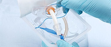


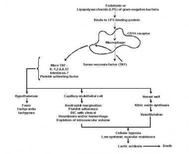


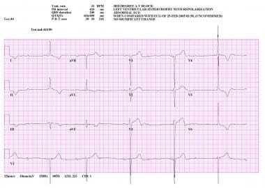


:quality(70)/cloudfront-us-east-1.images.arcpublishing.com/tronc/6YGAJLBALVGYVGJLZMCCEE3ZZI.JPG)