Central Nervous System Vascular Malformations
Vascular Malformations
Arteriovenous Malformations
Intracranial Arteriovenous Malformations
Central Nervous System
Hemangioma
Telangiectasia, Hereditary Hemorrhagic
Hemangioma, Cavernous, Central Nervous System
Lymphatic Abnormalities
Central Nervous System Diseases
Hemangioma, Cavernous
Double-Balloon Enteroscopy
Arteriovenous Fistula
Sclerotherapy
Port-Wine Stain
Angiodysplasia
Klippel-Trenaunay-Weber Syndrome
Skin Diseases, Vascular
Central Nervous System Neoplasms
Arnold-Chiari Malformation
Embolization, Therapeutic
Nervous System
Brain
Magnetic Resonance Imaging
Magnetic Resonance Angiography
Tomography, X-Ray Computed
Abnormalities, Multiple
Sturge-Weber Syndrome
Gastrointestinal Hemorrhage
Sclerosing Solutions
Cerebral Hemorrhage
Central Nervous System Venous Angioma
Angiomatosis
Telangiectasis
Central Nervous System Infections
Spinal Cord
Dura Mater
Proteus Syndrome
Spinal Cord Diseases
Angiography, Digital Subtraction
Hemangioendothelioma
Nevus, Blue
Blood Vessels
Cardiac Output, High
Cyanoacrylates
Cerebral Angiography
Nervous System Malformations
Peripheral Nervous System
Intracranial Hemorrhages
Vascular Headaches
Vascular Neoplasms
Azygos Vein
Pons
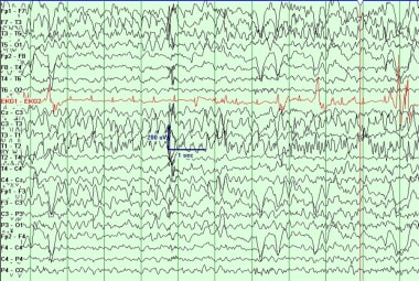
Clinically aggressive diffuse capillary telangiectasia of the brain stem: a clinical radiologic-pathologic case study. (1/221)
Capillary malformations or telangiectasias of the brain usually exhibit a benign clinical course, although occassionally they may be associated with mild to moderate symptomatology of uncertain origin. We report a case of an exceptionally aggressive capillary telangiectasia in a child, which was associated with progressive neurologic deterioration resulting in death. (+info)Cerebrovascular manifestations in 321 cases of hereditary hemorrhagic telangiectasia. (2/221)
BACKGROUND AND PURPOSE: Patients with hereditary hemorrhagic telangiectasia (HHT) are at risk for developing cerebral vascular malformations and pulmonary arteriovenous fistulae. We assessed the risk of neurological dysfunction from these malformations and fistulae. METHODS: Three hundred twenty-one consecutive patients with HHT seen at a single institution over a 20-year period were studied. Any evidence of prior neurological symptoms or presence of an intracranial vascular malformation was recorded. All cases of possible cerebral arteriovenous malformation were confirmed by conventional arteriography. RESULTS: Twelve patients (3.7%) had a history of cerebral vascular malformations. Ten patients had arteriovenous malformations, 1 had a dural arteriovenous fistula, and 1 had a cavernous malformation. Seven patients (2.1%) presented with intracranial hemorrhage, 2 presented with seizures alone, and 3 were discovered incidentally. The average age at the time of symptomatic intracranial hemorrhage was 25.4 years. All patients with a history of intracranial hemorrhage were classified as Rankin grade I or II at a mean follow-up interval of 6.0 years. A history of cerebral infarction or transient ischemic attack was found in 29.6% of patients with HHT and a pulmonary arteriovenous fistula. CONCLUSIONS: The risk of intracranial hemorrhage is low among people with HHT. Furthermore, a majority of these patients have a good functional outcome after hemorrhage. The data do not suggest a compelling indication for routine screening of patients with HHT for asymptomatic cerebral vascular malformations. By comparison, pulmonary arteriovenous fistulae are a much more frequent cause of neurological symptoms in this population. (+info)Identification of eight novel 5'-exons in cerebral capillary malformation gene-1 (CCM1) encoding KRIT1. (3/221)
Truncating mutations in the CCM1 gene encoding KRIT1 were recently found in patients affected by inherited cerebral capillary malformations, lesions that cause a wide variety of neurologic problems. However, CCM1 mutations have not been identified in all the families linked to CCM1. Here we demonstrate that the CCM1 gene contains eight additional exons which may thus encompass the missing mutations. (+info)Dural arteriovenous fistula in children: endovascular treatment and outcomes in seven cases. (4/221)
BACKGROUND AND PURPOSE: Dural AVF is a vascular anomaly that rarely occurs in children and is best treated by endovascular embolization. We report our experience using various endovascular embolization techniques in the treatment of dural AVF in a pediatric population. METHODS: Seven children with angiographically proven dural AVF were treated with endovascular embolization using microcoils, N-butylcyanoacrylate, detachable balloons, and/or silk suture. All imaging studies, embolization procedures, and patient charts were retrospectively reviewed. RESULTS: Seven children had been treated for dural AVF at our institution since 1987. Three newborns presented with congestive heart failure. Four older children (10 months-10 years) presented with signs referable to venous hypertension, including seizures, hydrocephalus, and proptosis. Embolization approaches included transarterial, transvenous, and direct puncture after neurosurgical exposure of a dural sinus. The number of embolizations ranged from 1 to 13 sessions per patient. All patients experienced symptomatic improvement after each embolization session. The three newborns showed marked improvement in cardiac function that allowed discharge to home. Clinical follow-up ranged from 3 weeks to 9 years (mean, 4.1 years). Two children with partially embolized dural AVF died, and one was lost to follow-up. Four children are alive after complete embolization of their dural AVF; two are developmentally normal, and two have mild developmental delay. CONCLUSION: Endovascular embolotherapy is the current treatment of choice for dural AVF. Embolization therapy may be life saving in the setting of cardiac failure and curative in cases of small or simple fistulae. Multiple, complex dural AVF are usually not curable, and treatment is aimed at symptomatic relief. Treatment strategies focus on the location and/or complexity of the fistula, the patient's clinical status, and the neurologic prognosis. (+info)Ultrastructural and immunocytochemical evidence that an incompetent blood-brain barrier is related to the pathophysiology of cavernous malformations. (5/221)
OBJECTIVES: Cerebral cavernous malformations are linked to mutations of the KRIT1 gene at the CCM1 locus and to mutations at two other loci, CCM2 and CCM3, for which genes are not yet identified. There is little information regarding the function of KRIT1. Histological and immunocytochemical analysis of cavernous malformations have not shed much light on their pathophysiology. METHODS: Morphological analysis of cavernous malformations was extended to the ultrastructural level by examining lesions from two patients by immunocytochemistry and electron microscopy. RESULTS: The lesions consisted of endothelial lined vascular sinusoids embedded in a collagen matrix. Nuclei belonging to cells distinct from endothelial cells were rare. The basal lamina of the endothelial cells consisted focally of multiple layers. No tight junctions at endothelial cell interfaces were found; however, several examined endothelial cell interfaces demonstrated apparent gaps between endothelial cell processes where basal lamina was exposed directly to the lumen of the sinusoids. Heavy hemosiderin deposits were found underlying the vascular channels within microns of the basal lamina without evidence of disrupted vessels. No astrocytic foot processes were seen within lesions. Glial fibrillary acidic protein immunocytochemistry confirmed that astrocyte processes stopped at the border of the lesions. CONCLUSIONS: The absence of blood-brain barrier components may lead to leakage of red blood cells into these lesions and the surrounding brain in the absence of major haemorrhage, thus accounting for the propensity of cavernous malformations to cause seizures. These data also raise the possibility that KRIT1 plays a part in the formation of endothelial cell junctions and expression of a mature vascular phenotype. (+info)MR imaging and histologic features of capillary telangiectasia of the basal ganglia. (6/221)
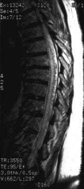
Capillary telangiectasias are being recognized with increasing frequency on MR imaging studies. Most are located in the brain stem and show slightly increased signal intensity on T2-weighted images, low signal intensity on T2*-weighted images (reflecting the presence of deoxyhemoglobin), and contrast enhancement. These findings are considered fairly typical for capillary telangiectasia, and pathologic correlation is not generally pursued. We present a case of a proven capillary telangiectasia in the basal ganglia. The imaging features of the lesion were identical to those described for capillary telangiectasias in the brain stem. (+info)Spinal dural arteriovenous fistulae--a diagnostic challenge. (7/221)
BACKGROUND: Spinal dural arteriovenous fistulae comprise the majority of spinal vascular malformations. The most common clinical presentation is that of progressive myeloradiculopathy, probably related to venous hypertension, which may lead to permanent disability and even death. OBJECTIVE: To report our clinical experience with spinal dural arteriovenous fistulae. METHODS: Nine patients with spinal dural AVF were managed at our center during a one year period (1998-1999). The patients, eight men and one woman ranging in age from 46 to 75 years, presented with initially fluctuating and eventually permanent and progressive paraparesis, sensory disturbances and sphincter dysfunction. The neurological signs generally began symmetrically and progressed from the distal to proximal limb regions. The duration of symptoms before diagnosis ranged from 6 to 36 months during which the patients underwent an extensive but fruitless work-up and even unnecessary operations due to misdiagnosis. All patients finally underwent magnetic resonance imaging and spinal angiography, which demonstrated the pathological vascular fistula. Interruption of the AVF was achieved by embolization or by surgical resection. RESULTS: Following treatment, six patients experienced improvement of gait and sphincter control, and the severe neurological deficits stabilized in the other three patients with long duration of illness. There was no further deterioration in any of the treated patients. CONCLUSIONS: The history, neurological findings and radiological changes on MRI scan should alert clinicians to the possibility of spinal dural AVF, leading to diagnostic spinal angiography. Early diagnosis and treatment may significantly improve outcome and prevent permanent disability and even mortality. (+info)Dural arteriovenous fistulae: noninvasive diagnosis with dynamic MR digital subtraction angiography. (8/221)
MR digital subtraction angiography (DSA) is a new diagnostic tool capable of producing dynamic images of the cerebral circulation with the injection of gadopentetate dimeglumine into a peripheral vein. Previous reports have concentrated on its potential as a noninvasive technique for the study of pial arteriovenous malformations. In this report, we present our early findings with MR DSA in the evaluation of intracranial dural arteriovenous fistulae. (+info)Central nervous system (CNS) vascular malformations are abnormal tangles or masses of blood vessels in the brain or spinal cord. These malformations can be congenital (present at birth) or acquired (develop later in life). They can vary in size, location, and symptoms, which may include headaches, seizures, weakness, numbness, difficulty speaking or understanding speech, and vision problems.
There are several types of CNS vascular malformations, including:
1. Arteriovenous malformations (AVMs): These are tangles of arteries and veins with a direct connection between them, bypassing the capillary network. AVMs can cause bleeding in the brain or spinal cord, leading to stroke or neurological deficits.
2. Cavernous malformations: These are clusters of dilated, thin-walled blood vessels that form a sac-like structure. They can rupture and bleed, causing symptoms such as seizures, headaches, or neurological deficits.
3. Developmental venous anomalies (DVAs): These are benign vascular malformations characterized by an abnormal pattern of veins that drain blood from the brain. DVAs are usually asymptomatic but can be associated with other vascular malformations.
4. Capillary telangiectasias: These are small clusters of dilated capillaries in the brain or spinal cord. They are usually asymptomatic and found incidentally during imaging studies.
5. Moyamoya disease: This is a rare, progressive cerebrovascular disorder characterized by the narrowing or blockage of the internal carotid arteries and their branches. This can lead to decreased blood flow to the brain, causing symptoms such as headaches, seizures, and strokes.
The diagnosis of CNS vascular malformations typically involves imaging studies such as MRI or CT scans, and sometimes angiography. Treatment options may include observation, medication, surgery, or endovascular procedures, depending on the type, location, and severity of the malformation.
Vascular malformations are abnormalities in the development and growth of blood vessels and lymphatic vessels that can occur anywhere in the body. They can be present at birth or develop later in life, and they can affect both the form and function of the affected tissues and organs. Vascular malformations can involve arteries, veins, capillaries, and/or lymphatic vessels, and they can range from simple, localized lesions to complex, multifocal disorders.
Vascular malformations are typically classified based on their location, size, flow characteristics, and the type of blood or lymphatic vessels involved. Some common types of vascular malformations include:
1. Capillary malformations (CMs): These are characterized by abnormal dilated capillaries that can cause red or pink discoloration of the skin, typically on the face or neck.
2. Venous malformations (VMs): These involve abnormal veins that can cause swelling, pain, and disfigurement in the affected area.
3. Lymphatic malformations (LMs): These involve abnormal lymphatic vessels that can cause swelling, infection, and other complications.
4. Arteriovenous malformations (AVMs): These involve a tangled mass of arteries and veins that can cause high-flow lesions, bleeding, and other serious complications.
5. Combined vascular malformations: These involve a combination of different types of blood or lymphatic vessels, such as capillary-lymphatic-venous malformations (CLVMs) or arteriovenous-lymphatic malformations (AVLMs).
The exact cause of vascular malformations is not fully understood, but they are believed to result from genetic mutations that affect the development and growth of blood vessels and lymphatic vessels. Treatment options for vascular malformations depend on the type, size, location, and severity of the lesion, as well as the patient's age and overall health. Treatment may include medication, compression garments, sclerotherapy, surgery, or a combination of these approaches.
Arteriovenous malformations (AVMs) are abnormal tangles of blood vessels that directly connect arteries and veins, bypassing the capillary system. This results in a high-flow and high-pressure circulation in the affected area. AVMs can occur anywhere in the body but are most common in the brain and spine. They can vary in size and may cause symptoms such as headaches, seizures, or bleeding in the brain. In some cases, AVMs may not cause any symptoms and may only be discovered during imaging tests for other conditions. Treatment options include surgery, radiation therapy, or embolization to reduce the flow of blood through the malformation and prevent complications.
Intracranial arteriovenous malformations (AVMs) are abnormal, tangled connections between the arteries and veins in the brain. These connections bypass the capillary system, which can lead to high-flow shunting and potential complications such as hemorrhage, stroke, or neurological deficits. AVMs are congenital conditions, meaning they are present at birth, although symptoms may not appear until later in life. They are relatively rare, affecting approximately 0.1% of the population. Treatment options for AVMs include surgery, radiation therapy, and endovascular embolization, depending on the size, location, and specific characteristics of the malformation.
The Central Nervous System (CNS) is the part of the nervous system that consists of the brain and spinal cord. It is called the "central" system because it receives information from, and sends information to, the rest of the body through peripheral nerves, which make up the Peripheral Nervous System (PNS).
The CNS is responsible for processing sensory information, controlling motor functions, and regulating various autonomic processes like heart rate, respiration, and digestion. The brain, as the command center of the CNS, interprets sensory stimuli, formulates thoughts, and initiates actions. The spinal cord serves as a conduit for nerve impulses traveling to and from the brain and the rest of the body.
The CNS is protected by several structures, including the skull (which houses the brain) and the vertebral column (which surrounds and protects the spinal cord). Despite these protective measures, the CNS remains vulnerable to injury and disease, which can have severe consequences due to its crucial role in controlling essential bodily functions.
A hemangioma is a benign (noncancerous) vascular tumor or growth that originates from blood vessels. It is characterized by an overgrowth of endothelial cells, which line the interior surface of blood vessels. Hemangiomas can occur in various parts of the body, but they are most commonly found on the skin and mucous membranes.
Hemangiomas can be classified into two main types:
1. Capillary hemangioma (also known as strawberry hemangioma): This type is more common and typically appears during the first few weeks of life. It grows rapidly for several months before gradually involuting (or shrinking) on its own, usually within the first 5 years of life. Capillary hemangiomas can be superficial, appearing as a bright red, raised lesion on the skin, or deep, forming a bluish, compressible mass beneath the skin.
2. Cavernous hemangioma: This type is less common and typically appears during infancy or early childhood. It consists of large, dilated blood vessels and can occur in various organs, including the skin, liver, brain, and gastrointestinal tract. Cavernous hemangiomas on the skin appear as a rubbery, bluish mass that does not typically involute like capillary hemangiomas.
Most hemangiomas do not require treatment, especially if they are small and not causing any significant problems. However, in cases where hemangiomas interfere with vital functions, impair vision or hearing, or become infected, various treatments may be considered, such as medication (e.g., corticosteroids, propranolol), laser therapy, surgical excision, or embolization.
Hereditary Hemorrhagic Telangiectasia (HHT) is a rare genetic disorder that affects the blood vessels. It is also known as Osler-Weber-Rendu syndrome. This condition is characterized by the formation of abnormal blood vessels called telangiectases, which are small red spots or tiny bulges that can be found in the skin, mucous membranes (like those inside the nose, mouth, and GI tract), and sometimes in vital organs like the lungs and brain.
These telangiectases have a tendency to bleed easily, leading to potentially serious complications such as anemia due to chronic blood loss, and in some cases, strokes or brain abscesses if the telangiectases in the brain rupture. HHT is typically inherited in an autosomal dominant pattern, meaning that a child has a 50% chance of inheriting the gene from an affected parent. There are several genes associated with HHT, the most common being ACVRL1, ENG, and SMAD4.
A cavernous hemangioma in the central nervous system (CNS) refers to a type of benign vascular tumor that is made up of dilated and thin-walled blood vessels. These tumors are called "cavernous" because they are filled with blood-filled sacs or "caverns."
When these hemangiomas occur in the CNS, which includes the brain and spinal cord, they can cause various neurological symptoms depending on their size and location. Small hemangiomas may not cause any symptoms at all, while larger ones can cause seizures, headaches, weakness, or sensory changes.
Cavernous hemangiomas in the CNS are typically congenital, meaning that they are present at birth. However, they may not become symptomatic until later in life. Treatment options for cavernous hemangiomas in the CNS include observation, surgery, or radiation therapy, depending on the size, location, and symptoms caused by the tumor.
Lymphatic abnormalities refer to conditions or defects that affect the lymphatic system, which is a part of the immune and circulatory systems. The lymphatic system includes a network of vessels, tissues, and organs that help rid the body of waste and toxins, fight infections, and maintain fluid balance.
Lymphatic abnormalities can occur due to genetic mutations, infections, inflammation, or cancer. These abnormalities may affect various components of the lymphatic system, including:
1. Lymph vessels: Abnormalities in lymph vessels can lead to a buildup of lymph fluid in certain parts of the body, causing swelling known as lymphedema.
2. Lymph nodes: Enlarged or abnormally shaped lymph nodes (lymphadenopathy) may indicate an infection, inflammation, or cancer.
3. Spleen: An enlarged spleen (splenomegaly) can be a sign of various conditions, such as infections, blood disorders, or cancer.
4. Thymus: Abnormalities in the thymus gland, which is part of the immune system, can lead to immunodeficiency disorders.
5. Tonsils and adenoids: Enlarged tonsils and adenoids can cause breathing and swallowing difficulties, especially in children.
6. Aggregated lymphatic tissue: Abnormalities in aggregated lymphatic tissue, such as Peyer's patches in the small intestine or the appendix, can increase the risk of infections and autoimmune disorders.
Lymphatic abnormalities can present with various symptoms, including swelling, pain, recurrent infections, and fatigue. Treatment depends on the underlying cause and may involve medications, surgery, or lifestyle changes.
The parotid region is the area on either side of the face, anterior to (in front of) the ear, and below the zygomatic arch (cheekbone). It is named after the parotid gland, which is a salivary gland located within this region. The parotid gland produces saliva that helps in digestion, particularly of starches.
The parotid region contains not only the parotid gland but also other important structures such as the facial nerve, external carotid artery, and retromandibular vein. Injuries or diseases affecting this region may cause problems with chewing, speaking, or moving the face.
Central nervous system (CNS) diseases refer to medical conditions that primarily affect the brain and spinal cord. The CNS is responsible for controlling various functions in the body, including movement, sensation, cognition, and behavior. Therefore, diseases of the CNS can have significant impacts on a person's quality of life and overall health.
There are many different types of CNS diseases, including:
1. Infectious diseases: These are caused by viruses, bacteria, fungi, or parasites that infect the brain or spinal cord. Examples include meningitis, encephalitis, and polio.
2. Neurodegenerative diseases: These are characterized by progressive loss of nerve cells in the brain or spinal cord. Examples include Alzheimer's disease, Parkinson's disease, and Huntington's disease.
3. Structural diseases: These involve damage to the physical structure of the brain or spinal cord, such as from trauma, tumors, or stroke.
4. Functional diseases: These affect the function of the nervous system without obvious structural damage, such as multiple sclerosis and epilepsy.
5. Genetic disorders: Some CNS diseases are caused by genetic mutations, such as spinal muscular atrophy and Friedreich's ataxia.
Symptoms of CNS diseases can vary widely depending on the specific condition and the area of the brain or spinal cord that is affected. They may include muscle weakness, paralysis, seizures, loss of sensation, difficulty with coordination and balance, confusion, memory loss, changes in behavior or mood, and pain. Treatment for CNS diseases depends on the specific condition and may involve medications, surgery, rehabilitation therapy, or a combination of these approaches.
A cavernous hemangioma is a type of benign vascular tumor that is made up of large, dilated blood vessels. It is characterized by the presence of large, "cavernous" spaces or sacs filled with blood. These lesions can occur in various parts of the body, but when they occur in the skin or mucous membranes, they appear as well-circumscribed rubbery masses that are compressible and blanchable (turn pale when pressed).
Cavernous hemangiomas are most commonly found on the face and neck, but they can also occur in other parts of the body such as the liver. They typically grow slowly during infancy or early childhood and then stabilize or even regress spontaneously over time. However, if they are located in critical areas such as the airway or near vital organs, they may require treatment to prevent complications.
Histologically, cavernous hemangiomas are composed of large, irregularly shaped vascular spaces lined by a single layer of endothelial cells and surrounded by fibrous tissue. Treatment options for cavernous hemangiomas include observation, compression therapy, laser therapy, surgical excision, or embolization.
Double-balloon enteroscopy (DBE) is a medical procedure used to examine the small intestine, which is difficult to reach with traditional endoscopes due to its length and twists and turns. DBE uses a specialized endoscope with two inflatable balloons on its tip. The endoscope is inserted through the mouth or the rectum and advanced slowly into the small intestine while alternately inflating and deflating the balloons to help move the endoscope forward and provide better visualization of the intestinal lining.
DBE can be used for diagnostic purposes, such as evaluating obscure gastrointestinal bleeding, Crohn's disease, tumors, or polyps in the small intestine. It can also be used for therapeutic interventions, such as removing polyps, taking biopsies, or placing feeding tubes.
The procedure is usually done under sedation and takes several hours to complete. While it is considered a safe procedure, potential risks include perforation of the intestinal wall, bleeding, and adverse reactions to the anesthesia.
An arteriovenous fistula is an abnormal connection or passageway between an artery and a vein. This connection causes blood to flow directly from the artery into the vein, bypassing the capillary network that would normally distribute the oxygen-rich blood to the surrounding tissues.
Arteriovenous fistulas can occur as a result of trauma, disease, or as a planned surgical procedure for patients who require hemodialysis, a treatment for advanced kidney failure. In hemodialysis, the arteriovenous fistula serves as a site for repeated access to the bloodstream, allowing for efficient removal of waste products and excess fluids.
The medical definition of an arteriovenous fistula is:
"An abnormal communication between an artery and a vein, usually created by surgical means for hemodialysis access or occurring as a result of trauma, congenital defects, or disease processes such as vasculitis or neoplasm."
Sclerotherapy is a medical procedure used to treat varicose veins and spider veins. It involves the injection of a solution (called a sclerosant) directly into the affected vein, which causes the vein to collapse and eventually fade away. The sclerosant works by irritating the lining of the vein, causing it to swell and stick together, which then leads to clotting and the eventual reabsorption of the vein by the body.
The procedure is typically performed in a doctor's office or outpatient setting and may require multiple sessions depending on the severity and number of veins being treated. Common side effects include bruising, swelling, and discomfort at the injection site, as well as the possibility of developing brownish pigmentation or small ulcers near the treatment area. However, these side effects are usually temporary and resolve on their own within a few weeks.
Sclerotherapy is considered a safe and effective treatment for varicose veins and spider veins, with high success rates and low complication rates. It is important to note that while sclerotherapy can improve the appearance of affected veins, it does not prevent new veins from developing in the future.
A port-wine stain is a type of birthmark that appears at birth or shortly thereafter. It's caused by an abnormal development of blood vessels in the skin, leading to a permanently reddish-purple discoloration. Port-wine stains are generally found on the face but can occur anywhere on the body. They tend to grow as the child grows and may become darker and thicker over time.
Unlike some other types of birthmarks, port-wine stains usually do not fade or go away on their own. In some cases, they can be associated with various syndromes or conditions that affect the development of blood vessels or nerves. Treatment options include laser therapy, which can help to reduce the size and color of the stain, especially when started in early childhood.
Cerebral veins are the blood vessels that carry deoxygenated blood from the brain to the dural venous sinuses, which are located between the layers of tissue covering the brain. The largest cerebral vein is the superior sagittal sinus, which runs along the top of the brain. Other major cerebral veins include the straight sinus, transverse sinus, sigmoid sinus, and cavernous sinus. These veins receive blood from smaller veins called venules that drain the surface and deep structures of the brain. The cerebral veins play an important role in maintaining normal circulation and pressure within the brain.
Angiodysplasia is a vascular disorder characterized by the dilation and abnormal formation of blood vessels, particularly in the gastrointestinal (GI) tract. These abnormal blood vessels are prone to leakage or rupture, which can lead to bleeding. Angiodysplasia is most commonly found in the colon but can occur in other parts of the GI tract as well. It is more common in older adults and can cause symptoms such as anemia, fatigue, and bloody stools. The exact cause of angiodysplasia is not known, but it may be associated with chronic low-grade inflammation or increased pressure in the blood vessels. Treatment options include endoscopic therapies to stop bleeding, medications to reduce acid production in the stomach, and surgery in severe cases.
Klippel-Trenaunay-Weber Syndrome (KTWS) is a rare and complex congenital vascular disorder that affects the development of blood vessels, soft tissues, and bones. It is also known as Klippel-Trenaunay syndrome or KTS.
The medical definition of KTWS includes the following features:
1. Port-wine stain (capillary malformation): A red or purple birthmark caused by an abnormal collection of blood vessels in the skin, often present at birth and usually affecting one limb or part of the body.
2. Venous and lymphatic abnormalities: Varicose veins, dilated veins, or abnormal vein patterns may be present, along with lymphatic malformations that can cause swelling in the affected area.
3. Soft tissue and bone hypertrophy: Overgrowth of soft tissues and bones in the affected limb or region, leading to asymmetry and sometimes functional impairment.
4. Other possible features: May include skin abnormalities, such as increased hair growth or changes in texture; joint deformities; and orthopedic problems, like scoliosis or hip dysplasia.
It is important to note that the severity of KTWS can vary significantly from person to person, ranging from mild symptoms to severe cases with significant functional impairment. The condition is not typically life-threatening but may require ongoing medical management and surveillance to address potential complications, such as infections, bleeding, or deep vein thrombosis.
Vascular skin diseases are a group of medical conditions that affect the blood vessels in the skin. These disorders can be caused by problems with the structure or function of the blood vessels, which can lead to various symptoms such as redness, discoloration, pain, itching, and ulcerations. Some examples of vascular skin diseases include:
1. Rosacea: a chronic skin condition that causes redness, flushing, and visible blood vessels in the face.
2. Eczema: a group of inflammatory skin conditions that can cause redness, itching, and dryness. Some types of eczema, such as varicose eczema, are associated with problems with the veins.
3. Psoriasis: an autoimmune condition that causes red, scaly patches on the skin. Some people with psoriasis may also develop psoriatic arthritis, which can affect the blood vessels in the skin and joints.
4. Vasculitis: a group of conditions that cause inflammation of the blood vessels. This can lead to symptoms such as redness, pain, and ulcerations.
5. Livedo reticularis: a condition that causes a net-like pattern of discoloration on the skin, usually on the legs. It is caused by abnormalities in the small blood vessels.
6. Henoch-Schönlein purpura: a rare condition that causes inflammation of the small blood vessels, leading to purple spots on the skin and joint pain.
7. Raynaud's phenomenon: a condition that affects the blood vessels in the fingers and toes, causing them to become narrow and restrict blood flow in response to cold temperatures or stress.
Treatment for vascular skin diseases depends on the specific condition and its severity. It may include medications, lifestyle changes, and in some cases, surgery.
Central nervous system (CNS) neoplasms refer to a group of abnormal growths or tumors that develop within the brain or spinal cord. These tumors can be benign or malignant, and their growth can compress or disrupt the normal functioning of surrounding brain or spinal cord tissue.
Benign CNS neoplasms are slow-growing and rarely spread to other parts of the body. However, they can still cause significant problems if they grow large enough to put pressure on vital structures within the brain or spinal cord. Malignant CNS neoplasms, on the other hand, are aggressive tumors that can invade and destroy surrounding tissue. They may also spread to other parts of the CNS or, rarely, to other organs in the body.
CNS neoplasms can arise from various types of cells within the brain or spinal cord, including nerve cells, glial cells (which provide support and insulation for nerve cells), and supportive tissues such as blood vessels. The specific type of CNS neoplasm is often used to help guide treatment decisions and determine prognosis.
Symptoms of CNS neoplasms can vary widely depending on the location and size of the tumor, but may include headaches, seizures, weakness or paralysis, vision or hearing changes, balance problems, memory loss, and changes in behavior or personality. Treatment options for CNS neoplasms may include surgery, radiation therapy, chemotherapy, or a combination of these approaches.
Arnold-Chiari malformation is a structural abnormality of the brain and skull base, specifically the cerebellum and brainstem. It is characterized by the descent of the cerebellar tonsils and sometimes parts of the brainstem through the foramen magnum (the opening at the base of the skull) into the upper spinal canal. This can cause pressure on the brainstem and cerebellum, potentially leading to a range of symptoms such as headaches, neck pain, unsteady gait, swallowing difficulties, hearing or balance problems, and in severe cases, neurological deficits. There are four types of Arnold-Chiari malformations, with type I being the most common and least severe form. Types II, III, and IV are progressively more severe and involve varying degrees of hindbrain herniation and associated neural tissue damage. Surgical intervention is often required to alleviate symptoms and prevent further neurological deterioration.
Therapeutic embolization is a medical procedure that involves intentionally blocking or obstructing blood vessels to stop excessive bleeding or block the flow of blood to a tumor or abnormal tissue. This is typically accomplished by injecting small particles, such as microspheres or coils, into the targeted blood vessel through a catheter, which is inserted into a larger blood vessel and guided to the desired location using imaging techniques like X-ray or CT scanning. The goal of therapeutic embolization is to reduce the size of a tumor, control bleeding, or block off abnormal blood vessels that are causing problems.
The nervous system is a complex, highly organized network of specialized cells called neurons and glial cells that communicate with each other via electrical and chemical signals to coordinate various functions and activities in the body. It consists of two main parts: the central nervous system (CNS), including the brain and spinal cord, and the peripheral nervous system (PNS), which includes all the nerves and ganglia outside the CNS.
The primary function of the nervous system is to receive, process, and integrate information from both internal and external environments and then respond by generating appropriate motor outputs or behaviors. This involves sensing various stimuli through specialized receptors, transmitting this information through afferent neurons to the CNS for processing, integrating this information with other inputs and memories, making decisions based on this processed information, and finally executing responses through efferent neurons that control effector organs such as muscles and glands.
The nervous system can be further divided into subsystems based on their functions, including the somatic nervous system, which controls voluntary movements and reflexes; the autonomic nervous system, which regulates involuntary physiological processes like heart rate, digestion, and respiration; and the enteric nervous system, which is a specialized subset of the autonomic nervous system that controls gut functions. Overall, the nervous system plays a critical role in maintaining homeostasis, regulating behavior, and enabling cognition and consciousness.
The brain is the central organ of the nervous system, responsible for receiving and processing sensory information, regulating vital functions, and controlling behavior, movement, and cognition. It is divided into several distinct regions, each with specific functions:
1. Cerebrum: The largest part of the brain, responsible for higher cognitive functions such as thinking, learning, memory, language, and perception. It is divided into two hemispheres, each controlling the opposite side of the body.
2. Cerebellum: Located at the back of the brain, it is responsible for coordinating muscle movements, maintaining balance, and fine-tuning motor skills.
3. Brainstem: Connects the cerebrum and cerebellum to the spinal cord, controlling vital functions such as breathing, heart rate, and blood pressure. It also serves as a relay center for sensory information and motor commands between the brain and the rest of the body.
4. Diencephalon: A region that includes the thalamus (a major sensory relay station) and hypothalamus (regulates hormones, temperature, hunger, thirst, and sleep).
5. Limbic system: A group of structures involved in emotional processing, memory formation, and motivation, including the hippocampus, amygdala, and cingulate gyrus.
The brain is composed of billions of interconnected neurons that communicate through electrical and chemical signals. It is protected by the skull and surrounded by three layers of membranes called meninges, as well as cerebrospinal fluid that provides cushioning and nutrients.
Medical Definition:
Magnetic Resonance Imaging (MRI) is a non-invasive diagnostic imaging technique that uses a strong magnetic field and radio waves to create detailed cross-sectional or three-dimensional images of the internal structures of the body. The patient lies within a large, cylindrical magnet, and the scanner detects changes in the direction of the magnetic field caused by protons in the body. These changes are then converted into detailed images that help medical professionals to diagnose and monitor various medical conditions, such as tumors, injuries, or diseases affecting the brain, spinal cord, heart, blood vessels, joints, and other internal organs. MRI does not use radiation like computed tomography (CT) scans.
Congenital abnormalities, also known as birth defects, are structural or functional anomalies that are present at birth. These abnormalities can develop at any point during fetal development, and they can affect any part of the body. They can be caused by genetic factors, environmental influences, or a combination of both.
Congenital abnormalities can range from mild to severe and may include structural defects such as heart defects, neural tube defects, and cleft lip and palate, as well as functional defects such as intellectual disabilities and sensory impairments. Some congenital abnormalities may be visible at birth, while others may not become apparent until later in life.
In some cases, congenital abnormalities may be detected through prenatal testing, such as ultrasound or amniocentesis. In other cases, they may not be diagnosed until after the baby is born. Treatment for congenital abnormalities varies depending on the type and severity of the defect, and may include surgery, therapy, medication, or a combination of these approaches.
Magnetic Resonance Angiography (MRA) is a non-invasive medical imaging technique that uses magnetic fields and radio waves to create detailed images of the blood vessels or arteries within the body. It is a type of Magnetic Resonance Imaging (MRI) that focuses specifically on the circulatory system.
MRA can be used to diagnose and evaluate various conditions related to the blood vessels, such as aneurysms, stenosis (narrowing of the vessel), or the presence of plaques or tumors. It can also be used to plan for surgeries or other treatments related to the vascular system. The procedure does not use radiation and is generally considered safe, although people with certain implants like pacemakers may not be able to have an MRA due to safety concerns.
X-ray computed tomography (CT or CAT scan) is a medical imaging method that uses computer-processed combinations of many X-ray images taken from different angles to produce cross-sectional (tomographic) images (virtual "slices") of the body. These cross-sectional images can then be used to display detailed internal views of organs, bones, and soft tissues in the body.
The term "computed tomography" is used instead of "CT scan" or "CAT scan" because the machines take a series of X-ray measurements from different angles around the body and then use a computer to process these data to create detailed images of internal structures within the body.
CT scanning is a noninvasive, painless medical test that helps physicians diagnose and treat medical conditions. CT imaging provides detailed information about many types of tissue including lung, bone, soft tissue and blood vessels. CT examinations can be performed on every part of the body for a variety of reasons including diagnosis, surgical planning, and monitoring of therapeutic responses.
In computed tomography (CT), an X-ray source and detector rotate around the patient, measuring the X-ray attenuation at many different angles. A computer uses this data to construct a cross-sectional image by the process of reconstruction. This technique is called "tomography". The term "computed" refers to the use of a computer to reconstruct the images.
CT has become an important tool in medical imaging and diagnosis, allowing radiologists and other physicians to view detailed internal images of the body. It can help identify many different medical conditions including cancer, heart disease, lung nodules, liver tumors, and internal injuries from trauma. CT is also commonly used for guiding biopsies and other minimally invasive procedures.
In summary, X-ray computed tomography (CT or CAT scan) is a medical imaging technique that uses computer-processed combinations of many X-ray images taken from different angles to produce cross-sectional images of the body. It provides detailed internal views of organs, bones, and soft tissues in the body, allowing physicians to diagnose and treat medical conditions.
'Abnormalities, Multiple' is a broad term that refers to the presence of two or more structural or functional anomalies in an individual. These abnormalities can be present at birth (congenital) or can develop later in life (acquired). They can affect various organs and systems of the body and can vary greatly in severity and impact on a person's health and well-being.
Multiple abnormalities can occur due to genetic factors, environmental influences, or a combination of both. Chromosomal abnormalities, gene mutations, exposure to teratogens (substances that cause birth defects), and maternal infections during pregnancy are some of the common causes of multiple congenital abnormalities.
Examples of multiple congenital abnormalities include Down syndrome, Turner syndrome, and VATER/VACTERL association. Acquired multiple abnormalities can result from conditions such as trauma, infection, degenerative diseases, or cancer.
The medical evaluation and management of individuals with multiple abnormalities depend on the specific abnormalities present and their impact on the individual's health and functioning. A multidisciplinary team of healthcare professionals is often involved in the care of these individuals to address their complex needs.
Sturge-Weber syndrome is a rare neurocutaneous disorder characterized by the combination of a facial port-wine birthmark and neurological abnormalities. The facial birthmark, which is typically located on one side of the face, occurs due to the malformation of small blood vessels (capillaries) in the skin and eye.
Neurological features often include seizures that begin in infancy, muscle weakness or paralysis on one side of the body (hemiparesis), developmental delay, and intellectual disability. These neurological symptoms are caused by abnormal blood vessel formation in the brain (leptomeningeal angiomatosis) leading to increased pressure, reduced blood flow, and potential damage to the brain tissue.
Sturge-Weber syndrome can also affect the eyes, with glaucoma being a common occurrence due to increased pressure within the eye. Early diagnosis and appropriate management of this condition are crucial for improving the quality of life and reducing potential complications.
Gastrointestinal (GI) hemorrhage is a term used to describe any bleeding that occurs in the gastrointestinal tract, which includes the esophagus, stomach, small intestine, large intestine, and rectum. The bleeding can range from mild to severe and can produce symptoms such as vomiting blood, passing black or tarry stools, or having low blood pressure.
GI hemorrhage can be classified as either upper or lower, depending on the location of the bleed. Upper GI hemorrhage refers to bleeding that occurs above the ligament of Treitz, which is a point in the small intestine where it becomes narrower and turns a corner. Common causes of upper GI hemorrhage include gastritis, ulcers, esophageal varices, and Mallory-Weiss tears.
Lower GI hemorrhage refers to bleeding that occurs below the ligament of Treitz. Common causes of lower GI hemorrhage include diverticulosis, colitis, inflammatory bowel disease, and vascular abnormalities such as angiodysplasia.
The diagnosis of GI hemorrhage is often made based on the patient's symptoms, medical history, physical examination, and diagnostic tests such as endoscopy, CT scan, or radionuclide scanning. Treatment depends on the severity and cause of the bleeding and may include medications, endoscopic procedures, surgery, or a combination of these approaches.
Sclerosing solutions are medications or substances that are used to intentionally cause the scarring and hardening (sclerosis) of tissue, usually in the context of treating various medical conditions. These solutions work by irritating the interior lining of blood vessels or other targeted tissues, leading to the formation of a fibrous scar and the eventual closure of the affected area.
One common use of sclerosing solutions is in the treatment of abnormal veins, such as varicose veins or spider veins. A solution like sodium tetradecyl sulfate or polidocanol is injected directly into the problematic vein, causing inflammation and eventual closure of the vein. The body then gradually absorbs the closed vein, reducing its appearance and associated symptoms.
Other medical applications for sclerosing solutions include the treatment of lymphatic malformations, hydroceles, and certain types of tumors or cysts. It is essential to administer these substances under the supervision of a qualified healthcare professional, as improper use can lead to complications such as infection, tissue damage, or embolism.
A cerebral hemorrhage, also known as an intracranial hemorrhage or intracerebral hemorrhage, is a type of stroke that results from bleeding within the brain tissue. It occurs when a weakened blood vessel bursts and causes localized bleeding in the brain. This bleeding can increase pressure in the skull, damage nearby brain cells, and release toxic substances that further harm brain tissues.
Cerebral hemorrhages are often caused by chronic conditions like hypertension (high blood pressure) or cerebral amyloid angiopathy, which weakens the walls of blood vessels over time. Other potential causes include trauma, aneurysms, arteriovenous malformations, illicit drug use, and brain tumors. Symptoms may include sudden headache, weakness, numbness, difficulty speaking or understanding speech, vision problems, loss of balance, and altered level of consciousness. Immediate medical attention is required to diagnose and manage cerebral hemorrhage through imaging techniques, supportive care, and possible surgical interventions.
A Central Nervous System Venous Angioma (CNS VA), also known as a cerebral venous angioma or developmental venous anomaly (DVA), is a benign vascular malformation of the central nervous system. It is a congenital condition, which means it is present at birth.
A CNS VA is characterized by a cluster of veins that converge into a single larger vein, creating a radial pattern that resembles a Medusa head or a spoked wheel. This venous anomaly typically drains blood from normal brain tissue and usually does not cause any symptoms or neurological deficits. However, in rare cases, CNS VAs may be associated with intracranial hemorrhage, seizures, or development of arteriovenous malformations (AVMs).
CNS VAs are usually discovered incidentally during imaging studies performed for other medical reasons. Diagnostic imaging techniques such as magnetic resonance imaging (MRI) and computed tomography (CT) scans with contrast can help identify and characterize CNS VAs. No specific treatment is required for asymptomatic CNS VAs, but follow-up imaging may be recommended to monitor the condition over time. In cases where symptoms are present or there is a risk of complications, various treatment options may be considered, including surgical removal, endovascular embolization, or radiation therapy.
Angiomatosis is a medical term that refers to a benign condition characterized by the proliferation of blood vessels in various tissues and organs. It is typically composed of small, tangled blood vessels called capillaries, which can form clusters or networks. The condition can affect skin, internal organs, bones, and other tissues.
Angiomatosis is often asymptomatic and may be discovered incidentally during medical imaging or surgical procedures. In some cases, it may cause symptoms such as pain, swelling, or bleeding, depending on the location and extent of the lesions.
While angiomatosis is generally a benign condition, in rare cases, it can be associated with malignant tumors or other medical conditions. Treatment options for angiomatosis depend on the size, location, and symptoms of the lesions and may include observation, medication, or surgical removal.
Telangiectasia is a medical term that refers to the dilation and widening of small blood vessels called capillaries, leading to their visibility under the skin or mucous membranes. These dilated vessels often appear as tiny red lines or patterns, measuring less than 1 millimeter in diameter.
Telangiectasias can occur in various parts of the body, such as the face, nose, cheeks, legs, and fingers. They are typically harmless but may cause cosmetic concerns for some individuals. In certain cases, telangiectasias can be a sign of an underlying medical condition, like rosacea, hereditary hemorrhagic telangiectasia (HHT), or liver disease.
It is essential to consult with a healthcare professional if you notice any unusual changes in your skin or mucous membranes, as they can provide appropriate evaluation and treatment recommendations based on the underlying cause of the telangiectasias.
Angiography is a medical procedure in which an x-ray image is taken to visualize the internal structure of blood vessels, arteries, or veins. This is done by injecting a radiopaque contrast agent (dye) into the blood vessel using a thin, flexible catheter. The dye makes the blood vessels visible on an x-ray image, allowing doctors to diagnose and treat various medical conditions such as blockages, narrowing, or malformations of the blood vessels.
There are several types of angiography, including:
* Cardiac angiography (also called coronary angiography) - used to examine the blood vessels of the heart
* Cerebral angiography - used to examine the blood vessels of the brain
* Peripheral angiography - used to examine the blood vessels in the limbs or other parts of the body.
Angiography is typically performed by a radiologist, cardiologist, or vascular surgeon in a hospital setting. It can help diagnose conditions such as coronary artery disease, aneurysms, and peripheral arterial disease, among others.
Central nervous system (CNS) infections refer to infectious processes that affect the brain, spinal cord, and their surrounding membranes, known as meninges. These infections can be caused by various microorganisms, including bacteria, viruses, fungi, and parasites. Examples of CNS infections are:
1. Meningitis: Inflammation of the meninges, usually caused by bacterial or viral infections. Bacterial meningitis is a medical emergency that requires immediate treatment.
2. Encephalitis: Inflammation of the brain parenchyma, often caused by viral infections. Some viruses associated with encephalitis include herpes simplex virus, enteroviruses, and arboviruses.
3. Meningoencephalitis: A combined inflammation of both the brain and meninges, commonly seen in certain viral infections or when bacterial pathogens directly invade the brain.
4. Brain abscess: A localized collection of pus within the brain caused by a bacterial or fungal infection.
5. Spinal epidural abscess: An infection in the space surrounding the spinal cord, usually caused by bacteria.
6. Myelitis: Inflammation of the spinal cord, which can result from viral, bacterial, or fungal infections.
7. Rarely, parasitic infections like toxoplasmosis and cysticercosis can also affect the CNS.
Symptoms of CNS infections may include fever, headache, stiff neck, altered mental status, seizures, focal neurological deficits, or meningeal signs (e.g., Brudzinski's and Kernig's signs). The specific symptoms depend on the location and extent of the infection, as well as the causative organism. Prompt diagnosis and treatment are crucial to prevent long-term neurological complications or death.
The spinal cord is a major part of the nervous system, extending from the brainstem and continuing down to the lower back. It is a slender, tubular bundle of nerve fibers (axons) and support cells (glial cells) that carries signals between the brain and the rest of the body. The spinal cord primarily serves as a conduit for motor information, which travels from the brain to the muscles, and sensory information, which travels from the body to the brain. It also contains neurons that can independently process and respond to information within the spinal cord without direct input from the brain.
The spinal cord is protected by the bony vertebral column (spine) and is divided into 31 segments: 8 cervical, 12 thoracic, 5 lumbar, 5 sacral, and 1 coccygeal. Each segment corresponds to a specific region of the body and gives rise to pairs of spinal nerves that exit through the intervertebral foramina at each level.
The spinal cord is responsible for several vital functions, including:
1. Reflexes: Simple reflex actions, such as the withdrawal reflex when touching a hot surface, are mediated by the spinal cord without involving the brain.
2. Muscle control: The spinal cord carries motor signals from the brain to the muscles, enabling voluntary movement and muscle tone regulation.
3. Sensory perception: The spinal cord transmits sensory information, such as touch, temperature, pain, and vibration, from the body to the brain for processing and awareness.
4. Autonomic functions: The sympathetic and parasympathetic divisions of the autonomic nervous system originate in the thoracolumbar and sacral regions of the spinal cord, respectively, controlling involuntary physiological responses like heart rate, blood pressure, digestion, and respiration.
Damage to the spinal cord can result in various degrees of paralysis or loss of sensation below the level of injury, depending on the severity and location of the damage.
Dura Mater is the thickest and outermost of the three membranes (meninges) that cover the brain and spinal cord. It provides protection and support to these delicate structures. The other two layers are called the Arachnoid Mater and the Pia Mater, which are thinner and more delicate than the Dura Mater. Together, these three layers form a protective barrier around the central nervous system.
Proteus Syndrome is a rare genetic disorder characterized by progressive overgrowth of skin, bones, muscles, and other tissues. It is caused by a mutation in the AKT1 gene, which regulates cell growth and division. The disorder is named after the Greek sea-god Proteus, who could change his shape at will, as people with this condition often have highly variable and asymmetric features.
The symptoms of Proteus Syndrome can vary widely from person to person, but may include:
1. Overgrowth of skin, which can lead to the formation of thickened, rough, or irregular areas of skin (known as "cerebriform" skin) and deep creases or folds.
2. Asymmetric overgrowth of bones, muscles, and other tissues, leading to differences in size and shape between the two sides of the body.
3. The formation of benign tumors (such as lipomas and lymphangiomas) and abnormal blood vessels.
4. Abnormalities of the brain, eyes, and other organs.
5. Increased risk of developing certain types of cancer.
Proteus Syndrome is typically diagnosed based on a combination of clinical features, medical imaging, and genetic testing. There is no cure for the disorder, but treatment is focused on managing symptoms and preventing complications. This may involve surgery to remove tumors or correct bone deformities, physical therapy to improve mobility and strength, and medications to control pain and other symptoms.
Spinal cord diseases refer to a group of conditions that affect the spinal cord, which is a part of the central nervous system responsible for transmitting messages between the brain and the rest of the body. These diseases can cause damage to the spinal cord, leading to various symptoms such as muscle weakness, numbness, pain, bladder and bowel dysfunction, and difficulty with movement and coordination.
Spinal cord diseases can be congenital or acquired, and they can result from a variety of causes, including infections, injuries, tumors, degenerative conditions, autoimmune disorders, and genetic factors. Some examples of spinal cord diseases include multiple sclerosis, spina bifida, spinal cord injury, herniated discs, spinal stenosis, and motor neuron diseases such as amyotrophic lateral sclerosis (ALS).
The treatment for spinal cord diseases varies depending on the underlying cause and severity of the condition. Treatment options may include medication, physical therapy, surgery, and rehabilitation. In some cases, the damage to the spinal cord may be irreversible, leading to permanent disability or paralysis.
Veins are blood vessels that carry deoxygenated blood from the tissues back to the heart. They have a lower pressure than arteries and contain valves to prevent the backflow of blood. Veins have a thin, flexible wall with a larger lumen compared to arteries, allowing them to accommodate more blood volume. The color of veins is often blue or green due to the absorption characteristics of light and the reduced oxygen content in the blood they carry.
Digital subtraction angiography (DSA) is a medical imaging technique used to visualize the blood vessels and blood flow within the body. It combines the use of X-ray technology with digital image processing to produce detailed images of the vascular system.
In DSA, a contrast agent is injected into the patient's bloodstream through a catheter, which is typically inserted into an artery in the leg and guided to the area of interest using fluoroscopy. As the contrast agent flows through the blood vessels, X-ray images are taken at multiple time points.
The digital subtraction process involves taking a baseline image without contrast and then subtracting it from subsequent images taken with contrast. This allows for the removal of background structures and noise, resulting in clearer images of the blood vessels. DSA can be used to diagnose and evaluate various vascular conditions, such as aneurysms, stenosis, and tumors, and can also guide interventional procedures such as angioplasty and stenting.
Hemangioendothelioma is a rare type of vascular tumor, which means it arises from the endothelial cells that line the blood vessels. It can occur in various parts of the body, but it most commonly involves the soft tissues and bones. Hemangioendotheliomas are often classified as borderline malignant tumors because they can behave either indolently (like a benign tumor) or aggressively (like a malignant tumor), depending on their specific type and location.
There are several subtypes of hemangioendothelioma, including:
1. Epithelioid hemangioendothelioma: This subtype typically affects young adults and can involve various organs, such as the liver, lungs, or soft tissues. It tends to have a more indolent course but can metastasize in some cases.
2. Kaposiform hemangioendothelioma: This is an aggressive subtype that usually occurs in infants and children. It often involves the skin and soft tissues, causing local invasion and consumptive coagulopathy (Kasabach-Merritt phenomenon).
3. Retiform hemangioendothelioma: A rare and low-grade malignant tumor that typically affects the skin and subcutaneous tissue of adults. It has a favorable prognosis with a low risk of metastasis.
4. Papillary intralymphatic angioendothelioma (PILA): This is a rare, slow-growing tumor that usually occurs in the head and neck region of children and young adults. It has an excellent prognosis with no reported cases of metastasis or recurrence after complete surgical resection.
Treatment for hemangioendotheliomas typically involves surgical excision when possible. Other treatment options, such as radiation therapy, chemotherapy, or targeted therapies, may be considered depending on the tumor's location, size, and behavior. Regular follow-up is essential to monitor for potential recurrence or metastasis.
A blue nevus, also known as a "naevus" or "mole," is a type of melanocytic nevus, which means it contains the pigment-producing cells called melanocytes. The term "blue" refers to its characteristic color, which results from the way light penetrates and scatters in the deep layers of the skin where the nevus is located.
Blue nevi are typically benign, meaning they are not cancerous and do not usually pose a threat to health. They can appear as solitary lesions or multiple lesions and may be present at birth (congenital) or develop during childhood or adulthood.
While blue nevi are generally harmless, it is important to monitor them for any changes in size, shape, color, or texture, as well as the development of new symptoms such as pain, itching, or bleeding. In rare cases, a blue nevus may undergo malignant transformation and develop into a type of skin cancer called melanoma.
If you have a blue nevus that is changing or causing concern, it is recommended to consult with a healthcare professional for further evaluation and management.
Blood vessels are the part of the circulatory system that transport blood throughout the body. They form a network of tubes that carry blood to and from the heart, lungs, and other organs. The main types of blood vessels are arteries, veins, and capillaries. Arteries carry oxygenated blood away from the heart to the rest of the body, while veins return deoxygenated blood back to the heart. Capillaries connect arteries and veins and facilitate the exchange of oxygen, nutrients, and waste materials between the blood and the body's tissues.
Cardiac output is a measure of the amount of blood that is pumped by the heart in one minute. It is calculated by multiplying the stroke volume (the amount of blood pumped by the left ventricle in each beat) by the heart rate (the number of times the heart beats per minute).
A "high" cardiac output refers to a situation where the cardiac output is greater than normal. This can occur in various conditions such as hyperthyroidism, anemia, fever, pregnancy, or any other condition that increases the body's metabolic demand and requires more blood flow to tissues. It can also be seen in patients with certain heart conditions like a severely narrowed aortic valve or high output cardiac failure.
However, it is important to note that while a high cardiac output may be beneficial in some cases, such as during exercise or pregnancy, chronically elevated levels can lead to increased workload on the heart and potentially contribute to heart failure over time.
Cyanoacrylates are a type of fast-acting adhesive that polymerize in the presence of moisture. They are commonly used in medical settings as tissue adhesives or surgical glues to close wounds and promote healing. The most well-known cyanoacrylate is probably "super glue," which is not intended for medical use.
In a medical context, cyanoacrylates are often used as an alternative to sutures or staples to close minor cuts and wounds. They can also be used in certain surgical procedures to help stop bleeding and hold tissue together while it heals. The adhesive forms a strong bond that helps to keep the wound closed and reduce the risk of infection.
It's important to note that cyanoacrylates should only be used under the direction of a healthcare professional, as improper use can lead to skin irritation or other complications. Additionally, cyanoacrylates are not suitable for all types of wounds, so it's important to follow your doctor's instructions carefully when using these products.
Cerebral angiography is a medical procedure that involves taking X-ray images of the blood vessels in the brain after injecting a contrast dye into them. This procedure helps doctors to diagnose and treat various conditions affecting the blood vessels in the brain, such as aneurysms, arteriovenous malformations, and stenosis (narrowing of the blood vessels).
During the procedure, a catheter is inserted into an artery in the leg and threaded through the body to the blood vessels in the neck or brain. The contrast dye is then injected through the catheter, and X-ray images are taken to visualize the blood flow through the brain's blood vessels.
Cerebral angiography provides detailed images of the blood vessels in the brain, allowing doctors to identify any abnormalities or blockages that may be causing symptoms or increasing the risk of stroke. Based on the results of the cerebral angiography, doctors can develop a treatment plan to address these issues and prevent further complications.
Nervous system malformations, also known as nervous system dysplasias or developmental anomalies, refer to structural abnormalities or defects in the development of the nervous system. These malformations can occur during fetal development and can affect various parts of the nervous system, including the brain, spinal cord, and peripheral nerves.
Nervous system malformations can result from genetic mutations, environmental factors, or a combination of both. They can range from mild to severe and may cause a wide variety of symptoms, depending on the specific type and location of the malformation. Some common examples of nervous system malformations include:
* Spina bifida: a defect in the closure of the spinal cord and surrounding bones, which can lead to neurological problems such as paralysis, bladder and bowel dysfunction, and hydrocephalus.
* Anencephaly: a severe malformation where the brain and skull do not develop properly, resulting in stillbirth or death shortly after birth.
* Chiari malformation: a structural defect in the cerebellum, the part of the brain that controls balance and coordination, which can cause headaches, neck pain, and difficulty swallowing.
* Microcephaly: a condition where the head is smaller than normal due to abnormal development of the brain, which can lead to intellectual disability and developmental delays.
* Hydrocephalus: a buildup of fluid in the brain that can cause pressure on the brain and lead to cognitive impairment, vision problems, and other neurological symptoms.
Treatment for nervous system malformations depends on the specific type and severity of the condition and may include surgery, medication, physical therapy, or a combination of these approaches.
The Peripheral Nervous System (PNS) is that part of the nervous system which lies outside of the brain and spinal cord. It includes all the nerves and ganglia ( clusters of neurons) outside of the central nervous system (CNS). The PNS is divided into two components: the somatic nervous system and the autonomic nervous system.
The somatic nervous system is responsible for transmitting sensory information from the skin, muscles, and joints to the CNS, and for controlling voluntary movements of the skeletal muscles.
The autonomic nervous system, on the other hand, controls involuntary actions, such as heart rate, digestion, respiratory rate, salivation, perspiration, pupillary dilation, and sexual arousal. It is further divided into the sympathetic and parasympathetic systems, which generally have opposing effects and maintain homeostasis in the body.
Damage to the peripheral nervous system can result in various medical conditions such as neuropathies, neuritis, plexopathies, and radiculopathies, leading to symptoms like numbness, tingling, pain, weakness, or loss of reflexes in the affected area.
Intracranial hemorrhage (ICH) is a type of stroke caused by bleeding within the brain or its surrounding tissues. It's a serious medical emergency that requires immediate attention and treatment. The bleeding can occur in various locations:
1. Epidural hematoma: Bleeding between the dura mater (the outermost protective covering of the brain) and the skull. This is often caused by trauma, such as a head injury.
2. Subdural hematoma: Bleeding between the dura mater and the brain's surface, which can also be caused by trauma.
3. Subarachnoid hemorrhage: Bleeding in the subarachnoid space, which is filled with cerebrospinal fluid (CSF) and surrounds the brain. This type of ICH is commonly caused by the rupture of an intracranial aneurysm or arteriovenous malformation.
4. Intraparenchymal hemorrhage: Bleeding within the brain tissue itself, which can be caused by hypertension (high blood pressure), amyloid angiopathy, or trauma.
5. Intraventricular hemorrhage: Bleeding into the brain's ventricular system, which contains CSF and communicates with the subarachnoid space. This type of ICH is often seen in premature infants but can also be caused by head trauma or aneurysm rupture in adults.
Symptoms of intracranial hemorrhage may include sudden severe headache, vomiting, altered consciousness, confusion, seizures, weakness, numbness, or paralysis on one side of the body, vision changes, or difficulty speaking or understanding speech. Rapid diagnosis and treatment are crucial to prevent further brain damage and potential long-term disabilities or death.
A vascular headache is a type of headache that is primarily caused by disturbances in the blood vessels that supply blood to the brain and surrounding tissues. The two most common types of vascular headaches are migraines and cluster headaches.
Migraines are characterized by intense, throbbing pain on one or both sides of the head, often accompanied by nausea, vomiting, sensitivity to light and sound, and visual disturbances known as auras. They can last from several hours to days.
Cluster headaches, on the other hand, are characterized by severe, one-sided pain around the eye or temple that occurs in clusters, meaning they occur several times a day for weeks or months, followed by periods of remission. Cluster headaches are often accompanied by symptoms such as redness and tearing of the eye, nasal congestion, and sweating on the affected side of the face.
Other types of vascular headaches include toxic headaches caused by exposure to certain substances or drugs, and headaches associated with high blood pressure or other medical conditions that affect the blood vessels in the brain.
A newborn infant is a baby who is within the first 28 days of life. This period is also referred to as the neonatal period. Newborns require specialized care and attention due to their immature bodily systems and increased vulnerability to various health issues. They are closely monitored for signs of well-being, growth, and development during this critical time.
Vascular neoplasms are a type of tumor that develops from cells that line the blood vessels or lymphatic vessels. These tumors can be benign (non-cancerous) or malignant (cancerous). Benign vascular neoplasms, such as hemangiomas and lymphangiomas, are usually harmless and may not require treatment unless they cause symptoms or complications. Malignant vascular neoplasms, on the other hand, are known as angiosarcomas and can be aggressive, spreading to other parts of the body and potentially causing serious health problems.
Angiosarcomas can develop in any part of the body but are most commonly found in the skin, particularly in areas exposed to radiation or chronic lymph edema. They can also occur in the breast, liver, spleen, and heart. Treatment for vascular neoplasms depends on the type, location, size, and stage of the tumor, as well as the patient's overall health. Treatment options may include surgery, radiation therapy, chemotherapy, or a combination of these approaches.
Cerebral arteries refer to the blood vessels that supply oxygenated blood to the brain. These arteries branch off from the internal carotid arteries and the vertebral arteries, which combine to form the basilar artery. The major cerebral arteries include:
1. Anterior cerebral artery (ACA): This artery supplies blood to the frontal lobes of the brain, including the motor and sensory cortices responsible for movement and sensation in the lower limbs.
2. Middle cerebral artery (MCA): The MCA is the largest of the cerebral arteries and supplies blood to the lateral surface of the brain, including the temporal, parietal, and frontal lobes. It is responsible for providing blood to areas involved in motor function, sensory perception, speech, memory, and vision.
3. Posterior cerebral artery (PCA): The PCA supplies blood to the occipital lobe, which is responsible for visual processing, as well as parts of the temporal and parietal lobes.
4. Anterior communicating artery (ACoA) and posterior communicating arteries (PComAs): These are small arteries that connect the major cerebral arteries, forming an important circulatory network called the Circle of Willis. The ACoA connects the two ACAs, while the PComAs connect the ICA with the PCA and the basilar artery.
These cerebral arteries play a crucial role in maintaining proper brain function by delivering oxygenated blood to various regions of the brain. Any damage or obstruction to these arteries can lead to serious neurological conditions, such as strokes or transient ischemic attacks (TIAs).
The azygos vein is a large, unpaired venous structure in the thoracic cavity of the human body. It begins as the ascending lumbar vein, which receives blood from the lower extremities and abdominal organs. As it enters the thorax through the diaphragm, it becomes the azygos vein and continues to ascend along the vertebral column.
The azygos vein receives blood from various tributaries, including the intercostal veins, esophageal veins, mediastinal veins, and bronchial veins. It then arches over the right mainstem bronchus and empties into the superior vena cava, which returns blood to the right atrium of the heart.
The azygos vein provides an important collateral pathway for venous return in cases where the inferior vena cava is obstructed or occluded. It also plays a role in the spread of certain thoracic diseases, such as tuberculosis and cancer.
The pons is a part of the brainstem that lies between the medulla oblongata and the midbrain. Its name comes from the Latin word "ponte" which means "bridge," as it serves to connect these two regions of the brainstem. The pons contains several important structures, including nerve fibers that carry signals between the cerebellum (the part of the brain responsible for coordinating muscle movements) and the rest of the nervous system. It also contains nuclei (clusters of neurons) that help regulate various functions such as respiration, sleep, and facial movements.
Neurosurgical procedures are operations that are performed on the brain, spinal cord, and peripheral nerves. These procedures are typically carried out by neurosurgeons, who are medical doctors with specialized training in the diagnosis and treatment of disorders of the nervous system. Neurosurgical procedures can be used to treat a wide range of conditions, including traumatic injuries, tumors, aneurysms, vascular malformations, infections, degenerative diseases, and congenital abnormalities.
Some common types of neurosurgical procedures include:
* Craniotomy: A procedure in which a bone flap is temporarily removed from the skull to gain access to the brain. This type of procedure may be performed to remove a tumor, repair a blood vessel, or relieve pressure on the brain.
* Spinal fusion: A procedure in which two or more vertebrae in the spine are fused together using bone grafts and metal hardware. This is often done to stabilize the spine and alleviate pain caused by degenerative conditions or spinal deformities.
* Microvascular decompression: A procedure in which a blood vessel that is causing pressure on a nerve is repositioned or removed. This type of procedure is often used to treat trigeminal neuralgia, a condition that causes severe facial pain.
* Deep brain stimulation: A procedure in which electrodes are implanted in specific areas of the brain and connected to a battery-operated device called a neurostimulator. The neurostimulator sends electrical impulses to the brain to help alleviate symptoms of movement disorders such as Parkinson's disease or dystonia.
* Stereotactic radiosurgery: A non-invasive procedure that uses focused beams of radiation to treat tumors, vascular malformations, and other abnormalities in the brain or spine. This type of procedure is often used for patients who are not good candidates for traditional surgery due to age, health status, or location of the lesion.
Neurosurgical procedures can be complex and require a high degree of skill and expertise. Patients considering neurosurgical treatment should consult with a qualified neurosurgeon to discuss their options and determine the best course of action for their individual situation.
 Central Nervous System Vascular Malformations Symptoms Causes Treatment - Yesdoct
 Intracranial Arteriovenous Malformation, Cavernous Angioma, Venous Angioma, and Capillary Telangiectasia: Practice Essentials,...
 Quadrigeminal cistern - Wikipedia
Arteriovenous Malformation Brain Imaging: Practice Essentials, Ultrasonography, Computed Tomography
 Evolution of treatment and a detailed analysis of occlusion, recurrence, and clinical outcomes in an endovascular library of...
Spinal Cord Hemorrhage: Background, Pathophysiology, Epidemiology
Cerebral Cavernous Malformation: What a Practicing Clinician Should Know
 Stereotactic radiosurgery - Mayo Clinic
Brain Cancer Staging: Grading of Central Nervous System Tumors
 maura ruzhnikov's Profile | Stanford Profiles
 Impossible? Can researchers develop 100 drugs in 10 years? | ScienceDaily
Internet Scientific Publications
 Chitra Venkatasubramanian, MBBS, MD, MSc, FNCS | Stanford Medicine
Edward Lebowitz | Stanford Medicine
 Mobile Stroke Unit Provides Life-Saving Care on the Scene | MemorialCare
Plus it
Medicine and Health Sciences | Open Access Articles | Digital Commons Network™
 Glossary of Terms: R
Brain Imaging in Arteriovenous Malformation: Practice Essentials, Radiography, Computed Tomography
MESH TREE NUMBER CHANGES - 2008 MeSH
MESH TREE NUMBER CHANGES - 2008 MeSH
MESH TREE NUMBER CHANGES - 2008 MeSH
MESH TREE NUMBER CHANGES - 2008 MeSH
HuGE Navigator|Genopedia|PHGKB
MESH TREE NUMBER CHANGES - 2008 MeSH
MESH TREE NUMBER CHANGES - 2008 MeSH
MESH TREE NUMBER CHANGES - 2008 MeSH
MESH TREE NUMBER CHANGES - 2008 MeSH
MESH TREE NUMBER CHANGES - 2008 MeSH
MESH TREE NUMBER CHANGES - 2008 MeSH
Congenital6
- Vein of Galen aneurysmal malformations (VGAMs) are rare congenital abnormalities that can cause severe morbidity and mortality, particularly in neonates but also in infants and older children (1−8). (ajnr.org)
- Cerebral cavernous malformation (CCM) is a congenital vascular anomaly that predominantly involves the central nervous system (CNS). (cancerindex.org)
- A long-held dogma in neurosurgery is that parenchymal arteriovenous malformations (AVMs) are congenital. (elsevierpure.com)
- Congenital arteriovenous malformation involving the VEIN OF GALEN, a large deep vein at the base of the brain. (ouhsc.edu)
- Congenital malformations in the MKG area often appear sporadically - is there a genetic (co-)cause? (amedes-genetics.de)
- Lip-jaw-palate (LKG) clefts are known to be the most common congenital malformations, accounting for ~15% of all cases. (amedes-genetics.de)
Lesions8
- A rare genetic vascular anomaly characterized by the presence of angiomatous lesions affecting the skin, brain, and spinal cord. (orpha.net)
- Lesions of the central nervous system have a marked tendency to bleed. (orpha.net)
- His presentations and publications include work in the surgical and endovascular management of intracranial aneurysms, vascular malformations of the central nervous system, minimally invasive approaches for skull base tumors, innovative surgical approaches to lesions of the brainstem previously deemed inoperable, among others. (oregonclinic.com)
- CAPNON may mimic more common vascular malformations or neoplasms and are often not considered in the differential diagnosis of calcified lesions. (ajnr.org)
- As their name suggests, calcifying pseudoneoplasms of the neuraxis (CAPNON) are non-neoplastic, calcified lesions occurring anywhere in the central nervous system. (ajnr.org)
- Vascular neurosurgery and Endovascular neurosurgery are both modalities to treat vascular lesions of the central nervous system such as aneurysms, arteriovenous malformations, tumors and fistulas. (maxicurehospitals.com)
- Cavernous malformations (CMs) are uncommon lesions occurring in the central nervous system, with an incidence of approximately 0.5% in the general population and constituting 5%e10% of all intracranial vascular malformations. (neurosurgery-blog.com)
- Both lesions demonstrate the mixed-signal intensities characteristic of a cavernous malformation. (jamanetwork.com)
Angiography2
- Up to now, spinal angiography remains the gold standard and the first choice for diagnosis and characterization of spinal vascular lesion. (jneuropsychiatry.org)
- We excluded arteriovenous malformation and dural fistulae with cerebral angiography. (bmj.com)
Cavernous malformation2
- Two Novel CCM2 Heterozygous Mutations Associated with Cerebral Cavernous Malformation in a Chinese Family. (cancerindex.org)
- the latter is called familial cerebral cavernous malformation (FCCM). (cancerindex.org)
Anomalies2
- Cerebral cavernous malformations are acquired vascular anomalies that constitute a common cause of central nervous system hemorrhage and stroke. (nih.gov)
- Developmental venous anomalies (DVAs) are the most common vascular malformation of the brain, representing 60% of all central nervous system (CNS) vascular malformations. (5minuteconsult.com)
AVMs2
- AVMs are more likely than other types of vascular malformations to be clinically symptomatic. (medscape.com)
- The incidence of brain arteriovenous malformations (AVMs) is 1.34 per 100,000 persons, and spinal AVMs are identified annually in approximately 1 in 1 million patients [ 5 , 6 ]. (jneuropsychiatry.org)
Venous malformations3
- Endovascular approaches have been widely applied in patients with brain arterio-venous malformations (AVM). (springeropen.com)
- Arterio-venous malformations (AVM) of the human brain represent a rare clinical entity. (springeropen.com)
- The syndrome involves multiple cutaneous and visceral venous malformations. (bmj.com)
Tangle of blood vessels3
- An arteriovenous malformation (AVM) is an abnormal tangle of blood vessels that causes problems with the connections between your arteries and veins. (nih.gov)
- In a brain arteriovenous malformation, blood passes directly from arteries to veins through a tangle of blood vessels. (mayoclinic.org)
- A brain arteriovenous malformation (AVM) is a tangle of blood vessels that connects arteries and veins in the brain. (mayoclinic.org)
Seizures1
- Q: How does a vascular malformation cause seizures? (5minuteconsult.com)
Computed Tomography1
- Schwartz ACWeaver RG JrBloomfield RTyler ME Cavernous hemangioma of the retina, cutaneous angiomas, and intracranial vascular lesion by computed tomography and nuclear magnetic resonance imaging. (jamanetwork.com)
Dural1
- On the other hand, we excluded patients with dural AVM, cavernous malformations, or known allergy to the injected dye. (springeropen.com)
Disorders2
- Effective treatments for central nervous system (CNS) injuries, diseases, and disorders remain a serious challenge for preclinical research scientists and clinicians This newsletter discusses some compounds that are in clinical trials or proteins/pathways that warrant consideration as therapeutic targets (Fig. 1). (cytoskeleton.com)
- BA-1049 is being evaluated as a treatment for glaucoma and cerebral vascular disorders (e.g., cerebral cavernous malformations) 3 . (cytoskeleton.com)
Vein6
- In an arteriovenous malformation (AVM), blood passes quickly from an artery to a vein, disrupting the usual blood flow and depriving the surrounding tissues of oxygen. (mayoclinic.org)
- Neonates with vein of Galen aneurysmal malformations (VGAMs) presenting with cardiac failure have high morbidity and mortality, and outcomes are significantly better in those presenting in later childhood. (ajnr.org)
- This graph shows the total number of publications written about "Vein of Galen Malformations" by people in this website by year, and whether "Vein of Galen Malformations" was a major or minor topic of these publications. (ouhsc.edu)
- Below are the most recent publications written about "Vein of Galen Malformations" by people in Profiles. (ouhsc.edu)
- Spontaneously disappearing pineal region mass: a rare manifestation of vein of galen malformation. (ouhsc.edu)
- The high risks of ventriculoperitoneal shunt procedures for hydrocephalus associated with vein of Galen malformations in childhood: case report and literature review. (ouhsc.edu)
Peripheral2
- This branch encompasses a range of conditions that impair the functioning of the muscles, either directly, being pathologies of the voluntary muscle, or indirectly, being pathologies of the peripheral nervous system or neuromuscular junctions. (maxicurehospitals.com)
- These abnormal sensations can be seen with various nerve injuries, both at the central and peripheral level. (neupsykey.com)
Diagnosis3
- This publication provides an overview of arteriovenous malformations, including common symptoms, diagnosis, and available therapies. (nih.gov)
- Although rare, BRBNS represents an important differential diagnosis for patients presenting with multiple and/or multisystem vascular malformations. (bmj.com)
- Even more extensive and complex facial malformations include the differential diagnosis of frontonasal dysplasia, facial clefts of different Tessier types and the Opitz G/BBB syndromes (with different inheritance modes), which are associated with laryngo-tracheo-esophageal defects and hypospadias in addition to variably pronounced facial cleft or hypertelorism. (amedes-genetics.de)
Diseases1
- Molecular biology has revolutionized the diagnostics of causing a variety of diseases, from monosymptomatic of infectious diseases through the introduction of febrile infection to severe infection in the central nervous more sensitive and specifi c diagnostic tests. (cdc.gov)
Angiomas1
- venous angiomas, a more common type of cerebrovascular malformation, account for 64% of cases. (medscape.com)
Lesion1
- A, Fundus photograph of a presumed choroidal hemangioma demonstrating a well-demarcated, flat, choroidal lesion harboring multiple deep vascular channels. (jamanetwork.com)
Spinal cord1
- An arteriovenous malformation can develop anywhere in the body but common locations include the brain and spinal cord - though overall, brain AVM s are rare. (mayoclinic.org)
Arteries2
- An arteriovenous malformation (AVM) is a tangled cluster of vessels, typically located in the supratentorial portion of the brain, in which arteries connect directly to veins without an intervening capillary bed. (medscape.com)
- This extremely detailed scan looks directly at all the arteries and veins in your brain to assess whether there has been any narrowing or malformations of them. (echelon.health)
Hemorrhage1
- A brain arteriovenous malformation may not cause any signs or symptoms until the AVM ruptures, resulting in hemorrhage. (mayoclinic.org)
Comprise2
- These organoid systems comprise an robust in vitro system which we are exploiting for the functional validation of putative oncogenic loci which are identified in whole-genome cancer surveys such as TCGA. (stanford.edu)
- Spinal vascular malformations comprise only approximately 5% to 9% of all vascular malformations affecting the central nervous system [ 4 ]. (jneuropsychiatry.org)
Clinical2
- Whether the EESAVM is different from other spinal AVM in its clinical presentation, natural history, and vascular anatomy has never been revised. (jneuropsychiatry.org)
- Using a combination of mouse genetics and cell culture systems, we have shown that endoglin is essential for cardiovascular development, is dynamically regulated during angiogenesis, and that depletion of endoglin in the context of an angiogenic trigger (such as developmental angiogenesis or inflammation) leads to arteriovenous malformations, a major clinical feature of HHT disease. (ncl.ac.uk)
Descriptor1
- Vascular Headaches" is a descriptor in the National Library of Medicine's controlled vocabulary thesaurus, MeSH (Medical Subject Headings) . (umassmed.edu)
Occlusion1
- In Situ Occlusion of a Galenic Arteriovenous Malformation in a Young Child: 2-Dimensional Operative Video. (ouhsc.edu)
Infections1
- To from 3 genera ( Enterovirus , Hepatovirus , and Parechovirus ) study a possible association between SAFV and infections have been fi rmly established as being capable of causing in the human central nervous system, we designed a real- time PCR for SAFV and tested cerebrospinal fl uid (CSF) clinically signifi cant disease in humans. (cdc.gov)
Incidence1
- The incidence of exclusively epidural spinal arteriovenous malformation (EESAVM) is extremely low and there are only a few case-reports in literature. (jneuropsychiatry.org)
Hemangioma2
- Pancurak JGoldberg MFFrenkel MCrowell RM Cavernous hemangioma of the retina: genetic and central nervous system involvement. (jamanetwork.com)
- Giuffre G Cavernous hemangioma of the retina and retinal telangiectasis: distinct or related vascular malformations? (jamanetwork.com)
Gene2
- Mutations in this gene result in cerebral cavernous malformations. (cancerindex.org)
- A gene responsible for cavernous malformations of the brain maps to chromosome 7q. (jamanetwork.com)