Choriocarcinoma
Choriocarcinoma, Non-gestational
Hydatidiform Mole
Trophoblastic Neoplasms
Trophoblasts
Hydatidiform Mole, Invasive
Gestational Trophoblastic Disease
Chorionic Gonadotropin, beta Subunit, Human
Chorionic Gonadotropin
Placenta
Pregnancy
Chorionic Villi
Glycoprotein Hormones, alpha Subunit
Tumor Cells, Cultured
Retinoic acid stimulates the expression of 11beta-hydroxysteroid dehydrogenase type 2 in human choriocarcinoma JEG-3 cells. (1/568)
The syncytiotrophoblasts of the human placenta express high levels of 11beta-hydroxysteroid dehydrogenase type 2 (11beta-HSD2), the enzyme responsible for the inactivation of glucocorticoids. It has been proposed that the placental 11beta-HSD2 serves as a barrier to protect the fetus from high levels of maternal cortisol. To examine the hypothesis that nutritional signals regulate the expression of 11beta-HSD2 in placental syncytiotrophoblasts, we investigated the effects of retinoic acids (RAs), the major metabolites of vitamin A, on the expression of 11beta-HSD2 using human choriocarcinoma JEG-3 cells as a model. This trophoblast-like cell line displays a number of functional similarities to the syncytiotrophoblast. Treatment for 24 h with all-trans RA (1-1000 nM) resulted in a dose-dependent increase in 11beta-HSD2 activity with a maximal effect (increase to 3-fold) at 100 nM. The effect of all-trans RA (100 nM) was also time-dependent in that the effect was detectable at 6 h and reached its maximum by 48 h. Similar increases in 11beta-HSD2 activity were observed when the cells were treated with 9-cis RA. Results from semi-quantitative reverse transcription-polymerase chain reaction demonstrated that there was a corresponding increase in 11beta-HSD2 mRNA after RA treatment. Moreover, treatment with actinomycin D (100 ng/ml) abrogated the increase in 11beta-HSD2 mRNA induced by RA, indicating an effect on transcription. In conclusion, the present study has demonstrated for the first time that RA, at physiological concentrations, induces 11beta-HSD2 gene expression and enzyme activity in JEG-3 cells. If this occurs in vivo, the present finding suggests that high expression of 11beta-HSD2 in the human placenta may be maintained, at least in part, by dietary intake of vitamin A. (+info)CD9 is involved in invasion of human trophoblast-like choriocarcinoma cell line, BeWo cells. (2/568)
The CD9 molecule is expressed on human extravillous trophoblasts, which invade the endometrium during implantation and placentation. To elucidate the role of CD9 in trophoblastic function, we investigated the expression of CD9 protein and mRNA in BeWo cells, a human trophoblast-like choriocarcinoma cell line, using immunohistochemistry, Western blotting and reverse transcription-polymerase chain reaction (RT-PCR). When BeWo cells were cultured with anti-CD9 monoclonal antibodies (mAb), their invasion through the extracellular matrices was significantly enhanced in a dose-dependent manner. Cell proliferation and human chorionic gonadotrophin production were unaffected. On the other hand, culture in the presence of mAb against integrins alpha3, alpha5 and beta1, which partially block the interaction with the extracellular matrices, inhibited BeWo cell invasion. Anti-CD9 monoclonal antibody had a stimulatory effect on BeWo cell invasion in the presence of anti-integrin alpha3 antibody. In contrast, it had no effect in the presence of mAb against integrins alpha5 and beta1, which were also highly expressed on BeWo cells. These findings suggest that CD9 has a function connected with the invasive properties of BeWo cells, which is partially mediated by integrin alpha5beta1. This may relate to the involvement of CD9 in trophoblastic invasion. (+info)Ubiquitous induction of p53 in tumor cells by antisense inhibition of MDM2 expression. (3/568)
BACKGROUND: The MDM2 oncogene functions as a negative feedback regulator of the p53 tumor suppressor. Abnormal expression of MDM2 in tumors may attenuate the p53-mediated growth arrest and apoptosis response, resulting in increased cell proliferation and resistance to chemotherapy. MATERIALS AND METHODS: We have developed phosphorothioate antisense oligodeoxynucleotides optimized for inhibition of MDM2 expression and investigated the role of MDM2 in a large panel of tumor cell lines. RESULTS: Inhibition of MDM2 expression in 15 tumor types containing wild-type p53 results in a significant induction of nuclear p53 accumulation. The increase in p53 level is due to prolonged half-life and is associated with an increase in p53 transcriptional activity, growth inhibition, or apoptosis. Inhibition of MDM2 expression is also sufficient to induce nuclear p53 accumulation in several cell lines with cytoplasmic p53. CONCLUSIONS: The MDM2 negative feedback loop is important for maintenance of p53 at a low level by promoting p53 degradation. Nuclear export and degradation by MDM2 may contribute to the p53 nuclear exclusion phenotype. Inhibition of MDM2 expression can effectively activate p53 in most tumor types, including those without MDM2 overexpression, and may have broad anti-tumor potential. (+info)The presentation and management of post-partum choriocarcinoma. (4/568)
Post-partum choriocarcinoma is a rare complication of pregnancy. We have analysed a series of nine consecutive patients presenting with choriocarcinoma after a full-term non-molar pregnancy. All patients were managed at the Supraregional Trophoblastic Disease Screening and Treatment Centre at Weston Park Hospital, Sheffield between 1987 and 1996. All presented with persistent primary or secondary post-partum haemorrhage. Treatment with multiagent chemotherapy (initially methotrexate, dactinomycin and etoposide) was successful in all cases. Early diagnosis is important because this rare condition is potentially curable with appropriate chemotherapy. (+info)Early pregnancy human chorionic gonadotropin (hCG) isoforms measured by an immunometric assay for choriocarcinoma-like hCG. (5/568)
Human chorionic gonadotropin (hCG) exhibits molecular heterogeneity in both its protein and carbohydrate moieties. This communication describes changes in hCG isoforms detected directly in clinical samples. These isoforms, quantified in blood or urine specimens, show a progression of change throughout normal pregnancy. Early pregnancy produces a type of hCG that resembles, in terms of immunoreactivity, a major form of hCG excreted in choriocarcinoma. The isoforms predominate for the first 5-6 weeks of gestation and then diminish, being replaced with the hCG isoforms which predominate throughout the remainder of pregnancy. The alteration in hCG isoform content occurs in both blood and urine. The progression of isoforms is best delineated by calculating the change in the ratio of the two forms, as many hCG assays either do not detect or fail to discriminate among these isoforms. An analogous pattern of hCG isoforms was observed in patients with in vitro fertilization pregnancies. hCG isolated from the pituitary displayed binding characteristics similar to those of the hCG derived from normal pregnancy urine. The early pregnancy hCG isoforms appear to have a differential expression in normal pregnancy as opposed to pregnancies which will not carry to term, suggesting that a determination of the relative balance of hCG isoforms may have diagnostic application in predicting pregnancy outcome. (+info)Comparison of mechanisms mediating uptake and efflux of thyroid hormones in the human choriocarcinoma cell line, JAR. (6/568)
We compared the specificities of transport mechanisms for uptake and efflux of thyroid hormones in cells of the human choriocarcinoma cell line, JAR, to determine whether triiodothyronine (T3), thyroxine (T4) and reverse T3 (rT3) are carried by the same transport mechanism. Uptake of 125I-T3, 125I-T4 and 125I-rT3 was saturable and stereospecific, but not specific for T3, T4 and rT3, as unlabelled L-stereoisomers of the thyroid hormones inhibited uptake of each of the radiolabelled hormones. Efflux of 125I-T3 was also saturable and stereospecific and was inhibited by T4 and rT3. Efflux of 125I-T4 or 125I-rT3 was, in contrast, not significantly inhibited by any of the unlabelled thyroid hormones tested. A range of compounds known to interfere with receptor-mediated thyroid hormone uptake in cells inhibited uptake of 125I-T3 and 125I-rT3, but not 125I-T4. We conclude that in JAR cells uptake and efflux of 125I-T3 are mediated by saturable and stereospecific membrane transport processes. In contrast, the uptake, but not the efflux, of 125I-T4 and 125I-rT3 is saturable and stereospecific, indicating that uptake and efflux of T4 and rT3 in JAR cells occur by different mechanisms. These results suggest that in JAR cells thyroid hormones may be transported by at least two types of transporters: a low affinity iodothyronine transporter (Michaelis constant, Km, around 1 microM) which interacts with T3, T4 and rT3, but not amino acids, and an amino acid transporter which takes up T3, but not T4 or rT3. Efflux of T4 and rT3 appears to occur by passive diffusion in these cells. (+info)Expression of CD1D mRNA transcripts in human choriocarcinoma cell lines and placentally derived trophoblast cells. (7/568)
Human placental trophoblast is critically involved in mediating maternal tolerance of the fetal semiallograft. Genes encoding highly polymorphic major histocompatibility complex (MHC) class I and class II antigens that could provoke maternal immune rejection responses are silenced in trophoblast. However, several MHC class I or class I-related products exhibiting reduced or negligible polymorphism are expressed and assumed to be functionally involved in maintaining pregnancy. The CD1 gene family encodes non-polymorphic MHC class I-like products that have the unusual ability to present non-peptide antigens to T cells. One member, CD1D, is expressed in certain epithelial cells and interacts with a specific T-cell subset that may promote the development of Th2-mediated responses believed to be associated with pregnancy. In this study we examined the expression of CD1D in human trophoblast cell lines and placentally derived trophoblast cells by reverse transcriptase-polymerase chain reaction using CD1D-specific oligonucleotide primers. We have found that CD1D mRNA transcripts are expressed in trophoblast cells and cell lines. We have also identified a novel alternatively spliced CD1D mRNA transcript lacking exon 4. Exon 4-intact and exon 4-deficient CD1D transcripts appear to be differentially expressed in different trophoblast and non-trophoblast cell populations. Our studies suggest that at least one member of the CD1 family is transcribed in human trophoblast. (+info)Human placental Na+-dependent multivitamin transporter. Cloning, functional expression, gene structure, and chromosomal localization. (8/568)
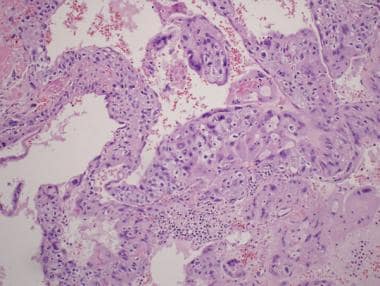
We have cloned the human Na+-dependent multivitamin transporter (SMVT), which transports the water-soluble vitamins pantothenate, biotin, and lipoate, from a placental choriocarcinoma cell line (JAR). The cDNA codes for a protein of 635 amino acids with 12 transmembrane domains and 4 putative sites for N-linked glycosylation. The human SMVT exhibits a high degree of homology (84% identity and 89% similarity) to the rat counterpart. When expressed in HRPE cells, the cDNA-induced transport process is obligatorily dependent on Na+ and accepts pantothenate, biotin, and lipoate as substrates. The relationship between the cDNA-specific uptake rate of pantothenate or biotin and Na+ concentration is sigmoidal with a Na+:vitamin stoichiometry of 2:1. The human SMVT, when expressed in Xenopus laevis oocytes, induces inward currents in the presence of pantothenate, biotin, and lipoate in a Na+-, concentration-, and potential-dependent manner. We also report here on the structural organization and chromosomal localization of the human SMVT gene. The SMVT gene is approximately 14 kilobase pairs in length and consists of 17 exons. The SMVT gene is located on chromosome 2p23 as evidenced by somatic cell hybrid analysis and fluorescence in situ hybridization. (+info)Choriocarcinoma is a rapidly growing and invasive type of gestational trophoblastic disease (GTD), which are abnormal growths that develop in the tissues that are supposed to become the placenta during pregnancy. It occurs when a malignant tumor develops from trophoblast cells, which are normally found in the developing embryo and help to form the placenta.
Choriocarcinoma can occur after any type of pregnancy, including normal pregnancies, molar pregnancies (a rare mass that forms inside the uterus after conception), or ectopic pregnancies (when a fertilized egg implants outside the uterus). It is characterized by the presence of both trophoblastic and cancerous cells, which can produce human chorionic gonadotropin (hCG) hormone.
Choriocarcinoma can spread quickly to other parts of the body, such as the lungs, liver, brain, or vagina, through the bloodstream. It is important to diagnose and treat choriocarcinoma early to prevent serious complications and improve the chances of a successful treatment outcome. Treatment typically involves surgery, chemotherapy, or radiation therapy.
Choriocarcinoma, non-gestational is a rare type of cancer that develops from the abnormal growth of cells that form the placenta. Unlike gestational choriocarcinoma, which arises during pregnancy or after it has ended, non-gestational choriocarcinoma is not related to pregnancy and can occur in both men and women. It typically occurs in the ovaries, testicles, or other organs where trophoblastic cells (cells that normally develop into the placenta) may be found.
Non-gestational choriocarcinoma is an aggressive cancer that can spread quickly to other parts of the body, such as the lungs, liver, and brain. It is usually treated with a combination of chemotherapy, surgery, and radiation therapy. The prognosis for non-gestational choriocarcinoma depends on several factors, including the stage of the cancer at diagnosis, the patient's age and overall health, and the response to treatment.
Uterine neoplasms refer to abnormal growths in the uterus, which can be benign (non-cancerous) or malignant (cancerous). These growths can originate from different types of cells within the uterus, leading to various types of uterine neoplasms. The two main categories of uterine neoplasms are endometrial neoplasms and uterine sarcomas.
Endometrial neoplasms develop from the endometrium, which is the inner lining of the uterus. Most endometrial neoplasms are classified as endometrioid adenocarcinomas, arising from glandular cells in the endometrium. Other types include serous carcinoma, clear cell carcinoma, and mucinous carcinoma.
Uterine sarcomas, on the other hand, are less common and originate from the connective tissue (stroma) or muscle (myometrium) of the uterus. Uterine sarcomas can be further divided into several subtypes, such as leiomyosarcoma, endometrial stromal sarcoma, and undifferentiated uterine sarcoma.
Uterine neoplasms can cause various symptoms, including abnormal vaginal bleeding or discharge, pelvic pain, and difficulty urinating or having bowel movements. The diagnosis typically involves a combination of imaging tests (such as ultrasound, CT, or MRI scans) and tissue biopsies to determine the type and extent of the neoplasm. Treatment options depend on the type, stage, and patient's overall health but may include surgery, radiation therapy, chemotherapy, or hormone therapy.
A hydatidiform mole, also known as a molar pregnancy, is a type of gestational trophoblastic disease (GTD), which is a group of rare disorders that involve abnormal growth of the placental tissue.
In a hydatidiform mole, there is an abnormal fertilization event leading to the growth of a mass of grapelike cysts in the uterus instead of a normal pregnancy. The chromosomes from the sperm and egg do not combine properly, resulting in an extra set of chromosomes, which leads to the development of the mole.
Hydatidiform moles can be complete or partial:
* Complete hydatidiform mole (CHM): This type arises when an egg without a nucleus is fertilized by one or two sperm, leading to the growth of abnormal placental tissue with no embryo. The chromosomes come from the father only, and there are typically 46 chromosomes, all of paternal origin.
* Partial hydatidiform mole (PHM): This type occurs when an egg is fertilized by two sperm or a single sperm that duplicates itself, resulting in an abnormal placenta with some fetal tissue. The chromosomes are of both maternal and paternal origin, and the placental tissue has a mix of normal and abnormal cells.
Hydatidiform moles can cause vaginal bleeding, rapid uterine enlargement, and high levels of human chorionic gonadotropin (hCG) hormone in the blood. They are usually detected during an ultrasound exam and require medical treatment to prevent complications such as gestational trophoblastic neoplasia, a malignant form of GTD that can spread to other organs.
Trophoblastic neoplasms are a group of rare tumors that originate from the trophoblast, which is the outer layer of cells that surrounds a developing embryo and helps to form the placenta during pregnancy. These tumors can be benign or malignant and are characterized by their ability to produce human chorionic gonadotropin (hCG), a hormone that is normally produced during pregnancy.
There are several types of trophoblastic neoplasms, including:
1. Hydatidiform mole: A benign growth that forms in the uterus when a fertilized egg implants but does not develop into a normal embryo. There are two types of hydatidiform moles: complete and partial. Complete moles have no fetal tissue, while partial moles have some fetal tissue.
2. Invasive mole: A malignant form of hydatidiform mole that invades the uterine wall and may spread to other parts of the body.
3. Choriocarcinoma: A rapidly growing and highly invasive malignant tumor that can arise from a hydatidiform mole, a normal pregnancy, or an ectopic pregnancy. It can spread quickly to other parts of the body, such as the lungs, liver, and brain.
4. Placental site trophoblastic tumor (PSTT): A rare type of trophoblastic neoplasm that arises from the cells that attach the placenta to the uterine wall. It is usually slow-growing but can be aggressive in some cases.
5. Epithelioid trophoblastic tumor (ETT): Another rare type of trophoblastic neoplasm that arises from the cells that form the placental villi. It is typically low-grade and has a good prognosis, but it can recur in some cases.
The treatment for trophoblastic neoplasms depends on the type and stage of the tumor. Treatment options may include surgery, chemotherapy, radiation therapy, or a combination of these approaches. Regular monitoring of hCG levels is also important to ensure that the tumor has been completely removed and to detect any recurrence early.
Trophoblasts are specialized cells that make up the outer layer of a blastocyst, which is a hollow ball of cells that forms in the earliest stages of embryonic development. In humans, this process occurs about 5-6 days after fertilization. The blastocyst consists of an inner cell mass (which will eventually become the embryo) and an outer layer of trophoblasts.
Trophoblasts play a crucial role in implantation, which is the process by which the blastocyst attaches to and invades the lining of the uterus. Once implanted, the trophoblasts differentiate into two main layers: the cytotrophoblasts (which are closer to the inner cell mass) and the syncytiotrophoblasts (which form a multinucleated layer that is in direct contact with the maternal tissues).
The cytotrophoblasts proliferate and fuse to form the syncytiotrophoblasts, which have several important functions. They secrete enzymes that help to degrade and remodel the extracellular matrix of the uterine lining, allowing the blastocyst to implant more deeply. They also form a barrier between the maternal and fetal tissues, helping to protect the developing embryo from the mother's immune system.
Additionally, trophoblasts are responsible for the formation of the placenta, which provides nutrients and oxygen to the developing fetus and removes waste products. The syncytiotrophoblasts in particular play a key role in this process by secreting hormones such as human chorionic gonadotropin (hCG), which helps to maintain pregnancy, and by forming blood vessels that allow for the exchange of nutrients and waste between the mother and fetus.
Abnormalities in trophoblast development or function can lead to a variety of pregnancy-related complications, including preeclampsia, intrauterine growth restriction, and gestational trophoblastic diseases such as hydatidiform moles and choriocarcinomas.
An invasive hydatidiform mole (IHM) is a rare and aggressive complication of a gestational trophoblastic disease (GTD), which itself originates from the abnormal proliferation of trophoblastic cells, the tissue that normally develops into the placenta during pregnancy. IHMs are characterized by the invasion of molar villi into the myometrium (the muscular layer of the uterus) and can potentially spread to other organs through the bloodstream, leading to distant metastases.
IHMs usually arise from a complete hydatidiform mole (CHM), which is an abnormal conceptus with no embryonic or fetal development. CHMs are typically diploid and originate from the fertilization of an egg without genetic material (an empty egg or an egg with two sets of paternal chromosomes) by one or two sperm cells. This results in a conceptus with only paternal chromosomes, which leads to uncontrolled proliferation of trophoblastic tissue and the formation of grapelike vesicles filled with fluid (hydatidiform moles).
Invasive hydatidiform moles can cause various symptoms, such as vaginal bleeding, pelvic pain, or the presence of an enlarged uterus. They also pose a risk for developing choriocarcinoma, another type of gestational trophoblastic neoplasia (GTN), which is a malignant tumor that can metastasize and spread to other organs. Proper diagnosis and timely treatment are crucial to prevent severe complications and improve the prognosis for patients with IHMs. Treatment usually involves surgical removal of the mole, followed by chemotherapy to eliminate any residual disease and reduce the risk of GTN development.
Gestational Trophoblastic Disease (GTD) is a group of rare pregnancy-related disorders that involve abnormal growth of cells inside a woman's uterus. These cells are part of the placenta, which provides nutrients to the developing fetus. GTD occurs when some of these cells grow in an uncontrolled way, forming tumors or tumor-like growths.
There are several types of GTD:
1. Hydatidiform Mole (HM): Also known as a molar pregnancy, this is the most common type of GTD. It occurs when an egg that has no genetic information is fertilized by a sperm and then divides into multiple copies. This results in a growth that resembles a cluster of grapes, rather than a developing fetus. There are two types of HMs: complete and partial. A complete HM forms when an empty egg is fertilized by two sperms, resulting in no fetal tissue. A partial HM forms when a normal egg is fertilized by two sperm or an abnormal egg with two sets of genetic material, resulting in some fetal tissue.
2. Invasive Mole: This type of GTD occurs when cells from a molar pregnancy invade the uterine wall and surrounding tissues. It can also spread to other parts of the body, such as the lungs or brain.
3. Choriocarcinoma: This is a rare form of GTD that develops from trophoblastic cells and forms a malignant tumor. It can grow rapidly and spread quickly to other organs.
4. Placental Site Trophoblastic Tumor (PSTT): This is an even rarer type of GTD that forms in the tissue where the placenta attaches to the uterus. PSTTs are usually slow-growing but can sometimes spread to other parts of the body.
5. Epithelioid Trophoblastic Tumor (ETT): This is a very rare type of GTD that forms in the tissue where the placenta attaches to the uterus. ETTs are usually slow-growing and have a good prognosis.
It's important to note that most molar pregnancies do not develop into more serious forms of GTD, but regular follow-up care is necessary to monitor for any signs of progression. Treatment options depend on the type and stage of GTD and may include surgery, chemotherapy, or radiation therapy.
Chorionic Gonadotropin, beta Subunit, Human (β-hCG) is a protein that is produced by the placenta during pregnancy. It is a component of human chorionic gonadotropin (hCG), which is a hormone that is composed of two subunits: alpha and beta. The β-hCG subunit is specific to hCG and is not found in other hormones, making it a useful marker for pregnancy and certain medical conditions.
During early pregnancy, the levels of β-hCG increase rapidly and can be detected in the blood and urine. This has led to the development of pregnancy tests that detect the presence of β-hCG to confirm pregnancy. In addition to its role in pregnancy, β-hCG is also used as a tumor marker for certain types of cancer, such as germ cell tumors and choriocarcinoma.
Elevated levels of β-hCG may indicate the presence of a molar pregnancy, a condition in which a fertilized egg implants in the uterus but does not develop properly. In some cases, a molar pregnancy can become cancerous and require treatment. Therefore, monitoring β-hCG levels during pregnancy is important for detecting any potential complications.
Chorionic Gonadotropin (hCG) is a hormone that is produced during pregnancy. It is produced by the placenta after implantation of the fertilized egg in the uterus. The main function of hCG is to prevent the disintegration of the corpus luteum, which is a temporary endocrine structure that forms in the ovary after ovulation and produces progesterone during early pregnancy. Progesterone is essential for maintaining the lining of the uterus and supporting the pregnancy.
hCG can be detected in the blood or urine as early as 10 days after conception, and its levels continue to rise throughout the first trimester of pregnancy. In addition to its role in maintaining pregnancy, hCG is also used as a clinical marker for pregnancy and to monitor certain medical conditions such as gestational trophoblastic diseases.
The placenta is an organ that develops in the uterus during pregnancy and provides oxygen and nutrients to the growing baby through the umbilical cord. It also removes waste products from the baby's blood. The placenta attaches to the wall of the uterus, and the baby's side of the placenta contains many tiny blood vessels that connect to the baby's circulatory system. This allows for the exchange of oxygen, nutrients, and waste between the mother's and baby's blood. After the baby is born, the placenta is usually expelled from the uterus in a process called afterbirth.
Pregnancy is a physiological state or condition where a fertilized egg (zygote) successfully implants and grows in the uterus of a woman, leading to the development of an embryo and finally a fetus. This process typically spans approximately 40 weeks, divided into three trimesters, and culminates in childbirth. Throughout this period, numerous hormonal and physical changes occur to support the growing offspring, including uterine enlargement, breast development, and various maternal adaptations to ensure the fetus's optimal growth and well-being.
Chorionic villi are finger-like projections of the chorion, which is the outermost extraembryonic membrane in a developing embryo. These structures are composed of both fetal and maternal tissues and play a crucial role in the early stages of pregnancy by providing a site for exchange of nutrients and waste products between the mother and the developing fetus.
Chorionic villi contain fetal blood vessels that are surrounded by stromal cells, trophoblasts, and connective tissue. They are formed during the process of implantation, when the fertilized egg attaches to the uterine wall. The chorionic villi continue to grow and multiply as the placenta develops, eventually forming a highly vascular and specialized organ that supports fetal growth and development throughout pregnancy.
One important function of chorionic villi is to serve as the site for the production of human chorionic gonadotropin (hCG), a hormone that can be detected in the mother's blood and urine during early pregnancy. This hormone plays a critical role in maintaining pregnancy by signaling the corpus luteum to continue producing progesterone, which helps to prevent menstruation and support fetal growth.
Abnormalities in chorionic villi can lead to various pregnancy complications, such as miscarriage, stillbirth, or intrauterine growth restriction. For this reason, chorionic villus sampling (CVS) is a diagnostic procedure that may be performed during early pregnancy to obtain fetal cells for genetic testing and diagnosis of chromosomal abnormalities or other genetic disorders.
Glycoprotein hormones are a group of hormones that share a similar structure and are made up of four subunits: two identical alpha subunits and two distinct beta subunits. The alpha subunit is common to all glycoprotein hormones, including thyroid-stimulating hormone (TSH), follicle-stimulating hormone (FSH), luteinizing hormone (LH), and human chorionic gonadotropin (hCG).
The alpha subunit of glycoprotein hormones is a 92 amino acid polypeptide chain that contains several disulfide bonds, which help to stabilize its structure. It is heavily glycosylated, meaning that it contains many carbohydrate groups attached to the protein backbone. The alpha subunit plays an important role in the biological activity of the hormone by interacting with a specific receptor on the target cell surface.
The alpha subunit contains several regions that are important for its function, including a signal peptide, a variable region, and a conserved region. The signal peptide is a short sequence of amino acids at the N-terminus of the protein that directs it to the endoplasmic reticulum for processing and secretion. The variable region contains several amino acid residues that differ between different glycoprotein hormones, while the conserved region contains amino acids that are identical or very similar in all glycoprotein hormones.
Together with the beta subunit, the alpha subunit forms the functional hormone molecule. The beta subunit determines the specificity of the hormone for its target cells and regulates its biological activity.
'Tumor cells, cultured' refers to the process of removing cancerous cells from a tumor and growing them in controlled laboratory conditions. This is typically done by isolating the tumor cells from a patient's tissue sample, then placing them in a nutrient-rich environment that promotes their growth and multiplication.
The resulting cultured tumor cells can be used for various research purposes, including the study of cancer biology, drug development, and toxicity testing. They provide a valuable tool for researchers to better understand the behavior and characteristics of cancer cells outside of the human body, which can lead to the development of more effective cancer treatments.
It is important to note that cultured tumor cells may not always behave exactly the same way as they do in the human body, so findings from cell culture studies must be validated through further research, such as animal models or clinical trials.
Testicular neoplasms are abnormal growths or tumors in the testicle that can be benign (non-cancerous) or malignant (cancerous). They are a type of genitourinary cancer, which affects the reproductive and urinary systems. Testicular neoplasms can occur in men of any age but are most commonly found in young adults between the ages of 15 and 40.
Testicular neoplasms can be classified into two main categories: germ cell tumors and non-germ cell tumors. Germ cell tumors, which arise from the cells that give rise to sperm, are further divided into seminomas and non-seminomas. Seminomas are typically slow-growing and have a good prognosis, while non-seminomas tend to grow more quickly and can spread to other parts of the body.
Non-germ cell tumors are less common than germ cell tumors and include Leydig cell tumors, Sertoli cell tumors, and lymphomas. These tumors can have a variety of clinical behaviors, ranging from benign to malignant.
Testicular neoplasms often present as a painless mass or swelling in the testicle. Other symptoms may include a feeling of heaviness or discomfort in the scrotum, a dull ache in the lower abdomen or groin, and breast enlargement (gynecomastia).
Diagnosis typically involves a physical examination, imaging studies such as ultrasound or CT scan, and blood tests to detect tumor markers. Treatment options depend on the type and stage of the neoplasm but may include surgery, radiation therapy, chemotherapy, or a combination of these modalities. Regular self-examinations of the testicles are recommended for early detection and improved outcomes.
 Choriocarcinoma
Gestational choriocarcinoma
Trophoblastic neoplasm
Ovary
History of cancer chemotherapy
Gestational trophoblastic disease
Professional Medical Film
Methylestradiol
Normethandrone
CABIN1
Min Chiu Li
Gestational pemphigoid
Methotrexate
H19 (gene)
Placenta
PAK4
Precocious puberty
Chorioangioma
National Cancer Institute
Giant-cell carcinoma of the lung
Trophoblast
ERV3
HOPX
Molar pregnancy
Alkaline phosphatase
Amphetamine
Ovarian cancer
Placental alkaline phosphatase
Germ cell tumor
Ovarian cyst
Choriocarcinoma - Wikipedia
 Testicular Choriocarcinoma: Practice Essentials, Pathophysiology and Etiology, Epidemiology
 Ovary: Choriocarcinoma
Treatment Of Choriocarcinoma - Net Health Book
 Choriocarcinoma (Gestational Trophoblastic Neoplasia) Global Clinical Trials Review, H1, 2016 | ClickPress
Choriocarcinoma | Profiles RNS
 Nuclear β-catenin and Ki-67 expression in choriocarcinoma and its pre-malignant form | Journal of Clinical Pathology
 Characterization and Cloning of MHC Class I Sequences From Choriocarcinoma JEG-3 Cells - Enlighten Theses
View of Primary Pancreatic Choriocarcinoma Presenting as Pancreatitis
Choriocarcinoma - MD Nexus
Extraovarian nongestational choriocarcinoma in a postmenopausal woman | International Journal of Gynecologic Cancer
 choriocarcinoma | Taber's Medical Dictionary
 Clinical characteristics and prognosis of 272 postterm choriocarcinoma patients at Peking Union Medical College Hospital: a...
PODCASTS - Choriocarcinoma Gestational Trophoblastic Disease
 Choriocarcinoma: Video, Anatomy, Definition & Function | Osmosis
 Uterine corpus choriocarcinoma (Concept Id: C1336904)
- MedGen - NCBI
Uterine corpus choriocarcinoma (Concept Id: C1336904)
- MedGen - NCBI
 Choriocarcinoma -
TC-Cancer.com - Testicular Cancer Information & Support Forum
Choriocarcinoma -
TC-Cancer.com - Testicular Cancer Information & Support Forum
 Ovarian cancer in young females: Causes and treatment
 Coriocarcinoma gástrico primario: reporte de caso<...
Evidence for a novel HLA antigen found on human extravillous trophoblast and a choriocarcinoma cell line. - Nuffield Department...
Neurologic Disease and Pregnancy: Overview, General Considerations, New-Onset Neurologic Complications
 Germ Cell Tumors (for Parents) - Children's Health System - Alabama (iFrame)
Germ Cell Tumors (for Parents) - CareSource
EP1144 Stage IV choriocarcinoma: case report and review of literature | International Journal of Gynecologic Cancer
 Choriocarcinoma with Pulmonary and Spinal Metastases : A Case Report | Syafar | Andalas Obstetrics And Gynecology Journal
 Testicular cancer: MedlinePlus Medical Encyclopedia
Isthmin A couple of can be decreased inside preeclampsia and very depicted in choriocarcinoma. | Cxcr Signal
Cerebral Amyloid Angiopathy: Overview, Diagnostic Guidelines, Etiology
 Diagnostic Challenges in Fine-Needle Aspiration Cytology of Mediastinal Tumors and Lesions | Archives of Pathology & Laboratory...
 Choriocarcinoma in Ongoing Pregnancy Presenting with Intracranial Metastasis. | J Clin Diagn Res;10(11): QD01-QD03, 2016 Nov. ...
Tumor13
- Elements of choriocarcinoma in a mixed testicular tumor have no prognostic importance. (wikipedia.org)
- Clinicopathologic analysis of choriocarcinoma as a pure or predominant component of germ cell tumor of the testis. (medscape.com)
- This tumor consisted of typical morphologic and immunophenotypic features of ETT and choriocarcinoma. (hindawi.com)
- Unlike other such cancers, choriocarcinoma metastasizes hematogenously, with the testicular primary tumor often small or even "burned-out. (medscape.com)
- In a series of 125 patients with a history or clinical evidence of cryptorchidism and testis tumor, 3 (2%) were pure choriocarcinoma, which is similar to the overall incidence of choriocarcinoma among GCTs. (medscape.com)
- We want to emphasize that if a neonate or infant has anemia and/or hepatomegaly, it is necessary to be aware of the possibility of infantile choriocarcinoma with the presence of an occult tumor in the mother and to examine the mother's and infant's hCG levels immediately. (indexindex.com)
- Infantile choriocarcinoma of the liver: This is a very rare tumor that starts in the placenta and spreads to the fetus . (vicc.org)
- 9. Choriocarcinoma presented as a vaginal tumor. (nih.gov)
- Embryonic germ cell tumors include teratoma , and extraembryonic germ cell tumors include Choriocarcinoma and Yolk sac tumor . (wikidoc.org)
- These tumors appear to arise from a pluripotent germ cell capable of differentiating into embryonic structures (teratoma and embryonal carcinoma), placental structures (yolk-sac tumor and choriocarcinoma) or seminoma (the most primitive germ cell tumor). (health.am)
- Specifically, he was diagnosed with choriocarcinoma, a rare tumor that forms in the testis. (iuhealth.org)
- Yes, there are different subtypes of GTN: invasive mole, choriocarcinoma, placental site trophoblastic tumor (PSTT) and epithelioid trophoblastic tumor (ETT). (isuog.org)
- The last two subtypes -- endodermal sinus tumor, also known as "yolk sac tumor," and Choriocarcinoma -- grow and spread rapidly, but they are very rare. (webmd.com)
Hydatidiform mole4
- High magnification Very high magnification Since gestational choriocarcinoma (which arises from a hydatidiform mole) contains paternal DNA (and thus paternal antigens), it is exquisitely sensitive to chemotherapy. (wikipedia.org)
- About one half of all women with a choriocarcinoma had a hydatidiform mole, or molar pregnancy. (medlineplus.gov)
- Serum human chorionic gonadotropin (hCG) was measured by a radioreceptorassay (RRA) and radioimmunoassay (RIA) and serum hCG-β and hCG-α by RIA in 10 patients with intact mole, 3 patients with choriocarcinoma, and 4 patients with hydatidiform mole during treatment. (lww.com)
- 2. Intraplacental choriocarcinoma arising in a second trimester placenta with partial hydatidiform mole. (nih.gov)
Pure choriocarcinoma7
- Although trophoblastic components are common components of mixed germ cell tumors, pure choriocarcinoma of the adult testis is rare. (wikipedia.org)
- Pure choriocarcinoma of the testis represents the most aggressive pathologic variant of germ cell tumors in adults, characteristically with early hematogenous and lymphatic metastatic spread. (wikipedia.org)
- Ramon y Cajal S, Pinango L, Barat A. Metastatic pure choriocarcinoma of the testis in an elderly man. (medscape.com)
- Puri S, Sood S, Mohindroo S, Kaushal V. Cytomorphology of lung metastasis of pure choriocarcinoma of testis in a 58-year-old male. (medscape.com)
- Pure choriocarcinoma of the testis is the exception to most of the rules established for testicular seminoma and all other forms of NSGCTs. (medscape.com)
- In a literature review of 10,000 cases of germinal testicular cell tumors, Ramon y Cajal et al found 54 (0.5%) cases of pure choriocarcinoma. (medscape.com)
- the oldest patient with a pure choriocarcinoma was 63 years old. (medscape.com)
Ovarian choriocarcinoma1
- I was diagnosed last February with ovarian choriocarcinoma after a miscarriage. (cancer.org)
Metastatic choriocarcinoma4
- Lepidini G, Biancari F, D'Andrea V. Severe thrombosis after chemotherapy for metastatic choriocarcinoma of the testis maintaining complete remission for a long period. (medscape.com)
- Metastases to the lung and brain usually occur in the mother, but metastatic choriocarcinoma in the fetus or neonates does occur. (medscape.com)
- It is introduced by two distinct terms in the literatures: breast cancer with choriocarcinomatous features and metastatic choriocarcinoma to the breast. (ac.ir)
- 4. Metastatic choriocarcinoma in a postmenopausal woman. (nih.gov)
Chemotherapy5
- The cure rates, even for metastatic gestational choriocarcinoma, is more than 90% when using chemotherapy for invasive mole and choriocarcinoma. (wikipedia.org)
- Choriocarcinoma arising in the testicle is rare, malignant and highly resistant to chemotherapy. (wikipedia.org)
- NaUCC-2, a choriocarcinoma cell line, was derived from a patient who had a very poor clinical response to combination chemotherapy. (nih.gov)
- These findings also suggest that the most commonly used combination chemotherapy for choriocarcinoma, dactinomycin and MTX, may not always be the best method. (nih.gov)
- hCG-α was higher than hCG by RRA and RIA and hCG-β in molar pregnancies, in the uterine venous blood draining a uterine choriocarcinoma, and during chemotherapy of choriocarcinoma. (lww.com)
Molar3
- High clinical suspicion should be maintained for choriocarcinoma in women with hemoptysis and molar pregnancy, current, or recent pregnancy, or irregular vaginal bleeding. (asianhhm.com)
- When my molar pregnancy disease progressed to aggressive malignant cancer choriocarcinoma, my anxiety was at its highest. (choriocarcinomamolarpregnancyawareness.com)
- 14. Cerebral venous sinus thrombosis presenting as cerebral metastasis in a patient with choriocarcinoma following a non-molar gestation. (nih.gov)
Malignant3
- Choriocarcinoma is a malignant, trophoblastic cancer, usually of the placenta. (wikipedia.org)
- Choriocarcinoma is a highly malignant gestational trophoblast neoplasia. (greek.doctor)
- A highly malignant CHORIOCARCINOMA derived from the non-placental origin such as the totipotent cells in the TESTIS, the OVARY, and the PINEAL GLAND. (bvsalud.org)
Testis2
- Requena L, Sanchez M, Aguilar A. Choriocarcinoma of the testis metastatic to the skin. (medscape.com)
- Choriocarcinoma and embryonal cell carcinoma of the testis. (healthdirect.gov.au)
Site trophoblastic1
- Gestational trophoblastic neoplasms (GTN) are a group of neoplasms from fetal trophoblastic cells including choriocarcinoma (CC), epithelioid trophoblastic tumors (ETT), and placental site trophoblastic tumors (PSTT) [ 1 ]. (hindawi.com)
Gestational trophob2
- Choriocarcinoma is a type of gestational trophoblastic disease . (medlineplus.gov)
- See the PDQ summary on Gestational Trophoblastic Disease Treatment for more information on the treatment of choriocarcinoma for the mother of the child. (vicc.org)
Pregnancy8
- Choriocarcinoma is a rare cancer that occurs as an abnormal pregnancy. (medlineplus.gov)
- Choriocarcinomas may also occur after an early pregnancy that does not continue (miscarriage). (medlineplus.gov)
- Cite this: Incidental Placental Choriocarcinoma in a Term Pregnancy: A Case Report - Medscape - Oct 16, 2008. (medscape.com)
- Choriocarcinoma is a rare, aggressive neoplastic type of trophoblastic disease, which may occur after miscarriage, abortion, ectopic pregnancy or term pregnancy. (asianhhm.com)
- 5. Incidental finding of placental choriocarcinoma after an uncomplicated term pregnancy: a case report with review of the literature. (nih.gov)
- 17. Intraplacental choriocarcinoma associated with pregnancy continuum: a diagnostic dilemma. (nih.gov)
- 19. Ectopic choriocarcinoma masquerading as a persisting pregnancy of unknown location: case report and review of the literature. (nih.gov)
- Choriocarcinoma and the very rare PSTT and ETT can follow any type of pregnancy. (isuog.org)
Infantile1
- 15. Intraplacental choriocarcinoma in a term placenta with both maternal and infantile metastases: a case report and review of the literature. (nih.gov)
Metastases5
- Maternal choriocarcinoma is usually diagnosed in symptomatic patients with metastases. (medscape.com)
- Of all forms of gestational choriocarcinoma, placental choriocarcinoma is the most rare and is usually diagnosed in symptomatic patients with metastases. (medscape.com)
- We present a case of gestational choriocarcinoma that developed in a pregnant woman with metastases to her baby. (indexindex.com)
- In 2 of 3 choriocarcinoma patients who eventually developed cerebral metastases, hCG-α increased while hCG and hCG-β were declining or negative. (lww.com)
- Hepatic metastases due to choriocarcinoma. (nih.gov)
Maternal1
- 10. Maternal and neonatal death from advanced choriocarcinoma due to a delay in diagnosis: a case report. (nih.gov)
Prognosis3
- Testicular choriocarcinoma has the worst prognosis of all germ-cell cancers. (wikipedia.org)
- In most reports, choriocarcinoma carries a dismal prognosis due to its early hematogenous spread. (medscape.com)
- The prognosis of choriocarcinoma has improved since the introduction of chemotherapeutic drugs. (greek.doctor)
Intraplacental6
- Her placenta revealed intraplacental choriocarcinoma. (medscape.com)
- 1. [Intraplacental choriocarcinoma]. (nih.gov)
- 6. Intraplacental choriocarcinoma: a case report. (nih.gov)
- 8. [Incidental intraplacental gestational choriocarcinoma on a full-term placenta]. (nih.gov)
- 11. Biochemical analysis of intraplacental choriocarcinoma and fetomaternal transfusion. (nih.gov)
- 16. A case of intraplacental choriocarcinoma associated with placental hemangioma. (nih.gov)
Pulmonary2
- Tatokoro M, Kawakami S, Sakura M, Kobayashi T, Kihara K, Akamatsu H. Successful management of life-threatening choriocarcinoma syndrome with rupture of pulmonary metastatic foci causing hemorrhagic shock. (medscape.com)
- A choriocarcinoma that is located_in the pulmonary artery. (jax.org)
Diagnosis2
- Early diagnosis of choriocarcinoma can improve the outcome. (medlineplus.gov)
- Due to the potential fatal outcome of placental choriocarcinoma, careful evaluation of both mother and infant after the diagnosis is made is important. (medscape.com)
Cells3
- Since choriocarcinomas include syncytiotrophoblasts (beta-HCG producing cells), they cause elevated blood levels of beta-human chorionic gonadotropin. (wikipedia.org)
- HCG MAb detects cells and tumors of trophoblastic origin such as choriocarcinoma. (neobiotechnologies.com)
- c-Rel Promotes Invasion of Choriocarcinoma Cells via PI3K/AKT Signaling. (genscript.com)
Invasive mole1
- Treatment of invasive mole is the same as for choriocarcinoma. (greek.doctor)
Testicular Cancer2
- Zeitjian VS, Arslan W, Borja-Alvarez A, Amar S. Choriocarcinoma Syndrome: A Potentially Fatal Complication of Testicular Cancer. (medscape.com)
- You may already know that Lance Armstrong is among the best known cancer survivors for having beaten advanced testicular cancer that was 60% choriocarcinoma -- before going on to win seven Tours de France. (testicularcancersociety.org)
Teratocarcinoma1
- Testicular Carcinoma Embryonal cell, choriocarcinoma, and teratocarcinoma. (cancermonthly.com)
Findings2
- Although additional studies are necessary to determine the mechanism responsible for this effect, these findings suggest that a mechanism other than drug uptake or DHFR activity must play a role in the drug resistance for choriocarcinoma. (nih.gov)
- In this case report, the history, physical examination, laboratory findings, imaging studies, and pathological findings of breast choriocarcinoma in a 41-year-old woman are described and previous literatures about choriocarcinoma in the breast are reviewed. (ac.ir)
Predominant1
- In a histologic review of 1010 orchiectomies from 1999 to 2011 from a single Mexican oncology institution, 0.6% were pure choriocarcinomas and 0.9% were mixed germ cell tumors with a predominant choriocarcinoma component. (medscape.com)
Occurs2
- Choriocarcinoma is a fast-growing cancer that occurs in a woman's uterus (womb). (medlineplus.gov)
- Gestational choriocarcinoma occurs in 1 in 40,000 pregnancies. (medscape.com)
Seminoma1
- Testicular choriocarcinoma is one of the histologic types of nonseminomatous germ cell tumors (NSGCTs), which along with testicular seminoma constitute the two major histologic groups of testicular cancers. (medscape.com)
Rare2
- Of all forms of gestational choriocarcinoma, placental choriocarcinoma is the most rare. (medscape.com)
- Choriocarcinoma is an extremely rare pathology among breast malignancies. (ac.ir)
Incidental1
- The incidental finding of a choriocarcinoma confined to the placenta with no evidence of dissemination to the mother, or infant is the least common scenario. (medscape.com)
Incidence1
- The incidence of placental choriocarcinoma may actually be higher than expected since it is not routine practice to send placentas for pathological evaluation after a normal spontaneous delivery. (medscape.com)
Syndrome1
- Rejlekova K, Cursano MC, De Giorgi U, Mego M. Severe Complications in Testicular Germ Cell Tumors: The Choriocarcinoma Syndrome. (medscape.com)
Ovaries1
- Choriocarcinomas can also occur in the ovaries and other organs. (wikipedia.org)
Pathology1
- The patient underwent a surgical resection of the cerebellar lesion and the anatomic pathology results were indicative of Choriocarcinoma. (asianhhm.com)
Treatment2
- A choriocarcinoma may come back within a few months to 3 years after treatment. (medlineplus.gov)
- Fertility-Sparing Treatment in Gestational Choriocarcinoma: Evaluating Oncological and Obstetrical Outcomes. (medscimonit.com)
Case Report1
- 7. Choriocarcinoma presenting as an intussusception--a case report. (nih.gov)
Cell line2
- Disruption of thyroid hormone sulfotransferase activity by brominated flame retardant chemicals in the human choriocarcinoma placenta cell line, BeWo. (nih.gov)
- In this study, we investigated the effects of two polybrominated diphenyl ethers (PBDEs), two hydroxylated PBDEs, and 2,4,6-tribromophenol (2,4,6-TBP) on TH SULT activity in a choriocarcinoma placenta cell line (BeWo). (nih.gov)
Organs1
- Chen X, Xu L, Chen X, Teng X, Zheng S. Testicular choriocarcinoma metastatic to skin and multiple organs. (medscape.com)