Clavicle
Acromioclavicular Joint
Sternoclavicular Joint
Cleidocranial Dysplasia
Fracture Fixation, Internal
Bone Plates
Pseudarthrosis
Fractures, Malunited
Fracture Healing
Manubrium
Scapula
Dislocations
Fractures, Ununited
Shoulder
Bone Wires
Gymnastics
Encyclopedias as Topic
Results of the Bosworth method for unstable fractures of the distal clavicle. (1/248)
Eleven consecutive Neer's type II unstable fractures of the distal third of the clavicle were treated by open reduction and internal fixation, using a temporary Bosworth-type screw. In all cases, fracture healing occurred within 10 weeks. Shoulder function was restored to the pre-injury level. A Bosworth-type screw fixation is a relatively easy and safe technique of open reduction and internal fixation of type II fractures of the distal third of the clavicle. (+info)Osteochondroma of the first rib presenting as a prominent clavicle. A report of 2 cases. (2/248)
We describe and discuss two patients with osteochondromas of the first rib which presented as prominence of the medial end of the clavicle. (+info)The prevalence and CT appearance of the levator claviculae muscle: a normal variant not to be mistaken for an abnormality. (3/248)
BACKGROUND AND PURPOSE: The levator claviculae muscle is an infrequently recognized variant in humans, occurring in 2% to 3% of the population, and has rarely been reported in the radiologic or anatomic literature. The importance of this muscle to radiologists is in distinguishing it from an abnormality; most commonly, cervical adenopathy. After discovering this muscle on the CT scans of two patients during routine clinical examinations, we conducted a study to determine the prevalence and appearance of the muscle on CT studies. METHODS: We evaluated 300 CT scans that adequately depicted the expected location of the muscle. The most superior level in which the muscle could be identified and the apparent location of insertion on the clavicle were recorded for all subjects in whom the muscle was detected. RESULTS: Seven levator claviculae muscles were identified in six subjects (2%). It was bilateral in one, on the left in four, and on the right in one. It was identified up to the level of the transverse process of C3 in all cases. The insertion was the middle third of the clavicle for two muscles and the lateral third of the clavicle for the remaining five muscles. CONCLUSION: Because the levator claviculae muscle will most likely be encountered during a radiologist's career, it is important to recognize this muscle as a variant and not as an abnormality. (+info)Early prenatal ultrasound diagnosis of cleidocranial dysplasia. (4/248)
A woman was referred in the first trimester of her third pregnancy because of a family history of cleidocranial dysplasia. An ultrasound examination at 14 weeks 4 days revealed a fetus with appropriate biometric measurements. However, the clavicles were noted to be hypoplastic and the cranium appeared less well ossified than expected for gestational age, suggesting the diagnosis of cleidocranial dysplasia. On subsequent examination at 21 weeks, the findings were essentially unchanged. Induced vaginal delivery owing to decreased amniotic fluid volume occurred at 37 weeks, and a female weighing 3200 g was delivered. The infant had clinical and X-ray signs of cleidocranial dysplasia. (+info)Surgical treatment of comminuted fractures of the distal clavicle using Wolter clavicular plates. (5/248)
Surgical reduction and fixation using Wolter clavicular plates was performed in 16 patients with distal clavicle fractures. Good bony union was obtained in all cases. The patients had neither postoperative pain nor muscle weakness. Compared with tension band wiring, the Wolter clavicular plates were better able to maintain the anatomic repositioning of comminuted distal clavicle fractures. However this method takes longer and requires a second operation for plate removal. Wolter clavicular plate placement is the treatment of choice for unstable distal clavicle fractures with small comminuted fragments. (+info)Genomic differentiation of Neanderthals and anatomically modern man allows a fossil-DNA-based classification of morphologically indistinguishable hominid bones. (6/248)
Southern blot hybridizations of genomic DNA were introduced as a relatively simple fossil-DNA-based approach to classify remains of Neanderthals. When hybridized with genomic DNA of either human or Neanderthal origin, DNA extracted from two Neanderthal finds-the Os parietale, from Warendorf-Neuwarendorf, Germany, and a clavicula, from Krapina, Croatia-was shown to yield hybridization signals that differ by at least a factor of two compared to the signals obtained with the use of fossil DNA of an early Homo sapiens from the Vogelherd cave (Stetten I), Germany. When labeled chimpanzee DNA was used as a probe, Neanderthal and human DNA, however, revealed hybridization signals of similar intensity. Thus, the genome of Neanderthals is expected to differ significantly from the genome of anatomically modern man, because of the contrasting composition of repetitive DNA. These data support the hypothesis that Neanderthals were not ancestors of anatomically modern man. (+info)Arthroscopic Mumford procedure variation of technique. (7/248)
Fifty-seven patients had arthroscopic Mumford procedures for acromioclavicular pain non-responsive to conservative treatment. Thirty-nine of these patients had concomitant rotator cuff repairs. All had significant improvement of their distal clavicular pain. Neither the amount nor the completeness of distal clavicle resection affected the results. Arthroscopic distal clavicle resection is a safe and effective method of alleviating acromioclavicular pain. (+info)Claviculectomy for subclavian venous repair: long-term functional results. (8/248)
OBJECTIVES: The purpose of this study was to determine the long-term functional results after medial claviculectomy and venous patch angioplasty or bypass grafting using internal jugular vein after incomplete thrombolysis of effort thrombosis of the subclavian vein. METHODS: The records of 11 patients with effort thrombosis who were treated over the past 9 years were reviewed. Patients have been followed up between 3 and 9 years at 6-month intervals with duplex imaging and contrast venography when indicated and have had an orthopedic evaluation of their shoulder function. RESULTS: All reconstructed veins are patent, and only one patient complains of any arm swelling after prolonged usage. This patient is one of three with postphlebitic changes at the site of repair and has similar findings in her basilic vein. All patients have returned to their prethrombosis vocation without limitation. Four of the 11 patients have jobs requiring heavy physical labor. No patient describes any limitations of shoulder function, but one man who works as a diesel mechanic complains of shoulder aching with overuse with repetitive pulling. Three patients describe upper extremity paresthesias when lying on the operated side. Two patients (one man and one woman) are bothered by the large scar and indentation at the site of the incision. Every patient considers the overall result completely successful from a functional standpoint. CONCLUSIONS: Early subclavian venous repair performed through a medial claviculectomy is a durable operation with excellent long-term functional results. Half of the patients noted minor but significant symptoms, but all are uniformly able to return to normal function. (+info)The clavicle, also known as the collarbone, is a long, slender bone that lies horizontally between the breastbone (sternum) and the shoulder blade (scapula). It is part of the shoulder girdle and plays a crucial role in supporting the upper limb. The clavicle has two ends: the medial end, which articulates with the sternum, and the lateral end, which articulates with the acromion process of the scapula. It is a common site of fracture due to its superficial location and susceptibility to direct trauma.
The acromioclavicular (AC) joint is the joint located between the acromion process of the scapula (shoulder blade) and the clavicle (collarbone). It allows for a small amount of movement between these two bones and participates in shoulder motion. Injuries to this joint, such as AC joint separations or sprains, are common and can occur due to falls, direct blows, or repetitive motions that cause the ligaments that support the AC joint to become stretched or torn.
The sternoclavicular joint is the joint where the clavicle (collarbone) meets the sternum (breastbone). It is the only joint that connects the upper limb to the trunk of the body. This joint allows for movement in multiple directions, including elevation and depression of the shoulder, as well as some degree of protraction and retraction. The sternoclavicular joint is supported by several ligaments, which provide stability and strength to the joint.
A bone fracture is a medical condition in which there is a partial or complete break in the continuity of a bone due to external or internal forces. Fractures can occur in any bone in the body and can vary in severity from a small crack to a shattered bone. The symptoms of a bone fracture typically include pain, swelling, bruising, deformity, and difficulty moving the affected limb. Treatment for a bone fracture may involve immobilization with a cast or splint, surgery to realign and stabilize the bone, or medication to manage pain and prevent infection. The specific treatment approach will depend on the location, type, and severity of the fracture.
Osteitis is a medical term that refers to the inflammation of bone tissue. It can occur as a result of various conditions, such as infection (osteomyelitis), trauma, or autoimmune disorders. The symptoms of osteitis may include pain, swelling, warmth, and redness in the affected area, as well as fever and general malaise. Treatment typically involves addressing the underlying cause of the inflammation, which may involve antibiotics for infection or anti-inflammatory medications for other causes. In some cases, surgery may be necessary to remove infected or damaged bone tissue.
Cleidocranial dysplasia is a genetic skeletal disorder that affects the development of bones and teeth. The condition is characterized by the underdevelopment or absence of the collarbones (clavicles), which can result in shoulder joints that are abnormally close together. This may allow the person to bring their shoulders around to touch or even overlap in front of their body.
People with cleidocranial dysplasia also often have a delayed closure of the fontanels (soft spots) on the skull, as well as an abnormal shape and size of the head. The facial bones may be underdeveloped, leading to a sunken appearance in the middle of the face and a prominent forehead. Dental abnormalities are also common, such as missing or delayed eruption of teeth, extra teeth, and misaligned teeth.
Cleidocranial dysplasia is caused by mutations in the CBFA1/RUNX2 gene and is inherited in an autosomal dominant manner, meaning that a child has a 50% chance of inheriting the condition if one of their parents is affected. However, many cases result from new mutations in the gene and occur in people with no family history of the disorder. Treatment typically involves surgical procedures to correct skeletal abnormalities and dental issues, as well as orthodontic treatment to align teeth.
Fracture fixation, internal, is a surgical procedure where a fractured bone is fixed using metal devices such as plates, screws, or rods that are implanted inside the body. This technique helps to maintain the alignment and stability of the broken bone while it heals. The implants may be temporarily or permanently left inside the body, depending on the nature and severity of the fracture. Internal fixation allows for early mobilization and rehabilitation, which can result in a faster recovery and improved functional outcome.
Bone plates are medical devices used in orthopedic surgery to stabilize and hold together fractured or broken bones during the healing process. They are typically made of surgical-grade stainless steel, titanium, or other biocompatible materials. The plate is shaped to fit the contour of the bone and is held in place with screws that are inserted through the plate and into the bone on either side of the fracture. This provides stability and alignment to the broken bones, allowing them to heal properly. Bone plates can be used to treat a variety of fractures, including those that are complex or unstable. After healing is complete, the bone plate may be left in place or removed, depending on the individual's needs and the surgeon's recommendation.
A comminuted fracture is a type of bone break where the bone is shattered into three or more pieces. This type of fracture typically occurs after high-energy trauma, such as a car accident or a fall from a great height. Commminuted fractures can also occur in bones that are weakened by conditions like osteoporosis or cancer. Because of the severity and complexity of comminuted fractures, they often require extensive treatment, which may include surgery to realign and stabilize the bone fragments using metal screws, plates, or rods.
Pseudarthrosis is a medical term that refers to a false joint or a nonunion of bones, meaning that the broken bone ends do not heal properly and continue to move at the fracture site. This condition can cause pain, instability, and deformity in the affected limb. It may require additional treatment such as surgery to promote bone healing and stabilization.
Malunited fractures refer to a type of fracture where the bones do not heal in their proper alignment or position. This can occur due to various reasons such as inadequate reduction of the fracture fragments during initial treatment, improper casting or immobilization, or failure of the patient to follow proper immobilization instructions. Malunited fractures can result in deformity, limited range of motion, and decreased functionality of the affected limb. Additional treatments such as surgery may be required to correct the malunion and restore normal function.
Fracture healing is the natural process by which a broken bone repairs itself. When a fracture occurs, the body responds by initiating a series of biological and cellular events aimed at restoring the structural integrity of the bone. This process involves the formation of a hematoma (a collection of blood) around the fracture site, followed by the activation of inflammatory cells that help to clean up debris and prepare the area for repair.
Over time, specialized cells called osteoblasts begin to lay down new bone matrix, or osteoid, along the edges of the broken bone ends. This osteoid eventually hardens into new bone tissue, forming a bridge between the fracture fragments. As this process continues, the callus (a mass of newly formed bone and connective tissue) gradually becomes stronger and more compact, eventually remodeling itself into a solid, unbroken bone.
The entire process of fracture healing can take several weeks to several months, depending on factors such as the severity of the injury, the patient's age and overall health, and the location of the fracture. In some cases, medical intervention may be necessary to help promote healing or ensure proper alignment of the bone fragments. This may include the use of casts, braces, or surgical implants such as plates, screws, or rods.
The manubrium is the upper and expanded part of the sternum (breastbone). It has a shape similar to a spoon or a shield, and it articulates with the clavicles (collarbones) and the first pair of ribs. The manubrium plays an essential role in protecting underlying organs such as the heart and major blood vessels.
The scapula, also known as the shoulder blade, is a flat, triangular bone located in the upper back region of the human body. It serves as the site of attachment for various muscles that are involved in movements of the shoulder joint and arm. The scapula has several important features:
1. Three borders (anterior, lateral, and medial)
2. Three angles (superior, inferior, and lateral)
3. Spine of the scapula - a long, horizontal ridge that divides the scapula into two parts: supraspinous fossa (above the spine) and infraspinous fossa (below the spine)
4. Glenoid cavity - a shallow, concave surface on the lateral border that articulates with the humerus to form the shoulder joint
5. Acromion process - a bony projection at the top of the scapula that forms part of the shoulder joint and serves as an attachment point for muscles and ligaments
6. Coracoid process - a hook-like bony projection extending from the anterior border, which provides attachment for muscles and ligaments
Understanding the anatomy and function of the scapula is essential in diagnosing and treating various shoulder and upper back conditions.
A closed fracture, also known as a simple fracture, is a type of bone break where the skin remains intact and there is no open wound. The bone may be broken in such a way that it does not pierce the skin, but still requires medical attention for proper diagnosis, treatment, and healing. Closed fractures can range from hairline cracks to complete breaks and can occur due to various reasons, including trauma, overuse, or weakened bones. It is important to seek immediate medical care if a closed fracture is suspected, as improper healing can lead to long-term complications such as decreased mobility, chronic pain, or deformity.
A dislocation is a condition in which a bone slips out of its normal position in a joint. This can happen as a result of trauma or injury, such as a fall or direct blow to the body. Dislocations can cause pain, swelling, and limited mobility in the affected area. In some cases, a dislocation may also damage surrounding tissues, such as ligaments, tendons, and nerves.
Dislocations are typically treated by reducing the dislocation, which means putting the bone back into its normal position. This is usually done with the help of medication to relieve pain and relaxation techniques to help the person stay still during the reduction. In some cases, surgery may be necessary to repair damaged tissues or if the dislocation cannot be reduced through other methods. After the dislocation has been reduced, the joint may be immobilized with a splint or sling to allow it to heal properly.
It is important to seek medical attention promptly if you suspect that you have a dislocation. If left untreated, a dislocation can lead to further complications, such as joint instability and chronic pain.
Ununited fracture is a medical term used to describe a fractured bone that has failed to heal properly. This condition is also known as a nonunion fracture. In a normal healing process, the broken ends of the bone will grow together, or "unite," over time as new bone tissue forms. However, in some cases, the bones may not reconnect due to various reasons such as infection, poor blood supply, excessive motion at the fracture site, or inadequate stabilization of the fracture.
Ununited fractures can cause significant pain, swelling, and deformity in the affected area. They may also lead to a decreased range of motion, weakness, and instability in the joint near the fracture. Treatment for ununited fractures typically involves surgical intervention to promote bone healing, such as bone grafting or internal fixation with screws or plates. In some cases, electrical stimulation or ultrasound therapy may also be used to help promote bone growth and healing.
In anatomical terms, the shoulder refers to the complex joint of the human body that connects the upper limb to the trunk. It is formed by the union of three bones: the clavicle (collarbone), scapula (shoulder blade), and humerus (upper arm bone). The shoulder joint is a ball-and-socket type of synovial joint, allowing for a wide range of movements such as flexion, extension, abduction, adduction, internal rotation, and external rotation.
The shoulder complex includes not only the glenohumeral joint but also other structures that contribute to its movement and stability, including:
1. The acromioclavicular (AC) joint: where the clavicle meets the acromion process of the scapula.
2. The coracoclavicular (CC) ligament: connects the coracoid process of the scapula to the clavicle, providing additional stability to the AC joint.
3. The rotator cuff: a group of four muscles (supraspinatus, infraspinatus, teres minor, and subscapularis) that surround and reinforce the shoulder joint, contributing to its stability and range of motion.
4. The biceps tendon: originates from the supraglenoid tubercle of the scapula and passes through the shoulder joint, helping with flexion, supination, and stability.
5. Various ligaments and capsular structures that provide additional support and limit excessive movement in the shoulder joint.
The shoulder is a remarkable joint due to its wide range of motion, but this also makes it susceptible to injuries and disorders such as dislocations, subluxations, sprains, strains, tendinitis, bursitis, and degenerative conditions like osteoarthritis. Proper care, exercise, and maintenance are essential for maintaining shoulder health and function throughout one's life.
I'm not aware of a medical term called "bone wires." The term "wiring" is used in orthopedic surgery to describe the use of metal wire to hold bones or fractures in place during healing. However, I couldn't find any specific medical definition or term related to "bone wires." It may be a colloquialism, a term used in a specific context, or a term from science fiction. If you could provide more context about where you encountered this term, I might be able to give a more accurate answer.
I'm sorry for any confusion, but "Gymnastics" is not a medical term. It is an athletic discipline consisting of physical exercises requiring balance, strength, flexibility, agility, coordination, and endurance. If you have any questions related to medical terminology or health concerns, I would be happy to try to help answer those for you!
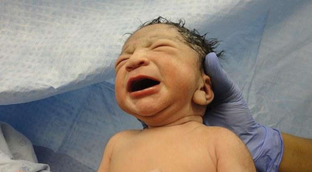
Birth injuries refer to damages or injuries that a baby suffers during the birthing process. These injuries can result from various factors, such as mechanical forces during delivery, medical negligence, or complications during pregnancy or labor. Some common examples of birth injuries include:
1. Brachial plexus injuries: Damage to the nerves that control movement and feeling in the arms and hands, often caused by excessive pulling or stretching during delivery.
2. Cephalohematoma: A collection of blood between the skull and the periosteum (the membrane covering the bone), usually caused by trauma during delivery.
3. Caput succedaneum: Swelling of the soft tissues of the baby's scalp, often resulting from pressure on the head during labor and delivery.
4. Fractures: Broken bones, such as a clavicle or skull fracture, can occur due to mechanical forces during delivery.
5. Intracranial hemorrhage: Bleeding in or around the brain, which can result from trauma during delivery or complications like high blood pressure in the mother.
6. Perinatal asphyxia: A lack of oxygen supply to the baby before, during, or immediately after birth, which can lead to brain damage and other health issues.
7. Subconjunctival hemorrhage: Bleeding under the conjunctiva (the clear membrane covering the eye), often caused by pressure on the head during delivery.
8. Spinal cord injuries: Damage to the spinal cord, which can result in paralysis or other neurological issues, may occur due to excessive force during delivery or medical negligence.
It's important to note that some birth injuries are unavoidable and may not be a result of medical malpractice. However, if a healthcare provider fails to provide the standard of care expected during pregnancy, labor, or delivery, they may be held liable for any resulting injuries.
An encyclopedia is a comprehensive reference work containing articles on various topics, usually arranged in alphabetical order. In the context of medicine, a medical encyclopedia is a collection of articles that provide information about a wide range of medical topics, including diseases and conditions, treatments, tests, procedures, and anatomy and physiology. Medical encyclopedias may be published in print or electronic formats and are often used as a starting point for researching medical topics. They can provide reliable and accurate information on medical subjects, making them useful resources for healthcare professionals, students, and patients alike. Some well-known examples of medical encyclopedias include the Merck Manual and the Stedman's Medical Dictionary.
Fracture fixation is a surgical procedure in orthopedic trauma surgery where a fractured bone is stabilized using various devices and techniques to promote proper healing and alignment. The goal of fracture fixation is to maintain the broken bone ends in correct anatomical position and length, allowing for adequate stability during the healing process.
There are two main types of fracture fixation:
1. Internal fixation: In this method, metal implants like plates, screws, or intramedullary rods are inserted directly into the bone to hold the fragments in place. These implants can be either removed or left in the body once healing is complete, depending on the type and location of the fracture.
2. External fixation: This technique involves placing pins or screws through the skin and into the bone above and below the fracture site. These pins are then connected to an external frame that maintains alignment and stability. External fixators are typically used when there is significant soft tissue damage, infection, or when internal fixation is not possible due to the complexity of the fracture.
The choice between internal and external fixation depends on various factors such as the type and location of the fracture, patient's age and overall health, surgeon's preference, and potential complications. Both methods aim to provide a stable environment for bone healing while minimizing the risk of malunion, nonunion, or deformity.
 Clavicle fracture
Well-Tempered Clavicle
Costal tuberosity of clavicle
Tiarajudens
Subclavius muscle
Sternohyoid muscle
Trapezius
Joint capsule
Pectoralis major
2014 Philadelphia Phillies season
2011 Milwaukee Brewers season
Birth injury
Cage Rage Championships
Neanderthal anatomy
Rumble on the Rock
Universal Reality Combat Championship
BAMMA
Extreme Fighting Championship
Oscar Möller
Ken Tsang
Beating of Ken Tsang
Scapular fracture
Pectoral fascia
Subclavian triangle
Cotylorhynchus
Mumford procedure
Glossary of medicine
Aerosaurus
Mike Scifres
Huanjing bunao
Clavicle fracture - Wikipedia
 Clavicle Fractures: Practice Essentials, Background, Anatomy
Class 2 Device Recall Locking Clavicle Plate
 Clavicle features, muscles, ligaments Quiz - By KeeperHes
 A to Z: Fracture, Clavicle
 Fractured Clavicle - SportMedBC
![Progressive displacement after clavicle fracture: prospective observational study [poster]](data:image/png;base64,iVBORw0KGgoAAAANSUhEUgAAABAAAAAQCAMAAAAoLQ9TAAAAQlBMVEVHcExZF5dTHZhDaZE9ftsAm0kAm0k9fts/fOAGmz8AnUc8gN09fts9ftsAm0kAm0kAm0kAnUc9ftsAm0lSI5hSIZhBqe7oAAAAEnRSTlMAI+AQqalWVx8f4drsqqryWtDTgttsAAAAXklEQVQYlZXMyxKAIAhAUcQeavYC+f9fLQxtWyyYOXcYAL5N1BVeb3yXQGvzyMwxENHwOjm3tFLtRbwV9emkFLGbifUDloL6hWZ4ikf0ZisxdNeSAHI3wLHrzt0/5wJAeQX65ICUdQAAAABJRU5ErkJggg==) Progressive displacement after clavicle fracture: prospective observational study [poster]
Progressive displacement after clavicle fracture: prospective observational study [poster]
 Clavicle Fracture (Broken Collarbone) - OrthoInfo - AAOS
 Cervical Clavicle
 Sternum, Clavicle, And Rib Labeling - ProProfs Quiz
 Elastic stable intramedullary nailing of the clavicle
3B Scientific® Clavicle | Boreal Science
Shoulder, Clavicle, Scapula, AC Joints Positioning - ProProfs Quiz
 ORIF distal third clavicle with CC ligament repair. | Medical Billing and Coding Forum - AAPC
Learn About Clavicle Fracture - Sports Injury Info
 Capuchin Clavicle - Bone Clones, Inc. - Osteological Reproductions
 Clavicle | Radiology Reference Article | Radiopaedia.org
 Clavicle Book (1st-2nd) - Poopsheet Foundation
 Acromioclavicular Joint Stabilisaton for instability following distal clavicle excision | ShoulderDoc
 A Beizi Shell Necklace For Women's Simple White Beimu Clavicle Chain Fashion And Sweet Lifetime Necklacelittle Fresh - Buy...
Hip Hop Butterfly Cuban Necklace Men Women Clavicle Chain Fashion Jewelry Steel Pet Collar For Big Dog Pet Collars & Leashes -...
Retrosternal dislocation of the clavicle: an important injury easily missed. | Emergency Medicine Journal
 TripleClicks.com: Sparkling Hand Gold or Silver Plated Clavicle Chain Women Pendant Necklace
 Baroque Crystal Pearl Clavicle Chain Necklace Elegant Party Dainty Jewelry Gift For Women From Linzhe310, $10.06 | DHgate.Com
Medical Science Monitor | Fracture of the clavicle after surgical treatment for congenital pseudarthrosis - Article abstract ...
 Adjustable Posture Clavicle Support Corrector Back Shoulders Brace
 Adjustable Intelligent Posture Trainer Smart Posture Corrector Upper Back Brace Clavicle Support for Men and Women Pain Relief...
 Fashion Double Layer Butterfly Pendant Clavicle Chain-Silver | Jumia Nigeria
Manayunk Bike Race: Ted King Crashes, Breaks Clavicle - SB Nation Philly
 "Congenital Pseudoarthrosis of the Clavicle with Multiple Vertebral Ano" by AHMET YİĞİT GÖKTAY, MUSTAFA SEÇİL et al.
Collarbone10
- A clavicle fracture, also known as a broken collarbone, is a bone fracture of the clavicle. (wikipedia.org)
- Sharp pain when any movement is made Referred pain: dull to extreme ache in and around clavicle area, including surrounding muscles Possible nausea, dizziness, and/or spotty vision due to extreme pain Clavicle fractures are commonly known as a breaking of the collarbone, and they are usually a result of injury or trauma. (wikipedia.org)
- A clavicle fracture , or broken collarbone, is one of the most common broken bones among kids and teens. (childrensmn.org)
- The collarbone (clavicle) runs between the top of the breastbone (sternum) and the front of the shoulder blade (scapula) and helps connect the arm to the rest of the body. (childrensmn.org)
- A clavicle fracture is a break in the collarbone, one of the main bones in the shoulder. (aaos.org)
- The clavicle, also known as the collarbone, is a long bone that connects the sternum to the scapula. (proprofs.com)
- This notch serves as an important landmark for the articulation of the clavicle (collarbone) with the sternum. (proprofs.com)
- The clavicle , also colloquially known as the collarbone , is the only bone connecting the pectoral girdle to the axial skeleton and is the only long bone that lies horizontally in the human skeleton. (radiopaedia.org)
- The clavicle or collarbone is a prominent bone that attaches to the scapula and ribcage, providing support to the shoulder joint and protecting important nerves that run behind it. (cuanschutz.edu)
- A break (fracture) of the collarbone (clavicle) is an injury that occurs mainly in children and especially male adolescents. (primomedico.com)
Midshaft Clavicle Fractures5
- The traditional treatment of midshaft clavicle fractures has been non operative. (aofoundation.org)
- Although minimally displaced fractures do well, outcome studies have shown higher incidences of fracture malunion, non-union, and patient dissatisfaction after non operative treatment of displaced midshaft clavicle fractures. (aofoundation.org)
- Percutaneous Pinning of Displaced Midshaft Clavicle Fractures. (aofoundation.org)
- Strong evidence that operative treatment of displaced midshaft clavicle fractures in adult patients is associated with higher union rates and better early patient-reported outcomes than non-operative treatment. (foreonline.org)
- In the absence of reliable evidence, the guideline notes that operative treatment in adolescent patients with displaced midshaft clavicle fractures may offer no benefit compared with non-operative treatment. (foreonline.org)
Closed clavicle fracture1
- A posterior view demonstrating a closed clavicle fracture tenting the skin (arrow), which can potentially lead to an open fracture. (medscape.com)
Bone26
- After fracture of the clavicle, the sternocleidomastoid muscle elevates the medial fragment of the bone. (wikipedia.org)
- citation needed] The clavicle is the bone that connects the trunk of the body to the arm, and it is located directly above the first rib. (wikipedia.org)
- The clavicle is the first bone in the body to ossify, beginning at the fifth week of gestation. (medscape.com)
- Usually, someone with a clavicle fracture wears a sling (which keeps the arm close to the body) or a special brace called a figure-of-eight bandage (which wraps around the shoulders) for several weeks while the bone heals. (childrensmn.org)
- Pain in the front of the shoulder along the clavicle, usually focused in the middle of the bone. (sportmedbc.com)
- Most clavicle fractures occur when a fall onto the shoulder or an outstretched arm puts enough pressure on the bone that it snaps or breaks. (aaos.org)
- Many clavicle fractures can be treated by wearing a sling to keep the arm and shoulder from moving while the bone heals. (aaos.org)
- With some clavicle fractures, however, the pieces of bone move far out of place when the injury occurs. (aaos.org)
- This illustration shows a clavicle fracture close to where the bone attaches to the scapula (shoulder blade). (aaos.org)
- In a clavicle fracture, the broken ends of the bone may cause tenting of the skin over the fracture site. (aaos.org)
- Label the sternum, clavicle, and ribs using pictures from the Human Bone Manual. (proprofs.com)
- The bone depicted in the image is the right clavicle. (proprofs.com)
- They both describe a shallow groove on the inferior surface of the clavicle bone, where the subclavius muscle attaches. (proprofs.com)
- This model is a realistic replica of the human clavicle bone. (boreal.com)
- This plastic clavicle bone is a great alternative to bone! (boreal.com)
- The clavicle is also known as the collar bone. (sports-injury-info.com)
- Another mechanism that can cause a broken clavicle is to take a direct blow or hit to the collar bone. (sports-injury-info.com)
- It takes quite a bit of force to fracture a bone, so not only will there be injury to the clavicle, but also to the surrounding soft tissue. (sports-injury-info.com)
- Looking at a clavicle fracture in the mirror, you may see a "step off deformity", where the bone seems to drop in the middle. (sports-injury-info.com)
- Clavicle fractures commonly occur in the middle of the bone and occasionally where it attaches to the shoulder blade or ribcage. (cuanschutz.edu)
- Your surgeon makes an incision in line with or perpendicular to the clavicle bone. (cuanschutz.edu)
- A metal plate is positioned along the reduced clavicle bone and held in place with tacks or clamps. (cuanschutz.edu)
- Class A fractures involve the middle third of the bone and account for about 80% of clavicle fractures. (msdmanuals.com)
- If the clavicle is fractured at the outer portion of the bone shaft, the injury is often accompanied by a tear of the ligaments that stabilizes the acromioclavicular joint. (primomedico.com)
- In such cases, the physician may detect the elevation of the clavicle, in which the outer portion of the bone protrudes upward in the direction of the head and can be resiliently pressed downward. (primomedico.com)
- The clavicle is an S-shaped bone that forms the anterior portion of the shoulder girdle that keeps the arm away from the trunk, allowing it to move freely. (medscape.com)
Fixation2
- In a distal clavicle fracture, stable fixation can be achieved in many ways, including through combinations of a coracoclavicular screw, Dacron or Mersilene tape, tension banding, a Kirschner wire (K-wire), and clavicular plates. (medscape.com)
- Use of the Titanium Elastic Nail (TEN) in displaced clavicle fractures offers a means of minimally invasive fixation that allows for early motion and function, immediate pain reduction and a lower rate of fracture non-union and malunion compared to non operative treatments. (aofoundation.org)
Lateral8
- The lateral fragment of the clavicle during a fracture is depressed by the weight of the arm and is pulled downward by the strong abductor muscles of the shoulder joint, especially the deltoid. (wikipedia.org)
- The clavicle consists of a medial end, a shaft, and a lateral end. (wikipedia.org)
- A special opening device, the bend awl allows to properly open the clavicle from either side to enable for adequate medial or lateral TEN insertion. (aofoundation.org)
- The clavicle is roughly "S-shaped" with a flattened, concave, lateral one-third and a thickened, convex, medial two-thirds. (radiopaedia.org)
- Moderate evidence that lateral locking plates may have fewer complications and better functional outcomes than hook plates for the treatment of lateral (Neer Type II) clavicle fractures in adults. (foreonline.org)
- Clavicle fractures usually result from a fall on the lateral shoulder or, less often, a direct blow. (msdmanuals.com)
- 26-year-old male with shotgun wound to the right shoulder presented with right distal open comminuted lateral third clavicle fracture with intact AC joint and large overlying skin defect. (biocomposites.com)
- With the arm in adduction, it is represented on the skin with the clavicle as the superior base, the skin of the thoracic cage medial, and the medial side of the upper as the lateral wall (see the image below). (medscape.com)
Treatment of clavicle fractures2
- Additional considerations outside the scope of the CPG are necessary for the treatment of clavicle fractures in the polytraumatized patient. (foreonline.org)
- The CPG for the treatment of clavicle fractures is available here . (foreonline.org)
Acromioclavicular joint4
- Reproduced and adapted from Nuber GW, Bowen MK: Acromioclavicular joint injuries and distal clavicle fractures. (aaos.org)
- The clavicle articulates with the acromion at the acromioclavicular joint laterally and the sternum at the sternoclavicular joint medially. (radiopaedia.org)
- Acromioclavicular joint ( ACJ ) instability following distal clavicle excision is an under-appreciated entity, which can result in considerable pain and dysfunction. (shoulderdoc.co.uk)
- The clavicle has 2 articulations, the sternoclavicular joint and the acromioclavicular joint. (medscape.com)
Clavicular1
- The clavicle fits into the clavicular notch, forming the sternoclavicular joint. (proprofs.com)
Heal5
- Although clavicle fractures are common and usually heal regardless of the selected treatment, complications are possible, warranting careful attention to these injuries. (medscape.com)
- The vast majority of clavicle fractures heal with nonoperative management, which includes the use of a simple shoulder sling. (medscape.com)
- With proper care, a clavicle fracture usually will heal completely. (childrensmn.org)
- While a clavicle fracture will generally heal itself, sometimes additional measures need to be taken to repair the damage and ensure proper healing. (justicestartshere.com)
- Most clavicle fractures occur without symptoms and heal on their own, and only in rare cases does the newborn require surgery. (primomedico.com)
Sternum3
- The clavicle is located between the ribcage (sternum) and the shoulder blade (scapula). (aaos.org)
- It is the uppermost segment of the sternum and connects to the clavicles and the first rib. (proprofs.com)
- The sternoclavicular joint is formed by the medial aspect of the clavicle articulating with the manubrium of the sternum. (medscape.com)
Injuries4
- Despite the innocuous appearance of clavicle fractures, however, potential treatment difficulties and possible complications warrant careful attention to these injuries. (medscape.com)
- Clavicle fractures are common sports injuries with sports like football and rugby. (sports-injury-info.com)
- Distal clavicle fractures are traumatic injuries usually caused by direct trauma to the shoulder from a fall in adults. (orthobullets.com)
- If your child has sustained a clavicle fracture, which is one of the most common birth injuries, the prognosis is most likely good. (justicestartshere.com)
Occur3
- Clavicle fractures most commonly occur in people under the age of 25 and those over the age of 70. (wikipedia.org)
- Clavicle fractures are fairly common and occur in people of all ages. (aaos.org)
- In a baby, a clavicle fracture can occur during the passage through the birth canal. (aaos.org)
Medial clavicle1
- On the inferior surface of the medial clavicle is the costal tuberosity and subclavian groove, which form the attachment sites for costoclavicular ligament and subclavius muscle, respectively. (radiopaedia.org)
Fractures account1
- Clavicle fractures account for nearly 5% of all adult fractures, with high-energy events such as a blow to the shoulder on a sports field, a bicycle crash, or a motor vehicle collision commonly causing these fractures. (foreonline.org)
Humerus2
- As your body weight lands on your arm, it forces the humerus into the shoulder joint, and the forces transferred up the arm, if great enough, can cause the clavicle to fracture. (sports-injury-info.com)
- The boundaries of the infraclavicular fossa are the pectoralis minor and major anteriorly, ribs medially, clavicle and coracoid process superiorly, and humerus laterally. (medscape.com)
Sternoclavicular joint1
- The sternoclavicular joint allows 30-35 º of upward elevation, 35 º of anteroposterior movement, and 44-50 º of rotation about the long axis of the clavicle. (medscape.com)
Distal third2
- ORIF distal third clavicle with CC ligament repair. (aapc.com)
- Without all of the Operative Report to review, it appears that the patient had a distal third of the Clavicle fracture, and an injury to the AC Joint (how severe is uncertain, but enough to warrant open treatment by his Coracoclavicular (CC) Ligament Repair). (aapc.com)
Blood vessels2
- Reasons for surgical repair include an open fracture, involvement of the nerves or blood vessels, or shortening of the clavicle by more than 1.5 cm in a young person. (wikipedia.org)
- The clavicle lies above several important nerves and blood vessels. (aaos.org)
Ligaments2
- Treatment is immobilization or surgery, depending on the displacement and stability of the distal clavicle, as determined by whether coracoclavicular (CC) ligaments (trapezoid and conoid) are intact. (orthobullets.com)
- PE teacher Paul Elser is recovering from a bicycle accident, which resulted in him tearing the ligaments that hold the clavicle to his shoulder. (cfschools.org)
Scapula6
- The clavicle forms a slight S-shaped curve where it curves from the sternal end laterally and anteriorly for near half its length, then forming a posterior curve to the acromion of the scapula. (wikipedia.org)
- In this quiz on human anatomy, we'll be focusing in on the shoulder, clavicle, scapula AC joints and their respective positions in the body. (proprofs.com)
- Another indication is a floating shoulder, in which not only the clavicle and the glenoid cavity of the scapula fractures, leading to a very unstable shoulder. (primomedico.com)
- These are the clavicle and scapula. (medscape.com)
- The acromioclavicular (AC) joint is the only articulation between the clavicle and scapula. (medscape.com)
- It is formed by the distal clavicle articulating with the acromion of the scapula. (medscape.com)
Direct blow1
- Clavicle fractures are most often caused by a direct blow to the shoulder. (aaos.org)
Inferior1
- Using a 3-5 drill bit, I drilled 4 cortices under fluoroscopic guidance through the inferior clavicle. (aapc.com)
Brace2
- Clavicle fractures may be treated with conservative, non-operative methods such as applying a sling or brace and physical therapy to restore motion. (cuanschutz.edu)
- In addition, the CPG suggests that a sling is preferred in most cases for immobilization of acute clavicle fractures as opposed to figure-of-8 brace. (foreonline.org)
Stabilize1
- This groove provides a location for the subclavius muscle to anchor and stabilize the clavicle. (proprofs.com)
Shoulder blade1
- Because the clavicle attaches to the shoulder blade at the AC joint, shoulder range of motion will often be limited and painful. (sports-injury-info.com)
Congenital2
- Background:Congenital pseudarthrosis of the clavicle is a rare disease. (medscimonit.com)
- Congenital Pseudoarthrosis of the Clavicle with Multiple Vertebral Ano" by AHMET YİĞİT GÖKTAY, MUSTAFA SEÇİL et al. (tubitak.gov.tr)
Surgical treatment3
- Surgical treatment of clavicle shaft fractures with an intramedullary nail or a single plate results in equivalent long-term clinical outcomes with similar complication rates. (foreonline.org)
- Happily, about a decade ago, dedicated clavicle fracture hardware was developed, and the surgical treatment was revolutionized. (centralmontgomeryorthopedics.com)
- In the case of surgical treatment of the clavicle fracture, the shoulder must first be immobilized and rested for up to 3 days. (primomedico.com)
Subcutaneous2
- Clavicle fractures are common and easily recognized because of their subcutaneous position, as shown in the images below. (medscape.com)
- The subcutaneous position of the clavicle lends itself to less invasive surgical techniques. (aofoundation.org)
Diagnosis1
- Clinical and radiographic abnormalities like short stature, hipoplastic clavicle of the right side, presence of an enlarged cranium, hypertelorism, hipoplastic maxillary bones as well as delayed permanent tooth eruption and the presence of supernumerary teeth confirmed the diagnosis of CCD. (bvsalud.org)
Adult1
- Now, most adult clavicle fractures that meet certain criteria associated with a protracted healing process are best treated with surgical stabilization. (centralmontgomeryorthopedics.com)
Acute1
- Moderate evidence that low-intensity pulsed ultrasound (LIPUS) should not be used for non-operative management of acute midshaft clavicle fracture, as it does not result in accelerated healing or lower rates of non-union. (foreonline.org)
Plexus4
- The plexus, artery, and fascial sheath continue down under the clavicle into the axilla. (medscape.com)
- Furthermore, the clavicle provides protection for the subclavian artery, subclavian vein, and brachial plexus posteriorly and inferiorly. (medscape.com)
- It blocks the brachial plexus below the level of the clavicle close to the coracoid process. (medscape.com)
- Under the clavicle, the plexus are set up as divisions, as described above. (medscape.com)
Proximal1
- The proximal one is under the clavicle at the midpoint. (medscape.com)
Orthopaedic2
- A recently updated clinical practice guideline (CPG) from the American Academy of Orthopaedic Surgeons (AAOS) is intended to help physicians develop an evidence-based approach to diagnosing and treating both skeletally immature and mature patients with clavicle fractures. (foreonline.org)
- This guideline is intended to be used by orthopaedic surgeons and other healthcare professionals treating isolated clavicle fractures. (foreonline.org)
Intramedullary1
- Intramedullary nailing of the clavicle was shown to have a higher union rate with a lower complication rate than plating (Wu). (aofoundation.org)
Broken clavicle1
- King has been diagnosed with a broken clavicle after a nasty spill. (sbnation.com)
Newborns3
- Newborns often present clavicle fractures following a difficult delivery[citation needed]. (wikipedia.org)
- In addition, clavicle fracture is the most common birth injury in newborns. (primomedico.com)
- A clavicle fracture is prevalent during birth in newborns. (primomedico.com)
Common4
- Incidents that may lead to a clavicle fracture include automobile accidents, biking accidents (especially common in mountain biking), vertical falls on the shoulder joint, or contact sports such as football, rugby, hurling, or wrestling. (wikipedia.org)
- The clavicle fracture can be a common sports injury, especially in contact or collision sports like football or rugby. (sports-injury-info.com)
- Clavicle fractures are among the most common fractures, particularly among children. (msdmanuals.com)
- Clavicle fractures are common injures and account for approximately 2.6%-5% of all fractures in adults. (drdavidgeier.com)
Operative1
- The operative technique as described by Rehm starts with a skin incision just above the sternal end of the clavicle. (aofoundation.org)
Bones1
- In former times, X-rays were taken of both clavicle bones for comparison purposes. (wikipedia.org)
Chest4
- A clavicle is located on each side of the front, upper part of the chest. (wikipedia.org)
- On a chest x-ray image, the clavicles are superimposed over the apex of both the lungs and obscure the subtle lesions. (radiopaedia.org)
- Chest x-rays are correctly aligned if the medial ends of clavicles are equidistant from the spinous process of vertebrae at the T4/5 level. (radiopaedia.org)
- X-ray of right shoulder revealed multiple radiopaque foreign bodies surrounding the clavicle, superior chest and neck. (biocomposites.com)