Coagulation Protein Disorders
Blood Coagulation
Blood Coagulation Factors
Vitamin K
Databases, Protein
Factor IX
Prothrombin
Sequence Analysis, Protein
Prothrombin Time
Factor X
Carbon-Carbon Ligases
Proteins
Disseminated Intravascular Coagulation
Factor XI
Blood Protein Disorders
Protein C
1-Carboxyglutamic Acid
Thromboplastin
Factor VIII
Factor V
Blood Coagulation Disorders
Computational Biology
Protein Conformation
Algorithms
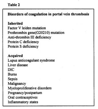
Deficiency of natural anticoagulant proteins C, S, and antithrombin in portal vein thrombosis: a secondary phenomenon? (1/21)
BACKGROUND: Hereditary deficiencies of natural anticoagulant proteins are implicated in the pathogenesis of portal vein thrombosis (PVT). Secondary deficiencies of these proteins have also been reported in PVT, making interpretation of concentrations difficult. AIMS: To characterise the coagulation profiles in adult patients with PVT and to investigate the possible mechanisms of natural anticoagulant protein deficiency. PATIENTS: Twenty nine adult patients with portal hypertension caused by PVT, and normal biochemical liver function tests. METHODS: Routine coagulation profiles and concentrations of proteins C, S, and antithrombin were measured; where indicated, corresponding concentrations in parents were also measured. Synchronous peripheral and hepatic or splenic vein concentrations were compared in seven patients undergoing interventional procedures, as were peripheral concentrations before and after shunt surgery in three patients. RESULTS: Deficiencies of one or more of the natural anticoagulant proteins occurred in 18 patients (62%), with six patients having combined deficiency of all three proteins. There were strong correlations between prothrombin and partial thromboplastin time ratios and concentrations of natural anticoagulant proteins. Family studies in nine cases of anticoagulant protein deficiency revealed possible hereditary deficiency in only three cases, and significantly lower concentrations of anticoagulant proteins in all PVT cases compared with parents. Levels of anticoagulant proteins tended to be lower in hepatic veins but higher in splenic veins compared with peripheral vein concentrations. Peripheral concentrations decreased after shunt surgery. CONCLUSIONS: Deficiency of natural anticoagulant proteins is common in PVT and is probably a secondary phenomenon in most cases, occurring as part of a global disturbance of coagulation variables. The mechanism for this remains unclear but may result from a combination of reduced hepatic blood flow and portosystemic shunting itself. (+info)Studies of the human factor VIII/von Willebrand factor protein. III. Qualitative defects in von Willebrand's disease. (2/21)
The Factor VIII/von Willebrand factor protein was characterized in two unrelated patients with von Willebrand's disease in whom procoagulant and Factor VIII/von Willebrand factor antigen levels were normal. In both patients evidence of an abnormal protein was observed on crossed antigen-antibody electrophoresis. In one patient the Factor VIII/von Willebrand factor protein eluted from Sepharose 4B in a position and distribution identical to normal with normal levels of procoagulant activity and antigen. However, the partially purified Factor VIII/von Willebrand factor protein had markedly reduced von Willebrand factor activity in a ristocetin assay. In the second patient the peak of Factor VIII/von Willebrand factor protein, antigen, and procoagulant activity eluted from a Sepharose 4B column with an estimated molecular weight of approximately half that of normal. This protein had no von Willebrand factor activity. In both patients the reduced Factor VIII/von Willebrand factor protein subunit was indistinguishable from normal on polyacrylamide gel electrophoresis. These studies indicate that in some patients with von Willebrand's disease there is a qualitative defect of the Factor VII/von Willebrand factor protein; the total amount of protein, antigen, and procoagulant activity are normal while the von Willebrand factor activity is deficient. (+info)Coagulation and bleeding disorders: review and update. (3/21)
Hemostasis is initiated by injury to the vascular wall, leading to the deposition of platelets adhering to components of the subendothelium. Platelet adhesion requires the presence of von Willebrand factor and platelet receptors (IIb/IIIa and Ib/IX). Additional platelets are recruited to the site of injury by release of platelet granular contents, including ADP. The "platelet plug" is stabilized by interaction with fibrinogen. In this review, I consider laboratory tests used to evaluate coagulation, including prothrombin time, activated partial thromboplastin time, thrombin time, and platelet count. I discuss hereditary disorders of platelets and/or coagulation proteins that lead to clinical bleeding as well as acquired disorders, including disseminated intravascular coagulation and acquired circulating anticoagulants. (+info)Activated partial thromboplastin time as a screening test of minor or moderate coagulation factor deficiencies for canine plasma: sensitivity of different commercial reagents. (4/21)
To determine the sensitivity for detection of coagulation factor deficiencies by commercial reagents for canine plasma, 5 commercial activated partial thromboplastin time (APTT) reagents with different types of contact activator and phospholipid of various origin were examined. Thirty canine plasma samples with minor or moderate deficiencies of coagualition factors that influence the APTT were examined. Significant differences were found for the sensitivity of various reagents, but no correlation was found with the type of contact activator. Following the test instructions provided by the manufacturers, the number of APTT results that were prolonged beyond the reference range varied between 20 and 30 (sensitivity = 0.67-1.00); the number of corresponding results using a standardized test protocol varied between 19 and 28 (sensitivity: 0.63-0.93). The most sensitive reagent contained kaolin as a contact activator and a human placental thromboplastin. The results of this study indicate that the APTT test optimized for human plasma is also a sensitive screening test of the intrinsic system of canine plasma, provided that a suitable reagent is used. (+info)Fibrinogen Ales: a homozygous case of dysfibrinogenemia (gamma-Asp(330)-->Val) characterized by a defective fibrin polymerization site "a". (5/21)
Congenital homozygous dysfibrinogenemia was diagnosed in a man with a history of 2 thrombotic strokes before age 30. His hemostatic profile was characterized by a dramatically prolonged plasma thrombin clotting time, and no clotting was observed with reptilase. Complete clotting of the abnormal fibrinogen occurred after a prolonged incubation of plasma with thrombin. The release of fibrinopeptides A and B by thrombin and of fibrinopeptide A by reptilase were both normal. Thrombin-induced fibrin polymerization was impaired, and no polymerization occurred with reptilase. The polymerization defect was characterized by a defective site "a," resulting in an absence of interaction between sites A and a, indicated by the lack of fragment D(1) (or fibrinogen) binding to normal fibrin monomers depleted in fibrinopeptide A only (Des-AA fm). By SDS-PAGE, the defect was detected on the gamma-chain and in its fragment D(1). The molecular defect determined by analysis of genomic DNA showed a single base change (A-->T) in exon VIII of the gamma-chain. The resulting change in the amino acid structure is gamma 330 aspartic acid (GAT) --> valine (GTT). It is concluded that the residue gamma-Asp(330) is essential for the normal functioning of the polymerization site a on the fibrinogen gamma-chain. (+info)Decreased coagulability has no clinically relevant effect on atherogenesis: observations in individuals with a hereditary bleeding tendency. (6/21)
BACKGROUND: Hemostasis affects ischemic cardiovascular disease through its role in formation of occluding arterial thrombi. Several studies suggest that hemostasis also might play a role in atherogenesis. We investigated whether individuals with an inherited bleeding tendency are protected against development of atherosclerosis. METHODS AND RESULTS: A total of 76 individuals with an inherited bleeding tendency (hemophilia and von Willebrand disease) and 142 healthy controls were included in the present study. Early atherosclerotic vessel-wall changes were quantified by measurement of intima-media thickness in the carotid and femoral arteries by B-mode ultrasonography. To validate intima-media thickness measurements, measurements also were performed in 77 individuals with clinically proven atherosclerosis and in 34 healthy, age-matched controls. A large difference in intima-media thickness was found between individuals with proven atherosclerosis and healthy controls, in particular for the femoral artery (difference for carotid artery, 0.16 mm; femoral artery, 0.53 mm). Comparison between patients with a bleeding tendency and healthy controls showed only minimally reduced intima-media in femoral artery in individuals with a bleeding tendency (adjusted difference, -0.078 mm; 95% CI, -0.17 to 0.018 mm). Subgroup analysis revealed that in subjects with moderate to severe hemophilia, vessel walls were thinnest (adjusted difference, -0.10 mm; 95% CI, -0.27 to 0.061 mm). CONCLUSIONS: Hypocoagulability caused by hemophilia or von Willebrand disease has at most a limited effect on atherogenesis. (+info)Neonatal coagulation problems. (7/21)
Bleeding problems often occur during the neonatal period. Although thrombocytopenia is the most common cause, coagulation problems often occur, and the two problems may co-exist. The causes, diagnosis, and management of coagulation problems in newborn infants are reviewed. (+info)Compound heterozygosity of novel missense mutations in the gamma-glutamyl-carboxylase gene causes hereditary combined vitamin K-dependent coagulation factor deficiency. (8/21)
Hereditary combined vitamin K-dependent (VKD) coagulation factor deficiency is an autosomal recessive bleeding disorder associated with defects in either the gamma-carboxylase, which carboxylates VKD proteins to render them active, or the vitamin K epoxide reductase (VKORC1), which supplies the reduced vitamin K cofactor required for carboxylation. Such deficiencies are rare, and we report the fourth case resulting from mutations in the carboxylase gene, identified in a Tunisian girl who exhibited impaired function in hemostatic VKD factors that was not restored by vitamin K administration. Sequence analysis of the proposita did not identify any mutations in the VKORC1 gene but, remarkably, revealed 3 heterozygous mutations in the carboxylase gene that caused the substitutions Asp31Asn, Trp157Arg, and Thr591Lys. None of these mutations have previously been reported. Family analysis showed that Asp31Asn and Thr591Lys were coallelic and maternally transmitted while Trp157Arg was transmitted by the father, and a genomic screen of 100 healthy individuals ruled out frequent polymorphisms. Mutational analysis indicated wild-type activity for the Asp31Asn carboxylase. In contrast, the respective Trp157Arg and Thr591Lys activities were 8% and 0% that of wild-type carboxylase, and their compound heterozygosity can therefore account for functional VKD factor deficiency. The implications for carboxylase mechanism are discussed. (+info)Coagulation protein disorders are a group of medical conditions that affect the body's ability to form blood clots properly. These disorders can be caused by genetic defects or acquired factors, such as liver disease or vitamin K deficiency.
The coagulation system is a complex process that involves various proteins called clotting factors. When there is an injury to a blood vessel, these clotting factors work together in a specific order to form a clot and prevent excessive bleeding. In coagulation protein disorders, one or more of these clotting factors are missing or not functioning properly, leading to abnormal bleeding or clotting.
There are several types of coagulation protein disorders, including:
1. Hemophilia: This is a genetic disorder that affects the clotting factor VIII or IX. People with hemophilia may experience prolonged bleeding after injuries, surgery, or dental work.
2. Von Willebrand disease: This is another genetic disorder that affects the von Willebrand factor, a protein that helps platelets stick together and form a clot. People with this condition may have nosebleeds, easy bruising, and excessive bleeding during menstruation or after surgery.
3. Factor XI deficiency: This is a rare genetic disorder that affects the clotting factor XI. People with this condition may experience prolonged bleeding after surgery or trauma.
4. Factor VII deficiency: This is a rare genetic disorder that affects the clotting factor VII. People with this condition may have nosebleeds, easy bruising, and excessive bleeding during menstruation or after surgery.
5. Acquired coagulation protein disorders: These are conditions that develop due to other medical factors, such as liver disease, vitamin K deficiency, or the use of certain medications. These disorders can affect one or more clotting factors and may cause abnormal bleeding or clotting.
Treatment for coagulation protein disorders depends on the specific condition and severity of symptoms. In some cases, replacement therapy with the missing clotting factor may be necessary to prevent excessive bleeding. Other treatments may include medications to control bleeding, such as desmopressin or antifibrinolytic agents, and lifestyle changes to reduce the risk of injury and bleeding.
Blood coagulation, also known as blood clotting, is a complex process that occurs in the body to prevent excessive bleeding when a blood vessel is damaged. This process involves several different proteins and chemical reactions that ultimately lead to the formation of a clot.
The coagulation cascade is initiated when blood comes into contact with tissue factor, which is exposed after damage to the blood vessel wall. This triggers a series of enzymatic reactions that activate clotting factors, leading to the formation of a fibrin clot. Fibrin is a protein that forms a mesh-like structure that traps platelets and red blood cells to form a stable clot.
Once the bleeding has stopped, the coagulation process is regulated and inhibited to prevent excessive clotting. The fibrinolytic system degrades the clot over time, allowing for the restoration of normal blood flow.
Abnormalities in the blood coagulation process can lead to bleeding disorders or thrombotic disorders such as deep vein thrombosis and pulmonary embolism.
Blood coagulation factors, also known as clotting factors, are a group of proteins that play a crucial role in the blood coagulation process. They are essential for maintaining hemostasis, which is the body's ability to stop bleeding after injury.
There are 13 known blood coagulation factors, and they are designated by Roman numerals I through XIII. These factors are produced in the liver and are normally present in an inactive form in the blood. When there is an injury to a blood vessel, the coagulation process is initiated, leading to the activation of these factors in a specific order.
The coagulation cascade involves two pathways: the intrinsic and extrinsic pathways. The intrinsic pathway is activated when there is damage to the blood vessel itself, while the extrinsic pathway is activated by tissue factor released from damaged tissues. Both pathways converge at the common pathway, leading to the formation of a fibrin clot.
Blood coagulation factors work together in a complex series of reactions that involve activation, binding, and proteolysis. When one factor is activated, it activates the next factor in the cascade, and so on. This process continues until a stable fibrin clot is formed.
Deficiencies or abnormalities in blood coagulation factors can lead to bleeding disorders such as hemophilia or thrombosis. Hemophilia is a genetic disorder that affects one or more of the coagulation factors, leading to excessive bleeding and difficulty forming clots. Thrombosis, on the other hand, occurs when there is an abnormal formation of blood clots in the blood vessels, which can lead to serious complications such as stroke or pulmonary embolism.
Vitamin K is a fat-soluble vitamin that plays a crucial role in blood clotting and bone metabolism. It is essential for the production of several proteins involved in blood clotting, including factor II (prothrombin), factor VII, factor IX, and factor X. Additionally, Vitamin K is necessary for the synthesis of osteocalcin, a protein that contributes to bone health by regulating the deposition of calcium in bones.
There are two main forms of Vitamin K: Vitamin K1 (phylloquinone), which is found primarily in green leafy vegetables and some vegetable oils, and Vitamin K2 (menaquinones), which is produced by bacteria in the intestines and is also found in some fermented foods.
Vitamin K deficiency can lead to bleeding disorders such as hemorrhage and excessive bruising. While Vitamin K deficiency is rare in adults, it can occur in newborns who have not yet developed sufficient levels of the vitamin. Therefore, newborns are often given a Vitamin K injection shortly after birth to prevent bleeding problems.
A protein database is a type of biological database that contains information about proteins and their structures, functions, sequences, and interactions with other molecules. These databases can include experimentally determined data, such as protein sequences derived from DNA sequencing or mass spectrometry, as well as predicted data based on computational methods.
Some examples of protein databases include:
1. UniProtKB: a comprehensive protein database that provides information about protein sequences, functions, and structures, as well as literature references and links to other resources.
2. PDB (Protein Data Bank): a database of three-dimensional protein structures determined by experimental methods such as X-ray crystallography and nuclear magnetic resonance (NMR) spectroscopy.
3. BLAST (Basic Local Alignment Search Tool): a web-based tool that allows users to compare a query protein sequence against a protein database to identify similar sequences and potential functional relationships.
4. InterPro: a database of protein families, domains, and functional sites that provides information about protein function based on sequence analysis and other data.
5. STRING (Search Tool for the Retrieval of Interacting Genes/Proteins): a database of known and predicted protein-protein interactions, including physical and functional associations.
Protein databases are essential tools in proteomics research, enabling researchers to study protein function, evolution, and interaction networks on a large scale.
Factor IX is also known as Christmas factor, which is a protein that plays a crucial role in the coagulation cascade, a series of chemical reactions that leads to the formation of a blood clot. It is one of the essential components required for the proper functioning of the body's natural blood-clotting mechanism.
Factor IX is synthesized in the liver and activated when it comes into contact with an injured blood vessel. Once activated, it collaborates with other factors to convert factor X to its active form, which then converts prothrombin to thrombin. Thrombin is responsible for converting fibrinogen to fibrin, forming a stable fibrin clot that helps stop bleeding and promote healing.
Deficiencies in Factor IX can lead to hemophilia B, a genetic disorder characterized by prolonged bleeding and an increased risk of spontaneous bleeding. Hemophilia B is inherited in an X-linked recessive pattern, meaning it primarily affects males, while females serve as carriers of the disease. Treatment for hemophilia B typically involves replacing the missing or deficient Factor IX through infusions to prevent or manage bleeding episodes.
Prothrombin is a protein present in blood plasma, and it's also known as coagulation factor II. It plays a crucial role in the coagulation cascade, which is a complex series of reactions that leads to the formation of a blood clot.
When an injury occurs, the coagulation cascade is initiated to prevent excessive blood loss. Prothrombin is converted into its active form, thrombin, by another factor called factor Xa in the presence of calcium ions, phospholipids, and factor Va. Thrombin then catalyzes the conversion of fibrinogen into fibrin, forming a stable clot.
Prothrombin levels can be measured through a blood test, which is often used to diagnose or monitor conditions related to bleeding or coagulation disorders, such as liver disease or vitamin K deficiency.
Protein sequence analysis is the systematic examination and interpretation of the amino acid sequence of a protein to understand its structure, function, evolutionary relationships, and other biological properties. It involves various computational methods and tools to analyze the primary structure of proteins, which is the linear arrangement of amino acids along the polypeptide chain.
Protein sequence analysis can provide insights into several aspects, such as:
1. Identification of functional domains, motifs, or sites within a protein that may be responsible for its specific biochemical activities.
2. Comparison of homologous sequences from different organisms to infer evolutionary relationships and determine the degree of similarity or divergence among them.
3. Prediction of secondary and tertiary structures based on patterns of amino acid composition, hydrophobicity, and charge distribution.
4. Detection of post-translational modifications that may influence protein function, localization, or stability.
5. Identification of protease cleavage sites, signal peptides, or other sequence features that play a role in protein processing and targeting.
Some common techniques used in protein sequence analysis include:
1. Multiple Sequence Alignment (MSA): A method to align multiple protein sequences to identify conserved regions, gaps, and variations.
2. BLAST (Basic Local Alignment Search Tool): A widely-used tool for comparing a query protein sequence against a database of known sequences to find similarities and infer function or evolutionary relationships.
3. Hidden Markov Models (HMMs): Statistical models used to describe the probability distribution of amino acid sequences in protein families, allowing for more sensitive detection of remote homologs.
4. Protein structure prediction: Methods that use various computational approaches to predict the three-dimensional structure of a protein based on its amino acid sequence.
5. Phylogenetic analysis: The construction and interpretation of evolutionary trees (phylogenies) based on aligned protein sequences, which can provide insights into the historical relationships among organisms or proteins.
Prothrombin time (PT) is a medical laboratory test that measures the time it takes for blood to clot. It's often used to evaluate the functioning of the extrinsic and common pathways of the coagulation system, which is responsible for blood clotting. Specifically, PT measures how long it takes for prothrombin (a protein produced by the liver) to be converted into thrombin, an enzyme that converts fibrinogen into fibrin and helps form a clot.
Prolonged PT may indicate a bleeding disorder or a deficiency in coagulation factors, such as vitamin K deficiency or the use of anticoagulant medications like warfarin. It's important to note that PT is often reported with an international normalized ratio (INR), which allows for standardization and comparison of results across different laboratories and reagent types.
Factor X is a protein that is essential for blood clotting, also known as coagulation. It is an enzyme that plays a crucial role in the coagulation cascade, which is a series of chemical reactions that lead to the formation of a blood clot. Factor X is activated by one of two pathways: the intrinsic pathway, which is initiated by damage to the blood vessels, or the extrinsic pathway, which is triggered by the release of tissue factor from damaged cells. Once activated, Factor X converts prothrombin to thrombin, which then converts fibrinogen to fibrin to form a stable clot.
Inherited deficiencies in Factor X can lead to bleeding disorders, while increased levels of Factor X have been associated with an increased risk of thrombosis or blood clots. Therefore, maintaining appropriate levels of Factor X is important for the proper balance between bleeding and clotting in the body.
Carbon-carbon ligases are a type of enzyme that catalyze the formation of carbon-carbon bonds between two molecules. These enzymes play important roles in various biological processes, including the biosynthesis of natural products and the metabolism of carbohydrates and lipids.
Carbon-carbon ligases can be classified into several categories based on the type of reaction they catalyze. For example, aldolases catalyze the condensation of an aldehyde or ketone with another molecule to form a new carbon-carbon bond and a new carbonyl group. Other examples include the polyketide synthases (PKSs) and nonribosomal peptide synthetases (NRPSs), which are large multienzyme complexes that catalyze the sequential addition of activated carbon units to form complex natural products.
Carbon-carbon ligases are important targets for drug discovery and development, as they play critical roles in the biosynthesis of many disease-relevant molecules. Inhibitors of these enzymes have shown promise as potential therapeutic agents for a variety of diseases, including cancer, infectious diseases, and metabolic disorders.
Proteins are complex, large molecules that play critical roles in the body's functions. They are made up of amino acids, which are organic compounds that are the building blocks of proteins. Proteins are required for the structure, function, and regulation of the body's tissues and organs. They are essential for the growth, repair, and maintenance of body tissues, and they play a crucial role in many biological processes, including metabolism, immune response, and cellular signaling. Proteins can be classified into different types based on their structure and function, such as enzymes, hormones, antibodies, and structural proteins. They are found in various foods, especially animal-derived products like meat, dairy, and eggs, as well as plant-based sources like beans, nuts, and grains.
Disseminated Intravascular Coagulation (DIC) is a complex medical condition characterized by the abnormal activation of the coagulation cascade, leading to the formation of blood clots in small blood vessels throughout the body. This process can result in the consumption of clotting factors and platelets, which can then lead to bleeding complications. DIC can be caused by a variety of underlying conditions, including sepsis, trauma, cancer, and obstetric emergencies.
The term "disseminated" refers to the widespread nature of the clotting activation, while "intravascular" indicates that the clotting is occurring within the blood vessels. The condition can manifest as both bleeding and clotting complications, which can make it challenging to diagnose and manage.
The diagnosis of DIC typically involves laboratory tests that evaluate coagulation factors, platelet count, fibrin degradation products, and other markers of coagulation activation. Treatment is focused on addressing the underlying cause of the condition while also managing any bleeding or clotting complications that may arise.
Factor XI, also known as plasma thromboplastin antecedent (PTA) or antihemophilic factor C, is a protein involved in blood coagulation. It is one of the factors in the intrinsic pathway of coagulation, which is activated when blood comes into contact with negatively charged surfaces, such as damaged blood vessels.
When Factor XI is activated (usually by thrombin or activated Factor XII), it activates more Factor XI and also activates Factor IX, leading to the formation of a complex that converts Factor X to its active form, Factor Xa. This ultimately leads to the formation of a fibrin clot and helps to stop bleeding.
Deficiencies in Factor XI can lead to an increased risk of bleeding, although the severity of the bleeding disorder can vary widely among individuals with Factor XI deficiency. Treatment for Factor XI deficiency typically involves replacement therapy with fresh frozen plasma or recombinant Factor XI concentrate.
Blood protein disorders refer to a group of medical conditions that affect the production or function of proteins in the blood. These proteins are crucial for maintaining the proper functioning of the body's immune system, transporting nutrients, and preventing excessive bleeding. Some examples of blood protein disorders include:
1. Hemophilia: A genetic disorder caused by a deficiency or absence of clotting factors in the blood, leading to prolonged bleeding and poor clot formation.
2. Von Willebrand disease: A genetic disorder characterized by abnormal or deficient von Willebrand factor, which is necessary for platelet function and proper clotting.
3. Dysproteinemias: Abnormal levels of certain proteins in the blood, such as immunoglobulins (antibodies) or paraproteins, which can indicate underlying conditions like multiple myeloma or macroglobulinemia.
4. Hypoproteinemia: Low levels of total protein in the blood, often caused by liver disease, malnutrition, or kidney disease.
5. Hyperproteinemia: Elevated levels of total protein in the blood, which can be caused by dehydration, inflammation, or certain types of cancer.
6. Hemoglobinopathies: Genetic disorders affecting the structure and function of hemoglobin, a protein found in red blood cells that carries oxygen throughout the body. Examples include sickle cell anemia and thalassemia.
7. Disorders of complement proteins: Abnormalities in the complement system, which is a group of proteins involved in the immune response, can lead to conditions like autoimmune disorders or recurrent infections.
Treatment for blood protein disorders varies depending on the specific condition and its severity but may include medications, transfusions, or other medical interventions.
Protein C is a vitamin K-dependent protease that functions as an important regulator of coagulation and inflammation. It is a plasma protein produced in the liver that, when activated, degrades clotting factors Va and VIIIa to limit thrombus formation and prevent excessive blood clotting. Protein C also has anti-inflammatory properties by inhibiting the release of pro-inflammatory cytokines and reducing endothelial cell activation. Inherited or acquired deficiencies in Protein C can lead to an increased risk of thrombosis, a condition characterized by abnormal blood clot formation within blood vessels.
1-Carboxyglutamic acid, also known as γ-carboxyglutamic acid, is a post-translational modification found on certain blood clotting factors and other calcium-binding proteins. It is formed by the carboxylation of glutamic acid residues in these proteins, which enhances their ability to bind to calcium ions. This modification is essential for the proper functioning of many physiological processes, including blood coagulation, bone metabolism, and wound healing.
Thromboplastin is a substance that activates the coagulation cascade, leading to the formation of a clot (thrombus). It's primarily found in damaged or injured tissues and blood vessels, as well as in platelets (thrombocytes). There are two types of thromboplastin:
1. Extrinsic thromboplastin (also known as tissue factor): This is a transmembrane glycoprotein that is primarily found in subendothelial cells and released upon injury to the blood vessels. It initiates the extrinsic pathway of coagulation by binding to and activating Factor VII, ultimately leading to the formation of thrombin and fibrin clots.
2. Intrinsic thromboplastin (also known as plasma thromboplastin or factor III): This term is used less frequently and refers to a labile phospholipid component present in platelet membranes, which plays a role in the intrinsic pathway of coagulation.
In clinical settings, the term "thromboplastin" often refers to reagents used in laboratory tests like the prothrombin time (PT) and activated partial thromboplastin time (aPTT). These reagents contain a source of tissue factor and calcium ions to initiate and monitor the coagulation process.
Factor VIII is a protein in the blood that is essential for normal blood clotting. It is also known as antihemophilic factor (AHF). Deficiency or dysfunction of this protein results in hemophilia A, a genetic disorder characterized by prolonged bleeding and easy bruising. Factor VIII works together with other proteins to help form a clot and stop bleeding at the site of an injury. It acts as a cofactor for another clotting factor, IX, in the so-called intrinsic pathway of blood coagulation. Intravenous infusions of Factor VIII concentrate are used to treat and prevent bleeding episodes in people with hemophilia A.
Factor V, also known as proaccelerin or labile factor, is a protein involved in the coagulation cascade, which is a series of chemical reactions that leads to the formation of a blood clot. Factor V acts as a cofactor for the activation of Factor X to Factor Xa, which is a critical step in the coagulation cascade.
When blood vessels are damaged, the coagulation cascade is initiated to prevent excessive bleeding. During this process, Factor V is activated by thrombin, another protein involved in coagulation, and then forms a complex with activated Factor X and calcium ions on the surface of platelets or other cells. This complex converts prothrombin to thrombin, which then converts fibrinogen to fibrin to form a stable clot.
Deficiency or dysfunction of Factor V can lead to bleeding disorders such as hemophilia B or factor V deficiency, while mutations in the gene encoding Factor V can increase the risk of thrombosis, as seen in the Factor V Leiden mutation.
Blood coagulation disorders, also known as bleeding disorders or clotting disorders, refer to a group of medical conditions that affect the body's ability to form blood clots properly. Normally, when a blood vessel is injured, the body's coagulation system works to form a clot to stop the bleeding and promote healing.
In blood coagulation disorders, there can be either an increased tendency to bleed due to problems with the formation of clots (hemorrhagic disorder), or an increased tendency for clots to form inappropriately even without injury, leading to blockages in the blood vessels (thrombotic disorder).
Examples of hemorrhagic disorders include:
1. Hemophilia - a genetic disorder that affects the ability to form clots due to deficiencies in clotting factors VIII or IX.
2. Von Willebrand disease - another genetic disorder caused by a deficiency or abnormality of the von Willebrand factor, which helps platelets stick together to form a clot.
3. Liver diseases - can lead to decreased production of coagulation factors, increasing the risk of bleeding.
4. Disseminated intravascular coagulation (DIC) - a serious condition where clotting and bleeding occur simultaneously due to widespread activation of the coagulation system.
Examples of thrombotic disorders include:
1. Factor V Leiden mutation - a genetic disorder that increases the risk of inappropriate blood clot formation.
2. Antithrombin III deficiency - a genetic disorder that impairs the body's ability to break down clots, increasing the risk of thrombosis.
3. Protein C or S deficiencies - genetic disorders that lead to an increased risk of thrombosis due to impaired regulation of the coagulation system.
4. Antiphospholipid syndrome (APS) - an autoimmune disorder where the body produces antibodies against its own clotting factors, increasing the risk of thrombosis.
Treatment for blood coagulation disorders depends on the specific diagnosis and may include medications to manage bleeding or prevent clots, as well as lifestyle changes and monitoring to reduce the risk of complications.
Thrombin is a serine protease enzyme that plays a crucial role in the coagulation cascade, which is a complex series of biochemical reactions that leads to the formation of a blood clot (thrombus) to prevent excessive bleeding during an injury. Thrombin is formed from its precursor protein, prothrombin, through a process called activation, which involves cleavage by another enzyme called factor Xa.
Once activated, thrombin converts fibrinogen, a soluble plasma protein, into fibrin, an insoluble protein that forms the structural framework of a blood clot. Thrombin also activates other components of the coagulation cascade, such as factor XIII, which crosslinks and stabilizes the fibrin network, and platelets, which contribute to the formation and growth of the clot.
Thrombin has several regulatory mechanisms that control its activity, including feedback inhibition by antithrombin III, a plasma protein that inactivates thrombin and other serine proteases, and tissue factor pathway inhibitor (TFPI), which inhibits the activation of factor Xa, thereby preventing further thrombin formation.
Overall, thrombin is an essential enzyme in hemostasis, the process that maintains the balance between bleeding and clotting in the body. However, excessive or uncontrolled thrombin activity can lead to pathological conditions such as thrombosis, atherosclerosis, and disseminated intravascular coagulation (DIC).
Blood coagulation tests, also known as coagulation studies or clotting tests, are a series of medical tests used to evaluate the blood's ability to clot. These tests measure the functioning of various clotting factors and regulatory proteins involved in the coagulation cascade, which is a complex process that leads to the formation of a blood clot to prevent excessive bleeding.
The most commonly performed coagulation tests include:
1. Prothrombin Time (PT): Measures the time it takes for a sample of plasma to clot after the addition of calcium and tissue factor, which activates the extrinsic pathway of coagulation. The PT is reported in seconds and can be converted to an International Normalized Ratio (INR) to monitor anticoagulant therapy.
2. Activated Partial Thromboplastin Time (aPTT): Measures the time it takes for a sample of plasma to clot after the addition of calcium, phospholipid, and a contact activator, which activates the intrinsic pathway of coagulation. The aPTT is reported in seconds and is used to monitor heparin therapy.
3. Thrombin Time (TT): Measures the time it takes for a sample of plasma to clot after the addition of thrombin, which directly converts fibrinogen to fibrin. The TT is reported in seconds and can be used to detect the presence of fibrin degradation products or abnormalities in fibrinogen function.
4. Fibrinogen Level: Measures the amount of fibrinogen, a protein involved in clot formation, present in the blood. The level is reported in grams per liter (g/L) and can be used to assess bleeding risk or the effectiveness of fibrinogen replacement therapy.
5. D-dimer Level: Measures the amount of D-dimer, a protein fragment produced during the breakdown of a blood clot, present in the blood. The level is reported in micrograms per milliliter (µg/mL) and can be used to diagnose or exclude venous thromboembolism (VTE), such as deep vein thrombosis (DVT) or pulmonary embolism (PE).
These tests are important for the diagnosis, management, and monitoring of various bleeding and clotting disorders. They can help identify the underlying cause of abnormal bleeding or clotting, guide appropriate treatment decisions, and monitor the effectiveness of therapy. It is essential to interpret these test results in conjunction with a patient's clinical presentation and medical history.
Computational biology is a branch of biology that uses mathematical and computational methods to study biological data, models, and processes. It involves the development and application of algorithms, statistical models, and computational approaches to analyze and interpret large-scale molecular and phenotypic data from genomics, transcriptomics, proteomics, metabolomics, and other high-throughput technologies. The goal is to gain insights into biological systems and processes, develop predictive models, and inform experimental design and hypothesis testing in the life sciences. Computational biology encompasses a wide range of disciplines, including bioinformatics, systems biology, computational genomics, network biology, and mathematical modeling of biological systems.
Protein folding is the process by which a protein molecule naturally folds into its three-dimensional structure, following the synthesis of its amino acid chain. This complex process is determined by the sequence and properties of the amino acids, as well as various environmental factors such as temperature, pH, and the presence of molecular chaperones. The final folded conformation of a protein is crucial for its proper function, as it enables the formation of specific interactions between different parts of the molecule, which in turn define its biological activity. Protein misfolding can lead to various diseases, including neurodegenerative disorders such as Alzheimer's and Parkinson's disease.
Protein conformation refers to the specific three-dimensional shape that a protein molecule assumes due to the spatial arrangement of its constituent amino acid residues and their associated chemical groups. This complex structure is determined by several factors, including covalent bonds (disulfide bridges), hydrogen bonds, van der Waals forces, and ionic bonds, which help stabilize the protein's unique conformation.
Protein conformations can be broadly classified into two categories: primary, secondary, tertiary, and quaternary structures. The primary structure represents the linear sequence of amino acids in a polypeptide chain. The secondary structure arises from local interactions between adjacent amino acid residues, leading to the formation of recurring motifs such as α-helices and β-sheets. Tertiary structure refers to the overall three-dimensional folding pattern of a single polypeptide chain, while quaternary structure describes the spatial arrangement of multiple folded polypeptide chains (subunits) that interact to form a functional protein complex.
Understanding protein conformation is crucial for elucidating protein function, as the specific three-dimensional shape of a protein directly influences its ability to interact with other molecules, such as ligands, nucleic acids, or other proteins. Any alterations in protein conformation due to genetic mutations, environmental factors, or chemical modifications can lead to loss of function, misfolding, aggregation, and disease states like neurodegenerative disorders and cancer.
The proteome is the entire set of proteins produced or present in an organism, system, organ, or cell at a certain time under specific conditions. It is a dynamic collection of protein species that changes over time, responding to various internal and external stimuli such as disease, stress, or environmental factors. The study of the proteome, known as proteomics, involves the identification and quantification of these protein components and their post-translational modifications, providing valuable insights into biological processes, functional pathways, and disease mechanisms.
An algorithm is not a medical term, but rather a concept from computer science and mathematics. In the context of medicine, algorithms are often used to describe step-by-step procedures for diagnosing or managing medical conditions. These procedures typically involve a series of rules or decision points that help healthcare professionals make informed decisions about patient care.
For example, an algorithm for diagnosing a particular type of heart disease might involve taking a patient's medical history, performing a physical exam, ordering certain diagnostic tests, and interpreting the results in a specific way. By following this algorithm, healthcare professionals can ensure that they are using a consistent and evidence-based approach to making a diagnosis.
Algorithms can also be used to guide treatment decisions. For instance, an algorithm for managing diabetes might involve setting target blood sugar levels, recommending certain medications or lifestyle changes based on the patient's individual needs, and monitoring the patient's response to treatment over time.
Overall, algorithms are valuable tools in medicine because they help standardize clinical decision-making and ensure that patients receive high-quality care based on the latest scientific evidence.
I am not aware of a widely accepted medical definition for the term "software," as it is more commonly used in the context of computer science and technology. Software refers to programs, data, and instructions that are used by computers to perform various tasks. It does not have direct relevance to medical fields such as anatomy, physiology, or clinical practice. If you have any questions related to medicine or healthcare, I would be happy to try to help with those instead!
 List of MeSH codes (C15)
Hematologic disease
Lonomia
Coagulopathy
Congenital afibrinogenemia
Bleeding diathesis
D-dimer
Congenital hypofibrinogenemia
Omega loop
Factor VIII
Hypertension
Hypoprothrombinemia
Sticky platelet syndrome
Iranian Blood Transfusion Organization
Recombinant DNA
MCFD2
Purpura fulminans
Purpura
Factor V
Factor VII deficiency
Bispecific monoclonal antibody
Factor X deficiency
Hematology
Aptamer
Thrombin time
Contact activation system
Octapharma
Plasmin-α2-antiplasmin complex
Protein S
Björn Dahlbäck
 Coagulation Protein Disorders in Animals - Circulatory System - MSD Veterinary Manual
 Group B streptococcal septicemia of the newborn: MedlinePlus Medical Encyclopedia
Factor VII Deficiency | Profiles RNS
 Dusky bullae on the lower abdomen - Clinical Advisor
List of MeSH codes (C15) - Wikipedia
Factor VII assay: MedlinePlus Medical Encyclopedia
 Study to Evaluate the Efficacy and Safety of Valoctocogene Roxaparvovec, With Prophylactic Steroids in Hemophilia A - Full Text...
 Partial thromboplastin time (PTT)
 Prothix Company Profile: Valuation, Funding & Investors | PitchBook
Detection of Severe Murine Typhus by Nanopore Targeted Sequencing, China - Volume 29, Number 6-June 2023 - Emerging Infectious...
 Browse School of Medicine | Stanford Profiles
DeCS - New Terms
DeCS - Termos Novos
DeCS - Términos Nuevos
 DeCS - New Terms
DeCS - New Terms
DeCS - Termos Novos
HuGE Navigator|Genopedia|PHGKB
DeCS - New Terms
DeCS - Términos Nuevos
DeCS - Termos Novos
DeCS - Términos Nuevos
DeCS - New Terms
DeCS - New Terms
Factor IX assay
 Metabolomics in COPD | Archivos de Bronconeumología
Protein S blood test
Overview of Coagulation Disorders - Hematology and Oncology - Merck Manuals Professional Edition
 Algal proteins. Medical search
Deficiency23
- In a severe deficiency or functional defect of coagulation proteins, clinical signs appear at an early age. (msdvetmanual.com)
- An autosomal recessive characteristic or a coagulation disorder acquired in association with VITAMIN K DEFICIENCY. (umassmed.edu)
- The test is also used to screen relatives of people who are known to have protein S deficiency . (ucsfbenioffchildrens.org)
- A lack (deficiency) of protein S can lead to excess clotting. (ucsfbenioffchildrens.org)
- A protein S deficiency may be inherited. (ucsfbenioffchildrens.org)
- Coagulation disorders include Hemophilia A and B (which are inherited disorders), and Hemophilia C (which is due to a deficiency of factor XI). (differencebetween.net)
- Coagulation factor VII deficiency is an inherited bleeding disorder affecting dogs. (pawprintgenetics.com)
- Deficiency of this factor most commonly results in a mild bleeding disorder. (pawprintgenetics.com)
- Veterinarians performing surgery on dogs that are known to have coagulation factor VII deficiency should have ready access to blood banked for transfusions. (pawprintgenetics.com)
- Genetic testing of the F7 gene in dogs will reliably determine whether a dog is a genetic Carrier of coagulation factor VII deficiency. (pawprintgenetics.com)
- Coagulation factor VII deficiency is inherited in an Autosomal Recessive manner in dogs meaning that they must receive two copies of the mutated gene (one from each parent) to develop the disease. (pawprintgenetics.com)
- Withnall E, Giger U. Effects of recombinant human activated factor VII and canine fresh frozen plasma in Beagles with hereditary coagulation factor VII deficiency. (pawprintgenetics.com)
- As a result, we identified a large number of genes encoding N. lugens pattern recognition proteins, modulation proteins in the prophenoloxidase (proPO) activating cascade, immune effectors, and the signal transduction molecules involved in the immune pathways, including the Toll, Immune deficiency (Imd) and Janus kinase signal transducers and activators of transcription (JAK-STAT) pathways. (biomedcentral.com)
- Von Willebrand disease (VWD) is a bleeding disorder caused by a congenital quantitative reduction, deficiency, or qualitative abnormality of the von Willebrand factor (VWF). (willebrandxpert.com)
- To assess coagulation function and assist in diagnosis of disorders such as thrombosis related to protein C and protein S deficiency. (unboundmedicine.com)
- Functional Protein C and Free Protein S antigen, free are recommended for initial screening of Protein C and Protein S deficiency. (unboundmedicine.com)
- The two are initially tested together because a deficiency in Protein S may affect Protein C activity. (unboundmedicine.com)
- Protein C antigen is used to further distinguish inherited type 1 from inherited type 2 protein C deficiency. (unboundmedicine.com)
- Protein S Free Antigen or Functional Protein S may be used to identify Protein S deficiency. (unboundmedicine.com)
- Protein S Total Antigen is used to further distinguish inherited types I, II, or III Protein S deficiency. (unboundmedicine.com)
- A high PT usually means that there is serious liver damage, vitamin K deficiency, or a coagulation factor deficiency (factor VII). (multimedilab.com)
- characterized by a deficiency of the coagulation factor and by mucosal bleeding. (wordinfo.info)
- solid course="kwd-title" Keywords: obtained element V inhibitor, bloodstream coagulation disorders, element V deficiency, obtained bleeding disorders Intro Factor V can be a coagulation proteins that is within blood plasma like a single-chain polypeptide (around 80%) and in platelet alpha-granules (around 20%).1 Element V is cleaved after binding to turned on platelets and Alpl acts as a cofactor for aspect Xa in the prothrombinase organic. (abt-888.net)
Thrombosis4
- This effect is related to the differential half-lives of protein C, protein S, and the vitamin K-dependent clotting factors II, VII, IX, and X. Long-term anticoagulation is definitely indicated for patients with recurrent venous thrombosis and/or persistent or irreversible risk factors. (medscape.com)
- Under physiological circumstances, the resistance of the endothelial cell lining to interactions with platelets and coagulation factors prevents thrombosis. (medscape.com)
- There are several conditions that can lead to portal vein thrombosis (PVT), including including infection, malignancies, and coagulation disorders. (bvsalud.org)
- We report the case of a non-cirrhotic 63-year-old male diagnosed with acute superior mesenteric vein thrombosis and PVT and combined deficiencies in proteins C and S, recanalized by short-term low molecular heparin plus oral warfarin therapy. (bvsalud.org)
Deficiencies7
- Most of the congenital coagulation protein disorders reported in domestic animals are deficiencies or abnormalities of a single factor. (msdvetmanual.com)
- Ragni MV. Hemorrhagic disorders: coagulation factor deficiencies. (ucsfbenioffchildrens.org)
- Hemophilia Hemophilias are common hereditary bleeding disorders caused by deficiencies of either clotting factor VIII or IX. (merckmanuals.com)
- When protein C and S deficiencies are present, disorders that lead to over clotting can occur and leave the body in a state where it is unable to stop factor V and VIII. (differencebetween.net)
- Hereditary bleeding disorders, such as hemophilia and von Willebrand disease (VWD), result from specific deficiencies or malformations in the coagulation cascade proteins. (willebrandxpert.com)
- Platelet disorders lead to defects in primary hemostasis and produce signs and symptoms different from coagulation factor deficiencies (disorders of secondary hemostasis). (medscape.com)
- Anew condition of interest is protein C and S deficiencies, associated with hypercoagulation and recurrent venous thromboembolism. (bvsalud.org)
Called the coagulation cascade2
- This is called the coagulation cascade. (ucsfhealth.org)
- The series of proteins is called the coagulation cascade in which each factor activate each other in chain reaction. (bartleby.com)
Hemostasis5
- Marked reductions in activity of coagulation proteins essential to hemostasis are usually fatal. (msdvetmanual.com)
- Overview of Hemostasis Hemostasis, the arrest of bleeding from an injured blood vessel, requires the combined activity of Vascular factors Platelets Plasma coagulation factors Regulatory mechanisms counterbalance. (merckmanuals.com)
- It was shown that there is a statistically significant positive correlation (R=0.369, p =0.018) between the acquired immunity parameter: the level of serum antibodies to myelin basic protein (BMP): abBMP parameter, and the main parameter of platelet hemostasis - the time of appearance of spontaneous clots (Tsp). (microbiomeprescription.com)
- 3 Primary Hemostasis Disorders as a Cause of Heavy Menstrual Bleeding in Women of Reproductive Age. (willebrandxpert.com)
- This negative surface provides binding sites for enzymes and cofactors of the coagulation system, resulting in the formation of a clot (secondary hemostasis). (medscape.com)
Willebrand4
- Von Willebrand disease (VWD) is a common bleeding disorder caused by mutations in the von Willebrand factor gene (VWF). (willebrandxpert.com)
- According to the National Hemophilia Foundation (n.d.), von Willebrand disease (VWD) is a genetic disorder caused by missing or defective von Willebrand factor (VWF), a clotting protein. (bartleby.com)
- An inherited bleeding disorder in which a clotting protein called von Willebrand factor is deficient or defective. (wordinfo.info)
- The Von Willebrand factor is also a carrier of clotting factor VIII, another protein that helps the blood to clot. (wordinfo.info)
Activation of the coagulation2
- Bauer KA, Mannucci PM, Gringeri A, Tradati F, Barzegar S, Kass BL, ten Cate H, Kestin AS, Brettler DB, Rosenberg RD. Factor IXa-factor VIIIa-cell surface complex does not contribute to the basal activation of the coagulation mechanism in vivo. (umassmed.edu)
- 2,8 Although circulating heparin-PF4 antibodies are often detected in individuals with heparin-induced skin necrosis, these patients rarely demonstrate profound thrombocytopenia or significant activation of the coagulation cascade, which is seen in classic HIT.2,7 It remains important to evaluate patients for these associations, however. (clinicaladvisor.com)
Platelet7
- Soon after, her body temperature, platelet count, and blood coagulation function returned to normal. (cdc.gov)
- Overview of Platelet Disorders Platelets are circulating cell fragments that function in the clotting system. (merckmanuals.com)
- Platelet agglutination is either artificially done as a diagnostic tool or it refers to a part of the coagulation process. (differencebetween.net)
- Modified from Blanchette VS, Rand ML: Platelet disorders in newborn infants: diagnosis and management. (oncohemakey.com)
- Platelet activation allows binding of these proteins, which bridges adjacent platelets. (medscape.com)
- The alpha granules contain hemostatic proteins such as fibrinogen, vWf, and growth factors (eg, platelet-derived growth factor and transforming growth factors). (medscape.com)
- VWF binds factor VIII, a key clotting protein, and platelets in blood vessel walls, which help form a platelet plug during the clotting process. (bartleby.com)
Hematology3
- Williams Manual of Hematology, Tenth Edition provides a concise, easy-to-navigate compilation of the pathogenic, diagnostic, and therapeutic essentials of blood cell and coagulation protein disorders. (medbookvn.com)
- Long revered for its comprehensiveness and extraordinary depth of detail, Williams Hematology provides essential coverage of the origins, pathophysiological mechanisms, and management of benign and malignant disorders of blood and marrow cells and coagulation proteins. (digibookee.com)
- This new edition contains everything that has made Williams Hematology the go-to resource for decades and has been updated with new chapters and critical new research into the molecular mechanisms responsible for hematological disorders and the impact on diagnosis and treatment. (digibookee.com)
Hemophilia12
- Hemophilia A patients are at risk of spontaneous bleeds because they lack clotting factor VIII, a protein necessary for coagulation. (nasdaq.com)
- Consequently, it's a much-needed protein for people with hemophilia and other blood-clotting disorders. (valentine.gr)
- Pacific Northwest researchers have produced coagulation factor VIII, which is critical to hemophilia therapies, as well as factor XIII and a substance called thrombin which are clotting enzymes that aid in healing wounds and offer an alternative to sutures and other surgical sealants. (valentine.gr)
- Acquired hemophilia is a rare but potentially life-threatening bleeding disorder caused by the development of autoantibodies (inhibitors) directed against plasma coagulation factors, most frequently factor VIII (FVIII). (medscape.com)
- [ 1 ] Acquired hemophilia can arise in the context of a variety of disorders, including autoimmune diseases and malignancies, or be due to medications, but approximately half of cases are idiopathic. (medscape.com)
- Defects in this gene results in hemophilia A, a common recessive X-linked coagulation disorder. (ilexlife.com)
- Hemophilia is a genetic disease where there is a defect in the series of protein that forms blood clots. (bartleby.com)
- Hemophilia is an X-linked recessive disease in which blood lacks blood-clotting proteins. (bartleby.com)
- The genetic disorder of Hemophilia is where the clotting factors of the blood are absent or deficient, causing it to be a dangerous disorder to the people who have it. (bartleby.com)
- Hemophilia A is an X-linked disorder caused by a deficient or defective clotting factor VIII (FVIII) protein, and characterized by spontaneous or traumatic bleeding into joints and muscles [Ragni]. (bartleby.com)
- Is Hemophilia A Known X Linked Autosomal Disorder? (bartleby.com)
- A genetic disorder that affects the blood vessels when injured that results in clotting is Hemophilia. (bartleby.com)
Cascade1
- This study showed that FXa activity, an important part of the coagulation cascade that is the target of drug treatments, could be quantitatively assessed by DBCM, a method that is easy to use and may be readily used in clinical treatment. (sciencedaily.com)
Human coagulation2
- and Andrew M, Paes B, Milner R, et al: Development of the human coagulation system in the full-term infant. (oncohemakey.com)
- Researchers are synthesizing human coagulation factor VIII, an ingredient in human blood that helps stop bleeding. (valentine.gr)
Pathways3
- The complement and coagulation pathways may be activated in the peripheral blood of children with ASD and play a key role in the pathogenesis of ASD. (microbiomeprescription.com)
- Then comes secondary homeostasis, which involves the coagulation pathways. (differencebetween.net)
- Bleeding time depends on various factors such as functions of platelets, endothelial cells of arteries, pathways of coagulation. (multimedilab.com)
Inhibitors2
- Molecular genetic methods were implemented into the screening examinations for thrombophilic disorders in the 1990's along with the first discoveries of coagulation inhibitors (AT, protein C and protein S). The discovery of the molecular cause of activated protein C (APC) resistance by Bertina in 1994 greatly expanded their utilization. (intechopen.com)
- discuss this uncommon disorder, its uncommon manifestation, and offer a mini-review of the existing literature regarding element V inhibitors. (abt-888.net)
Antibodies4
- The company's bio-pharmaceuticals are monoclonal antibody products that include humanization and de-immunization of human cells sensitized with anti-human leukocyte antigen antibodies, enabling medical professionals to monitor and treat thromboembolic, inflammatory, and coagulation disorders. (pitchbook.com)
- ITP associated with Varicella needs special caution: occasionally more complex coagulation disorders viz antibodies against proteins S +/or C. (scottishpaeds.org.uk)
- Cold agglutinin disease is an agglutination disorder characterized by a high concentration of cold sensitive antibodies circulating in the blood stream of the body. (differencebetween.net)
- Different blood plasma components are divided into numerous types such as antibodies (immunoglobulins), albumin and coagulation proteins and then further distributed according to medical need in complex acute and chronic indications spanning e.g. haematology, immunology and neurology. (pharmexec.com)
Factors9
- When you bleed, a series of actions involving many different proteins (clotting factors) take place in the body that helps the blood clot. (ucsfhealth.org)
- The PTT test looks at some of the proteins or factors involved in this process and measures their ability to help blood clot. (ucsfhealth.org)
- Overview of Vascular Bleeding Disorders Bleeding may result from abnormalities in Platelets Coagulation factors Blood vessels Vascular bleeding disorders result from defects in blood vessels, typically causing cutaneous or mucosal. (merckmanuals.com)
- Because all coagulation factors are made in the liver (by hepatocytes and hepatic sinusoidal endothelial cells), both the prothrombin time (PT) and partial thromboplastin time (PTT) are prolonged in severe liver disorders. (merckmanuals.com)
- There are several factors that lead to gastrointestinal disorders. (proprofs.com)
- Pacific Northwest researchers have produced two blood factors that are used to treat most patients with blood clotting disorders. (valentine.gr)
- Patents are pending on the production and composition of plant-derived human blood coagulation factors. (valentine.gr)
- The hemostatic system consists of platelets, coagulation factors, and the endothelial cells lining the blood vessels. (medscape.com)
- It is one of the clotting (coagulation) factors. (multimedilab.com)
20231
- 2017. https://nursing.unboundmedicine.com/nursingcentral/view/Davis-Lab-and-Diagnostic-Tests/425198/1/Protein_C_and_Protein_S. Accessed October 3, 2023. (unboundmedicine.com)
Liver4
- Insufficient production of coagulation proteins or limited access to vitamin K by the immature neonatal liver may exacerbate a coagulation defect. (msdvetmanual.com)
- Fatty liver of pregnancy Hepatic disorders in pregnancy may be Unique to pregnancy Preexisting Coincident with pregnancy and possibly exacerbated by pregnancy Jaundice may result from nonobstetric or obstetric conditions. (merckmanuals.com)
- Prothrombin is a protein made by the liver. (multimedilab.com)
- When liver is not making the right amount of blood clotting proteins, clotting process takes longer. (multimedilab.com)
Clotting11
- Disseminated intravascular coagulation (DIC): A serious disorder in which the proteins that control blood clotting are abnormally active. (medlineplus.gov)
- This test measures the function of a part of the coagulation (clotting) system. (ucsfhealth.org)
- Protein S is a normal substance in your body that prevents blood clotting. (ucsfbenioffchildrens.org)
- Protein S helps control blood clotting. (ucsfbenioffchildrens.org)
- Antithrombin lowers the level of activated factor X. In addition, protein C and S and also responsible to stop over-clotting and do so by preventing the activation of factor V and VIII. (differencebetween.net)
- Factor VII is an essential protein needed for normal blood clotting. (pawprintgenetics.com)
- This is the most common hereditary blood clotting disorder in dogs. (dogwellnet.com)
- A prothrombin time (PT) is a blood test used to help identify & diagnose bleeding disorders or clotting disorders in your circulatory system. (multimedilab.com)
- This inherited disorder is not contagious and is found through various lab tests such as a blood clotting test. (bartleby.com)
- Haemophilia is a genetic disorder that is passed through generations on the x chromosomes, that affects the clotting factor in the blood and makes patients more prone to spontaneous and injury-resulted bleeding which is usually internal. (bartleby.com)
- The activities of the enzyme are consistent with the known in vivo effects of Bitis gabonica envenoming, including bleeding disorders, clotting disorders and hypotension. (reading.ac.uk)
Neonate1
- If activity of any particular coagulation protein is 5%-10% of normal, the neonate may survive, but signs usually appear before 6 months of age. (msdvetmanual.com)
Clot2
- This is one of the proteins in the body that helps the blood clot. (medlineplus.gov)
- For instance,is a rare bleeding disorder in which the blood doesn't clot normally. (bartleby.com)
VIII3
- Since uncontrolled bleeding is life-threatening, nearly half of the 150,000 patients with this genetic disorder receive prophylactic infusions of factor VIII every week. (nasdaq.com)
- Dr. Brian Hooker is the man who's been working on cultivating the factor VIII protein. (valentine.gr)
- Coagulation Factor VIII Human, Recombinant Protein is produced in CHO cells and is a glycosylated polypeptide chain with 2322 amino acids. (ilexlife.com)
Symptoms2
- It is a common disorder in children, but is often not diagnosed because it may have no symptoms. (hrb.ie)
- Symptoms of a bleeding disorder include easy bruising, non-stop bleeding after applying pressure, heavy menstrual periods, blood in the urine, swollen or painful joints, nosebleeds etc. (multimedilab.com)
Assays1
- Accordingly, laboratory investigation of thrombophilic disorders has expanded due to incorporation of modern molecular assays. (intechopen.com)
Factor V Leid1
- Factor V Leiden is a disorder caused by a genetic mutation which results in a defect of factor V to the extent that protein C is unable to activate is. (differencebetween.net)
Thromboembolic1
- 1 This complication is uncommon but likely underreported and preferentially affects middle-aged women with a history of thromboembolic disorders. (clinicaladvisor.com)
Marrow1
- Repeat count within the first 7-10 d to check that there is no evidence of a serious marrow disorder emerging, eg aplasia. (scottishpaeds.org.uk)
Hemorrhagic2
- Hereditary hemorrhagic telangiectasia (also called Osler-Weber-Rendu syndrome) is a hereditary disorder of vascular malformation. (merckmanuals.com)
- Obtained factor V inhibitor is definitely a uncommon hemostatic disorder that displays with hemorrhagic manifestations in almost all individuals. (abt-888.net)
Venom1
- A detailed understanding of the functions of these enzymes is important both for acquiring a fuller understanding of the pathology of envenoming and because these venom proteins have shown potential in treating blood coagulation disorders. (reading.ac.uk)
Thrombocytopenia1
- Preliminary laboratory investigation demonstrated mild leukocytosis (13.86 × 10 9 cells/L), moderately elevated transaminase levels (alanine aminotransferase 197 U/L, aspartate aminotransferase 128 U/L), severe thrombocytopenia (12 × 10 9 platelets/L), coagulation disorder (D-dimer 49.8 µg/mL), elevated C-reactive protein (207.4 mg/L) and procalcitonin (4.65 ng/mL) levels, and respiratory failure (partial pressure of oxygen 58.9 mm Hg). (cdc.gov)
Plasma1
- the public sector is principally focused on blood collection (used for transfusion of red cells and blood platelets) whereas the private sector is focused mainly on plasma (used to purify and concentrate a large array of human plasma proteins), and they do not agree on many policies related to plasma collection and usage[8]. (pharmexec.com)
Clinical3
- Other clinical entities that can present similarly to heparin-induced skin necrosis include calciphylaxis, pyoderma gangrenosum, disseminated intravascular coagulation (DIC), leukocytoclastic vasculitis, and other bullous disorders. (clinicaladvisor.com)
- I am a high functioning autistic person (which is a major factor in microbiome prescription being created - some autism characteristics allowed me to be super focused and not bored creating it), I know part of my issues was low grade coagulation issues - not usually deemed clinical significant usually . (microbiomeprescription.com)
- Autoimmune disorders are conditions where the self-directed immune response results in clinical manifestations. (femelife.com)
Monoclonal2
- A monoclonal antibody raised against purified flagellar basal apparatuses from the green flagellate Spermatozopsis similis reacted with a protein of 210 kDa (p210) in western blots. (lookformedical.com)
- Serum proteins electrophoresis didn't indicate the current presence of a monoclonal proteins. (abt-888.net)
Hemostatic Disorders1
- Hence, primary hemostatic disorders are characterized by prolonged bleeding time, and the characteristic physical examination findings are petechiae and purpura. (medscape.com)
Vitamin3
- FACTOR VII is a Vitamin K dependent glycoprotein essential to the extrinsic pathway of coagulation. (umassmed.edu)
- The actions of vitamin K-dependent proteins require calcium. (msdmanuals.com)
- The vitamin K-dependent proteins, osteocalcin and matrix gamma-carboxy-glutamyl (Gla) protein, may have important roles in bone and other tissues. (msdmanuals.com)
Endocrine1
- This group is comprised of phenotypically diverse disorders affecting multiple systems including the central nervous system, muscle function, immunity, endocrine system, and coagulation. (orpha.net)
Mechanism2
- Coagulation on the other hand, is a broader process and mechanism whereby the body is kept in a steady state. (differencebetween.net)
- Coagulation is a mechanism of the body to maintain homeostasis (or a regulatory steady state). (differencebetween.net)
Autoantibodies1
- APS is caused by autoantibodies to certain phospholipid-binding proteins that would protect against excessive coagulation activation. (femelife.com)
Autoimmune disorder3
- Primary, this autoimmune disorder works by early (or premature) destruction of the red blood cells. (differencebetween.net)
- Pregnancy may trigger an autoimmune disorder in the women. (femelife.com)
- An existing autoimmune disorder can interfere with pregnancy, which is harmful to a fetus. (femelife.com)
Diagnosis1
- Since the very beginning of the diagnosis of thrombophilic disorders, which arose from the study of families with a high frequency of thrombophilic complications, it was apparent that in a number of cases, the disorder was due to dominantly inherited conditions. (intechopen.com)
Body's2
- ATIII, the body's primary anticoagulant, inactivates thrombin and inhibits the activity of activated factor X in the coagulation process. (medscape.com)
- Cold agglutination disease is also a rarely occurring disorder of the body's autoimmune system. (differencebetween.net)
Basal3
- A 210 kDa protein is located in a membrane-microtubule linker at the distal end of mature and nascent basal bodies. (lookformedical.com)
- Using a polyclonal antibody (anti-p210) raised against the C-terminal part of p210, it was shown that the protein was highly enriched in the basal apparatuses. (lookformedical.com)
- During deflagellation the protein remained at the basal body but we observed changes in its distribution, indicating that p210 partially moved to the tip of the basal body. (lookformedical.com)
Antibody2
- Agglutinin is an antibody or sugar-binding protein found in the blood. (differencebetween.net)
- A substance that inhibits or prevents hemagglutination: Judy found out that an antihemagglutinin could be an antibody that would suppress the coagulation of red blood cells. (wordinfo.info)
Anticoagulants1
- proteins C and S are anticoagulants. (msdmanuals.com)