Cochlea
Hair Cells, Auditory
Organ of Corti
Spiral Ganglion
Cochlear Diseases
Hair Cells, Auditory, Inner
Hair Cells, Auditory, Outer
Spiral Ligament of Cochlea
Hearing
Ear, Inner
Labyrinth Supporting Cells
Evoked Potentials, Auditory, Brain Stem
Stria Vascularis
Cochlear Nerve
Round Window, Ear
Cochlear Microphonic Potentials
Perilymph
Endolymph
Stapes
Otoacoustic Emissions, Spontaneous
Tectorial Membrane
Scala Tympani
Hearing Loss
Hearing Loss, Noise-Induced
Cochlear Duct
Temporal Bone
Chinchilla
Hearing Loss, Sensorineural
Gerbillinae
Guinea Pigs
Cochlear Implantation
Tympanic Membrane
Vestibulocochlear Nerve
Cochlear Implants
Ear, Middle
Ear Canal
Presbycusis
Auditory Pathways
Labyrinthine Fluids
Vestibule, Labyrinth
Evoked Potentials, Auditory
Sound
Endolymphatic Hydrops
Cochlear Nucleus
Petrous Bone
Ear Ossicles
Labyrinth Diseases
Mechanotransduction, Cellular
Mice, Inbred CBA
Efferent Pathways
Vibration
Acoustics
Hearing Disorders
Vestibulocochlear Nerve Diseases
Meniere Disease
Ear
Saccule and Utricle
Audiometry
Interferometry
Audiometry, Pure-Tone
Anatomy, Regional
Kanamycin
Endolymphatic Sac
Inferior Colliculi
Olivary Nucleus
Stereocilia
Tinnitus
3D MRI of the membranous labyrinth. An age related comparison of MR findings in patients with labyrinthine fibrosis and in persons without inner ear symptoms. (1/1700)
PURPOSE: We compared MRI of the membranous labyrinth in patients with chronic non-neoplastic inner ear disease and MR signs of labyrinthine fibrosis and controls depending on their age, in order to establish whether there were any MR differences regarding patient age groups, control age groups and between the patients and controls themselves. MATERIALS AND METHODS: Clinical ENT examinations as well as a T2* weighted 3D CISS (Constructive Interference in Steady State) sequence with a slice thickness of 0.7 mm were performed. Our collective was subdivided as follows: 0-19 years (10 controls, 3 patients with chronic non-neoplastic inner ear disease), 20-49 years (55 controls, 8 patients), 50 years and older (40 controls, 22 patients). Detectability of labyrinthine structures (e.g. cochlea, vestibule, semicircular canals) and filling defects were evaluated. RESULTS: In the 3 age-groups of the control collective no significant differences were observed in the membranous labyrinth. However differences concerning labyrinthine detectability emerged between controls and patients in both the 20-49 years and 50 years and older age groups. In the patient collective the 3 age groups showed no significant discrepancy in the mean number of lesions. CONCLUSION: Filling defects of the membranous labyrinth on 3D CISS MR images are pathological even in older persons. We would therefore recommend high resolution T2* weighted MRI in the case of suspected labyrinthine fibrosis. (+info)Inner ear and kidney anomalies caused by IAP insertion in an intron of the Eya1 gene in a mouse model of BOR syndrome. (2/1700)
A spontaneous mutation causing deafness and circling behavior was discovered in a C3H/HeJ colony of mice at the Jackson Laboratory. Pathological analysis of mutant mice revealed gross morphological abnormalities of the inner ear, and also dysmorphic or missing kidneys. The deafness and abnormal behavior were shown to be inherited as an autosomal recessive trait and mapped to mouse chromosome 1 near the position of the Eya1 gene. The human homolog of this gene, EYA1, has been shown to underly branchio-oto-renal (BOR) syndrome, an autosomal dominant disorder characterized by hearing loss with associated branchial and renal anomalies. Molecular analysis of the Eya1 gene in mutant mice revealed the insertion of an intracisternal A particle (IAP) element in intron 7. The presence of the IAP insertion was associated with reduced expression of the normal Eya1 message and formation of additional aberrant transcripts. The hypomorphic nature of the mutation may explain its recessive inheritance, if protein levels in homozygotes, but not heterozygotes, are below a critical threshold needed for normal developmental function. The new mouse mutation is designated Eya1(bor) to denote its similarity to human BOR syndrome, and will provide a valuable model for studying mutant gene expression and etiology. (+info)Synapses involving auditory nerve fibers in primate cochlea. (3/1700)
The anatomical mechanisms for processing auditory signals are extremely complex and incompletely understood, despite major advances already made with the use of electron microscopy. A major enigma, for example, is the presence in the mammalian cochlea of a double hair cell receptor system. A renewed attempt to discover evidence of synaptic coupling between the two systems in the primate cochlea, postulated from physiological studies, has failed. However, in the outer spiral bundle the narrow and rigid clefts seen between pairs of presumptive afferent fibers suggest the possibility of dendro-dendritic interaction confined to the outer hair cell system. The clustering of afferent processes within folds of supporting cells subjacent to outer hair cells is in contrast to the lack of such close associations in the inner hair cell region. The difference reinforces the suggestion of functional interaction of some sort between the outer hair cell afferent nerve processes. (+info)Development of acetylcholine-induced responses in neonatal gerbil outer hair cells. (4/1700)
Cochlear outer hair cells (OHCs) are dominantly innervated by efferents, with acetylcholine (ACh) being their principal neurotransmitter. ACh activation of the cholinergic receptors on isolated OHCs induces calcium influx through the ionotropic receptors, followed by a large outward K+ current through nearby Ca2+-activated K+ channels. The outward K+ current hyperpolarizes the cell, resulting in the fast inhibitory effects of efferent action. Although the ACh receptors (AChRs) in adult OHCs have been identified and the ACh-induced current responses have been characterized, it is unclear when the ACh-induced current responses occur during development. In this study we attempt to address this question by determining the time of onset of the ACh-induced currents in neonatal gerbil OHCs, using whole cell patch-clamp techniques. Developing gerbils ranging in age from 4 to 12 days were used in these experiments, because efferent synaptogenesis and functional maturation of OHCs occur after birth. Results show that the first detectable ACh-induced current occurred at 6 days after birth (DAB) in 12% of the basal turn cells with a small outward current. The fraction of responsive cells and the size of outward currents increased as development progressed. By 11 DAB, the fraction of responsive cells and the current size were comparable with those of adult OHCs. The results indicate that the maturation of the ACh-induced response begins around 6 DAB. It appears that the development of ACh-induced responses occur during the same time period when OHCs develop motility but before the onset of auditory function, which is around 12 DAB when cochlear microphonic potentials can first be evoked with acoustic stimulation in gerbils. (+info)Comparing in vitro, in situ, and in vivo experimental data in a three-dimensional model of mammalian cochlear mechanics. (5/1700)
Normal mammalian hearing is refined by amplification of the motion of the cochlear partition. This partition, comprising the organ of Corti sandwiched between the basilar and tectorial membranes, contains the outer hair cells that are thought to drive this amplification process. Force generation by outer hair cells has been studied extensively in vitro and in situ, but, to understand cochlear amplification fully, it is necessary to characterize the role played by each of the components of the cochlear partition in vivo. Observations of cochlear partition motion in vivo are severely restricted by its inaccessibility and sensitivity to surgical trauma, so, for the present study, a computer model has been used to simulate the operation of the cochlea under different experimental conditions. In this model, which uniquely retains much of the three-dimensional complexity of the real cochlea, the motions of the basilar and tectorial membranes are fundamentally different during in situ- and in vivo-like conditions. Furthermore, enhanced outer hair cell force generation in vitro leads paradoxically to a decrease in the gain of the cochlear amplifier during sound stimulation to the model in vivo. These results suggest that it is not possible to extrapolate directly from experimental observations made in vitro and in situ to the normal operation of the intact organ in vivo. (+info)Gene disruption of p27(Kip1) allows cell proliferation in the postnatal and adult organ of corti. (6/1700)
Hearing loss is most often the result of hair-cell degeneration due to genetic abnormalities or ototoxic and traumatic insults. In the postembryonic and adult mammalian auditory sensory epithelium, the organ of Corti, no hair-cell regeneration has ever been observed. However, nonmammalian hair-cell epithelia are capable of regenerating sensory hair cells as a consequence of nonsensory supporting-cell proliferation. The supporting cells of the organ of Corti are highly specialized, terminally differentiated cell types that apparently are incapable of proliferation. At the molecular level terminally differentiated cells have been shown to express high levels of cell-cycle inhibitors, in particular, cyclin-dependent kinase inhibitors [Parker, S. B., et al. (1995) Science 267, 1024-1027], which are thought to be responsible for preventing these cells from reentering the cell cycle. Here we report that the cyclin-dependent kinase inhibitor p27(Kip1) is selectively expressed in the supporting-cell population of the organ of Corti. Effects of p27(Kip1)-gene disruption include ongoing cell proliferation in postnatal and adult mouse organ of Corti at time points well after mitosis normally has ceased during embryonic development. This suggests that release from p27(Kip1)-induced cell-cycle arrest is sufficient to allow supporting-cell proliferation to occur. This finding may provide an important pathway for inducing hair-cell regeneration in the mammalian hearing organ. (+info)A changing pattern of brain-derived neurotrophic factor expression correlates with the rearrangement of fibers during cochlear development of rats and mice. (7/1700)
The reorganization of specific neuronal connections is a typical feature of the developing nervous system. It is assumed that the refinement of connections in sensory systems requires spontaneous activity before the onset of cochlear function and selective sensory experience during the ensuing period. The mechanism of refinement through sensory experience is currently postulated as being based on the selective reinforcement of active projections by neurotrophins. We studied a presumed role of neurotrophins for rearrangement of afferent and efferent fibers before the onset of sensory function in the precisely innervated auditory end organ, the cochlea. We observed a spatiotemporal change in the localization of brain-derived neurotrophic factor (BDNF) protein and mRNA, which correlated with the reorganization of fibers. Thus, BDNF decreased in target hair cells during fiber retraction and was subsequently upregulated in neurons, target hair cells, and adjacent supporting cells concomitant with the formation of new synaptic contacts. Analysis of the innervation pattern in BDNF gene-deleted mice by immunohistochemistry and confocal microscopy revealed a failure in the rearrangement of fibers and a BDNF dependency of distinct neuronal projections that reorganize in control animals. Our data suggest that, before the onset of auditory function, a spatiotemporal change in BDNF expression in sensory, epithelial, and neuronal cells may guide the initial steps of refinement of the innervation pattern. (+info)Dose dependent protection by lipoic acid against cisplatin-induced ototoxicity in rats: antioxidant defense system. (8/1700)
This study investigated the alterations that occur in auditory brainstem-evoked responses (ABRs) concurrent with changes in cochlear concentrations of glutathione (GSH), lipid peroxidation, and antioxidant enzyme activity in cisplatin-induced ototoxicity and in dose-dependent otoprotection by an antioxidant lipoate. Male Wistar rats were divided into different groups and were treated as follows, with: (1) vehicle (saline) control; (2) cisplatin (16 mg/kg, i.p.); (3) lipoate (100 mg/kg, i.p.) plus saline; (4) cisplatin plus lipoate (25 mg/kg); (5) cisplatin plus lipoate (50 mg/kg), and (6) cisplatin plus lipoate (100 mg/kg). Post-treatment ABRs were evaluated after three days, the rats were sacrificed, and cochleae were harvested and analyzed. The cisplatin-injected rats showed ABR threshold elevations above the pre-treatment thresholds. Rats treated with lipoate plus cisplatin did not show significant elevation of hearing thresholds. Cisplatin administration resulted in a depletion of cochlear GSH concentration (69% of control), whereas, cisplatin-plus-lipoate treatment increased GSH concentration close to control value. Cisplatin-treated rats showed a decrease in cochlear superoxide dismutase (SOD), catalase (CAT), glutathione peroxidase (GSH-Px), and glutathione reductase (GR) activities (57, 78, 59, and 58% of control, respectively), and an increase in malondialdehyde (MDA) concentration (196% of control). Cochlear SOD, CAT, GSH-Px, and GR activities and MDA concentrations were restored in the rats injected with cisplatin plus graded doses of lipoate than those with cisplatin alone. It is concluded that cisplatin-induced ototoxicity is related to impairment of the cochlear antioxidant defense system, and the dose-dependent otoprotection conferred by an antioxidant lipoate against cisplatin ototoxicity is associated with sparing of the cochlear antioxidant defense system. (+info)The cochlea is a part of the inner ear that is responsible for hearing. It is a spiral-shaped structure that looks like a snail shell and is filled with fluid. The cochlea contains hair cells, which are specialized sensory cells that convert sound vibrations into electrical signals that are sent to the brain.
The cochlea has three main parts: the vestibular canal, the tympanic canal, and the cochlear duct. Sound waves enter the inner ear and cause the fluid in the cochlea to move, which in turn causes the hair cells to bend. This bending motion stimulates the hair cells to generate electrical signals that are sent to the brain via the auditory nerve.
The brain then interprets these signals as sound, allowing us to hear and understand speech, music, and other sounds in our environment. Damage to the hair cells or other structures in the cochlea can lead to hearing loss or deafness.
Auditory hair cells are specialized sensory receptor cells located in the inner ear, more specifically in the organ of Corti within the cochlea. They play a crucial role in hearing by converting sound vibrations into electrical signals that can be interpreted by the brain.
These hair cells have hair-like projections called stereocilia on their apical surface, which are embedded in a gelatinous matrix. When sound waves reach the inner ear, they cause the fluid within the cochlea to move, which in turn causes the stereocilia to bend. This bending motion opens ion channels at the tips of the stereocilia, allowing positively charged ions (such as potassium) to flow into the hair cells and trigger a receptor potential.
The receptor potential then leads to the release of neurotransmitters at the base of the hair cells, which activate afferent nerve fibers that synapse with these cells. The electrical signals generated by this process are transmitted to the brain via the auditory nerve, where they are interpreted as sound.
There are two types of auditory hair cells: inner hair cells and outer hair cells. Inner hair cells are the primary sensory receptors responsible for transmitting information about sound to the brain. They make direct contact with afferent nerve fibers and are more sensitive to mechanical stimulation than outer hair cells.
Outer hair cells, on the other hand, are involved in amplifying and fine-tuning the mechanical response of the inner ear to sound. They have a unique ability to contract and relax in response to electrical signals, which allows them to adjust the stiffness of their stereocilia and enhance the sensitivity of the cochlea to different frequencies.
Damage or loss of auditory hair cells can lead to hearing impairment or deafness, as these cells cannot regenerate spontaneously in mammals. Therefore, understanding the structure and function of hair cells is essential for developing therapies aimed at treating hearing disorders.
The Organ of Corti is the sensory organ of hearing within the cochlea of the inner ear. It is a structure in the inner spiral sulcus of the cochlear duct and is responsible for converting sound vibrations into electrical signals that are sent to the brain via the auditory nerve.
The Organ of Corti consists of hair cells, which are sensory receptors with hair-like projections called stereocilia on their apical surfaces. These stereocilia are embedded in a gelatinous matrix and are arranged in rows of different heights. When sound vibrations cause the fluid in the cochlea to move, the stereocilia bend, which opens ion channels and triggers nerve impulses that are sent to the brain.
Damage or loss of hair cells in the Organ of Corti can result in hearing loss, making it a critical structure for maintaining normal auditory function.
The spiral ganglion is a structure located in the inner ear, specifically within the cochlea. It consists of nerve cell bodies that form the sensory component of the auditory nervous system. The spiral ganglion's neurons are bipolar and have peripheral processes that form synapses with hair cells in the organ of Corti, which is responsible for converting sound vibrations into electrical signals.
The central processes of these neurons then coalesce to form the cochlear nerve, which transmits these electrical signals to the brainstem and ultimately to the auditory cortex for processing and interpretation as sound. Damage to the spiral ganglion or its associated neural structures can lead to hearing loss or deafness.
Cochlear diseases refer to conditions that affect the structure or function of the cochlea, which is a part of the inner ear responsible for hearing. These diseases can cause various types and degrees of hearing loss, ranging from mild to profound. Some common cochlear diseases include:
1. Cochlear otosclerosis: A condition where there is abnormal bone growth in the cochlea, which can lead to conductive or sensorineural hearing loss.
2. Cochlear Meniere's disease: A disorder that affects the inner ear and causes vertigo, tinnitus, and fluctuating hearing loss.
3. Cochlear damage due to exposure to loud noises: Prolonged or sudden exposure to loud noises can cause permanent cochlear damage and hearing loss.
4. Presbycusis: Age-related hearing loss that affects the cochlea and other structures of the auditory system.
5. Cochlear nerve tumors: Rare benign or malignant growths on the cochlear nerve can cause hearing loss, tinnitus, and balance problems.
6. Infections: Bacterial or viral infections such as meningitis, labyrinthitis, or otitis media can damage the cochlea and lead to hearing loss.
7. Ototoxicity: Certain medications can be toxic to the cochlea and cause hearing loss, tinnitus, or balance problems.
8. Genetic factors: Inherited genetic mutations can cause various types of cochlear diseases, such as connexin 26 deficiency, Waardenburg syndrome, or Usher syndrome.
It is important to note that early diagnosis and treatment of cochlear diseases can help prevent or minimize hearing loss and other complications.
Auditory inner hair cells are specialized sensory receptor cells located in the inner ear, more specifically in the organ of Corti within the cochlea. They play a crucial role in hearing by converting mechanical sound energy into electrical signals that can be processed and interpreted by the brain.
Human ears have about 3,500 inner hair cells arranged in one row along the length of the basilar membrane in each cochlea. These hair cells are characterized by their stereocilia, which are hair-like projections on the apical surface that are embedded in a gelatinous matrix called the tectorial membrane.
When sound waves cause the basilar membrane to vibrate, the stereocilia of inner hair cells bend and deflect. This deflection triggers a cascade of biochemical events leading to the release of neurotransmitters at the base of the hair cell. These neurotransmitters then stimulate the afferent auditory nerve fibers (type I fibers) that synapse with the inner hair cells, transmitting the electrical signals to the brain for further processing and interpretation as sound.
Damage or loss of these inner hair cells can lead to significant hearing impairment or deafness, as they are essential for normal auditory function. Currently, there is no effective way to regenerate damaged inner hair cells in humans, making hearing loss due to their damage permanent.
Auditory outer hair cells are specialized sensory receptor cells located in the cochlea of the inner ear. They are part of the organ of Corti and play a crucial role in hearing by converting sound energy into electrical signals that can be interpreted by the brain.
Unlike the more numerous and simpler auditory inner hair cells, outer hair cells are equipped with unique actin-based molecular motors called "motile" or "piezoelectric" properties. These motors enable the outer hair cells to change their shape and length in response to electrical signals, which in turn amplifies the mechanical vibrations of the basilar membrane where they are located. This amplification increases the sensitivity and frequency selectivity of hearing, allowing us to detect and discriminate sounds over a wide range of intensities and frequencies.
Damage or loss of outer hair cells is a common cause of sensorineural hearing loss, which can result from exposure to loud noises, aging, genetics, ototoxic drugs, and other factors. Currently, there are no effective treatments to regenerate or replace damaged outer hair cells, making hearing loss an irreversible condition in most cases.
The spiral ligament of the cochlea is a fibrous structure located in the inner ear, more specifically in the cochlea. It is part of the membranous labyrinth and helps to maintain the shape and tension of the cochlear duct, which is essential for hearing.
The spiral ligament is attached to the bony wall of the cochlea and runs along the entire length of the cochlear duct, spiraling around it in a snail-like fashion. It consists of an outer, highly vascularized fibrous layer (the fibrous cap) and an inner, more cellular layer (the avascular zone).
The spiral ligament plays a crucial role in sound transmission and perception by helping to maintain the mechanical properties of the cochlear duct. The tension on the basilar membrane, where the sensory hair cells are located, is regulated by the spiral ligament's stiffness and elasticity. This tension affects the vibration amplitude and frequency selectivity of the basilar membrane, which in turn influences how we perceive different sounds and pitches.
Damage to the spiral ligament can result in hearing loss or impairment due to disrupted sound transmission and perception.
Hearing is the ability to perceive sounds by detecting vibrations in the air or other mediums and translating them into nerve impulses that are sent to the brain for interpretation. In medical terms, hearing is defined as the sense of sound perception, which is mediated by the ear and interpreted by the brain. It involves a complex series of processes, including the conduction of sound waves through the outer ear to the eardrum, the vibration of the middle ear bones, and the movement of fluid in the inner ear, which stimulates hair cells to send electrical signals to the auditory nerve and ultimately to the brain. Hearing allows us to communicate with others, appreciate music and sounds, and detect danger or important events in our environment.
The inner ear is the innermost part of the ear that contains the sensory organs for hearing and balance. It consists of a complex system of fluid-filled tubes and sacs called the vestibular system, which is responsible for maintaining balance and spatial orientation, and the cochlea, a spiral-shaped organ that converts sound vibrations into electrical signals that are sent to the brain.
The inner ear is located deep within the temporal bone of the skull and is protected by a bony labyrinth. The vestibular system includes the semicircular canals, which detect rotational movements of the head, and the otolith organs (the saccule and utricle), which detect linear acceleration and gravity.
Damage to the inner ear can result in hearing loss, tinnitus (ringing in the ears), vertigo (a spinning sensation), and balance problems.
Labyrinth supporting cells are specialized cells that are located in the inner ear and provide structural and functional support to the sensory hair cells within the labyrinth, which is the complex system of tubes and sacs responsible for maintaining balance and hearing. These supporting cells form a crucial part of the architecture of the inner ear and help to maintain the proper functioning of the sensory hair cells by providing mechanical support, contributing to the development and maintenance of the extracellular matrix, and playing a role in the recycling of neurotransmitters. Additionally, labyrinth supporting cells can also transform into new hair cells in certain circumstances, which has implications for potential regenerative therapies aimed at treating hearing loss and balance disorders.
Auditory brainstem evoked potentials (ABEPs or BAEPs) are medical tests that measure the electrical activity in the auditory pathway of the brain in response to sound stimulation. The test involves placing electrodes on the scalp and recording the tiny electrical signals generated by the nerve cells in the brainstem as they respond to clicks or tone bursts presented through earphones.
The resulting waveform is analyzed for latency (the time it takes for the signal to travel from the ear to the brain) and amplitude (the strength of the signal). Abnormalities in the waveform can indicate damage to the auditory nerve or brainstem, and are often used in the diagnosis of various neurological conditions such as multiple sclerosis, acoustic neuroma, and brainstem tumors.
The test is non-invasive, painless, and takes only a few minutes to perform. It provides valuable information about the functioning of the auditory pathway and can help guide treatment decisions for patients with hearing or balance disorders.
Stria vascularis is a highly vascularized (rich in blood vessels) structure located in the cochlea of the inner ear. It plays a crucial role in the process of hearing by maintaining the endocochlear potential, which is essential for the conversion of sound waves into electrical signals that can be interpreted by the brain. The stria vascularis is composed of three layers: the marginal cells, intermediate cells, and basal cells, which work together to maintain the ionic balance and generate the endocochlear potential. Damage to the stria vascularis can result in hearing loss.
The cochlear nerve, also known as the auditory nerve, is the sensory nerve that transmits sound signals from the inner ear to the brain. It consists of two parts: the outer spiral ganglion and the inner vestibular portion. The spiral ganglion contains the cell bodies of the bipolar neurons that receive input from hair cells in the cochlea, which is the snail-shaped organ in the inner ear responsible for hearing. These neurons then send their axons to form the cochlear nerve, which travels through the internal auditory meatus and synapses with neurons in the cochlear nuclei located in the brainstem.
Damage to the cochlear nerve can result in hearing loss or deafness, depending on the severity of the injury. Common causes of cochlear nerve damage include acoustic trauma, such as exposure to loud noises, viral infections, meningitis, and tumors affecting the nerve or surrounding structures. In some cases, cochlear nerve damage may be treated with hearing aids, cochlear implants, or other assistive devices to help restore or improve hearing function.
The round window ( membrana tympani rotunda) is a small, thin membrane-covered opening located in the inner ear between the middle ear and the cochlea. It serves as one of the two openings that lead into the cochlea, with the other being the oval window.
The round window's primary function is to help regulate and dampen the pressure changes within the cochlea that occur when sound waves reach the inner ear. This is accomplished through the movement of the fluid-filled spaces inside the cochlea (the scala vestibuli and scala tympani) caused by vibrations from the stapes bone, which connects to the oval window.
As the stapes bone moves in response to sound waves, it causes a corresponding motion in the perilymph fluid within the cochlea. This movement then creates pressure changes at the round window, causing it to bulge outward or move inward. The flexibility of the round window allows it to absorb and dissipate these pressure changes, which helps protect the delicate structures inside the inner ear from damage due to excessive pressure buildup.
It is important to note that any damage or dysfunction in the round window can negatively impact hearing ability and cause various hearing disorders.
Cochlear microphonic potentials (CMs) are electrical responses that originate from the hair cells in the cochlea, which is a part of the inner ear responsible for hearing. These potentials can be recorded using an electrode placed near the cochlea in response to sound stimulation.
The CMs are considered to be a passive response of the hair cells to the mechanical deflection caused by sound waves. They represent the receptor potential of the outer hair cells and are directly proportional to the sound pressure level. Unlike other electrical responses in the cochlea, such as the action potentials generated by the auditory nerve fibers, CMs do not require the presence of neurotransmitters or synaptic transmission.
Cochlear microphonic potentials have been used in research to study the biophysical properties of hair cells and their response to different types of sound stimuli. However, they are not typically used in clinical audiology due to their small amplitude and susceptibility to interference from other electrical signals in the body.
Deafness is a hearing loss that is so severe that it results in significant difficulty in understanding or comprehending speech, even when using hearing aids. It can be congenital (present at birth) or acquired later in life due to various causes such as disease, injury, infection, exposure to loud noises, or aging. Deafness can range from mild to profound and may affect one ear (unilateral) or both ears (bilateral). In some cases, deafness may be accompanied by tinnitus, which is the perception of ringing or other sounds in the ears.
Deaf individuals often use American Sign Language (ASL) or other forms of sign language to communicate. Some people with less severe hearing loss may benefit from hearing aids, cochlear implants, or other assistive listening devices. Deafness can have significant social, educational, and vocational implications, and early intervention and appropriate support services are critical for optimal development and outcomes.
Perilymph is a type of fluid found in the inner ear, more specifically within the bony labyrinth of the inner ear. It fills the space between the membranous labyrinth and the bony labyrinth in the cochlea and vestibular system. Perilymph is similar in composition to cerebrospinal fluid (CSF) and contains sodium, chloride, and protein ions. Its main function is to protect the inner ear from damage, maintain hydrostatic pressure, and facilitate the transmission of sound waves to the hair cells in the cochlea for hearing.
Endolymph is a specific type of fluid that is found within the inner ear, more specifically in the membranous labyrinth of the inner ear. This fluid plays a crucial role in maintaining balance and hearing functions. It helps in the stimulation of hair cells present in the inner ear which then transmit signals to the brain, enabling us to hear and maintain our balance. Any disturbance or changes in the composition or flow of endolymph can lead to various vestibular disorders and hearing problems.
The stapes is the smallest bone in the human body, which is a part of the middle ear. It is also known as the "stirrup" because of its U-shaped structure. The stapes connects the inner ear to the middle ear, transmitting sound vibrations from the ear drum to the inner ear. More specifically, it is the third bone in the series of three bones (the ossicles) that conduct sound waves from the air to the fluid-filled inner ear.
Spontaneous otoacoustic emissions (SOAEs) are low-level sounds that are produced by the inner ear (cochlea) without any external stimulation. They can be recorded in a quiet room using specialized microphones placed inside the ear canal. SOAEs are thought to arise from the motion of the hair cells within the cochlea, which generate tiny currents in response to sound. These currents then cause the surrounding fluid and tissue to vibrate, producing sound waves that can be detected with a microphone.
SOAEs are typically present in individuals with normal hearing, although their presence or absence is not a definitive indicator of hearing ability. They tend to occur at specific frequencies and can vary from person to person. In some cases, SOAEs may be absent or reduced in individuals with hearing loss or damage to the hair cells in the cochlea.
It's worth noting that SOAEs are different from evoked otoacoustic emissions (EOAEs), which are sounds produced by the inner ear in response to external stimuli, such as clicks or tones. Both types of otoacoustic emissions are used in hearing tests and research to assess cochlear function and health.
The tectorial membrane is a specialized structure in the inner ear, more specifically in the cochlea. It is a gelatinous, hair-like structure that is located above and parallel to the organ of Corti, which contains the sensory hair cells responsible for hearing. The tectorial membrane is composed of collagen fibers and a glycoprotein matrix.
The main function of the tectorial membrane is to deflect the stereocilia (hair-like projections) of the inner and outer hair cells as sound waves pass through the cochlea, which in turn triggers nerve impulses that are sent to the brain and interpreted as sound. The tectorial membrane moves in response to sound-induced vibrations of the fluid within the cochlea, causing shearing forces on the stereocilia, leading to the initiation of the hearing process.
The Scala Tympani is a part of the inner ear's bony labyrinth, specifically within the cochlea. It is one of the two channels (the other being the Scala Vestibuli) that make up the bony duct of the cochlea, through which sound waves are transmitted to the inner ear.
The Scala Tympani starts at the round window, which is a membrane-covered opening located on the cochlea's outer wall. It runs parallel to the Scala Vestibuli and connects with it at the helicotrema, a small opening at the apex or tip of the cochlea.
When sound waves reach the inner ear, they cause vibrations in the fluid-filled Scala Tympani and Scala Vestibuli, which stimulate hair cells within the organ of Corti, leading to the conversion of mechanical energy into electrical signals that are then transmitted to the brain via the auditory nerve.
It's important to note that any damage or dysfunction in the Scala Tympani or other parts of the inner ear can lead to hearing loss or other auditory disorders.
Hearing loss is a partial or total inability to hear sounds in one or both ears. It can occur due to damage to the structures of the ear, including the outer ear, middle ear, inner ear, or nerve pathways that transmit sound to the brain. The degree of hearing loss can vary from mild (difficulty hearing soft sounds) to severe (inability to hear even loud sounds). Hearing loss can be temporary or permanent and may be caused by factors such as exposure to loud noises, genetics, aging, infections, trauma, or certain medical conditions. It is important to note that hearing loss can have significant impacts on a person's communication abilities, social interactions, and overall quality of life.
Noise-induced hearing loss (NIHL) is a type of sensorineural hearing loss that occurs due to exposure to harmful levels of noise. The damage can be caused by a one-time exposure to an extremely loud sound or by continuous exposure to lower level sounds over time. NIHL can affect people of all ages and can cause permanent damage to the hair cells in the cochlea, leading to hearing loss, tinnitus (ringing in the ears), and difficulty understanding speech in noisy environments. Prevention measures include avoiding excessive noise exposure, wearing hearing protection, and taking regular breaks from noisy activities.
The cochlear duct, also known as the scala media, is a membranous duct located within the cochlea of the inner ear. It is one of three fluid-filled compartments in the cochlea, along with the vestibular duct (scala vestibuli) and the tympanic duct (scala tympani).
The cochlear duct contains endolymph, a specialized fluid that carries electrical signals to the auditory nerve. The organ of Corti, which is responsible for converting sound vibrations into electrical signals, is located within the cochlear duct.
The cochlear duct runs along the length of the cochlea and is separated from the vestibular duct by Reissner's membrane and from the tympanic duct by the basilar membrane. These membranes help to create a highly sensitive and selective environment for sound perception, allowing us to hear and distinguish different frequencies and intensities of sound.
The auditory threshold is the minimum sound intensity or loudness level that a person can detect 50% of the time, for a given tone frequency. It is typically measured in decibels (dB) and represents the quietest sound that a person can hear. The auditory threshold can be affected by various factors such as age, exposure to noise, and certain medical conditions. Hearing tests, such as pure-tone audiometry, are used to measure an individual's auditory thresholds for different frequencies.
The temporal bone is a paired bone that is located on each side of the skull, forming part of the lateral and inferior walls of the cranial cavity. It is one of the most complex bones in the human body and has several important structures associated with it. The main functions of the temporal bone include protecting the middle and inner ear, providing attachment for various muscles of the head and neck, and forming part of the base of the skull.
The temporal bone is divided into several parts, including the squamous part, the petrous part, the tympanic part, and the styloid process. The squamous part forms the lateral portion of the temporal bone and articulates with the parietal bone. The petrous part is the most medial and superior portion of the temporal bone and contains the inner ear and the semicircular canals. The tympanic part forms the lower and anterior portions of the temporal bone and includes the external auditory meatus or ear canal. The styloid process is a long, slender projection that extends downward from the inferior aspect of the temporal bone and serves as an attachment site for various muscles and ligaments.
The temporal bone plays a crucial role in hearing and balance, as it contains the structures of the middle and inner ear, including the oval window, round window, cochlea, vestibule, and semicircular canals. The stapes bone, one of the three bones in the middle ear, is entirely encased within the petrous portion of the temporal bone. Additionally, the temporal bone contains important structures for facial expression and sensation, including the facial nerve, which exits the skull through the stylomastoid foramen, a small opening in the temporal bone.
## I am not aware of a medical definition for the term "chinchilla."
A chinchilla is actually a type of rodent that is native to South America. They have thick, soft fur and are often kept as exotic pets or used in laboratory research. If you're looking for information about chinchillas in a medical context, such as their use in research or any potential health concerns related to keeping them as pets, I would be happy to help you try to find more information on those topics.
Acoustic stimulation refers to the use of sound waves or vibrations to elicit a response in an individual, typically for the purpose of assessing or treating hearing, balance, or neurological disorders. In a medical context, acoustic stimulation may involve presenting pure tones, speech sounds, or other types of auditory signals through headphones, speakers, or specialized devices such as bone conduction transducers.
The response to acoustic stimulation can be measured using various techniques, including electrophysiological tests like auditory brainstem responses (ABRs) or otoacoustic emissions (OAEs), behavioral observations, or functional imaging methods like fMRI. Acoustic stimulation is also used in therapeutic settings, such as auditory training programs for hearing impairment or vestibular rehabilitation for balance disorders.
It's important to note that acoustic stimulation should be administered under the guidance of a qualified healthcare professional to ensure safety and effectiveness.
Sensorineural hearing loss (SNHL) is a type of hearing impairment that occurs due to damage to the inner ear (cochlea) or to the nerve pathways from the inner ear to the brain. It can be caused by various factors such as aging, exposure to loud noises, genetics, certain medical conditions (like diabetes and heart disease), and ototoxic medications.
SNHL affects the ability of the hair cells in the cochlea to convert sound waves into electrical signals that are sent to the brain via the auditory nerve. As a result, sounds may be perceived as muffled, faint, or distorted, making it difficult to understand speech, especially in noisy environments.
SNHL is typically permanent and cannot be corrected with medication or surgery, but hearing aids or cochlear implants can help improve communication and quality of life for those affected.
Gerbillinae is a subfamily of rodents that includes gerbils, jirds, and sand rats. These small mammals are primarily found in arid regions of Africa and Asia. They are characterized by their long hind legs, which they use for hopping, and their long, thin tails. Some species have adapted to desert environments by developing specialized kidneys that allow them to survive on minimal water intake.
I must clarify that the term "Guinea Pigs" is not typically used in medical definitions. However, in colloquial or informal language, it may refer to people who are used as the first to try out a new medical treatment or drug. This is known as being a "test subject" or "in a clinical trial."
In the field of scientific research, particularly in studies involving animals, guinea pigs are small rodents that are often used as experimental subjects due to their size, cost-effectiveness, and ease of handling. They are not actually pigs from Guinea, despite their name's origins being unclear. However, they do not exactly fit the description of being used in human medical experiments.
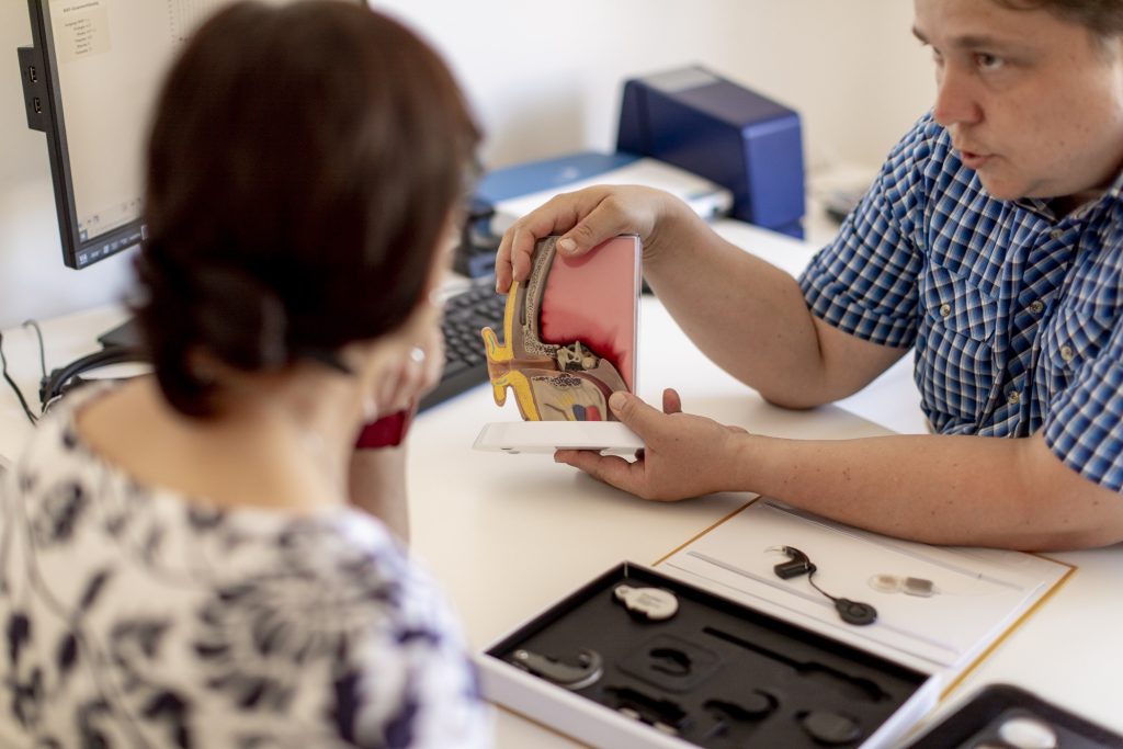
Cochlear implantation is a surgical procedure in which a device called a cochlear implant is inserted into the inner ear (cochlea) of a person with severe to profound hearing loss. The implant consists of an external component, which includes a microphone, processor, and transmitter, and an internal component, which includes a receiver and electrode array.
The microphone picks up sounds from the environment and sends them to the processor, which analyzes and converts the sounds into electrical signals. These signals are then transmitted to the receiver, which stimulates the electrode array in the cochlea. The electrodes directly stimulate the auditory nerve fibers, bypassing the damaged hair cells in the inner ear that are responsible for normal hearing.
The brain interprets these electrical signals as sound, allowing the person to perceive and understand speech and other sounds. Cochlear implantation is typically recommended for people who do not benefit from traditional hearing aids and can significantly improve communication, quality of life, and social integration for those with severe to profound hearing loss.
The tympanic membrane, also known as the eardrum, is a thin, cone-shaped membrane that separates the external auditory canal from the middle ear. It serves to transmit sound vibrations from the air to the inner ear, where they are converted into electrical signals that can be interpreted by the brain as sound. The tympanic membrane is composed of three layers: an outer layer of skin, a middle layer of connective tissue, and an inner layer of mucous membrane. It is held in place by several small bones and muscles and is highly sensitive to changes in pressure.
In the context of medicine, particularly in audiology and otolaryngology (ear, nose, and throat specialty), "noise" is defined as unwanted or disturbing sound in the environment that can interfere with communication, rest, sleep, or cognitive tasks. It can also refer to sounds that are harmful to hearing, such as loud machinery noises or music, which can cause noise-induced hearing loss if exposure is prolonged or at high enough levels.
In some medical contexts, "noise" may also refer to non-specific signals or interfering factors in diagnostic tests and measurements that can make it difficult to interpret results accurately.
The vestibulocochlear nerve, also known as the auditory-vestibular nerve or cranial nerve VIII, is a paired peripheral nerve that transmits sensory information from the inner ear to the brain. It has two distinct parts: the cochlear part and the vestibular part.
The cochlear part is responsible for hearing and transmits sound signals from the cochlea to the brain. The vestibular part, on the other hand, is responsible for maintaining balance and spatial orientation by transmitting information about head movement and position from the vestibular apparatus (utricle, saccule, and semicircular canals) in the inner ear to the brain.
Together, these two parts of the vestibulocochlear nerve play a crucial role in our ability to hear and maintain balance. Damage to this nerve can result in hearing loss, tinnitus (ringing in the ears), vertigo (dizziness), or balance problems.
Cochlear implants are medical devices that are surgically implanted in the inner ear to help restore hearing in individuals with severe to profound hearing loss. These devices bypass the damaged hair cells in the inner ear and directly stimulate the auditory nerve, allowing the brain to interpret sound signals. Cochlear implants consist of two main components: an external processor that picks up and analyzes sounds from the environment, and an internal receiver/stimulator that receives the processed information and sends electrical impulses to the auditory nerve. The resulting patterns of electrical activity are then perceived as sound by the brain. Cochlear implants can significantly improve communication abilities, language development, and overall quality of life for individuals with profound hearing loss.
The middle ear is the middle of the three parts of the ear, located between the outer ear and inner ear. It contains three small bones called ossicles (the malleus, incus, and stapes) that transmit and amplify sound vibrations from the eardrum to the inner ear. The middle ear also contains the Eustachian tube, which helps regulate air pressure in the middle ear and protects against infection by allowing fluid to drain from the middle ear into the back of the throat.
The ear canal, also known as the external auditory canal, is the tubular passage that extends from the outer ear (pinna) to the eardrum (tympanic membrane). It is lined with skin and tiny hairs, and is responsible for conducting sound waves from the outside environment to the middle and inner ear. The ear canal is typically about 2.5 cm long in adults and has a self-cleaning mechanism that helps to keep it free of debris and wax.
Presbycusis is an age-related hearing loss, typically characterized by the progressive loss of sensitivity to high-frequency sounds. It's a result of natural aging of the auditory system and is often seen as a type of sensorineural hearing loss. The term comes from the Greek words "presbus" meaning old man and "akousis" meaning hearing.
This condition usually develops slowly over many years and can affect both ears equally. Presbycusis can make understanding speech, especially in noisy environments, quite challenging. It's a common condition, and its prevalence increases with age. While it's not reversible, various assistive devices like hearing aids can help manage the symptoms.
Auditory pathways refer to the series of structures and nerves in the body that are involved in processing sound and transmitting it to the brain for interpretation. The process begins when sound waves enter the ear and cause vibrations in the eardrum, which then move the bones in the middle ear. These movements stimulate hair cells in the cochlea, a spiral-shaped structure in the inner ear, causing them to release neurotransmitters that activate auditory nerve fibers.
The auditory nerve carries these signals to the brainstem, where they are relayed through several additional structures before reaching the auditory cortex in the temporal lobe of the brain. Here, the signals are processed and interpreted as sounds, allowing us to hear and understand speech, music, and other environmental noises.
Damage or dysfunction at any point along the auditory pathway can lead to hearing loss or impairment.
Labyrinthine fluids, also known as endolymph and perilymph, are fluids that fill the inner ear structures, specifically the bony labyrinth. The bony labyrinth is divided into two main parts: the cochlea, responsible for hearing, and the vestibular system, responsible for balance.
Endolymph is a clear, plasma-like fluid found within the membranous labyrinth, which is a series of interconnected tubes and sacs that lie inside the bony labyrinth. Endolymph plays a crucial role in the functioning of both the cochlea and vestibular system by creating an electrochemical gradient necessary for the conversion of mechanical sound vibrations into electrical signals in the cochlea, as well as facilitating the detection of head movements and maintaining balance in the vestibular system.
Perilymph, on the other hand, is a clear, colorless fluid that fills the space between the bony labyrinth and the membranous labyrinth. It is similar in composition to cerebrospinal fluid (CSF) and serves as a protective cushion for the delicate inner ear structures. Perilymph also helps maintain the electrochemical gradient required for sound transduction in the cochlea.
Disorders related to these labyrinthine fluids, such as endolymphatic hydrops or perilymph fistula, can lead to hearing and balance problems.
The vestibular system is a part of the inner ear that contributes to our sense of balance and spatial orientation. It is made up of two main components: the vestibule and the labyrinth.
The vestibule is a bony chamber in the inner ear that contains two important structures called the utricle and saccule. These structures contain hair cells and fluid-filled sacs that help detect changes in head position and movement, allowing us to maintain our balance and orientation in space.
The labyrinth, on the other hand, is a more complex structure that includes the vestibule as well as three semicircular canals. These canals are also filled with fluid and contain hair cells that detect rotational movements of the head. Together, the vestibule and labyrinth work together to provide us with information about our body's position and movement in space.
Overall, the vestibular system plays a crucial role in maintaining our balance, coordinating our movements, and helping us navigate through our environment.
Efferent neurons are specialized nerve cells that transmit signals from the central nervous system (CNS), which includes the brain and spinal cord, to effector organs such as muscles or glands. These signals typically result in a response or action, hence the term "efferent," derived from the Latin word "efferre" meaning "to carry away."
Efferent neurons are part of the motor pathway and can be further classified into two types:
1. Somatic efferent neurons: These neurons transmit signals to skeletal muscles, enabling voluntary movements and posture maintenance. They have their cell bodies located in the ventral horn of the spinal cord and send their axons through the ventral roots to innervate specific muscle fibers.
2. Autonomic efferent neurons: These neurons are responsible for controlling involuntary functions, such as heart rate, digestion, respiration, and pupil dilation. They have a two-neuron chain arrangement, with the preganglionic neuron having its cell body in the CNS (brainstem or spinal cord) and synapsing with the postganglionic neuron in an autonomic ganglion near the effector organ. Autonomic efferent neurons can be further divided into sympathetic, parasympathetic, and enteric subdivisions based on their functions and innervation patterns.
In summary, efferent neurons are a critical component of the nervous system, responsible for transmitting signals from the CNS to various effector organs, ultimately controlling and coordinating numerous bodily functions and responses.
Auditory evoked potentials (AEP) are medical tests that measure the electrical activity in the brain in response to sound stimuli. These tests are often used to assess hearing function and neural processing in individuals, particularly those who cannot perform traditional behavioral hearing tests.
There are several types of AEP tests, including:
1. Brainstem Auditory Evoked Response (BAER) or Brainstem Auditory Evoked Potentials (BAEP): This test measures the electrical activity generated by the brainstem in response to a click or tone stimulus. It is often used to assess the integrity of the auditory nerve and brainstem pathways, and can help diagnose conditions such as auditory neuropathy and retrocochlear lesions.
2. Middle Latency Auditory Evoked Potentials (MLAEP): This test measures the electrical activity generated by the cortical auditory areas of the brain in response to a click or tone stimulus. It is often used to assess higher-level auditory processing, and can help diagnose conditions such as auditory processing disorders and central auditory dysfunction.
3. Long Latency Auditory Evoked Potentials (LLAEP): This test measures the electrical activity generated by the cortical auditory areas of the brain in response to a complex stimulus, such as speech. It is often used to assess language processing and cognitive function, and can help diagnose conditions such as learning disabilities and dementia.
Overall, AEP tests are valuable tools for assessing hearing and neural function in individuals who cannot perform traditional behavioral hearing tests or who have complex neurological conditions.
In the context of medicine, particularly in the field of auscultation (the act of listening to the internal sounds of the body), "sound" refers to the noises produced by the functioning of the heart, lungs, and other organs. These sounds are typically categorized into two types:
1. **Bradyacoustic sounds**: These are low-pitched sounds that are heard when there is a turbulent flow of blood or when two body structures rub against each other. An example would be the heart sound known as "S1," which is produced by the closure of the mitral and tricuspid valves at the beginning of systole (contraction of the heart's ventricles).
2. **High-pitched sounds**: These are sharper, higher-frequency sounds that can provide valuable diagnostic information. An example would be lung sounds, which include breath sounds like those heard during inhalation and exhalation, as well as adventitious sounds like crackles, wheezes, and pleural friction rubs.
It's important to note that these medical "sounds" are not the same as the everyday definition of sound, which refers to the sensation produced by stimulation of the auditory system by vibrations.
Endolymphatic hydrops is a term used to describe the abnormal accumulation of fluid (endolymph) within the inner ear. This condition is most commonly associated with Meniere's disease, but can also be seen in other disorders that affect the inner ear.
The inner ear is made up of two main parts: the cochlea, which is responsible for hearing, and the vestibular system, which helps to control balance. Both of these systems are filled with fluid, including endolymph, which is a watery fluid that bathes the sensory hair cells in these structures.
In endolymphatic hydrops, there is an overproduction or decreased absorption of endolymph, leading to an abnormal buildup of fluid within the inner ear. This can cause a variety of symptoms, including vertigo (a spinning sensation), tinnitus (ringing in the ears), hearing loss, and a feeling of fullness or pressure in the affected ear.
The exact cause of endolymphatic hydrops is not fully understood, but it is thought to be related to changes in the inner ear's fluid balance. Treatment options may include medications to help control symptoms, as well as surgical procedures to relieve pressure on the inner ear.
The cochlear nucleus is the first relay station in the auditory pathway within the central nervous system. It is a structure located in the lower pons region of the brainstem and receives sensory information from the cochlea, which is the spiral-shaped organ of hearing in the inner ear.
The cochlear nucleus consists of several subdivisions, each with distinct neuronal populations that process different aspects of auditory information. These subdivisions include the anteroventral cochlear nucleus (AVCN), posteroventral cochlear nucleus (PVCN), dorsal cochlear nucleus (DCN), and the granule cell domain.
Neurons in these subdivisions perform various computations on the incoming auditory signals, such as frequency analysis, intensity coding, and sound localization. The output of the cochlear nucleus is then sent via several pathways to higher brain regions for further processing and interpretation, including the inferior colliculus, medial geniculate body, and eventually the auditory cortex.
Damage or dysfunction in the cochlear nucleus can lead to hearing impairments and other auditory processing disorders.
The petrous bone is a part of the temporal bone, one of the 22 bones in the human skull. It is a thick and irregularly shaped bone located at the base of the skull and forms part of the ear and the cranial cavity. The petrous bone contains the cochlea, vestibule, and semicircular canals of the inner ear, which are responsible for hearing and balance. It also helps protect the brain from injury by forming part of the bony structure surrounding the brain.
The term "petrous" comes from the Latin word "petrosus," meaning "stony" or "rock-like," which describes the hard and dense nature of this bone. The petrous bone is one of the densest bones in the human body, making it highly resistant to fractures and other forms of damage.
In medical terminology, the term "petrous" may also be used to describe any structure that resembles a rock or is hard and dense, such as the petrous apex, which refers to the portion of the petrous bone that points towards the sphenoid bone.
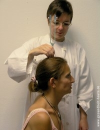
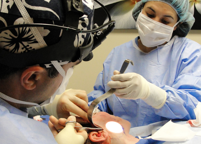
Otologic surgical procedures refer to a range of surgeries performed on the ear or its related structures. These procedures are typically conducted by otologists, who are specialists trained in diagnosing and treating conditions that affect the ears, balance system, and related nerves. The goal of otologic surgery can vary from repairing damaged bones in the middle ear to managing hearing loss, tumors, or chronic infections. Some common otologic surgical procedures include:
1. Stapedectomy/Stapedotomy: These are procedures used to treat otosclerosis, a condition where the stapes bone in the middle ear becomes fixed and causes conductive hearing loss. The surgeon creates an opening in the stapes footplate (stapedotomy) or removes the entire stapes bone (stapedectomy) and replaces it with a prosthetic device to improve sound conduction.
2. Myringoplasty/Tympanoplasty: These are surgeries aimed at repairing damaged eardrums (tympanic membrane). A myringoplasty involves grafting a piece of tissue over the perforation in the eardrum, while a tympanoplasty includes both eardrum repair and reconstruction of the middle ear bones if necessary.
3. Mastoidectomy: This procedure involves removing the mastoid air cells, which are located in the bony prominence behind the ear. A mastoidectomy is often performed to treat chronic mastoiditis, cholesteatoma, or complications from middle ear infections.
4. Ossiculoplasty: This procedure aims to reconstruct and improve the function of the ossicles (middle ear bones) when they are damaged due to various reasons such as infection, trauma, or congenital conditions. The surgeon uses prosthetic devices made from plastic, metal, or even bone to replace or support the damaged ossicles.
5. Cochlear implantation: This is a surgical procedure that involves placing an electronic device inside the inner ear to help individuals with severe to profound hearing loss. The implant consists of an external processor and internal components that directly stimulate the auditory nerve, bypassing the damaged hair cells in the cochlea.
6. Labyrinthectomy: This procedure involves removing the balance-sensing structures (vestibular system) inside the inner ear to treat severe vertigo or dizziness caused by conditions like Meniere's disease when other treatments have failed.
7. Acoustic neuroma removal: An acoustic neuroma is a benign tumor that grows on the vestibulocochlear nerve, which connects the inner ear to the brain. Surgical removal of the tumor is necessary to prevent hearing loss, balance problems, and potential neurological complications.
These are just a few examples of the various surgical procedures performed by otolaryngologists (ear, nose, and throat specialists) to treat conditions affecting the ear and surrounding structures. Each procedure has its specific indications, benefits, risks, and postoperative care requirements. Patients should consult with their healthcare providers to discuss the most appropriate treatment options for their individual needs.
The ear ossicles are the three smallest bones in the human body, which are located in the middle ear. They play a crucial role in the process of hearing by transmitting and amplifying sound vibrations from the eardrum to the inner ear. The three ear ossicles are:
1. Malleus (hammer): The largest of the three bones, it is shaped like a hammer and connects to the eardrum.
2. Incus (anvil): The middle-sized bone, it looks like an anvil and connects the malleus to the stapes.
3. Stapes (stirrup): The smallest and lightest bone in the human body, it resembles a stirrup and transmits vibrations from the incus to the inner ear.
Together, these tiny bones work to efficiently transfer sound waves from the air to the fluid-filled cochlea of the inner ear, enabling us to hear.
Labyrinth diseases refer to conditions that affect the inner ear's labyrinth, which is the complex system of fluid-filled channels and sacs responsible for maintaining balance and hearing. These diseases can cause symptoms such as vertigo (a spinning sensation), dizziness, nausea, hearing loss, and tinnitus (ringing in the ears). Examples of labyrinth diseases include Meniere's disease, labyrinthitis, vestibular neuronitis, and benign paroxysmal positional vertigo. Treatment for these conditions varies depending on the specific diagnosis but may include medications, physical therapy, or surgery.
Cellular mechanotransduction is the process by which cells convert mechanical stimuli into biochemical signals, resulting in changes in cell behavior and function. This complex process involves various molecular components, including transmembrane receptors, ion channels, cytoskeletal proteins, and signaling molecules. Mechanical forces such as tension, compression, or fluid flow can activate these components, leading to alterations in gene expression, protein synthesis, and cell shape or movement. Cellular mechanotransduction plays a crucial role in various physiological processes, including tissue development, homeostasis, and repair, as well as in pathological conditions such as fibrosis and cancer progression.
"CBA" is an abbreviation for a specific strain of inbred mice that were developed at the Cancer Research Institute in London. The "Inbred CBA" mice are genetically identical individuals within the same strain, due to many generations of brother-sister matings. This results in a homozygous population, making them valuable tools for research because they reduce variability and increase reproducibility in experimental outcomes.
The CBA strain is known for its susceptibility to certain diseases, such as autoimmune disorders and cancer, which makes it a popular choice for researchers studying those conditions. Additionally, the CBA strain has been widely used in studies related to transplantation immunology, infectious diseases, and genetic research.
It's important to note that while "Inbred CBA" mice are a well-established and useful tool in biomedical research, they represent only one of many inbred strains available for scientific investigation. Each strain has its own unique characteristics and advantages, depending on the specific research question being asked.
Efferent pathways refer to the neural connections that carry signals from the central nervous system (CNS), which includes the brain and spinal cord, to the peripheral effectors such as muscles and glands. These pathways are responsible for the initiation and control of motor responses, as well as regulating various autonomic functions.
Efferent pathways can be divided into two main types:
1. Somatic efferent pathways: These pathways carry signals from the CNS to the skeletal muscles, enabling voluntary movements and postural control. The final common pathway for somatic motor innervation is the alpha-motor neuron, which synapses directly onto skeletal muscle fibers.
2. Autonomic efferent pathways: These pathways regulate the function of internal organs, smooth muscles, and glands. They are further divided into two subtypes: sympathetic and parasympathetic. The sympathetic system is responsible for the 'fight or flight' response, while the parasympathetic system promotes rest and digestion. Both systems use a two-neuron chain to transmit signals from the CNS to the effector organs. The preganglionic neuron has its cell body in the CNS and synapses with the postganglionic neuron in an autonomic ganglion located near the effector organ. The postganglionic neuron then innervates the target organ or tissue.
In summary, efferent pathways are the neural connections that carry signals from the CNS to peripheral effectors, enabling motor responses and regulating various autonomic functions. They can be divided into somatic and autonomic efferent pathways, with further subdivisions within the autonomic system.
In the context of medicine and physiology, vibration refers to the mechanical oscillation of a physical body or substance with a periodic back-and-forth motion around an equilibrium point. This motion can be produced by external forces or internal processes within the body.
Vibration is often measured in terms of frequency (the number of cycles per second) and amplitude (the maximum displacement from the equilibrium position). In clinical settings, vibration perception tests are used to assess peripheral nerve function and diagnose conditions such as neuropathy.
Prolonged exposure to whole-body vibration or hand-transmitted vibration in certain occupational settings can also have adverse health effects, including hearing loss, musculoskeletal disorders, and vascular damage.
Acoustics is a branch of physics that deals with the study of sound, its production, transmission, and effects. In a medical context, acoustics may refer to the use of sound waves in medical procedures such as:
1. Diagnostic ultrasound: This technique uses high-frequency sound waves to create images of internal organs and tissues. It is commonly used during pregnancy to monitor fetal development, but it can also be used to diagnose a variety of medical conditions, including heart disease, cancer, and musculoskeletal injuries.
2. Therapeutic ultrasound: This technique uses low-frequency sound waves to promote healing and reduce pain and inflammation in muscles, tendons, and ligaments. It is often used to treat soft tissue injuries, arthritis, and other musculoskeletal conditions.
3. Otology: Acoustics also plays a crucial role in the field of otology, which deals with the study and treatment of hearing and balance disorders. The shape, size, and movement of the outer ear, middle ear, and inner ear all affect how sound waves are transmitted and perceived. Abnormalities in any of these structures can lead to hearing loss, tinnitus, or balance problems.
In summary, acoustics is an important field of study in medicine that has applications in diagnosis, therapy, and the understanding of various medical conditions related to sound and hearing.
Hearing disorders, also known as hearing impairments or auditory impairments, refer to conditions that affect an individual's ability to hear sounds in one or both ears. These disorders can range from mild to profound and may result from genetic factors, aging, exposure to loud noises, infections, trauma, or certain medical conditions.
There are mainly two types of hearing disorders: conductive hearing loss and sensorineural hearing loss. Conductive hearing loss occurs when there is a problem with the outer or middle ear, preventing sound waves from reaching the inner ear. Causes include earwax buildup, fluid in the middle ear, a perforated eardrum, or damage to the ossicles (the bones in the middle ear).
Sensorineural hearing loss, on the other hand, is caused by damage to the inner ear (cochlea) or the nerve pathways from the inner ear to the brain. This type of hearing loss is often permanent and can be due to aging (presbycusis), exposure to loud noises, genetics, viral infections, certain medications, or head injuries.
Mixed hearing loss is a combination of both conductive and sensorineural components. In some cases, hearing disorders can also involve tinnitus (ringing or other sounds in the ears) or vestibular problems that affect balance and equilibrium.
Early identification and intervention for hearing disorders are crucial to prevent further deterioration and to help individuals develop appropriate communication skills and maintain a good quality of life.
The vestibulocochlear nerve, also known as the 8th cranial nerve, is responsible for transmitting sound and balance information from the inner ear to the brain. Vestibulocochlear nerve diseases refer to conditions that affect this nerve and can result in hearing loss, vertigo, and balance problems.
These diseases can be caused by various factors, including genetics, infection, trauma, tumors, or degeneration. Some examples of vestibulocochlear nerve diseases include:
1. Vestibular neuritis: an inner ear infection that causes severe vertigo, nausea, and balance problems.
2. Labyrinthitis: an inner ear infection that affects both the vestibular and cochlear nerves, causing vertigo, hearing loss, and tinnitus.
3. Acoustic neuroma: a benign tumor that grows on the vestibulocochlear nerve, causing hearing loss, tinnitus, and balance problems.
4. Meniere's disease: a inner ear disorder that causes vertigo, hearing loss, tinnitus, and a feeling of fullness in the ear.
5. Ototoxicity: damage to the inner ear caused by certain medications or chemicals that can result in hearing loss and balance problems.
6. Vestibular migraine: a type of migraine that is associated with vertigo, dizziness, and balance problems.
Treatment for vestibulocochlear nerve diseases varies depending on the specific condition and its severity. It may include medication, physical therapy, surgery, or a combination of these approaches.
Menière disease is an inner ear disorder that is characterized by episodes of vertigo (a spinning sensation), tinnitus (ringing or buzzing in the ear), hearing loss, and aural fullness (a feeling of pressure or blockage in the ear). It is caused by an abnormal accumulation of endolymphatic fluid in the inner ear, which can lead to damage of the vestibular system and cochlea. The exact cause of this fluid buildup is not known, but it may be related to genetics, allergies, or autoimmune disorders. Menière disease is typically a chronic condition, with symptoms that can vary in frequency and severity over time. Treatment options include dietary modifications, diuretics, vestibular rehabilitation therapy, and, in some cases, surgery.
The ear is the sensory organ responsible for hearing and maintaining balance. It can be divided into three parts: the outer ear, middle ear, and inner ear. The outer ear consists of the pinna (the visible part of the ear) and the external auditory canal, which directs sound waves toward the eardrum. The middle ear contains three small bones called ossicles that transmit sound vibrations from the eardrum to the inner ear. The inner ear contains the cochlea, a spiral-shaped organ responsible for converting sound vibrations into electrical signals that are sent to the brain, and the vestibular system, which is responsible for maintaining balance.
The saccule and utricle are components of the vestibular system, which is responsible for maintaining balance and spatial orientation within the inner ear. Here are the medical definitions:
1. Saccule: A small sac-like structure located in the vestibular labyrinth of the inner ear. It is one of the two otolith organs (the other being the utricle) that detect linear acceleration and gravity. The saccule contains hair cells with stereocilia, which are embedded in a gelatinous matrix containing calcium carbonate crystals called otoconia. When the head changes position or moves linearly, the movement of these otoconia stimulates the hair cells, sending signals to the brain about the direction and speed of the motion.
2. Utricle: Another sac-like structure in the vestibular labyrinth, similar to the saccule but slightly larger. The utricle is also an otolith organ that detects linear acceleration and head tilts. It contains hair cells with stereocilia embedded in a gelatinous matrix filled with otoconia. When the head tilts or moves linearly, the movement of the otoconia stimulates the hair cells, providing information about the position and motion of the head to the brain.
In summary, both the saccule and utricle are essential for maintaining balance and spatial orientation by detecting linear acceleration and gravity through the movement of otoconia on their hair cell receptors.
"Newborn animals" refers to the very young offspring of animals that have recently been born. In medical terminology, newborns are often referred to as "neonates," and they are classified as such from birth until about 28 days of age. During this time period, newborn animals are particularly vulnerable and require close monitoring and care to ensure their survival and healthy development.
The specific needs of newborn animals can vary widely depending on the species, but generally, they require warmth, nutrition, hydration, and protection from harm. In many cases, newborns are unable to regulate their own body temperature or feed themselves, so they rely heavily on their mothers for care and support.
In medical settings, newborn animals may be examined and treated by veterinarians to ensure that they are healthy and receiving the care they need. This can include providing medical interventions such as feeding tubes, antibiotics, or other treatments as needed to address any health issues that arise. Overall, the care and support of newborn animals is an important aspect of animal medicine and conservation efforts.
Audiometry is the testing of a person's ability to hear different sounds, pitches, or frequencies. It is typically conducted using an audiometer, a device that emits tones at varying volumes and frequencies. The person being tested wears headphones and indicates when they can hear the tone by pressing a button or raising their hand.
There are two main types of audiometry: pure-tone audiometry and speech audiometry. Pure-tone audiometry measures a person's ability to hear different frequencies at varying volumes, while speech audiometry measures a person's ability to understand spoken words at different volumes and in the presence of background noise.
The results of an audiometry test are typically plotted on an audiogram, which shows the quietest sounds that a person can hear at different frequencies. This information can be used to diagnose hearing loss, determine its cause, and develop a treatment plan.
Interferometry is not specifically a medical term, but it is used in certain medical fields such as ophthalmology and optics research. Here is a general definition:
Interferometry is a physical method that uses the interference of waves to measure the differences in phase between two or more waves. In other words, it's a technique that combines two or more light waves to create an interference pattern, which can then be analyzed to extract information about the properties of the light waves, such as their wavelength, amplitude, and phase.
In ophthalmology, interferometry is used in devices like wavefront sensors to measure the aberrations in the eye's optical system. By analyzing the interference pattern created by the light passing through the eye, these devices can provide detailed information about the shape and curvature of the cornea and lens, helping doctors to diagnose and treat various vision disorders.
In optics research, interferometry is used to study the properties of light waves and materials that interact with them. By analyzing the interference patterns created by light passing through different materials or devices, researchers can gain insights into their optical properties, such as their refractive index, thickness, and surface roughness.
Pure-tone audiometry is a hearing test that measures a person's ability to hear different sounds, pitches, or frequencies. During the test, pure tones are presented to the patient through headphones or ear inserts, and the patient is asked to indicate each time they hear the sound by raising their hand, pressing a button, or responding verbally.
The softest sound that the person can hear at each frequency is recorded as the hearing threshold, and a graph called an audiogram is created to show the results. The audiogram provides information about the type and degree of hearing loss in each ear. Pure-tone audiometry is a standard hearing test used to diagnose and monitor hearing disorders.
Regional anatomy is a subfield of anatomy that focuses on the study of specific regions or parts of the human body, such as the head and neck, thorax, abdomen, or extremities. It involves the detailed examination of the structures and functions of the organs, tissues, and systems within these regions, including their relationships with adjacent regions. Regional anatomy is often taught in medical schools and other health professions programs to provide a foundation for understanding clinical conditions and performing medical procedures. It is also relevant for professionals in fields such as athletic training, physical therapy, and exercise science, who need to understand the regional anatomy of the body to design safe and effective exercise programs or rehabilitation plans.
Kanamycin is an aminoglycoside antibiotic that is derived from the bacterium Streptomyces kanamyceticus. It works by binding to the 30S subunit of the bacterial ribosome, thereby inhibiting protein synthesis and leading to bacterial cell death. Kanamycin is primarily used to treat serious infections caused by Gram-negative bacteria, such as Pseudomonas aeruginosa, Escherichia coli, and Klebsiella pneumoniae. It is also used in veterinary medicine to prevent bacterial infections in animals.
Like other aminoglycosides, kanamycin can cause ototoxicity (hearing loss) and nephrotoxicity (kidney damage) with prolonged use or high doses. Therefore, it is important to monitor patients closely for signs of toxicity and adjust the dose accordingly. Kanamycin is not commonly used as a first-line antibiotic due to its potential side effects and the availability of safer alternatives. However, it remains an important option for treating multidrug-resistant bacterial infections.
A hearing test is a procedure used to evaluate a person's ability to hear different sounds, pitches, or frequencies. It is performed by a hearing healthcare professional in a sound-treated booth or room with calibrated audiometers. The test measures a person's hearing sensitivity at different frequencies and determines the quietest sounds they can hear, known as their hearing thresholds.
There are several types of hearing tests, including:
1. Pure Tone Audiometry (PTA): This is the most common type of hearing test, where the person is presented with pure tones at different frequencies and volumes through headphones or ear inserts. The person indicates when they hear the sound by pressing a button or raising their hand.
2. Speech Audiometry: This test measures a person's ability to understand speech at different volume levels. The person is asked to repeat words presented to them in quiet and in background noise.
3. Tympanometry: This test measures the function of the middle ear by creating variations in air pressure in the ear canal. It can help identify issues such as fluid buildup or a perforated eardrum.
4. Acoustic Reflex Testing: This test measures the body's natural response to loud sounds and can help identify the location of damage in the hearing system.
5. Otoacoustic Emissions (OAEs): This test measures the sound that is produced by the inner ear when it is stimulated by a sound. It can help identify cochlear damage or abnormalities.
Hearing tests are important for diagnosing and monitoring hearing loss, as well as identifying any underlying medical conditions that may be causing the hearing problems.
The endolymphatic sac is a small, fluid-filled structure that is part of the inner ear. It is located near the vestibular aqueduct and is responsible for maintaining the balance of fluids in the inner ear. The endolymphatic sac also plays a role in the resorption of endolymph, which is the fluid that fills the membranous labyrinth of the inner ear. Disorders of the endolymphatic sac can lead to conditions such as Meniere's disease, which is characterized by vertigo, hearing loss, and tinnitus.
The inferior colliculi are a pair of rounded eminences located in the midbrain, specifically in the tectum of the mesencephalon. They play a crucial role in auditory processing and integration. The inferior colliculi receive inputs from various sources, including the cochlear nuclei, superior olivary complex, and cortical areas. They then send their outputs to the medial geniculate body, which is a part of the thalamus that relays auditory information to the auditory cortex.
In summary, the inferior colliculi are important structures in the auditory pathway that help process and integrate auditory information before it reaches the cerebral cortex for further analysis and perception.
The olivary nucleus is a structure located in the medulla oblongata, which is a part of the brainstem. It consists of two main parts: the inferior olive and the accessory olive. The inferior olive is further divided into several subnuclei.
The olivary nucleus plays an important role in the coordination of movements, particularly in the regulation of fine motor control and rhythmic movements. It receives input from various sources, including the cerebellum, spinal cord, and other brainstem nuclei, and sends output to the cerebellum via the climbing fibers.
Damage to the olivary nucleus can result in a variety of neurological symptoms, including ataxia (loss of coordination), tremors, and dysarthria (speech difficulties). Certain neurodegenerative disorders, such as multiple system atrophy, may also affect the olivary nucleus and contribute to its degeneration.
Stereocilia are hair-like projections found in the inner ear, more specifically in the organ of Corti within the cochlea. They are present on the sensory cells known as hair cells and are involved in hearing by converting sound vibrations into electrical signals that can be transmitted to the brain.
Stereocilia are arranged in rows of graded height, with the tallest ones located near the opening of the cochlea (the base) and the shortest ones closer to the apex. When sound waves reach the inner ear, they cause the fluid within the cochlea to move, which in turn causes stereocilia to bend. This bending action triggers the release of chemical signals that stimulate nerve fibers connected to the hair cells, ultimately transmitting information about the sound to the brain.
Damage or loss of stereocilia can result in hearing impairment or deafness, as seen in various forms of hearing disorders and age-related hearing loss.
Tinnitus is the perception of ringing or other sounds in the ears or head when no external sound is present. It can be described as a sensation of hearing sound even when no actual noise is present. The sounds perceived can vary widely, from a whistling, buzzing, hissing, swooshing, to a pulsating sound, and can be soft or loud.
Tinnitus is not a disease itself but a symptom that can result from a wide range of underlying causes, such as hearing loss, exposure to loud noises, ear infections, earwax blockage, head or neck injuries, circulatory system disorders, certain medications, and age-related hearing loss.
Tinnitus can be temporary or chronic, and it may affect one or both ears. While tinnitus is not usually a sign of a serious medical condition, it can significantly impact quality of life and interfere with daily activities, sleep, and concentration.
 Cochlea
Turritella cochlea
Euophrys cochlea
Carelia cochlea
Modiolus (cochlea)
Heteropsammia cochlea
Evolution of the cochlea
Aspidosiphon muelleri
Cochlear implant
List of Hawaiian animals extinct in the Holocene
Inner ear
Temporal envelope and fine structure
Cochlear amplifier
Sensory nervous system
Turritella
Critical band
Basilar membrane
Bern Riddles
Richard F. Lyon
Tiliqua rugosa
Johnston's organ
Cochlear duct
Analog ear
Oxymeris trochlea
Tectorial membrane
Spiral ganglion
Reticular membrane
Carver Mead
Johann Cochlaeus
Gabriele Faerno
Cochlea - Wikipedia
 Hearing and the cochlea - Health Video: MedlinePlus Medical Encyclopedia
 Cochlea - Dana Foundation
 A TWO-DIMENSIONAL COCHLEA FLUID MODEL - NEW RESULTS. - Nokia Bell Labs
 Cochlea definition | Psychology Glossary | AlleyDog.com
 Frontiers | Molecular Behavior of HMGB1 in the Cochlea Following Noise Exposure and in vitro
 Cochlea & Eustachia - Cochlea & Eustachia Comic book sc by Hans Rickheit Order online
 The Webcomic List - Comic Stats for COCHLEA & EUSTACHIA
 spiral canal of the cochlea - definition and meaning
 Ear Histology - Cochlea (labels) - histology illustration
 Baserange - Cochlea Bamboo Tee
- BONA DRAG
Baserange - Cochlea Bamboo Tee
- BONA DRAG
Higher order cochlea-like channelizing filters | MIT Lincoln Laboratory
 The Cochlea and Passages of the Ear | ClipArt ETC
Hepatocyte Growth Factor-c-MET Signaling Mediates the Development of Nonsensory Structures of the Mammalian Cochlea and Hearing...
Denis Rodd Embryo EP Cochlea Music | Big Shot Magazine
 Silicon cochlea : a digital VLSI implementation of a psychoacoustically and physiologically motivated speech preprocessor -...
 Cochlea - Latest cochlea , Information & Updates - Health -ET HealthWorld
 Spontaneous Cannabinoid Receptor 2 (CB2) Expression in the Cochlea of Adult Albino Rat and Its Up-Regulation after Cisplatin...
 Changing Cochleas, Part 3: Horns, Hisses & Hugs (Activation) | Hearing Health & Technology Matters
 Cochlear implants
| Cochlea
Cochlear implants
| Cochlea
 Figures and data in Gradients of glucose metabolism regulate morphogen signalling required for specifying tonotopic...
 Large Cochlea Shaped Shell Pendant - Audbling
Mechanical microcircuits in the cochlea<...
 To a new hardware design methodology : a case study of the cochlea model - University of Twente Student Theses
 Hearing Loss in children
| Cochlea
Hearing Loss in children
| Cochlea
Audition Promenade round Cochlea autour Cochl e oreille ear organ of Corti oreille ear CRIC Montpellier Remy Pujol U254
 Cochlea - Snuff Puppets
 COCHLEA studia | residency
Vestibular1
- The stapes (stirrup) ossicle bone of the middle ear transmits vibrations to the fenestra ovalis (oval window) on the outside of the cochlea, which vibrates the perilymph in the vestibular duct (upper chamber of the cochlea). (wikipedia.org)
Base of the cochlea5
- The organ of Corti is larger and the basilar membrane on which it sits is longer as it gets further away from the base of the cochlea. (bcm.edu)
- The shorter, smaller structures near the base of the cochlea respond best to high frequencies, while the longer, larger structures near the top of cochlea respond best to low frequencies. (bcm.edu)
- The wide base of the cochlea from which this segment comes is towards the bottom of the page. (bcm.edu)
- With either imaging technique, the cochlea appears as a spiral-shaped structure with 2.5 turns and a normal measured height of 5.1 mm (with a range of 4.4 to 5.9 mm). 1 The cochlear turns (basal, middle, and apical) are separated by interscalar septae, a bony plate radiating from the modiolus that forms the base of the cochlea ( Fig. 3.1 ). (neupsykey.com)
- Note the normal appearance of the modiolus ( star ) at the base of the cochlea. (neupsykey.com)
Length of the cochlea2
- This difference in size is consistent with the fact that different frequencies of sound result in greater vibrations of the organ of Corti depending on where along the length of the cochlea you are measuring. (bcm.edu)
- For many years, cochlear fluids were thought to be generated by filtration of blood or cerebrospinal fluid, which then flowed longitudinally down the length of the cochlea to be absorbed through the endolymphatic sac. (medscape.com)
Modiolus3
- The spiral canal of the cochlea is a section of the bony labyrinth of the inner ear that is approximately 30 mm long and makes 23⁄4 turns about the modiolus. (wikipedia.org)
- The spiral lamina that also projects from the modiolus is a microanatomical osseous structure that separates the spiral of the cochlea into scala tympani (inferiorly), scala media, and scala vestibuli (superiorly). (neupsykey.com)
- The fibers of the cochlear nerve originate from an aggregation of nerve cell bodies in the spiral ganglion, located in the modiolus of the cochlea. (medscape.com)
Organ5
- A core component of the cochlea is the Organ of Corti, the sensory organ of hearing, which is distributed along the partition separating the fluid chambers in the coiled tapered tube of the cochlea. (wikipedia.org)
- This action is passed onto the cochlea, a fluid-filled snail-like structure that contains the organ of Corti, the organ for hearing. (medlineplus.gov)
- The hearing organ in mammals is a spiraling structure called the "cochlea" from the Greek word for snail. (bcm.edu)
- Figure 5 shows a short section of the organ of Corti as it spirals in the cochlea. (bcm.edu)
- Within the cochlea is the organ of Corti, which consists, in part, of about 20,000 specialized cells called hair cells. (merckmanuals.com)
Cochlear nerve3
- Imaging of the cochlea, cochlear nerve, brainstem, and auditory system is central to proper candidate selection and surgical planning in the setting of an auditory brainstem implantation. (neupsykey.com)
- The evaluation of the cochlea and cochlear nerve determines the eligibility of the patient for the cochlear versus auditory brainstem implantation. (neupsykey.com)
- Fig. 3.2 Anatomy of the cochlea and cochlear nerve by high-resolution magnetic resonance imaging (MRI). (neupsykey.com)
Endolymph3
- The cochlea is filled with a watery liquid, the endolymph, which moves in response to the vibrations coming from the middle ear via the oval window. (wikipedia.org)
- The cochlear duct, filled with endolymph, lies in the middle of the cochlea, dividing it into thirds, with the scala vestibuli above and the scala tympani below the cochlear duct. (digitalhistology.org)
- The stria vascularis is a type of epithelium that is uniquely able to produce endolymph in the cochlea. (medscape.com)
Vibrate2
- Hair cells in different parts of the cochlea vibrate in response to different sound frequencies and convert the vibrations into nerve impulses. (merckmanuals.com)
- The sound vibrations cause the fluid and cilia in the cochlea to vibrate. (msdmanuals.com)
Hearing4
- The cochlea is the part of the inner ear involved in hearing. (wikipedia.org)
- The cochlea looks very much like a snail and is a vital component in hearing. (alleydog.com)
- The spiral shaped cochlea originates from one of the balance organs and contains the sensory epithelium for hearing. (bcm.edu)
- Ototoxicity involving the cochlea produces hearing loss, usually commencing with high frequencies but often eventually progressing to the lower frequencies that encompass speech. (medscape.com)
Anatomy3
- Anatomy of the cochlea, unlabeled. (alilamedicalmedia.com)
- Detailed anatomy of the cochlea of human ear in cross section, unlabeled version. (alilamedicalmedia.com)
- Fig. 3.1 Computed tomography (CT) anatomy of the cochlea. (neupsykey.com)
Vibrations2
- The cochlea receives sound in the form of vibrations, which cause the stereocilia to move. (wikipedia.org)
- These vibrations are transmitted to three small bones in the middle ear (called ossicles), then through the oval window into the inner ear, where they enter the cochlea. (msdmanuals.com)
Auditory2
- 2) Sensorineuronal (SNHL) Sound signals are not converted into neurological signals, often due to abnormalities of the inner ear (cochlea or auditory nerve). (cochlea.eu)
- Pure-tone (PT) audiometry measures throughout the outer ear, middle ear, cochlea, cranial nerve (CN) VIII, and central auditory system. (medscape.com)
Neurons1
- The neurons of the spiral ganglion are the first of 4 order neurons between the cochlea and the cerebrum. (medscape.com)
Frequencies2
- Position of the receptors for tones of varying frequencies in the spiral canal of the human cochlea. (siemens-stiftung.org)
- To differentiate these frequencies, the receptors for high tones are at the beginning of the canal, those for the low tones at the apex of the cochlea. (siemens-stiftung.org)
Electrode1
- Cochlear implantation involves the insertion of a thin electrode array inside the cochlea. (upf.edu)
Semicircular1
- Cochlea and semicircular canals. (cdc.gov)
Snail2
- The cochlea is a portion of the inner ear that looks like a snail shell (cochlea is Greek for snail). (wikipedia.org)
- The cochlea (from the Greek word meaning "snail") is a bony, spiral-shaped, fluid-filled tube in the inner ear through which sound waves travel and trigger nerve impulses. (alleydog.com)
Canal1
- Help support Wordnik (and make this page ad-free) by adopting the word spiral canal of the cochlea . (wordnik.com)
Scala vestibuli1
- [ 2 ] In contrast, the composition of perilymph resembles that of extracellular fluid and is high in Na + and low in K + . These differences in electrolyte concentrations remain fairly constant throughout the cochlea, although slight differences are noted in the electrolyte composition of scala vestibuli and scala tympani and between the basal and apical portions of scala media. (medscape.com)
Outer hair cells1
- These sounds are produced specifically by the cochlea and, most probably, by the cochlear outer hair cells as they expand and contract. (medscape.com)
Nerve1
- The electrical distribution around the cochlea is computed, and this is coupled with a model of the nerve fibres around the cochlea. (upf.edu)
Frequency1
- Fibers near the upper end of the cochlea resonate to lower frequency sound. (medlineplus.gov)
Hollow3
- The cochlea (PL: cochleae) is a spiraled, hollow, conical chamber of bone, in which waves propagate from the base (near the middle ear and the oval window) to the apex (the top or center of the spiral). (wikipedia.org)
- The walls of the hollow cochlea are made of bone, with a thin, delicate lining of epithelial tissue. (wikipedia.org)
- The cochlea, a hollow tube coiled in the shape of a snail's shell, is filled with fluid. (merckmanuals.com)
Noise1
- Loud noise can damage cells and membranes in the cochlea. (cdc.gov)
Implementation1
- The topic of the case study was the cochlea model, for which the goal was to design a working implementation of the cochlea model on an FPGA. (utwente.nl)
Consists2
- It consists of tiny hair cells that line the cochlea. (medlineplus.gov)
- The cochlea consists of 3 fluid-filled ducts or scalae (see the image below). (medscape.com)
Biology1
- Due to its relatively independent function accompanying its different cellular localization, a deeper understanding of HMGB1 biology in the cochlea is indispensable for guiding future precise therapeutic interventions. (frontiersin.org)
Acoustic2
- However, the type and function of DAMPs in the cochlea following acoustic trauma is rarely clear. (frontiersin.org)
- However, the spatiotemporal expression of HMGB1 in cochlea with acoustic injury has not been systemically investigated. (frontiersin.org)
Image1
- This is manifested by a standing current that can be recorded within the cochlea (see the first image below). (medscape.com)
Human3
- Cochlea draws on Snuff Puppets' 30-year legacy of giant public spectacles and presenting giant Human Body Parts as symbols of our shared humanity. (snuffpuppets.com)
- There are two and a half turns in the human cochlea and if you were to unwind the cochlea it would stretch to nearly an inch in length. (bcm.edu)
- Researchers at Technische Universität Ilmenau, Kiel University, University College Cork, Karlsruher Institute of Technology, and Fraunhofer Institute for Digital Media Technology Ilmenau have engineered a microelectromechanical sensor inspired by the human cochlea that can process sounds and detect individual signals in noisy environments more effectively than current microphones. (interestingengineering.com)
Axis1
- The central axis of the spiraling cochlea is to the left of the drawing. (bcm.edu)
Fluid2
- The ossicles are essential for efficient coupling of sound waves into the cochlea, since the cochlea environment is a fluid-membrane system, and it takes more pressure to move sound through fluid-membrane waves than it does through air. (wikipedia.org)
- The round window is a small, membrane-covered opening between the fluid-filled cochlea and the middle ear. (merckmanuals.com)
Segment1
- This article is the third in a series, "Changing Cochleas", in which I share this new segment in my lifelong pursuit of better communication. (hearinghealthmatters.org)
Sound3
- The high-quality, sterling silver jewelry is inspired by the shape of the cochlea, the part of the ear that gives us the ability to interpret sound. (audbling.com)
- This window helps dampen the pressure caused by sound waves in the cochlea. (merckmanuals.com)
- The response only emanates from the cochlea, but the outer and middle ear must be able to transmit the emitted sound back to the recording microphone. (medscape.com)
Year2
- COCHLEA studia invite dance-artists internationally to work in Athens all through the year. (cochleares.com)
- This graph shows the total number of publications written about "Cochlea" by people in this website by year, and whether "Cochlea" was a major or minor topic of these publications. (wakehealth.edu)
MODEL1
- The cochlea model in its original form proved to be too large to fit on the selected FPGA. (utwente.nl)
Area2
- Is the Subject Area "Cochlea" applicable to this article? (plos.org)
- COCHLEA studia are located at Lycabettus hill area, near Exarcheia & Kolonaki neighbourhoods, two of the most artistic, alive, and diverse places in central Athens, residential and of extraordinary urbanity, including a number of typical Athenian open-air cinemas on walking distance, affordable quality restaurants for every appetite (incl. (cochleares.com)
Previous work1
- The circuit topology extends previous work on cochlea-like channelizers by introducing multiple resonator-channel filter sections. (mit.edu)
Lower1
- From this go out on the upper side the semi-circular canals (x,y,z) and on the lower side of the passages of the cochlea (k). (usf.edu)