Cornea
Corneal Stroma
Epithelium, Corneal
Keratoconus
Corneal Opacity
Corneal Neovascularization
Corneal Ulcer
Keratoplasty, Penetrating
Corneal Edema
Descemet Membrane
Ophthalmic Nerve
Corneal Topography
Rabbits
Keratitis, Herpetic
Limbus Corneae
Keratan Sulfate
Lasers, Excimer
Conjunctiva
Tissue Preservation
Photorefractive Keratectomy
Corneal Dystrophies, Hereditary
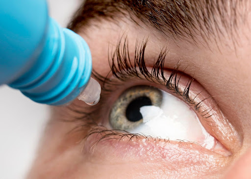
Ophthalmic Solutions
Sodium Hydroxide
Eye Injuries
Fluorescent Antibody Technique, Indirect
Fuchs' Endothelial Dystrophy
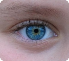
Eye
Organ Culture Techniques
Artificial Organs
Alkalies
Administration, Topical
Bowman Membrane
Keratomileusis, Laser In Situ
Eye Infections, Fungal
Keratitis, Dendritic
Keratin-12
Epithelium
Anterior Eye Segment
Lens, Crystalline
Collagen
Sclera
Microscopy, Confocal
Eye Proteins
Fluorophotometry
Organ Preservation
Corneal Perforation
Aqueous Humor
Descemet Stripping Endothelial Keratoplasty
Anterior Chamber
Iris
Onchocerciasis, Ocular
Cell Count
Contact Lenses, Hydrophilic
Fibrillar Collagens
Immunoenzyme Techniques
Microscopy, Acoustic
Cystic fibrosis transmembrane conductance regulator-mediated corneal epithelial cell ingestion of Pseudomonas aeruginosa is a key component in the pathogenesis of experimental murine keratitis. (1/5105)
Previous findings indicate that the cystic fibrosis transmembrane conductance regulator (CFTR) is a ligand for Pseudomonas aeruginosa ingestion into respiratory epithelial cells. In experimental murine keratitis, P. aeruginosa enters corneal epithelial cells. We determined the importance of CFTR-mediated uptake of P. aeruginosa by corneal cells in experimental eye infections. Entry of noncytotoxic (exoU) P. aeruginosa into human and rabbit corneal cell cultures was inhibited with monoclonal antibodies and peptides specific to CFTR amino acids 108 to 117. Immunofluorescence microscopy and flow cytometry demonstrated CFTR in the intact murine corneal epithelium, and electron microscopy showed that CFTR binds to P. aeruginosa following corneal cell ingestion. In experimental murine eye infections, multiple additions of 5 nM CFTR peptide 103-117 to inocula of either cytotoxic (exoU+) or noncytotoxic P. aeruginosa resulted in large reductions in bacteria in the eye and markedly lessened eye pathology. Compared with wild-type C57BL/6 mice, heterozygous DeltaF508 Cftr mice infected with P. aeruginosa had an approximately 10-fold reduction in bacterial levels in the eye and consequent reductions in eye pathology. Homozygous DeltaF508 Cftr mice were nearly completely resistant to P. aeruginosa corneal infection. CFTR-mediated internalization of P. aeruginosa by buried corneal epithelial cells is critical to the pathogenesis of experimental eye infection, while in the lung, P. aeruginosa uptake by surface epithelial cells enhances P. aeruginosa clearance from this tissue. (+info)First report of Thelazia sp. from a captive Oriental white stork (Ciconia boyciana) in Japan. (2/5105)
Nematodes of the genus Thelazia were recovered from the cornea and inferior conjunctival sac of an immature Oriental white stork (Ciconia boyciana). The bird hatched and reared at the Toyooka Oriental White Stork Breeding Center, Hyogo Prefecture, Japan, but died of chlamydiosis. There were neither gross nor histopathologic ophthalmic lesions. The eye worm from a bird is believed to be first reported in Japan. As regarding reintroduction plan for the Oriental white stork, control measures for prevent further infection with the eye worm will be needed. (+info)Pharmacological studies on root bark of mulberry tree (Morus alba L.) (3/5105)
Pharmacological studies were done on the root bark of mulberry tree and pharmacological effects were compared with the clinical effects of "Sohakuhi" in Chinese medicine. n-Butanol- and water-soluble fractions of mulberry root had similar effects except for those on the cadiovascular system. Both fractions showed cathartic, analgesic, diuretic, antitussive, antiedema, sedative, anticonvulsant, and hypotensive actions in mice, rats, guinea pigs and dogs. There appears to be a correlation between the experimental pharmacological results and the clinical applications of mulberry root found in the literature on Chinese medicine. (+info)Pathogenesis of experimental Pseudomonas keratitis in the guinea pig: bacteriologic, clinical, and microscopic observations. (4/5105)
Uniformly severe corneal infections were produced in guinea pigs by intracorneal injection of about 10 viable Pseudomonas aeruginosa. After a brief lag period, multiplication of bacteria was rapid, reaching geometric means of 280,000 after 24 hr and of 5 million after 48 hr. Within 8 hr after inoculation, polymorphonuclear leukocytes (PMNs) began to infiltrate the anterior two thirds of the stroma. Stromal cells adjacent to the injection site became necrotic and appeared to be engulfed by PMNs. By 14 to 16 hr, an abscess containing a dense aggregate of PMNs and multiplying bacteria developed in the central stroma. By 16 to 24 hr, collagen breakdown was apparent within and around the abscess. Ultrastructural evidence of collagen breakdown included loss of intact collagen fibrils, tactoid formation, and accumulation of amorphous electron-dense material. The area of liquefactive necrosis gradually enlarged, and many corneas perforated after 3 to 4 days. Because the course of infection is highly reproducible, this model should prove useful for many studies of experimental Pseudomonas keratitis. (+info)Freeze-fracture studies of the developing cell surface. II. Particle-free membrane blisters on glutaraldehyde-fixed corneal fibroblasts are artefacts. (5/5105)
We describe, in sections and by freeze-fracture, four classes of intramembrane particle (IMP)-free membrane blebs or "blisters" associated with glutaraldehyde-fixed embryonic corneal fibroblasts: (a) Single blisters attached to the cell membrane; (b) free (detached) vesicles; (c) myelin figures; (d) multivesicular protrusions which resemble the "mounds" described by others on nerve growth cones. The IMP-free, membrane-bounded blisters contain no ground cytoplasm or organelles, in contrast to blebs on trypsin-isolated fibroblasts, which we show here do contain cytoplasm and IMP-rich membranes. That the IMP-free membrane blisters in embryonic corneas are artefacts of fixation is demonstrated by (a) their absence in replicas of fibroblasts frozen and fractured without prior aldehyde fixation and (b) their absence in sections of fibroblasts fixed in a combination of glutaraldehyde and osmium tetroxide. We suggest that the addition of osmium prevents postfixation movement of membrane lipids, especially the negatively charged "fluid" lipids which others have shown are capable of considerable mobility after aldehyde fixation alone. Recent literature has implicated membrane blistering in secretory processes and in growth of nerves, but before the functional significance of such IMP-free blisters is assessed, membrane mobility of the type shown here should be taken into consideration. (+info)cDNA cloning of 15-lipoxygenase type 2 and 12-lipoxygenases of bovine corneal epithelium. (6/5105)
Bovine corneal epithelium contains arachidonate 12- and 15-lipoxygenase activity, while human corneal epithelium contains only 15-lipoxygenase activity. Our purpose was to identify the corneal 12- and 15-lipoxygenase isozymes. We used cDNA cloning to isolate the amino acid coding nucleotide sequences of two bovine lipoxygenases. The translated sequence of one lipoxygenase was 82% identical with human 15-lipoxygenase type 2 and 75% identical with mouse 8-lipoxygenase, whereas the other translated nucleotide sequence was 87% identical with human 12-lipoxygenase of the platelet type. Expression of 15-lipoxygenase type 2 and platelet type 12-lipoxygenase mRNAs were detected by Northern analysis. In addition to these two lipoxygenases, 12-lipoxygenase of leukocyte (tracheal) type was detected by polymerase chain reaction (PCR), sequencing, and Northern analysis. Finally, PCR and sequencing suggested that human corneal epithelium contains 15-lipoxygenase types 1 and 2. (+info)Characterization of proteoglycans synthesized by cultured corneal fibroblasts in response to transforming growth factor beta and fetal calf serum. (7/5105)
A culture system was developed to analyze the relationship between proteoglycans and growth factors during corneal injury. Specifically, the effects of transforming growth factor beta-1 (TGF-beta1) and fetal calf serum on proteoglycan synthesis in corneal fibroblasts were examined. Glycosaminoglycan synthesis and sulfation were determined using selective polysaccharidases. Proteoglycan core proteins were analyzed using gel electrophoresis and Western blotting. Cells cultured in 10% dialyzed fetal calf serum exhibited decreased synthesis of more highly sulfated chondroitin sulfate and heparan sulfate compared with cells cultured in 1% dialyzed fetal calf serum. The amount and sulfation of the glycosaminoglycans was not significantly influenced by TGF-beta1. The major proteoglycan species secreted into the media were decorin and perlecan. Decorin was glycanated with chondroitin sulfate. Perlecan was linked to either chondroitin sulfate, heparan sulfate, or both chondroitin sulfate and heparan sulfate. Decorin synthesis was reduced by either TGF-beta1 or serum. At early time points, both TGF-beta1 and serum induced substantial increases in perlecan bearing chondroitin sulfate and/or heparan sulfate chains. In contrast, after extended periods in culture, the amount of perlecan bearing heparan sulfate chains was unaffected by TGF-beta1 and decreased by serum. The levels of perlecan bearing chondroitin sulfate chains were elevated with TGF-beta1 treatment and were decreased with serum. Because both decorin and perlecan bind growth factors and are proposed to modulate their activity, changes in the expression of either of these proteoglycans could substantially affect the cellular response to injury. (+info)Effect of leukocytes on corneal cellular proliferation and wound healing. (8/5105)
PURPOSE: To establish whether fucoidin, by blocking the adhesion of leukocytes on the limbal vascular endothelium, prevents extravasation of the cells from the blood stream into the limbal stroma and the wounded area after corneal injury. Successful leukocyte blocking enabled investigation of the influence of leukocytes on corneal cellular proliferation after corneal wounding. METHODS: Thirty-two New Zealand White rabbits were used. Photorefractive keratectomy (PRK) and a standardized alkali corneal wound were used as models in two sets of experiments. In half of the injured rabbits fucoidin was used to prevent leukocytes from leaving the local vessels. The efficiency of the blocking technique was evaluated by counting the number of leukocytes in the limbal and wounded corneal areas. Proliferating cell nuclear antigen (PCNA) was used as a marker for proliferative activity. RESULTS: The infiltration of leukocytes into the limbus and the cornea after PRK and alkali injuries can be blocked by fucoidin. The healing rate of corneal epithelium after alkali burn was retarded in the absence of leukocytes. PCNA expression was enhanced in the presence of leukocytes. Fucoidin per se had no influence on corneal cell proliferation and wound healing. CONCLUSIONS: Polymorphonuclear leukocytes (PMNs) can be prevented from entering the cornea in vivo by fucoidin after PRK and after alkali burn. The corneal epithelial healing rate is delayed in the absence of PMNs in vivo, and PCNA expression increases in the presence of leukocytes. (+info)The cornea is the clear, dome-shaped surface at the front of the eye. It plays a crucial role in focusing vision. The cornea protects the eye from harmful particles and microorganisms, and it also serves as a barrier against UV light. Its transparency allows light to pass through and get focused onto the retina. The cornea does not contain blood vessels, so it relies on tears and the fluid inside the eye (aqueous humor) for nutrition and oxygen. Any damage or disease that affects its clarity and shape can significantly impact vision and potentially lead to blindness if left untreated.
The corneal stroma, also known as the substantia propria, is the thickest layer of the cornea, which is the clear, dome-shaped surface at the front of the eye. The cornea plays a crucial role in focusing vision.
The corneal stroma makes up about 90% of the cornea's thickness and is composed of parallel bundles of collagen fibers that are arranged in regular, repeating patterns. These fibers give the cornea its strength and transparency. The corneal stroma also contains a small number of cells called keratocytes, which produce and maintain the collagen fibers.
Disorders that affect the corneal stroma can cause vision loss or other eye problems. For example, conditions such as keratoconus, in which the cornea becomes thin and bulges outward, can distort vision and make it difficult to see clearly. Other conditions, such as corneal scarring or infection, can also affect the corneal stroma and lead to vision loss or other eye problems.
The corneal epithelium is the outermost layer of the cornea, which is the clear, dome-shaped surface at the front of the eye. It is a stratified squamous epithelium, consisting of several layers of flat, scale-like cells that are tightly packed together. The corneal epithelium serves as a barrier to protect the eye from microorganisms, dust, and other foreign particles. It also provides a smooth surface for the refraction of light, contributes to the maintenance of corneal transparency, and plays a role in the eye's sensitivity to touch and pain. The corneal epithelium is constantly being renewed through the process of cell division and shedding, with new cells produced by stem cells located at the limbus, the border between the cornea and the conjunctiva.
The endothelium of the cornea is the thin, innermost layer of cells that lines the inner surface of the cornea, which is the clear, dome-shaped structure at the front of the eye. This single layer of specialized cells is essential for maintaining the transparency and proper hydration of the cornea, allowing light to pass through it and focus on the retina.
The endothelial cells are hexagonal in shape and have tight junctions between them, creating a semi-permeable barrier that controls the movement of water and solutes between the corneal stroma (the middle layer of the cornea) and the anterior chamber (the space between the cornea and the iris). The endothelial cells actively pump excess fluid out of the cornea, maintaining a delicate balance of hydration that is critical for corneal clarity.
Damage to or dysfunction of the corneal endothelium can result in corneal edema (swelling), cloudiness, and loss of vision. Factors contributing to endothelial damage include aging, eye trauma, intraocular surgery, and certain diseases such as Fuchs' dystrophy and glaucoma.
Keratitis is a medical condition that refers to inflammation of the cornea, which is the clear, dome-shaped surface at the front of the eye. The cornea plays an essential role in focusing vision, and any damage or infection can cause significant visual impairment. Keratitis can result from various causes, including bacterial, viral, fungal, or parasitic infections, as well as trauma, allergies, or underlying medical conditions such as dry eye syndrome. Symptoms of keratitis may include redness, pain, tearing, sensitivity to light, blurred vision, and a feeling of something foreign in the eye. Treatment for keratitis depends on the underlying cause but typically includes antibiotics, antivirals, or anti-fungal medications, as well as measures to alleviate symptoms and promote healing.
Keratoconus is a degenerative non-inflammatory disorder of the eye, primarily affecting the cornea. It is characterized by a progressive thinning and steepening of the central or paracentral cornea, causing it to assume a conical shape. This results in irregular astigmatism, myopia, and scattering of light leading to blurred vision, visual distortions, and sensitivity to glare. The exact cause of keratoconus is unknown, but it may be associated with genetics, eye rubbing, and certain medical conditions. It typically starts in the teenage years and progresses into the third or fourth decade of life. Treatment options include glasses, contact lenses, cross-linking, and corneal transplantation in advanced cases.
Corneal opacity refers to a condition in which the cornea, the clear front part of the eye, becomes cloudy or opaque. This can occur due to various reasons such as injury, infection, degenerative changes, or inherited disorders. As a result, light is not properly refracted and vision becomes blurred or distorted. In some cases, corneal opacity can lead to complete loss of vision in the affected eye. Treatment options depend on the underlying cause and may include medication, corneal transplantation, or other surgical procedures.
Eye burns typically refer to injuries or damage to the eyes caused by exposure to harmful substances, extreme temperatures, or radiation. This can result in a variety of symptoms, including redness, pain, tearing, swelling, and blurred vision.
Chemical eye burns can occur when the eyes come into contact with strong acids, alkalis, or other irritants. These substances can cause damage to the cornea, conjunctiva, and other structures of the eye. The severity of the burn will depend on the type and concentration of the chemical, as well as the length of time it was in contact with the eye.
Thermal eye burns can result from exposure to hot or cold temperatures, such as steam, flames, or extreme cold. These types of burns can cause damage to the surface of the eye and may require medical attention to prevent further complications.
Radiation eye burns can occur after exposure to high levels of ultraviolet (UV) light, such as from welding torches, sun lamps, or tanning beds. Prolonged exposure to these sources can cause damage to the cornea and other structures of the eye, leading to symptoms like pain, redness, and sensitivity to light.
If you experience symptoms of an eye burn, it is important to seek medical attention as soon as possible. Treatment may include flushing the eyes with water or saline solution, administering medication to relieve pain and inflammation, or in severe cases, surgery to repair damaged tissue.
Corneal neovascularization is a medical condition that refers to the growth of new, abnormal blood vessels in the cornea, which is the clear, dome-shaped surface at the front of the eye. The cornea typically receives its nutrients from tears and oxygen in the air, so it does not have its own blood vessels. However, when the cornea is damaged or inflamed, it may trigger the growth of new blood vessels from the surrounding tissue into the cornea to promote healing.
Corneal neovascularization can occur due to various eye conditions such as infection, injury, inflammation, degenerative diseases, or contact lens wear. Excessive growth of blood vessels in the cornea can interfere with vision, cause scarring, and increase the risk of corneal transplant rejection. Treatment for corneal neovascularization depends on the underlying cause and may include topical medications, surgery, or other therapies to reduce inflammation, prevent further growth of blood vessels, and preserve vision.
A corneal ulcer is a medical condition that affects the eye, specifically the cornea. It is characterized by an open sore or lesion on the surface of the cornea, which can be caused by various factors such as bacterial or fungal infections, viruses, or injury to the eye.
The cornea is a transparent tissue that covers the front part of the eye and protects it from harmful particles, bacteria, and other foreign substances. When the cornea becomes damaged or infected, it can lead to the development of an ulcer. Symptoms of a corneal ulcer may include pain, redness, tearing, sensitivity to light, blurred vision, and a white spot on the surface of the eye.
Corneal ulcers require prompt medical attention to prevent further damage to the eye and potential loss of vision. Treatment typically involves antibiotics or antifungal medications to eliminate the infection, as well as pain management and measures to protect the eye while it heals. In severe cases, surgery may be necessary to repair the damage to the cornea.
Penetrating keratoplasty (PK) is a type of corneal transplant surgery where the entire thickness of the host's damaged or diseased cornea is removed and replaced with a similar full-thickness portion of a healthy donor's cornea. The procedure aims to restore visual function, alleviate pain, and improve the structural integrity of the eye. It is typically performed for conditions such as severe keratoconus, corneal scarring, or corneal ulcers that cannot be treated with other, less invasive methods. Following the surgery, patients may require extended recovery time and rigorous postoperative care to minimize the risk of complications and ensure optimal visual outcomes.
An Eye Bank is an organization that collects, stores, and distributes donated human eyes for corneal transplantation and other ocular medical research purposes. The eye bank's primary function is to ensure the quality of the donated tissue and make it available for those in need of sight-restoring procedures.
The cornea, the clear front part of the eye, can be surgically transplanted from a deceased donor to a recipient with corneal damage or disease, thereby improving or restoring their vision. The eye bank's role includes obtaining consent for donation, retrieving the eyes from the donor, evaluating the tissue for suitability, preserving it properly, and then allocating it to surgeons for transplantation.
Eye banks follow strict medical guidelines and adhere to ethical standards to ensure the safety and quality of the donated tissues. The process involves screening potential donors for infectious diseases and other conditions that may affect the quality or safety of the cornea. Once deemed suitable, the corneas are carefully removed, preserved in specific solutions, and stored until they are needed for transplantation.
In addition to corneal transplants, eye banks also support research and education in ophthalmology by providing human eye tissues for various studies aimed at advancing our understanding of eye diseases and developing new treatments.
Corneal edema is a medical condition characterized by the accumulation of fluid in the cornea, which is the clear, dome-shaped surface at the front of the eye. This buildup of fluid causes the cornea to swell and thicken, resulting in blurry or distorted vision. Corneal edema can be caused by various factors, including eye injuries, certain medications, eye surgeries, and diseases that affect the eye's ability to pump fluids out of the cornea. In some cases, corneal edema may resolve on its own or with treatment, but in severe cases, it may require a corneal transplant.
The Descemet membrane is the thin, transparent basement membrane that is produced by the corneal endothelial cells. It is located between the corneal stroma and the corneal endothelium, which is the innermost layer of the cornea. The Descemet membrane provides structural support for the corneal endothelium and helps to maintain the proper hydration and clarity of the cornea. It is named after the French physician Jean Descemet, who first described it in 1752.
Corneal keratocytes are specialized cells located within the stroma, which is the thickest layer of the cornea, which is the clear front "window" of the eye. These cells play a crucial role in maintaining the transparency and structural integrity of the cornea. Keratocytes are star-shaped cells that produce and maintain the extracellular matrix (ECM) of the corneal stroma, which consists mainly of collagen fibrils and proteoglycans.
In a healthy cornea, keratocytes exist in a quiescent state, but they can become activated and undergo phenotypic changes in response to injury or disease. Activated keratocytes can differentiate into fibroblasts or myofibroblasts, which participate in the wound healing process by synthesizing ECM components and contracting to help close wounds. However, an overactive or dysregulated wound healing response can lead to corneal opacity, scarring, and visual impairment.
Therefore, understanding the behavior and regulation of corneal keratocytes is essential for developing effective therapies and treatments for various corneal disorders and diseases.
The ophthalmic nerve, also known as the first cranial nerve or CN I, is a sensory nerve that primarily transmits information about vision, including light intensity and color, and sensation in the eye and surrounding areas. It is responsible for the sensory innervation of the upper eyelid, conjunctiva, cornea, iris, ciliary body, and nasal cavity. The ophthalmic nerve has three major branches: the lacrimal nerve, frontal nerve, and nasociliary nerve. Damage to this nerve can result in various visual disturbances and loss of sensation in the affected areas.
Corneal topography is a non-invasive medical imaging technique used to create a detailed map of the surface curvature of the cornea, which is the clear, dome-shaped surface at the front of the eye. This procedure provides valuable information about the shape and condition of the cornea, helping eye care professionals assess various eye conditions such as astigmatism, keratoconus, and other corneal abnormalities. It can also be used in contact lens fitting, refractive surgery planning, and post-surgical evaluation.
I believe there may be some confusion in your question. "Rabbits" is a common name used to refer to the Lagomorpha species, particularly members of the family Leporidae. They are small mammals known for their long ears, strong legs, and quick reproduction.
However, if you're referring to "rabbits" in a medical context, there is a term called "rabbit syndrome," which is a rare movement disorder characterized by repetitive, involuntary movements of the fingers, resembling those of a rabbit chewing. It is also known as "finger-chewing chorea." This condition is usually associated with certain medications, particularly antipsychotics, and typically resolves when the medication is stopped or adjusted.
Herpetic keratitis is a specific type of keratitis (inflammation of the cornea) that is caused by herpes simplex virus (HSV) infection. It is further divided into two types: dendritic and disciform keratitis. Dendritic keratitis is characterized by the development of branching ulcers on the surface of the cornea, while disciform keratitis involves inflammation and opacity in the stroma (middle layer) of the cornea. Both types of herpetic keratitis can cause symptoms such as eye pain, redness, sensitivity to light, tearing, and blurred vision. If left untreated, herpetic keratitis can lead to serious complications, including blindness.
The limbus cornea, also known as the corneoscleral junction, is the border between the transparent cornea and the opaque sclera in the eye. It's a circular, narrow region that contains cells called limbal stem cells, which are essential for maintaining the health and clarity of the cornea. These stem cells continuously regenerate and differentiate into corneal epithelial cells, replacing the outermost layer of the cornea. Any damage or disorder in this area can lead to vision impairment or loss.
Keratan sulfate is a type of glycosaminoglycan (GAG), which is a complex carbohydrate found in connective tissues, including the cornea and cartilage. It is composed of repeating disaccharide units of galactose and N-acetylglucosamine, with sulfate groups attached to some of the sugar molecules.
Keratan sulfate is unique among GAGs because it contains a high proportion of non-sulfated sugars and is often found covalently linked to proteins in structures called proteoglycans. In the cornea, keratan sulfate plays important roles in maintaining transparency and regulating hydration. In cartilage, it contributes to the elasticity and resilience of the tissue.
Abnormalities in keratan sulfate metabolism have been associated with several genetic disorders, including corneal dystrophies and skeletal dysplasias.
An excimer laser is a type of laser that is used in various medical procedures, particularly in ophthalmology and dermatology. The term "excimer" is derived from "excited dimer," which refers to a short-lived molecule formed when two atoms combine in an excited state.
Excimer lasers emit light at a specific wavelength that is determined by the type of gas used in the laser. In medical applications, excimer lasers typically use noble gases such as argon, krypton, or xenon, combined with halogens such as fluorine or chlorine. The most commonly used excimer laser in medical procedures is the excimer laser that uses a mixture of argon and fluoride gas to produce light at a wavelength of 193 nanometers (nm).
In ophthalmology, excimer lasers are primarily used for refractive surgery, such as LASIK and PRK, to correct vision problems like myopia, hyperopia, and astigmatism. The laser works by vaporizing tiny amounts of tissue from the cornea, reshaping its curvature to improve the way light is focused onto the retina.
In dermatology, excimer lasers are used for various skin conditions, including psoriasis, vitiligo, and atopic dermatitis. The laser works by emitting high-energy ultraviolet (UV) light that selectively targets and destroys the abnormal cells responsible for these conditions while leaving surrounding healthy tissue intact.
Excimer lasers are known for their precision, accuracy, and minimal side effects, making them a popular choice in medical procedures where fine detail and tissue preservation are critical.
The conjunctiva is the mucous membrane that lines the inner surface of the eyelids and covers the front part of the eye, also known as the sclera. It helps to keep the eye moist and protected from irritants. The conjunctiva can become inflamed or infected, leading to conditions such as conjunctivitis (pink eye).
Wound healing is a complex and dynamic process that occurs after tissue injury, aiming to restore the integrity and functionality of the damaged tissue. It involves a series of overlapping phases: hemostasis, inflammation, proliferation, and remodeling.
1. Hemostasis: This initial phase begins immediately after injury and involves the activation of the coagulation cascade to form a clot, which stabilizes the wound and prevents excessive blood loss.
2. Inflammation: Activated inflammatory cells, such as neutrophils and monocytes/macrophages, infiltrate the wound site to eliminate pathogens, remove debris, and release growth factors that promote healing. This phase typically lasts for 2-5 days post-injury.
3. Proliferation: In this phase, various cell types, including fibroblasts, endothelial cells, and keratinocytes, proliferate and migrate to the wound site to synthesize extracellular matrix (ECM) components, form new blood vessels (angiogenesis), and re-epithelialize the wounded area. This phase can last up to several weeks depending on the size and severity of the wound.
4. Remodeling: The final phase of wound healing involves the maturation and realignment of collagen fibers, leading to the restoration of tensile strength in the healed tissue. This process can continue for months to years after injury, although the tissue may never fully regain its original structure and function.
It is important to note that wound healing can be compromised by several factors, including age, nutrition, comorbidities (e.g., diabetes, vascular disease), and infection, which can result in delayed healing or non-healing chronic wounds.
Tissue preservation is the process of preventing decomposition or autolysis (self-digestion) of tissues after they have been removed from a living organism. This is typically achieved through the use of fixatives, such as formaldehyde or glutaraldehyde, which stabilize proteins and other cellular structures by creating cross-links between them. Other methods of tissue preservation include freezing, dehydration, and embedding in paraffin or plastic resins. Properly preserved tissues can be stored for long periods of time and used for various research and diagnostic purposes, such as histology, immunohistochemistry, and molecular biology studies.
Photorefractive Keratectomy (PRK) is a type of refractive surgery used to correct vision issues such as nearsightedness, farsightedness, and astigmatism. It works by reshaping the cornea using a laser, which alters how light enters the eye and focuses on the retina.
In PRK, the surgeon removes the thin outer layer of the cornea (epithelium) with an alcohol solution or a blunt surgical instrument before using the laser to reshape the underlying stromal layer. The epithelium then grows back during the healing process, which can take several days.
Compared to LASIK (another type of refractive surgery), PRK has a longer recovery time and may cause more discomfort in the first few days after surgery. However, it is an option for people who are not good candidates for LASIK due to thin corneas or other eye conditions.
It's important to note that while refractive surgeries like PRK can significantly improve vision and reduce dependence on glasses or contact lenses, they may not completely eliminate the need for corrective eyewear in all cases. Additionally, as with any surgical procedure, there are potential risks and complications associated with PRK, including infection, dry eye, and visual disturbances such as glare or halos around lights.
Corneal dystrophies, hereditary are a group of genetic disorders that affect the cornea, which is the clear, outermost layer at the front of the eye. These conditions are characterized by the buildup of abnormal material in the cornea, leading to decreased vision, pain, or cloudiness in the eye.
There are many different types of corneal dystrophies, each affecting a specific layer of the cornea and having its own pattern of inheritance. Some common types include:
1. Fuchs' endothelial dystrophy: This affects the inner lining of the cornea (endothelium) and causes swelling and cloudiness in the cornea. It is typically inherited in an autosomal dominant manner, meaning that a child has a 50% chance of inheriting the condition if one parent has it.
2. Granular dystrophy: This affects the stroma, which is the middle layer of the cornea. It causes the formation of opaque, grayish-white deposits in the cornea that can affect vision. It is typically inherited in an autosomal dominant or recessive manner.
3. Lattice dystrophy: This also affects the stroma and is characterized by the formation of a lattice-like pattern of fine, whitish lines in the cornea. It is typically inherited in an autosomal dominant manner.
4. Macular dystrophy: This affects the central part of the cornea (macula) and can cause cloudiness, leading to decreased vision. It is typically inherited in an autosomal recessive manner.
Treatment for corneal dystrophies may include eyedrops, medications, or surgery, depending on the severity of the condition and its impact on vision. In some cases, a corneal transplant may be necessary to restore vision.
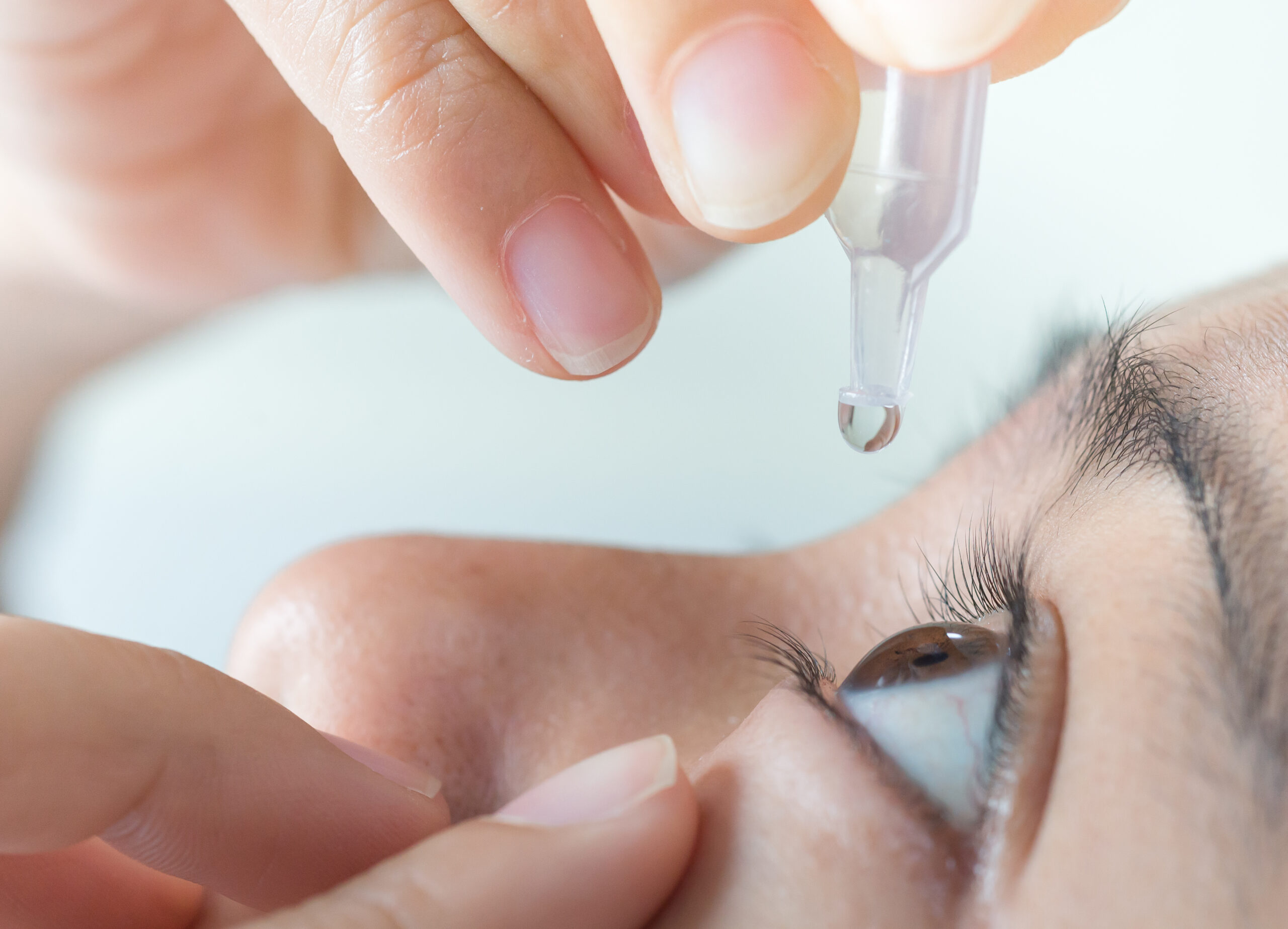
Ophthalmic solutions are sterile, single-use or multi-dose preparations in a liquid form that are intended for topical administration to the eye. These solutions can contain various types of medications, such as antibiotics, anti-inflammatory agents, antihistamines, or lubricants, which are used to treat or prevent ocular diseases and conditions.
The pH and osmolarity of ophthalmic solutions are carefully controlled to match the physiological environment of the eye and minimize any potential discomfort or irritation. The solutions may be packaged in various forms, including drops, sprays, or irrigations, depending on the intended use and administration route.
It is important to follow the instructions for use provided by a healthcare professional when administering ophthalmic solutions, as improper use can lead to eye injury or reduced effectiveness of the medication.
Sodium hydroxide, also known as caustic soda or lye, is a highly basic anhydrous metal hydroxide with the chemical formula NaOH. It is a white solid that is available in pellets, flakes, granules, or as a 50% saturated solution. Sodium hydroxide is produced in large quantities, primarily for the manufacture of pulp and paper, alcohols, textiles, soaps, detergents, and drain cleaners. It is used in many chemical reactions to neutralize acids and it is a strong bases that can cause severe burns and eye damage.
Eye injuries refer to any damage or trauma caused to the eye or its surrounding structures. These injuries can vary in severity and may include:
1. Corneal abrasions: A scratch or scrape on the clear surface of the eye (cornea).
2. Chemical burns: Occurs when chemicals come into contact with the eye, causing damage to the cornea and other structures.
3. Eyelid lacerations: Cuts or tears to the eyelid.
4. Subconjunctival hemorrhage: Bleeding under the conjunctiva, the clear membrane that covers the white part of the eye.
5. Hyphema: Accumulation of blood in the anterior chamber of the eye, which is the space between the cornea and iris.
6. Orbital fractures: Breaks in the bones surrounding the eye.
7. Retinal detachment: Separation of the retina from its underlying tissue, which can lead to vision loss if not treated promptly.
8. Traumatic uveitis: Inflammation of the uvea, the middle layer of the eye, caused by trauma.
9. Optic nerve damage: Damage to the optic nerve, which transmits visual information from the eye to the brain.
Eye injuries can result from a variety of causes, including accidents, sports-related injuries, violence, and chemical exposure. It is important to seek medical attention promptly for any suspected eye injury to prevent further damage and potential vision loss.
The Fluorescent Antibody Technique (FAT), Indirect is a type of immunofluorescence assay used to detect the presence of specific antigens in a sample. In this method, the sample is first incubated with a primary antibody that binds to the target antigen. After washing to remove unbound primary antibodies, a secondary fluorescently labeled antibody is added, which recognizes and binds to the primary antibody. This indirect labeling approach allows for amplification of the signal, making it more sensitive than direct methods. The sample is then examined under a fluorescence microscope to visualize the location and amount of antigen based on the emitted light from the fluorescent secondary antibody. It's commonly used in diagnostic laboratories for detection of various bacteria, viruses, and other antigens in clinical specimens.
Fuchs' Endothelial Dystrophy is a medical condition that affects the eye's cornea. It is a slowly progressing disorder that causes the endothelium, a thin layer of cells lining the inner surface of the cornea, to deteriorate and eventually fail to function properly. This results in swelling of the cornea, leading to cloudy vision, distorted vision, and sensitivity to light.
The condition is typically inherited and tends to affect both eyes. It is more common in women than in men and usually becomes apparent after the age of 50. There is no cure for Fuchs' Endothelial Dystrophy, but treatments such as corneal transplantation can help improve vision and alleviate symptoms.
The eye is the organ of sight, primarily responsible for detecting and focusing on visual stimuli. It is a complex structure composed of various parts that work together to enable vision. Here are some of the main components of the eye:
1. Cornea: The clear front part of the eye that refracts light entering the eye and protects the eye from harmful particles and microorganisms.
2. Iris: The colored part of the eye that controls the amount of light reaching the retina by adjusting the size of the pupil.
3. Pupil: The opening in the center of the iris that allows light to enter the eye.
4. Lens: A biconvex structure located behind the iris that further refracts light and focuses it onto the retina.
5. Retina: A layer of light-sensitive cells (rods and cones) at the back of the eye that convert light into electrical signals, which are then transmitted to the brain via the optic nerve.
6. Optic Nerve: The nerve that carries visual information from the retina to the brain.
7. Vitreous: A clear, gel-like substance that fills the space between the lens and the retina, providing structural support to the eye.
8. Conjunctiva: A thin, transparent membrane that covers the front of the eye and the inner surface of the eyelids.
9. Extraocular Muscles: Six muscles that control the movement of the eye, allowing for proper alignment and focus.
The eye is a remarkable organ that allows us to perceive and interact with our surroundings. Various medical specialties, such as ophthalmology and optometry, are dedicated to the diagnosis, treatment, and management of various eye conditions and diseases.
In medical terms, "tears" are a clear, salty liquid that is produced by the tear glands (lacrimal glands) in our eyes. They serve to keep the eyes moist, protect against dust and other foreign particles, and help to provide clear vision by maintaining a smooth surface on the front of the eye. Tears consist of water, oil, and mucus, which help to prevent evaporation and ensure that the tears spread evenly across the surface of the eye. Emotional or reflexive responses, such as crying or yawning, can also stimulate the production of tears.
Contact lenses are thin, curved plastic or silicone hydrogel devices that are placed on the eye to correct vision, replace a missing or damaged cornea, or for cosmetic purposes. They rest on the surface of the eye, called the cornea, and conform to its shape. Contact lenses are designed to float on a thin layer of tears and move with each blink.
There are two main types of contact lenses: soft and rigid gas permeable (RGP). Soft contact lenses are made of flexible hydrophilic (water-absorbing) materials that allow oxygen to pass through the lens to the cornea. RGP lenses are made of harder, more oxygen-permeable materials.
Contact lenses can be used to correct various vision problems, including nearsightedness, farsightedness, astigmatism, and presbyopia. They come in different shapes, sizes, and powers to suit individual needs and preferences. Proper care, handling, and regular check-ups with an eye care professional are essential for maintaining good eye health and preventing complications associated with contact lens wear.
Organ culture techniques refer to the methods used to maintain or grow intact organs or pieces of organs under controlled conditions in vitro, while preserving their structural and functional characteristics. These techniques are widely used in biomedical research to study organ physiology, pathophysiology, drug development, and toxicity testing.
Organ culture can be performed using a variety of methods, including:
1. Static organ culture: In this method, the organs or tissue pieces are placed on a porous support in a culture dish and maintained in a nutrient-rich medium. The medium is replaced periodically to ensure adequate nutrition and removal of waste products.
2. Perfusion organ culture: This method involves perfusing the organ with nutrient-rich media, allowing for better distribution of nutrients and oxygen throughout the tissue. This technique is particularly useful for studying larger organs such as the liver or kidney.
3. Microfluidic organ culture: In this approach, microfluidic devices are used to create a controlled microenvironment for organ cultures. These devices allow for precise control over the flow of nutrients and waste products, as well as the application of mechanical forces.
Organ culture techniques can be used to study various aspects of organ function, including metabolism, secretion, and response to drugs or toxins. Additionally, these methods can be used to generate three-dimensional tissue models that better recapitulate the structure and function of intact organs compared to traditional two-dimensional cell cultures.
Artificial organs are medical devices that are implanted in the human body to replace the function of a damaged, diseased, or failing organ. These devices can be made from a variety of materials, including metals, plastics, and synthetic biomaterials. They are designed to mimic the structure and function of natural organs as closely as possible, with the goal of improving the patient's quality of life and extending their lifespan.
Some examples of artificial organs include:
1. Artificial heart: A device that is implanted in the chest to replace the function of a failing heart. It can be used as a temporary or permanent solution for patients with end-stage heart failure.
2. Artificial pancreas: A device that is used to treat type 1 diabetes by regulating blood sugar levels. It consists of an insulin pump and a continuous glucose monitor, which work together to deliver insulin automatically based on the patient's needs.
3. Artificial kidney: A device that filters waste products from the blood, similar to a natural kidney. It can be used as a temporary or permanent solution for patients with end-stage renal disease.
4. Artificial lung: A device that helps patients with respiratory failure breathe by exchanging oxygen and carbon dioxide in the blood.
5. Artificial bladder: A device that is implanted in the body to help patients with bladder dysfunction urinate.
6. Artificial eyes: Prosthetic devices that are used to replace a missing or damaged eye, providing cosmetic and sometimes functional benefits.
It's important to note that while artificial organs can significantly improve the quality of life for many patients, they are not without risks. Complications such as infection, rejection, and device failure can occur, and ongoing medical care is necessary to monitor and manage these risks.
Alkalies are a type of basic compound that has a pH level greater than 7. They are also known as bases and can neutralize acids. Alkalies can react with acids to form salts and water. Some common alkalies include sodium hydroxide (lye), potassium hydroxide, and calcium hydroxide. When in solution, alkalies can increase the pH level of a substance, making it more basic or alkaline. They are widely used in various industries for different purposes such as cleaning, manufacturing, and processing.
Topical administration refers to a route of administering a medication or treatment directly to a specific area of the body, such as the skin, mucous membranes, or eyes. This method allows the drug to be applied directly to the site where it is needed, which can increase its effectiveness and reduce potential side effects compared to systemic administration (taking the medication by mouth or injecting it into a vein or muscle).
Topical medications come in various forms, including creams, ointments, gels, lotions, solutions, sprays, and patches. They may be used to treat localized conditions such as skin infections, rashes, inflammation, or pain, or to deliver medication to the eyes or mucous membranes for local or systemic effects.
When applying topical medications, it is important to follow the instructions carefully to ensure proper absorption and avoid irritation or other adverse reactions. This may include cleaning the area before application, covering the treated area with a dressing, or avoiding exposure to sunlight or water after application, depending on the specific medication and its intended use.
The Bowman membrane, also known as the Bowman's capsule, is a part of the nephron in the kidney. It is the outermost layer of the renal corpuscle and surrounds the glomerulus. The primary function of the Bowman membrane is to filter blood and produce urine.
The Bowman membrane is composed of two layers: an inner visceral layer, which is closely applied to the glomerular capillaries, and an outer parietal layer, which forms the inner lining of the Bowman's capsule. The space between these two layers is called the urinary space or Bowman's space.
The filtration process in the Bowman membrane allows for the passage of small molecules such as water, glucose, and amino acids from the blood into the urinary space, while larger molecules like proteins and blood cells are retained in the bloodstream. The fluid that passes through the Bowman membrane then flows into the tubular part of the nephron, where it is further modified before being excreted as urine.
Laser In Situ Keratomileusis (LASIK) is a type of refractive surgery used to correct vision issues such as myopia (nearsightedness), hyperopia (farsightedness), and astigmatism. The procedure involves reshaping the cornea, which is the clear, dome-shaped surface at the front of the eye, using an excimer laser.
In LASIK, a thin flap is created on the surface of the cornea using a femtosecond or microkeratome laser. The flap is then lifted, and the excimer laser is used to reshape the underlying tissue. After the reshaping is complete, the flap is replaced, allowing for quicker healing and visual recovery compared to other refractive surgery procedures.
LASIK is an outpatient procedure that typically takes about 30 minutes or less per eye. Most people can expect to see improved vision within a few days of the procedure, although it may take several weeks for vision to fully stabilize. LASIK has a high success rate and is generally considered safe when performed by a qualified surgeon. However, as with any surgical procedure, there are risks involved, including dry eye, infection, and visual complications such as glare or halos around lights.
Fungal eye infections, also known as fungal keratitis or ocular fungal infections, are caused by the invasion of fungi into the eye. The most common types of fungi that cause these infections include Fusarium, Aspergillus, and Candida. These infections can affect any part of the eye, including the cornea, conjunctiva, sclera, and vitreous humor.
Fungal eye infections often present with symptoms such as redness, pain, sensitivity to light, tearing, blurred vision, and discharge. In severe cases, they can lead to corneal ulcers, perforation of the eye, and even blindness if left untreated. Risk factors for fungal eye infections include trauma to the eye, contact lens wear, immunosuppression, and pre-existing eye conditions such as dry eye or previous eye surgery.
Diagnosis of fungal eye infections typically involves a thorough eye examination, including visual acuity testing, slit lamp examination, and sometimes corneal scrapings for microbiological culture and sensitivity testing. Treatment usually involves topical antifungal medications, such as natamycin or amphotericin B, and in some cases may require oral or intravenous antifungal therapy. In severe cases, surgical intervention may be necessary to remove infected tissue or repair any damage caused by the infection.
Dendritic keratitis is a specific form of keratitis, which is inflammation of the cornea. The term "dendritic" refers to the characteristic appearance of the lesion on the cornea, which resembles a branching tree or a dendrite.
Dendritic keratitis is most commonly caused by herpes simplex virus type 1 (HSV-1) infection, although other infectious and non-infectious etiologies can also produce similar lesions. The condition is characterized by the presence of a branching, dendrite-like ulcer on the corneal epithelium, often accompanied by symptoms such as eye pain, redness, photophobia (sensitivity to light), and tearing.
Treatment for dendritic keratitis typically involves antiviral medications to manage the underlying HSV-1 infection, as well as measures to promote corneal healing and reduce discomfort. It is essential to seek prompt medical attention if you suspect dendritic keratitis, as untreated or improperly managed cases can lead to serious complications, including corneal scarring, vision loss, and potential blindness.
Keratin-1
Epithelium is the tissue that covers the outer surface of the body, lines the internal cavities and organs, and forms various glands. It is composed of one or more layers of tightly packed cells that have a uniform shape and size, and rest on a basement membrane. Epithelial tissues are avascular, meaning they do not contain blood vessels, and are supplied with nutrients by diffusion from the underlying connective tissue.
Epithelial cells perform a variety of functions, including protection, secretion, absorption, excretion, and sensation. They can be classified based on their shape and the number of cell layers they contain. The main types of epithelium are:
1. Squamous epithelium: composed of flat, scalelike cells that fit together like tiles on a roof. It forms the lining of blood vessels, air sacs in the lungs, and the outermost layer of the skin.
2. Cuboidal epithelium: composed of cube-shaped cells with equal height and width. It is found in glands, tubules, and ducts.
3. Columnar epithelium: composed of tall, rectangular cells that are taller than they are wide. It lines the respiratory, digestive, and reproductive tracts.
4. Pseudostratified epithelium: appears stratified or layered but is actually made up of a single layer of cells that vary in height. The nuclei of these cells appear at different levels, giving the tissue a stratified appearance. It lines the respiratory and reproductive tracts.
5. Transitional epithelium: composed of several layers of cells that can stretch and change shape to accommodate changes in volume. It is found in the urinary bladder and ureters.
Epithelial tissue provides a barrier between the internal and external environments, protecting the body from physical, chemical, and biological damage. It also plays a crucial role in maintaining homeostasis by regulating the exchange of substances between the body and its environment.
The anterior eye segment refers to the front portion of the eye, which includes the cornea, iris, ciliary body, and lens. The cornea is the clear, dome-shaped surface at the front of the eye that refracts light entering the eye and provides protection. The iris is the colored part of the eye that controls the amount of light reaching the retina by adjusting the size of the pupil. The ciliary body is a muscle that changes the shape of the lens to focus on objects at different distances. The lens is a transparent structure located behind the iris that further refracts light to provide a clear image. Together, these structures work to focus light onto the retina and enable vision.
The crystalline lens is a biconvex transparent structure in the eye that helps to refract (bend) light rays and focus them onto the retina. It is located behind the iris and pupil and is suspended by small fibers called zonules that connect it to the ciliary body. The lens can change its shape to accommodate and focus on objects at different distances, a process known as accommodation. With age, the lens may become cloudy or opaque, leading to cataracts.
Collagen is the most abundant protein in the human body, and it is a major component of connective tissues such as tendons, ligaments, skin, and bones. Collagen provides structure and strength to these tissues and helps them to withstand stretching and tension. It is made up of long chains of amino acids, primarily glycine, proline, and hydroxyproline, which are arranged in a triple helix structure. There are at least 16 different types of collagen found in the body, each with slightly different structures and functions. Collagen is important for maintaining the integrity and health of tissues throughout the body, and it has been studied for its potential therapeutic uses in various medical conditions.
The sclera is the tough, white, fibrous outer coating of the eye in humans and other vertebrates, covering about five sixths of the eyeball's surface. It provides protection for the delicate inner structures of the eye and maintains its shape. The sclera is composed mainly of collagen and elastic fiber, making it strong and resilient. Its name comes from the Greek word "skleros," which means hard.
Confocal microscopy is a powerful imaging technique used in medical and biological research to obtain high-resolution, contrast-rich images of thick samples. This super-resolution technology provides detailed visualization of cellular structures and processes at various depths within a specimen.
In confocal microscopy, a laser beam focused through a pinhole illuminates a small spot within the sample. The emitted fluorescence or reflected light from this spot is then collected by a detector, passing through a second pinhole that ensures only light from the focal plane reaches the detector. This process eliminates out-of-focus light, resulting in sharp images with improved contrast compared to conventional widefield microscopy.
By scanning the laser beam across the sample in a raster pattern and collecting fluorescence at each point, confocal microscopy generates optical sections of the specimen. These sections can be combined to create three-dimensional reconstructions, allowing researchers to study cellular architecture and interactions within complex tissues.
Confocal microscopy has numerous applications in medical research, including studying protein localization, tracking intracellular dynamics, analyzing cell morphology, and investigating disease mechanisms at the cellular level. Additionally, it is widely used in clinical settings for diagnostic purposes, such as analyzing skin lesions or detecting pathogens in patient samples.
Eye proteins, also known as ocular proteins, are specific proteins that are found within the eye and play crucial roles in maintaining proper eye function and health. These proteins can be found in various parts of the eye, including the cornea, iris, lens, retina, and other structures. They perform a wide range of functions, such as:
1. Structural support: Proteins like collagen and elastin provide strength and flexibility to the eye's tissues, enabling them to maintain their shape and withstand mechanical stress.
2. Light absorption and transmission: Proteins like opsins and crystallins are involved in capturing and transmitting light signals within the eye, which is essential for vision.
3. Protection against damage: Some eye proteins, such as antioxidant enzymes and heat shock proteins, help protect the eye from oxidative stress, UV radiation, and other environmental factors that can cause damage.
4. Regulation of eye growth and development: Various growth factors and signaling molecules, which are protein-based, contribute to the proper growth, differentiation, and maintenance of eye tissues during embryonic development and throughout adulthood.
5. Immune defense: Proteins involved in the immune response, such as complement components and immunoglobulins, help protect the eye from infection and inflammation.
6. Maintenance of transparency: Crystallin proteins in the lens maintain its transparency, allowing light to pass through unobstructed for clear vision.
7. Neuroprotection: Certain eye proteins, like brain-derived neurotrophic factor (BDNF), support the survival and function of neurons within the retina, helping to preserve vision.
Dysfunction or damage to these eye proteins can contribute to various eye disorders and diseases, such as cataracts, age-related macular degeneration, glaucoma, diabetic retinopathy, and others.
Fluorophotometry is a medical diagnostic technique that measures the concentration of fluorescein dye in various tissues, particularly the eye. This technique utilizes a specialized instrument called a fluorophotometer which emits light at a specific wavelength that causes the fluorescein to emit light at a longer wavelength. The intensity of this emitted light is then measured and used to calculate the concentration of fluorescein in the tissue.
Fluorophotometry is often used in ophthalmology to assess the permeability of the blood-retinal barrier, which can be helpful in diagnosing and monitoring conditions such as diabetic retinopathy, age-related macular degeneration, and uveitis. It may also have applications in other medical fields for measuring the concentration of fluorescent markers in various tissues.
Organ preservation is a medical technique used to maintain the viability and functionality of an organ outside the body for a certain period, typically for transplantation purposes. This process involves cooling the organ to slow down its metabolic activity and prevent tissue damage, while using specialized solutions that help preserve the organ's structure and function. Commonly preserved organs include hearts, livers, kidneys, lungs, and pancreases. The goal of organ preservation is to ensure that the transplanted organ remains in optimal condition until it can be successfully implanted into a recipient.
Corneal perforation is a serious eye condition that refers to a hole or rupture in the cornea, which is the clear, dome-shaped surface at the front of the eye. The cornea plays an important role in protecting the eye and focusing light onto the retina. A perforation can result from trauma, infection, degenerative conditions, or surgical complications. It can lead to severe vision loss or blindness if not treated promptly and properly. Treatment typically involves surgery to repair or replace the damaged cornea.
Penetrating eye injuries are a type of ocular trauma where a foreign object or substance pierces the outer layers of the eye and damages the internal structures. This can result in serious harm to various parts of the eye, such as the cornea, iris, lens, or retina, and may potentially cause vision loss or blindness if not promptly treated.
The severity of a penetrating eye injury depends on several factors, including the type and size of the object that caused the injury, the location of the wound, and the extent of damage to the internal structures. Common causes of penetrating eye injuries include sharp objects, such as metal shards or glass fragments, projectiles, such as pellets or bullets, and explosive materials.
Symptoms of a penetrating eye injury may include pain, redness, sensitivity to light, blurred vision, floaters, or the presence of a foreign body in the eye. If you suspect that you have sustained a penetrating eye injury, it is essential to seek immediate medical attention from an ophthalmologist or other healthcare professional with experience in treating eye trauma.
Treatment for penetrating eye injuries may include removing any foreign objects or substances from the eye, repairing damaged tissues, and administering medications to prevent infection and reduce inflammation. In some cases, surgery may be necessary to repair the injury and restore vision. Preventing eye injuries is crucial, and appropriate protective eyewear should be worn when engaging in activities that pose a risk of eye trauma.
Aqueous humor is a clear, watery fluid that fills the anterior and posterior chambers of the eye. It is produced by the ciliary processes in the posterior chamber and circulates through the pupil into the anterior chamber, where it provides nutrients to the cornea and lens, maintains intraocular pressure, and helps to shape the eye. The aqueous humor then drains out of the eye through the trabecular meshwork and into the canal of Schlemm, eventually reaching the venous system.
Descemet Stripping Endothelial Keratoplasty (DSEK) is a type of corneal transplant surgery that involves replacing the damaged endothelium (inner layer) of the cornea with healthy endothelial cells from a donor. In this procedure, the surgeon removes the patient's Descemet's membrane (a thin, clear tissue beneath the endothelium) along with the damaged endothelium. Then, a thin disc of donor tissue, which includes both the endothelium and a small portion of the adjacent corneal stroma, is inserted into the eye and positioned using an air bubble. The new endothelial cells help to pump excess fluid out of the cornea, allowing it to become clear again. DSEK typically results in faster visual recovery and lower rejection rates compared to traditional full-thickness corneal transplantation.
The anterior chamber is the front portion of the eye, located between the cornea (the clear front "window" of the eye) and the iris (the colored part of the eye). It is filled with a clear fluid called aqueous humor that provides nutrients to the structures inside the eye and helps maintain its shape. The anterior chamber plays an important role in maintaining the overall health and function of the eye.
In medical terms, the iris refers to the colored portion of the eye that surrounds the pupil. It is a circular structure composed of thin, contractile muscle fibers (radial and circumferential) arranged in a regular pattern. These muscles are controlled by the autonomic nervous system and can adjust the size of the pupil in response to changes in light intensity or emotional arousal. By constricting or dilating the iris, the amount of light entering the eye can be regulated, which helps maintain optimal visual acuity under various lighting conditions.
The color of the iris is determined by the concentration and distribution of melanin pigments within the iris stroma. The iris also contains blood vessels, nerves, and connective tissue that support its structure and function. Anatomically, the iris is continuous with the ciliary body and the choroid, forming part of the uveal tract in the eye.
Onchocerciasis, Ocular is a medical condition that specifically refers to the eye manifestations caused by the parasitic infection, Onchocerca volvulus. Also known as "river blindness," this disease is spread through the bite of infected blackflies.
Ocular onchocerciasis affects various parts of the eye, including the conjunctiva, cornea, iris, and retina. The infection can cause symptoms such as itching, burning, and redness of the eyes. Over time, it may lead to more serious complications like punctate keratitis (small, scattered opacities on the cornea), cataracts, glaucoma, and ultimately, blindness.
The infection is diagnosed through a skin snip or blood test, which can detect the presence of microfilariae (the larval stage of the parasite) or antibodies against the parasite. Treatment typically involves administering oral medications such as ivermectin, which kills the microfilariae and reduces the risk of eye damage. However, it does not kill the adult worms, so multiple doses are often required to control the infection. In some cases, surgery may be necessary to remove advanced ocular lesions.
"Cell count" is a medical term that refers to the process of determining the number of cells present in a given volume or sample of fluid or tissue. This can be done through various laboratory methods, such as counting individual cells under a microscope using a specialized grid called a hemocytometer, or using automated cell counters that use light scattering and electrical impedance techniques to count and classify different types of cells.
Cell counts are used in a variety of medical contexts, including hematology (the study of blood and blood-forming tissues), microbiology (the study of microscopic organisms), and pathology (the study of diseases and their causes). For example, a complete blood count (CBC) is a routine laboratory test that includes a white blood cell (WBC) count, red blood cell (RBC) count, hemoglobin level, hematocrit value, and platelet count. Abnormal cell counts can indicate the presence of various medical conditions, such as infections, anemia, or leukemia.
Hydrophilic contact lenses are a type of contact lens that is designed to absorb and retain water. These lenses are made from materials that have an affinity for water, which helps them to remain moist and comfortable on the eye. The water content of hydrophilic contact lenses can vary, but typically ranges from 30-80% by weight.
Hydrophilic contact lenses are often used to correct refractive errors such as myopia (nearsightedness), hyperopia (farsightedness), and astigmatism. They can be made in a variety of materials, including soft hydrogel and silicone hydrogel.
One advantage of hydrophilic contact lenses is that they tend to be more comfortable to wear than other types of contacts, as they retain moisture and conform closely to the shape of the eye. However, they may also be more prone to deposits and buildup, which can lead to protein accumulation and discomfort over time. Proper care and cleaning are essential to maintain the health of the eyes when wearing hydrophilic contact lenses.
Fibrillar collagens are a type of collagen that form rope-like fibrils in the extracellular matrix of connective tissues. They are composed of three polypeptide chains, called alpha chains, which are coiled together in a triple helix structure. The most common types of fibrillar collagens are Type I, II, III, V, and XI. These collagens provide strength and support to tissues such as tendons, ligaments, skin, and bones. They also play important roles in the regulation of cell behavior and tissue development. Mutations in genes encoding fibrillar collagens can lead to a variety of connective tissue disorders, including osteogenesis imperfecta, Ehlers-Danlos syndrome, and Marfan syndrome.
Eye diseases are a range of conditions that affect the eye or visual system, causing damage to vision and, in some cases, leading to blindness. These diseases can be categorized into various types, including:
1. Refractive errors: These include myopia (nearsightedness), hyperopia (farsightedness), astigmatism, and presbyopia, which affect the way light is focused on the retina and can usually be corrected with glasses or contact lenses.
2. Cataracts: A clouding of the lens inside the eye that leads to blurry vision, glare, and decreased contrast sensitivity. Cataract surgery is the most common treatment for this condition.
3. Glaucoma: A group of diseases characterized by increased pressure in the eye, leading to damage to the optic nerve and potential blindness if left untreated. Treatment includes medications, laser therapy, or surgery.
4. Age-related macular degeneration (AMD): A progressive condition that affects the central part of the retina called the macula, causing blurry vision and, in advanced stages, loss of central vision. Treatment may include anti-VEGF injections, laser therapy, or nutritional supplements.
5. Diabetic retinopathy: A complication of diabetes that affects the blood vessels in the retina, leading to bleeding, leakage, and potential blindness if left untreated. Treatment includes laser therapy, anti-VEGF injections, or surgery.
6. Retinal detachment: A separation of the retina from its underlying tissue, which can lead to vision loss if not treated promptly with surgery.
7. Amblyopia (lazy eye): A condition where one eye does not develop normal vision, often due to a misalignment or refractive error in childhood. Treatment includes correcting the underlying problem and encouraging the use of the weaker eye through patching or other methods.
8. Strabismus (crossed eyes): A misalignment of the eyes that can lead to amblyopia if not treated promptly with surgery, glasses, or other methods.
9. Corneal diseases: Conditions that affect the transparent outer layer of the eye, such as keratoconus, Fuchs' dystrophy, and infectious keratitis, which can lead to vision loss if not treated promptly.
10. Uveitis: Inflammation of the middle layer of the eye, which can cause vision loss if not treated promptly with anti-inflammatory medications or surgery.
Eyelids are the thin folds of skin that cover and protect the front surface (cornea) of the eye when closed. They are composed of several layers, including the skin, muscle, connective tissue, and a mucous membrane called the conjunctiva. The upper and lower eyelids meet at the outer corner of the eye (lateral canthus) and the inner corner of the eye (medial canthus).
The main function of the eyelids is to protect the eye from foreign particles, light, and trauma. They also help to distribute tears evenly over the surface of the eye through blinking, which helps to keep the eye moist and healthy. Additionally, the eyelids play a role in facial expressions and non-verbal communication.
Ocular refraction is a medical term that refers to the bending of light as it passes through the optical media of the eye, including the cornea and lens. This process allows the eye to focus light onto the retina, creating a clear image. The refractive power of the eye is determined by the curvature and transparency of these structures.
In a normal eye, light rays are bent or refracted in such a way that they converge at a single point on the retina, producing a sharp and focused image. However, if the curvature of the cornea or lens is too steep or too flat, the light rays may not converge properly, resulting in a refractive error such as myopia (nearsightedness), hyperopia (farsightedness), or astigmatism.
Ocular refraction can be measured using a variety of techniques, including retinoscopy, automated refraction, and subjective refraction. These measurements are used to determine the appropriate prescription for corrective lenses such as eyeglasses or contact lenses. In some cases, ocular refractive errors may be corrected surgically through procedures such as LASIK or PRK.
Immunoenzyme techniques are a group of laboratory methods used in immunology and clinical chemistry that combine the specificity of antibody-antigen reactions with the sensitivity and amplification capabilities of enzyme reactions. These techniques are primarily used for the detection, quantitation, or identification of various analytes (such as proteins, hormones, drugs, viruses, or bacteria) in biological samples.
In immunoenzyme techniques, an enzyme is linked to an antibody or antigen, creating a conjugate. This conjugate then interacts with the target analyte in the sample, forming an immune complex. The presence and amount of this immune complex can be visualized or measured by detecting the enzymatic activity associated with it.
There are several types of immunoenzyme techniques, including:
1. Enzyme-linked Immunosorbent Assay (ELISA): A widely used method for detecting and quantifying various analytes in a sample. In ELISA, an enzyme is attached to either the capture antibody or the detection antibody. After the immune complex formation, a substrate is added that reacts with the enzyme, producing a colored product that can be measured spectrophotometrically.
2. Immunoblotting (Western blot): A method used for detecting specific proteins in a complex mixture, such as a protein extract from cells or tissues. In this technique, proteins are separated by gel electrophoresis and transferred to a membrane, where they are probed with an enzyme-conjugated antibody directed against the target protein.
3. Immunohistochemistry (IHC): A method used for detecting specific antigens in tissue sections or cells. In IHC, an enzyme-conjugated primary or secondary antibody is applied to the sample, and the presence of the antigen is visualized using a chromogenic substrate that produces a colored product at the site of the antigen-antibody interaction.
4. Immunofluorescence (IF): A method used for detecting specific antigens in cells or tissues by employing fluorophore-conjugated antibodies. The presence of the antigen is visualized using a fluorescence microscope.
5. Enzyme-linked immunosorbent assay (ELISA): A method used for detecting and quantifying specific antigens or antibodies in liquid samples, such as serum or culture supernatants. In ELISA, an enzyme-conjugated detection antibody is added after the immune complex formation, and a substrate is added that reacts with the enzyme to produce a colored product that can be measured spectrophotometrically.
These techniques are widely used in research and diagnostic laboratories for various applications, including protein characterization, disease diagnosis, and monitoring treatment responses.
Acoustic microscopy is a non-invasive imaging technique that uses sound waves to visualize and analyze the structure and properties of various materials, including biological samples. In the context of medical diagnostics and research, acoustic microscopy can be used to examine tissues, cells, and cellular components with high resolution, providing valuable information about their mechanical and physical properties.
In acoustic microscopy, high-frequency sound waves are focused onto a sample using a transducer. The interaction between the sound waves and the sample generates echoes, which contain information about the sample's internal structure and properties. These echoes are then recorded and processed to create an image of the sample.
Acoustic microscopy offers several advantages over other imaging techniques, such as optical microscopy or electron microscopy. For example, it does not require staining or labeling of samples, which can be time-consuming and potentially damaging. Additionally, acoustic microscopy can provide high-resolution images of samples in their native state, allowing researchers to study the effects of various treatments or interventions on living cells and tissues.
In summary, acoustic microscopy is a non-invasive imaging technique that uses sound waves to visualize and analyze the structure and properties of biological samples with high resolution, providing valuable information for medical diagnostics and research.
Acanthamoeba keratitis is a rare but serious infection of the cornea, which is the clear outer layer at the front of the eye. It's caused by a microscopic organism called Acanthamoeba, which is commonly found in water and soil.
The infection typically occurs in people who wear contact lenses, particularly those who do not clean and disinfect their lenses properly or who swim or shower while wearing their contacts. It can cause pain, redness, blurry vision, sensitivity to light, and a feeling like there's something in your eye.
If left untreated, Acanthamoeba keratitis can lead to serious complications, including corneal scarring, loss of vision, or even blindness. Treatment typically involves the use of specialized antimicrobial drops and sometimes requires a corneal transplant in severe cases. Prevention measures include proper contact lens hygiene, avoiding swimming or showering while wearing contacts, and regularly replacing contact lens storage cases.
 Cornea
Cornel Cornea
Euthria cornea
Cornea (surname)
Niebla cornea
Turbonilla cornea
Cryptogemma cornea
Cornea verticillata
Dimitrie Cornea
Alexandru Cornea
Cornea plana
Cornea (album)
Auricularia cornea
Gheorghe Cornea
Cornea (disambiguation)
Ovidiu Cornea
Turritella cornea
Amastra cornea
Calocera cornea
Atheist's Cornea
Isara cornea
Persicula cornea
Neritodryas cornea
Torellia cornea
Doina Cornea
Cornea, Caraș-Severin
Victor Vlad Cornea
Cornea plana 1
Stroma of cornea
Cornea plana 2
Cornea - Wikipedia
 Cornea and External Disease | Medscape
 Questions & Answers by Costea Cornea - CodeProject
Congenital Clouding of the Cornea: Background, Pathophysiology, Epidemiology
 Controversial Topics in Cornea - American Academy of Ophthalmology
 Descemet's Membrane Biomimetic Microtopography Differentiate... : Cornea
 After daughter's death, Tampa parents deliver her donated cornea
 CL-TL;DR: Upcoming Innovations in Cornea and Contact Lenses | AOA
Is my cornea thick enough for LASIK? - American Academy of Ophthalmology
 Kate Garraway health: Star experienced an inflamed cornea - symptoms | Express.co.uk
 Stem cell therapy used to restore transparency in corneas
 Regenerative approaches for the cornea
 Bausch + Lomb Will Present Scientific Data and Analyses During World Cornea Congress | BioSpace
 Altered epithelial-basement membrane interactions in diabetic corneas
 New Technique Uses Stem Cells To Treat Cornea Damage
 Cornea guidelines - Royal Alex
 Cloudy cornea Information | Mount Sinai - New York
 BostonSight Holds Fifth Annual FitAcademy Retreat for Cornea and Contact Lens Residents
 BostonSight Holds Fifth Annual FitAcademy Retreat for Cornea and Contact Lens Residents
 FELLOWSHIP IN CORNEA AND ANTERIOR SEGMENT - Secretariat of Postgraduate Studies
 Cornea (My Father's Eyes) - Sugarbutch
 Risk of cornea melt reviewed
Make an Appointment - Cornea Service | Wills Eye Hospital
Amyloidosis of the cornea. | British Journal of Ophthalmology
 Woman rips off cornea after wearing contacts for 10 hours, Health News - AsiaOne
 Artificial Cornea and Corneal Implant Market worth $599 million by 2026
 Cornea Research Foundation of America - Glaucoma
Cornea Research Foundation of America - Register
 Cornea and External Disease | UK Healthcare
 Cornea Program | Children's Hospital Los Angeles
Transplants12
- Each year in the US, over 40,000 people receive transplants of the cornea-the clear front part of the eye that light goes through first. (sciencefriday.com)
- IRA FLATOW: But first, each year in the United States, some-40,000 people receive transplants of the cornea. (sciencefriday.com)
- IRA FLATOW: Mm. Now, I mentioned cornea transplants, and they often are successful for many patients. (sciencefriday.com)
- On the basis of transplant type, the artificial cornea and corneal implants market is segmented into penetrating keratoplasty, endothelial keratoplasty, and other transplants (including anterior lamellar keratoplasty (ALK) and keratoprosthesis). (marketsandmarkets.com)
- March 20, 2015 - The Indianapolis Business Journal honored the Foundation with this distinguished award for pioneering advancements and training for DMEK cornea transplants. (cornea.org)
- The technique could be used in the future to ensure an unlimited supply of corneas, but it will be several years of testing before they could be used in transplants, according to the scientists. (technovelgy.com)
- For cataract surgery we use the Catalys system and for cornea transplants we were one of the first to use the Intralase femtosecond laser. (laser-prk.com)
- Without limbal cells and a healthy eye surface, patients are unable to undergo artificial cornea transplants, the current standard of vision rehabilitation. (worldhealth.net)
- Cornea transplants are performed to restore vision loss due to injuries, infection, diseases, or scarring of the cornea. (assileye.com)
- Are all cornea transplants the same? (assileye.com)
- There are several surgical approaches to cornea transplants (known as corneal keratoplasty). (assileye.com)
- At AGEI we perform three types of cornea transplants to address a wide variety of conditions. (assileye.com)
20232
- NEEDHAM, MA, UNITED STATES, September 19, 2023/ EINPresswire.com / -- BostonSight ® , a nonprofit eye healthcare organization that advances the treatment of diseased and damaged corneas and dry eyes held their annual FitAcademy ™ Retreat for Residents at New England College of Optometry (NECO) in Boston, September 15-16, 2023. (wtnh.com)
- Cornea;2023 Jun 15. (bvsalud.org)
Glaucoma2
- A common reason for congenital clouding of the cornea is congenital glaucoma . (medscape.com)
- We treat a full spectrum of conditions involving the cornea, lens, glaucoma and anterior segment. (chla.org)
Stem cells8
- Stem cells collected from human corneas restore transparency and don't trigger a rejection response when injected into eyes that are scarred and hazy, according to experiments conducted in mice by researchers at the University of Pittsburgh School of Medicine. (news-medical.net)
- A few years ago, Dr. Funderburgh and other University of Pittsburgh researchers identified stem cells in a layer of the cornea called the stroma, and they recently showed that even after many rounds of expansion in the lab, these cells continued to produce the biochemical components, or matrix, of the cornea. (news-medical.net)
- The cornea and its stromal stem cells themselves appear to be "immune privileged," meaning they don't trigger a significant immune response even when transplanted across species, as in the Pitt experiments. (news-medical.net)
- And the reason it is clear is because there are stem cells that reside in the periphery of the cornea. (sciencefriday.com)
- And these corneal stem cells prevent the white part of the eye, or conjunctiva, from growing onto the cornea and maintain the corneal clarity. (sciencefriday.com)
- Believe it or not, our bodies have stem cells even though we are adults, and they are the source of corneal epithelium or surface of a cornea. (sciencefriday.com)
- ULA JURKUNAS: So if you don't have stem cells, you can replace the central cornea with a new cornea, but it will never take as these stem cells will not be there to regenerate the front layer of the transplant. (sciencefriday.com)
- Stem cells (human corneal stromal cells) from a healthy donor s cornea were mixed together with alginate and collagen** to create a bio-ink solution. (technovelgy.com)
Transplantation8
- The current popularity of LASIK corrective eye surgery is expected to substantially reduce the availability of donor tissue because the procedure alters the cornea in a way that makes it unsuitable for transplantation. (news-medical.net)
- For decades, corneal transplantation using human donor corneas has been the only option for treating corneal blindness. (diva-portal.org)
- Despite recent improvement in surgical techniques, donor cornea transplantation remains plagued by risks of suboptimal optical results and visual acuity, immune rejection and eventually graft failure. (diva-portal.org)
- It is a made-in-India product by an Indian clinician-scientist team and the first 3D printed human cornea that is optically and physically suitable for transplantation. (indiatimes.com)
- Younger participants, those whose work or studies were unrelated to the medical field, and those with the least knowledge about cornea donation and transplantation were more likely to change their views towards donation by the end of the survey (42.3%, 44.8% and 82.1% increase in willingness to donate, respectively). (bmj.com)
- According to the Global Survey of Cornea Transplantation and Eye Banking, the cornea procurement rate per capita is only 0.92 × 10 −6 and Greece is classified as a non-sufficient country. (bmj.com)
- These can range from transplantation of just the inner corneal lining to transplantation of all layers of the cornea (called a full-thickness transplant). (assileye.com)
- https://en.wikipedia.org/wiki/Organ_transplantation. (who.int)
Conditions involving1
- We provide comprehensive medical and surgical management of conditions involving the cornea, conjunctiva and ocular surface, as well as disorders of the anterior chamber, iris and lens. (uky.edu)
Ophthalmology1
- Our early results suggest that CALEC might offer hope to patients who had been left with untreatable vision loss and pain associated with major cornea injuries," said principal investigator and lead study author Ula Jurkunas, MD, associate director of the Cornea Service at Mass Eye and Ear and an associate professor of ophthalmology at Harvard Medical School. (worldhealth.net)
Keratoconus6
- On the basis of disease indication, the artificial cornea and corneal implants market is segmented into fungal keratitis, Fuchs dystrophy, keratoconus, and other diseases. (marketsandmarkets.com)
- For example, if the average cornea is 520µm, the standard deviation to the thick side might be 20µm but only 7µm to the thin side (virtually all corneas that are 490µm thick have keratoconus, but those that are 600µm thick are normal). (crstoday.com)
- The ophthalmologists at Cornea Eye Institute, are laser vision specialists providing patients with the very latest treatments for myopia, astigmatism, cataracts and Keratoconus from their comfortable and innovative offices in Los Angeles, Beverly Hills and Orange County. (laser-prk.com)
- For patients with keratoconus we have been offering cornea cross-linking for the past 15 years and have treated over 2000 patients. (laser-prk.com)
- We provide consultations for Vision Correction, Cornea, Keratoconus, and Cataracts in Los Angeles and Orange County. (laser-prk.com)
- Recurrence of keratoconus in donor cornea 22 years after successful keratoplasty. (bmj.com)
Surgical3
- The cornea can be reshaped by surgical procedures such as LASIK. (wikipedia.org)
- Dr. Kugler, a surgical associate and cornea/refractive surgery fellow with Ming Wang, MD, PhD, in Nashville, TN, briefly described four cases of corneal melt that had one similarity, namely, that the complication developed in the area of the incision. (ophthalmologytimes.com)
- Dr Falguni Pati, Associate Professor, Department of Biomedical Engineering at IIT-Hyderabad, added "we used a biomimicking approach to provide an optimised micro-environment for stromal regeneration while maintaining the curvature and thickness of the bio-printed cornea to facilitate surgical implantation. (indiatimes.com)
Tissue12
- Transparency, avascularity, the presence of immature resident immune cells, and immunologic privilege makes the cornea a very special tissue. (wikipedia.org)
- Mice that lack the ability to produce lumican develop opaque areas of their corneas comparable to the scar tissue that human eyes form in response to trauma and inflammation, Dr. Funderburgh said. (news-medical.net)
- RA'ANANA, Israel , Jan. 11, 2021 /PRNewswire/ -- The CorNeat KPro, the first artificial cornea which completely integrates with the eye wall with no reliance on donor tissue, was successfully implanted in a human. (prnewswire.co.uk)
- People who experience chemical burns and other eye injuries may develop limbal stem cell deficiency, an irreversible loss of cells on the tissue surrounding the cornea. (worldhealth.net)
- A cornea transplant involves removing part or all of the cornea and replacing it with healthy tissue from a deceased human donor. (assileye.com)
- What's more, a cornea doesn't need to be tissue matched, like a heart or a kidney does. (assileye.com)
- This is because the cornea has no blood vessels which greatly decreases its risk of tissue rejection. (assileye.com)
- It does so by ensuring that the shape of the graft tissue taken from the donor exactly matches (like a puzzle) the graft deposit site in the recipient´s cornea. (assileye.com)
- PK is another full-thickness transplant procedure in which the surgeon removes and replaces all layers of the diseased cornea with clear, healthy donor tissue. (assileye.com)
- The differences between the healthy and the smoky cornea concerned the temperature ranges in which there appeared the decomposition of loosely-bound water and β-relaxation associated with polar side-chains relaxations on protein molecules of this tissue. (aaem.pl)
- The results are interpreted as caused by the toxic compounds of the tobacco smoke leading to higher ion transport in the nonhomogeneous structure of the cornea when compared to that of the control tissue. (aaem.pl)
- Recognition of the effect of frequency and temperature on the dielectric behaviour of the smoky cornea may be of interest for disease characterization of this tissue. (aaem.pl)
Sclera3
- The human cornea borders with the sclera at the corneal limbus. (wikipedia.org)
- In lampreys, the cornea is solely an extension of the sclera, and is separate from the skin above it, but in more advanced vertebrates it is always fused with the skin to form a single structure, albeit one composed of multiple layers. (wikipedia.org)
- The outer layer or tunic (sclera, or white, and cornea) is fibrous and protective. (mountsinai.org)
Keratoplasty3
- The report ' Artificial Cornea and Corneal Implant Market by Type (Human Cornea, Artificial Cornea), Transplant Type (Penetrating Keratoplasty, Endothelial Keratoplasty), Disease Indication, End Users (Hospitals, Specialty Clinics & ASCs) - Global Forecast to 2026' The global artificial cornea and corneal implant market is projected to reach USD 599 million by 2026 from USD 418 million in 2021, at a CAGR of 7.4% from 2021 to 2026. (marketsandmarkets.com)
- Penetrating keratoplasty segment accounted for the largest share of artificial cornea and corneal implant market in 2020. (marketsandmarkets.com)
- In 2020, the penetrating keratoplasty segment accounted for the largest share of the global artificial cornea and corneal implants market. (marketsandmarkets.com)
Disease3
- Primary corneal disease is developmental and may be isolated to the cornea or have a related systemic component. (medscape.com)
- 1 If the number of corneal endothelial cells (CECs) falls below a certain threshold because of traumatic injury, disease, or normal aging processes, functionality of the CE is decreased, and the cornea swells and becomes milky, leading to eventual vision loss. (lww.com)
- However, the lessons learned from the use of the cornea as a disease model will allow for extension of the biomaterials and techniques for regeneration of more complex organs such as the heart. (diva-portal.org)
20222
- In this interview from AAO 2022, Dr. Anthony Aldave talked about controversies in cornea discussed at the meeting such as the preferred treatment for recurrent corneal erosions, and the role of steroids in Acanthamoeba keratitis treatment. (aao.org)
- VAUGHAN, ON, Sept. 27, 2022 /PRNewswire/ -- Bausch + Lomb Corporation (NYSE/TSX: BLCO) ("Bausch + Lomb"), a leading global eye health company dedicated to helping people see better to live better, today announced the presentation of one podium and two poster presentations at the World Cornea Congress VIII meeting, which will take place in Chicago from Sept. 28-29, 2022. (biospace.com)
Transparency4
- Because transparency is of prime importance, the healthy cornea does not have or need blood vessels within it. (wikipedia.org)
- There are 2 theories of how transparency in the cornea comes about:- 1. (wikipedia.org)
- And when that happens, the cornea loses its transparency. (sciencefriday.com)
- A cloudy cornea is a loss of transparency of the cornea. (mountsinai.org)
Stromal2
- Other rarer causes of congenital clouding or opacity of the cornea include the following: corneal keloids, congenital corneal ectasia, congenital hereditary stromal dystrophy, posterior polymorphous dystrophy , and Fryns syndrome. (medscape.com)
- The cornea showed a large central epithelial defect with underlying stromal infiltrate and Descemet folds. (cdc.gov)
Refractive disorders2
- Cornea and external diseases range from refractive disorders to pink eye and dry eye. (uky.edu)
- As one of the few programs in the United States to offer specialized treatment in pediatric corneal and refractive disorders, the Cornea Program in The Vision Center at Children's Hospital Los Angeles serves as the major referral center for pediatric eye care. (chla.org)
Lens7
- Along with the anterior chamber and lens, the cornea refracts light, accounting for approximately two-thirds of the eye's total optical power. (wikipedia.org)
- This rapid-fire lecture will provide a brief summary of upcoming innovations and research into cornea and contact lens technology. (aoa.org)
- The educational program of FitAcademy offers our Cornea and Contact Lens residents skills to be life-changing providers for their future patients. (wtnh.com)
- This year marks the fifth anniversary of this free event for emerging clinicians in cornea and contact lens residencies and fellowships at schools and colleges of optometry and accredited fellowship sites throughout the United States and Canada. (wtnh.com)
- This year, 29 cornea and contact lens residents attended in person for the full curriculum, which included a hands-on wet lab training. (wtnh.com)
- The educational program of FitAcademy offers our Cornea and Contact Lens residents skills to be life-changing providers for their future patients," shares Dr. Jennifer Liao Assistant Professor of Optometry at NECO and Attending Optometrist at NECO Center for Eye Care. (wtnh.com)
- Most vertebrates have UV-sensitive vision, but the UV sensitivity of their eyes is limited by the transmittance of the ocular media, and the specific contribution of the different media (cornea, lens) has remained unclear. (lu.se)
Implantation2
- Dr. Kugler cited a report by Sépideh Samimi, MD, and associates of histopathologic findings in corneas from eyes that required a corneal transplant following implantation of the inserts. (ophthalmologytimes.com)
- The CorNeat KPro implant is designed to replace deformed, scarred or opacified corneas and is expected to fully and immediately rehabilitate the vision of corneally blind patients following a relatively simple implantation procedure. (prnewswire.co.uk)
Thickened stroma1
- dua's layer : the fourth layer of cornea is dua's layer which is nothing but a thickened stroma in the poster most part of the stroma this was recently declared as the newly added structure, thus making the cornea a 6 layered structure rather than just 5 layer structure. (wikipedia.org)
Weakens1
- The lamellar procedure weakens the cornea enough to allow for ectasia. (crstoday.com)
Blindness1
- The CorNeat KPro will transform global corneal therapy and provide, for the first time, a reliable and scalable synthetic substitute to the human cornea, significantly impacting the lives of millions of people with cornea-related visual impairments and blindness. (prnewswire.co.uk)
Artificial corneas3
- Still more patients with damaged corneas might receive artificial corneas to help restore clear vision. (sciencefriday.com)
- Other people receive artificial corneas to help restore clear vision. (sciencefriday.com)
- The scarcity of human eye donors has resulted in the development of innovative solutions such as artificial corneas. (marketsandmarkets.com)
Antibiotic Resistance1
- In Vitro Antibiotic Resistance among Bacterial Pathogens Sourced from the Cornea in ARMOR. (biospace.com)
Specialty1
- Based on end users, the artificial cornea and corneal implants market is segmented into hospitals and specialty clinics and ambulatory surgery centers (ASCs). (marketsandmarkets.com)
Patient's1
- Once delivered, a surgeon will cut out the patient's damaged or diseased cornea - an area roughly the size of a small button - and then stitch in the donated cornea. (tampabay.com)
Periphery2
- In humans, the cornea has a diameter of about 11.5 mm and a thickness of 0.5-0.6 mm in the center and 0.6-0.8 mm at the periphery. (wikipedia.org)
- In total sclerocornea, the entire cornea is involved, but the center of the cornea is clearer than the periphery. (medscape.com)
Opacification1
- Congenital clouding or opacification of the normally clear cornea can result from various genetic, metabolic, developmental, and idiopathic causes. (medscape.com)
Conjunctiva1
- And again, the conjunctiva will overgrow the clear part of the cornea. (sciencefriday.com)
Request an appointment1
- To request an appointment with a Wills Eye Cornea doctor, please complete the form below. (willseye.org)
Institute's2
- The institute's technicians cut the corneas to fit donors. (tampabay.com)
- It is Cornea Eye Institute's policy to respect your privacy regarding any information we may collect from you across our website, laser-prk.com , and other sites we own and operate. (laser-prk.com)
Infection2
- The bio-ink used to make this 3D printed cornea can be sight-saving for army personnel at the site of injury to seal corneal perforation and prevent infection during war-related injuries or in a remote area with no tertiary eye care facility. (indiatimes.com)
- Patient outcomes include permanent vision loss resulting from cornea infection, hospitalization, and one death due to systemic infection. (cdc.gov)
Thickness5
- Clinically, there can be no doubt that a cornea with a central thickness of 500µm has a much greater chance of developing irregular astigmatism (mild ectasia) after LASIK compared to a cornea with a preoperative central thickness of at least 515µm. (crstoday.com)
- In my opinion, it is prudent to perform a surface laser procedure on "normal" corneas that have a central corneal thickness of 505µm or less. (crstoday.com)
- Vision is recovered in a matter of weeks, as opposed to months required for a full-thickness cornea transplant. (assileye.com)
- OMT λ T0.5 is positively correlated with eye size, but λ T0.5 of corneas and lenses are not correlated with their thickness when controlled for phylogeny. (lu.se)
- of corneas and lenses are not correlated with their thickness when controlled for phylogeny. (lu.se)
Clouding1
- Clouding may affect all or part of the cornea. (mountsinai.org)
Specialists2
- Our team of top-level cornea and laser vision correction specialists proudly serves their patients with compassionate, and expert eye care. (laser-prk.com)
- Cornea specialists have been hindered by a lack of treatment options with a high safety profile to help our patients with chemical burns and injuries that render them unable to get an artificial cornea transplant. (worldhealth.net)
Anterior chamber1
- The cornea (PL: corneas or corneae) is the transparent front part of the eye that covers the iris, pupil, and anterior chamber. (wikipedia.org)
Vitro1
- To determine if the abnormal basement membrane in diabetes has altered epithelial recognition of sites for hemidesmosome formation and if diabetic epithelium can recognize sites for hemidesmosome formation on normal basement membrane, we recombined epithelial sheets and corneal stromas with denuded basement membranes in vitro using diabetic and control corneas. (nih.gov)
Collagen1
- Whether the percentage of thin corneas that have a hidden collagen defect is 5% or 50%, LASIK is too risky. (crstoday.com)
Diseases3
- Certain metabolic diseases, trauma, infectious diseases, nutritional deficiencies and environmental situations may cause the cornea to be less transparent and cloudy. (mountsinai.org)
- Market growth is largely driven by the The growing geriatric population and the rising prevalence of eye diseases are the major drivers for the artificial cornea and corneal implants market. (marketsandmarkets.com)
- Autoimmune diseases affecting the cornea and ocular surface. (uky.edu)
Organ2
- We live in an online era and incorporation of online tools and applications in awareness campaigns towards cornea and organ donation has become a necessity. (bmj.com)
- Educating the public using online tools can have a significant impact on raising willingness to become a cornea/organ donor. (bmj.com)
Heals1
- Sometimes when we get a scratch on the cornea, that heals right away. (sciencefriday.com)
Epithelial1
- Cultivated autologous limbal epithelial cells (CALEC) procedure shown to be safe and feasible with early positive results of restored cornea surfaces or vision gains in four patients with severe chemical burns. (worldhealth.net)
Corneal transplant2
- An artificial corneal transplant, also known as keratoprosthesis (KPro), enables the restoration of vision in conditions wherein the cornea and the eye surface is affected or damaged. (marketsandmarkets.com)
- According to the study published August 18 in Science Advances, the patients who were followed for 12 months experienced restored cornea surfaces -- two were able to undergo a corneal transplant and two reported significant improvements in vision without additional treatment. (worldhealth.net)
Recipients3
- Established in 1973, the Lions Eye Institute in Ybor services 61 of Florida's 67 counties, supplying corneas for 10,000 eye transplant recipients each year, said CEO Jason Woody. (tampabay.com)
- and so 2 kidneys and 2 corneas were transplanted to 4 recipients on May 27th and June 1st. (cdc.gov)
- Both cornea recipients received post-exposure prophylaxis immediately after it was confirmed that the cornea they received was from a donor suspected of dying from rabies. (cdc.gov)
LASIK5
- Then, I asked how they felt about performing LASIK on a cornea that had a central pachymetry of 500µm. (crstoday.com)
- I found the vote concerning LASIK and a thin cornea interesting and puzzling. (crstoday.com)
- The problem occurs because the cornea is not of normal strength preoperatively and cannot resist the IOP and retain the proper shape after LASIK. (crstoday.com)
- If the cornea is 500µm thick and of normal strength, then LASIK will be successful in the long term. (crstoday.com)
- This question of LASIK and the thin cornea points out that a true refractive surgeon must perform the procedure that is most appropriate for a given case. (crstoday.com)
India2
- The rising prevalence of eye disorders, growth in the geriatric population, increasing awareness about the innovative artificial corneal technology, the willingness of patients to spend more on advanced treatments, and the growing number of hospitals in developing countries such as India, China, and Brazil are some of the key factors driving the growth of the artificial cornea and corneal implants market for hospitals. (marketsandmarkets.com)
- For the first time in India, researchers have successfully 3D printed an artificial cornea and transplanted it into a rabbit's eye. (indiatimes.com)
Procedure1
- The appropriate procedure for you is determined following a comprehensive evaluation that includes specialized imaging of your cornea and a lengthy conversation about your eye health with your ophthalmologist. (assileye.com)
Human5
- The human cornea has six layers Corneas of other primates have five known layers. (wikipedia.org)
- From the anterior to posterior the layers of the human cornea are: 1. (wikipedia.org)
- Dr. Ula Jurkunas, associate director of the Cornea Service at Mass Eye and Ear, and Dr. Jerome Ritz, the executive director of the Connell and O'Reilly Families Cell Manipulation Core Facility at Dana-Farber Cancer Institute, join Ira to talk about how the process works, and the challenges of manufacturing stem cell tissues in the lab for use in the human body. (sciencefriday.com)
- On the basis of type, the artificial cornea and corneal implants market is segmented into human cornea and artificial cornea. (marketsandmarkets.com)
- Using a simple low-cost 3D bio-printer, the bio-ink was successfully extruded in concentric circles to form the shape of a human cornea in less than 10 minutes. (technovelgy.com)
Iris1
- The cornea is a transparent structure that covers the iris. (mountsinai.org)
Associate1
- Dr. Ula Jurkunas is associate director of the Cornea Service at Mass Eye and Ear in Boston, Massachusetts. (sciencefriday.com)
Vision4
- The cornea is the transparent front part of the eye that transmits light to the back of the eye to generate vision. (diva-portal.org)
- If the cornea becomes cloudy, it can lead to various degrees of vision loss. (mountsinai.org)
- Whether you're exploring laser refractive surgery to minimize reliance on glasses or contact lenses, or dealing with cataracts, dry eyes, or any corneal condition, the Cornea Eye Institute can provide treatment with the latest technology helping you achieve the quality of vision you long for. (laser-prk.com)
- If you are vision-impaired or have some other impairment covered by the Americans with Disabilities Act or a similar law, and you wish to discuss potential accommodations related to using this website, please contact our Accessibility Manager at (800) 8-CORNEA . (chicagocornea.com)
Hospital1
- The CALEC project is a collaboration between Jurkunas and colleagues in the Cornea Service at Mass Eye and Ear, researchers at Dana-Farber Cancer Institute, led by Jerome Ritz, MD, Boston Children's Hospital, led by Myriam Armant, Ph.D., and the JAEB Center for Health Research. (worldhealth.net)
Insights1
- With the recent FDA acceptance of the New Drug Application for NOV03, we look forward to attending the World Cornea Congress so we can share valuable insights from our NOV03 clinical development program with the ophthalmic community," said Christina Ackermann, president, Ophthalmic Pharmaceuticals, Bausch + Lomb. (biospace.com)


:strip_icc()/i.s3.glbimg.com/v1/AUTH_59edd422c0c84a879bd37670ae4f538a/internal_photos/bs/2020/H/A/JlP5hmTVS9v0tpAFvyaA/destaque-do-informe-2-.jpg)