Coronary Restenosis
Angioplasty, Balloon, Coronary
Coronary Angiography
Stents
Coronary Disease
Coronary Artery Disease
Sirolimus
Drug-Eluting Stents
Follow-Up Studies
Swine
Coronary Artery Bypass
Angioplasty, Balloon
Coronary Thrombosis
Treatment Outcome
Myocardial Infarction
Coronary Aneurysm
Ultrasonography, Interventional
Constriction, Pathologic
Tunica Intima
Risk Factors
Prospective Studies
Percutaneous Coronary Intervention
Coronary Occlusion
Hyperplasia
Cardiovascular Agents
Atherectomy, Coronary
Platelet Aggregation Inhibitors
Iridium Radioisotopes
Myocardial Revascularization
Coronary Care Units
Brachytherapy
Predictive Value of Tests
Catheterization
Retreatment
Myocardial Ischemia
Carotid Stenosis
Angioplasty
Neointima
Retrospective Studies
Risk Assessment
Electrocardiography
Coated Materials, Biocompatible
Angioplasty, Balloon, Laser-Assisted
Drug Implants
Cardiac Catheterization
Arterial Occlusive Diseases
Endothelium, Vascular
Paclitaxel
Endarterectomy, Carotid
Dogs
Severity of Illness Index
Ticlopidine
Hemodynamics
Coronary Artery Bypass, Off-Pump
Atherectomy
Iliac Artery
Aspirin
Tomography, X-Ray Computed
Biological Markers
Chi-Square Distribution
Multivariate Analysis
Metals
Postoperative Complications
Blood Flow Velocity
Vasodilation
Ultrasonography, Doppler, Duplex
Collateral Circulation
Arteriosclerosis
Incidence
Carotid Arteries
Serial intravascular ultrasound analysis of the impact of lesion length on the efficacy of intracoronary gamma-irradiation for preventing recurrent in-stent restenosis. (1/1466)
BACKGROUND: The relation between lesion length and effectiveness of brachytherapy is not well studied. METHODS AND RESULTS: We compared serial (postintervention and follow-up) intravascular ultrasound findings in 66 patients with native coronary artery in-stent restenosis (ISR) who were treated with (192)Ir (15 Gy delivered 2 mm away from the radiation source). Patients were enrolled in the Washington Radiation for In-Stent Restenosis Trial (WRIST; ISR length, 10 to 47 mm; n=36) or Long WRIST (ISR length, 36 to 80 mm; n=30). External elastic membrane, stent, lumen, and intimal hyperplasia (IH; stent minus lumen) areas and source-to-target (intravascular ultrasound catheter to external elastic membrane) distances were measured. Postintervention stent areas were larger in WRIST and smaller in Long WRIST patients (P:<0.0001). At follow-up, maximum IH area significantly increased in both WRIST and Long WRIST patients (P:<0.0001 for both), but this increase was greater in Long WRIST patients (P:=0.0006). Similarly, minimum lumen cross-sectional area significantly decreased in both WRIST and Long WRIST patients (P:<0.05 and P:<0.0001, respectively), but this decrease was more pronounced in Long WRIST patients (P:=0.0567). The maximum source-to-target distance was longer in Long WRIST than in WRIST, and it correlated directly with ISR length (r=0.547, P:<0.0001). Overall, the change in minimum lumen area and the change in maximum IH area correlated with the maximum source-to-target distance (r=0.352, P:=0.0038 and r=0.523, P:<0.0001 for WRIST and Long WRIST, respectively). The variability (maximum/minimum) in IH area at follow-up also correlated with the maximum source-to-target distance (r=0.378, P:<0.0001). CONCLUSIONS: Brachytherapy may be less effective in longer ISR lesions because of the greater variability and longer source-to-target distances in diffuse ISR. (+info)Lack of neointimal proliferation after implantation of sirolimus-coated stents in human coronary arteries: a quantitative coronary angiography and three-dimensional intravascular ultrasound study. (2/1466)
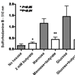
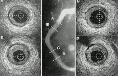
BACKGROUND: Restenosis remains an important limitation of interventional cardiology. Therefore, we aimed to determine the safety and efficacy of sirolimus (a cell-cycle inhibitor)-coated BX Velocity stents. METHODS AND RESULTS: Thirty patients with angina pectoris were electively treated with 2 different formulations of sirolimus-coated stents (slow release [SR], n=15, and fast release [FR], n=15). All stents were successfully delivered, and patients were discharged without clinical complications. Independent core laboratories analyzed angiographic and 3D volumetric intravascular ultrasound data (immediately after procedure and at 4-month follow-up). Eight-month clinical follow-up was obtained for all patients. There was minimal neointimal hyperplasia in both groups (11.0+/-3.0% in the SR group and 10.4+/-3.0% in the FR group, P:=NS) by ultrasound and quantitative coronary angiography (in-stent late loss, 0.09+/-0.3 mm [SR] and -0.02+/-0.3 mm [FR]; in-lesion late loss, 0.16+/-0.3 mm [SR] and -0.1+/-0.3 mm [FR]). No in-stent or edge restenosis (diameter stenosis >or=50%) was observed. No major clinical events (stent thrombosis, repeat revascularization, myocardial infarction, or death) had occurred by 8 months. CONCLUSIONS: The implantation of sirolimus-coated BX Velocity stents is feasible and safe and elicits minimal neointimal proliferation. Additional placebo-controlled trials are required to confirm these promising results. (+info)Oral anticoagulant therapy during and after coronary angioplasty the intensity and duration of anticoagulation are essential to reduce thrombotic complications. (3/1466)
BACKGROUND: In the randomized Balloon Angioplasty and Anticoagulation Study (BAAS), the addition of oral anticoagulants to aspirin significantly reduced early and late events after coronary angioplasty. However, bleeding episodes were increased. The present report studied the intensity and the duration of anticoagulation as predictors of thrombotic and bleeding events. METHODS AND RESULTS: A total of 530 patients, 34% of whom received a stent, were treated with aspirin plus coumarins. Half of the patients were randomized to angiographic follow-up. The target international normalized ratio (INR) was 2.1 to 4.8 during angioplasty and 6-month follow-up. Thrombotic events were death, myocardial infarction, target lesion revascularization, and thrombotic stroke. Bleeding complications were hemorrhagic stroke, major extracranial bleeding, and false aneurysm. "Optimal" anticoagulation was defined as an INR in the target range for at least 70% of the follow-up time. There were 17 early thrombotic events (3.2%), 7 early bleeding episodes (1.3%), and 10 false aneurysms (1.9%). The incidence rate for both early thrombotic and bleeding events was lowest in patients in the target range. A total of 61 late thrombotic events occurred (11.6%). Optimal anticoagulation was an independent predictor of late thrombotic events (relative risk, 0.33; 95% CI, 0.19 to 0.57) and was associated with a 0.21 mm (95% CI, 0.17 to 0.42) larger vessel lumen at 6 months. Late bleeding episodes (1.4%) were lowest in patients in the target range. CONCLUSIONS: Coumarins started before coronary angioplasty with a target INR of 2.1 to 4.8 led to the lowest procedural event rate, without an increase in bleeding episodes. During follow-up, optimal anticoagulation was associated with a decrease in the incidence of late events by 67% and a significant improvement in 6-month angiographic outcome. (+info)Relation of a common methylenetetrahydrofolate reductase mutation and plasma homocysteine with intimal hyperplasia after coronary stenting. (4/1466)
BACKGROUND: Hyperhomocysteinemia has been identified as an independent risk factor for coronary artery disease. Recent studies have shown that a common mutation (nucleotide 677 C-->T) in the methylenetetrahydrofolate reductase (MTHFR) gene may contribute to mild hyperhomocysteinemia and, therefore, to the incidence of coronary artery disease. No information exists, however, regarding the association between the mutation of the MTHFR gene or plasma homocysteine levels and morphological analysis of coronary atherosclerosis using intravascular ultrasound. METHODS AND RESULTS: To examine the potential influence of MTHFR genotype and homocysteine on coronaryarteries morphologically, we screened 62 patients with 65 lesions that were treated with 93 Palmaz-Schatz stents. The plasma homocysteine levels in the patients with the TT genotype were not significantly higher than those in the patients with non-TT (CC+CT) genotypes (13.1 +/- 5.5 versus 11.5 +/- 3.1 mmol/L, P=0.16). Angiographic analysis showed that the percent diameter stenosis in the patients with the TT genotype was significantly greater than that in those with non-TT genotypes (43.7 +/- 17.8% versus 29.0 +/- 22.0%, P=0.015). Intravascular ultrasound analysis showed that the TT genotype was significantly associated with greater intimal hyperplasia area (5.70 +/- 1.94 versus 3.72 +/- 1.38 mm2, P=0.001). In multiple stepwise regression analysis, the number of the T alleles was the only independent predictor of intimal hyperplasia after intervention (r2=0.21, P=0.004). CONCLUSIONS: The homozygous mutant genotype of the MTHFR gene may increase the risk of in-stent restenosis more than does the normal homozygous or heterozygous genotype. (+info)Long-term effects of intracoronary beta-radiation in balloon- and stent-injured porcine coronary arteries. (5/1466)
BACKGROUND: The data on the long-term safety and efficacy of intracoronary beta-radiation in animal models are limited. METHODS AND RESULTS: A total of 30 coronary arteries in 15 swine were subjected to balloon or stent injury followed by beta-radiation from a centered 32P source (2000 cGy to 1 mm beyond lumen surface) or a sham radiation procedure. The animals received aspirin for 6 months and ticlopidine for 30 days. Five of the 10 animals subjected to radiation died (at 5 days, 7 days, 3 months [n = 2], and 4 months) as a result of layered, occlusive thrombus at the intervention site (3 stent and 2 balloon injury sites). No deaths occurred in the control group. In the surviving animals, balloon-injured and irradiated vessels showed a trend toward larger lumens than controls (2.15 +/- 0.17 versus 1.80 +/- 0.08 mm2, P=0.06) and larger external elastic lamina areas (3.32 +/- 0.21 versus 2.62 +/- 0.10 mm2, P=0.003). In the stent-injured vessels from surviving animals, lumen, neointimal, and external elastic lamina areas were 3.58 +/- 0.33, 3.16 +/- 0.35, and 8.12 +/- 0.42 mm2 for irradiated vessel segments; these values were not different from those in controls (3.21 +/- 0.15, 2.84 +/- 0.27, and 7.76 +/- 0.28 mm2, respectively). Histologically, healing was complete in most survivors, although intramural fibrin and hemorrhage were occasionally seen. CONCLUSION: In the long-term (6 month) porcine model of restenosis, the inhibition by intracoronary beta-radiotherapy of the neointimal formation that is known to be present at 1 month is not sustained. This lack of effect on neointimal formation after balloon and stent arterial injury is accompanied by subacute and late thrombosis that leads to cardiac death on a background of continuous aspirin but relatively brief ticlopidine treatment. (+info)Lipid peroxidation may predict restenosis after coronary balloon angioplasty. (6/1466)
The present study assessed whether lipid peroxidation in plasma might predict restenosis after coronary balloon angioplasty. A total of 87 patients, who had undergone successful coronary balloon angioplasty using standard techniques, were enrolled. Fasting blood samples before the intervention were measured for plasma levels of thiobarbituric acid reactive substances (TBARS, an indicator of lipid peroxidation). Angiography was carried out before and 15 min after angioplasty, and at follow-up (4 months after angioplasty), and evaluated using a quantitative approach. There were 23 patients with restenosis (group R) and 64 patients without restenosis (group N) after coronary balloon angioplasty. The plasma TBARS level (mean+/-SEM) of 4.3+/-0.1 micromol/L in group R was significantly higher than that of 3.2+/-0.1 micromol/L in group N (p<0.01). There were no significant differences in other parameters, including plasma lipid levels, between the 2 groups. The plasma level of TBARS positively correlated with lumen loss of the coronary artery at the time of follow-up angiography (r=0.57, p<0.01). Our results suggest that oxidative stress contributes to restenosis and indicate that an elevated plasma level of TBARS may be a reliable predictor of restenosis. (+info)Histopathologic evaluation of coronary artery thrombi obtained by directional coronary atherectomy in patients with restenosis-induced unstable angina pectoris. (7/1466)
The pathogenesis of unstable angina pectoris (UAP) following percutaneous transluminal coronary angioplasty (PTCA) or directional coronary atherectomy (DCA) has not been adequately investigated, so the present study aimed to determine whether thrombi are present in restenotic lesions. The study group comprised 14 patients (16 arterial branches) with angina pectoris in whom either PTCA or DCA was performed and who had developed UAP associated with restenosis, and who then underwent DCA of the restenosed lesion (R-UAP group). The control groups comprised individuals with UAP undergoing DCA with no prior history of PTCA or DCA (P-UAP group; n=29, 29 branches), patients with acute myocardial infarction (AMI group; n=34, 34 branches), and patients with stable angina pectoris (SAP group; n=31, 33 branches). The presence of thrombi was determined by light microscopy of histologic specimens. Thrombus was present in only 1 of the 16 (6.3%) branches in the R-UAP group. 21 of the 29 (72.4%) branches in the P-UAP group, and in 25 of the 34 (73.5%) in the AMI group. In the SAP group, it was detected in only 2 of the 33 (7.1%) branches. The incidence of thrombus was significantly lower in the R-UAP group than in the P-UAP group. In conclusion, the role of thrombus is limited in causing post-interventional UAP at restenosed sites. (+info)Intracoronary brachytherapy in the treatment of in-stent restenosis. Initial experience in Brazil. (8/1466)
Intracoronary brachytherapy using beta or gamma radiation is currently the most efficient type of therapy for preventing the recurrence of coronary in-stent restenosis. Its implementation depends on the interaction among interventionists, radiotherapists, and physicists to assure the safety and quality of the method. The authors report the pioneering experience in Brazil of the treatment of 2 patients with coronary in-stent restenosis, in whom beta radiation was used as part of the international multicenter randomized PREVENT study (Proliferation REduction with Vascular ENergy Trial). The procedures were performed rapidly and did not require significant modifications in the traditional techniques used for conventional angioplasty. Alteration in the radiological protection devices of the hemodynamic laboratory were also not required, showing that intracoronary brachytherapy using beta radiation can be incorporated into the interventional tools of cardiology in our environment. (+info)Coronary restenosis is the re-narrowing or re-occlusion of a coronary artery after a previous successful procedure to open or widen the artery, such as angioplasty or stenting. This narrowing is usually caused by the excessive growth of scar tissue or smooth muscle cells in the artery lining, which can occur spontaneously or as a response to the initial procedure. Restenosis can lead to recurrent symptoms of coronary artery disease, such as chest pain or shortness of breath, and may require additional medical intervention.
Coronary balloon angioplasty is a minimally invasive medical procedure used to widen narrowed or obstructed coronary arteries (the blood vessels that supply oxygen-rich blood to the heart muscle) and improve blood flow to the heart. This procedure is typically performed in conjunction with the insertion of a stent, a small mesh tube that helps keep the artery open.
During coronary balloon angioplasty, a thin, flexible catheter with a deflated balloon at its tip is inserted into a blood vessel, usually through a small incision in the groin or arm. The catheter is then guided to the narrowed or obstructed section of the coronary artery. Once in position, the balloon is inflated to compress the plaque against the artery wall and widen the lumen (the inner space) of the artery. This helps restore blood flow to the heart muscle.
The procedure is typically performed under local anesthesia and conscious sedation to minimize discomfort. Coronary balloon angioplasty is a relatively safe and effective treatment for many people with coronary artery disease, although complications such as bleeding, infection, or re-narrowing of the artery (restenosis) can occur in some cases.
Coronary vessels refer to the network of blood vessels that supply oxygenated blood and nutrients to the heart muscle, also known as the myocardium. The two main coronary arteries are the left main coronary artery and the right coronary artery.
The left main coronary artery branches off into the left anterior descending artery (LAD) and the left circumflex artery (LCx). The LAD supplies blood to the front of the heart, while the LCx supplies blood to the side and back of the heart.
The right coronary artery supplies blood to the right lower part of the heart, including the right atrium and ventricle, as well as the back of the heart.
Coronary vessel disease (CVD) occurs when these vessels become narrowed or blocked due to the buildup of plaque, leading to reduced blood flow to the heart muscle. This can result in chest pain, shortness of breath, or a heart attack.
Coronary angiography is a medical procedure that uses X-ray imaging to visualize the coronary arteries, which supply blood to the heart muscle. During the procedure, a thin, flexible catheter is inserted into an artery in the arm or groin and threaded through the blood vessels to the heart. A contrast dye is then injected through the catheter, and X-ray images are taken as the dye flows through the coronary arteries. These images can help doctors diagnose and treat various heart conditions, such as blockages or narrowing of the arteries, that can lead to chest pain or heart attacks. It is also known as coronary arteriography or cardiac catheterization.
A stent is a small mesh tube that's used to treat narrow or weak arteries. Arteries are blood vessels that carry blood away from your heart to other parts of your body. A stent is placed in an artery as part of a procedure called angioplasty. Angioplasty restores blood flow through narrowed or blocked arteries by inflating a tiny balloon inside the blocked artery to widen it.
The stent is then inserted into the widened artery to keep it open. The stent is usually made of metal, but some are coated with medication that is slowly and continuously released to help prevent the formation of scar tissue in the artery. This can reduce the chance of the artery narrowing again.
Stents are also used in other parts of the body, such as the neck (carotid artery) and kidneys (renal artery), to help maintain blood flow and prevent blockages. They can also be used in the urinary system to treat conditions like ureteropelvic junction obstruction or narrowing of the urethra.
Coronary artery disease, often simply referred to as coronary disease, is a condition in which the blood vessels that supply oxygen-rich blood to the heart become narrowed or blocked due to the buildup of fatty deposits called plaques. This can lead to chest pain (angina), shortness of breath, or in severe cases, a heart attack.
The medical definition of coronary artery disease is:
A condition characterized by the accumulation of atheromatous plaques in the walls of the coronary arteries, leading to decreased blood flow and oxygen supply to the myocardium (heart muscle). This can result in symptoms such as angina pectoris, shortness of breath, or arrhythmias, and may ultimately lead to myocardial infarction (heart attack) or heart failure.
Risk factors for coronary artery disease include age, smoking, high blood pressure, high cholesterol, diabetes, obesity, physical inactivity, and a family history of the condition. Lifestyle changes such as quitting smoking, exercising regularly, eating a healthy diet, and managing stress can help reduce the risk of developing coronary artery disease. Medical treatments may include medications to control blood pressure, cholesterol levels, or irregular heart rhythms, as well as procedures such as angioplasty or bypass surgery to improve blood flow to the heart.
Recurrence, in a medical context, refers to the return of symptoms or signs of a disease after a period of improvement or remission. It indicates that the condition has not been fully eradicated and may require further treatment. Recurrence is often used to describe situations where a disease such as cancer comes back after initial treatment, but it can also apply to other medical conditions. The likelihood of recurrence varies depending on the type of disease and individual patient factors.
Coronary artery disease (CAD) is a medical condition in which the coronary arteries, which supply oxygen-rich blood to the heart muscle, become narrowed or blocked due to the buildup of cholesterol, fatty deposits, and other substances, known as plaque. Over time, this buildup can cause the arteries to harden and narrow (a process called atherosclerosis), reducing blood flow to the heart muscle.
The reduction in blood flow can lead to various symptoms and complications, including:
1. Angina (chest pain or discomfort) - This occurs when the heart muscle doesn't receive enough oxygen-rich blood, causing pain, pressure, or discomfort in the chest, arms, neck, jaw, or back.
2. Shortness of breath - When the heart isn't receiving adequate blood flow, it can't pump blood efficiently to meet the body's demands, leading to shortness of breath during physical activities or at rest.
3. Heart attack - If a piece of plaque ruptures or breaks off in a coronary artery, a blood clot can form and block the artery, causing a heart attack (myocardial infarction). This can damage or destroy part of the heart muscle.
4. Heart failure - Chronic reduced blood flow to the heart muscle can weaken it over time, leading to heart failure, a condition in which the heart can't pump blood efficiently to meet the body's needs.
5. Arrhythmias - Reduced blood flow and damage to the heart muscle can lead to abnormal heart rhythms (arrhythmias), which can be life-threatening if not treated promptly.
Coronary artery disease is typically diagnosed through a combination of medical history, physical examination, and diagnostic tests such as electrocardiograms (ECGs), stress testing, cardiac catheterization, and imaging studies like coronary computed tomography angiography (CCTA). Treatment options for CAD include lifestyle modifications, medications, medical procedures, and surgery.
Sirolimus is a medication that belongs to a class of drugs called immunosuppressants. It is also known as rapamycin. Sirolimus works by inhibiting the mammalian target of rapamycin (mTOR), which is a protein that plays a key role in cell growth and division.
Sirolimus is primarily used to prevent rejection of transplanted organs, such as kidneys, livers, and hearts. It works by suppressing the activity of the immune system, which can help to reduce the risk of the body rejecting the transplanted organ. Sirolimus is often used in combination with other immunosuppressive drugs, such as corticosteroids and calcineurin inhibitors.
Sirolimus is also being studied for its potential therapeutic benefits in a variety of other conditions, including cancer, tuberous sclerosis complex, and lymphangioleiomyomatosis. However, more research is needed to fully understand the safety and efficacy of sirolimus in these contexts.
It's important to note that sirolimus can have significant side effects, including increased risk of infections, mouth sores, high blood pressure, and kidney damage. Therefore, it should only be used under the close supervision of a healthcare provider.
Drug-eluting stents (DES) are medical devices used in the treatment of coronary artery disease. They are small, flexible tubes that are coated with a medication that is slowly released (eluted) over time to prevent the formation of scar tissue and reduce the risk of renarrowing (restenosis) of the artery after it has been treated with angioplasty and stenting.
The stent is typically placed in a narrowed or blocked coronary artery during a percutaneous coronary intervention (PCI) procedure, such as angioplasty, to open up the blood vessel and improve blood flow to the heart muscle. The medication on the DES helps to prevent the growth of smooth muscle cells and the formation of scar tissue in the artery, which can cause restenosis and require additional treatments.
The most commonly used medications on DES are sirolimus, paclitaxel, zotarolimus, and everolimus. These drugs work by inhibiting the growth of smooth muscle cells and reducing inflammation in the artery. While DES have been shown to reduce the risk of restenosis compared to bare-metal stents, they also carry a small increased risk of late stent thrombosis (blood clots forming in the stent), which can lead to serious complications such as heart attack or stroke. Therefore, patients who receive DES are typically prescribed long-term antiplatelet therapy to reduce this risk.
Coronary circulation refers to the circulation of blood in the coronary vessels, which supply oxygenated blood to the heart muscle (myocardium) and drain deoxygenated blood from it. The coronary circulation system includes two main coronary arteries - the left main coronary artery and the right coronary artery - that branch off from the aorta just above the aortic valve. These arteries further divide into smaller branches, which supply blood to different regions of the heart muscle.
The left main coronary artery divides into two branches: the left anterior descending (LAD) artery and the left circumflex (LCx) artery. The LAD supplies blood to the front and sides of the heart, while the LCx supplies blood to the back and sides of the heart. The right coronary artery supplies blood to the lower part of the heart, including the right ventricle and the bottom portion of the left ventricle.
The veins that drain the heart muscle include the great cardiac vein, the middle cardiac vein, and the small cardiac vein, which merge to form the coronary sinus. The coronary sinus empties into the right atrium, allowing deoxygenated blood to enter the right side of the heart and be pumped to the lungs for oxygenation.
Coronary circulation is essential for maintaining the health and function of the heart muscle, as it provides the necessary oxygen and nutrients required for proper contraction and relaxation of the myocardium. Any disruption or blockage in the coronary circulation system can lead to serious consequences, such as angina, heart attack, or even death.
Follow-up studies are a type of longitudinal research that involve repeated observations or measurements of the same variables over a period of time, in order to understand their long-term effects or outcomes. In medical context, follow-up studies are often used to evaluate the safety and efficacy of medical treatments, interventions, or procedures.
In a typical follow-up study, a group of individuals (called a cohort) who have received a particular treatment or intervention are identified and then followed over time through periodic assessments or data collection. The data collected may include information on clinical outcomes, adverse events, changes in symptoms or functional status, and other relevant measures.
The results of follow-up studies can provide important insights into the long-term benefits and risks of medical interventions, as well as help to identify factors that may influence treatment effectiveness or patient outcomes. However, it is important to note that follow-up studies can be subject to various biases and limitations, such as loss to follow-up, recall bias, and changes in clinical practice over time, which must be carefully considered when interpreting the results.
Coronary stenosis is a medical condition that refers to the narrowing of the coronary arteries, which supply oxygen-rich blood to the heart muscle. This narrowing is typically caused by the buildup of plaque, made up of fat, cholesterol, and other substances, on the inner walls of the arteries. Over time, as the plaque hardens and calcifies, it can cause the artery to become narrowed or blocked, reducing blood flow to the heart muscle.
Coronary stenosis can lead to various symptoms and complications, including chest pain (angina), shortness of breath, irregular heart rhythms (arrhythmias), and heart attacks. Treatment options for coronary stenosis may include lifestyle changes, medications, medical procedures such as angioplasty or bypass surgery, or a combination of these approaches. Regular check-ups and diagnostic tests, such as stress testing or coronary angiography, can help detect and monitor coronary stenosis over time.
"Swine" is a common term used to refer to even-toed ungulates of the family Suidae, including domestic pigs and wild boars. However, in a medical context, "swine" often appears in the phrase "swine flu," which is a strain of influenza virus that typically infects pigs but can also cause illness in humans. The 2009 H1N1 pandemic was caused by a new strain of swine-origin influenza A virus, which was commonly referred to as "swine flu." It's important to note that this virus is not transmitted through eating cooked pork products; it spreads from person to person, mainly through respiratory droplets produced when an infected person coughs or sneezes.
Coronary artery bypass surgery, also known as coronary artery bypass grafting (CABG), is a surgical procedure used to improve blood flow to the heart in patients with severe coronary artery disease. This condition occurs when the coronary arteries, which supply oxygen-rich blood to the heart muscle, become narrowed or blocked due to the buildup of fatty deposits, called plaques.
During CABG surgery, a healthy blood vessel from another part of the body is grafted, or attached, to the coronary artery, creating a new pathway for oxygen-rich blood to flow around the blocked or narrowed portion of the artery and reach the heart muscle. This bypass helps to restore normal blood flow and reduce the risk of angina (chest pain), shortness of breath, and other symptoms associated with coronary artery disease.
There are different types of CABG surgery, including traditional on-pump CABG, off-pump CABG, and minimally invasive CABG. The choice of procedure depends on various factors, such as the patient's overall health, the number and location of blocked arteries, and the presence of other medical conditions.
It is important to note that while CABG surgery can significantly improve symptoms and quality of life in patients with severe coronary artery disease, it does not cure the underlying condition. Lifestyle modifications, such as regular exercise, a healthy diet, smoking cessation, and medication therapy, are essential for long-term management and prevention of further progression of the disease.
Angioplasty, balloon refers to a medical procedure used to widen narrowed or obstructed blood vessels, particularly the coronary arteries that supply blood to the heart muscle. This procedure is typically performed using a catheter-based technique, where a thin, flexible tube called a catheter is inserted into an artery, usually through the groin or wrist, and guided to the site of the narrowing or obstruction in the coronary artery.
Once the catheter reaches the affected area, a small balloon attached to the tip of the catheter is inflated, which compresses the plaque against the artery wall and stretches the artery, thereby restoring blood flow. The balloon is then deflated and removed, along with the catheter.
Balloon angioplasty is often combined with the placement of a stent, a small metal mesh tube that helps to keep the artery open and prevent it from narrowing again. This procedure is known as percutaneous coronary intervention (PCI) or coronary angioplasty and stenting.
Overall, balloon angioplasty is a relatively safe and effective treatment for coronary artery disease, although complications such as bleeding, infection, or re-narrowing of the artery can occur in some cases.
Coronary thrombosis is a medical condition that refers to the formation of a blood clot (thrombus) inside a coronary artery, which supplies oxygenated blood to the heart muscle. The development of a thrombus can partially or completely obstruct blood flow, leading to insufficient oxygen supply to the heart muscle. This can cause chest pain (angina) or a heart attack (myocardial infarction), depending on the severity and duration of the blockage.
Coronary thrombosis often results from the rupture of an atherosclerotic plaque, a buildup of cholesterol, fat, calcium, and other substances in the inner lining (endothelium) of the coronary artery. The ruptured plaque exposes the underlying tissue to the bloodstream, triggering the coagulation cascade and resulting in the formation of a thrombus.
Immediate medical attention is crucial for managing coronary thrombosis, as timely treatment can help restore blood flow, prevent further damage to the heart muscle, and reduce the risk of complications such as heart failure or life-threatening arrhythmias. Treatment options may include medications, such as antiplatelet agents, anticoagulants, and thrombolytic drugs, or interventional procedures like angioplasty and stenting to open the blocked artery. In some cases, surgical intervention, such as coronary artery bypass grafting (CABG), may be necessary.
Coronary vasospasm refers to a sudden constriction (narrowing) of the coronary arteries, which supply oxygenated blood to the heart muscle. This constriction can reduce or block blood flow, leading to symptoms such as chest pain (angina) or, in severe cases, a heart attack (myocardial infarction). Coronary vasospasm can occur spontaneously or be triggered by various factors, including stress, smoking, and certain medications. It is also associated with conditions such as coronary artery disease and variant angina. Prolonged or recurrent vasospasms can cause damage to the heart muscle and increase the risk of cardiovascular events.
Treatment outcome is a term used to describe the result or effect of medical treatment on a patient's health status. It can be measured in various ways, such as through symptoms improvement, disease remission, reduced disability, improved quality of life, or survival rates. The treatment outcome helps healthcare providers evaluate the effectiveness of a particular treatment plan and make informed decisions about future care. It is also used in clinical research to compare the efficacy of different treatments and improve patient care.
Graft occlusion in the context of vascular surgery refers to the complete or partial blockage of a blood vessel that has been surgically replaced or repaired with a graft. The graft can be made from either synthetic materials or autologous tissue (taken from another part of the patient's body).
Graft occlusion can occur due to various reasons, including:
1. Thrombosis: Formation of a blood clot within the graft, which can obstruct blood flow.
2. Intimal hyperplasia: Overgrowth of the inner lining (intima) of the graft or the adjacent native vessel, causing narrowing of the lumen and reducing blood flow.
3. Atherosclerosis: Deposition of cholesterol and other substances in the walls of the graft, leading to hardening and narrowing of the vessel.
4. Infection: Bacterial or fungal infection of the graft can cause inflammation, weakening, and ultimately occlusion of the graft.
5. Mechanical factors: Kinking, twisting, or compression of the graft can lead to obstruction of blood flow.
Graft occlusion is a significant complication following vascular surgery, as it can result in reduced perfusion to downstream tissues and organs, leading to ischemia (lack of oxygen supply) and potential tissue damage or loss.
Myocardial infarction (MI), also known as a heart attack, is a medical condition characterized by the death of a segment of heart muscle (myocardium) due to the interruption of its blood supply. This interruption is most commonly caused by the blockage of a coronary artery by a blood clot formed on the top of an atherosclerotic plaque, which is a buildup of cholesterol and other substances in the inner lining of the artery.
The lack of oxygen and nutrients supply to the heart muscle tissue results in damage or death of the cardiac cells, causing the affected area to become necrotic. The extent and severity of the MI depend on the size of the affected area, the duration of the occlusion, and the presence of collateral circulation.
Symptoms of a myocardial infarction may include chest pain or discomfort, shortness of breath, nausea, lightheadedness, and sweating. Immediate medical attention is necessary to restore blood flow to the affected area and prevent further damage to the heart muscle. Treatment options for MI include medications, such as thrombolytics, antiplatelet agents, and pain relievers, as well as procedures such as percutaneous coronary intervention (PCI) or coronary artery bypass grafting (CABG).
A coronary aneurysm is a localized dilation or bulging of a portion of the wall of a coronary artery, which supplies blood to the muscle tissue of the heart. It's similar to a bubble or balloon-like structure that forms within the artery wall due to weakness in the arterial wall, leading to abnormal enlargement or widening.
Coronary aneurysms can vary in size and may be classified as true or false aneurysms based on their structure. True aneurysms involve all three layers of the artery wall, while false aneurysms (also known as pseudoaneurysms) only have one or two layers involved, with the remaining layer disrupted.
These aneurysms can lead to complications such as blood clots forming inside the aneurysm sac, which can then dislodge and cause blockages in smaller coronary arteries (embolism). Additionally, coronary aneurysms may rupture, leading to severe internal bleeding and potentially life-threatening situations.
Coronary aneurysms are often asymptomatic but can present with symptoms such as chest pain, shortness of breath, or palpitations, especially if the aneurysm causes a significant narrowing (stenosis) in the affected artery. They can be diagnosed through imaging techniques like coronary angiography, computed tomography (CT), or magnetic resonance imaging (MRI). Treatment options include medications to manage symptoms and prevent complications, as well as surgical interventions such as stenting or bypass grafting to repair or reroute the affected artery.
Interventional ultrasonography is a medical procedure that involves the use of real-time ultrasound imaging to guide minimally invasive diagnostic and therapeutic interventions. This technique combines the advantages of ultrasound, such as its non-ionizing nature (no radiation exposure), relatively low cost, and portability, with the ability to perform precise and targeted procedures.
In interventional ultrasonography, a specialized physician called an interventional radiologist or an interventional sonographer uses high-frequency sound waves to create detailed images of internal organs and tissues. These images help guide the placement of needles, catheters, or other instruments used during the procedure. Common interventions include biopsies (tissue sampling), fluid drainage, tumor ablation, and targeted drug delivery.
The real-time visualization provided by ultrasonography allows for increased accuracy and safety during these procedures, minimizing complications and reducing recovery time compared to traditional surgical approaches. Additionally, interventional ultrasonography can be performed on an outpatient basis, further contributing to its appeal as a less invasive alternative in many clinical scenarios.
Pathological constriction refers to an abnormal narrowing or tightening of a body passage or organ, which can interfere with the normal flow of blood, air, or other substances through the area. This constriction can occur due to various reasons such as inflammation, scarring, or abnormal growths, and can affect different parts of the body, including blood vessels, airways, intestines, and ureters. Pathological constriction can lead to a range of symptoms and complications depending on its location and severity, and may require medical intervention to correct.
Tunica intima, also known as the intima layer, is the innermost layer of a blood vessel, including arteries and veins. It is in direct contact with the flowing blood and is composed of simple squamous endothelial cells that form a continuous, non-keratinized, stratified epithelium. These cells play a crucial role in maintaining vascular homeostasis by regulating the passage of molecules and immune cells between the blood and the vessel wall, as well as contributing to the maintenance of blood fluidity and preventing coagulation.
The tunica intima is supported by a thin layer of connective tissue called the basement membrane, which provides structural stability and anchorage for the endothelial cells. Beneath the basement membrane lies a loose network of elastic fibers and collagen, known as the internal elastic lamina, that separates the tunica intima from the middle layer, or tunica media.
In summary, the tunica intima is the innermost layer of blood vessels, primarily composed of endothelial cells and a basement membrane, which regulates various functions to maintain vascular homeostasis.
Medical Definition:
"Risk factors" are any attribute, characteristic or exposure of an individual that increases the likelihood of developing a disease or injury. They can be divided into modifiable and non-modifiable risk factors. Modifiable risk factors are those that can be changed through lifestyle choices or medical treatment, while non-modifiable risk factors are inherent traits such as age, gender, or genetic predisposition. Examples of modifiable risk factors include smoking, alcohol consumption, physical inactivity, and unhealthy diet, while non-modifiable risk factors include age, sex, and family history. It is important to note that having a risk factor does not guarantee that a person will develop the disease, but rather indicates an increased susceptibility.
Prospective studies, also known as longitudinal studies, are a type of cohort study in which data is collected forward in time, following a group of individuals who share a common characteristic or exposure over a period of time. The researchers clearly define the study population and exposure of interest at the beginning of the study and follow up with the participants to determine the outcomes that develop over time. This type of study design allows for the investigation of causal relationships between exposures and outcomes, as well as the identification of risk factors and the estimation of disease incidence rates. Prospective studies are particularly useful in epidemiology and medical research when studying diseases with long latency periods or rare outcomes.
In the field of medicine, "time factors" refer to the duration of symptoms or time elapsed since the onset of a medical condition, which can have significant implications for diagnosis and treatment. Understanding time factors is crucial in determining the progression of a disease, evaluating the effectiveness of treatments, and making critical decisions regarding patient care.
For example, in stroke management, "time is brain," meaning that rapid intervention within a specific time frame (usually within 4.5 hours) is essential to administering tissue plasminogen activator (tPA), a clot-busting drug that can minimize brain damage and improve patient outcomes. Similarly, in trauma care, the "golden hour" concept emphasizes the importance of providing definitive care within the first 60 minutes after injury to increase survival rates and reduce morbidity.
Time factors also play a role in monitoring the progression of chronic conditions like diabetes or heart disease, where regular follow-ups and assessments help determine appropriate treatment adjustments and prevent complications. In infectious diseases, time factors are crucial for initiating antibiotic therapy and identifying potential outbreaks to control their spread.
Overall, "time factors" encompass the significance of recognizing and acting promptly in various medical scenarios to optimize patient outcomes and provide effective care.
Percutaneous Coronary Intervention (PCI), also known as coronary angioplasty, is a non-surgical procedure that opens up clogged coronary arteries to improve blood flow to the heart. It involves inserting a thin, flexible catheter into an artery in the groin or wrist and guiding it to the blocked artery in the heart. A small balloon is then inflated to widen the narrowed or blocked artery, and sometimes a stent (a tiny mesh tube) is placed to keep the artery open. This procedure helps to restore and maintain blood flow to the heart muscle, reducing symptoms of angina and improving overall cardiac function.
Coronary occlusion is the medical term used to describe a complete blockage in one or more of the coronary arteries, which supply oxygenated blood to the heart muscle. This blockage is usually caused by the buildup of fatty deposits, called plaques, inside the artery walls, a condition known as atherosclerosis. Over time, these plaques can rupture, leading to the formation of blood clots that completely obstruct the flow of blood through the coronary artery.
Coronary occlusion can lead to serious complications, such as a heart attack (myocardial infarction), angina (chest pain), or even sudden cardiac death, depending on the severity and duration of the blockage. Immediate medical attention is required in case of coronary occlusion to restore blood flow to the affected areas of the heart and prevent further damage. Treatment options may include medications, minimally invasive procedures like angioplasty and stenting, or surgical interventions such as coronary artery bypass grafting (CABG).
Beta particles, also known as beta rays, are a type of ionizing radiation that consist of high-energy electrons or positrons emitted from the nucleus of certain radioactive isotopes during their decay process. When a neutron in the nucleus decays into a proton, it results in an excess energy state and one electron is ejected from the atom at high speed. This ejected electron is referred to as a beta particle.
Beta particles can have both positive and negative charges, depending on the type of decay process. Negative beta particles (β−) are equivalent to electrons, while positive beta particles (β+) are equivalent to positrons. They possess kinetic energy that varies in range, with higher energies associated with greater penetrating power.
Beta particles can cause ionization and excitation of atoms and molecules they encounter, leading to chemical reactions and potential damage to living tissues. Therefore, appropriate safety measures must be taken when handling materials that emit beta radiation.
Hyperplasia is a medical term that refers to an abnormal increase in the number of cells in an organ or tissue, leading to an enlargement of the affected area. It's a response to various stimuli such as hormones, chronic irritation, or inflammation. Hyperplasia can be physiological, like the growth of breast tissue during pregnancy, or pathological, like in the case of benign or malignant tumors. The process is generally reversible if the stimulus is removed. It's important to note that hyperplasia itself is not cancerous, but some forms of hyperplasia can increase the risk of developing cancer over time.
Cardiovascular agents are a class of medications that are used to treat various conditions related to the cardiovascular system, which includes the heart and blood vessels. These agents can be further divided into several subcategories based on their specific mechanisms of action and therapeutic effects. Here are some examples:
1. Antiarrhythmics: These drugs are used to treat abnormal heart rhythms or arrhythmias. They work by stabilizing the electrical activity of the heart and preventing irregular impulses from spreading through the heart muscle.
2. Antihypertensives: These medications are used to lower high blood pressure, also known as hypertension. There are several classes of antihypertensive drugs, including diuretics, beta-blockers, calcium channel blockers, and angiotensin-converting enzyme (ACE) inhibitors.
3. Anticoagulants: These drugs are used to prevent blood clots from forming or growing larger. They work by interfering with the coagulation cascade, which is a series of chemical reactions that lead to the formation of a blood clot.
4. Antiplatelet agents: These medications are used to prevent platelets in the blood from sticking together and forming clots. They work by inhibiting the aggregation of platelets, which are small cells in the blood that help form clots.
5. Lipid-lowering agents: These drugs are used to lower cholesterol and other fats in the blood. They work by reducing the production or absorption of cholesterol in the body or increasing the removal of cholesterol from the bloodstream. Examples include statins, bile acid sequestrants, and PCSK9 inhibitors.
6. Vasodilators: These medications are used to widen blood vessels and improve blood flow. They work by relaxing the smooth muscle in the walls of blood vessels, causing them to dilate or widen. Examples include nitrates, calcium channel blockers, and ACE inhibitors.
7. Inotropes: These drugs are used to increase the force of heart contractions. They work by increasing the sensitivity of heart muscle cells to calcium ions, which are necessary for muscle contraction.
These are just a few examples of cardiovascular medications that are used to treat various conditions related to the heart and blood vessels. It is important to note that these medications can have side effects and should be taken under the guidance of a healthcare provider.
Atherectomy, coronary, is a medical procedure used to treat narrowed or blocked coronary arteries due to the buildup of plaque (atherosclerosis). The goal of coronary atherectomy is to improve blood flow to the heart muscle by removing the obstructive material within the vessel.
During the procedure, a specialized catheter with a cutting device on its tip is inserted into a peripheral artery, usually in the groin or arm, and advanced to the affected coronary artery. The cutting device can be a rotating blade, a high-speed spinning burr, or a laser fiber that is used to shave, drill, or vaporize the plaque, respectively. The removed material is collected in a chamber within the catheter or washed away by blood flow.
There are different types of coronary atherectomy devices, including:
1. Directional atherectomy (DCA): A rotating blade cuts and removes the plaque in a targeted direction.
2. Rotational atherectomy (Rotablator): A high-speed spinning burr is used to abrade and pulverize the plaque into tiny particles that can be safely carried away by blood flow.
3. Laser atherectomy: A laser fiber is used to vaporize or break down the plaque into gaseous or small particle form.
Coronary atherectomy is typically performed in conjunction with angioplasty and stenting, as it helps prepare the narrowed artery for these procedures by creating a larger lumen and reducing the risk of complications like dissections or restenosis (re-narrowing). However, its use may be limited to specific cases due to the potential risks, such as vessel trauma, distal embolization, or perforation.
It is essential to consult with a medical professional for detailed information and personalized treatment recommendations regarding coronary atherectomy.
Platelet aggregation inhibitors are a class of medications that prevent platelets (small blood cells involved in clotting) from sticking together and forming a clot. These drugs work by interfering with the ability of platelets to adhere to each other and to the damaged vessel wall, thereby reducing the risk of thrombosis (blood clot formation).
Platelet aggregation inhibitors are often prescribed for people who have an increased risk of developing blood clots due to various medical conditions such as atrial fibrillation, coronary artery disease, peripheral artery disease, stroke, or a history of heart attack. They may also be used in patients undergoing certain medical procedures, such as angioplasty and stenting, to prevent blood clot formation in the stents.
Examples of platelet aggregation inhibitors include:
1. Aspirin: A nonsteroidal anti-inflammatory drug (NSAID) that irreversibly inhibits the enzyme cyclooxygenase, which is involved in platelet activation and aggregation.
2. Clopidogrel (Plavix): A P2Y12 receptor antagonist that selectively blocks ADP-induced platelet activation and aggregation.
3. Prasugrel (Effient): A third-generation thienopyridine P2Y12 receptor antagonist, similar to clopidogrel but with faster onset and greater potency.
4. Ticagrelor (Brilinta): A direct-acting P2Y12 receptor antagonist that does not require metabolic activation and has a reversible binding profile.
5. Dipyridamole (Persantine): An antiplatelet agent that inhibits platelet aggregation by increasing cyclic adenosine monophosphate (cAMP) levels in platelets, which leads to decreased platelet reactivity.
6. Iloprost (Ventavis): A prostacyclin analogue that inhibits platelet aggregation and causes vasodilation, often used in the treatment of pulmonary arterial hypertension.
7. Cilostazol (Pletal): A phosphodiesterase III inhibitor that increases cAMP levels in platelets, leading to decreased platelet activation and aggregation, as well as vasodilation.
8. Ticlopidine (Ticlid): An older P2Y12 receptor antagonist with a slower onset of action and more frequent side effects compared to clopidogrel or prasugrel.
Iridium radioisotopes are unstable isotopes or variants of the element iridium that emit radiation as they decay into more stable forms. These isotopes can be used in various medical applications, such as brachytherapy, a type of cancer treatment where a small amount of radioactive material is placed inside the body near the tumor site to deliver targeted radiation therapy.
Iridium-192 is one commonly used iridium radioisotope for this purpose. It has a half-life of 74.2 days and emits gamma rays, making it useful for treating various types of cancer, including breast, gynecological, prostate, and head and neck cancers.
It's important to note that handling and using radioisotopes requires specialized training and equipment due to the potential radiation hazards associated with them.
Myocardial revascularization is a medical term that refers to the restoration of blood flow to the heart muscle (myocardium), typically through a surgical or interventional procedure. This is often performed in patients with coronary artery disease, where the buildup of plaque in the coronary arteries restricts blood flow to the heart muscle, causing symptoms such as chest pain (angina) or shortness of breath, and increasing the risk of a heart attack (myocardial infarction).
There are two main types of myocardial revascularization:
1. Coronary artery bypass grafting (CABG): This is a surgical procedure in which a healthy blood vessel from another part of the body is used to create a detour around the blocked or narrowed coronary artery, allowing blood to flow more freely to the heart muscle.
2. Percutaneous coronary intervention (PCI), also known as angioplasty and stenting: This is a minimally invasive procedure in which a thin catheter is inserted into an artery in the groin or arm and threaded up to the blocked or narrowed coronary artery. A balloon is then inflated to widen the artery, and a stent may be placed to keep it open.
Both procedures aim to improve symptoms, reduce the risk of heart attack, and prolong survival in appropriately selected patients with coronary artery disease.
Coronary Care Units (CCUs) are specialized hospital wards that provide intensive care to patients with severe, life-threatening heart conditions. These units are equipped with advanced monitoring and treatment technologies to continuously monitor a patient's cardiac function and provide immediate medical interventions when necessary. Common conditions treated in CCUs include acute myocardial infarction (heart attack), unstable angina, cardiac arrhythmias, and heart failure. The primary goal of a CCU is to stabilize the patient's condition, prevent further complications, and facilitate recovery.
Brachytherapy is a type of cancer treatment that involves placing radioactive material directly into or near the tumor site. The term "brachy" comes from the Greek word for "short," which refers to the short distance that the radiation travels. This allows for a high dose of radiation to be delivered directly to the tumor while minimizing exposure to healthy surrounding tissue.
There are two main types of brachytherapy:
1. Intracavitary brachytherapy: The radioactive material is placed inside a body cavity, such as the uterus or windpipe.
2. Interstitial brachytherapy: The radioactive material is placed directly into the tumor or surrounding tissue using needles, seeds, or catheters.
Brachytherapy can be used alone or in combination with other cancer treatments such as surgery, external beam radiation therapy, and chemotherapy. It may be recommended for a variety of cancers, including prostate, cervical, vaginal, vulvar, head and neck, and skin cancers. The specific type of brachytherapy used will depend on the size, location, and stage of the tumor.
The advantages of brachytherapy include its ability to deliver a high dose of radiation directly to the tumor while minimizing exposure to healthy tissue, which can result in fewer side effects compared to other forms of radiation therapy. Additionally, brachytherapy is often a shorter treatment course than external beam radiation therapy, with some treatments lasting only a few minutes or hours.
However, there are also potential risks and side effects associated with brachytherapy, including damage to nearby organs and tissues, bleeding, infection, and pain. Patients should discuss the benefits and risks of brachytherapy with their healthcare provider to determine if it is an appropriate treatment option for them.
A smooth muscle within the vascular system refers to the involuntary, innervated muscle that is found in the walls of blood vessels. These muscles are responsible for controlling the diameter of the blood vessels, which in turn regulates blood flow and blood pressure. They are called "smooth" muscles because their individual muscle cells do not have the striations, or cross-striped patterns, that are observed in skeletal and cardiac muscle cells. Smooth muscle in the vascular system is controlled by the autonomic nervous system and by hormones, and can contract or relax slowly over a period of time.
The Predictive Value of Tests, specifically the Positive Predictive Value (PPV) and Negative Predictive Value (NPV), are measures used in diagnostic tests to determine the probability that a positive or negative test result is correct.
Positive Predictive Value (PPV) is the proportion of patients with a positive test result who actually have the disease. It is calculated as the number of true positives divided by the total number of positive results (true positives + false positives). A higher PPV indicates that a positive test result is more likely to be a true positive, and therefore the disease is more likely to be present.
Negative Predictive Value (NPV) is the proportion of patients with a negative test result who do not have the disease. It is calculated as the number of true negatives divided by the total number of negative results (true negatives + false negatives). A higher NPV indicates that a negative test result is more likely to be a true negative, and therefore the disease is less likely to be present.
The predictive value of tests depends on the prevalence of the disease in the population being tested, as well as the sensitivity and specificity of the test. A test with high sensitivity and specificity will generally have higher predictive values than a test with low sensitivity and specificity. However, even a highly sensitive and specific test can have low predictive values if the prevalence of the disease is low in the population being tested.
Catheterization is a medical procedure in which a catheter (a flexible tube) is inserted into the body to treat various medical conditions or for diagnostic purposes. The specific definition can vary depending on the area of medicine and the particular procedure being discussed. Here are some common types of catheterization:
1. Urinary catheterization: This involves inserting a catheter through the urethra into the bladder to drain urine. It is often performed to manage urinary retention, monitor urine output in critically ill patients, or assist with surgical procedures.
2. Cardiac catheterization: A procedure where a catheter is inserted into a blood vessel, usually in the groin or arm, and guided to the heart. This allows for various diagnostic tests and treatments, such as measuring pressures within the heart chambers, assessing blood flow, or performing angioplasty and stenting of narrowed coronary arteries.
3. Central venous catheterization: A catheter is inserted into a large vein, typically in the neck, chest, or groin, to administer medications, fluids, or nutrition, or to monitor central venous pressure.
4. Peritoneal dialysis catheterization: A catheter is placed into the abdominal cavity for individuals undergoing peritoneal dialysis, a type of kidney replacement therapy.
5. Neurological catheterization: In some cases, a catheter may be inserted into the cerebrospinal fluid space (lumbar puncture) or the brain's ventricular system (ventriculostomy) to diagnose or treat various neurological conditions.
These are just a few examples of catheterization procedures in medicine. The specific definition and purpose will depend on the medical context and the particular organ or body system involved.
In medical terms, "retreatment" refers to the process of providing additional treatment or courses of therapy to an individual who has previously undergone a medical intervention but has not achieved the desired outcomes or has experienced a recurrence of symptoms. This may apply to various medical conditions and treatments, including dental procedures, cancer therapies, mental health treatments, and more.
In the context of dentistry, specifically endodontics (root canal treatment), retreatment is the process of repeating the root canal procedure on a tooth that has already been treated before. This may be necessary if the initial treatment was not successful in eliminating infection or if reinfection has occurred. The goal of retreatment is to preserve the natural tooth and alleviate any persistent pain or discomfort.
Myocardial ischemia is a condition in which the blood supply to the heart muscle (myocardium) is reduced or blocked, leading to insufficient oxygen delivery and potential damage to the heart tissue. This reduction in blood flow typically results from the buildup of fatty deposits, called plaques, in the coronary arteries that supply the heart with oxygen-rich blood. The plaques can rupture or become unstable, causing the formation of blood clots that obstruct the artery and limit blood flow.
Myocardial ischemia may manifest as chest pain (angina pectoris), shortness of breath, fatigue, or irregular heartbeats (arrhythmias). In severe cases, it can lead to myocardial infarction (heart attack) if the oxygen supply is significantly reduced or cut off completely, causing permanent damage or death of the heart muscle. Early diagnosis and treatment of myocardial ischemia are crucial for preventing further complications and improving patient outcomes.
Carotid stenosis is a medical condition that refers to the narrowing or constriction of the lumen (inner space) of the carotid artery. The carotid arteries are major blood vessels that supply oxygenated blood to the head and neck. Carotid stenosis usually results from the buildup of plaque, made up of fat, cholesterol, calcium, and other substances, on the inner walls of the artery. This process is called atherosclerosis.
As the plaque accumulates, it causes the artery to narrow, reducing blood flow to the brain. Severe carotid stenosis can increase the risk of stroke, as a clot or debris from the plaque can break off and travel to the brain, blocking a smaller blood vessel and causing tissue damage or death.
Carotid stenosis is typically diagnosed through imaging tests such as ultrasound, CT angiography, or MRI angiography. Treatment options may include lifestyle modifications (such as quitting smoking, controlling blood pressure, and managing cholesterol levels), medications to reduce the risk of clots, or surgical procedures like endarterectomy or stenting to remove or bypass the blockage.
Vascular patency is a term used in medicine to describe the state of a blood vessel (such as an artery or vein) being open, unobstructed, and allowing for the normal flow of blood. It is an important concept in the treatment and management of various cardiovascular conditions, such as peripheral artery disease, coronary artery disease, and deep vein thrombosis.
Maintaining vascular patency can help prevent serious complications like tissue damage, organ dysfunction, or even death. This may involve medical interventions such as administering blood-thinning medications to prevent clots, performing procedures to remove blockages, or using devices like stents to keep vessels open. Regular monitoring of vascular patency is also crucial for evaluating the effectiveness of treatments and adjusting care plans accordingly.
Angioplasty is a medical procedure used to open narrowed or blocked blood vessels, often referred to as coronary angioplasty when it involves the heart's blood vessels (coronary arteries). The term "angio" refers to an angiogram, which is a type of X-ray image that reveals the inside of blood vessels.
The procedure typically involves the following steps:
1. A thin, flexible catheter (tube) is inserted into a blood vessel, usually through a small incision in the groin or arm.
2. The catheter is guided to the narrowed or blocked area using real-time X-ray imaging.
3. Once in place, a tiny balloon attached to the tip of the catheter is inflated to widen the blood vessel and compress any plaque buildup against the artery walls.
4. A stent (a small mesh tube) may be inserted to help keep the blood vessel open and prevent it from narrowing again.
5. The balloon is deflated, and the catheter is removed.
Angioplasty helps improve blood flow, reduce symptoms such as chest pain or shortness of breath, and lower the risk of heart attack in patients with blocked arteries. It's important to note that angioplasty is not a permanent solution for coronary artery disease, and lifestyle changes, medications, and follow-up care are necessary to maintain long-term cardiovascular health.
Neointima is a term used in pathology and refers to the layer of tissue that forms inside a blood vessel as part of the healing process after an injury, such as angioplasty or stenting. This new tissue is composed mainly of smooth muscle cells and extracellular matrix and can grow inward, potentially causing restenosis (re-narrowing) of the vessel lumen.
In simpler terms, Neointima is a type of scar tissue that forms inside blood vessels as part of the healing process after an injury, but its growth can sometimes cause problems by narrowing the vessel and restricting blood flow.
The femoral artery is the major blood vessel that supplies oxygenated blood to the lower extremity of the human body. It is a continuation of the external iliac artery and becomes the popliteal artery as it passes through the adductor hiatus in the adductor magnus muscle of the thigh.
The femoral artery is located in the femoral triangle, which is bound by the sartorius muscle anteriorly, the adductor longus muscle medially, and the biceps femoris muscle posteriorly. It can be easily palpated in the groin region, making it a common site for taking blood samples, measuring blood pressure, and performing surgical procedures such as femoral artery catheterization and bypass grafting.
The femoral artery gives off several branches that supply blood to the lower limb, including the deep femoral artery, the superficial femoral artery, and the profunda femoris artery. These branches provide blood to the muscles, bones, skin, and other tissues of the leg, ankle, and foot.
Retrospective studies, also known as retrospective research or looking back studies, are a type of observational study that examines data from the past to draw conclusions about possible causal relationships between risk factors and outcomes. In these studies, researchers analyze existing records, medical charts, or previously collected data to test a hypothesis or answer a specific research question.
Retrospective studies can be useful for generating hypotheses and identifying trends, but they have limitations compared to prospective studies, which follow participants forward in time from exposure to outcome. Retrospective studies are subject to biases such as recall bias, selection bias, and information bias, which can affect the validity of the results. Therefore, retrospective studies should be interpreted with caution and used primarily to generate hypotheses for further testing in prospective studies.
Risk assessment in the medical context refers to the process of identifying, evaluating, and prioritizing risks to patients, healthcare workers, or the community related to healthcare delivery. It involves determining the likelihood and potential impact of adverse events or hazards, such as infectious diseases, medication errors, or medical devices failures, and implementing measures to mitigate or manage those risks. The goal of risk assessment is to promote safe and high-quality care by identifying areas for improvement and taking action to minimize harm.
Unstable angina is a term used in cardiology to describe chest pain or discomfort that occurs suddenly and unexpectedly, often at rest or with minimal physical exertion. It is caused by an insufficient supply of oxygen-rich blood to the heart muscle due to reduced blood flow, typically as a result of partial or complete blockage of the coronary arteries.
Unlike stable angina, which tends to occur predictably during physical activity and can be relieved with rest or nitroglycerin, unstable angina is more severe, unpredictable, and may not respond to traditional treatments. It is considered a medical emergency because it can be a sign of an impending heart attack or other serious cardiac event.
Unstable angina is often treated in the hospital with medications such as nitroglycerin, beta blockers, calcium channel blockers, and antiplatelet agents to improve blood flow to the heart and prevent further complications. In some cases, more invasive treatments such as coronary angioplasty or bypass surgery may be necessary to restore blood flow to the affected areas of the heart.
Electrocardiography (ECG or EKG) is a medical procedure that records the electrical activity of the heart. It provides a graphic representation of the electrical changes that occur during each heartbeat. The resulting tracing, called an electrocardiogram, can reveal information about the heart's rate and rhythm, as well as any damage to its cells or abnormalities in its conduction system.
During an ECG, small electrodes are placed on the skin of the chest, arms, and legs. These electrodes detect the electrical signals produced by the heart and transmit them to a machine that amplifies and records them. The procedure is non-invasive, painless, and quick, usually taking only a few minutes.
ECGs are commonly used to diagnose and monitor various heart conditions, including arrhythmias, coronary artery disease, heart attacks, and electrolyte imbalances. They can also be used to evaluate the effectiveness of certain medications or treatments.
Biocompatible coated materials refer to surfaces or substances that are treated or engineered with a layer or film designed to interact safely and effectively with living tissues or biological systems, without causing harm or adverse reactions. The coating material is typically composed of biomaterials that can withstand the conditions of the specific application while promoting a positive response from the body.
The purpose of these coatings may vary depending on the medical device or application. For example, they might be used to enhance the lubricity and wear resistance of implantable devices, reduce the risk of infection, promote integration with surrounding tissues, control drug release, or prevent the formation of biofilms.
Biocompatible coated materials must undergo rigorous testing and evaluation to ensure their safety and efficacy in various clinical settings. This includes assessing potential cytotoxicity, genotoxicity, sensitization, hemocompatibility, carcinogenicity, and other factors that could impact the body's response to the material.
Examples of biocompatible coating materials include:
1. Hydrogels: Cross-linked networks of hydrophilic polymers that can be used for drug delivery, tissue engineering, or as lubricious coatings on medical devices.
2. Self-assembling monolayers (SAMs): Organosilane or thiol-based molecules that form a stable, well-ordered film on surfaces, which can be further functionalized to promote specific biological interactions.
3. Poly(ethylene glycol) (PEG): A biocompatible polymer often used as a coating material due to its ability to reduce protein adsorption and cell attachment, making it useful for preventing biofouling or thrombosis on medical devices.
4. Bioactive glass: A type of biomaterial composed of silica-based glasses that can stimulate bone growth and healing when used as a coating material in orthopedic or dental applications.
5. Drug-eluting coatings: Biocompatible polymers impregnated with therapeutic agents, designed to release the drug over time to promote healing, prevent infection, or inhibit restenosis in various medical devices.
Laser-assisted angioplasty is a medical procedure used to open narrowed or blocked blood vessels. The term "angioplasty" refers to the use of a balloon to widen the affected blood vessel, while "laser-assisted" describes the use of a laser to help remove any blockages or obstructions in the vessel.
During the procedure, a catheter is inserted into a blood vessel through a small incision in the groin or arm. The catheter is then guided to the narrowed or blocked section of the blood vessel using imaging techniques such as X-ray or ultrasound. Once the catheter is in place, a laser fiber is passed through the catheter and directed at the blockage.
The laser emits high-energy light that vaporizes the blockage, allowing it to be removed from the blood vessel. After the blockage has been removed, a balloon angioplasty may be performed to widen the blood vessel and improve blood flow. The catheter is then removed and the incision is closed.
Laser-assisted angioplasty is typically used in cases where traditional balloon angioplasty is not effective or when the blockage is composed of materials that are difficult to remove with conventional methods, such as calcified plaque. It may also be used in patients who have complex lesions or multiple blockages in their blood vessels.
While laser-assisted angioplasty is generally safe and effective, it does carry some risks, including bleeding, infection, damage to the blood vessel, and recurrence of the blockage. As with any medical procedure, it is important for patients to discuss the potential benefits and risks with their healthcare provider before undergoing treatment.
Calcinosis is a medical condition characterized by the abnormal deposit of calcium salts in various tissues of the body, commonly under the skin or in the muscles and tendons. These calcium deposits can form hard lumps or nodules that can cause pain, inflammation, and restricted mobility. Calcinosis can occur as a complication of other medical conditions, such as autoimmune disorders, kidney disease, and hypercalcemia (high levels of calcium in the blood). In some cases, the cause of calcinosis may be unknown. Treatment for calcinosis depends on the underlying cause and may include medications to manage calcium levels, physical therapy, and surgical removal of large deposits.
A drug implant is a medical device that is specially designed to provide controlled release of a medication into the body over an extended period of time. Drug implants can be placed under the skin or in various body cavities, depending on the specific medical condition being treated. They are often used when other methods of administering medication, such as oral pills or injections, are not effective or practical.
Drug implants come in various forms, including rods, pellets, and small capsules. The medication is contained within the device and is released slowly over time, either through diffusion or erosion of the implant material. This allows for a steady concentration of the drug to be maintained in the body, which can help to improve treatment outcomes and reduce side effects.
Some common examples of drug implants include:
1. Hormonal implants: These are small rods that are inserted under the skin of the upper arm and release hormones such as progestin or estrogen over a period of several years. They are often used for birth control or to treat conditions such as endometriosis or uterine fibroids.
2. Intraocular implants: These are small devices that are placed in the eye during surgery to release medication directly into the eye. They are often used to treat conditions such as age-related macular degeneration or diabetic retinopathy.
3. Bone cement implants: These are specially formulated cements that contain antibiotics and are used to fill bone defects or joint spaces during surgery. The antibiotics are released slowly over time, helping to prevent infection.
4. Implantable pumps: These are small devices that are placed under the skin and deliver medication directly into a specific body cavity, such as the spinal cord or the peritoneal cavity. They are often used to treat chronic pain or cancer.
Overall, drug implants offer several advantages over other methods of administering medication, including improved compliance, reduced side effects, and more consistent drug levels in the body. However, they may also have some disadvantages, such as the need for surgical placement and the potential for infection or other complications. As with any medical treatment, it is important to discuss the risks and benefits of drug implants with a healthcare provider.
Cardiac catheterization is a medical procedure used to diagnose and treat cardiovascular conditions. In this procedure, a thin, flexible tube called a catheter is inserted into a blood vessel in the arm or leg and threaded up to the heart. The catheter can be used to perform various diagnostic tests, such as measuring the pressure inside the heart chambers and assessing the function of the heart valves.
Cardiac catheterization can also be used to treat certain cardiovascular conditions, such as narrowed or blocked arteries. In these cases, a balloon or stent may be inserted through the catheter to open up the blood vessel and improve blood flow. This procedure is known as angioplasty or percutaneous coronary intervention (PCI).
Cardiac catheterization is typically performed in a hospital cardiac catheterization laboratory by a team of healthcare professionals, including cardiologists, radiologists, and nurses. The procedure may be done under local anesthesia with sedation or general anesthesia, depending on the individual patient's needs and preferences.
Overall, cardiac catheterization is a valuable tool in the diagnosis and treatment of various heart conditions, and it can help improve symptoms, reduce complications, and prolong life for many patients.
Prosthesis design is a specialized field in medical device technology that involves creating and developing artificial substitutes to replace a missing body part, such as a limb, tooth, eye, or internal organ. The design process typically includes several stages: assessment of the patient's needs, selection of appropriate materials, creation of a prototype, testing and refinement, and final fabrication and fitting of the prosthesis.
The goal of prosthesis design is to create a device that functions as closely as possible to the natural body part it replaces, while also being comfortable, durable, and aesthetically pleasing for the patient. The design process may involve collaboration between medical professionals, engineers, and designers, and may take into account factors such as the patient's age, lifestyle, occupation, and overall health.
Prosthesis design can be highly complex, particularly for advanced devices such as robotic limbs or implantable organs. These devices often require sophisticated sensors, actuators, and control systems to mimic the natural functions of the body part they replace. As a result, prosthesis design is an active area of research and development in the medical field, with ongoing efforts to improve the functionality, comfort, and affordability of these devices for patients.
Arterial occlusive diseases are medical conditions characterized by the blockage or narrowing of the arteries, which can lead to a reduction in blood flow to various parts of the body. This reduction in blood flow can cause tissue damage and may result in serious complications such as tissue death (gangrene), organ dysfunction, or even death.
The most common cause of arterial occlusive diseases is atherosclerosis, which is the buildup of plaque made up of fat, cholesterol, calcium, and other substances in the inner lining of the artery walls. Over time, this plaque can harden and narrow the arteries, restricting blood flow. Other causes of arterial occlusive diseases include blood clots, emboli (tiny particles that travel through the bloodstream and lodge in smaller vessels), inflammation, trauma, and certain inherited conditions.
Symptoms of arterial occlusive diseases depend on the location and severity of the blockage. Common symptoms include:
* Pain, cramping, or fatigue in the affected limb, often triggered by exercise and relieved by rest (claudication)
* Numbness, tingling, or weakness in the affected limb
* Coldness or discoloration of the skin in the affected area
* Slow-healing sores or wounds on the toes, feet, or legs
* Erectile dysfunction in men
Treatment for arterial occlusive diseases may include lifestyle changes such as quitting smoking, exercising regularly, and eating a healthy diet. Medications to lower cholesterol, control blood pressure, prevent blood clots, or manage pain may also be prescribed. In severe cases, surgical procedures such as angioplasty, stenting, or bypass surgery may be necessary to restore blood flow.
The endothelium is a thin layer of simple squamous epithelial cells that lines the interior surface of blood vessels, lymphatic vessels, and heart chambers. The vascular endothelium, specifically, refers to the endothelial cells that line the blood vessels. These cells play a crucial role in maintaining vascular homeostasis by regulating vasomotor tone, coagulation, platelet activation, inflammation, and permeability of the vessel wall. They also contribute to the growth and repair of the vascular system and are involved in various pathological processes such as atherosclerosis, hypertension, and diabetes.
Paclitaxel is a chemotherapeutic agent derived from the bark of the Pacific yew tree (Taxus brevifolia). It is an antimicrotubule agent that promotes the assembly and stabilization of microtubules, thereby interfering with the normal dynamic reorganization of the microtubule network that is essential for cell division.
Paclitaxel is used in the treatment of various types of cancer including ovarian, breast, lung, and pancreatic cancers. It works by inhibiting the disassembly of microtubules, which prevents the separation of chromosomes during mitosis, leading to cell cycle arrest and apoptosis (programmed cell death).
Common side effects of paclitaxel include neutropenia (low white blood cell count), anemia (low red blood cell count), alopecia (hair loss), peripheral neuropathy (nerve damage causing numbness or tingling in the hands and feet), myalgias (muscle pain), arthralgias (joint pain), and hypersensitivity reactions.
Carotid endarterectomy is a surgical procedure to remove plaque buildup (atherosclerosis) from the carotid arteries, which are the major blood vessels that supply oxygen-rich blood to the brain. The surgery involves making an incision in the neck, opening the carotid artery, and removing the plaque from the inside of the artery wall. The goal of the procedure is to restore normal blood flow to the brain and reduce the risk of stroke caused by the narrowing or blockage of the carotid arteries.
Vasodilator agents are pharmacological substances that cause the relaxation or widening of blood vessels by relaxing the smooth muscle in the vessel walls. This results in an increase in the diameter of the blood vessels, which decreases vascular resistance and ultimately reduces blood pressure. Vasodilators can be further classified based on their site of action:
1. Systemic vasodilators: These agents cause a generalized relaxation of the smooth muscle in the walls of both arteries and veins, resulting in a decrease in peripheral vascular resistance and preload (the volume of blood returning to the heart). Examples include nitroglycerin, hydralazine, and calcium channel blockers.
2. Arterial vasodilators: These agents primarily affect the smooth muscle in arterial vessel walls, leading to a reduction in afterload (the pressure against which the heart pumps blood). Examples include angiotensin-converting enzyme (ACE) inhibitors, angiotensin receptor blockers (ARBs), and direct vasodilators like sodium nitroprusside.
3. Venous vasodilators: These agents primarily affect the smooth muscle in venous vessel walls, increasing venous capacitance and reducing preload. Examples include nitroglycerin and other organic nitrates.
Vasodilator agents are used to treat various cardiovascular conditions such as hypertension, heart failure, angina, and pulmonary arterial hypertension. It is essential to monitor their use carefully, as excessive vasodilation can lead to orthostatic hypotension, reflex tachycardia, or fluid retention.
I believe there might be a misunderstanding in your question. "Dogs" is not a medical term or condition. It is the common name for a domesticated carnivore of the family Canidae, specifically the genus Canis, which includes wolves, foxes, and other extant and extinct species of mammals. Dogs are often kept as pets and companions, and they have been bred in a wide variety of forms and sizes for different purposes, such as hunting, herding, guarding, assisting police and military forces, and providing companionship and emotional support.
If you meant to ask about a specific medical condition or term related to dogs, please provide more context so I can give you an accurate answer.
A Severity of Illness Index is a measurement tool used in healthcare to assess the severity of a patient's condition and the risk of mortality or other adverse outcomes. These indices typically take into account various physiological and clinical variables, such as vital signs, laboratory values, and co-morbidities, to generate a score that reflects the patient's overall illness severity.
Examples of Severity of Illness Indices include the Acute Physiology and Chronic Health Evaluation (APACHE) system, the Simplified Acute Physiology Score (SAPS), and the Mortality Probability Model (MPM). These indices are often used in critical care settings to guide clinical decision-making, inform prognosis, and compare outcomes across different patient populations.
It is important to note that while these indices can provide valuable information about a patient's condition, they should not be used as the sole basis for clinical decision-making. Rather, they should be considered in conjunction with other factors, such as the patient's overall clinical presentation, treatment preferences, and goals of care.
Cineangiography is a medical imaging technique used to visualize the blood flow in the heart and cardiovascular system. It involves the injection of a contrast agent into the bloodstream while X-ray images are taken in quick succession, creating a movie-like sequence that shows the movement of the contrast through the blood vessels and chambers of the heart. This technique is often used to diagnose and evaluate various heart conditions, such as coronary artery disease, valvular heart disease, and congenital heart defects.
The procedure typically involves threading a catheter through a blood vessel in the arm or leg and guiding it to the heart. Once in place, the contrast agent is injected, and X-ray images are taken using a specialized X-ray machine called a fluoroscope. The images captured during cineangiography can help doctors identify areas of narrowing or blockage in the coronary arteries, abnormalities in heart valves, and other cardiovascular problems.
Cineangiography is an invasive procedure that carries some risks, such as bleeding, infection, and reactions to the contrast agent. However, it can provide valuable information for diagnosing and treating heart conditions, and may be recommended when other diagnostic tests have been inconclusive.
Ticlopidine is defined as a platelet aggregation inhibitor drug, which works by preventing certain types of blood cells (platelets) from sticking together to form clots. It is used to reduce the risk of stroke and heart attack in patients who have already had a stroke or have peripheral arterial disease.
Ticlopidine is a thienopyridine derivative that selectively inhibits platelet activation and aggregation by blocking the ADP (adenosine diphosphate) receptor on the platelet surface. This action prevents the formation of platelet plugs, which can lead to the development of blood clots in the arteries.
Ticlopidine is available in oral form as tablets and is typically taken twice daily. Common side effects include diarrhea, skin rash, and itching. More serious side effects, such as neutropenia (low white blood cell count), thrombotic thrombocytopenic purpura (TTP), and aplastic anemia, are rare but can be life-threatening.
Due to the risk of serious side effects, ticlopidine is usually reserved for use in patients who cannot tolerate or have failed other antiplatelet therapies, such as aspirin or clopidogrel. It is important to monitor patients taking ticlopidine closely for signs of adverse reactions and to follow the prescribing instructions carefully.
Hemodynamics is the study of how blood flows through the cardiovascular system, including the heart and the vascular network. It examines various factors that affect blood flow, such as blood volume, viscosity, vessel length and diameter, and pressure differences between different parts of the circulatory system. Hemodynamics also considers the impact of various physiological and pathological conditions on these variables, and how they in turn influence the function of vital organs and systems in the body. It is a critical area of study in fields such as cardiology, anesthesiology, and critical care medicine.
Coronary artery bypass, off-pump refers to a surgical procedure used to treat coronary artery disease (CAD), which is the narrowing or blockage of the coronary arteries due to the buildup of fatty deposits called plaques. This procedure is also known as off-pump coronary artery bypass (OPCAB) or beating heart bypass surgery.
In a coronary artery bypass, off-pump procedure, the surgeon creates a new pathway for blood to flow around the blocked or narrowed portion of the coronary artery using a healthy blood vessel from another part of the body, such as the chest wall (internal mammary artery) or the leg (saphenous vein). This allows oxygen-rich blood to bypass the blockage and reach the heart muscle directly.
The key difference between on-pump and off-pump coronary artery bypass surgery is that in an off-pump procedure, the heart continues to beat during the operation, and no heart-lung machine (cardiopulmonary bypass) is used. This approach has several potential advantages over on-pump CABG, including reduced risks of bleeding, stroke, and kidney failure. However, it may not be suitable for all patients, particularly those with complex or extensive coronary artery disease.
Overall, coronary artery bypass, off-pump surgery is a safe and effective treatment option for many patients with CAD, and can help improve symptoms, quality of life, and long-term outcomes.
Atherectomy is a medical procedure in which the accumulated plaque or deposits in the inner lining of the artery (the endothelium) are removed using a specialized catheter with a cutting device on its tip. The goal of this procedure is to improve blood flow through the artery by physically removing the obstruction, as opposed to other procedures like angioplasty and stenting which use balloons and/or metal scaffolds to open up the artery.
There are several types of atherectomy devices available, including:
1. Directional atherectomy (DA): A rotating blade cuts and removes plaque from the artery wall into a collection chamber within the catheter.
2. Rotational atherectomy (RA): A high-speed burr-like device abrades and pulverizes the plaque, which is then carried away by blood flow.
3. Laser atherectomy: A laser beam vaporizes the plaque, turning it into gas that is absorbed or removed through irrigation.
4. Orbital atherectomy: A high-speed spinning diamond-coated crown abrades and removes plaque while minimizing the risk of damaging the artery wall.
Atherectomy can be an effective treatment option for peripheral arterial disease (PAD) and coronary artery disease (CAD), particularly in cases where angioplasty and stenting are not feasible or have failed. However, like any medical procedure, atherectomy carries certain risks, such as bleeding, infection, perforation of the artery, and distal embolization (the release of plaque particles downstream). Proper patient selection, careful technique, and close follow-up are essential for successful outcomes.
The saphenous vein is a term used in anatomical description to refer to the great or small saphenous veins, which are superficial veins located in the lower extremities of the human body.
The great saphenous vein (GSV) is the longest vein in the body and originates from the medial aspect of the foot, ascending along the medial side of the leg and thigh, and drains into the femoral vein at the saphenofemoral junction, located in the upper third of the thigh.
The small saphenous vein (SSV) is a shorter vein that originates from the lateral aspect of the foot, ascends along the posterior calf, and drains into the popliteal vein at the saphenopopliteal junction, located in the popliteal fossa.
These veins are often used as conduits for coronary artery bypass grafting (CABG) surgery due to their consistent anatomy and length.
Angiography is a medical procedure in which an x-ray image is taken to visualize the internal structure of blood vessels, arteries, or veins. This is done by injecting a radiopaque contrast agent (dye) into the blood vessel using a thin, flexible catheter. The dye makes the blood vessels visible on an x-ray image, allowing doctors to diagnose and treat various medical conditions such as blockages, narrowing, or malformations of the blood vessels.
There are several types of angiography, including:
* Cardiac angiography (also called coronary angiography) - used to examine the blood vessels of the heart
* Cerebral angiography - used to examine the blood vessels of the brain
* Peripheral angiography - used to examine the blood vessels in the limbs or other parts of the body.
Angiography is typically performed by a radiologist, cardiologist, or vascular surgeon in a hospital setting. It can help diagnose conditions such as coronary artery disease, aneurysms, and peripheral arterial disease, among others.
Thrombosis is the formation of a blood clot (thrombus) inside a blood vessel, obstructing the flow of blood through the circulatory system. When a clot forms in an artery, it can cut off the supply of oxygen and nutrients to the tissues served by that artery, leading to damage or tissue death. If a thrombus forms in the heart, it can cause a heart attack. If a thrombus breaks off and travels through the bloodstream, it can lodge in a smaller vessel, causing blockage and potentially leading to damage in the organ that the vessel supplies. This is known as an embolism.
Thrombosis can occur due to various factors such as injury to the blood vessel wall, abnormalities in blood flow, or changes in the composition of the blood. Certain medical conditions, medications, and lifestyle factors can increase the risk of thrombosis. Treatment typically involves anticoagulant or thrombolytic therapy to dissolve or prevent further growth of the clot, as well as addressing any underlying causes.
The iliac arteries are major branches of the abdominal aorta, the large artery that carries oxygen-rich blood from the heart to the rest of the body. The iliac arteries divide into two branches, the common iliac arteries, which further bifurcate into the internal and external iliac arteries.
The internal iliac artery supplies blood to the lower abdomen, pelvis, and the reproductive organs, while the external iliac artery provides blood to the lower extremities, including the legs and feet. Together, the iliac arteries play a crucial role in circulating blood throughout the body, ensuring that all tissues and organs receive the oxygen and nutrients they need to function properly.
Aspirin is the common name for acetylsalicylic acid, which is a medication used to relieve pain, reduce inflammation, and lower fever. It works by inhibiting the activity of an enzyme called cyclooxygenase (COX), which is involved in the production of prostaglandins, hormone-like substances that cause inflammation and pain. Aspirin also has an antiplatelet effect, which means it can help prevent blood clots from forming. This makes it useful for preventing heart attacks and strokes.
Aspirin is available over-the-counter in various forms, including tablets, capsules, and chewable tablets. It is also available in prescription strengths for certain medical conditions. As with any medication, aspirin should be taken as directed by a healthcare provider, and its use should be avoided in children and teenagers with viral infections due to the risk of Reye's syndrome, a rare but serious condition that can affect the liver and brain.
X-ray computed tomography (CT or CAT scan) is a medical imaging method that uses computer-processed combinations of many X-ray images taken from different angles to produce cross-sectional (tomographic) images (virtual "slices") of the body. These cross-sectional images can then be used to display detailed internal views of organs, bones, and soft tissues in the body.
The term "computed tomography" is used instead of "CT scan" or "CAT scan" because the machines take a series of X-ray measurements from different angles around the body and then use a computer to process these data to create detailed images of internal structures within the body.
CT scanning is a noninvasive, painless medical test that helps physicians diagnose and treat medical conditions. CT imaging provides detailed information about many types of tissue including lung, bone, soft tissue and blood vessels. CT examinations can be performed on every part of the body for a variety of reasons including diagnosis, surgical planning, and monitoring of therapeutic responses.
In computed tomography (CT), an X-ray source and detector rotate around the patient, measuring the X-ray attenuation at many different angles. A computer uses this data to construct a cross-sectional image by the process of reconstruction. This technique is called "tomography". The term "computed" refers to the use of a computer to reconstruct the images.
CT has become an important tool in medical imaging and diagnosis, allowing radiologists and other physicians to view detailed internal images of the body. It can help identify many different medical conditions including cancer, heart disease, lung nodules, liver tumors, and internal injuries from trauma. CT is also commonly used for guiding biopsies and other minimally invasive procedures.
In summary, X-ray computed tomography (CT or CAT scan) is a medical imaging technique that uses computer-processed combinations of many X-ray images taken from different angles to produce cross-sectional images of the body. It provides detailed internal views of organs, bones, and soft tissues in the body, allowing physicians to diagnose and treat medical conditions.
A biological marker, often referred to as a biomarker, is a measurable indicator that reflects the presence or severity of a disease state, or a response to a therapeutic intervention. Biomarkers can be found in various materials such as blood, tissues, or bodily fluids, and they can take many forms, including molecular, histologic, radiographic, or physiological measurements.
In the context of medical research and clinical practice, biomarkers are used for a variety of purposes, such as:
1. Diagnosis: Biomarkers can help diagnose a disease by indicating the presence or absence of a particular condition. For example, prostate-specific antigen (PSA) is a biomarker used to detect prostate cancer.
2. Monitoring: Biomarkers can be used to monitor the progression or regression of a disease over time. For instance, hemoglobin A1c (HbA1c) levels are monitored in diabetes patients to assess long-term blood glucose control.
3. Predicting: Biomarkers can help predict the likelihood of developing a particular disease or the risk of a negative outcome. For example, the presence of certain genetic mutations can indicate an increased risk for breast cancer.
4. Response to treatment: Biomarkers can be used to evaluate the effectiveness of a specific treatment by measuring changes in the biomarker levels before and after the intervention. This is particularly useful in personalized medicine, where treatments are tailored to individual patients based on their unique biomarker profiles.
It's important to note that for a biomarker to be considered clinically valid and useful, it must undergo rigorous validation through well-designed studies, including demonstrating sensitivity, specificity, reproducibility, and clinical relevance.
The Chi-square distribution is a continuous probability distribution that is often used in statistical hypothesis testing. It is the distribution of a sum of squares of k independent standard normal random variables. The resulting quantity follows a chi-square distribution with k degrees of freedom, denoted as χ²(k).
The probability density function (pdf) of the Chi-square distribution with k degrees of freedom is given by:
f(x; k) = (1/ (2^(k/2) * Γ(k/2))) \* x^((k/2)-1) \* e^(-x/2), for x > 0 and 0, otherwise.
Where Γ(k/2) is the gamma function evaluated at k/2. The mean and variance of a Chi-square distribution with k degrees of freedom are k and 2k, respectively.
The Chi-square distribution has various applications in statistical inference, including testing goodness-of-fit, homogeneity of variances, and independence in contingency tables.
Multivariate analysis is a statistical method used to examine the relationship between multiple independent variables and a dependent variable. It allows for the simultaneous examination of the effects of two or more independent variables on an outcome, while controlling for the effects of other variables in the model. This technique can be used to identify patterns, associations, and interactions among multiple variables, and is commonly used in medical research to understand complex health outcomes and disease processes. Examples of multivariate analysis methods include multiple regression, factor analysis, cluster analysis, and discriminant analysis.
In the context of medicine, there is no specific medical definition for 'metals.' However, certain metals have significant roles in biological systems and are thus studied in physiology, pathology, and pharmacology. Some metals are essential to life, serving as cofactors for enzymatic reactions, while others are toxic and can cause harm at certain levels.
Examples of essential metals include:
1. Iron (Fe): It is a crucial component of hemoglobin, myoglobin, and various enzymes involved in energy production, DNA synthesis, and electron transport.
2. Zinc (Zn): This metal is vital for immune function, wound healing, protein synthesis, and DNA synthesis. It acts as a cofactor for over 300 enzymes.
3. Copper (Cu): Copper is essential for energy production, iron metabolism, antioxidant defense, and connective tissue formation. It serves as a cofactor for several enzymes.
4. Magnesium (Mg): Magnesium plays a crucial role in many biochemical reactions, including nerve and muscle function, protein synthesis, and blood pressure regulation.
5. Manganese (Mn): This metal is necessary for bone development, protein metabolism, and antioxidant defense. It acts as a cofactor for several enzymes.
6. Molybdenum (Mo): Molybdenum is essential for the function of certain enzymes involved in the metabolism of nucleic acids, proteins, and drugs.
7. Cobalt (Co): Cobalt is a component of vitamin B12, which plays a vital role in DNA synthesis, fatty acid metabolism, and nerve function.
Examples of toxic metals include:
1. Lead (Pb): Exposure to lead can cause neurological damage, anemia, kidney dysfunction, and developmental issues.
2. Mercury (Hg): Mercury is highly toxic and can cause neurological problems, kidney damage, and developmental issues.
3. Arsenic (As): Arsenic exposure can lead to skin lesions, cancer, neurological disorders, and cardiovascular diseases.
4. Cadmium (Cd): Cadmium is toxic and can cause kidney damage, bone demineralization, and lung irritation.
5. Chromium (Cr): Excessive exposure to chromium can lead to skin ulcers, respiratory issues, and kidney and liver damage.
Postoperative complications refer to any unfavorable condition or event that occurs during the recovery period after a surgical procedure. These complications can vary in severity and may include, but are not limited to:
1. Infection: This can occur at the site of the incision or inside the body, such as pneumonia or urinary tract infection.
2. Bleeding: Excessive bleeding (hemorrhage) can lead to a drop in blood pressure and may require further surgical intervention.
3. Blood clots: These can form in the deep veins of the legs (deep vein thrombosis) and can potentially travel to the lungs (pulmonary embolism).
4. Wound dehiscence: This is when the surgical wound opens up, which can lead to infection and further complications.
5. Pulmonary issues: These include atelectasis (collapsed lung), pneumonia, or respiratory failure.
6. Cardiovascular problems: These include abnormal heart rhythms (arrhythmias), heart attack, or stroke.
7. Renal failure: This can occur due to various reasons such as dehydration, blood loss, or the use of certain medications.
8. Pain management issues: Inadequate pain control can lead to increased stress, anxiety, and decreased mobility.
9. Nausea and vomiting: These can be caused by anesthesia, opioid pain medication, or other factors.
10. Delirium: This is a state of confusion and disorientation that can occur in the elderly or those with certain medical conditions.
Prompt identification and management of these complications are crucial to ensure the best possible outcome for the patient.
Blood flow velocity is the speed at which blood travels through a specific part of the vascular system. It is typically measured in units of distance per time, such as centimeters per second (cm/s) or meters per second (m/s). Blood flow velocity can be affected by various factors, including cardiac output, vessel diameter, and viscosity of the blood. Measuring blood flow velocity is important in diagnosing and monitoring various medical conditions, such as heart disease, stroke, and peripheral vascular disease.
Vasodilation is the widening or increase in diameter of blood vessels, particularly the involuntary relaxation of the smooth muscle in the tunica media (middle layer) of the arteriole walls. This results in an increase in blood flow and a decrease in vascular resistance. Vasodilation can occur due to various physiological and pathophysiological stimuli, such as local metabolic demands, neural signals, or pharmacological agents. It plays a crucial role in regulating blood pressure, tissue perfusion, and thermoregulation.
Ultrasonography, Doppler, and Duplex are diagnostic medical techniques that use sound waves to create images of internal body structures and assess their function. Here are the definitions for each:
1. Ultrasonography: Also known as ultrasound, this is a non-invasive imaging technique that uses high-frequency sound waves to produce images of internal organs and tissues. A small handheld device called a transducer is placed on the skin surface, which emits and receives sound waves. The returning echoes are then processed to create real-time visual images of the internal structures.
2. Doppler: This is a type of ultrasound that measures the velocity and direction of blood flow in the body by analyzing the frequency shift of the reflected sound waves. It can be used to assess blood flow in various parts of the body, such as the heart, arteries, and veins.
3. Duplex: Duplex ultrasonography is a combination of both gray-scale ultrasound and Doppler ultrasound. It provides detailed images of internal structures, as well as information about blood flow velocity and direction. This technique is often used to evaluate conditions such as deep vein thrombosis, carotid artery stenosis, and peripheral arterial disease.
In summary, ultrasonography is a diagnostic imaging technique that uses sound waves to create images of internal structures, Doppler is a type of ultrasound that measures blood flow velocity and direction, and duplex is a combination of both techniques that provides detailed images and information about blood flow.
Collateral circulation refers to the alternate blood supply routes that bypass an obstructed or narrowed vessel and reconnect with the main vascular system. These collateral vessels can develop over time as a result of the body's natural adaptation to chronic ischemia (reduced blood flow) caused by various conditions such as atherosclerosis, thromboembolism, or vasculitis.
The development of collateral circulation helps maintain adequate blood flow and oxygenation to affected tissues, minimizing the risk of tissue damage and necrosis. In some cases, well-developed collateral circulations can help compensate for significant blockages in major vessels, reducing symptoms and potentially preventing the need for invasive interventions like revascularization procedures. However, the extent and effectiveness of collateral circulation vary from person to person and depend on factors such as age, overall health status, and the presence of comorbidities.
Arteriosclerosis is a general term that describes the hardening and stiffening of the artery walls. It's a progressive condition that can occur as a result of aging, or it may be associated with certain risk factors such as high blood pressure, high cholesterol, diabetes, smoking, and a sedentary lifestyle.
The process of arteriosclerosis involves the buildup of plaque, made up of fat, cholesterol, calcium, and other substances, in the inner lining of the artery walls. Over time, this buildup can cause the artery walls to thicken and harden, reducing the flow of oxygen-rich blood to the body's organs and tissues.
Arteriosclerosis can affect any of the body's arteries, but it is most commonly found in the coronary arteries that supply blood to the heart, the cerebral arteries that supply blood to the brain, and the peripheral arteries that supply blood to the limbs. When arteriosclerosis affects the coronary arteries, it can lead to heart disease, angina, or heart attack. When it affects the cerebral arteries, it can lead to stroke or transient ischemic attack (TIA). When it affects the peripheral arteries, it can cause pain, numbness, or weakness in the limbs, and in severe cases, gangrene and amputation.
In epidemiology, the incidence of a disease is defined as the number of new cases of that disease within a specific population over a certain period of time. It is typically expressed as a rate, with the number of new cases in the numerator and the size of the population at risk in the denominator. Incidence provides information about the risk of developing a disease during a given time period and can be used to compare disease rates between different populations or to monitor trends in disease occurrence over time.
The carotid arteries are a pair of vital blood vessels in the human body that supply oxygenated blood to the head and neck. Each person has two common carotid arteries, one on each side of the neck, which branch off from the aorta, the largest artery in the body.
The right common carotid artery originates from the brachiocephalic trunk, while the left common carotid artery arises directly from the aortic arch. As they ascend through the neck, they split into two main branches: the internal and external carotid arteries.
The internal carotid artery supplies oxygenated blood to the brain, eyes, and other structures within the skull, while the external carotid artery provides blood to the face, scalp, and various regions of the neck.
Maintaining healthy carotid arteries is crucial for overall cardiovascular health and preventing serious conditions like stroke, which can occur when the arteries become narrowed or blocked due to the buildup of plaque or fatty deposits (atherosclerosis). Regular check-ups with healthcare professionals may include monitoring carotid artery health through ultrasound or other imaging techniques.
In medical terms, the heart is a muscular organ located in the thoracic cavity that functions as a pump to circulate blood throughout the body. It's responsible for delivering oxygen and nutrients to the tissues and removing carbon dioxide and other wastes. The human heart is divided into four chambers: two atria on the top and two ventricles on the bottom. The right side of the heart receives deoxygenated blood from the body and pumps it to the lungs, while the left side receives oxygenated blood from the lungs and pumps it out to the rest of the body. The heart's rhythmic contractions and relaxations are regulated by a complex electrical conduction system.
"Miniature Swine" is not a medical term per se, but it is commonly used in the field of biomedical research to refer to certain breeds or types of pigs that are smaller in size compared to traditional farm pigs. These miniature swine are often used as animal models for human diseases due to their similarities with humans in terms of anatomy, genetics, and physiology. Examples of commonly used miniature swine include the Yucatan, Sinclair, and Göttingen breeds. It is important to note that while these animals are often called "miniature," they can still weigh between 50-200 pounds depending on the specific breed or age.
 Cardiology in Review
Bioresorbable stent
Dual therapy stent
Everolimus
MMP3
Sean P. Pinney
Trapidil
Endothelial dysfunction
Benjamin Abella
Paclitaxel
Neointimal hyperplasia
Irreversible electroporation
Reviparin sodium
Alan Heldman
Interleukin 36 receptor antagonist
Zotarolimus
Strontium-90
MTOR inhibitors
Daljeet Singh Gambhir
Brachytherapy
Sirolimus
Phosphorylcholine
Percutaneous coronary intervention
Stent
Concept Medical
Cardiac allograft vasculopathy
Angioplasty
CKLF-like MARVEL transmembrane domain-containing 5
Tranilast
Ticlopidine
 Previous cytomegalovirus or Chlamydia pneumoniae infection and risk of restenosis after percutaneous transluminal coronary...
Dual source coronary computed tomography angiography for detecting in-stent restenosis | Heart
 Impact of biomechanical factors on prediction of coronary plaque rupture location. Endocoronary Biomechanics and Restenosis |...
Coronary restenosis after implantation of drug-eluting stents - Minerva Cardioangiologica 2005 February;53(1):43-8 - Minerva...
 ESC 365 - Exercise testing can predict restenosis in patients after coronary revascularization
 Randomised trial of coronary intervention with antibody against platelet IIb/IIIa integrin for reduction of clinical restenosis...
 Homocysteine and malondialdehyde as predictors of restenosis following percutaneous coronary intervention
 Biodegradable Polymer-Based Sirolimus Coating Stent in a Porcine Coronary Restenosis Model
 Drug-Coated Balloon Better for High-Risk In-Stent Restenosis
Effect of lipid levels and lipid-lowering therapy on Restenosis after coronary artery stenting
Cardiology in Review - Wikipedia
 Right-sided chest leads in exercise testing for detection of coronary restenosis. - Radcliffe Department of Medicine
Percutaneous coronary interventional strategies for treatment of in-stent restenosis: a network meta-analysis. - Nuffield...
Peri-Procedural Tight Glycemic Control during Early Percutaneous Coronary Intervention Is Associated with a Lower Rate of In...
Association between cha2ds2vasc score with in stent restenosis in acute coronary syndrome patients undergoing percutaneous...
 IN STENT RESTENOSIS (ISR) IN PATIENTS UNDERGOING PERCUTANEOUS CORONARY INTERVENTION (PCI) FOR CORONARY ARTERY DISEASE (CAD)
...
IN STENT RESTENOSIS (ISR) IN PATIENTS UNDERGOING PERCUTANEOUS CORONARY INTERVENTION (PCI) FOR CORONARY ARTERY DISEASE (CAD)
...
Klues HG[au] - Search Results - PubMed
 Vitamin B6: MedlinePlus Supplements
 Related Articles | Annals Singapore
 PDF) Hemodynamics in Idealized Stented Coronary Arteries: Important Stent Design Considerations
NHANES 2003-2004: Vitamin B6 Data Documentation, Codebook, and Frequencies
 Location-Specific Comparison Between a 3D In-Stent Restenosis Model and Micro-CT and Histology Data from Porcine In Vivo...
JCI - A selective microRNA-based strategy inhibits restenosis while preserving endothelial function
 The importance of the endothelium in atherothrombosis and coronary stenting | Nature Reviews Cardiology
 Philips debuts new innovation at TCT 2021 - News | Philips
 Cardiology Research Unit, Research, Department of Medicine | University of Otago
 Jeffrey A Brinker, M.D., Professor of Medicine | Johns Hopkins Medicine
 AJR Podcast Series
Cytomegalovirus seropositivity and incident ischaemic heart disease in the Caerphilly prospective heart disease study | Heart
Coronary Bare-Metal Stent: Products, Design Features, Indications
Angioplasty21
- Restenosis after coronary angioplasty occurs in at least 30% of patients in the first six months and, as yet, there is no known treatment to decrease this event. (duke.edu)
- With maintenance of the double-blind state, patients were followed-up for at least 6 months to determine the need for repeat angioplasty or surgical coronary revascularisation and the occurrence of ischaemic events. (duke.edu)
- Approximate 50% reductions in the rates of target lesion restenosis and target vessel myocardial infarction (MI) accounted for the superior findings with the AGENT DCB over conventional balloon angioplasty. (medscape.com)
- The Cardiology Research Unit's areas of research involve contributing to multi-centre trials, including trials of new devices for coronary angioplasty, and clinical trials of new drugs for management of heart attacks, cholesterol levels, and arrhythmia. (otago.ac.nz)
- A randomized comparison of coronary-stent placement and balloon angioplasty in the treatment of coronary artery disease. (hopkinsmedicine.org)
- Treatment with Bivalirudin (Hirulog) as Compared with Heparin During Coronary Angioplasty for Unstable or Postinfarction Angina. (hopkinsmedicine.org)
- As a recent review has pointed out, 8 most of the evidence relates to the development of accelerated coronary artery disease during immunosuppression in transplanted human hearts, or to coronary restenosis after balloon angioplasty. (bmj.com)
- Whereas CMV DNA was detected in 11 of 24 restenoses after angioplasty, none was detected in 11 primary lesions. (bmj.com)
- Angiotensin II (AngII) stimulates oxidative stress in VSMC, inducing VSMC proliferation and migration, which is a critical factor in both developments of hypertension and angioplasty-induced arterial restenosis. (hindawi.com)
- Hypertensive patients undergoing angioplasty of percutaneous coronary intervention (PCI) are highly predisposed to vascular restenosis [ 1 , 2 ]. (hindawi.com)
- Therefore, despite the existing anti-RAAS medicines, finding a novel downstream molecule exclusively targeting to inhibit neointimal formation and vascular remodeling may be a promising strategy for the treatment of vascular restenosis after angioplasty in hypertension. (hindawi.com)
- Angioplasty using percutaneous coronary intervention techniques revealed satisfying result compared to conservative medical treatment. (researchandmarkets.com)
- All consecutive patients with diabetes mellitus in Sweden who underwent percutaneous coronary intervention were entered into the Swedish Coronary Angiography and Angioplasty Registry (SCAAR) during 2003-06 with complete follow-up for 1-4 years (median 2.5). (lu.se)
- Vitamin E supplementation, plasma lipids and incidence of restenosis after percutaneous transluminal coronary angioplasty PTCA. (vitalraadet.dk)
- In 2011, Abbott Laboratories introduced a XIENCE V (XIENCE nano)to minimize the risk of restenosis associated with small-vessel coronary angioplasty. (marketsandmarkets.com)
- Stents revolutionized the treatment of atherosclerosis in coronary and peripheral arteries, but they did little to address one of the chief problems with balloon angioplasty. (technologyreview.com)
- 1987) Intravascular Stents to Prevent Occlusion and Restenosis after Transluminal Angioplasty. (scirp.org)
- 1994) A Comparison of Balloon-Expandable-Stent Implantation with Balloon Angioplasty in Patients with Coronary Artery Disease. (scirp.org)
- Percutaneous coronary interventions (PCI) include percutaneous transluminal coronary angioplasty (PTCA) with or without stent insertion. (msdmanuals.com)
- Percutaneous coronary intervention (PCI), also known as coronary angioplasty, is a nonsurgical technique for treating obstructive coronary artery disease, including unstable angina, acute myocardial infarction (MI), and multivessel coronary artery disease (CAD). (medscape.com)
- Example of an intravascular ultrasound (IVUS) image in percutaneous transluminal coronary angioplasty (PTCA). (medscape.com)
Stents17
- Pigs were randomized into three groups in which the coronary arteries (15 pigs, 10 coronaries in each group) had either sirolimus-eluting stents with PLA (SEP, n = 10), biolimus A9-eluting stents with PLA (BEP, n = 10), or PLA-coated stents (PCS, n = 10). (e-ceth.org)
- Durable polymer-based first-generation sirolimus-eluting stents (SES) reduce the risks of in-stent restenosis (ISR) and revascularization compared to bare metal stents (BMS)[ 1 , 2 ]. (e-ceth.org)
- Therefore, drug eluting stents (DES), such as those using sirolimus and paclitaxel, have increasingly become available and used for the treatment of atherosclerotic coronary artery stenosis lesions. (e-ceth.org)
- BACKGROUND: Percutaneous coronary intervention (PCI) with drug-eluting stents is the standard of care for treatment of native coronary artery stenoses, but optimum treatment strategies for bare metal stent and drug-eluting stent in-stent restenosis (ISR) have not been established. (ox.ac.uk)
- INTERPRETATION: These findings suggest that two strategies should be considered for treatment of any type of coronary ISR: PCI with everolimus-eluting stents because of the best angiographic and clinical outcomes, and DCB because of its ability to provide favourable results without adding a new stent layer. (ox.ac.uk)
- There was statistically significant association (p-value=0.02) between stent length and frequency of In-Stent Restenosis and it was commonest in stents longer than 30 mm. (pafmj.org)
- Impairment of vasorelaxation in nonstented proximal and distal segments of stented coronary arteries is more severe with drug-eluting stents than bare-metal stents, and stent-induced flow disturbances resulting in complex spatiotemporal shear stress can also contribute to increased thrombogenicity and inflammation. (nature.com)
- Bare-metal coronary artery stents are used in percutaneous coronary intervention (PCI) for a variety of indications, including stable and unstable angina, acute myocardial infarction (MI), and multiple-vessel disease. (medscape.com)
- DCBs are also used to treat in-stent restenosis due to scar tissue proliferation inside stents, which can cause a vessel to occlude. (researchandmarkets.com)
- Stents reduced the restenosis rate slightly, but it was still high. (technologyreview.com)
- Drug-eluting stents now command more than 90 percent of the $3 billion U.S. coronary-stent market, according to the Millennium Research Group. (technologyreview.com)
- Unfortunately 1/3 of patients with bare metal stents (BMS) suffer from restenosis of the coronary artery, and about 1-2% of patients with DES suffer from in-stent thrombosis, leading to significant morbidity and mortality. (umass.edu)
- Future research planned includes determining the molecular basis for fluid flow-induced differences in endothelial cell migration, optimization of streamlined stent design, and pre-clinical studies of streamlined stents in coronary heart disease animal models. (umass.edu)
- Thus new improvements to balloons and stents are always necessary to achieve the best results from percutaneous coronary intervention (PCI). (scirp.org)
- Garg, S. and Serruys, P.W. (2010) Coronary Stents: Current Status. (scirp.org)
- Optical coherence tomography study of healing characteristics of paclitaxel-eluting balloons vs. everolimus-eluting stents for in-stent restenosis: the SEDUCE (Safety and Efficacy of a Drug elUting balloon in Coronary artery rEstenosis) randomised clinical trial. (umassmed.edu)
- Stents for coronary arteries are expandable wire mesh cylinders that help hold stenotic areas open. (msdmanuals.com)
Arteries5
- Stent induced hemodynamic changes in the coronary arteries are associated with higher risk of adverse clinical outcome. (researchgate.net)
- We report on an approach for validation of an in silico 3D model of in-stent restenosis in porcine coronary arteries and illustrate this approach by comparing the modelling results to in vivo data for 14 and 28 days post-stenting. (springer.com)
- The new updates, aimed at innovating and advancing procedures including percutaneous coronary intervention (PCI) to treat the narrowing of coronary arteries, are being announced at the Transcatheter Cardiovascular Therapeutics (TCT) annual meeting (Orlando, USA, November 4-6). (philips.com)
- 9 The CMV DNA found in transplanted hearts is localised to the medial and adventitial layers of the coronary arteries and not in the intima. (bmj.com)
- Today, restenosis in coronary arteries afflicts less than 10 percent of patients thanks to the development of the drug-eluting stent (DES), which slowly releases a drug that inhibits the growth of scar tissue. (technologyreview.com)
Stenosis9
- The percent area of stenosis using micro-computed tomography demonstrated similar restenosis rates with histopathological stenosis between SEP and BEP (36.7±13.77% in SEP vs. 41.2±20.00% in BEP vs. 86.5±10.92% in PCS). (e-ceth.org)
- Recordings during exercise were obtained with the standard 12-leads plus V(3)R through V(5)R. RESULTS: Out of 172 patients, 106 had stenosis in RCA, 35 in LCX, and 31 in both vessels while 6 months later, restenosis was detected in 8 (for RCA), 3 (for LCX), and 3 (for both vessels) patients respectively. (ox.ac.uk)
- Local research includes assessment of biomarkers as predictors of coronary stent re-stenosis after implantation. (otago.ac.nz)
- IVUS-derived negative remodeling is associated with adverse long-term clinical outcome in stable patients with intermediate coronary artery stenosis. (hindawi.com)
- Risk stratification and management strategy of intermediate coronary lesions, defined as 50-70% diameter stenosis (DS) at coronary angiography [ 1 , 2 ], remain a challenging issue [ 3 , 4 ]. (hindawi.com)
- ISR was defined as a diameter stenosis of 50% within a coronary stent. (summit-tctap.com)
- There was 80% stenosis of the proximal left anterior descending (LAD) coronary artery and total occlusion in the middle segment of the LAD. (salesandmarketingnetwork.com)
- The distal segment of the right coronary artery showed 50% stenosis. (salesandmarketingnetwork.com)
- Gruntzig, A. (1978) Trans Luminal Dilatation of Coronary-Artery Stenosis. (scirp.org)
Right corona3
- HYPOTHESIS: The purpose of this study was to evaluate any improvement in the ability of exercise testing in detecting restenosis, using additional V(3)R through V(5)R leads, in asymptomatic patients undergoing percutaneous coronary intervention (PCI) in the right coronary artery (RCA) or/and left circumflex (LCX). (ox.ac.uk)
- Left Anterior Descending was the commonest coronary artery that developed In-Stent Restenosis, followed by Right Coronary Artery and Left Circumflex respectively. (pafmj.org)
- Anomalous origin of the right coronary artery: preoperative and postoperative hemodynamics. (nih.gov)
Disease26
- Approximately 28%-32% had had a prior coronary artery bypass graft , 20%-22% had previous congestive heart failure , and about 22% had a history of left main coronary artery disease . (medscape.com)
- To study the frequency of In-Stent Restenosis and its treatment in patients undergoing Percutaneous Coronary Intervention for coronary artery disease. (pafmj.org)
- All the patients who underwent Percutaneous Coronary Intervention for Coronary Artery Disease at our institute were considered for this study. (pafmj.org)
- Sixty four percent of the patients (n=32) had Single Vessel Coronary Artery Disease, 14 (28%) had Double Vessel Coronary Artery Disease and 4 (8%) had Tripple Vessel Coronary Artery Disease. (pafmj.org)
- Polymorphisms--genetic risk factors for coronary heart disease? (nih.gov)
- Coronary artery disease is one of the most widespread causes of mortality in industrialized countries. (springer.com)
- IntraSight Mobile brings together imaging and physiology applications on a mobile system for coronary and peripheral artery disease therapy. (philips.com)
- These observations linking CMV infection to arterial disease have led to speculation that CMV may be implicated in coronary atheroma. (bmj.com)
- 10 The relevance of links between CMV and post-transplant or postangioplasty lesions to the aetiology of primary coronary artery disease is therefore uncertain. (bmj.com)
- 11 In a case-control study of angiographically proven coronary heart disease, 90% of cases and 86% of controls were seropositive for CMV, but fewer of the cases than controls had high CMV titres (14% v 21%, respectively). (bmj.com)
- Accordingly, we sought to investigate the impact of arterial remodeling of intermediate lesions on long-term prognosis in patients with stable coronary artery disease. (hindawi.com)
- Consecutive patients with stable coronary heart disease who received IVUS examination between January 2011 and January 2014 were retrospectively screened. (hindawi.com)
- Other clinical scenarios requiring IVUS examination (restenosis, acute coronary syndrome, left main disease, and chronic total occlusion) were excluded. (hindawi.com)
- The new balloons broadened physicians' treatment options for peripheral artery disease (PAD) patients with a high risk of restenosis and expand the Stellarex portfolio. (researchandmarkets.com)
- This study aims to assess the prognostic value of Gensini, CHA2DS2-VASc, and CHA2DS2-VASc-HSF scores on ISR in patients who had DES implantation for coronary heart disease (CAD). (summit-tctap.com)
- Patients with diabetes mellitus have more extensive coronary artery disease, more disease progression, and restenosis. (lu.se)
- This real-life registry study shows that restenosis was halved by DES in diabetic patients with stable or unstable coronary disease, with similar risk of death or MI up to 4 years compared with BMS. (lu.se)
- Vitamin E and coronary heart disease: The European paradox. (vitalraadet.dk)
- The exercise electrocardiogram (ECG) is used in the evaluation of symptomatic patients to predict the presence and extent of coronary artery disease (CAD) [1,2]. (who.int)
- Coronary heart disease is a major cause of death worldwide, and stenting has become one of the preferred therapies for treatment. (umass.edu)
- The patient is a 48-year-old male, who was diagnosed with coronary artery disease (CAD), heart failure, type II diabetes, and post-PCI before surgery. (salesandmarketingnetwork.com)
- These benefits and lower costs compared to surgical treatment make the DES an attractive alternative for the treatment of coronary artery disease. (scirp.org)
- Shamiri, M. , Al Mobeirek, A. , Albackr, H. and Albacker, T. (2014) Percutaneous Coronary Interventional Treatment for Coronary Artery Disease and the Role of Antiplaplatelets Therapy: A Review of the Literature. (scirp.org)
- 2007) Optimal Medical Therapy with or without PCI for Stable Coronary Disease. (scirp.org)
- A Heart Team approach (involving interventional cardiologists and cardiac surgeons) should be used in patients with diabetes and multivessel coronary artery disease and in patients with severe left main disease and a high Syntax score. (medscape.com)
- Coronary artery bypass grafting (CABG) is performed for patients with coronary artery disease (CAD) to improve quality of life and reduce cardiac-related mortality. (medscape.com)
Risk of restenosis1
- everolimus , ridaforolimus, zotarolimus) bonded to the metal that limit neointimal proliferation to reduce the risk of restenosis. (msdmanuals.com)
Porcine coronary3
- The aim of this study was to compare a sirolimus- and a biolimus A9-eluting stent using the same biodegradable polymer (PLA, poly-L-lactic acid) in a porcine coronary overstretch restenosis model. (e-ceth.org)
- SEP and BEP showed a superior neointima suppressive effect compared to PCS at one month after stenting in a porcine coronary restenosis model. (e-ceth.org)
- To evaluate their in vitro biological responses, we conducted experiments involving porcine coronary artery endothelial cells (PCAECs) and smooth muscle cells (PCASMCs). (programmaster.org)
Treatment of in-stent restenosis2
Thrombosis4
- As a rule, percutaneous coronary intervention (PCI) with drug-eluting stent implantation for every intermediate lesion could increase the risk of stent thrombosis or restenosis, whereas deferral revascularization of high-risk intermediate lesions might be associated with a higher risk of long-term events [ 5 , 6 ]. (hindawi.com)
- In coronary stenting procedures, in-stent restenosis and thrombosis pose ongoing challenges. (programmaster.org)
- These findings underscore the potential of TMS+NH3/O2 nanocoatings to mitigate both restenosis and thrombosis in coronary stent applications. (programmaster.org)
- My research explores how blood flow perturbations caused by the stent design contribute to in-stent restenosis and thrombosis, studying the impact of the fluid forces on blood components and endothelial cells. (umass.edu)
Lesions7
- However, there are important histological differences between these secondary lesions and primary coronary atheroma. (bmj.com)
- Treatment of coronary intermediate lesions remains a controversy, and the role of arterial remodeling patterns determined by intravascular ultrasound in intermediate lesion is still not well known. (hindawi.com)
- The aim of this study was to investigate the impact of arterial remodeling of intermediate coronary lesions on long-term clinical outcomes. (hindawi.com)
- Intravascular ultrasound (IVUS) is superior to coronary angiography in terms of accurate assessment of lumen area and plaque burden and thus is commonly applied to evaluate intermediate stenotic lesions and guide the decision about revascularization in real-world practice [ 4 , 7 ]. (hindawi.com)
- The number of lesions treated with DES to prevent one restenosis ranged from 11 to 47 in various subgroups. (lu.se)
- Wolverine (3.5*10mm) cutting balloon was used to dilate the lesions in the left main coronary artery, and Bingo (3.5*20mm) drug balloon was used to expand and release drugs. (salesandmarketingnetwork.com)
- Uses of balloon catheters or BMS for the treatment of coronary artery lesions shows good short-term results but long-term follow up revealed restenosis in up to 20%-30% of patients. (scirp.org)
Bypass2
- To understand why, we will compare the function of individual human right atrial and left ventricular cardiac cells (cardiomyocytes) from coronary bypass patients who have diabetes and those who do not. (otago.ac.nz)
- Lipid peroxidation and changes in vitamin E levels during coronary artery bypass grafting. (vitalraadet.dk)
Myocardial infarction3
- Objective:We examined the effects of peri-procedural intensive glycemic control (IGC) during early percutaneous coronary intervention (PCI) on restenosis rate in hyperglycemic patients with ST-segment elevation myocardial infarction (STEMI).Research Design and Methods:A total of 165 hyperglycemic patients (glucose ≥140 mg/dl) with first STEMI undergoing PCI were studied. (unisa.it)
- The clinical unit is located at the Department of Cardiology and focuses on prospective studies of patients with ST-elevation myocardial infarction (STEMI) who are treated with primary percutaneous coronary intervention. (gu.se)
- Infarct extent Acute myocardial infarction is myocardial necrosis resulting from acute obstruction of a coronary artery. (msdmanuals.com)
Drug-eluti1
- In 2012, Boston Scientific acquired BridgePoint Medical to integrate BridgePoint s first-of-its-kind crossing and re-entry systems for the treatment of coronary chronic total occlusions (CTOs).The drug-eluting stent segment also gainedsignificant attention from leading players inthe North American interventional cardiology and peripheral devices market. (marketsandmarkets.com)
Revascularization2
- However, data regarding the long-term clinical outcomes of IVUS-guided deferral of coronary revascularization are limited. (hindawi.com)
- Patients who will experience little benefit from coronary revascularization are also excluded. (medscape.com)
Artery restenosis1
- Coronary artery restenosis is an important side effect of percutaneous coronary intervention. (springer.com)
Occlusion1
- Chronic total coronary occlusion recanalization: Current techniques and new devices. (nih.gov)
19941
- In August 1994, the FDA approved the second coronary stent, the Palmaz-Schatz stent. (medscape.com)
Evidence of restenosis1
- Exercise tolerance tests were performed at two weeks, and six months post-procedure to determine if there was any evidence of restenosis. (usask.ca)
Percutaneous coronary interventions1
- In-stent restenosis is clinically challenging and accounts for about 10% of all percutaneous coronary interventions. (medscape.com)
Atherosclerosis1
- 8 Cytomegalovirus seropositivity was not independently related to coronary atherosclerosis in 314 hearts removed at transplantation, although a stronger and statistically significant association emerged in younger subjects. (bmj.com)
Acute4
- These results extend the benefit of c7E3 bolus/c7E3 infusion from reducing abrupt closure and acute-phase adverse outcomes to a diminished need for subsequent coronary revascularisation procedures. (duke.edu)
- Acute coronary syndrome is one of many primary causes of mortality and morbidity in the world. (ijisrt.com)
- Objective: The objective of this study was to determine whether the incidence of ISR in patients with acute coronary syndrome undergoing percutaneous coronary intervention at Haji Adam Malik General Hospital Medan was correlated with the CHA2DS2-VASc score. (ijisrt.com)
- CHA2DS2-VASc Score, In-Stent Restenosis, Acute Coronary Syndrome. (ijisrt.com)
Vascular2
- [ 6 ] Julio C Palmaz, an interventional vascular radiologist, and Richard Schatz, an interventional cardiologist, designed this bare-metal coronary stent, which was manufactured and sold by Cordis. (medscape.com)
- Neointimal hyperplasia after vascular injury plays a critical role in the process of vascular restenosis, but the mechanism has not been fully elucidated yet [ 3 ]. (hindawi.com)
Neointimal2
- Therefore, sirolimus with biodegradable polymers can be a useful and selective drug for coating the coronary stent to inhibit neointimal hyperplasia. (e-ceth.org)
- Paclitaxel Stent Coating Inhibits Neointimal Hyperplasia at 4 Weeks in a Porcine Model of Coronary Restenosis. (hopkinsmedicine.org)
Predictors3
- The results suggest that pre-procedural levels of plasma homocysteine and serum MDA were not predictors of restenosis following PCI. (usask.ca)
- However, the post-PCI six-month levels of both homocysteine and MDA are predictors of restenosis. (usask.ca)
- Moreover, the post-PCI levels of MDA were better predictors of restenosis than the post-PCI levels of homocysteine because the increase in MDA levels were greater at six months than the rise in homocysteine levels at the same time interval. (usask.ca)
Peripheral3
- Drug-coated balloons (DCB), also referred to as drug-eluting balloons (DEB), were created as a way to reduce very high restenosis rates in peripheral vessels. (researchandmarkets.com)
- Notable product launches in the North American interventional cardiology and peripheral devices market include Abbott s XIENCE Xpedition Everolimus Eluting Coronary Stent Systemand Covidien s Viance crossing catheter and Enteer Re-entry System. (marketsandmarkets.com)
- A guiding catheter is inserted into a large peripheral artery and threaded to the appropriate coronary ostium. (msdmanuals.com)
Cardiology1
- The cardiology clinic at Sahlgrenska University Hospital is the largest percutaneous coronary intervention center in Sweden. (gu.se)
Oxidative Stress1
- In-stent restenosis was positively associated with mean plasma glucose levels as well as oxidative stress and inflammatory markers during the insulin infusion period.Conclusions:In hyperglycemic patients with STEMI, optimal peri-procedural glycemic control by reducing oxidative stress and inflammation may improve the outcome after PCI. (unisa.it)
Study Investigators1
- Stent Restenosis Study Investigators. (hopkinsmedicine.org)
Endothelial cell1
- The conflicting reports may be due to oxidative factors (stimulation of polymorphonuclear leukocyte [PMNL]-induced reactive oxygen species generation, xanthine- xanthine oxidase, and arachidonic acid metabolism) other than homocysteine which could cause endothelial cell dysfunction leading to restenosis. (usask.ca)
Cardiac1
- The primary endpoint was target lesion failure at 1 year, a composite of target lesion restenosis, target vessel-related MI, or cardiac death. (medscape.com)
Diabetes1
- History of diabetes mellitus and greater stent length were major risk factors in developing In-Stent Restenosis in our study. (pafmj.org)
Outcomes1
- Restenosis is one of the major adverse outcomes of Percutaneous Coronary Intervention (PCI). (usask.ca)
Clinical trials1
- They also have been investigated in clinical trials to prevent coronary artery in-stent restenosis. (researchandmarkets.com)
Diameter1
- ISR was assessed by finding ≥50% narrowing of the stent or peri-stent segments luminal diameter at follow-up coronary angiography. (ijisrt.com)
Interventional2
- Interventional therapy has been known to cause complications, which is in-stent restenosis (ISR). (ijisrt.com)
- [ 5 ] Cesar Gianturco, a radiologist, and Gary Roubin, an interventional cardiologist, designed this bare-metal coronary stent, which was manufactured and sold by Cook Inc. The Gianturco-Roubin stent was a balloon-expandable and coil-type stent manufactured using a flat 316 L stainless steel wire coil attached to a single longitudinal strut. (medscape.com)