Craniosynostoses
Suture Techniques
Parietal Bone
Frontal Bone
Twist Transcription Factor
Synostosis
Skull Fractures
Dura Mater
Craniofacial Dysostosis
Receptor, Fibroblast Growth Factor, Type 2
Hematoma, Epidural, Cranial
Suture Anchors
Hematoma, Subdural
Osteoblasts
Receptors, Fibroblast Growth Factor
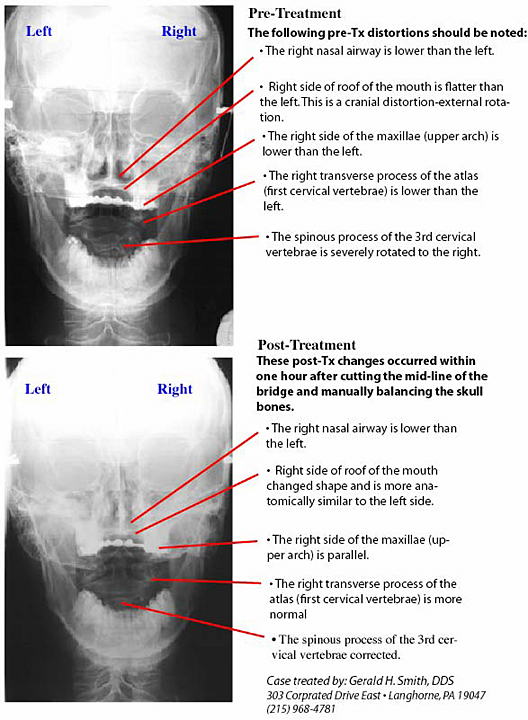
Midpalatal suture of osteopetrotic (op/op) mice exhibits immature fusion. (1/206)
The midpalatal suture was observed histologically in both toothless osteopetrotic (op/op) and normal (control) mice. The normal mice had a mature sutural structure, which consists of a well-developed cartilage cell zone and palatal bone. In contrast, the thickness of the cartilage cell zone was substantially greater in the op/op mice than that in the controls. Moreover, the cartilage cells in the op/op mice were frequently found in the palatal bone as well as in the sutural space, exhibiting an imperfect fusion. It seems that immature fusion at the sutural interface in the op/op mice is related to a decrease in biting or masticatory force accompanied by the failure of tooth eruption in addition to an essential defect in osteoclast differentiation, which is a congenital symptom in op/op mice. (+info)Craniofacial sutures: morphology, growth, and in vivo masticatory strains. (2/206)
The growth and morphology of craniofacial sutures are thought to reflect their functional environment. However, little is known about in vivo sutural mechanics. The present study investigates the strains experienced by the internasal, nasofrontal, and anterior interfrontal sutures during masticatory activity in 4-6-month-old miniature swine (Sus scrofa). Measurements of the bony/fibrous arrangements and growth rates of these sutures were then examined in the context of their mechanical environment. Large tensile strains were measured in the interfrontal suture (1,036 microepsilon +/- 400 SD), whereas the posterior internasal suture was under moderate compression (-440 microepsilon +/- 238) and the nasofrontal suture experienced large compression (-1,583 microepsilon +/- 506). Sutural interdigitation was associated with compressive strain. The collagen fibers of the internasal and interfrontal sutures were clearly arranged to resist compression and tension, respectively, whereas those of the nasofrontal suture could not be readily characterized as either compression or tension resisting. The average linear rate of growth over a 1-week period at the nasofrontal suture (133.8 micrometer, +/- 50.9 S.D) was significantly greater than that of both the internasal and interfrontal sutures (39.2 micrometer +/- 11.4 and 65. 5 micrometer +/- 14.0, respectively). Histological observations suggest that the nasofrontal suture contains chondroid tissue, which may explain the unexpected combination of high compressive loading and rapid growth in this suture. (+info)Craniofacial skeletal abnormalities in anomalous calves with clefts of the face. (3/206)
Thirteen anomalous calves with clefts of the face were morphologically examined, and craniofacial skeletons were studied in detail. According to the type and site of the cleft, four groups could be distinguished: median cleft lip and jaw (CLJ); median cleft lip, jaw, and palate (CLJP); lateral CLJ; and cleft palate (CP), including unilateral and bilateral type. Craniofacial skeletal abnormalities were observed in several bones at the roof, wall, and floor of the nasal cavity and at the boundary portion between the nasal and cranial cavities. Fissure formation at the cranial sutures, partial absence of the nasal process of the incisive bone, and opening of the bony palate were characteristic changes in median CLJ and CLJP, lateral CLJ, and CP, respectively. Furthermore, various associated changes were recognized in the median and paramedian skeletal elements of the face and other organs. The morphological changes of craniofacial skeletons with various types of clefts of the face depended on the site and degree of the cleft formation and reflected developmental errors of the facial embryonic segments. These changes would suggest disorders of the correlated development of facial processes and of other fetal organs of the face. For these conditions, etiologically hereditary cases were negative. (+info)Expression patterns of Twist and Fgfr1, -2 and -3 in the developing mouse coronal suture suggest a key role for twist in suture initiation and biogenesis. (4/206)
Sutural growth depends on maintenance of a balance between proliferation of osteogenic stem cells and their differentiation to form new bone, so that the stem cell population is maintained until growth of the skull is complete. The identification of heterozygous mutations in FGFR1, -2 and -3 and TWIST as well as microdeletions of TWIST in human craniosynostosis syndromes has highlighted these genes as playing important roles in maintaining the suture as a growth centre. In contrast to Drosophila, a molecular relationship between human (or other vertebrate) TWIST and FGFR genes has not yet been established. TWIST mutations exert their effect via haploinsufficiency whereas FGFR mutations have a gain-of-function mechanism of action. To investigate the biological basis of FGFR signalling pathways in the developing calvarium we compared the expression patterns of Twist with those of Fgfr1, -2 and -3 in the fetal mouse coronal suture over the course of embryonic days 14-18, as the suture is initiated and matures. Our results show that: (1) Twist expression precedes that of Fgfr genes at the time of initiation of the coronal suture; (2) in contrast to Fgfr transcripts, which are localised within and around the developing bone domains, Twist is expressed by the midsutural mesenchyme cells. Twist expression domains show some overlap with those of Fgfr2, which is expressed in the most immature (proliferating) osteogenic tissue. (+info)Location of the glenoid fossa after a period of unilateral masticatory function in young rabbits. (5/206)
Changes in glenoid fossa position and skull morphology after a period of unilateral masticatory function were studied. The right-side maxillary and mandibular molars in twenty-seven 10-day-old rabbits were ground down under general anaesthesia. The procedure was repeated twice a week, until the rabbits were 50 days old. Fourteen rabbits were then killed and 13 left to grow to age 100 days. Nine 50-day-old and sixteen 100-day-old rabbits with unmodified occlusions served as controls. Three-dimensional measurements were made using a machine-vision technique and a video-imaging camera. The glenoid fossa position become more anterior in both groups of animals subjected to molar grinding as compared with controls (P < 0.01 in the 50-day-old group and P < 0.05 in 100-day-old group). In the 100-day-old group the right-side fossa was also in a more inferior position (P < 0.01). The glenoid fossa was more anteriorly located on the right than on the left side of individual animals in the group in which the right-side molars had been ground down (P < 0.001). (+info)Strain in the braincase and its sutures during function. (6/206)
The skull is distinguished from other parts of the skeleton by its composite construction. The sutures between bony elements provide for interstitial growth of the cranium, but at the same time they alter the transmission of stress and strain through the skull. Strain gages were bonded to the frontal and parietal bones of miniature pigs and across the interfrontal, interparietal and coronal sutures. Strains were recorded 1) during natural mastication in conjunction with electromyographic activity from the jaw muscles and 2) during stimulation of various cranial muscles in anesthetized animals. Vault sutures exhibited vastly higher strains than did the adjoining bones. Further, bone strain primarily reflected torsion of the braincase set up by asymmetrical muscle contraction; the tensile axis alternated between +45 degrees and -45 degrees depending on which diagonal masseter/temporalis pair was most active. However, suture strains were not related to overall torsion but instead were responses to local muscle actions. Only the coronal suture showed significant strain (tension) during jaw opening; this was caused by the contraction of neck muscles. All sutures showed strain during jaw closing, but polarity depended on the pattern of muscle usage. For example, masseter contraction tensed the coronal suture and the anterior part of the interfrontal suture, whereas the temporalis caused compression in these locations. Peak tensile strains were larger than peak compressive strains. Histology suggested that the skull is bent at the sutures, with the ectocranial surface tensed and the endocranial surface predominantly compressed. Collectively, these results indicate that skulls with patent sutures should be analyzed as complexes of independent parts rather than solid structures. (+info)Trigonocephaly in rabbits with familial interfrontal suture synostosis: the multiple effects of premature single-suture fusion. (7/206)
Previous studies from our laboratory have characterized the craniofacial morphology and growth patterns of an inbred strain of rabbits with autosomal dominant coronal suture synostosis. A number of rabbit perinates from this colony have been collected sporadically over a 5-year period with premature interfrontal suture synostosis. The present study describes the very early onset of craniofacial dysmorphology of these rabbits and compares them to similar-aged normal control rabbits. A total of 40 perinatal New Zealand White rabbits were used in the present study. Twenty-one comprised the sample with interfrontal suture synostosis and ranged in age from 27 to 38 days postconception (term = 31 days) with a mean age of 33.53 days (+/-2.84 days). Nineteen rabbits served as age-matched, normal controls (mean age = 33.05 days +/-2.79 days). Lateral and dorsoventral radiographs were collected from each rabbit. The radiographs were traced, computer digitized, and 12 craniofacial measurements, angles, and indices were obtained. Mean measures were compared using an unpaired Student's t-test. All synostosed rabbits were stillborn or died shortly after birth. Grossly, these rabbits exhibited extreme frontal bossing, trigonocephaly with sagittal keeling, and midfacial shortening. No somatic anomalies were noted. Radiographically, rabbits with interfrontal suture synostosis had significantly (P < 0.05) narrower bifrontal widths, shorter cranial vault lengths, kyphotic cranial base angles, and different cranial vault indices (shapes) compared to controls. Results reveal severe and early pathological and compensatory cranial vault changes associated with premature interfrontal suture synostosis in this rabbit model. The 100% mortality rate noted in this condition may be related to the inheritance of a lethal genetic mutation or to neural compression from reduced intracranial volume. Results are discussed in light of current pathogenic hypotheses for human infants with premature metopic suture synostosis. (+info)Compressive force promotes chondrogenic differentiation and hypertrophy in midpalatal suture cartilage in growing rats. (8/206)
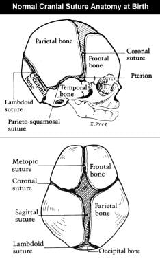
Midpalatal suture cartilage (MSC) is secondary cartilage located between the bilateral maxillary bones and has been utilized in the analysis of the biomechanical characteristics of secondary cartilage. The present study was designed to investigate the effects of compressive force on the differentiation of cartilage in midpalatal suture cartilage in rats. Forces of various magnitudes were applied to the midpalatal suture cartilage in 4-week-old male Wistar rats for 1, 2, 4, 7, or 14 days, mediated through the bilateral 1st molars using orthodontic wires. The differentiation pathways in the MSC cells were examined by immunohistochemistry for the differentiation markers type I, type II and type X collagen, and glycosaminoglycans (GAGs), chondroitin-4-sulfate, chondroitin-6-sulfate and keratan sulfate. Histologically and immunohistochemically, the midpalatal suture cartilage in control rats had the characteristic appearance of secondary cartilage. In the experimental groups, the center of the midpalatal suture cartilage that contained osteo-chondro progenitor cells seemed to become mature cartilage and its immuno-reaction to type II and X collagen and GAGs increased as the experiment progressed. This differentiation was dependent upon the magnitude and duration of the force applied to the midpalatal suture cartilage; i.e., cartilaginous differentiation progressed more rapidly as the applied force increased. The present results suggest that the differentiation of osteo-chondro progenitor cells into mature and hypertrophic chondrocytes in the precartilaginous cell layer is promoted by compressive force. (+info)Cranial sutures are the fibrous joints that connect and hold together the bones of the skull (cranium) in humans and other animals. These sutures provide flexibility for the skull during childbirth and growth, allowing the skull to expand as the brain grows in size, especially during infancy and early childhood.
There are several cranial sutures in the human skull, including:
1. The sagittal suture, which runs along the midline of the skull, connecting the two parietal bones.
2. The coronal suture, which connects the frontal bone to the two parietal bones.
3. The lambdoid suture, which connects the occipital bone to the two parietal bones.
4. The squamosal suture, which connects the temporal bone to the parietal bone.
5. The frontosphenoidal and sphenoethmoidal sutures, which connect the frontal bone, sphenoid bone, and ethmoid bone in the anterior cranial fossa.
These sutures are typically made up of a specialized type of connective tissue called Sharpey's fibers, which interdigitate with each other to form a strong yet flexible joint. Over time, as the skull bones fully fuse together, these sutures become less prominent and eventually ossify (turn into bone). In some cases, abnormalities in cranial suture development or fusion can lead to medical conditions such as craniosynostosis.
Craniosynostosis is a medical condition that affects the skull of a developing fetus or infant. It is characterized by the premature closure of one or more of the fibrous sutures between the bones of the skull (cranial sutures). These sutures typically remain open during infancy to allow for the growth and development of the brain.
When a suture closes too early, it can restrict the growth of the surrounding bones and cause an abnormal shape of the head. The severity of craniosynostosis can vary depending on the number of sutures involved and the extent of the premature closure. In some cases, craniosynostosis can also lead to increased pressure on the brain, which can cause a range of neurological symptoms.
There are several types of craniosynostoses, including:
1. Sagittal synostosis: This is the most common type and involves the premature closure of the sagittal suture, which runs from front to back along the top of the head. This can cause the skull to grow long and narrow, a condition known as scaphocephaly.
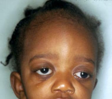
2. Coronal synostosis: This type involves the premature closure of one or both of the coronal sutures, which run from the temples to the front of the head. When one suture is affected, it can cause the forehead to bulge and the eye socket on that side to sink in (anterior plagiocephaly). When both sutures are affected, it can cause a flattened appearance of the forehead and a prominent back of the head (brachycephaly).
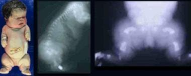
3. Metopic synostosis: This type involves the premature closure of the metopic suture, which runs from the top of the forehead to the bridge of the nose. It can cause a triangular shape of the forehead and a prominent ridge along the midline of the skull (trigonocephaly).
4. Lambdoid synostosis: This is the least common type and involves the premature closure of the lambdoid suture, which runs along the back of the head. It can cause an asymmetrical appearance of the head and face, as well as possible neurological symptoms.
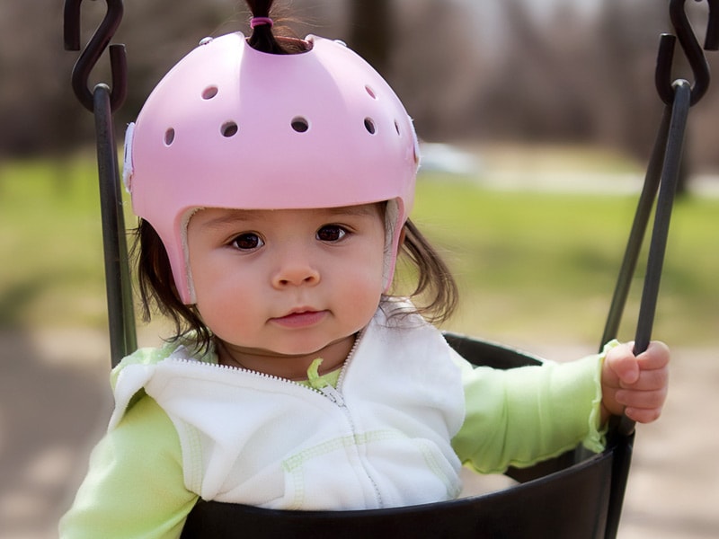
In some cases, multiple sutures may be affected, leading to more complex craniofacial abnormalities. Treatment for craniosynostosis typically involves surgery to release the fused suture(s) and reshape the skull. The timing of the surgery depends on the type and severity of the condition but is usually performed within the first year of life. Early intervention can help prevent further complications, such as increased intracranial pressure and developmental delays.
In medical terms, sutures are specialized surgical threads made from various materials such as absorbable synthetic or natural fibers, or non-absorbable materials like nylon or silk. They are used to approximate and hold together the edges of a wound or incision in the skin or other tissues during the healing process. Sutures come in different sizes, types, and shapes, each designed for specific uses and techniques depending on the location and type of tissue being sutured. Properly placed sutures help to promote optimal healing, minimize scarring, and reduce the risk of infection or other complications.
The skull is the bony structure that encloses and protects the brain, the eyes, and the ears. It is composed of two main parts: the cranium, which contains the brain, and the facial bones. The cranium is made up of several fused flat bones, while the facial bones include the upper jaw (maxilla), lower jaw (mandible), cheekbones, nose bones, and eye sockets (orbits).
The skull also provides attachment points for various muscles that control chewing, moving the head, and facial expressions. Additionally, it contains openings for blood vessels, nerves, and the spinal cord to pass through. The skull's primary function is to protect the delicate and vital structures within it from injury and trauma.
Suture techniques refer to the various methods used by surgeons to sew or stitch together tissues in the body after an injury, trauma, or surgical incision. The main goal of suturing is to approximate and hold the edges of the wound together, allowing for proper healing and minimizing scar formation.
There are several types of suture techniques, including:
1. Simple Interrupted Suture: This is one of the most basic suture techniques where the needle is passed through the tissue at a right angle, creating a loop that is then tightened to approximate the wound edges. Multiple stitches are placed along the length of the incision or wound.
2. Continuous Locking Suture: In this technique, the needle is passed continuously through the tissue in a zigzag pattern, with each stitch locking into the previous one. This creates a continuous line of sutures that provides strong tension and support to the wound edges.
3. Running Suture: Similar to the continuous locking suture, this technique involves passing the needle continuously through the tissue in a straight line. However, instead of locking each stitch, the needle is simply passed through the previous loop before being tightened. This creates a smooth and uninterrupted line of sutures that can be easily removed after healing.
4. Horizontal Mattress Suture: In this technique, two parallel stitches are placed horizontally across the wound edges, creating a "mattress" effect that provides additional support and tension to the wound. This is particularly useful in deep or irregularly shaped wounds.
5. Vertical Mattress Suture: Similar to the horizontal mattress suture, this technique involves placing two parallel stitches vertically across the wound edges. This creates a more pronounced "mattress" effect that can help reduce tension and minimize scarring.
6. Subcuticular Suture: In this technique, the needle is passed just below the surface of the skin, creating a smooth and barely visible line of sutures. This is particularly useful in cosmetic surgery or areas where minimizing scarring is important.
The choice of suture technique depends on various factors such as the location and size of the wound, the type of tissue involved, and the patient's individual needs and preferences. Proper suture placement and tension are crucial for optimal healing and aesthetic outcomes.
The parietal bone is one of the four flat bones that form the skull's cranial vault, which protects the brain. There are two parietal bones in the skull, one on each side, located posterior to the frontal bone and temporal bone, and anterior to the occipital bone. Each parietal bone has a squamous part, which forms the roof and sides of the skull, and a smaller, wing-like portion called the mastoid process. The parietal bones contribute to the formation of the coronal and lambdoid sutures, which are fibrous joints that connect the bones in the skull.
The frontal bone is the bone that forms the forehead and the upper part of the eye sockets (orbits) in the skull. It is a single, flat bone that has a prominent ridge in the middle called the superior sagittal sinus, which contains venous blood. The frontal bone articulates with several other bones, including the parietal bones at the sides and back, the nasal bones in the center of the face, and the zygomatic (cheek) bones at the lower sides of the orbits.
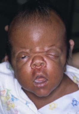
Acrocephalosyndactyly is a genetic disorder that affects the development of the skull and limbs. The term comes from the Greek words "acros," meaning extremity, "cephale," meaning head, and "syndactylia," meaning webbed or fused fingers or toes.
There are several types of acrocephalosyndactyly, but the most common is Type 1, also known as Apert syndrome. People with Apert syndrome have a characteristic appearance, including a high, prominent forehead (acrocephaly), widely spaced eyes (hypertelorism), and underdeveloped upper jaw and midface (maxillary hypoplasia). They also have webbed or fused fingers and toes (syndactyly) and may have other skeletal abnormalities.
Acrocephalosyndactyly is caused by a mutation in the FGFR2 gene, which provides instructions for making a protein that is involved in the development of bones and tissues. The mutation leads to overactive signaling of the FGFR2 protein, which can cause abnormal bone growth and fusion.
Treatment for acrocephalosyndactyly typically involves a team of specialists, including geneticists, orthopedic surgeons, craniofacial surgeons, and other healthcare professionals. Surgery may be necessary to correct skeletal abnormalities, improve function, and enhance appearance. Speech therapy, occupational therapy, and other supportive care may also be recommended.
A Twist Transcription Factor is a family of proteins that regulate gene expression through the process of transcription. The name "Twist" comes from the Drosophila melanogaster (fruit fly) gene, which was first identified due to its role in causing twisted or spiral patterns during embryonic development.
The Twist protein is a basic helix-loop-helix (bHLH) transcription factor that binds to specific DNA sequences and regulates the expression of target genes. It forms homodimers or heterodimers with other bHLH proteins, which then recognize and bind to E-box motifs in the promoter regions of target genes.
Twist proteins have been shown to play critical roles in various biological processes, including cell differentiation, proliferation, migration, and survival. In particular, they have been implicated in cancer progression and metastasis, as they can promote epithelial-mesenchymal transition (EMT), a key step in tumor invasion and dissemination.
Abnormal expression or mutations of Twist transcription factors have been associated with several human diseases, including various types of cancer, developmental disorders, and neurological conditions.
Synostosis is a medical term that refers to the abnormal or physiological fusion of adjacent bones. It's derived from two Greek words, "syn" meaning together and "osteon" meaning bone. In a normal physiological process, synostosis occurs during growth and development, where the growth of certain bones is stopped by the fusion of neighboring bones at specific sites known as sutures or fontanelles.
However, abnormal synostosis can occur due to various reasons such as injuries, infections, or genetic conditions. This can lead to restricted movement and growth disturbances in the affected area. Common examples include craniosynostosis, where the skull bones fuse prematurely, and syndactyly, where fingers or toes are fused together. Treatment for abnormal synostosis may involve surgery to correct the fusion and prevent further complications.
A skull fracture is a break in one or more of the bones that form the skull. It can occur from a direct blow to the head, penetrating injuries like gunshot wounds, or from strong rotational forces during an accident. There are several types of skull fractures, including:
1. Linear Skull Fracture: This is the most common type, where there's a simple break in the bone without any splintering, depression, or displacement. It often doesn't require treatment unless it's near a sensitive area like an eye or ear.
2. Depressed Skull Fracture: In this type, a piece of the skull is pushed inward toward the brain. Surgery may be needed to relieve pressure on the brain and repair the fracture.
3. Diastatic Skull Fracture: This occurs along the suture lines (the fibrous joints between the skull bones) that haven't fused yet, often seen in infants and young children.
4. Basilar Skull Fracture: This involves fractures at the base of the skull. It can be serious due to potential injury to the cranial nerves and blood vessels located in this area.
5. Comminuted Skull Fracture: In this severe type, the bone is shattered into many pieces. These fractures usually require extensive surgical repair.
Symptoms of a skull fracture can include pain, swelling, bruising, bleeding (if there's an open wound), and in some cases, clear fluid draining from the ears or nose (cerebrospinal fluid leak). Severe fractures may cause brain injury, leading to symptoms like confusion, loss of consciousness, seizures, or neurological deficits. Immediate medical attention is necessary for any suspected skull fracture.
Dura Mater is the thickest and outermost of the three membranes (meninges) that cover the brain and spinal cord. It provides protection and support to these delicate structures. The other two layers are called the Arachnoid Mater and the Pia Mater, which are thinner and more delicate than the Dura Mater. Together, these three layers form a protective barrier around the central nervous system.
Craniofacial dysostosis is a term used to describe a group of rare genetic disorders that affect the development of the skull and face. These conditions are characterized by cranial and facial abnormalities, including a misshapen head, wide-set eyes, a beaked nose, and underdeveloped jaws.
The most common type of craniofacial dysostosis is Crouzon syndrome, which is caused by mutations in the FGFR2 gene. Other types include Apert syndrome (caused by mutations in the FGFR2 or FGFR3 gene), Pfeiffer syndrome (caused by mutations in the FGFR1 or FGFR2 gene), and Saethre-Chotzen syndrome (caused by mutations in the TWIST1 gene).
These conditions can vary in severity, but they often cause complications such as breathing difficulties, vision problems, hearing loss, and developmental delays. Treatment typically involves a team of specialists, including craniofacial surgeons, orthodontists, ophthalmologists, and audiologists, and may include surgery to correct the structural abnormalities and improve function.
Fibroblast Growth Factor Receptor 2 (FGFR2) is a type of receptor tyrosine kinase that plays a crucial role in various biological processes such as cell survival, proliferation, differentiation, and migration. Specifically, FGFR2 is activated by binding to its specific ligands, fibroblast growth factors (FGFs), leading to the activation of downstream signaling pathways.
FGFR2 has several isoforms generated by alternative splicing, including FGFR2-IIIb and FGFR2-IIIc. These isoforms differ in their extracellular ligand-binding domains and have distinct expression patterns and functions. FGFR2-IIIb is primarily expressed in epithelial cells and binds to FGFs 1, 3, 7, 10, and 22, while FGFR2-IIIc is mainly expressed in mesenchymal cells and binds to FGFs 1, 2, 4, 6, 9, 10, and 22.
Mutations in the FGFR2 gene have been associated with various human diseases, including developmental disorders, cancers, and fibrosis. In particular, activating mutations or amplifications of FGFR2 have been identified in several types of cancer, such as breast, lung, gastric, and endometrial cancers, making it an attractive therapeutic target for cancer treatment.
An epidural cranial hematoma is a specific type of hematoma, which is defined as an abnormal accumulation of blood in a restricted space, occurring between the dura mater (the outermost layer of the meninges that covers the brain and spinal cord) and the skull in the cranial region. This condition is often caused by trauma or head injury, which results in the rupture of blood vessels, allowing blood to collect in the epidural space. The accumulation of blood can compress the brain tissue and cause various neurological symptoms, potentially leading to serious complications if not promptly diagnosed and treated.
A suture anchor is a medical device used in surgical procedures, particularly in orthopedic and cardiovascular surgeries. It is typically made of biocompatible materials such as metal (titanium or absorbable steel) or polymer (absorbable or non-absorbable). The suture anchor serves to attach a suture to bone securely, providing a stable fixation point for soft tissue reattachment or repair.
Suture anchors come in various shapes and sizes, including screws, hooks, or buttons, designed to fit specific surgical needs. Surgeons insert the anchor into a predrilled hole in the bone, and then pass the suture through the eyelet or loop of the anchor. Once the anchor is securely in place, the surgeon can tie the suture to attach tendons, ligaments, or other soft tissues to the bone.
The use of suture anchors has revolutionized many surgical procedures by providing a more reliable and less invasive method for reattaching soft tissues to bones compared to traditional methods such as drill holes and staples.
Osteogenesis is the process of bone formation or development. It involves the differentiation and maturation of osteoblasts, which are bone-forming cells that synthesize and deposit the organic matrix of bone tissue, composed mainly of type I collagen. This organic matrix later mineralizes to form the inorganic crystalline component of bone, primarily hydroxyapatite.
There are two primary types of osteogenesis: intramembranous and endochondral. Intramembranous osteogenesis occurs directly within connective tissue, where mesenchymal stem cells differentiate into osteoblasts and form bone tissue without an intervening cartilage template. This process is responsible for the formation of flat bones like the skull and clavicles.
Endochondral osteogenesis, on the other hand, involves the initial development of a cartilaginous model or template, which is later replaced by bone tissue. This process forms long bones, such as those in the limbs, and occurs through several stages involving chondrocyte proliferation, hypertrophy, and calcification, followed by invasion of blood vessels and osteoblasts to replace the cartilage with bone tissue.
Abnormalities in osteogenesis can lead to various skeletal disorders and diseases, such as osteogenesis imperfecta (brittle bone disease), achondroplasia (a form of dwarfism), and cleidocranial dysplasia (a disorder affecting skull and collarbone development).
A subdural hematoma is a type of hematoma (a collection of blood) that occurs between the dura mater, which is the outermost protective covering of the brain, and the brain itself. It is usually caused by bleeding from the veins located in this potential space, often as a result of a head injury or trauma.
Subdural hematomas can be classified as acute, subacute, or chronic based on their rate of symptom progression and the time course of their appearance on imaging studies. Acute subdural hematomas typically develop and cause symptoms rapidly, often within hours of the head injury. Subacute subdural hematomas have a more gradual onset of symptoms, which can occur over several days to a week after the trauma. Chronic subdural hematomas may take weeks to months to develop and are often seen in older adults or individuals with chronic alcohol abuse, even after minor head injuries.
Symptoms of a subdural hematoma can vary widely depending on the size and location of the hematoma, as well as the patient's age and overall health. Common symptoms include headache, altered mental status, confusion, memory loss, weakness or numbness, seizures, and in severe cases, coma or even death. Treatment typically involves surgical evacuation of the hematoma, along with management of any underlying conditions that may have contributed to its development.
Osteoblasts are specialized bone-forming cells that are derived from mesenchymal stem cells. They play a crucial role in the process of bone formation and remodeling. Osteoblasts synthesize, secrete, and mineralize the organic matrix of bones, which is mainly composed of type I collagen.
These cells have receptors for various hormones and growth factors that regulate their activity, such as parathyroid hormone, vitamin D, and transforming growth factor-beta. When osteoblasts are not actively producing bone matrix, they can become trapped within the matrix they produce, where they differentiate into osteocytes, which are mature bone cells that play a role in maintaining bone structure and responding to mechanical stress.
Abnormalities in osteoblast function can lead to various bone diseases, such as osteoporosis, osteogenesis imperfecta, and Paget's disease of bone.
Fibroblast growth factor (FGF) receptors are a group of cell surface tyrosine kinase receptors that play crucial roles in various biological processes, including embryonic development, tissue repair, and tumor growth. There are four high-affinity FGF receptors (FGFR1-4) in humans, which share a similar structure, consisting of an extracellular ligand-binding domain, a transmembrane region, and an intracellular tyrosine kinase domain.
These receptors bind to FGFs with different specificities and affinities, triggering a cascade of intracellular signaling events that regulate cell proliferation, differentiation, migration, and survival. Aberrant FGFR signaling has been implicated in several diseases, such as cancer, developmental disorders, and fibrotic conditions. Dysregulation of FGFRs can occur through various mechanisms, including genetic mutations, amplifications, or aberrant expression, leading to uncontrolled cell growth and malignant transformation. Therefore, FGFRs are considered promising targets for therapeutic intervention in several diseases.
 Chignon (medical term)
Frontal suture
Skull
Craniosynostosis
Orang-Outang, sive Homo Sylvestris
Fontanelle
Skull fracture
Fibrous joint
Daya Reddy
Anatomical terms of bone
Zygomatic arch
Skeletal changes of vertebrates transitioning from water to land
Mortuary archaeology
Hajdu-Cheney syndrome
Box counting
Odobenocetops
Theories of craniofacial growth
Hypophosphatasia
Molera
Infant
Sharpey's fibres
Asterion (anatomy)
Iufaa
McGillivray syndrome
Bird
Aralosaurus
Sambungmacan crania
Pycnodysostosis
Craniofacial surgery
Sphenofrontal suture
Treatment of cranial cruciate ligament1
- Comparison of outcomes associated with tibial plateau levelling osteotomy and a modified technique for tibial tuberosity advancement for the treatment of cranial cruciate ligament disease in dogs: a randomized clinical study. (thieme-connect.com)
Premature9
- Craniosynostosis is the premature fusion of one or more calvarial growth sites (sutures) prior to the completion of brain expansion. (elsevierpure.com)
- High dose exposed mice showed diminished area of the coronal and widening of the sagittal sutures indicative of premature fusion and compensatory growth. (elsevierpure.com)
- Two new loci for premature fusion of the cranial sutures in humans suggest a common endpoint in osteoblast regulation, linking upregulation of phosphorylated ERK1/2 and TWIST1 haploinsufficiency. (nature.com)
- The appearance of a new suture long after the normal time period for suture formation in utero indicates that the craniosynostosis may just as well be caused by disturbed formation of the suture as actual premature closure. (nih.gov)
- Craniosynostosis is the premature (early) closure of one or more cranial sutures. (memorialhermann.org)
- Saethre-Chotzen syndrome is an autosomal dominant inherited disorder with premature fusion of cranial sutures. (uni-marburg.de)
- Abstract Craniosynostosis is a common congenital craniofacial deformity caused by premature ossification and closure of one or more cranial sutures. (techscience.com)
- These occur with premature fusion of sagittal, metopic, or coronal sutures, with the coronal sutures being the most common. (medscape.com)
- The membranous neurocranium is derived from mesenchyme that invests the developing brain, a relationship that may help explain the etiology of diseases of premature suture fusion. (medscape.com)
Bone18
- The spatial scale spans eight orders of magnitude, from the macroarchitectural level (the entire cranium), through the mesoarchitectural (the local/regional bone-suture-bone complex) and microarchitectural levels (tissues and cells), to the nanoarchitectural level (molecules within and outside the cells). (uky.edu)
- Suture tissue is unique in that it facilitates growth and remodeling of bone connected to it in the presence of applied mechanical stimuli, and will generally fuse to become bone over time. (isbweb.org)
- In its unfused form, cranial sutures are a complex structure consisting primarily of vasculature, collagen fibres, and extracellular matrix, and may have varying levels of bone present throughout as they begin to fuse over time. (isbweb.org)
- Overall research goals in this area are to better understand the mechanical response of suture tissue to applied loading and how this in turn drives the biological response to facilitate bone remodeling and growth at suture sites. (isbweb.org)
- Bone formation and suture closure were enhanced in an organ culture of Runx2+/- calvariae with ligands or agonists of hedgehog , Fgf, Wnt and Pthlh signaling, while they were suppressed and suture mesenchymal cell proliferation was decreased in an organ culture of wild-type calvariae with their antagonists. (bvsalud.org)
- [1] The skull is composed of four types of bone i.e., cranial bones, facial bones, ear ossicles and hyoid bone. (wikipedia.org)
- Sometimes there can be extra bone pieces within the suture known as wormian bones or sutural bones . (wikipedia.org)
- Individual ossicles (small bone) are named for its associated suture. (eskeletons.org)
- Bridging of bone over a suture, an indistinct suture, or sclerosis along the suture margins indicates fusion. (aafp.org)
- The AN construct consisted of 3 bone anchors connected with monofilament nylon suture. (avma.org)
- The TN and TU constructs involved the creation of 3 bone tunnels and use of nylon or UHMWPE suture, respectively. (avma.org)
- In a baby's head, there is a strong elastic tissue which stretches between the seven plates of bone: these are called cranial sutures. (medic8.com)
- By the time the individual matures and the skull no longer needs to continue to expand, the sutures permanently fuse creating a continuous covering of bone rather than the mosaic effect that the plates of a baby present. (medic8.com)
- Skull sutures close up as the connective tissue is invaded by bone-making cells that, during development, either fuse or 'zipper' the skull bones together. (icr.org)
- It provides structural support as well as a small amount of blood supply to the cranial bone which it adheres to. (3d4medical.com)
- The equine skull is a giant jigsaw puzzle made of 26 individual plates of bone joined together by sutures. (holistichorse.com)
- Craniosynostosis refers to a specific aberration of the growth process in which bone growth at a particular suture site (or sites) is arrested prematurely, resulting in a specific skull shape deformity. (medscape.com)
- The mendosal is an obscure suture in the occipital bone running horizontally from the medial portion of the lambdoid suture. (medscape.com)
Ruptured cranial cruciate ligament1
- 7 Böddeker J, Drüen S, Meyer-Lindenberg A, Fehr M, Nolte I, Wefstaedt P. Computer-assisted gait analysis of the dog: comparison of two surgical techniques for the ruptured cranial cruciate ligament. (thieme-connect.com)
Frontal5
- The closure of posterior frontal (PF) and sagittal (SAG) sutures was completely interrupted in Runx2+/- mice , and the proliferation of suture mesenchymal cells and their condensation were less than those in wild-type mice . (bvsalud.org)
- Except for the metopic suture between the frontal bones, which closes at two years of age, the sutures remain open until brain growth ceases in the second decade of life. (aafp.org)
- One of the key differences of skull development between humans and apes is the time of closure for the frontal ( metopic ) suture-a line of dense connective tissue at the front of the skull (see image below). (icr.org)
- Anteriorly, the widened frontal and sagittal sutures can be seen. (medscape.com)
- The median frontal, or metopic, suture usually closes by 2-6 years. (medscape.com)
Coronal suture1
- Those with sagittal suture involvement have long, thin skulls, while those with coronal suture involvement have a flat, short head on one or both sides, depending on whether one or both coronal sutures are involved. (memorialhermann.org)
Skulls3
- Both fetal heads contain skulls with palpable cranial sutures and anterior and posterior fontanels. (anatomywarehouse.com)
- I got inspired by this my sophomore summer when I did research on craniosynostosis, which is a congenital disorder in which the cranial sutures of babies' skulls fuse prematurely, leading to abnormal brain development. (princeton.edu)
- Though this purpose for cranial sutures seems teleologically sound, Murray observes that cranial sutures are present in the skulls of chickens who do not suffer the forces of passage through a birth canal. (medscape.com)
Closure5
- Runx2 regulates cranial suture closure by inducing hedgehog, Fgf, Wnt and Pthlh signaling pathway gene expressions in suture mesenchymal cells. (bvsalud.org)
- However, it currently remains unclear why suture closure is severely impaired in CCD patients . (bvsalud.org)
- The next most common group is those infants who present with early closure of the cranial sutures ( craniosynostosis ). (memorialhermann.org)
- There is a distinctive head shape associated with early closure of specific sutures which differs from the head shape of infants with positional molding. (memorialhermann.org)
- While the molding function of sutures may be questioned, normal growth relies on their patency and appropriately timed closure. (medscape.com)
Craniosynostosis7
- The discovery of RAB23 mutations in patients with Carpenter syndrome implicates HH signaling in cranial-suture biogenesis--an unexpected finding, given that craniosynostosis is not usually associated with mutations of other HH-pathway components--and provides a new molecular target for studies of obesity. (ox.ac.uk)
- Craniosynostosis, and the consequent skull shape deformities, is treated with surgery including osteotomies of the fused sutures. (nih.gov)
- Additionally, in 7 patients (8%) a new suture appeared in a part of the suture that had a discernible suture prior to surgery.In conclusion, in this consecutive and well-defined patient cohort operated for craniosynostosis, the formation of a neosuture is not a rare, and speculatively not a random, event. (nih.gov)
- In children with multiple affected sutures or with sagittal craniosynostosis, surgery is performed prior to three months of age. (memorialhermann.org)
- In craniosynostosis, a hard ridge grows along the sutures far too soon and the fontanelle either disappears or feels changed. (medic8.com)
- in cases of craniosynostosis where just one suture is affected, just 15% of infants suffer raised ICP. (medic8.com)
- Craniosynostosis can be subdivided into a number of separate categories, differentiated by the sutures which are involved in the deformity. (medic8.com)
Metopic suture2
- Thus, neither of the predicted features necessary to support the form of metopic suture in the Taung Child suggested by Falk and colleagues are observed in the present study. (icr.org)
- 2012. Metopic suture of Taung (Australopithecus africanus) and its implications for hominin brain evolution. (icr.org)
Lambdoid suture1
- Most commonly these are found in the course of the lambdoid suture . (wikipedia.org)
Connective tissue2
- Cranial sutures are the soft connective tissue that joins bones in the skull. (isbweb.org)
- Fontanels are the fibrous, membrane-covered gaps created when more than two cranial bones are juxtaposed, as opposed to sutures, which are narrow seams of fibrous connective tissue that separate the flat bones of the skull. (aafp.org)
Fuse2
- In children whose cranial sutures have yet to fuse, there is a rapid enlargement of the head circumference 2 . (radiopaedia.org)
- The cartilaginous neurocranium (or chondrocranium) comprises a number of cartilaginous plates that grow, ossify, and fuse to form most of the cranial base. (medscape.com)
Fontanelles4
- The sutures and fontanelles are needed for the infant's brain growth and development. (medlineplus.gov)
- Without flexible sutures and fontanelles, the child's brain could not grow enough. (medlineplus.gov)
- Feeling the cranial sutures and fontanelles is one way that health care providers follow the child's growth and development. (medlineplus.gov)
- Because humans have larger brains than apes, these sutures (also called fontanelles ) are much larger at birth and allow for the baby's skull to slightly compress at the time of delivery through the birth canal. (icr.org)
Wormian bones1
- wormian bones] extra, small bones occuring within cranial sutures. (eskeletons.org)
Limb1
- Effect of surgical technique on limb function after surgery for rupture of the cranial cruciate ligament in dogs. (thieme-connect.com)
Tissues2
- These bones are held together by strong, fibrous, elastic tissues called sutures. (medlineplus.gov)
- Experimental, analytical modeling, and numerical modeling approaches are used to study materials such as cranial suture and periodontal ligament tissues as well as a number of other dental-based restorative materials. (isbweb.org)
Anterior2
- Case 1 was a 50-year-old man who presented with recurrent epistaxis and was diagnosed with an olfactory neuroblastoma that extended from the nasal cavity to the anterior cranial base and infiltrated the right anterior cranial fossa. (thejns.org)
- 8 The newborn's skull should be evaluated for shape, circumference, suture ridges, and size of anterior and posterior fontanels. (aafp.org)
Osteogenesis1
- Presence of thyroid receptors was confirmed for the murine cranial suture and markers of proliferation and osteogenesis were increased in sutures from exposed mice. (elsevierpure.com)
Skeleton4
- The skull consists of three parts, of different embryological origin-the neurocranium , the sutures , and the facial skeleton (also called the membraneous viscerocranium ). (wikipedia.org)
- The human skull is generally considered to consist of twenty-two bones -eight cranial bones and fourteen facial skeleton bones. (wikipedia.org)
- The presence of sutures in the cranial skeleton allows for compression and overriding of cranial bones during birth. (medscape.com)
- The facial skeleton, consisting of bones situated between the cranial base and the mandibular region. (bvsalud.org)
Scalp1
- Separating the scalp and meninges are the cranial bones. (3d4medical.com)
MeSH1
- This study demonstrates the value of 3D MRI study with 3D finite element mesh reconstruction during the second stage of labor to reveal how the fetal brain is impacted by the molding of the cranial bones. (plos.org)
Tissue4
- Cranial sutures are fibrous bands of tissue that connect the bones of the skull. (medlineplus.gov)
- Dr. Romanyk of the Department of Mechanical Engineering at the University of Alberta invites applications and queries for a full-time Doctoral research assistant position in the area of Cranial Suture Tissue Mechanics . (isbweb.org)
- This specific research project will focus on studying the mechanical response of cranial suture tissue using advanced experimental and modeling techniques. (isbweb.org)
- The process isn't fully understood, but there's evidence suggesting that the braincase shrinks as tissue within cranial sutures is resorbed. (eurekalert.org)
Utero1
- The formation of the cranial sutures, in utero, occurs when the ossification of the skull bones reaches predestined positions around gestational week 15 to 20. (nih.gov)
Joints4
- The sutures are fairly rigid joints between bones of the neurocranium. (wikipedia.org)
- Except for the mandible , all of the bones of the skull are joined by sutures - synarthrodial (immovable) joints formed by bony ossification , with Sharpey's fibres permitting some flexibility. (wikipedia.org)
- There are 8 cranial bones, held together by joints called sutures. (3d4medical.com)
- Sutures act like joints between the cranial bones and are designed to help disperse the energy from an impact to the skull. (holistichorse.com)
Ridges1
- This is because the distinctive ridges along the suture lines will be evidence, and this combined in some cases with displacement of the ears can be more than adequate signs. (medic8.com)
Morphology2
- Overall our data suggest that maternal exogenous thyroxine exposure can drive calvarial growth alterations and altered suture morphology. (elsevierpure.com)
- The mechanical strain experienced by the sutures eventually alters the morphology of the sutures. (uky.edu)
Abnormalities1
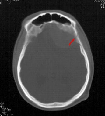
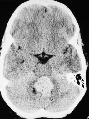
- 13 A computed tomographic (CT) scan can detect a fused suture, dilated ventricles, enlarged subarachnoid space, brain size, or an intracranial or extracranial mass. 14 Magnetic resonance imaging (MRI) can detect cortical and white-matter abnormalities, such as degenerative diseases, and document the extent of calvarial masses. (aafp.org)
Braincase1
- The neurocranium (or braincase ) forms the protective cranial cavity that surrounds and houses the brain and brainstem . (wikipedia.org)
Hedgehog1
- RAB23 mutations in Carpenter syndrome imply an unexpected role for hedgehog signaling in cranial-suture development and obesity. (ox.ac.uk)
Fusion4
- Skull X-rays and computerized tomography are used to evaluate the presence of early fusion of the cranial sutures. (memorialhermann.org)
- The fusion of the involved sutures can be seen. (medscape.com)
- Three-dimensional CT scans can be produced but yield no more information than standard scans, although suture fusion can be graphically displayed. (medscape.com)
- The correction of transverse demonstrating its efficacy, the understanding of the maturation maxillary atresia has long been a topic of discussion in the and fusion processes of the midpalatal suture (MPS) are literature. (bvsalud.org)
Craniosynostoses1
- These portions of the skull undergo endochondral ossification and form the greater portion of the cranial base, contributing little to the cranial suture involved in most syndromal and nonsyndromal craniosynostoses. (medscape.com)
Constructs1
- Ultimate strength was greatest for the UHMWPE-suture constructs. (avma.org)
Gene1
- Increased Htra1 and Igf1 gene expression were found in sutures from high dose exposed individuals. (elsevierpure.com)
Evaluate2
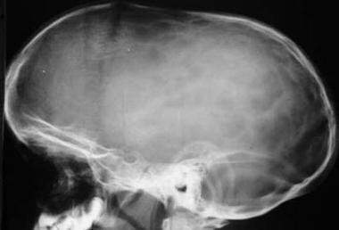
- Plain radiographs of the skull are the least expensive way to evaluate the sutures and cranial bones, but they are limited by the lack of mineralization of the neonatal cranium. (aafp.org)
- The CBCT scans were applied to evaluate midpalatal suture maturation status and comprised stages previously classified as B (29), C (92) and D (37). (bvsalud.org)
Perpendicular1
- 6 Once a suture is fused, growth perpendicular to that suture is restricted. (aafp.org)
Viscerocranium1
- The neurocranium (calvaria in the adult) and viscerocranium (facial bones and portions of cranial base in the adult) combine to form the skull and grow independently through separate mechanisms. (medscape.com)
Skull bones1
- The human skull fully develops two years after birth.The junctions of the skull bones are joined by structures called sutures . (wikipedia.org)
Approaches1
- ANSYS or ABAQUS) to further develop cranial suture modeling approaches. (isbweb.org)
Closes1
- The mendosal suture closes first, several days after birth. (medscape.com)
Abnormal1
- These children present with an abnormal head shape that varies according to the suture involved. (memorialhermann.org)
Infants1
- However in cases where there are multiple sutures which are involved, up to 60% of infants will also suffer from raised ICP. (medic8.com)
Mechanical1
- Outcomes from this research will significantly advance the fundamental understanding around cranial suture viscoelastic response to applied loading, and better inform clinicians and researchers as to how mechanical stimulus drives a biological response at suture sites. (isbweb.org)
Brain2
- During childbirth, the flexibility of the sutures allows the bones to overlap so the baby's head can pass through the birth canal without pressing on and damaging their brain. (medlineplus.gov)
- And these sutures are what provides the skull with the flexibility to be able to allow the brain room to grow. (medic8.com)
Form3
- This article reviews the form and function of cranial sutures across the temporal and spatial scales. (uky.edu)
- [6] The upper areas of the cranial bones form the calvaria (skullcap). (wikipedia.org)
- It begins with growth from 5 primary ossification centers meeting to form 6 main suture sites. (medscape.com)
Ossification1
- The flexible membranous junctions between neurocranial bones, termed sutures, are formed where growth from two ossification centers meet. (medscape.com)
Physical examination1
- Following the patient history is a physical examination, which focuses on ridging of the sutures, shape of the head and neck, and other possible deformities associated with syndromes. (memorialhermann.org)
Surgery1
- Surgery involves removing the fused suture and repositioning the skull and/or face. (memorialhermann.org)
Surgical1
- Four surgical intact rats and nine rats that had different surgical procedures for the closing of cranial sutures were used as subjects. (bvsalud.org)
Development1
- Beyond having an impact in the basic science and engineering community, this work will have application to better understanding cranial growth and development, and have implications in areas such as head trauma or in treatments such as orthodontic expansion of the upper jaw. (isbweb.org)