Dental Enamel Hypoplasia
Dental Enamel
Tooth, Deciduous
Molar
Enamel Organ
Amelogenesis
Ameloblasts
Dental Enamel Permeability
Amelogenin
Tooth Calcification
Amelogenesis Imperfecta
Matrix Metalloproteinase 20
Tooth Demineralization
Dental Caries
Dental Care
Hardness
Tooth Remineralization
Unusual indelible enamel staining following fixed appliance treatment. (1/76)
Two cases are described of indelible enamel staining following fixed appliance therapy. The acquired pigmentation occurred in patients with an identifiable enamel defect prior to treatment. The interaction of factors to cause the staining is discussed and it's prevention in future cases highlighted. Subsequent restoration of the affected teeth is shown. (+info)Oral manifestations of Ehlers-Danlos syndrome. (2/76)
Ehlers-Danlos syndrome is a rare hereditary disease of the connective tissue which can present oral manifestations. A brief history of the disease is presented along with the epidemiology and characteristics of the 8 main phenotypes of the syndrome. The article also describes the case of a 12-year-old patient presenting with hypermobility of the temporo-mandibular joint and capillary fragility, and highlights the precautions to take when treating patients with this syndrome. (+info)A case of amelogenesis imperfecta of deciduous and all permanent teeth. (3/76)
We experienced a case with severe enamel defects of both the deciduous teeth and all the permanent teeth. In order to clarify the etiology of enamel defects in this patient, we performed a DNA analysis in addition to conventional examinations. Although we suspected a variety of systemic factors causing enamel defects, there was no evidence suggesting disturbances of amelogenesis. In the present case, we suspected a mutation in the amelogenin gene and performed nucleotide sequencing of the exons of the amelogenin gene, but we could not find any evidence of mutation. We suggest that a mutation of some other gene related to enamel formation or the adventitious factors contributed to the amelogenesis imperfecta in this case. (+info)Temporal trends in demographic profiles and stress levels in medieval (6th-13th century) population samples from continental Croatia. (4/76)
AIM: To analyze and compare the demographic profiles and disease frequencies of early (6th-9th century) and late (10th-13th century) medieval skeletal series from continental Croatia. METHODS: Age and sex distributions in three early (n=277) and six late (n=175) medieval skeletal series were compared. All skeletons were analyzed for the presence of dental enamel hypoplasia, periostitis, trauma, and presence of Schmorl s depressions in vertebral bodies. RESULTS: Data collected from the skeletal series suggested significantly higher stress in the late medieval period. This stress may have affected mortality, as evidenced by significantly higher subadult mortality and shorter adult average life span. Men in the late medieval series, in particular, seem to have been under greater stress. They exhibited significantly higher mortality in the 21-25 years age category, and significantly higher frequencies of periosteal lesions, cranial and postcranial trauma, and Schmorl s depressions. CONCLUSION: The frequencies of all skeletal indicators of stress increased significantly during the late medieval period. This was accompanied by a significant increase in subadult mortality and shortening of the average life span of adult men and women. (+info)Oral health and related factors in cystic fibrosis and other chronic respiratory disorders. (5/76)
AIM: To compare the prevalence of dental caries, dental calculus, and enamel defects in children with cystic fibrosis (CF) and children with other chronic respiratory disorders. METHODS: A cross sectional observational survey. One examiner (AN) undertook oral examinations to assess dental caries, periodontal health, and enamel defects in children attending respiratory outpatient clinics. RESULTS: A total of 74 patients with CF (35 male; mean age 10.7 years, range 2.5-16.5) were compared with a control group of 106 patients with other chronic respiratory disorders (52 male; mean age 9.1 years, range 3.0-16.5). There were significantly more defects of enamel in the permanent teeth of CF patients, compared with the teeth of those children with other chronic respiratory disorders. In addition, non-significant trends towards a lower caries prevalence in both dentitions, increased numbers of sextants with calculus deposits, and a reduced number of healthy gingival sextants were observed in the patients with cystic fibrosis. CONCLUSIONS: Enamel defects, particularly enamel opacities, which can be disfiguring, are more common in CF patients. Early, regular dental visits may prevent such defects becoming dentally disabling and would also permit the removal of dental calculus deposits. The use of long term antibiotics and pancreatic enzymes may confer some protection against the development and progression of dental caries in patients with cystic fibrosis. The inclusion of a specialist paediatric dentist, as part of the multiprofessional team managing the care of these children, would be an advantage. (+info)The oral health status of children undergoing hemodialysis treatment. (6/76)
In this study, we investigated the oral status of children suffering from end-stage renal disease (ESRD) with the aim of determining the causes of low caries prevalence in this population (using the CRT bacteria and buffer test), and compared results with a control group (n=38). In the study group, there were 38 children (aged 4-17 years) who were being treated in pediatric nephrology units at three different hospitals in Izmir, Turkey. The study and control groups did not significantly differ in daily tooth brushing frequency and periodic dental check-up frequency. Severe enamel hypoplasia was present in the study group. Dmft, DMFT, gingival and plaque indices were compared statistically in mixed dentition stage with the control group and dmft and gingival status showed a statistically significant difference (p<0.05). The differences among groups for DMFT and plaque indices were not statistically significant. In the study group, high salivary buffer capacity was found in 89.5% of patients. Salivary levels of cariogenic streptococcus mutans and lactobacilli in the study group were significantly lower than in the control group. In conclusion, probably due to increased concentrations of antibacterial chemicals such as urea in the saliva of children with ESRD, decreased levels of cariogenic microorganisms were detected. Therefore, although dental treatment need is not high, these children should receive dental health education, including oral hygiene instruction, in order to improve their overall oral health. (+info)Enamel hypoplasia in a litter of rats with alloxan-induced diabetes mellitus. (7/76)
Enamel hypoplasia is an important clinical problem commonly seen in children born to diabetic women. We aimed to characterize the enamel hypoplasia in Wistar rats born to alloxan-induced diabetes mellitus rats. Groups consisted of pregnant rats supplemented (ISDR) or not (NISDR) with insulin and controls, in which sterile saline solution was administered instead of alloxan or insulin. The mandibular incisors of one-month-old rats born to these mothers were analyzed. Whitish defective enamel was found macroscopically in both experimental groups (ISDR = 37.5%, NISDR = 33.3%) but not in the control group. Mild to severe enamel hypoplasia was observed by scanning electron microscopy (ISDR = 93.8%; NISDR = 100%, control = 4.2%). The severity of hypoplasia correlated positively with the maternal level of blood glucose. In conclusion, the intensity of enamel hypoplasia in the teeth of the litter born to alloxan-induced diabetic rats was variable and was dependent on the glycemic level of the pregnant rat. (+info)Dental enamel growth, perikymata and hypoplasia in ancient tooth crowns. (8/76)
This paper describes the hypoplastic defects commonly seen on the surface of ancient human tooth crowns, excavated from archaeological sites, and presents a new method for estimating the ages at which these defects were initiated during life. The method is based upon examination of microscopic incremental structures on the enamel surface and it is possible also to apply it to reconstruction of the sequence and timing of dental crown development. The method of examination is non-destructive and allows full use to be made of the large numbers of complete, unworn dentitions which are found amongst archaeological remains. (+info)Dental enamel hypoplasia is a condition characterized by the deficiency or reduction in the thickness of the tooth's enamel surface. This results in the enamel being thin, weak, and prone to wear, fractures, and dental cavities. The appearance of teeth with enamel hypoplasia may be yellowish, brownish, or creamy white, and they can have pits, grooves, or bands of varying widths and shapes.
Enamel hypoplasia can occur due to various factors, including genetics, premature birth, low birth weight, malnutrition, infections during childhood (such as measles or chickenpox), trauma, exposure to environmental toxins, and certain medical conditions that affect enamel formation.
The condition is usually diagnosed through a dental examination, where the dentist can observe and assess the appearance and structure of the teeth. Treatment options depend on the severity of the hypoplasia and may include fluoride treatments, sealants, fillings, crowns, or extractions in severe cases. Preventive measures such as maintaining good oral hygiene, a balanced diet, and regular dental check-ups can help reduce the risk of developing enamel hypoplasia.
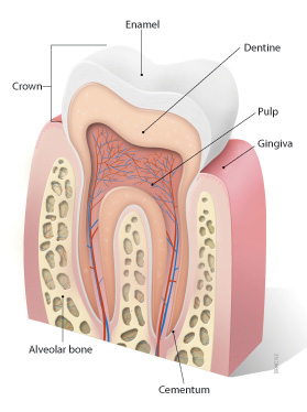
Dental enamel is the hard, white, outermost layer of a tooth. It is a highly mineralized and avascular tissue, meaning it contains no living cells or blood vessels. Enamel is primarily composed of calcium and phosphate minerals and serves as the protective covering for the crown of a tooth, which is the portion visible above the gum line.
Enamel is the hardest substance in the human body, and its primary function is to provide structural support and protection to the underlying dentin and pulp tissues of the tooth. It also plays a crucial role in chewing and biting by helping to distribute forces evenly across the tooth surface during these activities.
Despite its hardness, dental enamel can still be susceptible to damage from factors such as tooth decay, erosion, and abrasion. Once damaged or lost, enamel cannot regenerate or repair itself, making it essential to maintain good oral hygiene practices and seek regular dental checkups to prevent enamel damage and protect overall oral health.
Dental enamel is the hard, outermost layer of a tooth that protects the dentin and pulp inside. It is primarily made up of minerals, mainly hydroxyapatite, and contains very little organic material. However, during the formation of dental enamel, proteins are synthesized and secreted by ameloblast cells, which help in the development and mineralization of the enamel. These proteins play a crucial role in the proper formation and structure of the enamel.
Some of the main dental enamel proteins include:
1. Amelogenin: This is the most abundant protein found in developing enamel, accounting for about 90% of the organic matrix. Amelogenin helps regulate the growth and organization of hydroxyapatite crystals during mineralization. It also plays a role in determining the final hardness and structure of the enamel.
2. Enamelin: This protein is the second most abundant protein in developing enamel, accounting for about 5-10% of the organic matrix. Enamelin is involved in the elongation and thickening of hydroxyapatite crystals during mineralization. It also helps maintain the stability of the enamel structure.
3. Ameloblastin: This protein is produced by ameloblast cells and is essential for proper enamel formation. Ameloblastin plays a role in regulating crystal growth, promoting adhesion between crystals, and maintaining the structural integrity of the enamel.
4. Tuftelin: This protein is found in both dentin and enamel but is more abundant in enamel. Tuftelin is involved in the initiation of mineralization and helps regulate crystal growth during this process.
5. Dentin sialophosphoprotein (DSPP): Although primarily associated with dentin formation, DSPP is also found in developing enamel. It plays a role in regulating crystal growth and promoting adhesion between crystals during mineralization.
After the formation of dental enamel is complete, these proteins are largely degraded and removed, leaving behind the highly mineralized and hard tissue that characterizes mature enamel. However, traces of these proteins may still be present in the enamel and could potentially play a role in its structure and properties.
A deciduous tooth, also known as a baby tooth or primary tooth, is a type of temporary tooth that humans and some other mammals develop during childhood. They are called "deciduous" because they are eventually shed and replaced by permanent teeth, much like how leaves on a deciduous tree fall off and are replaced by new growth.
Deciduous teeth begin to form in the womb and start to erupt through the gums when a child is around six months old. By the time a child reaches age three, they typically have a full set of 20 deciduous teeth, including incisors, canines, and molars. These teeth are smaller and less durable than permanent teeth, but they serve important functions such as helping children chew food properly, speak clearly, and maintain space in the jaw for the permanent teeth to grow into.
Deciduous teeth usually begin to fall out around age six or seven, starting with the lower central incisors. This process continues until all of the deciduous teeth have been shed, typically by age 12 or 13. At this point, the permanent teeth will have grown in and taken their place, with the exception of the wisdom teeth, which may not erupt until later in adolescence or early adulthood.
Tooth abnormalities refer to any variations or irregularities in the size, shape, number, structure, or development of teeth that deviate from the typical or normal anatomy. These abnormalities can occur in primary (deciduous) or permanent teeth and can be caused by genetic factors, environmental influences, systemic diseases, or localized dental conditions during tooth formation.
Some examples of tooth abnormalities include:
1. Microdontia - teeth that are smaller than normal in size.
2. Macrodontia - teeth that are larger than normal in size.
3. Peg-shaped teeth - teeth with a narrow, conical shape.
4. Talon cusps - additional cusps or points on the biting surface of a tooth.
5. Dens invaginatus - an abnormal development where the tooth crown has an extra fold or pouch that can trap bacteria and cause dental problems.
6. Taurodontism - teeth with large pulp chambers and short roots.
7. Supernumerary teeth - having more teeth than the typical number (20 primary and 32 permanent teeth).
8. Hypodontia - missing one or more teeth due to a failure of development.
9. Germination - two adjacent teeth fused together, usually occurring in the front teeth.
10. Fusion - two separate teeth that have grown together during development.
Tooth abnormalities may not always require treatment unless they cause functional, aesthetic, or dental health issues. A dentist can diagnose and manage tooth abnormalities through various treatments, such as fillings, extractions, orthodontic care, or restorative procedures.
Dental caries susceptibility refers to the likelihood or predisposition of an individual to develop dental caries, also known as tooth decay or cavities. It is influenced by various factors such as oral hygiene practices, dietary habits, saliva composition, and the presence of certain bacteria in the mouth, particularly mutans streptococci and lactobacilli.
People with a higher dental caries susceptibility may have thinner or softer enamel, reduced saliva flow, or a greater concentration of cavity-causing bacteria in their mouths. Regular dental check-ups and good oral hygiene practices, such as brushing twice a day, flossing daily, and using fluoride toothpaste, can help reduce the risk of developing dental caries. Additionally, a balanced diet that limits sugary and starchy foods and beverages can also help lower the likelihood of tooth decay.
In the context of dentistry, a molar is a type of tooth found in the back of the mouth. They are larger and wider than other types of teeth, such as incisors or canines, and have a flat biting surface with multiple cusps. Molars are primarily used for grinding and chewing food into smaller pieces that are easier to swallow. Humans typically have twelve molars in total, including the four wisdom teeth.
In medical terminology outside of dentistry, "molar" can also refer to a unit of mass in the apothecaries' system of measurement, which is equivalent to 4.08 grams. However, this usage is less common and not related to dental or medical anatomy.
The enamel organ is a structure found in the developing teeth of vertebrates. It is responsible for the formation of enamel, which is the hard, outermost layer of the tooth crown. The enamel organ is derived from the dental papilla and is composed of several layers: the outer enamel epithelium, the stellate reticulum, the stratum intermedium, and the inner enamel epithelium. These layers work together to produce the enamel matrix, which is then mineralized to form the hard tissue that covers the tooth's crown. The enamel organ disappears after the formation of enamel is complete, leaving only the hardened enamel layer behind.
Amelogenesis is the biological process of forming enamel, which is the hard and highly mineralized outer layer of teeth. Enamel is primarily made up of calcium and phosphate minerals and is the toughest substance in the human body. Amelogenesis involves the synthesis, secretion, and maturation of enamel proteins by specialized cells called ameloblasts.
The medical definition of 'Amelogenesis' refers to a genetic disorder that affects the development and formation of tooth enamel. This condition is also known as Amelogenesis Imperfecta (AI) and can result in teeth that are discolored, sensitive, and prone to decay. There are several types of Amelogenesis Imperfecta, each with its own set of symptoms and genetic causes.
In summary, 'Amelogenesis' is the biological process of enamel formation, while 'Amelogenesis Imperfecta' is a genetic disorder that affects this process, leading to abnormal tooth enamel development.
Ameloblasts are the specialized epithelial cells that are responsible for the formation of enamel, which is the hard, outermost layer of a tooth. These cells are a part of the dental lamina and are present in the developing tooth's crown region. They align themselves along the surface of the developing tooth and secrete enamel proteins and minerals to form the enamel rods and interrod enamel. Once the enamel formation is complete, ameloblasts undergo programmed cell death, leaving behind the hard, mineralized enamel matrix. Any damage or abnormality in the functioning of ameloblasts can lead to developmental defects in the enamel, such as hypoplasia or hypocalcification, which may affect the tooth's structure and function.
Dental enamel permeability refers to the ability of substances to pass through the dental enamel, which is the hard, outermost layer of a tooth. The permeability of dental enamel can be affected by various factors such as its mineral content, structure, and the pH level of the oral environment.
Under normal conditions, dental enamel is relatively impermeable to substances due to its highly mineralized structure. However, when the enamel is exposed to acidic environments, such as those created by bacterial plaque, the minerals in the enamel can dissolve, creating microscopic pores that increase its permeability. This process, known as demineralization, can lead to tooth decay and other dental problems.
On the other hand, certain treatments and materials used in dentistry may temporarily increase the permeability of dental enamel, such as etching with acid before bonding procedures. This intentional increase in permeability allows for better adhesion of filling materials or sealants to the tooth surface. However, it is important to manage and control the permeability of dental enamel to maintain its structural integrity and protect oral health.
I'm not aware of a medical definition for "DMF Index." The abbreviation "DMF" could potentially stand for many things, as it is used in various contexts across different fields. In the field of dentistry, DMF stands for Decayed, Missing, and Filled teeth/surfaces, which is a method for measuring dental caries or tooth decay. However, there is no standard medical definition for "DMF Index." If you could provide more context or specify the field of study or practice, I would be happy to help further!
Amelogenin is a protein that plays a crucial role in the formation and mineralization of enamel, which is the hard, calcified tissue that covers the outer surface of teeth. It is expressed during tooth development and is secreted by ameloblasts, the cells responsible for producing enamel.
Amelogenin makes up approximately 90% of the organic matrix of developing enamel and guides the growth and organization of hydroxyapatite crystals, which are the primary mineral component of enamel. The protein is subsequently degraded and removed as the enamel matures and becomes fully mineralized.
Mutations in the gene that encodes amelogenin (AMELX on the X chromosome) can lead to various inherited enamel defects, such as amelogenesis imperfecta, which is characterized by thin, soft, or poorly formed enamel. Additionally, because of its high expression in developing teeth and unique size and structure, amelogenin has been widely used as a marker in forensic dentistry for human identification and sex determination.
Tooth calcification, also known as dental calculus or tartar formation, refers to the hardening of plaque on the surface of teeth. This process occurs when minerals from saliva combine with bacterial deposits and dental plaque, resulting in a hard, calcified substance that adheres to the tooth surface. Calcification can occur both above and below the gum line, and if not removed through professional dental cleanings, it can lead to periodontal disease, tooth decay, and other oral health issues.
Amelogenesis Imperfecta is a group of inherited dental disorders that affect the structure and appearance of tooth enamel. It is caused by mutations in various genes involved in the development and formation of enamel. The condition can be characterized by small, discolored, and poorly formed teeth that are prone to rapid wear, decay, and sensitivity. There are several types of Amelogenesis Imperfecta, which vary in their severity and the specific symptoms they present. Treatment typically focuses on managing the symptoms and improving the appearance and function of the teeth through restorative dental procedures.
Matrix metalloproteinase-20 (MMP-20) is a type of enzyme that belongs to the matrix metalloproteinase (MMP) family. MMPs are involved in the breakdown and remodeling of extracellular matrix components, such as collagen and elastin.
MMP-20, also known as Enamelysin, is primarily expressed in developing teeth and plays a crucial role in tooth development and mineralization. It is responsible for the degradation of enamel proteins during tooth formation, helping to shape and harden the enamel matrix. MMP-20 is secreted by ameloblasts, which are the cells that produce enamel.
Defects in MMP-20 have been associated with dental disorders such as Amelogenesis imperfecta, a group of genetic conditions characterized by abnormalities in tooth enamel formation and structure.
Tooth demineralization is a process that involves the loss of minerals, such as calcium and phosphate, from the hard tissues of the teeth. This process can lead to the development of dental caries or tooth decay. Demineralization occurs when acids produced by bacteria in the mouth attack the enamel of the tooth, dissolving its mineral content. Over time, these attacks can create holes or cavities in the teeth. Fluoride, found in many toothpastes and public water supplies, can help to remineralize teeth and prevent decay. Good oral hygiene practices, such as brushing and flossing regularly, can also help to prevent demineralization by removing plaque and bacteria from the mouth.
Dental caries, also known as tooth decay or cavities, refers to the damage or breakdown of the hard tissues of the teeth (enamel, dentin, and cementum) due to the activity of acid-producing bacteria. These bacteria ferment sugars from food and drinks, producing acids that dissolve and weaken the tooth structure, leading to cavities.
The process of dental caries development involves several stages:
1. Demineralization: The acidic environment created by bacterial activity causes minerals (calcium and phosphate) to be lost from the tooth surface, making it weaker and more susceptible to decay.
2. Formation of a white spot lesion: As demineralization progresses, a chalky white area appears on the tooth surface, indicating early caries development.
3. Cavity formation: If left untreated, the demineralization process continues, leading to the breakdown and loss of tooth structure, resulting in a cavity or hole in the tooth.
4. Infection and pulp involvement: As the decay progresses deeper into the tooth, it can reach the dental pulp (the soft tissue containing nerves and blood vessels), causing infection, inflammation, and potentially leading to toothache, abscess, or even tooth loss.
Preventing dental caries involves maintaining good oral hygiene, reducing sugar intake, using fluoride toothpaste and mouthwash, and having regular dental check-ups and cleanings. Early detection and treatment of dental caries can help prevent further progression and more severe complications.
Dental enamel solubility refers to the degree to which the mineral crystals that make up dental enamel can be dissolved or eroded by acidic substances. Dental enamel is the hard, outermost layer of a tooth that helps protect it from damage. It is primarily made up of minerals, including hydroxyapatite, which can dissolve in an acidic environment.
When the pH in the mouth drops below 5.5, the oral environment becomes acidic and dental enamel begins to demineralize or lose its mineral content. This process is known as dental caries or tooth decay. Over time, if left untreated, dental caries can lead to cavities, tooth sensitivity, and even tooth loss.
Certain factors can increase the solubility of dental enamel, including a diet high in sugar and starch, poor oral hygiene, and the presence of certain bacteria in the mouth that produce acid as a byproduct of their metabolism. On the other hand, fluoride exposure can help to reduce dental enamel solubility by promoting remineralization and making the enamel more resistant to acid attack.
Dental care refers to the practice of maintaining and improving the oral health of the teeth and gums. It involves regular check-ups, cleanings, and treatments by dental professionals such as dentists, hygienists, and dental assistants. Dental care also includes personal habits and practices, such as brushing and flossing, that help prevent tooth decay and gum disease.
Regular dental care is important for preventing common dental problems like cavities, gingivitis, and periodontal disease. It can also help detect early signs of more serious health issues, such as oral cancer or diabetes, which can have symptoms that appear in the mouth.
Dental care may involve a range of treatments, from routine cleanings and fillings to more complex procedures like root canals, crowns, bridges, and implants. Dental professionals use various tools and techniques to diagnose and treat dental problems, including X-rays, dental impressions, and local anesthesia.
Overall, dental care is a critical component of overall health and wellness, as poor oral health has been linked to a range of systemic health issues, including heart disease, stroke, and respiratory infections.
In the context of medical terminology, "hardness" is not a term that has a specific or standardized definition. It may be used in various ways to describe the firmness or consistency of a tissue, such as the hardness of an artery or tumor, but it does not have a single authoritative medical definition.
In some cases, healthcare professionals may use subjective terms like "hard," "firm," or "soft" to describe their tactile perception during a physical examination. For example, they might describe the hardness of an enlarged liver or spleen by comparing it to the feel of their knuckles when gently pressed against the abdomen.
However, in other contexts, healthcare professionals may use more objective measures of tissue stiffness or elasticity, such as palpation durometry or shear wave elastography, which provide quantitative assessments of tissue hardness. These techniques can be useful for diagnosing and monitoring conditions that affect the mechanical properties of tissues, such as liver fibrosis or cancer.
Therefore, while "hardness" may be a term used in medical contexts to describe certain physical characteristics of tissues, it does not have a single, universally accepted definition.
Tooth remineralization is a natural process by which minerals, such as calcium and phosphate, are redeposited into the microscopic pores (hydroxyapatite crystals) in the enamel of a tooth. This process can help to repair early decay and strengthen the teeth. It occurs when the mouth's pH is neutral or slightly alkaline, which allows the minerals in our saliva, fluoride from toothpaste or other sources, and calcium and phosphate ions from foods to be absorbed into the enamel. Remineralization can be promoted through good oral hygiene practices, such as brushing with a fluoride toothpaste, flossing, and eating a balanced diet that includes foods rich in calcium and phosphate.
Tooth bleaching, also known as tooth whitening, is a cosmetic dental procedure that aims to lighten the color of natural teeth and remove stains or discoloration. It's important to note that this process doesn't involve physically removing the tooth structure but rather uses various agents containing bleaching chemicals like hydrogen peroxide or carbamide peroxide to oxidize the stain molecules, breaking them down and making the teeth appear whiter and brighter.
The procedure can be performed in a dental office under professional supervision (in-office bleaching), at home using custom-made trays provided by a dentist (at-home or take-home bleaching), or through over-the-counter products such as whitening toothpaste, strips, and gels. However, it is always recommended to consult with a dental professional before starting any tooth bleaching treatment to ensure safety, effectiveness, and suitability for your specific oral health condition.
 Pitting enamel hypoplasia
Linear enamel hypoplasia
Enamel hypoplasia
Plane-form enamel hypoplasia
History of Isan
Kernicterus
2023 in paleontology
Tooth pathology
Deinotherium
Alan H. Goodman
2022 in paleomammalogy
Gault (archaeological site)
2019 in paleomammalogy
Carlo Catassi
Bili light
Anorexia nervosa
Eating disorder
Near Eastern bioarchaeology
Bog body
Bioarchaeology
Albright's hereditary osteodystrophy
List of MeSH codes (C07)
Saethre-Chotzen syndrome
Doxycycline
Bioelectricity
List of MeSH codes (C16)
Tooth enamel
Ehlers-Danlos syndromes
Amelogenesis imperfecta
Index of oral health and dental articles
enamel hypoplasia | Intelligent Dental
 Enamel hypoplasia: How to perform a non-aggressive prophylaxis - Be International Dental Expert
Pitting enamel hypoplasia - Wikipedia
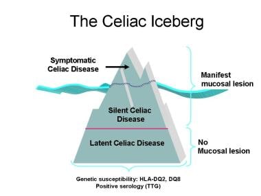
 Pediatric Celiac Disease (Sprue): Practice Essentials, Background, Pathophysiology
Ehlers-Danlos syndromes - Wikipedia
 Frontiers | Endocrine Diseases of Newborn: Epidemiology, Pathogenesis, Therapeutic Options, and Outcome "Current Insights Into...
 Oral-Facial-Digital Syndrome Type I - GeneReviews® - NCBI Bookshelf
 Call Transcript: May 12, 2016|Clinicians Outreach and Outreach Communication (COCA)
Hemolytic Disease of the Newborn Treatment & Management: Approach Considerations, Medical Care, Complications
 FLNB Disorders
 How to Get Rid of White Spots on Teeth: 5 Common Treatments
- GO SMILE
How to Get Rid of White Spots on Teeth: 5 Common Treatments
- GO SMILE
 The Backbone of History: Health and Nutrition in the Western Hemisphere
 Distinguishing predisposing factors for enamel hypoplasia and molar-incisor hypomineralization in children in Ile-Ife, Nigeria
 Apert syndrome: MedlinePlus Genetics
 CYTAG® CGH Labeling kit - ENZ-42671 - Enzo Life Sciences
Call Transcript: May 12, 2016|Clinicians Outreach and Outreach Communication (COCA)
 Supporting Oral Health Among Breastfed Infants - Dimensions of Dental Hygiene | Magazine
meifong | Intelligent Dental
Moreno Lopez, R.<...
 Vibramycin Intravenous (Doxycycline hyclate): Uses, Dosage, Side Effects, Interactions, Warning
 doxycycline hyclate capsules
WHO EMRO | Prevalence of oro-dental anomalies among schoolchildren in Sana'a city, Yemen | Volume 22, issue 1 | EMHJ volume 22,...
DailyMed - DOXYCYCLINE HYCLATE capsule
HuGE Navigator|Genopedia|PHGKB
STIM1base: stromal interaction molecule | Public Database
Heterozygous COL17A1 variants are a frequent cause of amelogenesis imperfecta | Journal of Medical Genetics
Bio2Vec
Celiac Disease: Complications | Calgary Guide
Fluorosis12
- For example, dental fluorosis is a condition where someone consumed too much fluoride as a child before their teeth broke through the gums. (gosmile.com)
- This goes especially for conditions like enamel hypoplasia or dental fluorosis, which is why we've listed several treatment options to help achieve a bright smile. (gosmile.com)
- Like dental fluorosis, enamel hypoplasia is a condition a bit beyond your control. (gosmile.com)
- Unfortunately, unlike harmless fluorosis, hypoplasia can increase the risk of tooth decay. (gosmile.com)
- Topical fluoride is a recommended treatment of dental fluorosis and enamel hypoplasia. (gosmile.com)
- This treatment is ideal mostly for dental fluorosis, as it reduces the appearance of non-harmful, more cosmetic-related white spots. (gosmile.com)
- For an unlucky minority, white spots don't come from demineralization or fluorosis, but from enamel hypoplasia, a condition that leaves the teeth with thinner enamel than usual and therefore more vulnerable to stains and decay. (gutierrezdentistry.com)
- A number of epidemiological studies have reported that chronic exposure to high concentrations of fluoride not only causes dental and skeletal fluorosis but additionally affects serum levels of reproductive hormones. (fluoridealert.org)
- Of the 98 students, the predominant success was dental fluorosis, where 93.9% answered the diagnosis. (bvsalud.org)
- Professors and students of the Dentistry course had difficulty in making treatment decisions on teeth with amelogenesis imperfecta, with mild dental fluorosis and ease on teeth with hypoplasia and dental caries. (bvsalud.org)
- Enamel Defects due to Fluorosis: Fluorosis occurs when children are exposed to excessive fluoride during tooth development. (chestnutdental.com)
- Fluorosis can cause white or brown spots, lines, or mottled appearance on the tooth enamel. (chestnutdental.com)
Crooked teeth1
- While dental veneers can be used in a variety of oral care treatments for a variety of reasons, such as chipped or crooked teeth or gaps, they work exceptionally well at getting rid of white spots on teeth. (gosmile.com)
Developmental4
- 18,19 In addition, developmental defects-including enamel hypoplasia-have been associated with caries in the primary dentition. (dimensionsofdentalhygiene.com)
- 230) cause junctional epidermolysis bullosa (JEB), a rare, genetically heterogeneous, mucocutaneous blistering disease with amelogenesis imperfecta (AI), a developmental enamel defect. (bmj.com)
- Developmental Dental Defect (D3) is also known as Enamel Hypoplasia. (asmilebydesign.com)
- By understanding the developmental process of tooth enamel, we can appreciate the importance of providing proper dental care and nutrition to support the optimal growth and strength of children's enamel. (chestnutdental.com)
Dentition3
- Severe Plane-Form Enamel Hypoplasia in a Dentition from Roman Britain" (PDF). (wikipedia.org)
- Inadequate levels of vitamin D during rapid phases of enamel formation (in utero for primary dentition, and post-natal for permanent dentition) increase the risk of interruptions in the enamel matrix, which can lead to defects, such as enamel hypoplasia. (dimensionsofdentalhygiene.com)
- These include acquired enamel disorders or dentinogenesis imperfecta , which is an autosomal dominant inherited structural disorder of tooth dentition (teeth eruption from the jaw). (medicinelearners.com)
Fluoride5
- This fluoride treatment can be done by your dentist and encourages enamel growth on the teeth, minimizing white spots, and also helps to prevent tooth decay overall. (gosmile.com)
- Fluoride is considered one of the best ingredients used in dental care today because of its many health benefits. (smilesolutions.us)
- Overconsumption of fluoride while enamel is still developing causes subsurface porosities . (cdhp.org)
- Fluoride has been a hot topic in the dental community for quite some time. (cosmeticdentistahwatukee.com)
- whitening process, we can also provide fluoride therapies and other treatments to reduce dental sensitivity often experienced following teeth whitening. (sierrasmiles4u.com)
Hypomineralization2
- It is characterized by asymmetrical enamel defects with severe hypomineralization. (bvsalud.org)
- Enamel Hypomineralization: Hypomineralization occurs when the enamel does not fully mineralize, making it softer and more vulnerable to damage. (chestnutdental.com)
Molars2
- A probable genetic origin for pitting enamel hypoplasia on the molars of Paranthropus robustus" (PDF). (wikipedia.org)
- Chemical, mechanical and morphological properties of hypomineralized enamel of permanent first molars. (edu.krd)
Amelogenesis6
- When Amelogenesis imperfecta is a genetic dental disease. (medicinelearners.com)
- According to PSYKNOWHOW.COM , the main cause of the development of amelogenesis imperfecta is the disruption of enamel formation. (medicinelearners.com)
- The congenital disruption of tooth enamel formation that occurs in the course of amelogenesis imperfecta is caused by proteins that are malfunctioning. (medicinelearners.com)
- The main cause of the development of amelogenesis imperfecta is the disruption of enamel formation. (medicinelearners.com)
- Amelogenesis imperfecta is a congenital defect that destroys tooth enamel. (medicinelearners.com)
- The process of enamel formation, known as amelogenesis , involves specialized cells called ameloblasts. (chestnutdental.com)
Abnormalities8
- [ 3 , 4 ] Their original classification system stratified the ectodermal dysplasias into different subgroups according to the presence or absence of (1) hair anomalies or trichodysplasias, (2) dental abnormalities, (3) nail abnormalities or onychodysplasias, and (4) eccrine gland dysfunction or dyshidrosis. (medscape.com)
- The main long-term consequence of IOM is future dental abnormalities. (huji.ac.il)
- Supernumerary teeth and dental fusion are abnormalities of dental development whose causes have not yet been elucidated with certainty. (bvsalud.org)
- Skin abnormalities may include dermal hypoplasia, fat nodules yellowish-pink color under the skin, aplasia cutis, telangiectasias, stripes slightly darker or lighter and skin papillomas. (ivami.com)
- Many individuals with focal dermal hypoplasia have abnormalities in hands and feet including oligodactyly, syndactyly, ectrodactilia and striated osteopathy. (ivami.com)
- Moreover, eye abnormalities are common in people with focal dermal hypoplasia, including microphthalmia, Anophthalmia, problems with the tear ducts and incomplete development of the retina or optic nerve. (ivami.com)
- Moreover, about half of individuals with focal dermal hypoplasia have dental abnormalities, especially enamel. (ivami.com)
- Signs and symptoms vary widely focal dermal hypoplasia, although nearly all affected individuals have skin abnormalities. (ivami.com)
Lesions2
- The possibility of co-existence of enamel hypoplasia and MIH/DMH makes it imperative to find ways to distinguish between the lesions. (bvsalud.org)
- Among the three periods, the highest frequency of enamel hypoplastic lesions was found out within individuals of the Lengyel culture (18.8%), whereas individuals of the LBK displayed lower values (13.5%), and individuals of the STK period even the lowest rate (12.5%) of DEH. (mzm.cz)
Systemic3
- It is characterized by demarcated enamel opacities with unknown systemic causation (idiopathic). (bvsalud.org)
- Systemic issues and medications like high fever, malnutrition, infectious diseases, trauma to developing tooth bud, chemotherapy, and anti-epileptics during infancy and early childhood disrupt enamel formation. (cdhp.org)
- It can be caused by factors such as malnutrition, certain medications, infections, or systemic conditions that affect enamel development. (chestnutdental.com)
Caries and dental2
- Kosma I, Kevrekidou A, Boka V, Arapostathis K, Kotsanos N. Molar incisor hypomineralisation (MIH): correlation with dental caries and dental fear. (edu.krd)
- Dental caries and dental wear were scored in order to find out basic characteristics of consumed food, and DEH was scored in order to find out the extent of non-specific stressors (i.e. indicators of metabolic and nutritional disruptions) within the Neolithic period. (mzm.cz)
Cavities5
- Also, there is a well established correlation between Rickets and Dental Enamel Hypoplasia - a condition of faulty development of the dental enamel that allows cavities to be more easily established. (easy-immune-health.com)
- This irregularity can lead to weakened enamel, increased tooth sensitivity, and a higher risk of cavities. (chestnutdental.com)
- Dental caries, also known as cavities, are caused by a bacterial infection of the tooth enamel. (americanmdcenter.com)
- The bacteria feed on sugars and starches from food and drink, producing acid that can erode the enamel and cause cavities. (americanmdcenter.com)
- Bacteria in the mouth that cause tooth decay create acids that eat away at tooth enamel and result in cavities. (americanmdcenter.com)
Defects7
- It has been suggested that because it is relatively rare to have both linear enamel hypoplasia and PEH, these types of defects may be commonly caused by different factors. (wikipedia.org)
- Sometimes, only a couple of ameloblasts stop forming enamel, leading to small PEH defects, with large pits forming when hundreds of these enamel-forming cells stop production. (wikipedia.org)
- 14 These enamel defects increase susceptibility to caries, a process that is led by Streptococcus mutans bacteria colonized within dental plaque. (dimensionsofdentalhygiene.com)
- 14,18 While caries is a multifactorial process, vitamin D-deficient infants have a greater risk for developing enamel defects, thus increasing caries risk. (dimensionsofdentalhygiene.com)
- While positive outcomes are possible with prompt intervention, certain types of enamel and dentin defects cannot be completely reversed. (cdhp.org)
- Result is undermineralized enamel prone to defects like translucency. (cdhp.org)
- Genetic defects in dentin formation result in teeth that are blue-grey or yellow brown in color with amber translucency concentrated in the incisal third and thinned enamel. (cdhp.org)
Affect enamel development1
- Exclusively breastfed babies may be at increased risk for vitamin D deficiency, which can affect enamel development and caries risk. (dimensionsofdentalhygiene.com)
Prevalence3
- Aim: To determine if the prevalence of enamel hypoplasia, molar-incisor hypomineralisation (MIH) and deciduous molar hypomineralisation (DMH) is associated with the socioeconomic status of the child and to determine the prevalence of enamel hypoplasia and MIH/DMH comorbidity in the study population. (bvsalud.org)
- Associations between sex, socioeconomic status and the prevalence of enamel hypoplasia, MIH and DMH were determined. (bvsalud.org)
- In this study all these individuals were scored for dental caries, dental wear, and the prevalence of dental enamel hypoplasia (DEH). (mzm.cz)
Abnormal1
- Abnormal fear or dread of visiting the dentist for preventive care or therapy and unwarranted anxiety over dental procedures. (lookformedical.com)
Erosion2
- Enamel Erosion: Enamel erosion occurs when the enamel gradually wears away due to the exposure of teeth to acids. (chestnutdental.com)
- Acidic foods and drinks, frequent vomiting, or gastric reflux can contribute to enamel erosion. (chestnutdental.com)
Thickness5
- The AI phenotype was consistent with enamel of near normal thickness and variable focal hypoplasia with surface irregularities including pitting. (bmj.com)
- The thickness of the enamel is often reduced at certain points or over an area. (medicinelearners.com)
- Chipping injuries later in life remove enamel thickness. (cdhp.org)
- This genetic condition disrupts normal enamel thickness , mineral content, and structure. (cdhp.org)
- This process continues until the enamel reaches its full thickness and density. (chestnutdental.com)
Etiology1
- Other nonendocrine clues to the presence of this autoimmune etiology include vitiligo and dental enamel hypoplasia. (medscape.com)
Demineralization3
- Demineralization is the gradual leaching of crucial minerals like calcium from the tooth enamel. (gutierrezdentistry.com)
- Preventing demineralization is all about good brushing and flossing habits, as well as regular dental visits. (gutierrezdentistry.com)
- Dental caries manifesting as barely noticeable frosty or opaque spots represents early demineralization of enamel minerals by oral bacteria. (cdhp.org)
Defect3
- PEH can be associated with other types of hypoplasia, but it is often the only defect observed. (wikipedia.org)
- We describe different outcomes of this practice, such as enamel hypoplasia and crown deformations with later necrosis and infection of the root canal system, severe discolouration, immature root apex, impaction of a canine, failure of development and missing lower permanent incisors and canines, an odontoma-like structure, severe periodontal defect and root dilaceration. (huji.ac.il)
- DGS is the clinical term used for patients who have the classic phenotype of cardiac defect, hypocalcemia and thymic hypoplasia leading to T-cell immunodeficiency. (immunodeficiencysearch.com)
Thin layer of enamel2
- With microabrasion, a thin layer of enamel is scraped away to restore the tooth's uniform appearance. (gutierrezdentistry.com)
- At birth, the crowns of primary (baby) teeth are already partially developed with a thin layer of enamel. (chestnutdental.com)
Dentin3
- If left unchecked, the cavity may penetrate the enamel and dentin and reach the pulp. (lookformedical.com)
- Cavitated caries confirmed as the cause reflects more advanced destruction into the enamel and dentin layers. (cdhp.org)
- Damage to the pulp tissue inside the tooth triggers inflammatory destruction and resorption of root canal dentin first and then enamel from inside. (cdhp.org)
Decay14
- The most common problem in pediatric dentistry is dental caries or tooth decay. (intelligentdental.com)
- When people tell us they have soft teeth, they usually mean they feel more susceptible to dental decay. (asmilebydesign.com)
- The great news is your dental team is educated and equipped to help you better understand what is causing your dental decay. (asmilebydesign.com)
- Disturbances in the oral microbiome can increase the risk of dental problems such as tooth decay. (cosmeticdentistahwatukee.com)
- Both dressings are very acidic, which can only stay in the enamel, but can wear it down, leaving a more susceptible to tooth decay if you consume these dressings in large quantities. (cosmeticdentistahwatukee.com)
- Among the various factors that contribute to dental health, tooth enamel plays a crucial role in protecting teeth from decay, sensitivity, and damage. (chestnutdental.com)
- Enamel hypoplasia may lead to increased tooth sensitivity, discoloration, and an increased risk of dental decay. (chestnutdental.com)
- Teeth with hypomineralized enamel may appear yellow or brown, and they are more susceptible to decay, sensitivity, and enamel breakdown. (chestnutdental.com)
- It's important to note that these enamel irregularities may vary in severity, and their effects on dental health can range from mild cosmetic concerns to increased vulnerability to tooth decay and sensitivity. (chestnutdental.com)
- Periodontal disease, dental caries, and enamel hypoplasia are common illnesses that result in tooth decay. (americanmdcenter.com)
- Common diseases that can cause tooth decay include periodontal disease, dental caries, and enamel hypoplasia. (americanmdcenter.com)
- The most popular method of treating dental decay is fillings . (americanmdcenter.com)
- This can help remove the bacteria and plaque that cause decay and strengthen the tooth's enamel. (americanmdcenter.com)
- Good dental hygiene can help prevent tooth decay, but individuals who don't take care of their teeth are more prone to experience it. (americanmdcenter.com)
Teeth's1
- If your spots are a result of dental hypoplasia, however, it's important to take care of your teeth's health and brush on a regular basis. (gosmile.com)
Irregularities4
- However, children can sometimes experience irregularities in their tooth enamel, which can have implications for their oral health. (chestnutdental.com)
- By gaining a deeper understanding of enamel irregularities, we can take proactive steps to promote the long-term dental well-being of our little ones. (chestnutdental.com)
- There are several enamel irregularities that can occur in children, which may impact their dental health. (chestnutdental.com)
- Proper diagnosis and treatment by a dental professional are essential to address these enamel irregularities and prevent further complications. (chestnutdental.com)
Dentistry3
- Pediatric dentistry refers to dental matters on children. (intelligentdental.com)
- The total of dental diagnostic, preventive, and restorative services provided to meet the needs of a patient (from Illustrated Dictionary of Dentistry , 1982). (lookformedical.com)
- Many of the city's dental clinics in Dubai offer various services, such as general dentistry , cosmetic dentistry, orthodontics , and implantology . (americanmdcenter.com)
Anomalies2
Inadequate1
- Enamel Hypoplasia: This condition refers to inadequate enamel formation, resulting in thin or pitted enamel. (chestnutdental.com)
Genetic1
- Focal Dermal Hypoplasia is a genetic disorder that primarily affects the skin, skeleton, eyes and face. (ivami.com)
Thins1
- However, some degree of translucency in the incisors is normal as we age and enamel thins. (cdhp.org)
Condition4
- And more research from 1973 called Enamel hypoplasia of the teeth associated with neonatal tetany: manifestation of maternal vitamin deficiency showed that infants born with vitamin D deficiency were prone to a tooth condition called 'enamel hypoplasia' that predisposes them to bad teeth for the rest of their life! (easy-immune-health.com)
- Visible as a pinkish spot shining through enamel, this condition requires radiographs to confirm size, location, and progression over monitoring visits. (cdhp.org)
- The term "soft teeth" is a layman's phrase often used to describe a condition known as "dental enamel hypoplasia. (cosmeticdentistahwatukee.com)
- o A Child Sample Person Questionnaire (CSPQ), for sample persons 6 months through 11 years that included sections on a number of health status issues (including dental condition and care), health care utilization, infant feeding practices, participation in meal programs, school attendance, and language use. (cdc.gov)
Microabrasion1
- Enamel microabrasion is used to correct discolored enamels, removing surface stains and yellowing, but it can also be used to remove the white spots. (gosmile.com)
Atrophic1
- Generalized non-Herlitz-type junctional epidermolysis bullosa is a form of non-Herlitz-type junctional epidermolysis bullosa (JEB-nH, see this term) characterized by generalized skin blistering, atrophic scarring, nail dystrophy or nail absence, and enamel hypoplasia, with extracutaneous involvement. (mendelian.co)
Turner's1
- It also affects the coloration of the tooth, which takes on a brown, yellow or even whiter than normal (Turner's hypoplasia). (bedentalexpert.com)
Bacteria2
- 15 Within this plaque, bacteria can easily adhere to a defective enamel surface, creating an environment prime for lesion development. (dimensionsofdentalhygiene.com)
- Composed primarily of minerals, such as hydroxyapatite, enamel acts as a shield, guarding the inner, more sensitive layers of the tooth against bacteria, acids, and mechanical forces. (chestnutdental.com)
Prenatal1
- In the context of vitamin D levels and oral health, discuss dental professionals' role in prenatal and postnatal care. (dimensionsofdentalhygiene.com)
Oral hygiene1
- To prevent enamel hypoplasia, practicing good oral hygiene and maintaining a balanced diet that includes adequate calcium and vitamin D is essential. (americanmdcenter.com)
Focal4
- They have identified at least 29 mutations in the gene responsible PORCN focal dermal hypoplasia. (ivami.com)
- Focal Dermal Hypoplasia is inherited as a dominant X - linked pattern In women, a mutation in one of the two copies of the gene in each cell is sufficient to express the disease. (ivami.com)
- It is believed that the distribution of active and inactive X chromosomes may play a role in determining the severity of the focal dermal hypoplasia in women. (ivami.com)
- A male may be born with focal dermal hypoplasia if a gene mutation has PORCN only in some cells (mosaicism). (ivami.com)
Medications1
- In addition, enamel hypoplasia can be caused by certain medications or illnesses. (americanmdcenter.com)
Disruption1
- Enamel hypoplasia can take a variety of forms, but all types are associated with a reduction of enamel formation due to disruption in ameloblast production. (wikipedia.org)
Diagnosis and treatment1
- Eight new cases of IOM are presented in this case series, with emphasis on dental diagnosis and treatment modalities. (huji.ac.il)
Calcium2
- The mineralization of the enamel matrix occurs through the deposition of minerals, primarily calcium and phosphate ions, which crystallize and harden the enamel. (chestnutdental.com)
- Adequate intake of essential minerals, such as calcium and phosphorus, along with vitamins like vitamin D, is essential for the formation of strong and healthy tooth enamel. (chestnutdental.com)
Ameloblasts2
- Each pit is linked to the ceasing of ameloblasts at a particular point in enamel formation. (wikipedia.org)
- As the enamel matrix matures, the ameloblasts eventually degenerate and are not replaced, meaning that once enamel is formed, it cannot regenerate or repair itself naturally. (chestnutdental.com)
Chronic2
- 14,16 Dental caries is considered the most common preventable chronic disease of childhood, with 21.4% of children younger than 5 experiencing disease. (dimensionsofdentalhygiene.com)
- Dental care for patients with chronic diseases. (lookformedical.com)
Tooth loss1
- Online searches were made to find literature for the study of other tissues and organs and on oral diseases, e.g. dental caries, perio- as a potential source of pathology affecting dontal diseases and tooth loss, and on NCD, other systems and organs" [ 6 ]. (who.int)
Affects1
- Another reason that Vitamin D affects periodontal disease in the Vitamin D and Teeth equation is that, according to The Boston University Goldman School of Dental Medicine , Vitamin D reduces gingivitis because of the anti inflammatory effects of the vitamin. (easy-immune-health.com)
Early childhood3
- While there are many ways you can remove stains to achieve a brighter smile, here's how to get rid of white spots on teeth, from vanquishing them with a consistent teeth whitening routine to wearing veneers to completely conceal speckled enamel caused by early childhood development. (gosmile.com)
- Cracks, fractures, displacement injuries in early childhood can damage developing enamel. (cdhp.org)
- It is important to note that proper nutrition, especially during early childhood, plays a crucial role in enamel development. (chestnutdental.com)
Development5
- Describe the relationship between vitamin D deficiency, enamel development and caries risk in infants and young children. (dimensionsofdentalhygiene.com)
- 4-8 This paper will describe the relationship between vitamin D deficiency and enamel development, provide national supplementation recommendations for mothers and children, and discuss the role of oral health professionals in promoting dental health among these patients. (dimensionsofdentalhygiene.com)
- Given the relationship between vitamin D and enamel development, oral health professionals must provide expectant mothers and caregivers with the resources needed to minimize the risk of vitamin D deficiency in infants who will be exclusively breastfed. (dimensionsofdentalhygiene.com)
- During childhood, tooth enamel undergoes a remarkable development process. (chestnutdental.com)
- As a child grows, the enamel development continues, particularly during the first few years of life. (chestnutdental.com)
Stains2
- Through your dental provider, you can get professionally fitted for custom veneers that will conceal any stains at all. (gosmile.com)
- Surface stains that affect the tooth enamel sometimes appear on a tooth that is otherwise healthy. (gutierrezdentistry.com)
Pits1
- In rare cases the enamel does not develop properly and can erupt with small holes (pits and fissures) or be thin or underdeveloped. (asmilebydesign.com)
Minerals2
- Tooth enamel is mainly composed of minerals, the structure of which is controlled by certain protein components. (medicinelearners.com)
- These cells secrete proteins and minerals that gradually build up the enamel matrix. (chestnutdental.com)
Suffer2
- If you consistently suffer from dental anxiety, then you're not alone - approximately 36 percent of U.S. people fear getting dental treatment. (smilesolutions.us)
- Most people that tell us they have 'soft teeth' do not suffer from Enamel Hypoplasia. (asmilebydesign.com)
Prophylaxis1
- Persons trained in an accredited school or dental college and licensed by the state in which they reside to provide dental prophylaxis under the direction of a licensed dentist. (lookformedical.com)
Injuries1
- Signs of dental injuries, whether immediate or long-term, should always be treated. (smilesolutions.us)
Acidic1
- Well, those white spots on your teeth can be caused by several bad habits, like drinking and eating acidic and sugary foods ( coffee and red wine , for instance) and poor dental hygiene. (gosmile.com)
Dentist2
- The Imagine Dental team of professionals have decades of combined experience between them, they understand that people go to a dentist because they need to, not necessarily because they want to. (cosmeticdentistahwatukee.com)
- Choose the Best Dental Clinic in Dubai as their expert dentist can help you to tell which product suits your teeth. (americanmdcenter.com)
Common1
- One of the most common types, Pitting Enamel Hypoplasia (PEH), ranges from small circular pinpricks to larger irregular depressions. (wikipedia.org)
Surface3
- Localized destruction of the tooth surface initiated by decalcification of the enamel followed by enzymatic lysis of organic structures and leading to cavity formation. (lookformedical.com)
- The enamel surface can shine and tend to split quickly. (medicinelearners.com)
- Cupping, flattening or notching of the enamel surface may also be seen. (cdhp.org)