Dentin Sensitivity
Dentin
Dentin Permeability
Dentin Desensitizing Agents
Sensitivity and Specificity
Dentin-Bonding Agents
Dental Bonding
Methacrylates
Dentin Dysplasia
Odontoblasts
Resin Cements
Acid Etching, Dental
Materials Testing
Smear Layer
Tensile Strength
Dentinogenesis
Tooth Calcification
Dental Stress Analysis
Phosphoric Acids
Dental Enamel
Tooth Demineralization
Bisphenol A-Glycidyl Methacrylate
Dental Pulp
Dental Cavity Lining
Tooth Root
Molar
Dental Cavity Preparation
Microscopy, Electron, Scanning
Dentinal Fluid
Dental Cements
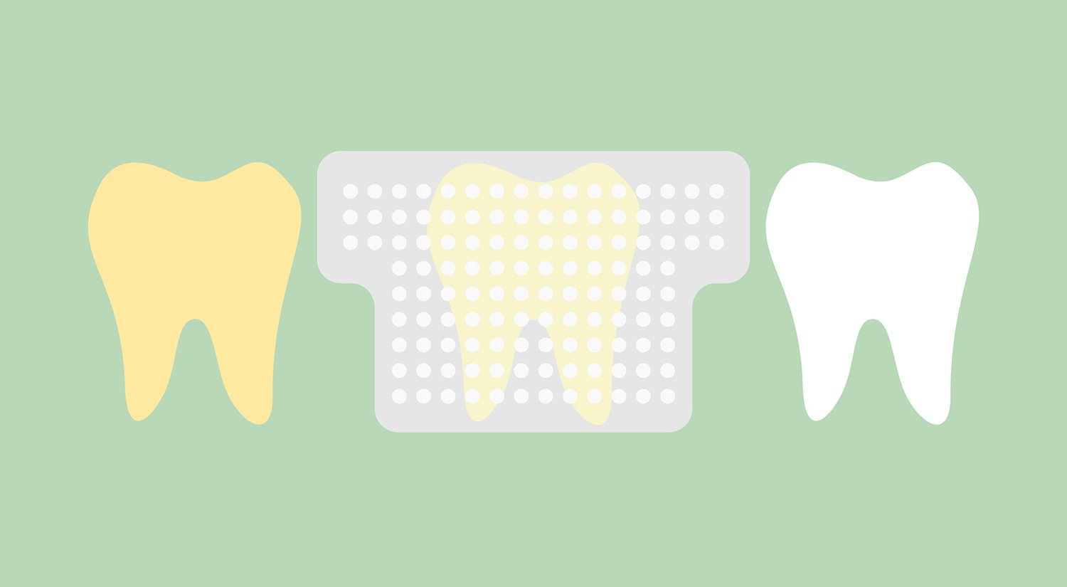
Tooth Remineralization
Hardness
Dental Materials
Surface Properties
Dental Leakage
Dental Caries
Dental Marginal Adaptation
Adhesives
Incisor
Sialoglycoproteins
Tooth Attrition
Tooth, Deciduous
Polymethacrylic Acids
Shear Strength
Pain
Pain Management
Chronic Pain
Pain Measurement
Dentistry
Dentinal tubule occlusion with lanthanum fluoride and powdered apatite glass ceramics in vitro. (1/87)
To simulate hypersensitive dentin, the smear layer and dentinal plugs of bovine root dentin specimens were removed by immersion in 10% phosphoric acid, polishing with hydroxyapatite particles, and ultrasonic cleansing. The fluoride-tannic acid-lanthanum-apatite (FTLA) group was treated with acidulated phosphate fluoride (APF) containing tannic acid followed by rubbing with a paste of lanthanum chloride (LaCl3) and powdered apatite glass ceramics. The treated specimens were immersed in a remineralizing solution that mimics saliva for 6 weeks. The SEM observations revealed that the treated surfaces of the FTLA group were completely covered with fine spherical compounds and the dentinal tubules were occluded with plugs to a depth of about 3 microns. Fluoride and lanthanum were detected to a depth of over 20 microns by EPMA observation. After the remineralization, the surface of FTLA-treated specimen did not have any opened tubules and showed a remarkable increase in the number of fine spherical deposits in the dentinal tubules. These results suggest that the reaction products produced by sequential treatment with acidic fluoride and LaCl3 and powdered apatite glass ceramics are able to effectively occlude dentinal tubules. (+info)Clinical evaluation of an electron-ionizing toothbrush with a tooth paste containing stannous fluoride in treatment of dentine hypersensitivity following periodontal surgery. (2/87)
The purpose of this study was to examine the effect of an electro-ionizing toothbrush with stannous fluoride in the treatment of dentin hypersensitivity following periodontal surgery. Thirty-two volunteers with dentin hypersensitivity were divided in two equal groups each using different methods: (Group I) stannous fluoride dentifrice and hyG Brnde ionizing brush without a battery and (Group II) stannous fluoride dentifrice and hyG Brnde ionizing brush with a battery. The volunteers brushed their teeth for 3 minutes twice a day for 12 weeks following one either of the test protocols. Mechanical (No 23 dental explorer), chemical (lemon juice) and thermal (dental air-water syringe) tests were used for the evaluation of the degree of dentin hypersensitivity. A subjective assessment of the degree of hypersensitivity for each stimulus was recorded. The evaluations were repeated at 4, 8 and 12 weeks after surgical treatment. The second group showed significantly less sensitivity than the first group. The findings appear to suggest that the ionizing brush may be an effective adjunct for the treatment of dentin hypersensitivity in post-periodontal surgery. (+info)Gingival prostheses--a review. (3/87)
Gingival replacement is often a component of comprehensive prosthodontics. Gingival prostheses may be fixed or removable and may be made from acrylics, composite resins, silicones or porcelain-based materials. Undercuts or dental attachments are used to secure removable prostheses, which are esthetically pleasing and easy to maintain. This paper describes several clinical situations in which gingival prostheses were used effectively. (+info)Consensus-based recommendations for the diagnosis and management of dentin hypersensitivity. (4/87)
These consensus recommendations for the diagnosis and management of dentin hypersensitivity were developed by a broadly constituted board of dentists and dental hygienists drawn from general dental practice, specialist practice, academia and research from across Canada, joined by 2 international dentists with subject matter expertise. The need for consensus recommendations was made evident by the lack of clear and robust evidence in the dental literature, as well as confusion about diagnosis and management demonstrated by an educational needs assessment survey. High prevalence of the condition, underdiagnosis and widespread availability of noninvasive, efficacious and inexpensive preventive treatment further underscored the need for direction. This paper outlines the key elements of the scientific basis for the causes, diagnosis and management of dentin hypersensitivity; where such evidence is deficient, the document relies on the compound experience of the board. A simple algorithm was developed to guide clinicians through the diagnostic process and assist them in determining appropriate case management. Finally, the board makes a series of recommendations to raise awareness, to improve dental education, to develop symbols for charting, to develop an index for case assessment and for further research. (+info)Tooth bleaching--a critical review of the biological aspects. (5/87)
Present tooth-bleaching techniques are based upon hydrogen peroxide as the active agent. It is applied directly, or produced in a chemical reaction from sodium perborate or carbamide peroxide. More than 90% immediate success has been reported for intracoronal bleaching of non-vital teeth, and in the period of 1-8 years' observation time, from 10 to 40% of the initially successfully treated teeth needed re-treatment. Cervical root resorption is a possible consequence of internal bleaching and is more frequently observed in teeth treated with the thermo-catalytic procedure. When the external tooth-bleaching technique is used, the first subjective change in tooth color may be observed after 2-4 nights of tooth bleaching, and more than 90% satisfactory results have been reported. Tooth sensitivity is a common side-effect of external tooth bleaching observed in 15%-78% of the patients, but clinical studies addressing the risk of other adverse effects are lacking. Direct contact with hydrogen peroxide induced genotoxic effects in bacteria and cultured cells, whereas the effect was reduced or abolished in the presence of metabolizing enzymes. Several tumor-promoting studies, including the hamster cheek pouch model, indicated that hydrogen peroxide might act as a promoter. Multiple exposures of hydrogen peroxide have resulted in localized effects on the gastric mucosa, decreased food consumption, reduced weight gain, and blood chemistry changes in mice and rats. Our risk assessment revealed that a sufficient safety level was not reached in certain clinical situations of external tooth bleaching, such as bleaching one tooth arch with 35% carbamide peroxide, using several applications per day of 22% carbamide peroxide, and bleaching both arches simultaneously with 22% carbamide peroxide. The recommendation is to avoid using concentrations higher than 10% carbamide peroxide when one performs external bleaching. We advocate a selective use of external tooth bleaching based on high ethical standards and professional judgment. (+info)Gingival recessions caused by lip piercing: case report. (6/87)
Fear of losing the teeth is common among patients presenting with gingival recession. This report describes a case in which unusual gingival recessions were caused by lip piercing. Periodontal treatment involved removal of the causative agent, hygiene instruction, scaling and root planing, and coverage of the root with a subepithelial connective tissue graft. The therapeutic measures applied in this case yielded satisfactory root coverage, an increase in the width of the keratinized gingiva, improvement in hygiene status and absence of dental hypersensitivity. (+info)Influence of natural fruit juices in removing the smear layer from root surfaces--an in vitro study. (7/87)
Certain elements of a patient's diet may be associated with dentin hypersensitivity. The intent of this study was to evaluate the degree of removal of the smear layer from dentin surfaces by various fruit juices. A smear layer was created on extracted human teeth by manual scaling. The roots were reduced and distributed into 8 experimental groups. Distilled water was the negative control. The juices were applied by 2 methods: topical application and topical application with friction. Specimens were photomicrographed and graded according to an index of smear layer removal. With topical application, all but 2 of the tested substances resulted in significantly greater removal of the smear layer and opening of dentinal tubules than was the case with the negative control (p = 0.05); the exceptions were Gala apple and Italian grape juices, which were no different from the control. For the active application (with friction), most substances removed more smear layer than the control (p < 0.05); Gala apple, Italian grape and orange juices were similar to the control. For each of the tested substances, removal of the smear layer did not differ with the method of application (topical vs. friction; p > 0.05). It is concluded that natural fruit juices can remove the smear layer from dentin surfaces, and the efficacy of this removal varies with the type of juice. (+info)Durability of FTLA treatment as a medicament for dentin hypersensitivity--abrasion resistance and profiles of fluoride release. (8/87)
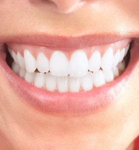
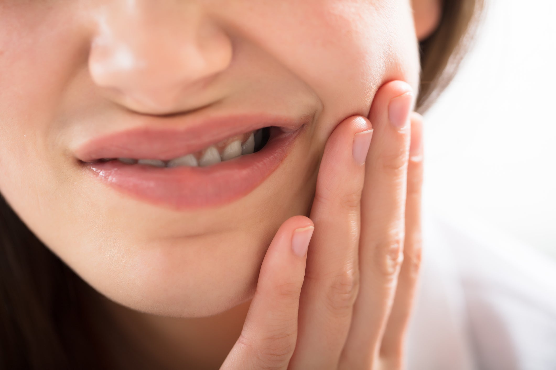
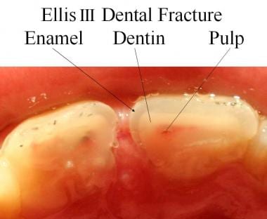
The purpose of this study was to evaluate the durability of tubules occluded with FTLA treatment by toothbrush abrasion test on the applied surface and by measuring fluoride release from the FTLA components. Dentin specimens with simulated hypersensitive surfaces were treated with APF containing tannic acid. After which, the specimens received lanthanum-chloride-with-powdered-fluoroapatite-glass-ceramics treatment. The specimens were subjected to toothbrush abrasion test up to 6,000 strokes. SEM observation revealed that dentinal tubules of the FTLA treated specimens were completely occluded with fine deposits even after toothbrush abrasion of 6,000 strokes. EPMA analysis revealed that fluoride, lanthanum, and aluminum were the main FTLA components on the dentin surface after 6,000-stroke abrasion. To measure fluoride release from the FTLA components, a slurry was enclosed in a cellulose tube and suspended in deionized water at 37 degrees C. After fluoride was dialyzed against deionized water, a high concentration of fluoride was found to be released from FTLA the components, indicating FTLA treatment's prominent durability. These results suggested that FTLA treatment has a superior resistance against toothbrush abrasion and a high fluoride-releasing performance. These characteristics lend much weight to showing that the FTLA method is an effective and durable medicament for dentin hypersensitivity. (+info)Dentin sensitivity is a common dental condition characterized by the short, sharp pain or discomfort in response to external stimuli, such as cold air, hot or cold foods and drinks, sweet or sour substances, and physical touch. This pain is typically caused by the exposure of dentin, the hard tissue beneath the tooth's enamel, due to receding gums, tooth decay, or other factors that wear down or damage the protective enamel layer.
When the dentin is exposed, the microscopic tubules within it become sensitive to temperature and pressure changes, allowing external stimuli to reach the nerve endings inside the tooth. This results in the characteristic pain or discomfort associated with dentin sensitivity. Dentin sensitivity can be managed through various treatments, including desensitizing toothpaste, fluoride applications, and dental restorations, depending on the underlying cause of the condition.
Dentin is the hard, calcified tissue that lies beneath the enamel and cementum of a tooth. It forms the majority of the tooth's structure and is composed primarily of mineral salts (hydroxyapatite), collagenous proteins, and water. Dentin has a tubular structure, with microscopic channels called dentinal tubules that radiate outward from the pulp chamber (the center of the tooth containing nerves and blood vessels) to the exterior of the tooth. These tubules contain fluid and nerve endings that are responsible for the tooth's sensitivity to various stimuli such as temperature changes, pressure, or decay. Dentin plays a crucial role in protecting the dental pulp while also providing support and structure to the overlying enamel and cementum.
Dentin permeability refers to the ability of various substances to penetrate or diffuse through the dentin, which is the hard, calcified tissue that lies beneath the enamel and forms the bulk of a tooth. Dentin is composed of microscopic tubules that run from the pulp chamber (which contains the dental pulp) to the exterior of the tooth. These tubules contain fluid and are lined with odontoblastic processes, which are extensions of the cells that form dentin.
When the dentin is exposed due to tooth decay, wear, or other factors, various substances can penetrate through these tubules and cause sensitivity, discomfort, or pain. The permeability of dentin can be influenced by several factors, including the diameter and number of tubules, the thickness and composition of the dentinal tissue, and the presence of dental sealants or other protective coatings.
In general, a higher dentin permeability is associated with increased susceptibility to tooth decay, sensitivity, and other dental problems. Therefore, understanding the factors that influence dentin permeability and developing strategies to reduce it is an important area of research in dental medicine.
Dentin desensitizing agents are chemical substances or materials applied to the teeth to reduce sensitivity in the dental tissues, specifically in the dentin. Dentin is a calcified tissue that lies beneath the tooth's enamel and cementum. It has numerous microscopic tubules that, when exposed due to various factors like gum recession, tooth wear, or dental procedures, can lead to hypersensitivity.
Dentin desensitizing agents work by occluding these dentinal tubules, thus preventing the stimuli (like cold, heat, or touch) from reaching the nerve endings inside the pulp chamber. These agents may contain various active ingredients like fluorides, strontium salts, calcium sodium phosphosilicate, potassium nitrate, arginine, and oxalates. They can be found in different forms, such as toothpaste, gels, varnishes, or bonding agents, and are often used in dental treatments and at-home oral care to alleviate dentinal hypersensitivity.
Sensitivity and specificity are statistical measures used to describe the performance of a diagnostic test or screening tool in identifying true positive and true negative results.
* Sensitivity refers to the proportion of people who have a particular condition (true positives) who are correctly identified by the test. It is also known as the "true positive rate" or "recall." A highly sensitive test will identify most or all of the people with the condition, but may also produce more false positives.
* Specificity refers to the proportion of people who do not have a particular condition (true negatives) who are correctly identified by the test. It is also known as the "true negative rate." A highly specific test will identify most or all of the people without the condition, but may also produce more false negatives.
In medical testing, both sensitivity and specificity are important considerations when evaluating a diagnostic test. High sensitivity is desirable for screening tests that aim to identify as many cases of a condition as possible, while high specificity is desirable for confirmatory tests that aim to rule out the condition in people who do not have it.
It's worth noting that sensitivity and specificity are often influenced by factors such as the prevalence of the condition in the population being tested, the threshold used to define a positive result, and the reliability and validity of the test itself. Therefore, it's important to consider these factors when interpreting the results of a diagnostic test.
Dentin-bonding agents are substances used in dentistry to create a strong and durable bond between the dental restoration material (such as composite resin, glass ionomer cement, or crowns) and the dentin surface of a tooth. Dentin is the hard tissue that lies beneath the enamel and consists of microscopic tubules filled with fluid.
The primary function of dentin-bonding agents is to improve the adhesion of restorative materials to the tooth structure, enhancing the retention and durability of dental fillings, crowns, veneers, and other types of restorations. These agents typically contain one or more types of bonding resins, such as hydroxyethyl methacrylate (HEMA), 4-methacryloxyethyl trimellitate anhydride (4-META), and/or phosphoric acid ester monomers.
The application process for dentin-bonding agents usually involves several steps, including:
1. Etching the dentin surface with a mild acid to remove the smear layer and expose the collagen network within the dentin tubules.
2. Applying a primer that penetrates into the etched dentin and promotes the infiltration of bonding resins into the dentinal tubules.
3. Applying an adhesive, which is typically a mixture of hydrophilic and hydrophobic monomers, to form a stable bond between the tooth structure and the restoration material.
4. Light-curing the adhesive to polymerize the resin and create a strong mechanical bond with the dentin surface.
Dentin-bonding agents have significantly improved the clinical success of various dental restorations by enhancing their retention, reducing microleakage, and minimizing postoperative sensitivity. However, they may still be susceptible to degradation over time due to factors such as moisture contamination, enzymatic degradation, or hydrolysis, which can lead to the failure of dental restorations. Therefore, continuous advancements in dentin-bonding technology are essential for improving the long-term success and durability of dental restorations.
Secondary dentin is a type of dentin that is formed after the initial development of the tooth. It is produced in response to stimuli such as tooth wear or injury and continues to form throughout an individual's life. Unlike primary dentin, which is laid down during tooth development and has a more uniform structure, secondary dentin is often deposited in a less organized manner and can vary in thickness. The formation of secondary dentin can help to protect the pulp tissue within the tooth from further damage or infection.
Dental bonding is a cosmetic dental procedure in which a tooth-colored resin material (a type of plastic) is applied and hardened with a special light, which ultimately "bonds" the material to the tooth to improve its appearance. According to the American Dental Association (ADA), dental bonding can be used for various purposes, including:
1. Repairing chipped or cracked teeth
2. Improving the appearance of discolored teeth
3. Closing spaces between teeth
4. Protecting a portion of the tooth's root that has been exposed due to gum recession
5. Changing the shape and size of teeth
Dental bonding is generally a quick and painless procedure, often requiring little to no anesthesia. The surface of the tooth is roughened and conditioned to help the resin adhere properly. Then, the resin material is applied, molded, and smoothed to the desired shape. A special light is used to harden the material, which typically takes only a few minutes. Finally, the bonded material is trimmed, shaped, and polished to match the surrounding teeth.
While dental bonding can be an effective solution for minor cosmetic concerns, it may not be as durable or long-lasting as other dental restoration options like veneers or crowns. The lifespan of a dental bonding procedure typically ranges from 3 to 10 years, depending on factors such as oral habits, location of the bonded tooth, and proper care. Regular dental checkups and good oral hygiene practices can help extend the life of dental bonding.
Methacrylates are a group of chemical compounds that contain the methacrylate functional group, which is a vinyl group (CH2=CH-) with a carbonyl group (C=O) at the β-position. This structure gives them unique chemical and physical properties, such as low viscosity, high reactivity, and resistance to heat and chemicals.
In medical terms, methacrylates are used in various biomedical applications, such as dental restorative materials, bone cements, and drug delivery systems. For example, methacrylate-based resins are commonly used in dentistry for fillings, crowns, and bridges due to their excellent mechanical properties and adhesion to tooth structures.
However, there have been concerns about the potential toxicity of methacrylates, particularly their ability to release monomers that can cause allergic reactions, irritation, or even mutagenic effects in some individuals. Therefore, it is essential to use these materials with caution and follow proper handling and safety protocols.
Dentin dysplasia is a rare genetic disorder that affects the development and formation of dentin, which is the hard tissue beneath the tooth's enamel. There are two types of dentin dysplasia: type I and type II.
Type I dentin dysplasia is also known as "radicular dentin dysplasia" and primarily affects the roots of the teeth. The roots may be short, thin, or even absent, which can make the teeth appear darkened or discolored. Despite the abnormal root structure, the teeth are often resistant to decay.
Type II dentin dysplasia is also known as "coronal dentin dysplasia" and primarily affects the crowns of the teeth. The teeth may appear normal in size and shape, but they can be prone to fractures and abscesses due to the thinness or absence of dentin beneath the tooth's enamel.
Both types of dentin dysplasia are inherited in an autosomal dominant manner, which means that a child has a 50% chance of inheriting the disorder if one parent is affected. Treatment for dentin dysplasia typically involves restorative dental procedures to address any tooth decay or fractures, and regular dental checkups to monitor the health of the teeth and gums.
Odontoblasts are defined as columnar-shaped cells that are located in the pulp tissue of teeth, specifically within the predentin region. They are responsible for the formation of dentin, one of the main components of a tooth, by synthesizing and depositing collagenous and non-collagenous proteins, as well as the mineral hydroxyapatite.
Odontoblasts have a single process that extends into the dentinal tubules, which are microscopic channels within the dentin matrix. These cells play a crucial role in sensing external stimuli, such as heat, cold, or pressure, and transmitting signals to the nerves located in the pulp tissue, thereby contributing to the tooth's sensitivity.
In summary, odontoblasts are specialized dental cells that produce dentin, provide structural support for teeth, and contribute to their sensory functions.
Resin cements are dental materials used to bond or cement restorations, such as crowns, bridges, and orthodontic appliances, to natural teeth or implants. They are called "resin" cements because they are made of a type of synthetic resin material that can be cured or hardened through the use of a chemical reaction or exposure to light.
Resin cements typically consist of three components: a base, a catalyst, and a filler. The base and catalyst are mixed together to create a putty-like consistency, which is then applied to the restoration or tooth surface. Once the cement is in place, it is exposed to light or allowed to chemically cure, which causes it to harden and form a strong bond between the restoration and the tooth.
Resin cements are known for their excellent adhesive properties, as well as their ability to withstand the forces of biting and chewing. They can also be color-matched to natural teeth, making them an aesthetically pleasing option for dental restorations. However, they may not be suitable for all patients or situations, and it is important for dental professionals to carefully consider the specific needs and conditions of each patient when choosing a cement material.
Acid etching in dental terminology refers to a surface treatment technique used in dentistry, particularly for bonding procedures. This process involves the application of a mild acid (usually phosphoric or maleic acid) onto the enamel or dentin surface of a tooth. The acid etches the surface by selectively removing the minerals and creating microscopic irregularities or porosities.
This etched surface provides an increased surface area and better mechanical retention for bonding agents, resin composites, or dental cements. As a result, the bond between the tooth and the restorative material becomes stronger and more durable. Acid etching is widely used in various dental procedures such as direct and indirect tooth-colored restorations, veneers, crowns, bridges, and orthodontic attachments.
I'm happy to help! However, I must clarify that "materials testing" is not a medical term per se. It is a term used in various fields, including engineering and materials science, to describe the process of subjecting materials to different tests to evaluate their properties and performance under various conditions.
In the context of medical devices or implants, materials testing may refer to the evaluation of the physical and mechanical properties of materials used in their construction. These tests can include assessments of strength, durability, biocompatibility, and other factors that are critical to ensuring the safety and efficacy of medical devices.
Medical device manufacturers must comply with regulatory standards for materials testing to ensure that their products meet specific requirements for performance, safety, and quality. These standards may vary depending on the type of device, its intended use, and the country or region in which it will be marketed and sold.
A smear layer is a thin, amorphous layer of debris that forms on the dentin surface when it comes into contact with instruments or solutions during dental procedures such as cavity preparation, root canal treatment, or biopsies. This layer is composed of organic and inorganic components, including dentinal cuttings, pulp tissue, bacteria, and materials from the irrigating solution. The smear layer can occlude the dentinal tubules, affecting the adhesion of filling materials and sealing ability of obturation points. Therefore, it is often removed during root canal preparation using various methods such as chemical dissolution, ultrasonic agitation, or laser ablation to ensure proper disinfection and seal of the root canal system.
Composite resins, also known as dental composites or filling materials, are a type of restorative material used in dentistry to restore the function, integrity, and morphology of missing tooth structure. They are called composite resins because they are composed of a combination of materials, including a resin matrix (usually made of bisphenol A-glycidyl methacrylate or urethane dimethacrylate) and filler particles (commonly made of silica, quartz, or glass).
The composite resins are widely used in modern dentistry due to their excellent esthetic properties, ease of handling, and ability to bond directly to tooth structure. They can be used for a variety of restorative procedures, including direct and indirect fillings, veneers, inlays, onlays, and crowns.
Composite resins are available in various shades and opacities, allowing dentists to match the color and translucency of natural teeth closely. They also have good wear resistance, strength, and durability, making them a popular choice for both anterior and posterior restorations. However, composite resins may be prone to staining over time and may require more frequent replacement compared to other types of restorative materials.
Tensile strength is a material property that measures the maximum amount of tensile (pulling) stress that a material can withstand before failure, such as breaking or fracturing. It is usually measured in units of force per unit area, such as pounds per square inch (psi) or pascals (Pa). In the context of medical devices or biomaterials, tensile strength may be used to describe the mechanical properties of materials used in implants, surgical tools, or other medical equipment. High tensile strength is often desirable in these applications to ensure that the material can withstand the stresses and forces it will encounter during use.
Dentinogenesis is the process of dentin formation, which is one of the main components of teeth. Dentin is a hard, calcified tissue that lies beneath the tooth's enamel and cementum layers, providing structural support and protection to the pulp tissue containing nerves and blood vessels. The process of dentinogenesis involves the differentiation and activation of odontoblasts, which are specialized cells that synthesize and secrete the organic and inorganic components of dentin matrix. These components include collagenous proteins and hydroxyapatite crystals, which form a highly mineralized tissue that is both strong and flexible. Dentinogenesis continues throughout life as new layers of dentin are formed in response to various stimuli such as tooth wear, dental caries, or injury.
Dentin solubility refers to the degree or extent to which dentin, a hard tissue that makes up the majority of a tooth's structure, can be dissolved or eroded by acidic substances. Dentin is primarily made up of mineral content (hydroxyapatite), organic material, and water. When exposed to acidic environments, such as those caused by bacterial acids produced during dental caries (tooth decay), the hydroxyapatite in dentin can dissolve, leading to loss of tooth structure and potential weakening of the tooth. Understanding dentin solubility is important for developing strategies to prevent or treat dental caries and other conditions that affect the integrity of teeth.
Tooth calcification, also known as dental calculus or tartar formation, refers to the hardening of plaque on the surface of teeth. This process occurs when minerals from saliva combine with bacterial deposits and dental plaque, resulting in a hard, calcified substance that adheres to the tooth surface. Calcification can occur both above and below the gum line, and if not removed through professional dental cleanings, it can lead to periodontal disease, tooth decay, and other oral health issues.
Dental stress analysis is a method used in dentistry to evaluate the amount and distribution of forces that act upon teeth and surrounding structures during biting, chewing, or other functional movements. This analysis helps dental professionals identify areas of excessive stress or strain that may lead to dental problems such as tooth fracture, mobility, or periodontal (gum) disease. By identifying these areas, dentists can develop treatment plans to reduce the risk of dental issues and improve overall oral health.
Dental stress analysis typically involves the use of specialized equipment, such as strain gauges, T-scan occlusal analysis systems, or finite element analysis software, to measure and analyze the forces that act upon teeth during various functional movements. The results of the analysis can help dentists determine the best course of treatment, which may include adjusting the bite, restoring damaged teeth with crowns or fillings, or fabricating custom-made oral appliances to redistribute the forces evenly across the dental arch.
Overall, dental stress analysis is an important tool in modern dentistry that helps dental professionals diagnose and treat dental problems related to occlusal (bite) forces, ensuring optimal oral health and function for their patients.
Phosphoric acids are a group of mineral acids known chemically as orthophosphoric acid and its salts or esters. The chemical formula for orthophosphoric acid is H3PO4. It is a weak acid that partially dissociates in solution to release hydrogen ions (H+), making it acidic. Phosphoric acid has many uses in various industries, including food additives, fertilizers, and detergents.
In the context of medical definitions, phosphoric acids are not typically referred to directly. However, they can be relevant in certain medical contexts, such as:
* In dentistry, phosphoric acid is used as an etching agent to prepare tooth enamel for bonding with dental materials.
* In nutrition, phosphorus is an essential mineral that plays a crucial role in many bodily functions, including energy metabolism, bone and teeth formation, and nerve function. Phosphoric acid is one form of phosphorus found in some foods and beverages.
* In medical research, phosphoric acids can be used as buffers to maintain a stable pH in laboratory experiments or as reagents in various analytical techniques.
Dental enamel is the hard, white, outermost layer of a tooth. It is a highly mineralized and avascular tissue, meaning it contains no living cells or blood vessels. Enamel is primarily composed of calcium and phosphate minerals and serves as the protective covering for the crown of a tooth, which is the portion visible above the gum line.
Enamel is the hardest substance in the human body, and its primary function is to provide structural support and protection to the underlying dentin and pulp tissues of the tooth. It also plays a crucial role in chewing and biting by helping to distribute forces evenly across the tooth surface during these activities.
Despite its hardness, dental enamel can still be susceptible to damage from factors such as tooth decay, erosion, and abrasion. Once damaged or lost, enamel cannot regenerate or repair itself, making it essential to maintain good oral hygiene practices and seek regular dental checkups to prevent enamel damage and protect overall oral health.
Tooth demineralization is a process that involves the loss of minerals, such as calcium and phosphate, from the hard tissues of the teeth. This process can lead to the development of dental caries or tooth decay. Demineralization occurs when acids produced by bacteria in the mouth attack the enamel of the tooth, dissolving its mineral content. Over time, these attacks can create holes or cavities in the teeth. Fluoride, found in many toothpastes and public water supplies, can help to remineralize teeth and prevent decay. Good oral hygiene practices, such as brushing and flossing regularly, can also help to prevent demineralization by removing plaque and bacteria from the mouth.
Bisphenol A-Glycidyl Methacrylate (BPAGM) is a type of chemical compound that belongs to the class of organic compounds known as glycidyl methacrylates. It is created by the reaction between bisphenol A and glycidyl methacrylate.
BPAGM is used in various industrial applications, including the production of coatings, adhesives, and resins. In the medical field, it has been used as a component in some dental materials, such as bonding agents and composite resins. However, due to concerns about its potential health effects, including its possible estrogenic activity and potential to cause reproductive toxicity, its use in dental materials has become more restricted in recent years.
It is important to note that exposure to BPAGM should be limited as much as possible, and appropriate safety measures should be taken when handling this chemical compound.
Dental pulp is the soft tissue located in the center of a tooth, surrounded by the dentin. It contains nerves, blood vessels, and connective tissue, and plays a vital role in the development and health of the tooth. The dental pulp helps to form dentin during tooth development and continues to provide nourishment to the tooth throughout its life. It also serves as a sensory organ, allowing the tooth to detect hot and cold temperatures and transmit pain signals to the brain. Injury or infection of the dental pulp can lead to serious dental problems, such as tooth decay or abscesses, and may require root canal treatment to remove the damaged tissue and save the tooth.
A tooth is a hard, calcified structure found in the jaws (upper and lower) of many vertebrates and used for biting and chewing food. In humans, a typical tooth has a crown, one or more roots, and three layers: the enamel (the outermost layer, hardest substance in the body), the dentin (the layer beneath the enamel), and the pulp (the innermost layer, containing nerves and blood vessels). Teeth are essential for proper nutrition, speech, and aesthetics. There are different types of teeth, including incisors, canines, premolars, and molars, each designed for specific functions in the mouth.
A dental cavity lining, also known as a dental restoration or filling, refers to the material used to fill and seal a tooth after decay has been removed. The purpose of the lining is to restore the function, integrity, and morphology of the tooth, while preventing further decay and infection. Common materials used for dental cavity linings include:
1. Amalgam: A mixture of metals, such as silver, tin, copper, and mercury, amalgam fillings are strong, durable, and resistant to wear. They are often used for posterior teeth that undergo heavy chewing forces. However, due to their dark color, they may be less aesthetically pleasing compared to other materials.
2. Composite resin: A tooth-colored material made of a mixture of plastic and glass particles, composite resins provide a more natural appearance and are often used for anterior teeth or cosmetic restorations. They bond directly to the tooth structure, which can help reinforce the remaining tooth structure. However, they may be less durable than amalgam fillings and may wear down or discolor over time.
3. Glass ionomer: A tooth-colored material made of acrylic and a type of glass, glass ionomers release fluoride, which can help protect the tooth from further decay. They are often used for fillings near the gum line, for cementing crowns or orthodontic appliances, or as a base layer under other restorative materials. Glass ionomers are less durable than composite resins and amalgam fillings and may not withstand heavy chewing forces as well.
4. Gold: A precious metal used for dental restorations, gold is highly durable, non-reactive, and resistant to corrosion. It can be used for inlays, onlays, or crowns and provides excellent longevity. However, due to its high cost and less desirable aesthetics, it is not as commonly used as other materials.
5. Porcelain: A ceramic material that can be matched to the color of natural teeth, porcelain is often used for inlays, onlays, crowns, or veneers. It provides excellent aesthetics and durability but may be more brittle than other materials and requires a skilled dental technician for fabrication.
Ultimately, the choice of restorative material depends on several factors, including the location and extent of the decay, the patient's oral health status, aesthetic preferences, and budget. Dentists will consider these factors when recommending the most appropriate material for a specific situation.
A tooth root is the part of a tooth that is embedded in the jawbone and cannot be seen when looking at a person's smile. It is the lower portion of a tooth that typically has a conical shape and anchors the tooth to the jawbone through a periodontal ligament. The tooth root is covered by cementum, a specialized bone-like tissue, and contains nerve endings and blood vessels within its pulp chamber.
The number of roots in a tooth can vary depending on the type of tooth. For example, incisors typically have one root, canines may have one or two roots, premolars usually have one or two roots, and molars often have two to four roots. The primary function of the tooth root is to provide stability and support for the crown of the tooth, allowing it to withstand the forces of biting and chewing.
In the context of dentistry, a molar is a type of tooth found in the back of the mouth. They are larger and wider than other types of teeth, such as incisors or canines, and have a flat biting surface with multiple cusps. Molars are primarily used for grinding and chewing food into smaller pieces that are easier to swallow. Humans typically have twelve molars in total, including the four wisdom teeth.
In medical terminology outside of dentistry, "molar" can also refer to a unit of mass in the apothecaries' system of measurement, which is equivalent to 4.08 grams. However, this usage is less common and not related to dental or medical anatomy.
Dental cavity preparation is the process of removing decayed and damaged tissue from a tooth and shaping the remaining healthy structure in order to prepare it for the placement of a filling or a crown. The goal of cavity preparation is to remove all traces of decay and create a clean, stable surface for the restoration to bond with, while also maintaining as much of the natural tooth structure as possible.
The process typically involves the use of dental drills and other tools to remove the decayed tissue and shape the tooth. The size and depth of the preparation will depend on the extent of the decay and the type of restoration that will be used. After the preparation is complete, the dentist will place the filling or crown, restoring the function and integrity of the tooth.
Scanning electron microscopy (SEM) is a type of electron microscopy that uses a focused beam of electrons to scan the surface of a sample and produce a high-resolution image. In SEM, a beam of electrons is scanned across the surface of a specimen, and secondary electrons are emitted from the sample due to interactions between the electrons and the atoms in the sample. These secondary electrons are then detected by a detector and used to create an image of the sample's surface topography. SEM can provide detailed images of the surface of a wide range of materials, including metals, polymers, ceramics, and biological samples. It is commonly used in materials science, biology, and electronics for the examination and analysis of surfaces at the micro- and nanoscale.
Dentinal fluid refers to the fluid present within the dentinal tubules, which are tiny microscopic channels that run through the dentin layer of a tooth. Dentin is the hard, calcified tissue that lies beneath the tooth's enamel and cementum layers and forms the majority of the tooth's structure.
The dentinal fluid is primarily made up of water and various organic components, including proteins and other molecules. It flows through the dentinal tubules in response to changes in pressure or temperature, which can stimulate nerve endings within the dentin and cause a sensation of pain or discomfort. This phenomenon is known as dentinal hypersensitivity.
The movement of dentinal fluid also plays a role in the transmission of sensory information from the tooth to the nervous system, allowing us to perceive different sensations such as hot, cold, or pressure. Understanding the properties and behavior of dentinal fluid is important for developing effective treatments for dental conditions such as tooth sensitivity and decay.
Dental cements are materials used in dentistry to bond or seal restorative dental materials, such as crowns, fillings, and orthodontic appliances, to natural tooth structures. They can be made from various materials including glass ionomers, resin-modified glass ionomers, zinc oxide eugenol, polycarboxylate, and composite resins. The choice of cement depends on the specific clinical situation and the properties required, such as strength, durability, biocompatibility, and esthetics.
Tooth remineralization is a natural process by which minerals, such as calcium and phosphate, are redeposited into the microscopic pores (hydroxyapatite crystals) in the enamel of a tooth. This process can help to repair early decay and strengthen the teeth. It occurs when the mouth's pH is neutral or slightly alkaline, which allows the minerals in our saliva, fluoride from toothpaste or other sources, and calcium and phosphate ions from foods to be absorbed into the enamel. Remineralization can be promoted through good oral hygiene practices, such as brushing with a fluoride toothpaste, flossing, and eating a balanced diet that includes foods rich in calcium and phosphate.
In the context of medical terminology, "hardness" is not a term that has a specific or standardized definition. It may be used in various ways to describe the firmness or consistency of a tissue, such as the hardness of an artery or tumor, but it does not have a single authoritative medical definition.
In some cases, healthcare professionals may use subjective terms like "hard," "firm," or "soft" to describe their tactile perception during a physical examination. For example, they might describe the hardness of an enlarged liver or spleen by comparing it to the feel of their knuckles when gently pressed against the abdomen.
However, in other contexts, healthcare professionals may use more objective measures of tissue stiffness or elasticity, such as palpation durometry or shear wave elastography, which provide quantitative assessments of tissue hardness. These techniques can be useful for diagnosing and monitoring conditions that affect the mechanical properties of tissues, such as liver fibrosis or cancer.
Therefore, while "hardness" may be a term used in medical contexts to describe certain physical characteristics of tissues, it does not have a single, universally accepted definition.
Dental materials are substances that are used in restorative dentistry, prosthodontics, endodontics, orthodontics, and preventive dentistry to restore or replace missing tooth structure, improve the function and esthetics of teeth, and protect the oral tissues from decay and disease. These materials can be classified into various categories based on their physical and chemical properties, including metals, ceramics, polymers, composites, cements, and alloys.
Some examples of dental materials include:
1. Amalgam: a metal alloy used for dental fillings that contains silver, tin, copper, and mercury. It is strong, durable, and resistant to wear but has been controversial due to concerns about the toxicity of mercury.
2. Composite: a tooth-colored restorative material made of a mixture of glass or ceramic particles and a bonding agent. It is used for fillings, veneers, and other esthetic dental treatments.
3. Glass ionomer cement: a type of cement used for dental restorations that releases fluoride ions and helps prevent tooth decay. It is often used for fillings in children's teeth or as a base under crowns and bridges.
4. Porcelain: a ceramic material used for dental crowns, veneers, and other esthetic restorations. It is strong, durable, and resistant to staining but can be brittle and prone to fracture.
5. Gold alloy: a metal alloy used for dental restorations that contains gold, copper, and other metals. It is highly biocompatible, corrosion-resistant, and malleable but can be expensive and less esthetic than other materials.
6. Acrylic resin: a type of polymer used for dental appliances such as dentures, night guards, and orthodontic retainers. It is lightweight, flexible, and easy to modify but can be less durable than other materials.
The choice of dental material depends on various factors, including the location and extent of the restoration, the patient's oral health status, their esthetic preferences, and their budget. Dental professionals must consider these factors carefully when selecting the appropriate dental material for each individual case.
Surface properties in the context of medical science refer to the characteristics and features of the outermost layer or surface of a biological material or structure, such as cells, tissues, organs, or medical devices. These properties can include physical attributes like roughness, smoothness, hydrophobicity or hydrophilicity, and electrical conductivity, as well as chemical properties like charge, reactivity, and composition.
In the field of biomaterials science, understanding surface properties is crucial for designing medical implants, devices, and drug delivery systems that can interact safely and effectively with biological tissues and fluids. Surface modifications, such as coatings or chemical treatments, can be used to alter surface properties and enhance biocompatibility, improve lubricity, reduce fouling, or promote specific cellular responses like adhesion, proliferation, or differentiation.
Similarly, in the field of cell biology, understanding surface properties is essential for studying cell-cell interactions, cell signaling, and cell behavior. Cells can sense and respond to changes in their environment, including variations in surface properties, which can influence cell shape, motility, and function. Therefore, characterizing and manipulating surface properties can provide valuable insights into the mechanisms of cellular processes and offer new strategies for developing therapies and treatments for various diseases.
Dental leakage, also known as "microleakage" in dental terminology, refers to the seepage or penetration of fluids, bacteria, or other substances between the walls of a dental restoration (such as a filling, crown, or bridge) and the prepared tooth structure. This occurs due to the presence of microscopic gaps or spaces at the interface of the restoration and the tooth.
Dental leakage can lead to several problems, including:
1. Recurrent decay: The seepage of fluids, bacteria, and sugars from the oral environment can cause secondary tooth decay around the margins of the restoration.
2. Sensitivity: Microleakage may result in temperature sensitivity or pain when consuming hot or cold foods and beverages due to fluid movement within the gap.
3. Discoloration: Over time, dental leakage might lead to staining of the tooth structure around the restoration, resulting in an unaesthetic appearance.
4. Failed restorations: Persistent dental leakage can weaken the bond between the restoration and the tooth, increasing the risk of restoration failure and the need for replacement.
To prevent dental leakage, dentists employ various techniques during restoration placement, such as using appropriate adhesives, following meticulous preparation protocols, and ensuring a tight seal around the margins of the restoration. Regular dental check-ups and professional cleanings are essential to monitor the condition of existing restorations and address any issues before they become more severe.
'Adhesiveness' is a term used in medicine and biology to describe the ability of two surfaces to stick or adhere to each other. In medical terms, it often refers to the property of tissues or cells to adhere to one another, as in the case of scar tissue formation where healing tissue adheres to adjacent structures.
In the context of microbiology, adhesiveness can refer to the ability of bacteria or other microorganisms to attach themselves to surfaces, such as medical devices or human tissues, which can lead to infection and other health problems. Adhesives used in medical devices, such as bandages or wound dressings, also have adhesiveness properties that allow them to stick to the skin or other surfaces.
Overall, adhesiveness is an important property in many areas of medicine and biology, with implications for wound healing, infection control, and the design and function of medical devices.
Dental caries, also known as tooth decay or cavities, refers to the damage or breakdown of the hard tissues of the teeth (enamel, dentin, and cementum) due to the activity of acid-producing bacteria. These bacteria ferment sugars from food and drinks, producing acids that dissolve and weaken the tooth structure, leading to cavities.
The process of dental caries development involves several stages:
1. Demineralization: The acidic environment created by bacterial activity causes minerals (calcium and phosphate) to be lost from the tooth surface, making it weaker and more susceptible to decay.
2. Formation of a white spot lesion: As demineralization progresses, a chalky white area appears on the tooth surface, indicating early caries development.
3. Cavity formation: If left untreated, the demineralization process continues, leading to the breakdown and loss of tooth structure, resulting in a cavity or hole in the tooth.
4. Infection and pulp involvement: As the decay progresses deeper into the tooth, it can reach the dental pulp (the soft tissue containing nerves and blood vessels), causing infection, inflammation, and potentially leading to toothache, abscess, or even tooth loss.
Preventing dental caries involves maintaining good oral hygiene, reducing sugar intake, using fluoride toothpaste and mouthwash, and having regular dental check-ups and cleanings. Early detection and treatment of dental caries can help prevent further progression and more severe complications.
Dental marginal adaptation refers to the way in which a dental restoration, such as a filling or crown, fits precisely and accurately along the margin or edge where it meets the tooth structure. The term "marginal" describes the border between the restoration and the tooth. Ideally, this junction should be tight and smooth, without any gaps or spaces that could allow for the accumulation of bacteria, food debris, or dental plaque.
Achieving good marginal adaptation is crucial to ensure the longevity and success of a dental restoration. When the margin is well-adapted, it helps prevent microleakage, secondary tooth decay, and sensitivity. It also contributes to the overall seal and integrity of the restoration, minimizing the risk of recurrent caries or other complications.
The process of achieving optimal marginal adaptation involves careful preparation of the tooth structure, precise impression-taking techniques, and meticulous fabrication of the dental restoration. The use of high-quality materials and modern technologies, such as digital impressions and CAD/CAM systems, can further enhance the accuracy and predictability of the marginal adaptation.
I'm not able to provide a specific medical definition for the term "Decalcification Technique" as it is not a standard term in medical or scientific literature. However, decalcification is a process that is commonly used in histology (the study of the microscopic structure of tissues) to prepare calcium-containing tissue samples for sectioning and staining.
Decalcification involves removing the calcium salts from the tissue using a weak acid solution, such as formic acid or acetic acid. This process makes it possible to cut thin sections of the tissue with a microtome (a tool used to cut thin slices of tissue for examination under a microscope).
The decalcification technique may refer to the specific method or protocol used to decalcify tissue samples, including the type and concentration of acid used, the duration of decalcification, and the temperature at which the process is carried out. The choice of decalcification technique will depend on the type and size of the tissue sample being prepared, as well as the specific research or diagnostic questions being addressed.
Adhesives are substances that are used to bind two surfaces together. They can be composed of a variety of materials, including natural substances like tree sap or animal glue, or synthetic substances like cyanoacrylates (super glues) or epoxies. Adhesives can be classified based on their chemical composition, how they cure (set), and their properties such as strength, flexibility, and resistance to environmental factors. In a medical context, adhesives may be used in a variety of applications, such as wound closure, securing medical devices, or attaching bandages or dressings. It's important to choose the right type of adhesive for each application to ensure proper adhesion, safety, and effectiveness.
An incisor is a type of tooth that is primarily designed for biting off food pieces rather than chewing or grinding. They are typically chisel-shaped, flat, and have a sharp cutting edge. In humans, there are eight incisors - four on the upper jaw and four on the lower jaw, located at the front of the mouth. Other animals such as dogs, cats, and rodents also have incisors that they use for different purposes like tearing or gnawing.
Sialglycoproteins are a type of glycoprotein that have sialic acid as the terminal sugar in their oligosaccharide chains. These complex molecules are abundant on the surface of many cell types and play important roles in various biological processes, including cell recognition, cell-cell interactions, and protection against proteolytic degradation.
The presence of sialic acid on the outermost part of these glycoproteins makes them negatively charged, which can affect their interaction with other molecules such as lectins, antibodies, and enzymes. Sialglycoproteins are also involved in the regulation of various physiological functions, including blood coagulation, inflammation, and immune response.
Abnormalities in sialglycoprotein expression or structure have been implicated in several diseases, such as cancer, autoimmune disorders, and neurodegenerative conditions. Therefore, understanding the biology of sialoglycoproteins is important for developing new diagnostic and therapeutic strategies for these diseases.
Tooth attrition is a type of wear on the teeth that results from normal dental occlusal forces during biting, chewing, and grinding of food. It involves the loss of tooth structure by mechanical forces and is typically seen as a flattening or reduction in the vertical height of the crowns of teeth.
Attrition differs from other types of tooth wear such as abrasion (which is caused by external factors like toothbrush bristles, toothpaste, or habitual pen/pencil biting), erosion (which is caused by chemical dissolution of tooth structure due to acid exposure), and abfraction (which is caused by flexural forces leading to cervical lesions).
While some degree of attrition is considered a normal part of the aging process, excessive attrition can lead to dental sensitivity, aesthetic concerns, and even affect the functionality of the teeth and overall oral health. Dental professionals may recommend various treatments such as fillings, crowns, or even orthodontic interventions to manage the consequences of severe tooth attrition.
A deciduous tooth, also known as a baby tooth or primary tooth, is a type of temporary tooth that humans and some other mammals develop during childhood. They are called "deciduous" because they are eventually shed and replaced by permanent teeth, much like how leaves on a deciduous tree fall off and are replaced by new growth.
Deciduous teeth begin to form in the womb and start to erupt through the gums when a child is around six months old. By the time a child reaches age three, they typically have a full set of 20 deciduous teeth, including incisors, canines, and molars. These teeth are smaller and less durable than permanent teeth, but they serve important functions such as helping children chew food properly, speak clearly, and maintain space in the jaw for the permanent teeth to grow into.
Deciduous teeth usually begin to fall out around age six or seven, starting with the lower central incisors. This process continues until all of the deciduous teeth have been shed, typically by age 12 or 13. At this point, the permanent teeth will have grown in and taken their place, with the exception of the wisdom teeth, which may not erupt until later in adolescence or early adulthood.
Polymethacrylic acids are not typically referred to as a medical term, but rather as a chemical one. They are a type of synthetic polymer made up of repeating units of methacrylic acid (MAA). These polymers have various applications in different industries, including the medical field.
In medicine, polymethacrylates are often used in the formulation of controlled-release drug delivery systems, such as beads or microspheres, due to their ability to swell and shrink in response to changes in pH or temperature. This property allows for the gradual release of drugs encapsulated within these polymers over an extended period.
Polymethacrylates are also used in dental applications, such as in the production of artificial teeth and dentures, due to their durability and resistance to wear. Additionally, they can be found in some surgical sealants and adhesives.
While polymethacrylic acids themselves may not have a specific medical definition, their various forms and applications in medical devices and drug delivery systems contribute significantly to the field of medicine.
Shear strength is a property of a material that describes its ability to withstand forces that cause internal friction and sliding of one portion of the material relative to another. In the context of human tissues, shear strength is an important factor in understanding how tissues respond to various stresses and strains, such as those experienced during physical activities or injuries.
For example, in the case of bones, shear strength is a critical factor in determining their ability to resist fractures under different types of loading conditions. Similarly, in soft tissues like ligaments and tendons, shear strength plays a crucial role in maintaining the integrity of these structures during movement and preventing excessive deformation or injury.
It's worth noting that measuring the shear strength of human tissues can be challenging due to their complex structure and anisotropic properties. As such, researchers often use specialized techniques and equipment to quantify these properties under controlled conditions in the lab.
Pain is an unpleasant sensory and emotional experience associated with actual or potential tissue damage, or described in terms of such damage. It is a complex phenomenon that can result from various stimuli, such as thermal, mechanical, or chemical irritation, and it can be acute or chronic. The perception of pain involves the activation of specialized nerve cells called nociceptors, which transmit signals to the brain via the spinal cord. These signals are then processed in different regions of the brain, leading to the conscious experience of pain. It's important to note that pain is a highly individual and subjective experience, and its perception can vary widely among individuals.
Pain management is a branch of medicine that focuses on the diagnosis and treatment of pain and improvement in the quality of life of patients with chronic pain. The goal of pain management is to reduce pain levels, improve physical functioning, and help patients cope mentally and emotionally with their pain. This may involve the use of medications, interventional procedures, physical therapy, psychological therapy, or a combination of these approaches.
The definition of pain management can vary depending on the medical context, but it generally refers to a multidisciplinary approach that addresses the complex interactions between biological, psychological, and social factors that contribute to the experience of pain. Pain management specialists may include physicians, nurses, physical therapists, psychologists, and other healthcare professionals who work together to provide comprehensive care for patients with chronic pain.
Chronic pain is defined as pain that persists or recurs for a period of 3 months or longer, beyond the normal healing time for an injury or illness. It can be continuous or intermittent and range from mild to severe. Chronic pain can have various causes, such as nerve damage, musculoskeletal conditions, or chronic diseases like cancer. It can significantly impact a person's quality of life, causing limitations in mobility, sleep disturbances, mood changes, and decreased overall well-being. Effective management of chronic pain often involves a multidisciplinary approach, including medications, physical therapy, psychological interventions, and complementary therapies.
Pain measurement, in a medical context, refers to the quantification or evaluation of the intensity and/or unpleasantness of a patient's subjective pain experience. This is typically accomplished through the use of standardized self-report measures such as numerical rating scales (NRS), visual analog scales (VAS), or categorical scales (mild, moderate, severe). In some cases, physiological measures like heart rate, blood pressure, and facial expressions may also be used to supplement self-reported pain ratings. The goal of pain measurement is to help healthcare providers better understand the nature and severity of a patient's pain in order to develop an effective treatment plan.
Dentistry is the branch of medicine that is concerned with the examination, diagnosis, prevention, and treatment of diseases, disorders, and conditions of the oral cavity (mouth), including the teeth, gums, and other supporting structures. Dentists use a variety of treatments and procedures to help patients maintain good oral health and prevent dental problems from developing or worsening. These may include:
* Routine cleanings and checkups to remove plaque and tartar and detect any potential issues early on
* Fillings, crowns, and other restorative treatments to repair damaged teeth
* Root canal therapy to treat infected or inflamed tooth pulp
* Extractions of severely decayed or impacted teeth
* Dentures, bridges, and implants to replace missing teeth
* Orthodontic treatment to align crooked or misaligned teeth
* Treatment for temporomandibular joint (TMJ) disorders and other issues affecting the jaw and surrounding muscles
Dental health is an important part of overall health and well-being. Poor oral health has been linked to a variety of systemic conditions, including heart disease, diabetes, and respiratory infections. Regular dental checkups and good oral hygiene practices can help prevent these and other dental problems from developing.
Pain threshold is a term used in medicine and research to describe the point at which a stimulus begins to be perceived as painful. It is an individual's subjective response and can vary from person to person based on factors such as their pain tolerance, mood, expectations, and cultural background.
The pain threshold is typically determined through a series of tests where gradually increasing levels of stimuli are applied until the individual reports feeling pain. This is often used in research settings to study pain perception and analgesic efficacy. However, it's important to note that the pain threshold should not be confused with pain tolerance, which refers to the maximum level of pain a person can endure.
Postoperative pain is defined as the pain or discomfort experienced by patients following a surgical procedure. It can vary in intensity and duration depending on the type of surgery performed, individual pain tolerance, and other factors. The pain may be caused by tissue trauma, inflammation, or nerve damage resulting from the surgical intervention. Proper assessment and management of postoperative pain is essential to promote recovery, prevent complications, and improve patient satisfaction.
 Oligopeptide P11-4
Remineralisation of teeth
Hydrodynamic theory (dentistry)
Hydroxyapatite
Toothache
Nova Southeastern University College of Dental Medicine
Gluma
Pulp (tooth)
Carl Wedl
Dentin hypersensitivity
Sports injury
Strontium chloride
Amelogenesis imperfecta
Regenerative endodontics
Dental composite
Gingival grafting
Dental attrition
Hypersensitivity (disambiguation)
Dental erosion
Ameloblast
Luting agent
Vertical root fracture
Dental bonding
Pulp capping
Odontoblast
Zinc oxide eugenol
Dental pulp test
Osteocalcin
Tooth enamel
Aesthetic anterior composite restoration
Dentin Tooth Sensitivity Treatments
Dentin Tooth Sensitivity Treatments
Dentin Tooth Sensitivity Treatments
 Dentin sensitivity and the impact on the quality of life of patients with chronic periodontitis at the Federal University of...
 IJMS | Free Full-Text | Ion Channels Involved in Tooth Pain
Oligopeptide P11-4 - Wikipedia
 3M™ Scotchbond™ Universal Adhesive Intro Kit - Vial, 41254 | 3M United States
 AGD Statement on American Medical Association's Approval of a New Category III CPT Code
 Tooth Enamel: Erosion and Restoration
 Why Are My Teeth Sensitive Sometimes But Not All The Time? (Treatments & Preventions)
 Sensitive Gums vs. Sensitive Teeth | Colgate®
 Pain Management in Dentistry: Overview, Pain Definitions, Current Knowledge of Pain Mechanisms
 Tooth sensitivity after a filling: What is normal?
 Sensitivity Treatment Options Guide | Colgate® Professional
 The Journal of Contemporary Dental Practice
 Sensitive Teeth Remedy
 Sensodyne Rapid Relief Whitening Toothpaste on sale at AllStarHealth.com
 Dental Cavities: Why Do They Happen? | HealthNews
 Whitening
Treatment Options For Tooth Sensitivity | Colgate®
 TEETHMATE DESENSITIZER (6-month) - The Dental Advisor
 Over-the-counter teeth whitening strips damage teeth and gums
 Crest debuts Crest Pro-Health Sensitive Shield | Dentistry IQ
 Pinterest
 Can You Fix Worn-Down Teeth? Treatment & Causes
Effect of Papain and Bromelain Enzymes on Shear Bond Strength of Composite to Superficial Dentin in Different Adhesive Systems
 Using a Night Guard to Avoid Clenching - Dental Partners of Boston
 Receding Gums: Causes, Treatment, and Prevention
 Teeth Cleaning in Skopje, North Macedonia • Check Prices & Reviews
 Signs of a Cavity: What Are the Symptoms of Tooth Decay?
Hypersensitivity12
- Treatment of initial caries lesions Regenerating enamel Dentin hypersensitivity Acid protection Availability of products containing P11-4 vary by country, with some products available only to dentists, and others available to the retail public. (wikipedia.org)
- Tooth sensitivity, also known as dentin hypersensitivity, is a common condition that affects millions of people. (cdhp.org)
- What is dentin hypersensitivity? (cdhp.org)
- Before exploring why tooth sensitivity comes and goes, it helps to understand what causes dentin hypersensitivity in the first place. (cdhp.org)
- Things like eroded enamel, receding gums, tooth grinding, and dental procedures can lead to surfaces of exposed dentin and result in hypersensitivity. (cdhp.org)
- Use this guide as a conversation starter to to screen your patients for dentin hypersensitivity and discuss the treatment options. (colgateprofessional.com)
- Consensus-based recommendations for the diagnosis and management of dentin hypersensitivity. (thejcdp.com)
- TEETHMATE DESENSITIZER is indicated for reduction of tooth hypersensitivity and is designed to seal the dentin tubules and microcracks in the enamel. (dentaladvisor.com)
- 27 patients who reported dentin hypersensitivity were randomly selected. (dentaladvisor.com)
- Dentin hypersensitivity is commonly known as Tooth Sensitivity, and is a common complaint of many dental patients. (doctorwaynesuway.com)
- Tooth Sensitivity is a common name for dentin hypersensitivity or root sensitivity - when one experiences sharp, intense pains in their teeth. (lumino.co.nz)
- Dentin hypersensitivity after teeth bleaching with in-office systems. (bvsalud.org)
Tubules12
- It also occludes open dentin tubules and thus reduces the dental sensitivity. (wikipedia.org)
- Dentin contains thousands of microscopic tubules that allow sensations from foods, drinks, and temperature changes to reach the inner tooth nerves. (cdhp.org)
- These triggers cause fluid shifts and movement within the open dentin tubules, stimulating the nerves and causing temporary pain . (cdhp.org)
- Sensitivity is primarily caused by enamel loss and gum recession that exposes dentin tubules. (dentistryiq.com)
- If you're experiencing tooth sensitivity, your enamel might have worn down enough to expose these tubules. (caortho.org)
- The self-adhesive formula syringes easily into place, minimizing placement and post-operative sensitivity by sealing dentin tubules. (denmat.com)
- The calcium peroxyphosphate compounds are capable of releasing, in an aqueous environment (such as exposure to saliva), whitening and remineralization effective amounts of calcium ion, phosphate ion, and active oxygen, that whiten the teeth while simultaneously remineralizing teeth to prevent and/or repair weaknesses including dental caries, exposed dentin tubules, and voids resulting from stain removal. (ada.org)
- The sensitive Dentin is a gentler tissue that involves minor tubules, which allows the temperature and sensation of pressure to the tooth's nerve. (dentalclinicinwhitefield.com)
- Dentin contains microscopic tubules that connect to nerve endings. (stephaniedigiusto.com)
- Dentin contains tiny tubules that are filled with fluid. (stephaniedigiusto.com)
- The temperature change causes the fluid inside the dentin tubules to move. (stephaniedigiusto.com)
- Special toothpaste designed for sensitive teeth can help block the dentin tubules, reducing sensitivity over time. (stephaniedigiusto.com)
Teeth with sensit1
- The distribution of teeth with sensitivity before desensitizing is shown in Figure 2. (dentaladvisor.com)
Fluoride15
- Medical device for caries treatment and enamel regeneration: CURODONT REPAIR (EU) REGENAMEL (CH) Cosmetic products for acid protection and dentin desensitization: CURODONT PROTECT (EU) EMOFLUOR PROTECT GEL PROFESSIONAL (CH) CURODONT D'SENZ (EU & CH) EMOFLUOR DESENS GEL PROFESSIONAL (CH) Candida Protect Professional (CH) Medicine portal Amorphous calcium phosphate (Recaldent) Remineralisation of teeth Oligopeptide Biomimetic materials Fluoride Brunton, P.A. (wikipedia.org)
- Your dental professional may recommend applying a desensitizing serum or fluoride varnish during your dental visit to help reduce your tooth sensitivity. (colgate.com)
- There was no significant difference between fluoride varnish and potassium nitrate in the reduction of pre-cementation sensitivity while one week after cementation, sensitivity was more relieved by potassium nitrate compared to fluoride varnish (p = 0.023). (thejcdp.com)
- Give a fluoride rinse to your mouth once a day to help decrease sensitivity. (fatfreekitchen.com)
- Crest Pro-Health Sensitive Shield contains the active ingredient stannous fluoride, which reduces sensitivity by blocking the painful triggers from stimulating nerves inside the tooth. (dentistryiq.com)
- Crest Pro-Health Sensitive Shield Paste: Smoother than the original Pro-Health paste, Sensitive Shield contains stannous fluoride to protect teeth from sensitivity and to care for healthy teeth and gums. (dentistryiq.com)
- A 1994 textbook for dental students instructs students that fluoride in the prophylaxis paste decreases thermal sensitivity. (rdhmag.com)
- Yes, your dentist may recommend treatments like fluoride varnishes, dental bonding, or dental sealants to alleviate tooth sensitivity. (noffn.org)
- Fluoride can strengthen enamel and reduce sensitivity by promoting remineralization. (stephaniedigiusto.com)
- If you're looking for a fluoride-free toothpaste that won't cause any irritation, Fluoridex Sensitivity Relief Toothpaste is definitely worth checking out! (dudepins.com)
- Fluoridex is a toothpaste that is designed to provide sensitivity relief for people who are highly sensitive to fluoride. (dudepins.com)
- Fluoridex contains a low concentration of fluoride, which is intended to minimize the risk of sensitivity. (dudepins.com)
- There are a variety of fluoride toothpastes on the market that are designed to relieve sensitivity, and many experts recommend choosing one that contains low levels of sodium lauryl sulfate (SLS). (dudepins.com)
- Colgate Total Whitening Toothpaste with Stannous Fluoride and Zinc, Sensitivity Relief and Cavity. (dudepins.com)
- For your oral health - PerioSciences Sensitive Toothpaste delivers potassium nitrate and fluoride to combat sensitivity to heat, cold, and sweets. (dudepins.com)
Recession5
- This layer that surrounds the nerve of the tooth is usually covered by gum tissue, but when recession takes place, the dentin can get exposed and the pain begins. (bradfielddental.com)
- As gum recession worsens over time, teeth become more prone to sensitivity due to increasing areas of exposed roots. (cdhp.org)
- Tooth sensitivity is due to the exposure of dentin, the part of the tooth which covers the nerve, either through loss of the enamel layer or recession of the gums. (colgate.com)
- provided excellent initial relief from sensitivity related to gingival recession. (dentaladvisor.com)
- 2. Gum recession: When your gums recede, the roots of your teeth become exposed, leading to sensitivity. (noffn.org)
Cavities8
- Crest Pro-Health is a toothpaste that aims to protect against teeth sensitivity and other areas dentists check: cavities, gingivitis, tartar, plaque, fresh breath, and whitening. (dentistryiq.com)
- You might experience symptoms like persistent toothache, sensitivity to hot and cold, bad breath, or visible cavities. (cityislanders.com)
- 1. Tooth decay: Cavities that reach the inner layers of the tooth can cause sensitivity. (noffn.org)
- Several people are experiencing teeth problems such as tooth sensitivity, cavities, and so on. (dentalclinicinwhitefield.com)
- Common teeth problems are tooth sensitivity, cavities, and teeth damage. (dentalclinicinwhitefield.com)
- Cavities that penetrate the enamel can reach the dentin, causing sensitivity. (stephaniedigiusto.com)
- Exposed dentin can also lead to increased potential for cavities . (newbeauty.com)
- If your tooth sensitivity stems from poor dental hygiene, cavities, gum disease, or rough toothbrushing, it's probably time to pick up a new toothbrush. (healthline.com)
Exposure9
- Tooth sensitivity begins when the dentin develops some exposure. (bradfielddental.com)
- What causes dentin exposure and sensitivity? (cdhp.org)
- Now that we understand how external triggers provoke sensitive teeth , what causes dentin exposure in the first place? (cdhp.org)
- Once there's dentin exposure, tooth sensitivity is triggered. (colgate.com)
- They can look for the signs of dentin exposure, and run tests to determine what the true cause of the sensitivity is. (colgate.com)
- This exposure allows external triggers such as hot and cold temperatures to reach deep inside the tooth and stimulate the nerves, causing pain and sensitivity. (dentistryiq.com)
- In conclusion, tooth sensitivity is a result of dentin exposure, which allows external stimuli to affect the nerve endings in the pulp. (stephaniedigiusto.com)
- Such injuries can lead to tooth sensitivity with heat or cold exposure. (healthline.com)
- However, dentin it is sometimes so severe that the treatment is exposure may be a factor in tooth sensitivity as it is discontinued (KUGEL et al. (bvsalud.org)
Fillings5
- Why do fillings cause tooth sensitivity? (medicalnewstoday.com)
- and treatment of prepared dentin for fillings and/or prosthetic restorations. (dentaladvisor.com)
- 6. Dental procedures: Some dental treatments, such as teeth whitening or fillings, can cause temporary sensitivity. (noffn.org)
- Similarly, fillings are a type of dental treatment that's linked to tooth sensitivity. (healthline.com)
- Tooth decay, worn leaky fillings and broken teeth that expose the dentin of your tooth. (lumino.co.nz)
Toothpaste6
- Use a desensitizing toothpaste which will block the tubes in the teeth that are connected to nerves, thus protecting the exposed dentin and treat sensitivity. (fatfreekitchen.com)
- Toothpaste offers oral-care solution with sensitivity protection. (dentistryiq.com)
- Abrasive toothpaste can also cause abrasion of the tooth's enamel surface and expose the dentin layer of the tooth. (doctorwaynesuway.com)
- For patients with mild tooth sensitivity, use of a toothpaste made for sensitive teeth might be recommended by our team. (mygreatsmile.com)
- And if you're looking for a toothpaste that isn't going to cause any sensitivity issues, Fluoridex Sensitivity Relief Toothpaste is a great option. (dudepins.com)
- Sensitive' toothpaste can help to reduce sensitivity in teeth, but only for as long as it is used. (lumino.co.nz)
Gums15
- When gums recede or pull away from the teeth, it leads to loss of gum tissue and exposed dentin on the tooth roots. (cdhp.org)
- Gum sensitivity is some form of irritation that originates from your gums. (colgate.com)
- But with time, you may realize that the location of your sensitivity is in your gums and hasn't reached your teeth. (colgate.com)
- Gum sensitivity can result from gingivitis, an inflammation of the gums that signals the gum disease's initial stage. (colgate.com)
- It is caused when the enamel of teeth is worn down or gums have receded, stimulating cells within tubes located in the dentin. (fatfreekitchen.com)
- The dentin is protected by gums and enamel shell of the teeth. (fatfreekitchen.com)
- Other times, the cause of the sensitivity is because the enamel has been lost through abrasion or erosion, or the gums have receded, causing the roots to be exposed. (colgate.com)
- S UNR I S E D EN T I S T R Y https://sunrise-dentistry.com/ While people always focus on their teeth when it comes to oral health, the gums are just as important. (edocr.com)
- Did you know that sensitivity and bad breath can be caused by receding gums? (edocr.com)
- Tooth sensitivity is typically caused by receding gums or exposed dentin of the tooth. (doctorwaynesuway.com)
- Under normal conditions, the underlying dentin of the tooth (the layer that immediately surrounds the nerve) is covered by the enamel in the tooth crown, and the gums that surround the tooth. (colgateprofessional.ca)
- The gums can also recede over time, exposing the underlying root surface dentin. (colgateprofessional.ca)
- However, your gums recede, you will surely experience the root problems or sensitivity as well. (dentalclinicinwhitefield.com)
- Receding gums can expose the dentin near the tooth's roots. (stephaniedigiusto.com)
- As well, the layer covering the root, cementum, can be exposed and cause sensitivity due to receding gums. (healthline.com)
Discomfort12
- We look forward to seeing you to help alleviate discomfort and solve your tooth sensitivity. (bradfielddental.com)
- The presence or absence of discomfort in response to the air blast was made at chair-side to confirm the sensitivity. (dentaladvisor.com)
- Sensitivity was measured by the presence or absence of discomfort in response to an air blast. (dentaladvisor.com)
- Worn-down teeth can cause sensitivity and discomfort. (medicinenet.com)
- Dentin is about 6 times softer than enamel, so the rate of wear typically speeds up once it is exposed, causing discomfort and sensitivity. (medicinenet.com)
- Similar to temperature sensitivity, a lingering discomfort from sweets is often a result of damage to the enamel and, more specifically, the start of a cavity. (healthline.com)
- They can help identify the cause of your sensitivity and provide solutions to alleviate your discomfort. (noffn.org)
- Sensitivity means you are severing from discomfort while eating or drinking something hot or cold. (dentalclinicinwhitefield.com)
- Tooth sensitivity is a common dental condition that can cause discomfort and inconvenience in our daily lives. (stephaniedigiusto.com)
- The nerve endings interpret this movement as pain or discomfort, leading to the sensation of tooth sensitivity. (stephaniedigiusto.com)
- Tooth sensitivity may occur as minor dental discomfort or as a painful sign of a more serious issue. (healthline.com)
- Home treatments can help ease some discomfort caused by tooth sensitivity. (healthline.com)
Caries6
- BACKGROUND: Members of the practice-based research network Practitioners Engaged in Applied Research and Learning (PEARL) Network investigated the dentin caries activity in early occlusal lesions and its relationship to patient age, preoperative tooth sensitivity and radiographic appearance, as well as its influence on preparation depth and volume. (emmes.com)
- RESULTS: P-Is found rapidly progressing dentin caries in 38.5 percent (258 of 671) of lesions and slowly progressing (and potentially inactive dentin) caries in the remainder of the lesions. (emmes.com)
- CONCLUSION: Rapidly progressing dentin caries, while present in only 38.5 percent of lesions, was related to the lesion's radiographic appearance but not to the participant's age or the study tooth's pre-operative sensitivity. (emmes.com)
- CLINICAL IMPLICATIONS: On the basis of the low level of rapidly progressing dentin caries in this study population and the fact that slowly progressing caries can be inactive or remineralizing, the authors advise sealing versus operative treatment of early or shallow occlusal lesions. (emmes.com)
- Deeper preps tend to cause sensitivity for a variety of reasons, but 2 of the most common were reducing/removing the amount of insulating tooth structure and causing pulpal inflammation from the process of removing the caries. (dentalproductsreport.com)
- In addition to the high prevalence of dental enamel anomalies, in many enamel alterations, its presence is absent or in small amount, and therefore there is a greater possibility of dental caries, since the dentin is unprotected 3 , which hinders or overlaps diagnosis of the initial lesion. (bvsalud.org)
Temporary sensitivity2
- Some temporary sensitivity after a tooth filling is common. (medicalnewstoday.com)
- Teeth whitening procedures can cause temporary sensitivity, but it usually resolves on its own. (noffn.org)
Underneath7
- Each tooth has multiple layers, including the hard external enamel, the dentin underneath, and the inner soft pulp containing nerves and blood vessels. (cdhp.org)
- When the outer enamel layer wears down or erodes, the softer dentin underneath becomes exposed. (cdhp.org)
- The enamel is the strongest part of a tooth, helping shield the sensitive dentin underneath. (cdhp.org)
- Sensitivity occurs when the dentin underneath tooth enamel becomes exposed. (allstarhealth.com)
- Underneath the dentin lies the connective tissue that binds the tooth with the gum. (news-medical.net)
- Underneath that hard layer of enamel is dentin, which is softer and more yellowish . (caortho.org)
- If your teeth becomes extremely yellow, this is probably a sign that the dentin, which is found underneath the tooth, is already exposed. (harcourthealth.com)
Pulp3
- If a bacterial infection occurs, the decay may extend from the dentin to the pulp. (healthnews.com)
- It also had the benefit of being antibacterial, as well as the ability to stimulate secondary dentin formation in proximity to the pulp. (dentalproductsreport.com)
- Increased concern has been given to the enamel and dentin to the pulp where it directly aesthetic appearance in dentistry in recent years. (bvsalud.org)
Causes of Tooth Sensitivity3
- We also look at other possible causes of tooth sensitivity. (medicalnewstoday.com)
- What are the Main Causes of Tooth Sensitivity? (dentalclinicinwhitefield.com)
- Causes of tooth sensitivity. (dentalclinicinwhitefield.com)
Treatments for tooth sensitivity1
- 7. Are there any dental treatments for tooth sensitivity? (noffn.org)
Persistent3
- Keep reading to understand the potential reasons behind temporary versus persistent tooth sensitivity. (cdhp.org)
- However, persistent or severe sensitivity after a filling is likely due to other causes that need treating. (medicalnewstoday.com)
- Tooth sensitivity can be indicative of a cavity, especially if the pain is persistent and lingers after eating or drinking. (noffn.org)
Decrease the sensitivity1
- Desensitizing agents and dentin bonding can help decrease the sensitivity of an exposed root. (edocr.com)
Restorations2
- The longevity of restorations and their bond strength depends on the extent of polymerized resin infiltrating into the demineralized dentin. (thejcdp.com)
- Patients with broken teeth or restorations will need to have these replaced and restored, and sensitivity caused by bruxism will require the use of a nightguard. (mygreatsmile.com)
Cavity6
- Sometimes, the sensitivity is due to a cavity or gum disease - these can be treated to address the sensitivity. (colgate.com)
- If the sensitivity is due to a cavity, a restoration can be placed. (colgate.com)
- Potential symptoms of a cavity include hot and cold sensitivity, toothache, sensitivity to sweets, staining on your tooth, or a hole or pit in your tooth. (healthline.com)
- Sensitivity that lingers after eating hot or cold food could be a sign that you have a cavity. (healthline.com)
- Although hot and cold are the most common sensitivities when you have a cavity, Dr. Inna Chern, DDS, founder of New York General Dentistry, says a lingering sensitivity to sweets and sugary drinks can also point to tooth decay. (healthline.com)
- Is tooth sensitivity a sign of a cavity? (noffn.org)
Wears5
- As enamel wears away, dentin becomes more visible . (cdhp.org)
- If something wears away or damages these protective layers, that's when dentin is at risk of becoming exposed. (colgate.com)
- Bruxing wears away at your enamel and can expose your dentin, causing pain and sensitivity as well as a darker appearance to the teeth. (dentalpartnersofboston.com)
- It occurs when the enamel that protects your teeth wears down, exposing the underlying dentin and causing sensitivity to hot, cold, sweet, or acidic foods and drinks. (noffn.org)
- As enamel wears away, teeth are more prone to sensitivity . (newbeauty.com)
Occurs6
- Some individuals only experience tooth sensitivity occasionally, while for others it occurs more regularly. (cdhp.org)
- Tooth sensitivity occurs when the dentin loses its protective covering and becomes exposed to the oral environment. (cdhp.org)
- Now that we know the main causes, what accounts for tooth sensitivity that only occurs sometimes rather than all the time? (cdhp.org)
- Short-term tooth sensitivity after a filling usually occurs because the filling procedure has aggravated or caused inflammation in the nerve inside the tooth. (medicalnewstoday.com)
- Understanding the science behind teeth sensitivity can shed light on why it occurs and how to manage it effectively. (stephaniedigiusto.com)
- Teeth sensitivity typically occurs when dentin becomes exposed. (stephaniedigiusto.com)
Tooth enamel1
- Three new studies have shown that hydrogen peroxide, which is commonly found in teeth whitening products, can damage dentin within tooth enamel. (news-medical.net)
Intermittent sensitivity1
- But what causes this intermittent sensitivity, and why do teeth hurt during some instances but not all the time? (cdhp.org)
Prevent tooth sensitivity1
- Yes, maintaining good oral hygiene, avoiding acidic foods, and using a soft-bristled toothbrush can help prevent tooth sensitivity. (noffn.org)
Severe5
- Although gum and tooth sensitivity can lead to severe mouth health issues, both are treatable and preventable. (colgate.com)
- This can cause pain and sensitivity that is often more severe than typical post-filling sensitivity. (medicalnewstoday.com)
- If a person experiences severe sensitivity or has difficulty eating or putting their teeth together, they should ask their dentist to check the bite. (medicalnewstoday.com)
- If the sensitivity persists for more than a few weeks or is severe, it is advisable to see a dentist to identify and treat the underlying cause. (noffn.org)
- In severe cases, dental procedures like bonding, veneers, or gum grafts may be necessary to address exposed dentin. (stephaniedigiusto.com)
Protective4
- Cracks or fractures in the teeth compromise the protective enamel and dentin layers. (cdhp.org)
- This thin protective layer does not provide you better insulation, and then you start experiencing sensitivity. (dentalclinicinwhitefield.com)
- Not only can grinding wear down dental enamel (the protective layer that shelters dentin), which Beverly Hills, CA cosmetic dentist Laurence Rifkin, DDS says can lead to exposed dentin, but it can also alter the color of the teeth. (newbeauty.com)
- Tooth sensitivity triggered by heat or cold tends to occur when a tooth's outer protective layer, the enamel, has worn down . (healthline.com)
Dentist10
- In this article, we look at why a person may have tooth sensitivity after a filling, how to treat it, and when to see a dentist. (medicalnewstoday.com)
- Many people have tooth sensitivity that comes and goes over time, however any time a patient experiences tooth sensitivity it should be reported to the dentist. (doctorwaynesuway.com)
- Tooth sensitivity can be a cause of damage to the tooth or exposed tooth roots, and a dentist can offer treatment options and preventative measures for sensitive teeth. (doctorwaynesuway.com)
- Contact Wayne G. Suway, DDS, MAGD today if you have tooth sensitivity, any oral health issue, or if you are looking for a dentist who is dedicated to his patients. (doctorwaynesuway.com)
- 2. When should I see a dentist for tooth sensitivity? (noffn.org)
- Remember, if you experience tooth sensitivity, it is important to consult with your dentist for an accurate diagnosis and appropriate treatment. (noffn.org)
- Los Altos, CA cosmetic dentist Joseph Field, DDS adds that the other issue with grinding is that "once the dentin is exposed, the wear rate compared to enamel goes up three of four times, so the teeth break down much more quickly. (newbeauty.com)
- According to Miami cosmetic dentist Ryan Ziegler, DMD , when sensitivity becomes a major issue, there can be tiny cracks in the enamel in addition to a loss of dentin, which can be painful. (newbeauty.com)
- However, to permanently address tooth sensitivity, you'll need to see a dentist. (healthline.com)
- A dentist can help determine a treatment plan to address the underlying dental issue causing sensitivity. (healthline.com)
Nerve7
- As the nerve heals, the sensitivity will go away. (medicalnewstoday.com)
- But when this protection is reduced due to improper brushing or teeth disease or chipping or cracking of tooth, the microscopic holes in dentin transmits heat to the tooth nerve. (fatfreekitchen.com)
- When exposed dentin comes into contact with hot or cold, sweet or sour, or even your toothbrush, it can trigger the nerve causing a short, sharp pain. (allstarhealth.com)
- The dentin contains a large numbers of pores or tubes that run from the outside of the tooth to the nerve in the center. (colgate.com)
- Dentin, the layer beneath your tooth's enamel, contains tiny tubes that lead to the tooth's nerve, causing sensitivity when exposed. (cityislanders.com)
- When there isn't enough enamel to protect the dentin, foods that are hot, cold, sticky, or acidic can stimulate the cells and nerve inside your tooth. (healthline.com)
- 7. Cracked teeth: A cracked tooth can expose the nerve and lead to sensitivity. (noffn.org)
Acidic foods1
- 5. Acidic foods and drinks: Consuming acidic substances can weaken the enamel and cause sensitivity. (noffn.org)
Phosphoric acid2
- observed that the treatment of dentin with 10% NaOCl after etching with 40% phosphoric acid enhanced the tensile strength of adhesive to dentin. (hindawi.com)
- As materials and techniques for dentin bonding evolved, a 15-second phosphoric acid treatment time also yielded good bond values to this substrate. (allenpress.com)
Layer7
- The above list can cause dentin, the inner layer of our teeth, to become exposed. (colgate.com)
- The outer layer is the enamel beneath which is the protein rich layer of the dentin. (news-medical.net)
- Each of your teeth possesses an outer layer of hard, bright enamel, and a darker inner layer of a substance called dentin. (dentalpartnersofboston.com)
- When the enamel on your tooth starts to wear away, it can affect the dentin, which is the hard tissue layer below the enamel. (healthline.com)
- The dentin layer covers the roots of the tooth for protection, but it is also made up of tiny holes. (doctorwaynesuway.com)
- When the dentin layer is exposed, hot and cold substances pass through the tiny holes and contact the sensitive roots. (doctorwaynesuway.com)
- A loss of enamel can expose the sensitive dentin of the tooth, the layer below the enamel that allows heat and cold to stimulate nerves. (healthline.com)
Sensitive teeth1
- While sensitive teeth are common, it can be difficult for a patient alone to determine the cause of their sensitivity. (mygreatsmile.com)
Softer1
- Once the outer enamel of your teeth begins to erode, the softer inside part of the tooth, called dentin, begins to wear away at a faster rate. (medicinenet.com)
Tissue3
- At the baseline visit, patients received an oral soft tissue exam to evaluate the overall health of the mouth and then were assessed to confirm tooth sensitivity and the level of sensitivity was documented. (dentaladvisor.com)
- Like bone, dentin is calcified living tissue. (caortho.org)
- Beneath the enamel lies dentin, a porous tissue that makes up the bulk of the tooth's structure. (stephaniedigiusto.com)
Premolars2
- In this in vitro experimental study, 60 extracted human upper premolars were cut 0.5 mm deep under occlusal DEJ and were randomly divided into six groups ( n = 10) based on the method of dentin treatment and adhesive system: (A) two steps self-etch adhesive system (Clearfil SE bond) and (B) two steps of total etch bonding system (Adper single bond 2). (thejcdp.com)
- We emphasize that from the teeth evaluated, the premolars showed higher sensitivity index, followed by the canines and incisors. (bvsalud.org)
Treatment of dentin2
- Treatment of dentin tooth sensitivity begins by making an appointment with the doctor. (bradfielddental.com)
- In literatures, there are conflicting results on how the treatment of dentin with NaOCl affects the subsequent bond strength. (hindawi.com)
Persists1
- pain/sensitivity still persists after 4 weeks of use. (allstarhealth.com)
Nerves1
- Other factors like grinding, chewing hard foods, and mouth trauma can also activate the exposed dentin and inner nerves. (cdhp.org)
Temperature2
- When the dentin is exposed, these tubes can be stimulated by changes in temperature or certain foods. (colgate.com)
- When you consume something hot or cold, for example, the temperature change is transmitted through the enamel to the dentin. (stephaniedigiusto.com)
Experience sensitivity2
- Once the numbing agent has worn off, a person may experience sensitivity around the filling. (medicalnewstoday.com)
- If they experience sensitivity, they should not eat or drink very hot or very cold food till the sensitivity abates. (whatclinic.com)
Penetrate2
- The studies reveal that the hydrogen peroxide present in these products can penetrate enamel and dentin. (news-medical.net)
- 1 , 2 The dentin bonding agents such as total etch and self-etch, which are employed may not completely penetrate into the demineralized dentin. (thejcdp.com)
Conclusion2
- Conclusion: Dentin sensitivity affected a portion of the periodontal patients, with low to medium impact on one's quality of life. (bvsalud.org)
- Conclusion: The application of both desensitizers is effective in sensitivity reduction of vital abutment teeth. (thejcdp.com)
Shear bond strength2
- The aim of this study was to compare different dentin treatments and adhesive systems on shear bond strength (SBS) of composite to superficial dentin. (thejcdp.com)
- This study examined the effect of different enamel and dentin conditioning times on the shear bond strength of a resin composite using etchand-rinse and self-etch adhesive systems. (allenpress.com)
Crowns1
- Larger areas of exposed dentin can be repaired through a combination of bonding, replacement crowns or other specialized operative techniques. (medicinenet.com)
Clinically2
- Clinically, dentin sensitivity was examined by an air-water spray and a #5 dental explorer, using scores 0-3. (bvsalud.org)
- Sensodyne Rapid Relief is clinically proven for fast relief of sensitivity pain. (allstarhealth.com)
Tubes1
- Dentin contains lots of microscopic little hollow tubes. (healthline.com)
Pain8
- Tooth sensitivity has several potential dental causes, some of which are similar and to gum pain, as observed by the ADA. (colgate.com)
- Some people experience tooth pain or sensitivity after a filling. (medicalnewstoday.com)
- It can trigger a sensitivity reaction and pain. (healthnews.com)
- Damage to the dentin can lead to teeth sensitivity and pain on eating hot or cold foods. (news-medical.net)
- Oftentimes, sensitivity sufferers fail to brush their teeth thoroughly due to the pain they feel while brushing. (dentistryiq.com)
- Patients may experience pain and sensitivity during the teeth cleaning procedure. (whatclinic.com)
- Patients who experience pain, sensitivity or bleeding after professional teeth cleaning usually recover within a few days. (whatclinic.com)
- If you experience a sharp or sudden pain in your teeth while eating or drinking, you may be suffering from tooth sensitivity. (noffn.org)
Total etch2
- Additionally, the application of bromelain enzyme as dentin treatment before two adhesive systems and papain before total etch adhesive system had no effect on the SBS of composite to superficial dentin. (thejcdp.com)
- 2-5 Steady advancement in adhesive systems continued and, by the late 1980s, dentin bonding became possible with the introduction of the total-etch technique. (allenpress.com)
Periodontal disease1
- Gum sensitivity resulting from gingivitis or periodontal disease (an advanced form of gum disease in which your teeth may loosen) is typically caused by inadequate oral hygiene. (colgate.com)