Dextrocardia
Situs Inversus
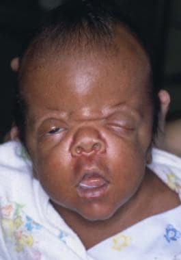
Poland Syndrome
Spinal Curvatures
Pulmonary Subvalvular Stenosis
Abnormalities, Multiple
Segmental spinal dysgenesis: neuroradiologic findings with clinical and embryologic correlation. (1/85)
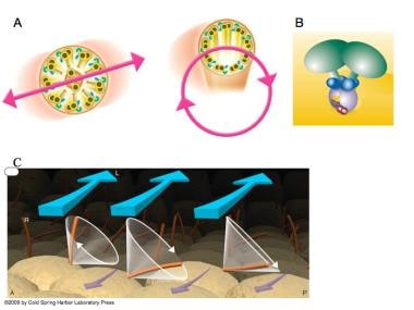
BACKGROUND AND PURPOSE: Segmental spinal dysgenesis (SSD) is a rare congenital abnormality in which a segment of the spine and spinal cord fails to develop properly. Our goal was to investigate the neuroradiologic features of this condition in order to correlate our findings with the degree of residual spinal cord function, and to provide insight into the embryologic origin of this disorder. We also aimed to clarify the relationship between SSD and other entities, such as multiple vertebral segmentation defects, congenital vertebral displacement, and caudal regression syndrome (CRS). METHODS: The records of patients treated at our institutions for congenital spinal anomalies were reviewed, and 10 cases were found to satisfy the inclusion criteria for SSD. Plain radiographs were available for review in all cases. MR imaging was performed in eight patients, one of whom also underwent conventional myelography. Two other patients underwent only conventional myelography. RESULTS: Segmental vertebral anomalies involved the thoracolumbar, lumbar, or lumbosacral spine. The spinal cord at the level of the abnormality was thinned or even indiscernible, and a bulky, low-lying cord segment was present caudad to the focal abnormality in most cases. Closed spinal dysraphisms were associated in five cases, and partial sacrococcygeal agenesis in three. Renal anomalies were detected in four cases, and dextrocardia in one; all patients had a neurogenic bladder. CONCLUSION: SSD is an autonomous entity with characteristic clinical and neuroradiologic features; however, SSD and CRS probably represent two faces of a single spectrum of segmental malformations of the spine and spinal cord. The neuroradiologic picture depends on the severity of the malformation and on its segmental level along the longitudinal embryonic axis. The severity of the morphologic derangement correlates with residual spinal cord function and with severity of the clinical deficit. (+info)A role of the cryptic gene in the correct establishment of the left-right axis. (2/85)
During vertebrate embryogenesis, a left-right axis is established. The heart, associated vessels and inner organs adopt asymmetric spatial arrangements and morphologies. Secreted growth factors of the TGF-beta family, including nodal, lefty-1 and lefty-2, play crucial roles in establishing left-right asymmetries [1] [2] [3]. In zebrafish, nodal signalling requires the presence of one-eyed pinhead (oep), a member of the EGF-CFC family of membrane-associated proteins [4]. We have generated a mutant allele of cryptic, a mouse EGF-CFC gene [5]. Homozygous cryptic mutants developed to birth, but the majority died during the first week of life because of complex cardiac malformations such as malpositioning of the great arteries, and atrial-ventricular septal defects. Moreover, laterality defects, including right isomerism of the lungs, right or left positioning of the stomach and splenic hypoplasia were observed. Nodal gene expression in the node was initiated in cryptic mutant mice, but neither nodal, lefty-2 nor Pitx2 were expressed in the left lateral plate mesoderm. The laterality defects observed in cryptic(-/-) mice resemble those of mice lacking the type IIB activin receptor or the homeobox-containing factor Pitx2 [6] [7] [8] [9], and are reminiscent of the human asplenic syndrome [10]. Our results provide genetic evidence for a role of cryptic in the signalling cascade that determines left-right asymmetry. (+info)Neonatal arterial switch operation for transposition of the great arteries in a patient with mirror image dextrocardia and situs inversus totalis. (3/85)
The neonatal arterial switch operation has become the standard therapy for D-transposition of the great arteries in the absence of left ventricular outflow tract obstruction. We describe our experience of successful arterial switch operation after balloon atrial septostomy in a 5-day-old infant girl who had atrial and visceral situs inversus totalis, mirror image dextrocardia, and D-transposition of the great arteries. To our knowledge, ours is the first report of this operation in a patient with this anatomy. (+info)Fetal cardiac dextroposition in the absence of an intrathoracic mass: sign of significant right lung hypoplasia. (4/85)
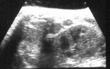
We reviewed our experience of fetal cardiac dextroposition in the absence of an intrathoracic mass. Ten cases were found by fetal echocardiography to have a normal cardiac axis, but the heart was shifted into the right chest and the amount of right lung tissue was reduced. At birth seven of the infants had confirmed structural heart disease (70%), including three with scimitar syndrome. Two infants had additional extracardiac anomalies (20%). Seven infants born at term had clinical pulmonary hypertension with a diagnosis of right lung hypoplasia in all of them. Two neonates died owing to significant heart disease (one with scimitar syndrome and the other with hypoplastic left heart syndrome). Of the three pregnancies that were terminated, the two fetuses with autopsies had severe right lung hypoplasia. Fetal cardiac dextroposition and right pulmonary artery hypoplasia in the absence of an intrathoracic mass are important signs of right lung hypoplasia, which can be associated with significant pathologic cardiac and extracardiac conditions. (+info)Multiple coronary artery bypass grafting in dextrocardia: case report. (5/85)
This is a case report of an unusual case of a patient with dextrocardia and "situs inversus totalis" who presented with unstable angina. Coronary angiography revealed severe main stem and severe triple vessel coronary artery disease. The patient later underwent successful emergency coronary artery bypass graft surgery. To the authors' knowledge this is the first reported case in Malaysia and also, the first ever report in the literature of multiple vessel coronary artery grafting, including the use of the right internal mammary artery. (+info)Reflections upon the aetiology of congenital pseudarthrosis of the clavicle. With a note on cranio-cleido dysostosis. (6/85)
The cause of pseudarthrosis of the clavicle is obscure. Right-sidedness is an almost constant feature. We have proposed that the lesion is sometimes due to pressure upon the developing clavicle by the subclavian artery which is normally at a higher level on the right side. This may be accentuated in the presence of cervical ribs or unduly elevated first ribs, both of which we have observed in association with pseudarthrosis. We have also noted pseudarthrosis on the left side in association with dextrocardia (when the relative positions of the subclavian arteries are reversed) and in the presence of a large left cervical rib. We have speculated upon the nature of the clavicular defect in cranio-cleido dysostosis, in which disorder the first ribs are habitually elevated. A similar mechanism may be involved. (+info)Cardiopulmonary malformations in the inv/inv mouse. (7/85)
The inv/inv mouse carries an insertional mutation in the inversin gene, (inv, for inversion of embryonic turning). Previously it had been reported that almost 100% of the homozygous offspring (inv/inv) were characterized by situs inversus totalis. In this report we identify the spectrum of cardiopulmonary anatomical abnormalities in inv/inv mice surviving to birth to determine whether the abnormalities seen are of the categories classically associated with human situs abnormalities. Stillborn mice, offspring that died unexpectedly (within 48 hr after birth), and neonates with phenotypic characteristics of situs inversus (right-sided stomachs, growth failure or jaundice) were processed for standard histological examination. Of 173 offspring, 34 (20%) neonates (11 stillborn, 9 unexpected deaths, and 14 mice with situs inversus phenotype) were examined, 27 of which were genotyped to be inv/inv. Interestingly, three inv/inv mice (11%) were found to have situs solitus. Twenty-four had situs inversus with normal, mirror-image cardiac anatomy (dextrocardia with atrioventricular concordance, ventriculoarterial concordance and a right aortic arch). The overall incidence of cardiovascular anomalies observed was 10 out of 27 (37%). The most frequent severe malformation, identified in 3 out of 27 animals, was a complex consisting of pulmonary infundibular stenosis/atresia with absence of pulmonary valve tissue and a ventricular septal defect. The pulmonary phenotype in inv/inv mice was situs inversus with occasional minor lobar abnormalities. We conclude that 1) cardiopulmonary malformations in inv/inv mice are not rare (37%), 2) the cardiopulmonary malformations observed in inv/inv specimens are not of the spectrum typically associated with human heterotaxia. In particular, inv/inv mice have a propensity for defects in the development of the right ventricular outflow tract and the interventricular septum, and 3) approximately one out of ten inv/inv mice is born with situs solitus and shows cardiac anomalies that correspond to those observed in inv/inv specimens with situs inversus. Our data therefore suggest that inversin, the product of the inv locus, may have specific roles in cardiac morphogenesis independent of its role in situs determination. (+info)Liver transplantation from situs inversus to situs inversus. (8/85)
Congenital anatomic anomalies often present technical obstacles during liver transplantation. Biliary atresia (BA) is the most common indication for liver transplantation in children, and up to 28% of children with situs inversus are complicated by BA. A boy aged 2 years 11 months with BA, situs inversus, and dextrocardia received a liver transplant from his father. The donor also had situs inversus and dextrocardia without other anomalies. Graft function was excellent postoperatively, and no significant complications were encountered. This is only the second report of the successful use of a living related donor graft for a patient with BA and situs inversus. This case was particularly rare because the donor also had situs inversus, which made the present procedure more feasible. (+info)Dextrocardia is a medical condition in which the heart is positioned on the right side of the chest instead of the left side. This is a congenital condition, meaning it is present at birth. In people with dextrocardia, the heart's structure and function are usually normal, but the orientation of the heart within the chest is reversed.
There are two main types of dextrocardia:
1. Dextrocardia without visceral situs inversus: In this type, the heart is on the right side of the chest, but the other organs in the chest and abdomen are in their normal positions. This is a rare condition and can be associated with other congenital heart defects.
2. Dextrocardia with visceral situs inversus: In this type, the heart is on the right side of the chest, and the other organs in the chest and abdomen are mirrored or reversed from their normal positions. This is a less common form of dextrocardia and is often referred to as "situs inversus totalis."
It's important to note that while dextrocardia itself is not a life-threatening condition, people with this condition may have other heart defects or medical issues that require treatment. If you or someone you know has been diagnosed with dextrocardia, it's essential to consult with a healthcare professional for proper evaluation and management.
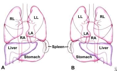
Situs Inversus is a congenital condition in which the major visceral organs are situated in mirror-image positions to their normal locations. Instead of being on the left side, the heart and its large blood vessels are on the right side, while the liver is on the left side and the lungs are reversed. The stomach, spleen, and pancreas may also be affected. It's important to note that this condition is generally asymptomatic and often goes unnoticed unless there are complications or associated abnormalities.
There are two types of Situs Inversus: total (complete reversal of all organs) and partial (reversal of only some organs). Total Situs Inversus is also sometimes referred to as "mirror-image dextrocardia" because the heart, which is usually on the left side, is located on the right side in a mirrored position.
While Situs Inversus itself does not typically cause health problems, people with this condition may have an increased risk for certain medical conditions, such as congenital heart defects or primary ciliary dyskinesia (PCD), which can lead to chronic respiratory infections and infertility.
Poland Syndrome is a rare congenital anomaly characterized by the absence or underdevelopment of the chest muscle (pectoralis major) on one side of the body, often associated with webbing or absence of the fingers (cutaneous syndactyly) and shortening of the arm on the same side. It was first described by Alfred Poland, a British surgeon, in 1841. The exact cause of this condition is not known, but it is believed to be due to an interruption of blood flow to the developing fetus during early pregnancy. Treatment typically involves reconstructive surgery and physical therapy.
Spinal curvatures refer to the normal or abnormal curvature patterns of the spine as viewed from the side. The human spine has four distinct curves that form an "S" shape when viewed from the side: cervical, thoracic, lumbar, and sacral. These natural curves provide strength, flexibility, and balance to the spine, allowing us to stand upright, maintain proper posture, and absorb shock during movement.
Abnormal spinal curvatures are often referred to as spinal deformities and can be classified into two main categories: hyperkyphosis (increased kyphosis) and hyperlordosis (increased lordosis). Examples of such conditions include:
1. Kyphosis: An excessive curvature in the thoracic or sacral regions, leading to a hunchback or rounded appearance. Mild kyphosis is common and usually not problematic, but severe cases can cause pain, breathing difficulties, and neurological issues.
2. Lordosis: An abnormal increase in the curvature of the lumbar or cervical spine, resulting in an exaggerated swayback posture. This can lead to lower back pain, muscle strain, and difficulty maintaining proper balance.
3. Scoliosis: A lateral (side-to-side) spinal curvature that causes the spine to twist and rotate, forming a C or S shape when viewed from behind. Most scoliosis cases are idiopathic (of unknown cause), but they can also be congenital (present at birth) or secondary to other medical conditions.
These abnormal spinal curvatures may require medical intervention, such as physical therapy, bracing, or surgery, depending on the severity and progression of the condition.
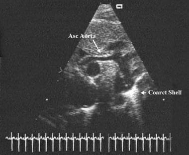
Pulmonary subvalvular stenosis is a rare cardiac condition that refers to the narrowing or obstruction of the pulmonary valve or the outflow tract below it, within the right ventricle of the heart. This results in restricted blood flow from the right ventricle to the pulmonary artery and subsequently to the lungs.
The narrowing can be caused by various factors such as a membranous shelf-like structure (dysplasia), a fibrous ring, or a tunnel-like narrowing of the outflow tract (tunneling). The severity of the stenosis may vary from mild to severe, and symptoms can range from shortness of breath, fatigue, and chest pain to more serious complications like heart failure or arrhythmias.
Diagnosis typically involves imaging tests such as echocardiography, cardiac MRI, or cardiac catheterization. Treatment options depend on the severity of the stenosis and may include monitoring, medications, or invasive procedures such as balloon dilation or surgical repair.
'Abnormalities, Multiple' is a broad term that refers to the presence of two or more structural or functional anomalies in an individual. These abnormalities can be present at birth (congenital) or can develop later in life (acquired). They can affect various organs and systems of the body and can vary greatly in severity and impact on a person's health and well-being.
Multiple abnormalities can occur due to genetic factors, environmental influences, or a combination of both. Chromosomal abnormalities, gene mutations, exposure to teratogens (substances that cause birth defects), and maternal infections during pregnancy are some of the common causes of multiple congenital abnormalities.
Examples of multiple congenital abnormalities include Down syndrome, Turner syndrome, and VATER/VACTERL association. Acquired multiple abnormalities can result from conditions such as trauma, infection, degenerative diseases, or cancer.
The medical evaluation and management of individuals with multiple abnormalities depend on the specific abnormalities present and their impact on the individual's health and functioning. A multidisciplinary team of healthcare professionals is often involved in the care of these individuals to address their complex needs.
Angiocardiography is a medical procedure used to examine the heart and blood vessels, particularly the chambers of the heart and the valves between them. It involves injecting a contrast agent into the bloodstream and taking X-ray images as the agent flows through the heart. This allows doctors to visualize any abnormalities such as blockages, narrowing, or leakage in the heart valves or blood vessels.
There are different types of angiocardiography, including:
* Left heart catheterization (LHC): A thin tube called a catheter is inserted into a vein in the arm or groin and threaded through to the left side of the heart to measure pressure and oxygen levels.
* Right heart catheterization (RHC): Similar to LHC, but the catheter is threaded through to the right side of the heart to measure pressure and oxygen levels there.
* Selective angiocardiography: A catheter is used to inject the contrast agent into specific blood vessels or chambers of the heart to get a more detailed view.
Angiocardiography can help diagnose and evaluate various heart conditions, including congenital heart defects, coronary artery disease, cardiomyopathy, and valvular heart disease. It is an invasive procedure that carries some risks, such as bleeding, infection, and damage to blood vessels or heart tissue. However, it can provide valuable information for diagnosing and treating heart conditions.
Kartagener Syndrome is a rare genetic disorder that primarily affects the respiratory system. It is characterized by the triad of chronic sinusitis, bronchiectasis (damage and widening of the airways in the lungs), and situs inversus totalis - a condition where the major visceral organs are mirrored or reversed from their normal positions.
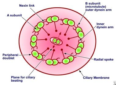
In Kartagener Syndrome, the cilia (tiny hair-like structures) lining the respiratory tract are abnormal or dysfunctional, which impairs their ability to clear mucus and other particles. This leads to recurrent respiratory infections, bronchiectasis, and ultimately, progressive lung damage.
The condition is inherited as an autosomal recessive trait, meaning that an individual must inherit two copies of the defective gene - one from each parent - to develop the syndrome. Kartagener Syndrome is a subtype of primary ciliary dyskinesia (PCD), a group of disorders affecting ciliary structure and function.
 Dextrocardia
Fredrick Arthur Willius
HES7 gene
Laterality
Shajarur Kanta (2015 film)
Carme Chacón
Maude Abbott
Pulmonary hypoplasia
Daisy Pearce
Right axis deviation
Stratton Parker syndrome
Harvey N. Middleton
Alexander Savvas
Wake therapy
Situs solitus
DNAH11
Levocardia
Diffuse idiopathic skeletal hyperostosis
Apex beat
Cardiac examination
Pulmonary agenesis
Polysplenia
Dr. No (novel)
18p-
Cold Hands, Warm Heart (novel)
Julius No
Johanson-Blizzard syndrome
Laryngeal cleft
Outline of cardiology
Keshava (film)
Anomalous pulmonary venous return2
- Tetralogy of fallot, dextrocardia, and situs inversus associated with total anomalous pulmonary venous return. (nih.gov)
- Modified Senning Procedure for Correction of Atrioventricular Discordance With Total Anomalous Pulmonary Venous Return, Atrial Situs Inversus, Dextrocardia, and Bilateral Superior Venae Cavae. (childrens.com)
Situs inversu3
- For all visceral organs to be mirrored, the correct term is dextrocardia situs inversus totalis. (wikipedia.org)
- 19. [Coronary artery bypass in dextrocardia with situs inversus totalis--a case report]. (nih.gov)
- 20. Coronary artery bypass grafting in dextrocardia with situs inversus totalis. (nih.gov)
Types of dextrocardia4
- There are two main types of dextrocardia: dextrocardia of embryonic arrest (also known as isolated dextrocardia)[citation needed] and dextrocardia situs inversus. (wikipedia.org)
- There are several types of dextrocardia. (medlineplus.gov)
- In the more common types of dextrocardia, other heart defects are also present. (medlineplus.gov)
- Types of dextrocardia. (nih.gov)
Congenital dextrocardia2
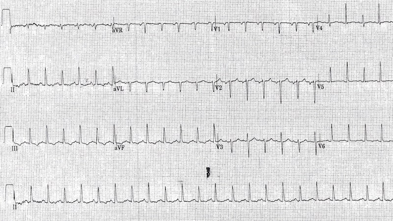
- Medical diagnosis of the two forms of congenital dextrocardia can be made by ECG or imaging. (wikipedia.org)
- Four cases of congenital dextrocardia, including a case with sino-auricular block. (nih.gov)
Transposition1
- Ivy was born with congenitally corrected transposition of the great arteries and dextrocardia. (cookchildrens.org)
Abnormalities1
- In rare cases, severely affected individuals have abnormalities of internal organs such as a lung or a kidney, or the heart is abnormally located in the right side of the chest (dextrocardia). (nih.gov)
Hypoplasia1
- Itoh M, Yada Y, Hashimoto U, Sasaki Y, Ohga K, Oka T. A case of intralobar pulmonary sequesteration associated with ASD, dextrocardia, hypoplasia of the right lung & eventration of the diaphragm. (edu.pk)
Levocardia1
- Defecto congénito en el que el corazón se localiza en el lado derecho del TÓRAX en lugar de en el lado izquierdo (levocardia, la posición normal). (bvsalud.org)
Pulmonary stenosis2
- Although statistically people with dextrocardia do not have any medical problems from the disorder, they may be prone to a number of bowel, esophageal, bronchial and cardiovascular disorders (such as double outlet right ventricle, endocardial cushion defect and pulmonary stenosis). (wikipedia.org)
- In addition, he's got a Ventricular Septal Defect (VSD) and Pulmonary Stenosis (PS) as well as Dextrocardia. (littlehearts.org)
Abnormal2
- The abdominal and chest organs in babies with dextrocardia may be abnormal and may not work correctly. (medlineplus.gov)
- The condition is called dextrocardia , an abnormal location of the heart, and is usually the result of a birth defect. (listverse.com)
Kartagener1
- citation needed] Kartagener syndrome may also be present in patients with dextrocardia but this must be in the setting of situs inversus and may include male infertility. (wikipedia.org)
Chest4
- Dextrocardia refers to a heart positioned in the right side of the chest. (wikipedia.org)
- Dextrocardia is a condition in which the heart is pointed toward the right side of the chest. (medlineplus.gov)
- The echocardiogram showed that the child's heart was on the right side, but it was not an ordinary dextrocardia, which made the doctor suspicious, in connection with which a contrast computed tomography of his chest was performed. (news.am)
- During her preoperative evaluation, a chest X-ray showed dextrocardia. (sages.org)
Visceral1
- Dextrocardia com transposição visceral completa. (nih.gov)
Organs1
- Dextrocardia may adversely affect other thoracic organs. (bvsalud.org)
Rarely1
- medical citation needed] In contrast to dextrocardia situs inversus which is only rarely associated with congenital heart disease, dextrocardia situs ambiguus is often associated with intracardiac anomalies. (wikipedia.org)
Coronary1
- Coronary artery bypass grafting in a case with dextrocardia and situs inversus. (florence.com.tr)
Symptoms1
- There are no symptoms of dextrocardia if the heart is normal. (medlineplus.gov)
Heart9
- Dextrocardia (from Latin dextro, meaning "right hand side," and Greek kardia, meaning "heart") is a rare congenital condition in which the apex of the heart is located on the right side of the body, rather than the more typical placement towards the left. (wikipedia.org)
- In this form of dextrocardia, the heart is simply placed further right in the thorax than is normal. (wikipedia.org)
- Dextrocardia situs inversus refers to the heart being a mirror image situated on the right side. (wikipedia.org)
- In the simplest type of dextrocardia, the heart is a mirror image of the normal heart and there are no other problems. (medlineplus.gov)
- A complete mirror image dextrocardia with no heart defects requires no treatment. (medlineplus.gov)
- The type of treatment needed depends on the heart or physical problems the infant may have in addition to dextrocardia. (medlineplus.gov)
- If heart defects are present with dextrocardia, the baby will most likely need surgery. (medlineplus.gov)
- Abhigyan was diagnosed with a critical heart malformation called Silus Solitus Dextrocardia and was admitted to a hospital. (cgi.com)
- When dextrocardia is accompanied with inverted HEART ATRIA , a right-sided STOMACH , and a left-sided LIVER , the combination is called dextrocardia with SITUS INVERSUS . (bvsalud.org)
Risk factors1
- Possible risk factors for dextrocardia include a family history of the condition. (medlineplus.gov)
Poland4
Mirror image1
- Some people with mirror-image dextrocardia have a problem with the fine hairs (cilia) that filter the air going into their nose and air passages. (medlineplus.gov)
True1
- A case of true dextrocardia. (nih.gov)
Person2
- citation needed] ECG leads must be placed in reversed positions on a person with dextrocardia. (wikipedia.org)
- Can a person live with dextrocardia? (prodietcare.com)
Addition1
- In addition, when defibrillating someone with dextrocardia, the pads should be placed in reverse positions. (wikipedia.org)
Common1
- dextrocardia is common. (msdmanuals.com)
Left1
- Certain cardiovascular and pulmonary disorders related to dextrocardia can be life-threatening if left unchecked. (wikipedia.org)