Diencephalon
Mesencephalon
Telencephalon
Prosencephalon
Gene Expression Regulation, Developmental
Metencephalon
Zebrafish
Optic Chiasm
Zebrafish Proteins
In Situ Hybridization
Body Patterning
Subcommissural Organ
Homeodomain Proteins
Brain
Otx Transcription Factors
Thalamus
Mamillary Bodies
Hedgehog Proteins
Embryo, Nonmammalian
Chick Embryo
Nerve Tissue Proteins
Fibroblast Growth Factor 8
Central Nervous System
Paired Box Transcription Factors
Coturnix
Tectum Mesencephali
Rhombencephalon
Hypothalamus
Habenula
Pituitary Gland
Transcription Factors
Encephalopsin: a novel mammalian extraretinal opsin discretely localized in the brain. (1/397)
We have identified a mammalian opsin, encephalopsin, that shows strong and specific expression in the brain. Encephalopsin defines a new family of opsins and shows highest homology to vertebrate retinal and pineal opsins. Encephalopsin is highly expressed in the preoptic area and paraventricular nucleus of the hypothalamus, both regions implicated in encephalic photoreception in nonmammalian vertebrates. In addition, encephalopsin shows highly patterned expression in other regions of the brain, being enriched in selected regions of the cerebral cortex, cerebellar Purkinje cells, a subset of striatal neurons, selected thalamic nuclei, and a subset of interneurons in the ventral horn of the spinal cord. Rostrocaudal gradients of encephalopsin expression are present in the cortex, cerebellum, and striatum. Radial stripes of encephalopsin expression are seen in the cerebellum. In the cortex and cerebellum, encephalopsin expression is considerably higher and more highly patterned in the adult than in the neonate. Encephalopsin is the first putative extraocular opsin identified in mammals and may play a role in encephalic photoreception. (+info)Anterior cephalic neural crest is required for forebrain viability. (2/397)
The prosencephalon, or embryonic forebrain, grows within a mesenchymal matrix of local paraxial mesoderm and of neural crest cells (NCC) derived from the posterior diencephalon and mesencephalon. Part of this NCC population forms the outer wall of capillaries within the prosencephalic leptomeninges and neuroepithelium itself. The surgical removal of NCC from the anterior head of chick embryos leads to massive cell death within the forebrain neuroepithelium during an interval that precedes its vascularization by at least 36 hours. During this critical period, a mesenchymal layer made up of intermingled mesodermal cells and NCC surround the neuroepithelium. This layer is not formed after anterior cephalic NCC ablation. The neuroepithelium then undergoes massive apoptosis. Cyclopia ensues after forebrain deterioration and absence of intervening frontonasal bud derivatives. The deleterious effect of ablation of the anterior NC cannot be interpreted as a deficit in vascularization because it takes place well before the time when blood vessels start to invade the neuroepithelium. Thus the mesenchymal layer itself exerts a trophic effect on the prosencephalic neuroepithelium. In an assay to rescue the operated phenotype, we found that the rhombencephalic but not the truncal NC can successfully replace the diencephalic and mesencephalic NC. Moreover, any region of the paraxial cephalic mesoderm can replace NCC in their dual function: in their early trophic effect and in providing pericytes to the forebrain meningeal blood vessels. The assumption of these roles by the cephalic neural crest may have been instrumental in the rostral expansion of the vertebrate forebrain over the course of evolution. (+info)The midbrain-hindbrain boundary genetic cascade is activated ectopically in the diencephalon in response to the widespread expression of one of its components, the medaka gene Ol-eng2. (3/397)
In vertebrates, the engrailed genes are expressed at early neurula stage in a narrow stripe encompassing the midbrain-hindbrain boundary (MHB), a region from which a peculiar structure, the isthmus, is formed. Knock-out experiments in mice demonstrated that these genes are essential for the development of this structure and of its derivatives. In contrast, little is known about the effect of an overexpression of engrailed genes in vertebrate development. Here we report the isolation of Ol-eng2, a medaka fish (Oryzias latipes) engrailed gene. We have monitored the effects of its widespread expression following mRNA injections in 1- and 2-cell medaka and Xenopus embryos. We found that the ectopic expression of Ol-eng2 predominantly results in an altered development of the anterior brain, including an inhibition of optic vesicle formation. No change in the patterns of mesencephalic and telencephalic markers were observed. In contrast, expressions of markers of the diencephalon were strongly repressed in injected embryos. Furthermore, the endogenous Ol-eng2, Pax2, Wnt1 and Fgf8, which are essential components of the MHB genetic cascade, were ectopically expressed in this region. Therefore, we propose that Ol-eng2 induces de novo formation of an isthmus-like structure, which correlates with the development of ectopic midbrain structures, including optic tectum. A competence of the diencephalon to change to a midbrain fate has been demonstrated in isthmic graft experiments. Our data demonstrate that this change can be mimicked by ectopic engrailed expression alone. (+info)CNS involvement in neuro-Behcet syndrome: an MR study. (4/397)
BACKGROUND AND PURPOSE: Behcet disease (BD) is a multisystem vasculitis of unknown origin in which neurologic involvement has been reported in the range of 5% to 10% in large series. Reports on clinical and radiologic aspects of neuro-Behcet syndrome (NBS) are in general limited in number. Our purpose was to determine the MR patterns in patients with NBS who had neural parenchymal involvement and to correlate our findings with possible vascular pathophysiology. METHODS: The MR images of 65 patients with NBS and neural parenchymal involvement were reviewed. In a subgroup of patients who had serial MR studies, we evaluated the anatomic-radiologic location and distribution of the lesions and whether they corresponded to any vascular territory, and studied their extension, enhancement patterns, and temporal course. RESULTS: The most common imaging finding in NBS patients who had neural parenchymal involvement was a mesodiencephalic junction lesion with edema extending along certain long tracts in the brain stem and diencephalon in 46% of the patients. The next most common location of involvement was the pontobulbar region, seen in 40% of the cases. Three primary cervical spinal cord lesions and one case of isolated optic nerve involvement were observed. CONCLUSION: The parenchymal distribution of lesions in NBS appears to support the hypothesis of small-vessel vasculitis; mainly, venular involvement. The anatomic distribution of intraaxial veins of the CNS explains the predominant involvement of the brain stem structures observed in our patients. This pattern of lesion distribution might help to differentiate NBS from other vasculitides as well as from the inflammatory-demyelinating diseases of the CNS, such as multiple sclerosis. (+info)Expression of three Rx homeobox genes in embryonic and adult zebrafish. (5/397)
The paired-class homeobox gene, Rx, is important in eye development. In this study we analyze expression patterns of three zebrafish Rx genes (Zrx1, 2, 3) in embryos and adults. All three genes show dynamic spatiotemporal patterns of expression. Zrx3 is expressed earliest, in the anteriormost region of the neural plate, in regions that give rise to ventral diencephalon and retinae. As development proceeds, Zrx3 expression is reduced in the lateral optic primordia, and is absent in the optic cup, but is retained at the ventral midline of the diencephalon, and is expressed in hypothalamus in the adult. As the neural retina begins to differentiate, Zrx3 is re-expressed in a subset of cells in the inner nuclear layer, presumably bipolar cells, and this expression is retained in the adult. In contrast, Zrx1/2 have a slightly later onset of expression, are initially coincident with Zrx3, but then become complementary, remaining on in the optic primordia but disappearing from the ventral midline of the diencephalon. Zrx1/2 are down-regulated as the retina differentiates, except in the outer nuclear layer where they continue to be expressed at high levels in cone, but not rod, photoreceptors. This is the first transcription factor described that distinguishes between cone and rod photoreceptors. (+info)Tissue interactions in the induction of anterior pituitary: role of the ventral diencephalon, mesenchyme, and notochord. (6/397)
Rathke's pouch, the epithelial primordium of the anterior pituitary, differentiates in close topographical and functional association with the ventral diencephalon. It is still not known whether the ventral diencephalon acts as the initial inducer of pituitary development. The roles of the adjacent mesenchyme and notochord, two other tissues located in close proximity to Rathke's pouch, in this process are even less clear. In this report we describe an in vitro experimental system that reproduces the earliest steps of anterior pituitary development. We provide evidence that the ventral diencephalon from 2- to 4-day-old chick embryos is able to function as an inducer of pituitary development and can convert early chick embryonic head ectoderm, which is not involved normally in pituitary development, into typical anterior pituitary tissue. This induction is contact-dependent. In our experimental system, there is a requirement for the supporting action of mesenchyme, which is independent of the mesenchyme source. Transplantation of the notochord into the lateral head region of a six-somite chick embryo induces an epithelial invagination, suggesting that the notochord induces the outpouching of the roof of the stomodeal ectoderm that results in formation of Rathke's pouch and causes the close contact between this ectoderm and the ventral diencephalon. Finally, we demonstrate that the ventral diencephalon from e9.5-e11.5 mouse embryos is also an efficient inducer of anterior pituitary differentiation in chick embryonic lateral head ectoderm, suggesting that the mechanism of anterior pituitary induction is conserved between mammals and birds, using the same, or similar, signaling pathways. (+info)Expression of a zebrafish iroquois homeobox gene, Ziro3, in the midline axial structures and central nervous system. (7/397)
We describe a zebrafish gene, Ziro3, which is highly homologous to Xenopus and mouse iroquois3. Ziro3 expression starts during gastrulation in the dorsal axial mesoderm that develops into the notochord. Later, the expression is limited to the chordo-neural hinge in the tailbud. Ziro3 expression also occurs in the central nervous system (CNS), excluding the telencephalon. The level of Ziro3 expression differs in odd and even rhombomeres. In the midbrain-hindbrain boundary (MHB) and rhombomere 6, Ziro3 transcripts appear only after the formation of the cerebellum and otic vesicle, respectively. (+info)Leptin increases serotonin turnover by inhibition of brain nitric oxide synthesis. (8/397)
Leptin administration inhibits diencephalic nitric oxide synthase (NOS) activity and increases brain serotonin (5-HT) metabolism in mice. We evaluated food intake, body-weight gain, diencephalic NOS activity, and diencephalic content of tryptophan (TRP), 5-HT, hydroxyindoleacetic acid (5-HIAA), and 5-HIAA/5-HT ratio after intracerebroventricular (ICV) or intraperitoneal (IP) leptin injection in mice. Five consecutive days of ICV or IP leptin injections induced a significant reduction in neuronal NOS (nNOS) activity, and caused a dose-dependent increase of 5-HT, 5-HIAA, and the 5-HIAA/5-HT ratio. Diencephalic 5-HT metabolism showed a significant increase in 5-HT, 5-HIAA, and the 5-HIAA/5-HT ratio 3 hours after a single leptin injection. This effect was maintained for 3 hours and had disappeared by 12 hours after injection. After a single IP leptin injection, the peak for 5-HT, 5-HIAA, and the 5-HIAA/5-HT ratio was achieved at 6 hours. Single injections of ICV or IP leptin significantly increased diencephalic 5-HT content. Leptin-induced 5-HT increase was antagonized by the coadministration of L-arginine only when the latter was ICV injected, whereas D-arginine did not influence leptin effects on brain 5-HT content. Finally, in nNOS-knockout mice, the appetite-suppressant activity of leptin was strongly reduced, and the leptin-induced increase in brain 5-HT metabolism was completely abolished. Our results indicate that the L-arginine/NO pathway is involved in mediating leptin effects on feeding behavior, and demonstrate that nNOS activity is required for the effects of leptin on brain 5-HT turnover. (+info)The diencephalon is a term used in anatomy to refer to the part of the brain that lies between the cerebrum and the midbrain. It includes several important structures, such as the thalamus, hypothalamus, epithalamus, and subthalamus.
The thalamus is a major relay station for sensory information, receiving input from all senses except smell and sending it to the appropriate areas of the cerebral cortex. The hypothalamus plays a crucial role in regulating various bodily functions, including hunger, thirst, body temperature, and sleep-wake cycles. It also produces hormones that regulate mood, growth, and development.
The epithalamus contains the pineal gland, which produces melatonin, a hormone that helps regulate sleep-wake cycles. The subthalamus is involved in motor control and coordination.
Overall, the diencephalon plays a critical role in integrating sensory information, regulating autonomic functions, and modulating behavior and emotion.
The mesencephalon, also known as the midbrain, is the middle portion of the brainstem that connects the hindbrain (rhombencephalon) and the forebrain (prosencephalon). It plays a crucial role in several important functions including motor control, vision, hearing, and the regulation of consciousness and sleep-wake cycles. The mesencephalon contains several important structures such as the cerebral aqueduct, tectum, tegmentum, cerebral peduncles, and several cranial nerve nuclei (III and IV).
The telencephalon is the most anterior (front) region of the embryonic brain, which eventually develops into the largest portion of the adult human brain, including the cerebral cortex, basal ganglia, and olfactory bulbs. It is derived from the prosencephalon (forebrain) during embryonic development and is responsible for higher cognitive functions such as thinking, perception, and language. The telencephalon can be further divided into two hemispheres, each containing regions associated with different functions.
The prosencephalon is a term used in the field of neuroembryology, which refers to the developmental stage of the forebrain in the embryonic nervous system. It is one of the three primary vesicles that form during the initial stages of neurulation, along with the mesencephalon (midbrain) and rhombencephalon (hindbrain).
The prosencephalon further differentiates into two secondary vesicles: the telencephalon and diencephalon. The telencephalon gives rise to structures such as the cerebral cortex, basal ganglia, and olfactory bulbs, while the diencephalon develops into structures like the thalamus, hypothalamus, and epithalamus.
It is important to note that 'prosencephalon' itself is not used as a medical term in adult neuroanatomy, but it is crucial for understanding the development of the human brain during embryogenesis.
Developmental gene expression regulation refers to the processes that control the activation or repression of specific genes during embryonic and fetal development. These regulatory mechanisms ensure that genes are expressed at the right time, in the right cells, and at appropriate levels to guide proper growth, differentiation, and morphogenesis of an organism.
Developmental gene expression regulation is a complex and dynamic process involving various molecular players, such as transcription factors, chromatin modifiers, non-coding RNAs, and signaling molecules. These regulators can interact with cis-regulatory elements, like enhancers and promoters, to fine-tune the spatiotemporal patterns of gene expression during development.
Dysregulation of developmental gene expression can lead to various congenital disorders and developmental abnormalities. Therefore, understanding the principles and mechanisms governing developmental gene expression regulation is crucial for uncovering the etiology of developmental diseases and devising potential therapeutic strategies.
The metencephalon is a term used in the field of neuroanatomy, which refers to the portion of the brain that develops from the anterior rhombencephalic vesicle during embryonic development. It gives rise to two major structures in the adult brain: the pons and the cerebellum.
The pons is a region located in the brainstem that plays important roles in relaying sensory information, regulating respiration, and controlling facial movements. The cerebellum, on the other hand, is a structure located at the back of the brain that is responsible for coordinating muscle movements, maintaining balance, and contributing to cognitive functions such as attention and language processing.
Overall, the metencephalon is an essential part of the brain that plays critical roles in sensory perception, motor control, and various other physiological processes.
A zebrafish is a freshwater fish species belonging to the family Cyprinidae and the genus Danio. Its name is derived from its distinctive striped pattern that resembles a zebra's. Zebrafish are often used as model organisms in scientific research, particularly in developmental biology, genetics, and toxicology studies. They have a high fecundity rate, transparent embryos, and a rapid development process, making them an ideal choice for researchers. However, it is important to note that providing a medical definition for zebrafish may not be entirely accurate or relevant since they are primarily used in biological research rather than clinical medicine.
The optic chiasm is a structure in the brain where the optic nerves from each eye meet and cross. This allows for the integration of visual information from both eyes into the brain's visual cortex, creating a single, combined image of the visual world. The optic chiasm plays an important role in the processing of visual information and helps to facilitate depth perception and other complex visual tasks. Damage to the optic chiasm can result in various visual field deficits, such as bitemporal hemianopsia, where there is a loss of vision in the outer halves (temporal fields) of both eyes' visual fields.
Zebrafish proteins refer to the diverse range of protein molecules that are produced by the organism Danio rerio, commonly known as the zebrafish. These proteins play crucial roles in various biological processes such as growth, development, reproduction, and response to environmental stimuli. They are involved in cellular functions like enzymatic reactions, signal transduction, structural support, and regulation of gene expression.
Zebrafish is a popular model organism in biomedical research due to its genetic similarity with humans, rapid development, and transparent embryos that allow for easy observation of biological processes. As a result, the study of zebrafish proteins has contributed significantly to our understanding of protein function, structure, and interaction in both zebrafish and human systems.
Some examples of zebrafish proteins include:
* Transcription factors that regulate gene expression during development
* Enzymes involved in metabolic pathways
* Structural proteins that provide support to cells and tissues
* Receptors and signaling molecules that mediate communication between cells
* Heat shock proteins that assist in protein folding and protect against stress
The analysis of zebrafish proteins can be performed using various techniques, including biochemical assays, mass spectrometry, protein crystallography, and computational modeling. These methods help researchers to identify, characterize, and understand the functions of individual proteins and their interactions within complex networks.
In situ hybridization (ISH) is a molecular biology technique used to detect and localize specific nucleic acid sequences, such as DNA or RNA, within cells or tissues. This technique involves the use of a labeled probe that is complementary to the target nucleic acid sequence. The probe can be labeled with various types of markers, including radioisotopes, fluorescent dyes, or enzymes.
During the ISH procedure, the labeled probe is hybridized to the target nucleic acid sequence in situ, meaning that the hybridization occurs within the intact cells or tissues. After washing away unbound probe, the location of the labeled probe can be visualized using various methods depending on the type of label used.
In situ hybridization has a wide range of applications in both research and diagnostic settings, including the detection of gene expression patterns, identification of viral infections, and diagnosis of genetic disorders.
"Body patterning" is a general term that refers to the process of forming and organizing various tissues and structures into specific patterns during embryonic development. This complex process involves a variety of molecular mechanisms, including gene expression, cell signaling, and cell-cell interactions. It results in the creation of distinct body regions, such as the head, trunk, and limbs, as well as the organization of internal organs and systems.
In medical terminology, "body patterning" may refer to specific developmental processes or abnormalities related to embryonic development. For example, in genetic disorders such as Poland syndrome or Holt-Oram syndrome, mutations in certain genes can lead to abnormal body patterning, resulting in the absence or underdevelopment of certain muscles, bones, or other structures.
It's important to note that "body patterning" is not a formal medical term with a specific definition, but rather a general concept used in developmental biology and genetics.
The subcommissural organ (SCO) is a small neuroendocrine gland located at the caudal end of the third ventricle in the brain. It is situated in the vicinity of the posterior commissure, hence its name. The SCO is primarily composed of ependymal cells and produces a variety of neuropeptides and proteins that are released into the cerebrospinal fluid (CSF).
The main function of the subcommissural organ is to secrete a glycoprotein called SCO-spondin, which plays a role in the formation and maintenance of the cerebral aqueduct and the rostral part of the central canal of the spinal cord. The CSF flow through these structures is facilitated by the presence of SCO-spondin, which has been shown to have adhesive properties that help prevent the collapse of these narrow channels.
Dysfunction or abnormalities in the subcommissural organ may contribute to various neurological disorders, such as hydrocephalus and other conditions associated with impaired CSF flow. However, further research is needed to fully understand the role of this intriguing structure in brain physiology and pathology.
Homeodomain proteins are a group of transcription factors that play crucial roles in the development and differentiation of cells in animals and plants. They are characterized by the presence of a highly conserved DNA-binding domain called the homeodomain, which is typically about 60 amino acids long. The homeodomain consists of three helices, with the third helix responsible for recognizing and binding to specific DNA sequences.
Homeodomain proteins are involved in regulating gene expression during embryonic development, tissue maintenance, and organismal growth. They can act as activators or repressors of transcription, depending on the context and the presence of cofactors. Mutations in homeodomain proteins have been associated with various human diseases, including cancer, congenital abnormalities, and neurological disorders.
Some examples of homeodomain proteins include PAX6, which is essential for eye development, HOX genes, which are involved in body patterning, and NANOG, which plays a role in maintaining pluripotency in stem cells.
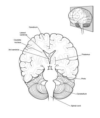
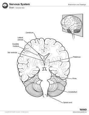
The brain is the central organ of the nervous system, responsible for receiving and processing sensory information, regulating vital functions, and controlling behavior, movement, and cognition. It is divided into several distinct regions, each with specific functions:
1. Cerebrum: The largest part of the brain, responsible for higher cognitive functions such as thinking, learning, memory, language, and perception. It is divided into two hemispheres, each controlling the opposite side of the body.
2. Cerebellum: Located at the back of the brain, it is responsible for coordinating muscle movements, maintaining balance, and fine-tuning motor skills.
3. Brainstem: Connects the cerebrum and cerebellum to the spinal cord, controlling vital functions such as breathing, heart rate, and blood pressure. It also serves as a relay center for sensory information and motor commands between the brain and the rest of the body.
4. Diencephalon: A region that includes the thalamus (a major sensory relay station) and hypothalamus (regulates hormones, temperature, hunger, thirst, and sleep).
5. Limbic system: A group of structures involved in emotional processing, memory formation, and motivation, including the hippocampus, amygdala, and cingulate gyrus.
The brain is composed of billions of interconnected neurons that communicate through electrical and chemical signals. It is protected by the skull and surrounded by three layers of membranes called meninges, as well as cerebrospinal fluid that provides cushioning and nutrients.
OTX (Orthodenticle homeobox) transcription factors are a family of proteins that regulate gene expression during embryonic development, particularly in the eye, forebrain, and midbrain. They play crucial roles in the development and differentiation of these tissues, including the specification of eye field identity, the determination of dorsoventral patterning in the neural tube, and the regulation of neurogenesis.
OTX transcription factors contain a highly conserved DNA-binding domain called the homeodomain, which allows them to recognize and bind to specific DNA sequences. In humans, there are four known OTX transcription factors (OTX1, OTX2, OTX3, and CRX), each with distinct expression patterns and functions.
Mutations in OTX genes have been associated with various developmental disorders, such as microphthalmia, anophthalmia, and severe eye malformations, highlighting their importance in normal eye development. Additionally, OTX transcription factors have also been implicated in the pathogenesis of certain cancers, including medulloblastoma and retinoblastoma.
The thalamus is a large, paired structure in the brain that serves as a relay station for sensory and motor signals to the cerebral cortex. It is located in the dorsal part of the diencephalon and is made up of two symmetrical halves, each connected to the corresponding cerebral hemisphere.
The thalamus receives inputs from almost all senses, except for the olfactory system, and processes them before sending them to specific areas in the cortex. It also plays a role in regulating consciousness, sleep, and alertness. Additionally, the thalamus is involved in motor control by relaying information between the cerebellum and the motor cortex.
The thalamus is divided into several nuclei, each with distinct connections and functions. Some of these nuclei are involved in sensory processing, while others are involved in motor function or regulation of emotions and cognition. Overall, the thalamus plays a critical role in integrating information from various brain regions and modulating cognitive and emotional processes.
The mamillary bodies are a pair of small, round structures located in the hypothalamus region of the brain. They play a crucial role in the limbic system, which is involved in emotions, memory, and learning. Specifically, the mamillary bodies are part of the circuit that forms the Papez circuit, a neural network responsible for memory and cognitive functions.
The mamillary bodies receive inputs from several brain regions, including the hippocampus, anterior thalamic nuclei, and cingulate gyrus. They then project this information to the thalamus, which in turn sends it to the cerebral cortex for further processing.
Damage to the mamillary bodies can result in memory impairment, as seen in patients with Korsakoff's syndrome, a condition often associated with chronic alcohol abuse.
Hedgehog proteins are a group of signaling molecules that play crucial roles in the development and regulation of various biological processes in animals. They are named after the hedgehog mutant fruit flies, which have spiky bristles due to defects in this pathway. These proteins are involved in cell growth, differentiation, and tissue regeneration. They exert their effects by binding to specific receptors on the surface of target cells, leading to a cascade of intracellular signaling events that ultimately influence gene expression and cell behavior.
There are three main types of Hedgehog proteins in mammals: Sonic hedgehog (Shh), Indian hedgehog (Ihh), and Desert hedgehog (Dhh). These protecules undergo post-translational modifications, including cleavage and lipid modification, which are essential for their activity. Dysregulation of Hedgehog signaling has been implicated in various diseases, including cancer, developmental abnormalities, and degenerative disorders.
A nonmammalian embryo refers to the developing organism in animals other than mammals, from the fertilized egg (zygote) stage until hatching or birth. In nonmammalian species, the developmental stages and terminology differ from those used in mammals. The term "embryo" is generally applied to the developing organism up until a specific stage of development that is characterized by the formation of major organs and structures. After this point, the developing organism is referred to as a "larva," "juvenile," or other species-specific terminology.
The study of nonmammalian embryos has played an important role in our understanding of developmental biology and evolutionary developmental biology (evo-devo). By comparing the developmental processes across different animal groups, researchers can gain insights into the evolutionary origins and diversification of body plans and structures. Additionally, nonmammalian embryos are often used as model systems for studying basic biological processes, such as cell division, gene regulation, and pattern formation.
A chick embryo refers to the developing organism that arises from a fertilized chicken egg. It is often used as a model system in biological research, particularly during the stages of development when many of its organs and systems are forming and can be easily observed and manipulated. The study of chick embryos has contributed significantly to our understanding of various aspects of developmental biology, including gastrulation, neurulation, organogenesis, and pattern formation. Researchers may use various techniques to observe and manipulate the chick embryo, such as surgical alterations, cell labeling, and exposure to drugs or other agents.
Nerve tissue proteins are specialized proteins found in the nervous system that provide structural and functional support to nerve cells, also known as neurons. These proteins include:
1. Neurofilaments: These are type IV intermediate filaments that provide structural support to neurons and help maintain their shape and size. They are composed of three subunits - NFL (light), NFM (medium), and NFH (heavy).
2. Neuronal Cytoskeletal Proteins: These include tubulins, actins, and spectrins that provide structural support to the neuronal cytoskeleton and help maintain its integrity.
3. Neurotransmitter Receptors: These are specialized proteins located on the postsynaptic membrane of neurons that bind neurotransmitters released by presynaptic neurons, triggering a response in the target cell.
4. Ion Channels: These are transmembrane proteins that regulate the flow of ions across the neuronal membrane and play a crucial role in generating and transmitting electrical signals in neurons.
5. Signaling Proteins: These include enzymes, receptors, and adaptor proteins that mediate intracellular signaling pathways involved in neuronal development, differentiation, survival, and death.
6. Adhesion Proteins: These are cell surface proteins that mediate cell-cell and cell-matrix interactions, playing a crucial role in the formation and maintenance of neural circuits.
7. Extracellular Matrix Proteins: These include proteoglycans, laminins, and collagens that provide structural support to nerve tissue and regulate neuronal migration, differentiation, and survival.
Fibroblast Growth Factor 8 (FGF-8) is a growth factor that belongs to the fibroblast growth factor family. It plays crucial roles in various biological processes, including embryonic development, tissue repair, and cancer progression. Specifically, FGF-8 has been implicated in the regulation of cell proliferation, differentiation, migration, and survival.
During embryonic development, FGF-8 is involved in the formation of the nervous system, limbs, and other organs. It acts as a signaling molecule that helps to establish patterns of gene expression and cell behavior during development. In tissue repair, FGF-8 can stimulate the proliferation and migration of cells involved in wound healing, such as fibroblasts and endothelial cells.
In cancer, FGF-8 has been shown to promote tumor growth, angiogenesis (the formation of new blood vessels), and metastasis. It can do this by activating signaling pathways that promote cell proliferation, survival, and migration. Overexpression of FGF-8 has been found in various types of cancer, including breast, lung, prostate, and ovarian cancer.
In summary, Fibroblast Growth Factor 8 (FGF-8) is a signaling molecule that plays important roles in embryonic development, tissue repair, and cancer progression by regulating cell proliferation, differentiation, migration, and survival.
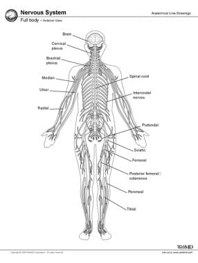
The Central Nervous System (CNS) is the part of the nervous system that consists of the brain and spinal cord. It is called the "central" system because it receives information from, and sends information to, the rest of the body through peripheral nerves, which make up the Peripheral Nervous System (PNS).
The CNS is responsible for processing sensory information, controlling motor functions, and regulating various autonomic processes like heart rate, respiration, and digestion. The brain, as the command center of the CNS, interprets sensory stimuli, formulates thoughts, and initiates actions. The spinal cord serves as a conduit for nerve impulses traveling to and from the brain and the rest of the body.
The CNS is protected by several structures, including the skull (which houses the brain) and the vertebral column (which surrounds and protects the spinal cord). Despite these protective measures, the CNS remains vulnerable to injury and disease, which can have severe consequences due to its crucial role in controlling essential bodily functions.
Paired box (PAX) transcription factors are a group of proteins that regulate gene expression during embryonic development and in some adult tissues. They are characterized by the presence of a paired box domain, a conserved DNA-binding motif that recognizes specific DNA sequences. PAX proteins play crucial roles in various developmental processes, such as the formation of the nervous system, eyes, and pancreas. Dysregulation of PAX genes has been implicated in several human diseases, including cancer.
"Coturnix" is a genus of birds that includes several species of quails. The most common species is the Common Quail (Coturnix coturnix), which is also known as the European Quail or the Eurasian Quail. This small ground-dwelling bird is found throughout Europe, Asia, and parts of Africa, and it is known for its distinctive call and its migratory habits. Other species in the genus Coturnix include the Rain Quail (Coturnix coromandelica), the Stubble Quail (Coturnix pectoralis), and the Harlequin Quail (Coturnix delegorguei). These birds are all similar in appearance and behavior, with small, round bodies, short wings, and strong legs that are adapted for running and scratching in leaf litter. They are also known for their cryptic coloration, which helps them blend in with their surroundings and avoid predators. Quails are popular game birds and are also kept as pets and for ornamental purposes in some parts of the world.
The "tectum mesencephali" is a term used in anatomy to refer to the roof or dorsal portion of the midbrain, which is a part of the brainstem. It plays a crucial role in visual and auditory processing, as well as motor coordination. The tectum mesencephali contains several important structures, including the superior colliculi and the inferior colliculi, which are involved in the reflexive responses to visual and auditory stimuli, respectively. Additionally, the tectum mesencephali is connected to various other regions of the brain, allowing for the integration of sensory information and the coordination of motor responses.
The rhombencephalon is a term used in the field of neuroanatomy, which refers to the most posterior region of the developing brain during embryonic development. It is also known as the hindbrain and it gives rise to several important structures in the adult brain.
More specifically, the rhombencephalon can be further divided into two main parts: the metencephalon and the myelencephalon. The metencephalon eventually develops into the pons and cerebellum, while the myelencephalon becomes the medulla oblongata.
The rhombencephalon plays a crucial role in several critical functions of the nervous system, including regulating heart rate and respiration, maintaining balance and posture, and coordinating motor movements. Defects or abnormalities in the development of the rhombencephalon can lead to various neurological disorders, such as cerebellar hypoplasia, Chiari malformation, and certain forms of brainstem tumors.
The hypothalamus is a small, vital region of the brain that lies just below the thalamus and forms part of the limbic system. It plays a crucial role in many important functions including:
1. Regulation of body temperature, hunger, thirst, fatigue, sleep, and circadian rhythms.
2. Production and regulation of hormones through its connection with the pituitary gland (the hypophysis). It controls the release of various hormones by producing releasing and inhibiting factors that regulate the anterior pituitary's function.
3. Emotional responses, behavior, and memory formation through its connections with the limbic system structures like the amygdala and hippocampus.
4. Autonomic nervous system regulation, which controls involuntary physiological functions such as heart rate, blood pressure, and digestion.
5. Regulation of the immune system by interacting with the autonomic nervous system.
Damage to the hypothalamus can lead to various disorders like diabetes insipidus, growth hormone deficiency, altered temperature regulation, sleep disturbances, and emotional or behavioral changes.
The habenula is a small, paired nucleus located in the epithalamus region of the brain. It plays a crucial role in the modulation of various functions such as mood, reward, and motivation. The habenula can be further divided into two subregions: the medial and lateral habenula.
The medial habenula is involved in the regulation of emotional behaviors, including responses to stress and anxiety. It receives inputs from several brain regions associated with emotion, such as the amygdala and hippocampus, and projects to the interpeduncular nucleus (IPN) in the midbrain.
The lateral habenula is primarily involved in processing aversive stimuli and modulating dopaminergic reward pathways. It receives inputs from various regions associated with motivation, learning, and memory, such as the prefrontal cortex, basal ganglia, and thalamus. The lateral habenula then projects to the midbrain's dopamine-producing neurons in the ventral tegmental area (VTA) and substantia nigra pars compacta (SNc), which are critical components of the brain's reward system.
Dysfunction of the habenula has been implicated in several neurological and psychiatric disorders, including depression, anxiety, addiction, and schizophrenia.
The pituitary gland is a small, endocrine gland located at the base of the brain, in the sella turcica of the sphenoid bone. It is often called the "master gland" because it controls other glands and makes the hormones that trigger many body functions. The pituitary gland measures about 0.5 cm in height and 1 cm in width, and it weighs approximately 0.5 grams.
The pituitary gland is divided into two main parts: the anterior lobe (adenohypophysis) and the posterior lobe (neurohypophysis). The anterior lobe is further divided into three zones: the pars distalis, pars intermedia, and pars tuberalis. Each part of the pituitary gland has distinct functions and produces different hormones.
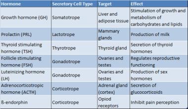
The anterior pituitary gland produces and releases several important hormones, including:
* Growth hormone (GH), which regulates growth and development in children and helps maintain muscle mass and bone strength in adults.
* Thyroid-stimulating hormone (TSH), which controls the production of thyroid hormones by the thyroid gland.
* Adrenocorticotropic hormone (ACTH), which stimulates the adrenal glands to produce cortisol and other steroid hormones.
* Follicle-stimulating hormone (FSH) and luteinizing hormone (LH), which regulate reproductive function in both males and females.
* Prolactin, which stimulates milk production in pregnant and lactating women.
The posterior pituitary gland stores and releases two hormones that are produced by the hypothalamus:
* Antidiuretic hormone (ADH), which helps regulate water balance in the body by controlling urine production.
* Oxytocin, which stimulates uterine contractions during childbirth and milk release during breastfeeding.
Overall, the pituitary gland plays a critical role in maintaining homeostasis and regulating various bodily functions, including growth, development, metabolism, and reproductive function.
Transcription factors are proteins that play a crucial role in regulating gene expression by controlling the transcription of DNA to messenger RNA (mRNA). They function by binding to specific DNA sequences, known as response elements, located in the promoter region or enhancer regions of target genes. This binding can either activate or repress the initiation of transcription, depending on the properties and interactions of the particular transcription factor. Transcription factors often act as part of a complex network of regulatory proteins that determine the precise spatiotemporal patterns of gene expression during development, differentiation, and homeostasis in an organism.
An axon is a long, slender extension of a neuron (a type of nerve cell) that conducts electrical impulses (nerve impulses) away from the cell body to target cells, such as other neurons or muscle cells. Axons can vary in length from a few micrometers to over a meter long and are typically surrounded by a myelin sheath, which helps to insulate and protect the axon and allows for faster transmission of nerve impulses.
Axons play a critical role in the functioning of the nervous system, as they provide the means by which neurons communicate with one another and with other cells in the body. Damage to axons can result in serious neurological problems, such as those seen in spinal cord injuries or neurodegenerative diseases like multiple sclerosis.
Morphogenesis is a term used in developmental biology and refers to the process by which cells give rise to tissues and organs with specific shapes, structures, and patterns during embryonic development. This process involves complex interactions between genes, cells, and the extracellular environment that result in the coordinated movement and differentiation of cells into specialized functional units.
Morphogenesis is a dynamic and highly regulated process that involves several mechanisms, including cell proliferation, death, migration, adhesion, and differentiation. These processes are controlled by genetic programs and signaling pathways that respond to environmental cues and regulate the behavior of individual cells within a developing tissue or organ.
The study of morphogenesis is important for understanding how complex biological structures form during development and how these processes can go awry in disease states such as cancer, birth defects, and degenerative disorders.
 Diencephalon
 The Diencephalon and Optic Tectum of the Longnose Gar, Lepisosteus osseus (L): Cytoarchitectonics and Distribution of...
 Development of the Diencephalon
 Diencephalon - The Behavioral Scientist
 Diencephalon: Thalamus, Hypothalamus, Epithalamus | SchoolWorkHelper
 SLITXOX's 'Diencephalon': A New Wave in Alternative Hip-Hop | Tunepical
 Bassett Collection - Lane Medical Library - Stanford University School of Medicine
 Otx1l, Otx2 and Irx1b establish and position the ZLI in the diencephalon | Development | The Company of Biologists
 Retina-Type Rhodopsin Gene Expressed in the Brain of a Teleost, Ayu (Plecoglossus altivelis)
 5 Brain Divisoons Cheat Sheet by drax - Download free from Cheatography - Cheatography.com: Cheat Sheets For Every Occasion
 Expression dynamics of the LIM-homeobox genes, Lhx1 and Lhx9, in the diencephalon during chick development | The International...
![Mokayes N[au] - Search Results - PubMed](data:image/png;base64,iVBORw0KGgoAAAANSUhEUgAAABAAAAAQCAMAAAAoLQ9TAAAARVBMVEVHcEwoU45gYmYAUpQAUpRPYGVgYmZLXnJgYmYAUZUAUpRJXnIAUpQAUpRgYmYAUpRgYmZgYmZhYmYAUpQAUpQAUpRgYmaDiPJuAAAAFXRSTlMADOJ+6QewGO8/uTRqtH7GdFJ11p1bCL3TAAAAZUlEQVQYlV2PVw7AIAxDTeney7n/UcsoldX3E+VJOAboEi7MBpHWMs1ADlG8u7UYWauwyZFeRQVPOhG2o+aiwhByJxUx91Jxhje3iJSqGfHuLKI0+0TpXvY1twCOPlFh5pa/++MB0vIOBm+1zaoAAAAASUVORK5CYII=) Mokayes N[au] - Search Results - PubMed
Mokayes N[au] - Search Results - PubMed Structure of the nervous system (video) | Khan Academy
 Holoprosencephaly: Practice Essentials, Anatomy, Pathophysiology
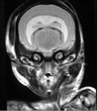
 Autograded Labs in Courseware
 musculo/neuro quiz Flashcards
 Cerebral Subcortex
 Profile | Biosciences | University of Exeter
 Interactive Prenatal Development Timeline - Advanced
 Advanced Search Results - Public Health Image Library(PHIL)
 BRAINMAPS.ORG - BRAIN ATLAS, BRAIN MAPS, BRAIN STRUCTURE,
NEUROINFORMATICS, BRAIN, STEREOTAXIC ATLAS, NEUROSCIENCE
BRAINMAPS.ORG - BRAIN ATLAS, BRAIN MAPS, BRAIN STRUCTURE,
NEUROINFORMATICS, BRAIN, STEREOTAXIC ATLAS, NEUROSCIENCE