Embryonic Induction
Gastrula
Mesoderm
Gene Expression Regulation, Developmental
Embryo, Nonmammalian
Body Patterning
Nervous System
Xenopus Proteins
Morphogenesis
In Situ Hybridization
Chick Embryo
Bone Morphogenetic Proteins
Sea Urchins
Neural Crest
Bone Morphogenetic Protein 4
Homeodomain Proteins
Germ Layers
Xenopus
Neural Plate
Blastula
Xenopus laevis
Embryonic Structures
Fibroblast Growth Factor 8
Fibroblast Growth Factors
Gastrulation
Cell Differentiation
Limb Buds
Transcription Factors
Tissue Transplantation
Quail
Signal Transduction
Notochord
Wnt Proteins
Lens, Crystalline
Paired Box Transcription Factors
Otx Transcription Factors
Genes, Homeobox
Zebrafish Proteins
Nodal Protein
Branchial Region
Hedgehog Proteins
Head
Cell Lineage
Embryo, Mammalian
Central Nervous System
Triturus
Molecular Sequence Data
Strongylocentrotus purpuratus
Bone Morphogenetic Protein Receptors
SOXB1 Transcription Factors
Trans-Activators
Epidermis
Organizers, Embryonic
Somites
Drosophila Proteins
Drosophila
Fibroblast Growth Factor 4
HMGB Proteins
DNA-Binding Proteins
Zebrafish
Chordata, Nonvertebrate
Activins
Blastocyst
MSX1 Transcription Factor
Base Sequence
Blastoderm
Proteins
Amino Acid Sequence
Repressor Proteins
Rhombencephalon
Neural Tube
Limb Deformities, Congenital
Wnt1 Protein
Models, Biological
Culture Techniques
T-Box Domain Proteins
Nerve Tissue Proteins
Eye
RNA, Messenger
Nodal Signaling Ligands
Pleurodeles
Coturnix
Axis
Wnt3 Protein
Hemicentrotus
Goosecoid Protein
Bone Morphogenetic Protein 2
LIM-Homeodomain Proteins
Feathers
Bone Morphogenetic Protein 7
Prosencephalon
Amphibians
Receptors, Fibroblast Growth Factor
Microinjections
Eye Proteins
The surface ectoderm is essential for nephric duct formation in intermediate mesoderm. (1/1870)
The nephric duct is the first epithelial tubule to differentiate from intermediate mesoderm that is essential for all further urogenital development. In this study we identify the domain of intermediate mesoderm that gives rise to the nephric duct and demonstrate that the surface ectoderm is required for its differentiation. Removal of the surface ectoderm resulted in decreased levels of Sim-1 and Pax-2 mRNA expression in mesenchymal nephric duct progenitors, and caused inhibition of nephric duct formation and subsequent kidney development. The surface ectoderm expresses BMP-4 and we show that it is required for the maintenance of high-level BMP-4 expression in lateral plate mesoderm. Addition of a BMP-4-coated bead to embryos lacking the surface ectoderm restored normal levels of Sim-1 and Pax-2 mRNA expression in nephric duct progenitors, nephric duct formation and the initiation of nephrogenesis. Thus, BMP-4 signaling can substitute for the surface ectoderm in supporting nephric duct morphogenesis. Collectively, these data suggest that inductive interactions between the surface ectoderm, lateral mesoderm and intermediate mesoderm are essential for nephric duct formation and the initiation of urogenital development. (+info)Regulation of neurotrophin-3 expression by epithelial-mesenchymal interactions: the role of Wnt factors. (2/1870)
Neurotrophins regulate survival, axonal growth, and target innervation of sensory and other neurons. Neurotrophin-3 (NT-3) is expressed specifically in cells adjacent to extending axons of dorsal root ganglia neurons, and its absence results in loss of most of these neurons before their axons reach their targets. However, axons are not required for NT-3 expression in limbs; instead, local signals from ectoderm induce NT-3 expression in adjacent mesenchyme. Wnt factors expressed in limb ectoderm induce NT-3 in the underlying mesenchyme. Thus, epithelial-mesenchymal interactions mediated by Wnt factors control NT-3 expression and may regulate axonal growth and guidance. (+info)Fish swimbladder: an excellent mesodermal inductor in primary embryonic induction. (3/1870)
Swimbladder of the crucian carp, Carassius auratus, was found to be better as a vegatalizing tissue than other tissues, such as guinea-pig bone marrow, when presumptive ectoderm of Triturus gastrulae was used as reacting tissue. Swimbladder usually induced assemblies of highly organized mesodermal tissues, such as notochord, somites and pronephric tubules, some of which were covered by mesodermal epithelium without any epidermal covering. A special character of the effect of swimbladder was the rather frequent induction of solid balls of undifferentiated cells, which were identified as mesodermal or mesodermal and probably endodermal. These findings show that swimbladder has a strong and fast spreading vegetalizing effect on the responding presumptive ectoderm. (+info)Embryological study of a T/t locus mutation (tw73) affecting trophectoderm development. (4/1870)
Mouse embryos homozygous for the recessive lethal mutation tw73 show specific defects in trophectoderm shortly after implantation. The trophectoderm and ectoplacental cone fail to form the usual close association with the uterine decidua, and proliferation is markedly reduced. The embryo proper ceases to develop beyond the two-layered stage and degenerates and dies within 5 days of implantation. (+info)Bmp4 is required for the generation of primordial germ cells in the mouse embryo. (5/1870)
In many organisms the allocation of primordial germ cells (PGCs) is determined by the inheritance of maternal factors deposited in the egg. However, in mammals, inductive cell interactions are required around gastrulation to establish the germ line. Here, we show that Bmp4 homozygous null embryos contain no PGCs. They also lack an allantois, an extraembryonic mesodermal tissue derived, like the PGCs, from precursors in the proximal epiblast. Heterozygotes have fewer PGCs than normal, due to a reduction in the size of the founding population and not to an effect on its subsequent expansion. Analysis of beta-galactosidase activity in Bmp4(lacZneo) embryos reveals that prior to gastrulation, Bmp4 is expressed in the extraembryonic ectoderm. Later, Bmp4 is expressed in the extraembryonic mesoderm, but not in PGCs. Chimera analysis indicates that it is the Bmp4 expression in the extraembryonic ectoderm that regulates the formation of allantois and primordial germ cell precursors, and the size of the founding population of PGCs. The initiation of the germ line in the mouse therefore depends on a secreted signal from the previously segregated, extraembryonic, trophectoderm lineage. (+info)BMP7 acts in murine lens placode development. (6/1870)
Targeted inactivation of the Bmp7 gene in mouse leads to eye defects with late onset and variable penetrance (A. T. Dudley et al., 1995, Genes Dev. 9, 2795-2807; G. Luo et al., 1995, Genes Dev. 9, 2808-2820). Here we report that the expressivity of the Bmp7 mutant phenotype markedly increases in a C3H/He genetic background and that the phenotype implicates Bmp7 in the early stages of lens development. Immunolocalization experiments show that BMP7 protein is present in the head ectoderm at the time of lens placode induction. Using an in vitro culture system, we demonstrate that addition of BMP7 antagonists during the period of lens placode induction inhibits lens formation, indicating a role for BMP7 in lens placode development. Next, to integrate Bmp7 into a developmental pathway controlling formation of the lens placode, we examined the expression of several early lens placode-specific markers in Bmp7 mutant embryos. In these embryos, Pax6 head ectoderm expression is lost just prior to the time when the lens placode should appear, while in Pax6-deficient (Sey/Sey) embryos, Bmp7 expression is maintained. These results could suggest a simple linear pathway in placode induction in which Bmp7 functions upstream of Pax6 and regulates lens placode induction. At odds with this interpretation, however, is the finding that expression of secreted Frizzled Related Protein-2 (sFRP-2), a component of the Wnt signaling pathway which is expressed in prospective lens placode, is absent in Sey/Sey embryos but initially present in Bmp7 mutants. This suggests a different model in which Bmp7 function is required to maintain Pax6 expression after induction, during a preplacodal stage of lens development. We conclude that Bmp7 is a critical component of the genetic mechanism(s) controlling lens placode formation. (+info)Gap junction signalling mediated through connexin-43 is required for chick limb development. (7/1870)
During chick limb development the gap junction protein Connexin-43 (Cx43) is expressed in discrete spatially restricted domains in the apical ectodermal ridge (AER) and mesenchyme of the zone of polarising activity. Antisense oligonucleotides (ODNs) were used to investigate the role of Connexin-43 (Cx43) in the development of the chick limb bud. We have used unmodified ODNs in Pluronic F-127 gel, which is liquid at low temperature but sets at room temperature and so remains situated at the point of application. As a mild surfactant, the gel increases antisense ODN penetration and supplies ODNs to the embryo continually for 12-18 h. We have shown a strong decrease in Cx43 protein expression after application of specific antisense oligonucleotides but the abundance of a closely related protein, Connexin-32 (Cx32), was not affected. Application of antisense Cx43 ODNs at stages 8-15 HH before limb outgrowth resulted in dramatic limb phenotypes. About 40% of treated embryos exhibited defects such as truncation of the limb bud, fragmentation into two or more domains, or complete splitting of the limb bud into two or three branches. Molecular analysis of antisense treated embryos failed to detect Shh or Bmp-2 in anterior structures and suggested that extra lobes seen in nicked and split limbs were not a result of establishment of new signalling centres as found after the application of FGF to the flank. However, examination of markers for the AER showed a number of abnormalities. In severely truncated specimens we were unable to detect the expression of either Fgf-4 or Fgf-8. In both nicked and split limbs the expression of these genes was discontinuous. Down-regulation of Cx43 after the antisense application could be comparable to AER removal and results in distal truncation of the limb bud. Taken together these data suggest the existence of a feedback loop between the FGFs and signalling mediated by Cx43. (+info)Chick Barx2b, a marker for myogenic cells also expressed in branchial arches and neural structures. (8/1870)
We have isolated a new chicken gene, cBarx2b, which is related to mBarx2 in sequence, although the expression patterns of the two genes are quite different from one another. The cBarx2b gene is expressed in craniofacial structures, regions of the neural tube, and muscle groups in the limb, neck and cloaca. Perturbation of anterior muscle pattern by application of Sonic Hedgehog protein results in a posteriorization of cBarx2b expression. (+info)Ectoderm is the outermost of the three primary germ layers in a developing embryo, along with the endoderm and mesoderm. The ectoderm gives rise to the outer covering of the body, including the skin, hair, nails, glands, and the nervous system, which includes the brain, spinal cord, and peripheral nerves. It also forms the lining of the mouth, anus, nose, and ears. Essentially, the ectoderm is responsible for producing all the epidermal structures and the neural crest cells that contribute to various derivatives such as melanocytes, adrenal medulla, smooth muscle, and peripheral nervous system components.
Embryonic induction is a process that occurs during the development of a multicellular organism, where one group of cells in the embryo signals and influences the developmental fate of another group of cells. This interaction leads to the formation of specific structures or organs in the developing embryo. The signaling cells that initiate the process are called organizers, and they release signaling molecules known as morphogens that bind to receptors on the target cells and trigger a cascade of intracellular signals that ultimately lead to changes in gene expression and cell fate. Embryonic induction is a crucial step in the development of complex organisms and plays a key role in establishing the body plan and organizing the different tissues and organs in the developing embryo.
A gastrula is a stage in the early development of many animals, including humans, that occurs following fertilization and cleavage of the zygote. During this stage, the embryo undergoes a process called gastrulation, which involves a series of cell movements that reorganize the embryo into three distinct layers: the ectoderm, mesoderm, and endoderm. These germ layers give rise to all the different tissues and organs in the developing organism.
The gastrula is characterized by the presence of a central cavity called the archenteron, which will eventually become the gut or gastrointestinal tract. The opening of the archenteron is called the blastopore, which will give rise to either the mouth or anus, depending on the animal group.
In summary, a gastrula is a developmental stage in which an embryo undergoes gastrulation to form three germ layers and a central cavity, which will eventually develop into various organs and tissues of the body.
In medical and embryological terms, the mesoderm is one of the three primary germ layers in the very early stages of embryonic development. It forms between the ectoderm and endoderm during gastrulation, and it gives rise to a wide variety of cell types, tissues, and organs in the developing embryo.
The mesoderm contributes to the formation of structures such as:
1. The connective tissues (including tendons, ligaments, and most of the bones)
2. Muscular system (skeletal, smooth, and cardiac muscles)
3. Circulatory system (heart, blood vessels, and blood cells)
4. Excretory system (kidneys and associated structures)
5. Reproductive system (gonads, including ovaries and testes)
6. Dermis of the skin
7. Parts of the eye and inner ear
8. Several organs in the urogenital system
Dysfunctions or abnormalities in mesoderm development can lead to various congenital disorders and birth defects, highlighting its importance during embryogenesis.
Developmental gene expression regulation refers to the processes that control the activation or repression of specific genes during embryonic and fetal development. These regulatory mechanisms ensure that genes are expressed at the right time, in the right cells, and at appropriate levels to guide proper growth, differentiation, and morphogenesis of an organism.
Developmental gene expression regulation is a complex and dynamic process involving various molecular players, such as transcription factors, chromatin modifiers, non-coding RNAs, and signaling molecules. These regulators can interact with cis-regulatory elements, like enhancers and promoters, to fine-tune the spatiotemporal patterns of gene expression during development.
Dysregulation of developmental gene expression can lead to various congenital disorders and developmental abnormalities. Therefore, understanding the principles and mechanisms governing developmental gene expression regulation is crucial for uncovering the etiology of developmental diseases and devising potential therapeutic strategies.
A nonmammalian embryo refers to the developing organism in animals other than mammals, from the fertilized egg (zygote) stage until hatching or birth. In nonmammalian species, the developmental stages and terminology differ from those used in mammals. The term "embryo" is generally applied to the developing organism up until a specific stage of development that is characterized by the formation of major organs and structures. After this point, the developing organism is referred to as a "larva," "juvenile," or other species-specific terminology.
The study of nonmammalian embryos has played an important role in our understanding of developmental biology and evolutionary developmental biology (evo-devo). By comparing the developmental processes across different animal groups, researchers can gain insights into the evolutionary origins and diversification of body plans and structures. Additionally, nonmammalian embryos are often used as model systems for studying basic biological processes, such as cell division, gene regulation, and pattern formation.
"Body patterning" is a general term that refers to the process of forming and organizing various tissues and structures into specific patterns during embryonic development. This complex process involves a variety of molecular mechanisms, including gene expression, cell signaling, and cell-cell interactions. It results in the creation of distinct body regions, such as the head, trunk, and limbs, as well as the organization of internal organs and systems.
In medical terminology, "body patterning" may refer to specific developmental processes or abnormalities related to embryonic development. For example, in genetic disorders such as Poland syndrome or Holt-Oram syndrome, mutations in certain genes can lead to abnormal body patterning, resulting in the absence or underdevelopment of certain muscles, bones, or other structures.
It's important to note that "body patterning" is not a formal medical term with a specific definition, but rather a general concept used in developmental biology and genetics.
The nervous system is a complex, highly organized network of specialized cells called neurons and glial cells that communicate with each other via electrical and chemical signals to coordinate various functions and activities in the body. It consists of two main parts: the central nervous system (CNS), including the brain and spinal cord, and the peripheral nervous system (PNS), which includes all the nerves and ganglia outside the CNS.
The primary function of the nervous system is to receive, process, and integrate information from both internal and external environments and then respond by generating appropriate motor outputs or behaviors. This involves sensing various stimuli through specialized receptors, transmitting this information through afferent neurons to the CNS for processing, integrating this information with other inputs and memories, making decisions based on this processed information, and finally executing responses through efferent neurons that control effector organs such as muscles and glands.
The nervous system can be further divided into subsystems based on their functions, including the somatic nervous system, which controls voluntary movements and reflexes; the autonomic nervous system, which regulates involuntary physiological processes like heart rate, digestion, and respiration; and the enteric nervous system, which is a specialized subset of the autonomic nervous system that controls gut functions. Overall, the nervous system plays a critical role in maintaining homeostasis, regulating behavior, and enabling cognition and consciousness.
Endoderm is the innermost of the three primary germ layers in a developing embryo, along with the ectoderm and mesoderm. The endoderm gives rise to several internal tissues and organs, most notably those found in the digestive system and respiratory system. Specifically, it forms the lining of the gut tube, which eventually becomes the epithelial lining of the gastrointestinal tract, liver, pancreas, lungs, and other associated structures.
During embryonic development, the endoderm arises from the inner cell mass of the blastocyst, following a series of cell divisions and migrations that help to establish the basic body plan of the organism. As the embryo grows and develops, the endoderm continues to differentiate into more specialized tissues and structures, playing a critical role in the formation of many essential bodily functions.
"Xenopus proteins" refer to the proteins that are expressed or isolated from the Xenopus species, which are primarily used as model organisms in biological and biomedical research. The most commonly used Xenopus species for research are the African clawed frogs, Xenopus laevis and Xenopus tropicalis. These proteins play crucial roles in various cellular processes and functions, and they serve as valuable tools to study different aspects of molecular biology, developmental biology, genetics, and biochemistry.
Some examples of Xenopus proteins that are widely studied include:
1. Xenopus Histones: These are the proteins that package DNA into nucleosomes, which are the fundamental units of chromatin in eukaryotic cells. They play a significant role in gene regulation and epigenetic modifications.
2. Xenopus Cyclins and Cyclin-dependent kinases (CDKs): These proteins regulate the cell cycle and control cell division, differentiation, and apoptosis.
3. Xenopus Transcription factors: These proteins bind to specific DNA sequences and regulate gene expression during development and in response to various stimuli.
4. Xenopus Signaling molecules: These proteins are involved in intracellular signaling pathways that control various cellular processes, such as cell growth, differentiation, migration, and survival.
5. Xenopus Cytoskeletal proteins: These proteins provide structural support to the cells and regulate their shape, motility, and organization.
6. Xenopus Enzymes: These proteins catalyze various biochemical reactions in the cell, such as metabolic pathways, DNA replication, transcription, and translation.
Overall, Xenopus proteins are essential tools for understanding fundamental biological processes and have contributed significantly to our current knowledge of molecular biology, genetics, and developmental biology.
Morphogenesis is a term used in developmental biology and refers to the process by which cells give rise to tissues and organs with specific shapes, structures, and patterns during embryonic development. This process involves complex interactions between genes, cells, and the extracellular environment that result in the coordinated movement and differentiation of cells into specialized functional units.
Morphogenesis is a dynamic and highly regulated process that involves several mechanisms, including cell proliferation, death, migration, adhesion, and differentiation. These processes are controlled by genetic programs and signaling pathways that respond to environmental cues and regulate the behavior of individual cells within a developing tissue or organ.
The study of morphogenesis is important for understanding how complex biological structures form during development and how these processes can go awry in disease states such as cancer, birth defects, and degenerative disorders.
In situ hybridization (ISH) is a molecular biology technique used to detect and localize specific nucleic acid sequences, such as DNA or RNA, within cells or tissues. This technique involves the use of a labeled probe that is complementary to the target nucleic acid sequence. The probe can be labeled with various types of markers, including radioisotopes, fluorescent dyes, or enzymes.
During the ISH procedure, the labeled probe is hybridized to the target nucleic acid sequence in situ, meaning that the hybridization occurs within the intact cells or tissues. After washing away unbound probe, the location of the labeled probe can be visualized using various methods depending on the type of label used.
In situ hybridization has a wide range of applications in both research and diagnostic settings, including the detection of gene expression patterns, identification of viral infections, and diagnosis of genetic disorders.
A chick embryo refers to the developing organism that arises from a fertilized chicken egg. It is often used as a model system in biological research, particularly during the stages of development when many of its organs and systems are forming and can be easily observed and manipulated. The study of chick embryos has contributed significantly to our understanding of various aspects of developmental biology, including gastrulation, neurulation, organogenesis, and pattern formation. Researchers may use various techniques to observe and manipulate the chick embryo, such as surgical alterations, cell labeling, and exposure to drugs or other agents.
Bone Morphogenetic Proteins (BMPs) are a group of growth factors that play crucial roles in the development, growth, and repair of bones and other tissues. They belong to the Transforming Growth Factor-β (TGF-β) superfamily and were first discovered when researchers found that certain proteins extracted from demineralized bone matrix had the ability to induce new bone formation.
BMPs stimulate the differentiation of mesenchymal stem cells into osteoblasts, which are the cells responsible for bone formation. They also promote the recruitment and proliferation of these cells, enhancing the overall process of bone regeneration. In addition to their role in bone biology, BMPs have been implicated in various other biological processes, including embryonic development, wound healing, and the regulation of fat metabolism.
There are several types of BMPs (BMP-2, BMP-4, BMP-7, etc.) that exhibit distinct functions and expression patterns. Due to their ability to stimulate bone formation, recombinant human BMPs have been used in clinical applications, such as spinal fusion surgery and non-healing fracture treatment. However, the use of BMPs in medicine has been associated with certain risks and complications, including uncontrolled bone growth, inflammation, and cancer development, which necessitates further research to optimize their therapeutic potential.
I'm happy to help! However, it seems there might be a bit of confusion here. "Sea urchins" is a common name that refers to a type of marine invertebrate animal, typically characterized by their round, spiny shells. They belong to the class Echinoidea within the phylum Echinodermata.
Medically speaking, there isn't a specific definition for "sea urchins." However, if you come into contact with sea urchins while swimming or diving and accidentally step on them, their spines can puncture your skin and potentially cause an infection. In this case, medical attention may be necessary to remove the embedded spines and treat any resulting infection.
If you were referring to a specific medical term related to sea urchins, could you please clarify? I'm here to help!
The neural crest is a transient, multipotent embryonic cell population that originates from the ectoderm (outermost layer) of the developing neural tube (precursor to the central nervous system). These cells undergo an epithelial-to-mesenchymal transition and migrate throughout the embryo, giving rise to a diverse array of cell types and structures.
Neural crest cells differentiate into various tissues, including:
1. Peripheral nervous system (PNS) components: sensory neurons, sympathetic and parasympathetic ganglia, and glial cells (e.g., Schwann cells).
2. Facial bones and cartilage, as well as connective tissue of the skull.
3. Melanocytes, which are pigment-producing cells in the skin.
4. Smooth muscle cells in major blood vessels, heart, gastrointestinal tract, and other organs.
5. Secretory cells in endocrine glands (e.g., chromaffin cells of the adrenal medulla).
6. Parts of the eye, such as the cornea and iris stroma.
7. Dental tissues, including dentin, cementum, and dental pulp.
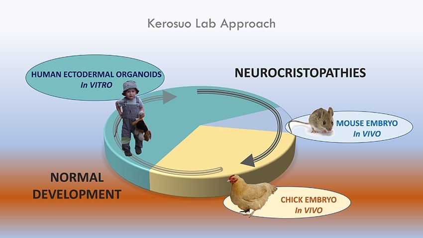
Due to their wide-ranging contributions to various tissues and organs, neural crest cells play a crucial role in embryonic development and organogenesis. Abnormalities in neural crest cell migration or differentiation can lead to several congenital disorders, such as neurocristopathies.
Bone Morphogenetic Protein 4 (BMP-4) is a growth factor that belongs to the transforming growth factor-beta (TGF-β) superfamily. It plays crucial roles in various biological processes, including embryonic development, cell growth, and differentiation. In the skeletal system, BMP-4 stimulates the formation of bone and cartilage by inducing the differentiation of mesenchymal stem cells into chondrocytes and osteoblasts. It also regulates the maintenance and repair of bones throughout life. An imbalance in BMP-4 signaling has been associated with several skeletal disorders, such as heterotopic ossification and osteoarthritis.
Homeodomain proteins are a group of transcription factors that play crucial roles in the development and differentiation of cells in animals and plants. They are characterized by the presence of a highly conserved DNA-binding domain called the homeodomain, which is typically about 60 amino acids long. The homeodomain consists of three helices, with the third helix responsible for recognizing and binding to specific DNA sequences.
Homeodomain proteins are involved in regulating gene expression during embryonic development, tissue maintenance, and organismal growth. They can act as activators or repressors of transcription, depending on the context and the presence of cofactors. Mutations in homeodomain proteins have been associated with various human diseases, including cancer, congenital abnormalities, and neurological disorders.
Some examples of homeodomain proteins include PAX6, which is essential for eye development, HOX genes, which are involved in body patterning, and NANOG, which plays a role in maintaining pluripotency in stem cells.
Germ layers refer to the primary layers of cells that form during embryonic development and give rise to the various tissues and organs in the body. In humans, there are three germ layers: the ectoderm, mesoderm, and endoderm. Each germ layer differentiates into distinct cell types and structures during the process of gastrulation. The ectoderm gives rise to the nervous system, sensory organs, and skin; the mesoderm forms muscles, bones, blood vessels, and the circulatory system; and the endoderm develops into the respiratory and digestive systems, including the lungs, liver, and pancreas.
"Xenopus" is not a medical term, but it is a genus of highly invasive aquatic frogs native to sub-Saharan Africa. They are often used in scientific research, particularly in developmental biology and genetics. The most commonly studied species is Xenopus laevis, also known as the African clawed frog.
In a medical context, Xenopus might be mentioned when discussing their use in research or as a model organism to study various biological processes or diseases.
The neural plate is a structure formed during the embryonic development of vertebrates. It is a thickened plate of ectodermal cells located on the dorsal surface of the developing embryo. The neural plate gives rise to the central nervous system, including the brain and spinal cord.
The process of neural plate formation begins with the specification of ectodermal cells into neural fated cells, a process that is regulated by various signaling molecules. Once specified, these cells undergo morphological changes, resulting in the thickening of the ectoderm to form the neural plate.
The neural plate then undergoes a series of folding movements, leading to the formation of the neural tube, which eventually develops into the brain and spinal cord. The edges of the neural plate, known as the neural folds, come together and fuse, forming a closed tube. Failure of the neural folds to fuse properly can result in neural tube defects, such as spina bifida.
Overall, the neural plate is a critical structure in the development of the nervous system in vertebrates, and its formation and subsequent development are tightly regulated by various genetic and environmental factors.
A blastula is a stage in the early development of many animals, including mammals. It is a hollow ball of cells that forms as a result of cleavage, which is the process of cell division during embryonic development. The blastula is typically characterized by the presence of a fluid-filled cavity called the blastocoel, which is surrounded by a single layer of cells known as the blastoderm.
In mammals, the blastula stage follows the morula stage, which is a solid mass of cells that results from cleavage of the fertilized egg. During further cell division and rearrangement, the cells in the morula become organized into an inner cell mass and an outer layer of cells, called the trophoblast. The inner cell mass will eventually give rise to the embryo proper, while the trophoblast will contribute to the formation of the placenta.
As the morula continues to divide and expand, it forms a cavity within the inner cell mass, which becomes the blastocoel. The single layer of cells surrounding the blastocoel is called the blastoderm. At this stage, the blastula is capable of further development through a process called gastrulation, during which the three germ layers of the embryo (ectoderm, mesoderm, and endoderm) are formed.
It's important to note that not all animals go through a blastula stage in their development. Some animals, such as insects and nematodes, have different patterns of early development that do not include a blastula stage.
"Xenopus laevis" is not a medical term itself, but it refers to a specific species of African clawed frog that is often used in scientific research, including biomedical and developmental studies. Therefore, its relevance to medicine comes from its role as a model organism in laboratories.
In a broader sense, Xenopus laevis has contributed significantly to various medical discoveries, such as the understanding of embryonic development, cell cycle regulation, and genetic research. For instance, the Nobel Prize in Physiology or Medicine was awarded in 1963 to John R. B. Gurdon and Sir Michael J. Bishop for their discoveries concerning the genetic mechanisms of organism development using Xenopus laevis as a model system.
Embryonic structures refer to the various parts and components that develop during the embryonic stage of prenatal development, which occurs from fertilization to the end of the 8th week of gestation. These structures include the primitive streak, notochord, neural tube, heart, somites, and limb buds, among others.
During this stage, the embryo undergoes rapid cell division, differentiation, and organization to form these structures, which will eventually develop into the various organs and systems of the human body. The embryonic structures are formed through a complex process of gene expression, signaling pathways, and interactions between cells and tissues.
Understanding the development of embryonic structures is crucial for understanding normal human development, as well as for identifying abnormalities or defects that may occur during this critical period. This knowledge can also inform medical interventions and treatments to address developmental issues and improve health outcomes for individuals throughout their lives.
The term "extremities" in a medical context refers to the most distant parts of the body, including the hands and feet (both fingers and toes), as well as the arms and legs. These are the farthest parts from the torso and head. Medical professionals may examine a patient's extremities for various reasons, such as checking circulation, assessing nerve function, or looking for injuries or abnormalities.
Fibroblast Growth Factor 8 (FGF-8) is a growth factor that belongs to the fibroblast growth factor family. It plays crucial roles in various biological processes, including embryonic development, tissue repair, and cancer progression. Specifically, FGF-8 has been implicated in the regulation of cell proliferation, differentiation, migration, and survival.
During embryonic development, FGF-8 is involved in the formation of the nervous system, limbs, and other organs. It acts as a signaling molecule that helps to establish patterns of gene expression and cell behavior during development. In tissue repair, FGF-8 can stimulate the proliferation and migration of cells involved in wound healing, such as fibroblasts and endothelial cells.
In cancer, FGF-8 has been shown to promote tumor growth, angiogenesis (the formation of new blood vessels), and metastasis. It can do this by activating signaling pathways that promote cell proliferation, survival, and migration. Overexpression of FGF-8 has been found in various types of cancer, including breast, lung, prostate, and ovarian cancer.
In summary, Fibroblast Growth Factor 8 (FGF-8) is a signaling molecule that plays important roles in embryonic development, tissue repair, and cancer progression by regulating cell proliferation, differentiation, migration, and survival.
Fibroblast Growth Factors (FGFs) are a family of growth factors that play crucial roles in various biological processes, including cell survival, proliferation, migration, and differentiation. They bind to specific tyrosine kinase receptors (FGFRs) on the cell surface, leading to intracellular signaling cascades that regulate gene expression and downstream cellular responses. FGFs are involved in embryonic development, tissue repair, and angiogenesis (the formation of new blood vessels). There are at least 22 distinct FGFs identified in humans, each with unique functions and patterns of expression. Some FGFs, like FGF1 and FGF2, have mitogenic effects on fibroblasts and other cell types, while others, such as FGF7 and FGF10, are essential for epithelial-mesenchymal interactions during organ development. Dysregulation of FGF signaling has been implicated in various pathological conditions, including cancer, fibrosis, and developmental disorders.
Gastrulation is a fundamental process in embryonic development, characterized by the transformation of a initially flat layer of cells called the blastula into a three-layered structure known as the gastrula. This complex series of cellular movements and rearrangements establishes the foundation for the formation of the three primary germ layers: the ectoderm, mesoderm, and endoderm. These germ layers further differentiate to give rise to all the diverse cell types and tissues in the developing organism, including the nervous system, muscles, bones, and internal organs.
The precise mechanisms of gastrulation vary among different animal groups; however, common features include:
1. Formation of a blastopore: A small indentation or opening that forms on the surface of the blastula, which eventually develops into the primitive gut or anus in the gastrula.
2. Invagination: The process by which cells at the blastopore fold inward and migrate towards the interior of the embryo, forming the endodermal layer.
3. Epiboly: A coordinated movement of cells that spreads over and encloses the yolk within the embryo, contributing to the formation of the ectodermal layer.
4. Delamination: The separation and migration of cells from the epiblast (the outer layer of the blastula) to form the mesodermal layer in between the ectoderm and endoderm.
Gastrulation is a critical period in embryonic development, as errors during this process can lead to severe congenital abnormalities or even embryonic lethality. A thorough understanding of gastrulation has important implications for regenerative medicine, stem cell research, and the study of evolutionary developmental biology (Evo-Devo).
Cell differentiation is the process by which a less specialized cell, or stem cell, becomes a more specialized cell type with specific functions and structures. This process involves changes in gene expression, which are regulated by various intracellular signaling pathways and transcription factors. Differentiation results in the development of distinct cell types that make up tissues and organs in multicellular organisms. It is a crucial aspect of embryonic development, tissue repair, and maintenance of homeostasis in the body.
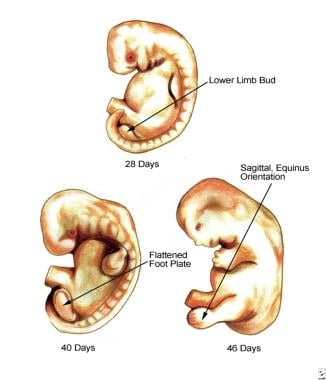
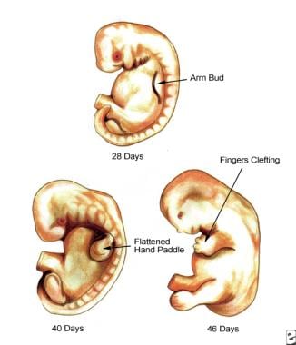
Limb buds are embryological structures that develop in the early stages of fetal growth and give rise to future limbs. In humans, they appear around the 4th week of gestation as thickenings on the sides of the body trunk. These buds consist of a core of mesenchymal tissue surrounded by ectoderm. The mesenchyme will later differentiate into bones, muscles, tendons, ligaments, and cartilages, while the ectoderm will form the skin and nervous tissues, including sensory organs in the limbs.
The development of limb buds is regulated by a complex interplay of genetic and molecular factors that control their outgrowth, patterning, and differentiation into specific limb components. Abnormalities during this process can lead to various congenital limb defects or deformations.
Transcription factors are proteins that play a crucial role in regulating gene expression by controlling the transcription of DNA to messenger RNA (mRNA). They function by binding to specific DNA sequences, known as response elements, located in the promoter region or enhancer regions of target genes. This binding can either activate or repress the initiation of transcription, depending on the properties and interactions of the particular transcription factor. Transcription factors often act as part of a complex network of regulatory proteins that determine the precise spatiotemporal patterns of gene expression during development, differentiation, and homeostasis in an organism.
Tissue transplantation is a medical procedure where tissues from one part of the body or from another individual's body are removed and implanted in a recipient to replace damaged, diseased, or missing tissues. The tissues may include skin, bone, tendons, ligaments, heart valves, corneas, or even entire organs such as hearts, lungs, livers, and kidneys.
The donor tissue must be compatible with the recipient's body to reduce the risk of rejection, which is the immune system attacking and destroying the transplanted tissue. This often requires matching certain proteins called human leukocyte antigens (HLAs) found on the surface of most cells in the body.
Tissue transplantation can significantly improve a patient's quality of life or, in some cases, save their life. However, it does carry risks such as infection, bleeding, and rejection, which require careful monitoring and management.
I believe there may be some confusion in your question. "Quail" is typically used to refer to a group of small birds that belong to the family Phasianidae and the subfamily Perdicinae. There is no established medical definition for "quail."
However, if you're referring to the verb "to quail," it means to shrink back, draw back, or cower, often due to fear or intimidation. In a medical context, this term could be used metaphorically to describe a patient's psychological response to a threatening situation, such as receiving a difficult diagnosis. But again, "quail" itself is not a medical term.
Signal transduction is the process by which a cell converts an extracellular signal, such as a hormone or neurotransmitter, into an intracellular response. This involves a series of molecular events that transmit the signal from the cell surface to the interior of the cell, ultimately resulting in changes in gene expression, protein activity, or metabolism.
The process typically begins with the binding of the extracellular signal to a receptor located on the cell membrane. This binding event activates the receptor, which then triggers a cascade of intracellular signaling molecules, such as second messengers, protein kinases, and ion channels. These molecules amplify and propagate the signal, ultimately leading to the activation or inhibition of specific cellular responses.
Signal transduction pathways are highly regulated and can be modulated by various factors, including other signaling molecules, post-translational modifications, and feedback mechanisms. Dysregulation of these pathways has been implicated in a variety of diseases, including cancer, diabetes, and neurological disorders.
The notochord is a flexible, rod-shaped structure that is present in the embryos of chordates, including humans. It is composed of cells called chordocytes and is surrounded by a sheath. The notochord runs along the length of the body, providing support and flexibility. In human embryos, the notochord eventually becomes part of the discs between the vertebrae in the spine. An abnormal or absent notochord can lead to developmental problems with the spine and nervous system.
Wnt proteins are a family of secreted signaling molecules that play crucial roles in the regulation of fundamental biological processes, including cell proliferation, differentiation, migration, and survival. They were first discovered in 1982 through genetic studies in Drosophila melanogaster (fruit flies) and have since been found to be highly conserved across various species, from invertebrates to humans.
Wnt proteins exert their effects by binding to specific receptors on the target cell surface, leading to the activation of several intracellular signaling pathways:
1. Canonical Wnt/β-catenin pathway: In the absence of Wnt ligands, β-catenin is continuously degraded by a destruction complex consisting of Axin, APC (Adenomatous polyposis coli), and GSK3β (Glycogen synthase kinase 3 beta). When Wnt proteins bind to their receptors Frizzled and LRP5/6, the formation of a "signalosome" complex leads to the inhibition of the destruction complex, allowing β-catenin to accumulate in the cytoplasm and translocate into the nucleus. Here, it interacts with TCF/LEF (T-cell factor/lymphoid enhancer-binding factor) transcription factors to regulate the expression of target genes involved in cell proliferation, differentiation, and survival.
2. Non-canonical Wnt pathways: These include the Wnt/Ca^2+^ pathway and the planar cell polarity (PCP) pathway. In the Wnt/Ca^2+^ pathway, Wnt ligands bind to Frizzled receptors and activate heterotrimeric G proteins, leading to an increase in intracellular Ca^2+^ levels and activation of downstream targets such as protein kinase C (PKC) and calcium/calmodulin-dependent protein kinase II (CAMKII). These signaling events ultimately regulate cell movement, adhesion, and gene expression. In the PCP pathway, Wnt ligands bind to Frizzled receptors and coreceptor complexes containing Ror2 or Ryk, leading to activation of small GTPases such as RhoA and Rac1, which control cytoskeletal organization and cell polarity.
Dysregulation of Wnt signaling has been implicated in various human diseases, including cancer, developmental disorders, and degenerative conditions. In cancer, aberrant activation of the canonical Wnt/β-catenin pathway contributes to tumor initiation, progression, and metastasis by promoting cell proliferation, survival, and epithelial-mesenchymal transition (EMT). Inhibitors targeting different components of the Wnt signaling pathway are currently being developed as potential therapeutic strategies for cancer treatment.
The crystalline lens is a biconvex transparent structure in the eye that helps to refract (bend) light rays and focus them onto the retina. It is located behind the iris and pupil and is suspended by small fibers called zonules that connect it to the ciliary body. The lens can change its shape to accommodate and focus on objects at different distances, a process known as accommodation. With age, the lens may become cloudy or opaque, leading to cataracts.
Paired box (PAX) transcription factors are a group of proteins that regulate gene expression during embryonic development and in some adult tissues. They are characterized by the presence of a paired box domain, a conserved DNA-binding motif that recognizes specific DNA sequences. PAX proteins play crucial roles in various developmental processes, such as the formation of the nervous system, eyes, and pancreas. Dysregulation of PAX genes has been implicated in several human diseases, including cancer.
OTX (Orthodenticle homeobox) transcription factors are a family of proteins that regulate gene expression during embryonic development, particularly in the eye, forebrain, and midbrain. They play crucial roles in the development and differentiation of these tissues, including the specification of eye field identity, the determination of dorsoventral patterning in the neural tube, and the regulation of neurogenesis.
OTX transcription factors contain a highly conserved DNA-binding domain called the homeodomain, which allows them to recognize and bind to specific DNA sequences. In humans, there are four known OTX transcription factors (OTX1, OTX2, OTX3, and CRX), each with distinct expression patterns and functions.
Mutations in OTX genes have been associated with various developmental disorders, such as microphthalmia, anophthalmia, and severe eye malformations, highlighting their importance in normal eye development. Additionally, OTX transcription factors have also been implicated in the pathogenesis of certain cancers, including medulloblastoma and retinoblastoma.
Homeobox genes are a specific class of genes that play a crucial role in the development and regulation of an organism's body plan. They encode transcription factors, which are proteins that regulate the expression of other genes. The homeobox region within these genes contains a highly conserved sequence of about 180 base pairs that encodes a DNA-binding domain called the homeodomain. This domain is responsible for recognizing and binding to specific DNA sequences, thereby controlling the transcription of target genes.
Homeobox genes are particularly important during embryonic development, where they help establish the anterior-posterior axis and regulate the development of various organs and body segments. They also play a role in maintaining adult tissue homeostasis and have been implicated in certain diseases, including cancer. Mutations in homeobox genes can lead to developmental abnormalities and congenital disorders.
Some examples of homeobox gene families include HOX genes, PAX genes, and NKX genes, among others. These genes are highly conserved across species, indicating their fundamental role in the development and regulation of body plans throughout the animal kingdom.
Zebrafish proteins refer to the diverse range of protein molecules that are produced by the organism Danio rerio, commonly known as the zebrafish. These proteins play crucial roles in various biological processes such as growth, development, reproduction, and response to environmental stimuli. They are involved in cellular functions like enzymatic reactions, signal transduction, structural support, and regulation of gene expression.
Zebrafish is a popular model organism in biomedical research due to its genetic similarity with humans, rapid development, and transparent embryos that allow for easy observation of biological processes. As a result, the study of zebrafish proteins has contributed significantly to our understanding of protein function, structure, and interaction in both zebrafish and human systems.
Some examples of zebrafish proteins include:
* Transcription factors that regulate gene expression during development
* Enzymes involved in metabolic pathways
* Structural proteins that provide support to cells and tissues
* Receptors and signaling molecules that mediate communication between cells
* Heat shock proteins that assist in protein folding and protect against stress
The analysis of zebrafish proteins can be performed using various techniques, including biochemical assays, mass spectrometry, protein crystallography, and computational modeling. These methods help researchers to identify, characterize, and understand the functions of individual proteins and their interactions within complex networks.
A nodal protein, in the context of molecular biology and genetics, refers to a protein that plays a role in signal transmission within a cell at a node or junction point of a signaling pathway. These proteins are often involved in regulatory processes, such as activating or inhibiting downstream effectors in response to specific signals received by the cell. Nodal proteins can be activated or deactivated through various mechanisms, including phosphorylation, ubiquitination, and interactions with other signaling molecules.
In a more specific context, nodal proteins are also known as nodal factors, which are members of the transforming growth factor-beta (TGF-β) superfamily of signaling molecules that play critical roles in embryonic development and tissue homeostasis. Nodal is a secreted protein that acts as a morphogen, inducing different cellular responses depending on its concentration gradient. It is involved in establishing left-right asymmetry during embryonic development and regulates various processes such as cell proliferation, differentiation, and apoptosis.
In summary, nodal proteins can refer to any protein that functions at a node or junction point of a signaling pathway, but they are also specifically known as nodal factors, which are TGF-β superfamily members involved in embryonic development and tissue homeostasis.
The branchial region, also known as the pharyngeal region or viscerocranium, is a term used in human anatomy to refer to the area of the developing embryo that gives rise to structures derived from the branchial (or pharyngeal) arches. The branchial arches are a series of paired, rod-like structures that appear early in embryonic development and give rise to various head and neck structures, including the bones and muscles of the face, jaws, and neck, as well as the associated nerves, blood vessels, and connective tissues.
The branchial region is divided into several subregions, each corresponding to a specific branchial arch. The first branchial arch gives rise to structures such as the mandible (lower jaw), maxilla (upper jaw), and muscles of mastication (chewing). The second branchial arch forms the stapes and styloid process in the ear, as well as some neck muscles. The third and fourth branchial arches contribute to the formation of the larynx, thyroid cartilage, and other structures in the neck.
Abnormalities in the development of the branchial region can lead to a variety of congenital defects, such as cleft palate, micrognathia (small jaw), and branchial cysts or sinuses. These conditions may require surgical intervention to correct.
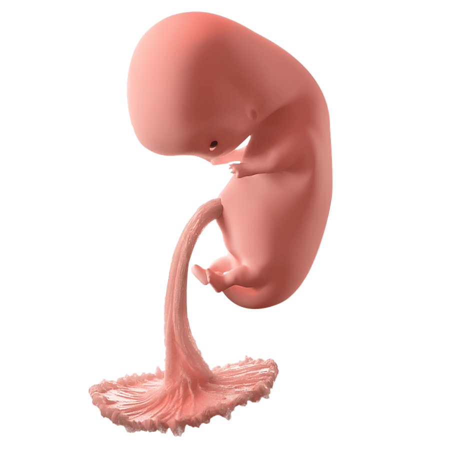
Embryonic and fetal development is the process of growth and development that occurs from fertilization of the egg (conception) to birth. The terms "embryo" and "fetus" are used to describe different stages of this development:
* Embryonic development: This stage begins at fertilization and continues until the end of the 8th week of pregnancy. During this time, the fertilized egg (zygote) divides and forms a blastocyst, which implants in the uterus and begins to develop into a complex structure called an embryo. The embryo consists of three layers of cells that will eventually form all of the organs and tissues of the body. During this stage, the basic structures of the body, including the nervous system, heart, and gastrointestinal tract, begin to form.
* Fetal development: This stage begins at the end of the 8th week of pregnancy and continues until birth. During this time, the embryo is called a fetus, and it grows and develops rapidly. The organs and tissues that were formed during the embryonic stage continue to mature and become more complex. The fetus also begins to move and kick, and it can hear and respond to sounds from outside the womb.
Overall, embryonic and fetal development is a complex and highly regulated process that involves the coordinated growth and differentiation of cells and tissues. It is a critical period of development that lays the foundation for the health and well-being of the individual throughout their life.
A chimera, in the context of medicine and biology, is a single organism that is composed of cells with different genetics. This can occur naturally in some situations, such as when fraternal twins do not fully separate in utero and end up sharing some organs or tissues. The term "chimera" can also refer to an organism that contains cells from two different species, which can happen in certain types of genetic research or medical treatments. For example, a patient's cells might be genetically modified in a lab and then introduced into their body to treat a disease; if some of these modified cells mix with the patient's original cells, the result could be a chimera.
It's worth noting that the term "chimera" comes from Greek mythology, where it referred to a fire-breathing monster that was part lion, part goat, and part snake. In modern scientific usage, the term has a specific technical meaning related to genetics and organisms, but it may still evoke images of fantastical creatures for some people.
Hedgehog proteins are a group of signaling molecules that play crucial roles in the development and regulation of various biological processes in animals. They are named after the hedgehog mutant fruit flies, which have spiky bristles due to defects in this pathway. These proteins are involved in cell growth, differentiation, and tissue regeneration. They exert their effects by binding to specific receptors on the surface of target cells, leading to a cascade of intracellular signaling events that ultimately influence gene expression and cell behavior.
There are three main types of Hedgehog proteins in mammals: Sonic hedgehog (Shh), Indian hedgehog (Ihh), and Desert hedgehog (Dhh). These protecules undergo post-translational modifications, including cleavage and lipid modification, which are essential for their activity. Dysregulation of Hedgehog signaling has been implicated in various diseases, including cancer, developmental abnormalities, and degenerative disorders.
Embryonic development is the series of growth and developmental stages that occur during the formation and early growth of the embryo. In humans, this stage begins at fertilization (when the sperm and egg cell combine) and continues until the end of the 8th week of pregnancy. During this time, the fertilized egg (now called a zygote) divides and forms a blastocyst, which then implants into the uterus. The cells in the blastocyst begin to differentiate and form the three germ layers: the ectoderm, mesoderm, and endoderm. These germ layers will eventually give rise to all of the different tissues and organs in the body.
Embryonic development is a complex and highly regulated process that involves the coordinated interaction of genetic and environmental factors. It is characterized by rapid cell division, migration, and differentiation, as well as programmed cell death (apoptosis) and tissue remodeling. Abnormalities in embryonic development can lead to birth defects or other developmental disorders.
It's important to note that the term "embryo" is used to describe the developing organism from fertilization until the end of the 8th week of pregnancy in humans, after which it is called a fetus.
In medical terms, the "head" is the uppermost part of the human body that contains the brain, skull, face, eyes, nose, mouth, and ears. It is connected to the rest of the body by the neck and is responsible for many vital functions such as sight, hearing, smell, taste, touch, and thought processing. The head also plays a crucial role in maintaining balance, speech, and eating.
'Cell lineage' is a term used in biology and medicine to describe the developmental history or relationship of a cell or group of cells to other cells, tracing back to the original progenitor or stem cell. It refers to the series of cell divisions and differentiation events that give rise to specific types of cells in an organism over time.
In simpler terms, cell lineage is like a family tree for cells, showing how they are related to each other through a chain of cell division and specialization events. This concept is important in understanding the development, growth, and maintenance of tissues and organs in living beings.
A mammalian embryo is the developing offspring of a mammal, from the time of implantation of the fertilized egg (blastocyst) in the uterus until the end of the eighth week of gestation. During this period, the embryo undergoes rapid cell division and organ differentiation to form a complex structure with all the major organs and systems in place. This stage is followed by fetal development, which continues until birth. The study of mammalian embryos is important for understanding human development, evolution, and reproductive biology.
The Central Nervous System (CNS) is the part of the nervous system that consists of the brain and spinal cord. It is called the "central" system because it receives information from, and sends information to, the rest of the body through peripheral nerves, which make up the Peripheral Nervous System (PNS).
The CNS is responsible for processing sensory information, controlling motor functions, and regulating various autonomic processes like heart rate, respiration, and digestion. The brain, as the command center of the CNS, interprets sensory stimuli, formulates thoughts, and initiates actions. The spinal cord serves as a conduit for nerve impulses traveling to and from the brain and the rest of the body.
The CNS is protected by several structures, including the skull (which houses the brain) and the vertebral column (which surrounds and protects the spinal cord). Despite these protective measures, the CNS remains vulnerable to injury and disease, which can have severe consequences due to its crucial role in controlling essential bodily functions.
I'm sorry for any confusion, but "Triturus" is not a medical term. It is actually the genus name for newts that are native to Europe. These newts belong to the family Salamandridae and are known for their ability to regenerate various body parts. If you have any questions related to medical terminology or healthcare, I would be happy to try and help answer those for you.
Molecular sequence data refers to the specific arrangement of molecules, most commonly nucleotides in DNA or RNA, or amino acids in proteins, that make up a biological macromolecule. This data is generated through laboratory techniques such as sequencing, and provides information about the exact order of the constituent molecules. This data is crucial in various fields of biology, including genetics, evolution, and molecular biology, allowing for comparisons between different organisms, identification of genetic variations, and studies of gene function and regulation.
I apologize for any confusion, but "Strongylocentrotus purpuratus" is not a medical term. It is the scientific name for a species of sea urchin that is often used in biological research. The purple sea urchin, as it is commonly known, is native to the Pacific coast of North America and is widely studied in developmental biology, genetics, and evolution due to its simple and well-understood anatomy.
Bone morphogenetic protein (BMP) receptors are a type of cell surface receptor that play a crucial role in bone and cartilage development, as well as in other biological processes such as wound healing and embryonic development. These receptors are part of the TGF-β (transforming growth factor-beta) superfamily and are composed of two types of subunits: type I and type II.
Type I BMP receptors include BMPR1A, BMPR1B, and ACTRIIA/B. Type II BMP receptors include BMPR2, ACVR2A, and ACVR2B. When BMPs bind to these receptors, they initiate a signaling cascade that leads to the activation of downstream targets involved in bone formation, cartilage development, and other processes.
Mutations in BMP receptor genes have been associated with various genetic disorders, including fibrodysplasia ossificans progressiva (FOP), a rare condition characterized by the abnormal formation of bone in muscles, tendons, and ligaments. Additionally, dysregulation of BMP signaling has been implicated in diseases such as cancer, where it can contribute to tumor growth and metastasis.
SOXB1 transcription factors are a subgroup of the SOX (SRY-related HMG box) family of transcription factors, which are characterized by a conserved high mobility group (HMG) box DNA-binding domain. The SOXB1 subfamily includes SOX1, SOX2, and SOX3, which play crucial roles during embryonic development and in the maintenance of stem cells. They regulate gene expression by binding to specific DNA sequences and interacting with other transcription factors and cofactors. SOXB1 proteins have been implicated in various biological processes, such as neurogenesis, eye development, and sex determination. Dysregulation of SOXB1 transcription factors has been associated with several human diseases, including cancer.
Trans-activators are proteins that increase the transcriptional activity of a gene or a set of genes. They do this by binding to specific DNA sequences and interacting with the transcription machinery, thereby enhancing the recruitment and assembly of the complexes needed for transcription. In some cases, trans-activators can also modulate the chromatin structure to make the template more accessible to the transcription machinery.
In the context of HIV (Human Immunodeficiency Virus) infection, the term "trans-activator" is often used specifically to refer to the Tat protein. The Tat protein is a viral regulatory protein that plays a critical role in the replication of HIV by activating the transcription of the viral genome. It does this by binding to a specific RNA structure called the Trans-Activation Response Element (TAR) located at the 5' end of all nascent HIV transcripts, and recruiting cellular cofactors that enhance the processivity and efficiency of RNA polymerase II, leading to increased viral gene expression.
The epidermis is the outermost layer of the skin, composed mainly of stratified squamous epithelium. It forms a protective barrier that prevents water loss and inhibits the entry of microorganisms. The epidermis contains no blood vessels, and its cells are nourished by diffusion from the underlying dermis. The bottom-most layer of the epidermis, called the stratum basale, is responsible for generating new skin cells that eventually move up to replace dead cells on the surface. This process of cell turnover takes about 28 days in adults.
The most superficial part of the epidermis consists of dead cells called squames, which are constantly shed and replaced. The exact rate at which this happens varies depending on location; for example, it's faster on the palms and soles than elsewhere. Melanocytes, the pigment-producing cells, are also located in the epidermis, specifically within the stratum basale layer.
In summary, the epidermis is a vital part of our integumentary system, providing not only physical protection but also playing a crucial role in immunity and sensory perception through touch receptors called Pacinian corpuscles.
Embryonic organizers are specialized cells or tissues in developing embryos that provide critical signals to guide the organization and development of surrounding cells and tissues. They play a crucial role in establishing the body plan and patterning of the organism during embryogenesis. A well-known example is the Spemann-Mangold organizer, first described in amphibians, which induces the formation of the neural tissue and organizes the surrounding tissues to form the body axis. Embryonic organizers have been identified in various animal models, including mammals, birds, and fish, and they are essential for normal embryonic development.
Somites are transient, segmentally repeated embryonic structures that form along the anterior-posterior body axis during vertebrate development. They are derived from the paraxial mesoderm and give rise to various tissues, including the sclerotome (which forms the vertebrae and ribs), myotome (which forms the skeletal muscles of the back and limbs), and dermatome (which forms the dermis of the skin).
Each somite is a block-like structure that is arranged in a repeating pattern along the notochord, which is a flexible rod-like structure that provides mechanical support to the developing embryo. The formation of somites is a critical step in the development of the vertebrate body plan, as they help to establish the segmental organization of the musculoskeletal system and contribute to the formation of other important structures such as the dermis and the circulatory system.
The process of somitogenesis, or the formation of somites, is a highly regulated and coordinated event that involves the interaction of various signaling molecules and genetic pathways. Defects in somite formation can lead to a range of developmental abnormalities, including spinal deformities, muscle weakness, and skin defects.
'Drosophila proteins' refer to the proteins that are expressed in the fruit fly, Drosophila melanogaster. This organism is a widely used model system in genetics, developmental biology, and molecular biology research. The study of Drosophila proteins has contributed significantly to our understanding of various biological processes, including gene regulation, cell signaling, development, and aging.
Some examples of well-studied Drosophila proteins include:
1. HSP70 (Heat Shock Protein 70): A chaperone protein involved in protein folding and protection from stress conditions.
2. TUBULIN: A structural protein that forms microtubules, important for cell division and intracellular transport.
3. ACTIN: A cytoskeletal protein involved in muscle contraction, cell motility, and maintenance of cell shape.
4. BETA-GALACTOSIDASE (LACZ): A reporter protein often used to monitor gene expression patterns in transgenic flies.
5. ENDOGLIN: A protein involved in the development of blood vessels during embryogenesis.
6. P53: A tumor suppressor protein that plays a crucial role in preventing cancer by regulating cell growth and division.
7. JUN-KINASE (JNK): A signaling protein involved in stress response, apoptosis, and developmental processes.
8. DECAPENTAPLEGIC (DPP): A member of the TGF-β (Transforming Growth Factor Beta) superfamily, playing essential roles in embryonic development and tissue homeostasis.
These proteins are often studied using various techniques such as biochemistry, genetics, molecular biology, and structural biology to understand their functions, interactions, and regulation within the cell.
"Drosophila" is a genus of small flies, also known as fruit flies. The most common species used in scientific research is "Drosophila melanogaster," which has been a valuable model organism for many areas of biological and medical research, including genetics, developmental biology, neurobiology, and aging.
The use of Drosophila as a model organism has led to numerous important discoveries in genetics and molecular biology, such as the identification of genes that are associated with human diseases like cancer, Parkinson's disease, and obesity. The short reproductive cycle, large number of offspring, and ease of genetic manipulation make Drosophila a powerful tool for studying complex biological processes.
Fibroblast Growth Factor 4 (FGF4) is a growth factor that belongs to the fibroblast growth factor family. It plays a crucial role in various biological processes, including embryonic development, cell survival, proliferation, and differentiation. Specifically, FGF4 has been implicated in the development of the musculoskeletal system, where it helps regulate the growth and patterning of limbs and bones.
FGF4 exerts its effects by binding to specific receptors on the surface of target cells, known as fibroblast growth factor receptors (FGFRs). This interaction triggers a cascade of intracellular signaling events that ultimately lead to changes in gene expression and cell behavior.
In addition to its role in development, FGF4 has also been implicated in various pathological processes, including cancer. For example, elevated levels of FGF4 have been observed in certain types of tumors, where it may contribute to tumor growth and progression by promoting the survival and proliferation of cancer cells.
High Mobility Group Box (HMGB) proteins are a family of nuclear proteins that are highly conserved and expressed in eukaryotic cells. They play a crucial role in the regulation of gene expression, DNA repair, and maintenance of nucleosome structure. HMGB proteins contain two positively charged DNA-binding domains (HMG boxes) and a negatively charged acidic tail. These proteins can bind to DNA in a variety of ways, bending it and altering its structure, which in turn affects the binding of other proteins and the transcriptional activity of genes. HMGB proteins can also be released from cells under conditions of stress or injury, where they act as damage-associated molecular patterns (DAMPs) and contribute to the inflammatory response.
DNA-binding proteins are a type of protein that have the ability to bind to DNA (deoxyribonucleic acid), the genetic material of organisms. These proteins play crucial roles in various biological processes, such as regulation of gene expression, DNA replication, repair and recombination.
The binding of DNA-binding proteins to specific DNA sequences is mediated by non-covalent interactions, including electrostatic, hydrogen bonding, and van der Waals forces. The specificity of binding is determined by the recognition of particular nucleotide sequences or structural features of the DNA molecule.
DNA-binding proteins can be classified into several categories based on their structure and function, such as transcription factors, histones, and restriction enzymes. Transcription factors are a major class of DNA-binding proteins that regulate gene expression by binding to specific DNA sequences in the promoter region of genes and recruiting other proteins to modulate transcription. Histones are DNA-binding proteins that package DNA into nucleosomes, the basic unit of chromatin structure. Restriction enzymes are DNA-binding proteins that recognize and cleave specific DNA sequences, and are widely used in molecular biology research and biotechnology applications.
A zebrafish is a freshwater fish species belonging to the family Cyprinidae and the genus Danio. Its name is derived from its distinctive striped pattern that resembles a zebra's. Zebrafish are often used as model organisms in scientific research, particularly in developmental biology, genetics, and toxicology studies. They have a high fecundity rate, transparent embryos, and a rapid development process, making them an ideal choice for researchers. However, it is important to note that providing a medical definition for zebrafish may not be entirely accurate or relevant since they are primarily used in biological research rather than clinical medicine.
Chordata is a phylum in the animal kingdom that includes animals with a notochord, dorsal hollow nerve cord, pharyngeal gill slits, and a post-anal tail at some point during their development. Nonvertebrate Chordates include two classes: Tunicata (sea squirts and salps) and Cephalochordata (lancelets). These animals do not have a backbone or vertebral column, which is why they are considered nonvertebrate. Despite the lack of a vertebral column, these animals share other common characteristics with Vertebrates, such as a circulatory system and a complex nervous system.
Activins are a type of protein that belongs to the transforming growth factor-beta (TGF-β) superfamily. They are produced and released by various cells in the body, including those in the ovaries, testes, pituitary gland, and other tissues. Activins play important roles in regulating several biological processes, such as cell growth, differentiation, and apoptosis (programmed cell death).
Activins bind to specific receptors on the surface of cells, leading to the activation of intracellular signaling pathways that control gene expression. They are particularly well-known for their role in reproductive biology, where they help regulate follicle stimulation and hormone production in the ovaries and testes. Activins also have been implicated in various disease processes, including cancer, fibrosis, and inflammation.
There are three main isoforms of activin in humans: activin A, activin B, and inhibin A. While activins and inhibins share similar structures and functions, they have opposite effects on the activity of the pituitary gland. Activins stimulate the production of follicle-stimulating hormone (FSH), while inhibins suppress it. This delicate balance between activins and inhibins helps regulate reproductive function and other physiological processes in the body.
A blastocyst is a stage in the early development of a fertilized egg, or embryo, in mammals. It occurs about 5-6 days after fertilization and consists of an outer layer of cells called trophoblasts, which will eventually form the placenta, and an inner cell mass, which will give rise to the fetus. The blastocyst is characterized by a fluid-filled cavity called the blastocoel. This stage is critical for the implantation of the embryo into the uterine lining.
MSX1 (Homeobox protein MSX-1) is a transcription factor that belongs to the muscle segment homebox gene family, also known as the msh homeobox genes. These genes are involved in the development and differentiation of various tissues, including muscle, bone, and neural crest derivatives.
MSX1 plays crucial roles during embryonic development, such as regulating cell proliferation, differentiation, and apoptosis. It is widely expressed in the developing embryo, particularly in the oral ectoderm, neural crest, and mesenchyme. In the oral region, MSX1 helps control tooth development by interacting with other transcription factors and signaling molecules.
As a transcription factor, MSX1 binds to specific DNA sequences called homeobox response elements (HREs) in the promoter regions of its target genes. This binding either activates or represses gene expression, depending on the context and interacting partners. Dysregulation of MSX1 has been implicated in various developmental disorders and diseases, such as tooth agenesis, cleft lip/palate, and cancer.
A base sequence in the context of molecular biology refers to the specific order of nucleotides in a DNA or RNA molecule. In DNA, these nucleotides are adenine (A), guanine (G), cytosine (C), and thymine (T). In RNA, uracil (U) takes the place of thymine. The base sequence contains genetic information that is transcribed into RNA and ultimately translated into proteins. It is the exact order of these bases that determines the genetic code and thus the function of the DNA or RNA molecule.
The blastoderm is the layer of cells that forms on the surface of a developing embryo, during the blastula stage of embryonic development. In mammals, this layer of cells is also known as the epiblast. The blastoderm is responsible for giving rise to all of the tissues and organs of the developing organism. It is formed by the cleavage of the fertilized egg, or zygote, and is typically a single layer of cells that surrounds a fluid-filled cavity called the blastocoel. The blastoderm plays a critical role in the early stages of embryonic development, and any disruptions to its formation or function can lead to developmental abnormalities or death of the embryo.
Fetal proteins are a type of proteins that are produced by the fetus during pregnancy and can be detected in various biological samples, such as amniotic fluid or maternal blood. These proteins can provide valuable information about the health and development of the fetus. One commonly studied fetal protein is human chorionic gonadotropin (hCG), which is produced by the placenta and can be used as a marker for pregnancy and to detect potential complications, such as Down syndrome or spinal cord defects. Other examples of fetal proteins include alpha-fetoprotein (AFP) and human placental lactogen (hPL).
Proteins are complex, large molecules that play critical roles in the body's functions. They are made up of amino acids, which are organic compounds that are the building blocks of proteins. Proteins are required for the structure, function, and regulation of the body's tissues and organs. They are essential for the growth, repair, and maintenance of body tissues, and they play a crucial role in many biological processes, including metabolism, immune response, and cellular signaling. Proteins can be classified into different types based on their structure and function, such as enzymes, hormones, antibodies, and structural proteins. They are found in various foods, especially animal-derived products like meat, dairy, and eggs, as well as plant-based sources like beans, nuts, and grains.
An amino acid sequence is the specific order of amino acids in a protein or peptide molecule, formed by the linking of the amino group (-NH2) of one amino acid to the carboxyl group (-COOH) of another amino acid through a peptide bond. The sequence is determined by the genetic code and is unique to each type of protein or peptide. It plays a crucial role in determining the three-dimensional structure and function of proteins.
Repressor proteins are a type of regulatory protein in molecular biology that suppress the transcription of specific genes into messenger RNA (mRNA) by binding to DNA. They function as part of gene regulation processes, often working in conjunction with an operator region and a promoter region within the DNA molecule. Repressor proteins can be activated or deactivated by various signals, allowing for precise control over gene expression in response to changing cellular conditions.
There are two main types of repressor proteins:
1. DNA-binding repressors: These directly bind to specific DNA sequences (operator regions) near the target gene and prevent RNA polymerase from transcribing the gene into mRNA.
2. Allosteric repressors: These bind to effector molecules, which then cause a conformational change in the repressor protein, enabling it to bind to DNA and inhibit transcription.
Repressor proteins play crucial roles in various biological processes, such as development, metabolism, and stress response, by controlling gene expression patterns in cells.
The rhombencephalon is a term used in the field of neuroanatomy, which refers to the most posterior region of the developing brain during embryonic development. It is also known as the hindbrain and it gives rise to several important structures in the adult brain.
More specifically, the rhombencephalon can be further divided into two main parts: the metencephalon and the myelencephalon. The metencephalon eventually develops into the pons and cerebellum, while the myelencephalon becomes the medulla oblongata.
The rhombencephalon plays a crucial role in several critical functions of the nervous system, including regulating heart rate and respiration, maintaining balance and posture, and coordinating motor movements. Defects or abnormalities in the development of the rhombencephalon can lead to various neurological disorders, such as cerebellar hypoplasia, Chiari malformation, and certain forms of brainstem tumors.
The Neural Tube is a structure that forms during the development of an embryo and eventually becomes the brain, spinal cord, and other parts of the nervous system. It is a narrow channel that runs along the back of the embryo, forming from the ectoderm (one of the three germ layers) and closing around the 23rd or 26th day after conception. Defects in the closure of the neural tube can lead to conditions such as spina bifida and anencephaly.
Congenital limb deformities refer to abnormalities in the structure, position, or function of the arms or legs that are present at birth. These deformities can vary greatly in severity and may affect any part of the limb, including the bones, muscles, joints, and nerves.
Congenital limb deformities can be caused by genetic factors, exposure to certain medications or chemicals during pregnancy, or other environmental factors. Some common types of congenital limb deformities include:
1. Clubfoot: A condition in which the foot is twisted out of shape, making it difficult to walk normally.
2. Polydactyly: A condition in which a person is born with extra fingers or toes.
3. Radial clubhand: A rare condition in which the radius bone in the forearm is missing or underdeveloped, causing the hand to turn inward and the wrist to bend.
4. Amniotic band syndrome: A condition in which strands of the amniotic sac wrap around a developing limb, restricting its growth and leading to deformities.
5. Agenesis: A condition in which a limb or part of a limb is missing at birth.
Treatment for congenital limb deformities may include surgery, bracing, physical therapy, or other interventions depending on the severity and nature of the deformity. In some cases, early intervention and treatment can help to improve function and reduce the impact of the deformity on a person's daily life.
Wnt1 protein is a member of the Wnt family, which is a group of secreted signaling proteins that play crucial roles in embryonic development and tissue homeostasis in adults. Specifically, Wnt1 is a highly conserved gene that encodes a glycoprotein with a molecular weight of approximately 40 kDa. It is primarily expressed in the developing nervous system, where it functions as a key regulator of neural crest cell migration and differentiation during embryogenesis.
Wnt1 protein mediates its effects by binding to Frizzled receptors on the surface of target cells, leading to the activation of several intracellular signaling pathways, including the canonical Wnt/β-catenin pathway and non-canonical Wnt/planar cell polarity (PCP) pathway. In the canonical pathway, Wnt1 protein stabilizes β-catenin, which then translocates to the nucleus and interacts with TCF/LEF transcription factors to regulate gene expression.
Dysregulation of Wnt1 signaling has been implicated in several human diseases, including cancer. For example, aberrant activation of the Wnt/β-catenin pathway by Wnt1 protein has been observed in various types of tumors, such as medulloblastomas and breast cancers, leading to uncontrolled cell proliferation and tumor growth. Therefore, understanding the molecular mechanisms underlying Wnt1 signaling is essential for developing novel therapeutic strategies for treating these diseases.
Biological models, also known as physiological models or organismal models, are simplified representations of biological systems, processes, or mechanisms that are used to understand and explain the underlying principles and relationships. These models can be theoretical (conceptual or mathematical) or physical (such as anatomical models, cell cultures, or animal models). They are widely used in biomedical research to study various phenomena, including disease pathophysiology, drug action, and therapeutic interventions.
Examples of biological models include:
1. Mathematical models: These use mathematical equations and formulas to describe complex biological systems or processes, such as population dynamics, metabolic pathways, or gene regulation networks. They can help predict the behavior of these systems under different conditions and test hypotheses about their underlying mechanisms.
2. Cell cultures: These are collections of cells grown in a controlled environment, typically in a laboratory dish or flask. They can be used to study cellular processes, such as signal transduction, gene expression, or metabolism, and to test the effects of drugs or other treatments on these processes.
3. Animal models: These are living organisms, usually vertebrates like mice, rats, or non-human primates, that are used to study various aspects of human biology and disease. They can provide valuable insights into the pathophysiology of diseases, the mechanisms of drug action, and the safety and efficacy of new therapies.
4. Anatomical models: These are physical representations of biological structures or systems, such as plastic models of organs or tissues, that can be used for educational purposes or to plan surgical procedures. They can also serve as a basis for developing more sophisticated models, such as computer simulations or 3D-printed replicas.
Overall, biological models play a crucial role in advancing our understanding of biology and medicine, helping to identify new targets for therapeutic intervention, develop novel drugs and treatments, and improve human health.
Culture techniques are methods used in microbiology to grow and multiply microorganisms, such as bacteria, fungi, or viruses, in a controlled laboratory environment. These techniques allow for the isolation, identification, and study of specific microorganisms, which is essential for diagnostic purposes, research, and development of medical treatments.
The most common culture technique involves inoculating a sterile growth medium with a sample suspected to contain microorganisms. The growth medium can be solid or liquid and contains nutrients that support the growth of the microorganisms. Common solid growth media include agar plates, while liquid growth media are used for broth cultures.
Once inoculated, the growth medium is incubated at a temperature that favors the growth of the microorganisms being studied. During incubation, the microorganisms multiply and form visible colonies on the solid growth medium or turbid growth in the liquid growth medium. The size, shape, color, and other characteristics of the colonies can provide important clues about the identity of the microorganism.
Other culture techniques include selective and differential media, which are designed to inhibit the growth of certain types of microorganisms while promoting the growth of others, allowing for the isolation and identification of specific pathogens. Enrichment cultures involve adding specific nutrients or factors to a sample to promote the growth of a particular type of microorganism.
Overall, culture techniques are essential tools in microbiology and play a critical role in medical diagnostics, research, and public health.
T-box domain proteins are a family of transcription factors that share a highly conserved DNA-binding domain, known as the T-box. The T-box domain is a DNA-binding motif that specifically recognizes and binds to T-box binding elements (TBEs) in the regulatory regions of target genes. These proteins play crucial roles during embryonic development, particularly in the formation of specific tissues and organs, such as the heart, limbs, and brain. Mutations in T-box domain proteins can lead to various congenital defects and developmental disorders. Some examples of T-box domain proteins include TBX1, TBX5, and TBX20.
Nerve tissue proteins are specialized proteins found in the nervous system that provide structural and functional support to nerve cells, also known as neurons. These proteins include:
1. Neurofilaments: These are type IV intermediate filaments that provide structural support to neurons and help maintain their shape and size. They are composed of three subunits - NFL (light), NFM (medium), and NFH (heavy).
2. Neuronal Cytoskeletal Proteins: These include tubulins, actins, and spectrins that provide structural support to the neuronal cytoskeleton and help maintain its integrity.
3. Neurotransmitter Receptors: These are specialized proteins located on the postsynaptic membrane of neurons that bind neurotransmitters released by presynaptic neurons, triggering a response in the target cell.
4. Ion Channels: These are transmembrane proteins that regulate the flow of ions across the neuronal membrane and play a crucial role in generating and transmitting electrical signals in neurons.
5. Signaling Proteins: These include enzymes, receptors, and adaptor proteins that mediate intracellular signaling pathways involved in neuronal development, differentiation, survival, and death.
6. Adhesion Proteins: These are cell surface proteins that mediate cell-cell and cell-matrix interactions, playing a crucial role in the formation and maintenance of neural circuits.
7. Extracellular Matrix Proteins: These include proteoglycans, laminins, and collagens that provide structural support to nerve tissue and regulate neuronal migration, differentiation, and survival.
The eye is the organ of sight, primarily responsible for detecting and focusing on visual stimuli. It is a complex structure composed of various parts that work together to enable vision. Here are some of the main components of the eye:
1. Cornea: The clear front part of the eye that refracts light entering the eye and protects the eye from harmful particles and microorganisms.
2. Iris: The colored part of the eye that controls the amount of light reaching the retina by adjusting the size of the pupil.
3. Pupil: The opening in the center of the iris that allows light to enter the eye.
4. Lens: A biconvex structure located behind the iris that further refracts light and focuses it onto the retina.
5. Retina: A layer of light-sensitive cells (rods and cones) at the back of the eye that convert light into electrical signals, which are then transmitted to the brain via the optic nerve.
6. Optic Nerve: The nerve that carries visual information from the retina to the brain.
7. Vitreous: A clear, gel-like substance that fills the space between the lens and the retina, providing structural support to the eye.
8. Conjunctiva: A thin, transparent membrane that covers the front of the eye and the inner surface of the eyelids.
9. Extraocular Muscles: Six muscles that control the movement of the eye, allowing for proper alignment and focus.
The eye is a remarkable organ that allows us to perceive and interact with our surroundings. Various medical specialties, such as ophthalmology and optometry, are dedicated to the diagnosis, treatment, and management of various eye conditions and diseases.
Messenger RNA (mRNA) is a type of RNA (ribonucleic acid) that carries genetic information copied from DNA in the form of a series of three-base code "words," each of which specifies a particular amino acid. This information is used by the cell's machinery to construct proteins, a process known as translation. After being transcribed from DNA, mRNA travels out of the nucleus to the ribosomes in the cytoplasm where protein synthesis occurs. Once the protein has been synthesized, the mRNA may be degraded and recycled. Post-transcriptional modifications can also occur to mRNA, such as alternative splicing and addition of a 5' cap and a poly(A) tail, which can affect its stability, localization, and translation efficiency.
Nodal signaling ligands refer to a group of proteins that play a crucial role in the developmental processes of organisms, particularly during embryogenesis. Nodal is a member of the transforming growth factor-beta (TGF-β) superfamily and functions as a key morphogen in establishing left-right asymmetry, inducing mesoderm formation, and promoting cell differentiation and proliferation.
Nodal signals are transmitted through a complex network of intracellular signaling pathways involving type I and type II receptors, regulatory Smad proteins (Smad2 and Smad3), and co-activators or co-repressors. The activation of Nodal signaling ligands is tightly regulated both spatially and temporally to ensure proper embryonic development.
Abnormalities in Nodal signaling have been implicated in various human congenital disorders, such as heterotaxy syndrome, which affects the normal asymmetry of internal organs. Additionally, deregulated Nodal signaling has also been associated with certain types of cancer, including ovarian and colorectal cancers.
"Pleurodeles" is not a medical term. It is the genus name for a group of fire-bellied newts, also known as Iberian ribbed newts, that are native to southwestern Europe. They belong to the family Salamandridae and are known for their distinctive orange or red belly with black spots. If you have any questions about biology or zoology, I would be happy to help answer those!
"Coturnix" is a genus of birds that includes several species of quails. The most common species is the Common Quail (Coturnix coturnix), which is also known as the European Quail or the Eurasian Quail. This small ground-dwelling bird is found throughout Europe, Asia, and parts of Africa, and it is known for its distinctive call and its migratory habits. Other species in the genus Coturnix include the Rain Quail (Coturnix coromandelica), the Stubble Quail (Coturnix pectoralis), and the Harlequin Quail (Coturnix delegorguei). These birds are all similar in appearance and behavior, with small, round bodies, short wings, and strong legs that are adapted for running and scratching in leaf litter. They are also known for their cryptic coloration, which helps them blend in with their surroundings and avoid predators. Quails are popular game birds and are also kept as pets and for ornamental purposes in some parts of the world.
In medical terms, "axis" is used to describe a line or lines along which a structure or body part can move or around which it is oriented. It is often used in anatomical context to refer to specific axes of movement or alignment for various parts of the body. For example:
* The axial skeleton, also known as the upright skeleton, includes the skull, vertebral column, and chest cage.
* In neurology, the term "axis" is used to describe the second cervical vertebra (C2), which is also called the axis because it serves as a pivot point for head movement.
* The term "longitudinal axis" is used to describe an imaginary line that runs from the head to the foot, passing through the center of the body.
* In imaging studies such as X-rays or MRIs, the term "axis" may be used to describe a specific orientation or alignment for the image.
Overall, the term "axis" is used in medicine to describe lines or planes that serve as reference points for movement, alignment, or orientation of various body structures and parts.
Wnt3 protein is a member of the Wnt family of signaling proteins, which are secreted signaling molecules that play crucial roles in embryonic development and tissue homeostasis in adults. Specifically, Wnt3 is involved in the regulation of cell fate decisions, proliferation, and differentiation during embryogenesis. It binds to receptors on the target cells and activates a signaling pathway known as the canonical Wnt pathway, leading to the stabilization and nuclear accumulation of β-catenin, which then interacts with transcription factors to regulate gene expression. Defects in Wnt3 have been implicated in various developmental disorders, including some forms of congenital scoliosis and spina bifida.
I'm sorry for any confusion, but "Hemicentrotus" is not a medical term. It is the genus name of a group of sea urchins, including the Pacific Sea Urchin (Hemicentrotus pulcherrimus), which are commonly studied in biological research. They are not associated with human health or disease.
Goosecoid protein is not a term that has a specific medical definition. However, it is a biological term related to the field of developmental biology and genetics.
Goosecoid protein is a transcription factor that plays a crucial role in embryonic development, particularly during gastrulation - an early stage of embryogenesis where the three germ layers (ectoderm, mesoderm, and endoderm) are formed. The goosecoid gene encodes this protein, and it is primarily expressed in the Spemann-Mangold organizer, a structure located in the dorsal blastopore lip of amphibian embryos. This organizer region is essential for establishing the body axis and inducing the formation of the central nervous system.
In humans, goosecoid protein homologs have been identified, and they are involved in various developmental processes, including limb development and craniofacial morphogenesis. Dysregulation of goosecoid protein expression or function has been implicated in several congenital disorders and cancer types. However, a direct medical definition focusing on 'Goosecoid Protein' is not available due to its broader biological context.
Bone Morphogenetic Protein 2 (BMP-2) is a growth factor that belongs to the transforming growth factor-beta (TGF-β) superfamily. It plays a crucial role in bone and cartilage formation, as well as in the regulation of wound healing and embryonic development. BMP-2 stimulates the differentiation of mesenchymal stem cells into osteoblasts, which are cells responsible for bone formation.
BMP-2 has been approved by the US Food and Drug Administration (FDA) as a medical device to promote bone growth in certain spinal fusion surgeries and in the treatment of open fractures that have not healed properly. It is usually administered in the form of a collagen sponge soaked with recombinant human BMP-2 protein, which is a laboratory-produced version of the natural protein.
While BMP-2 has shown promising results in some clinical applications, its use is not without risks and controversies. Some studies have reported adverse effects such as inflammation, ectopic bone formation, and increased rates of cancer, which have raised concerns about its safety and efficacy. Therefore, it is essential to weigh the benefits and risks of BMP-2 therapy on a case-by-case basis and under the guidance of a qualified healthcare professional.
LIM-homeodomain proteins are a family of transcription factors that contain both LIM domains and homeodomains. LIM domains are cysteine-rich motifs that function in protein-protein interactions, often mediating the formation of multimeric complexes. Homeodomains are DNA-binding domains that recognize and bind to specific DNA sequences, thereby regulating gene transcription.
LIM-homeodomain proteins play important roles in various developmental processes, including cell fate determination, differentiation, and migration. They have been implicated in the regulation of muscle, nerve, and cardiovascular development, as well as in cancer and other diseases. Some examples of LIM-homeodomain proteins include LMX1A, LHX2, and ISL1.
These proteins are characterized by the presence of two LIM domains at the N-terminus and a homeodomain at the C-terminus. The LIM domains are involved in protein-protein interactions, while the homeodomain is responsible for DNA binding and transcriptional regulation. Some LIM-homeodomain proteins also contain other functional domains, such as zinc fingers or leucine zippers, which contribute to their diverse functions.
Overall, LIM-homeodomain proteins are important regulators of gene expression and play critical roles in various developmental and disease processes.
Feathers are not a medical term, but they are a feature found in birds and some extinct theropod dinosaurs. Feathers are keratinous structures that grow from the skin and are used for various functions such as insulation, flight, waterproofing, and display. They have a complex structure consisting of a central shaft with barbs branching off on either side, which further divide into smaller barbules. The arrangement and modification of these feather structures vary widely among bird species to serve different purposes.
Bone Morphogenetic Protein 7 (BMP-7) is a growth factor belonging to the transforming growth factor-beta (TGF-β) superfamily. It plays crucial roles in the development and maintenance of various tissues, including bones, cartilages, and kidneys. In bones, BMP-7 stimulates the differentiation of mesenchymal stem cells into osteoblasts, which are bone-forming cells, thereby promoting bone formation and regeneration. It also has potential therapeutic applications in the treatment of various musculoskeletal disorders, such as fracture healing, spinal fusion, and osteoporosis.
The prosencephalon is a term used in the field of neuroembryology, which refers to the developmental stage of the forebrain in the embryonic nervous system. It is one of the three primary vesicles that form during the initial stages of neurulation, along with the mesencephalon (midbrain) and rhombencephalon (hindbrain).
The prosencephalon further differentiates into two secondary vesicles: the telencephalon and diencephalon. The telencephalon gives rise to structures such as the cerebral cortex, basal ganglia, and olfactory bulbs, while the diencephalon develops into structures like the thalamus, hypothalamus, and epithalamus.
It is important to note that 'prosencephalon' itself is not used as a medical term in adult neuroanatomy, but it is crucial for understanding the development of the human brain during embryogenesis.
Genes in insects refer to the hereditary units of DNA that are passed down from parents to offspring and contain the instructions for the development, function, and reproduction of an organism. These genetic materials are located within the chromosomes in the nucleus of insect cells. They play a crucial role in determining various traits such as physical characteristics, behavior, and susceptibility to diseases.
Insect genes, like those of other organisms, consist of exons (coding regions) that contain information for protein synthesis and introns (non-coding regions) that are removed during the process of gene expression. The expression of insect genes is regulated by various factors such as transcription factors, enhancers, and silencers, which bind to specific DNA sequences to activate or repress gene transcription.
Understanding the genetic makeup of insects has important implications for various fields, including agriculture, public health, and evolutionary biology. For example, genes associated with insect pests' resistance to pesticides can be identified and targeted to develop more effective control strategies. Similarly, genes involved in disease transmission by insect vectors such as mosquitoes can be studied to develop novel interventions for preventing the spread of infectious diseases.
Amphibians are a class of cold-blooded vertebrates that include frogs, toads, salamanders, newts, and caecilians. They are characterized by their four-limbed body structure, moist skin, and double circulation system with three-chambered hearts. Amphibians are unique because they have a life cycle that involves two distinct stages: an aquatic larval stage (usually as a tadpole or larva) and a terrestrial adult stage. They typically start their lives in water, undergoing metamorphosis to develop lungs and legs for a land-dwelling existence. Many amphibians are also known for their complex reproductive behaviors and vocalizations.
Fibroblast growth factor (FGF) receptors are a group of cell surface tyrosine kinase receptors that play crucial roles in various biological processes, including embryonic development, tissue repair, and tumor growth. There are four high-affinity FGF receptors (FGFR1-4) in humans, which share a similar structure, consisting of an extracellular ligand-binding domain, a transmembrane region, and an intracellular tyrosine kinase domain.
These receptors bind to FGFs with different specificities and affinities, triggering a cascade of intracellular signaling events that regulate cell proliferation, differentiation, migration, and survival. Aberrant FGFR signaling has been implicated in several diseases, such as cancer, developmental disorders, and fibrotic conditions. Dysregulation of FGFRs can occur through various mechanisms, including genetic mutations, amplifications, or aberrant expression, leading to uncontrolled cell growth and malignant transformation. Therefore, FGFRs are considered promising targets for therapeutic intervention in several diseases.
The skull is the bony structure that encloses and protects the brain, the eyes, and the ears. It is composed of two main parts: the cranium, which contains the brain, and the facial bones. The cranium is made up of several fused flat bones, while the facial bones include the upper jaw (maxilla), lower jaw (mandible), cheekbones, nose bones, and eye sockets (orbits).
The skull also provides attachment points for various muscles that control chewing, moving the head, and facial expressions. Additionally, it contains openings for blood vessels, nerves, and the spinal cord to pass through. The skull's primary function is to protect the delicate and vital structures within it from injury and trauma.
Microinjection is a medical technique that involves the use of a fine, precise needle to inject small amounts of liquid or chemicals into microscopic structures, cells, or tissues. This procedure is often used in research settings to introduce specific substances into individual cells for study purposes, such as introducing DNA or RNA into cell nuclei to manipulate gene expression.
In clinical settings, microinjections may be used in various medical and cosmetic procedures, including:
1. Intracytoplasmic Sperm Injection (ICSI): A type of assisted reproductive technology where a single sperm is injected directly into an egg to increase the chances of fertilization during in vitro fertilization (IVF) treatments.
2. Botulinum Toxin Injections: Microinjections of botulinum toxin (Botox, Dysport, or Xeomin) are used for cosmetic purposes to reduce wrinkles and fine lines by temporarily paralyzing the muscles responsible for their formation. They can also be used medically to treat various neuromuscular disorders, such as migraines, muscle spasticity, and excessive sweating (hyperhidrosis).
3. Drug Delivery: Microinjections may be used to deliver drugs directly into specific tissues or organs, bypassing the systemic circulation and potentially reducing side effects. This technique can be particularly useful in treating localized pain, delivering growth factors for tissue regeneration, or administering chemotherapy agents directly into tumors.
4. Gene Therapy: Microinjections of genetic material (DNA or RNA) can be used to introduce therapeutic genes into cells to treat various genetic disorders or diseases, such as cystic fibrosis, hemophilia, or cancer.
Overall, microinjection is a highly specialized and precise technique that allows for the targeted delivery of substances into small structures, cells, or tissues, with potential applications in research, medical diagnostics, and therapeutic interventions.
Eye proteins, also known as ocular proteins, are specific proteins that are found within the eye and play crucial roles in maintaining proper eye function and health. These proteins can be found in various parts of the eye, including the cornea, iris, lens, retina, and other structures. They perform a wide range of functions, such as:
1. Structural support: Proteins like collagen and elastin provide strength and flexibility to the eye's tissues, enabling them to maintain their shape and withstand mechanical stress.
2. Light absorption and transmission: Proteins like opsins and crystallins are involved in capturing and transmitting light signals within the eye, which is essential for vision.
3. Protection against damage: Some eye proteins, such as antioxidant enzymes and heat shock proteins, help protect the eye from oxidative stress, UV radiation, and other environmental factors that can cause damage.
4. Regulation of eye growth and development: Various growth factors and signaling molecules, which are protein-based, contribute to the proper growth, differentiation, and maintenance of eye tissues during embryonic development and throughout adulthood.
5. Immune defense: Proteins involved in the immune response, such as complement components and immunoglobulins, help protect the eye from infection and inflammation.
6. Maintenance of transparency: Crystallin proteins in the lens maintain its transparency, allowing light to pass through unobstructed for clear vision.
7. Neuroprotection: Certain eye proteins, like brain-derived neurotrophic factor (BDNF), support the survival and function of neurons within the retina, helping to preserve vision.
Dysfunction or damage to these eye proteins can contribute to various eye disorders and diseases, such as cataracts, age-related macular degeneration, glaucoma, diabetic retinopathy, and others.
 Ectoderm
Surface ectoderm
Ectoderm specification
Embryo
Hydra viridissima
Neurulation
Germline development
Mesoderm
Polycomb protein EED
Germ layer
Invagination
Dullard protein
Human tooth
Spermalege
Opiliones anatomy
Neural tube
Laminar organization
Cerebral organoid
Follistatin
Ablepharon macrostomia syndrome
John W. Saunders Jr.
Myoepithelial cell
Tissue (biology)
Capsule of lens
BioTapestry
Spinal cord
Neuromere
Neurula
Prenatal memory
Eye development
Ectoderm - Wikipedia
E130304I02Rik embryo ectoderm gene expression
Cep15 embryo ectoderm gene expression
 insect anterior ectoderm - Ontology Browser - Rat Genome Database
 Xenopus Anatomy Ontology: Summary for ectoderm
Ectoderm | Embryo Project Encyclopedia
 An essential role of Xenopus Foxi1a for ventral specification of the cephalic ectoderm during gastrulation | Development | The...
 Base64 Encoding of "ectoderm" - Online
endoderm ectoderm mesoderm Archives - Top Online Generals
 Ectoderm-heural cortex protein 1 (ENC-1), Mouse Monoclonal Antibody
 What does the ectoderm layer give rise to? - Sheppard-arts.com
Highly restricted expression at the ectoderm-endoderm boundary of PIHbox 9, a sea urchin homeobox gene related to the human HB9...
Keratin - Wikipedia
Make a list of organs formed by ectoderm and endoderm during embryonic development. - Biology Discussion
 BIOL 327 Developmental Biology I: Cell Communication, Model Systems, and Ectoderm Development | FAES Academic Programs
 The Digestive Canal of the American Alligator
![Rad54l2 RAD54 like 2 (S. cerevisiae) [Mus musculus (house mouse)] - Gene - NCBI](data:image/png;base64,iVBORw0KGgoAAAANSUhEUgAAABAAAAAQCAYAAAAf8/9hAAAB1ElEQVQ4jaWSPWgTcRjGf/ehuWhobO2JxGJRY3taTTRV2yoqSpW6iIWO4iAoUsRBioNDKUWKLU7i4KA4OfhVREQnETRia03k7IdiS0LaQYKJQg3mLtfc30GySNUDn/V5nx/vy/vAf0pqad3db2xquiBJku93s2Tb2eEHdw1rTcsxol23sObTjN7oIp9KVmaU9kMdTxcLAyiqGtA0bfms+XKQULSdQG2EmnUx0q9ughAA8p/CFW0IN3Sv0vUI5p2zIMpUrd5JeP/Jii//80ZJUlrb9lyV8qn3zI5dB8A4MoBWtcITAKBmZe3eRmPzccYf9uIUsyzx6zQd7fMMAIjFdgxpkuPy4clFANbu6qa6fouybXtznxeAoqoBn0/zz5kvBqVQ5DBasJ5gXaPnDQAWFpwCkiwLZekyAMp2wTPAsqy5d8nEZcIHThPQo7jlIua9854BibdvekqKX8PouARAOn6F+c8pT4Bc7svz6U8f77O1cwDVV439PcPU4yHw8AUhhDPyOn4OfWOMuuZfBZp41INTLACorhC2/Jc2zsxMX8vl8lMcPBUHFL5mnpEZGa748sS42esKYS8WLtl2NjE22s/6fScIhtr48W2S5O0zIFwvp3vST6Z+myCvkaonAAAAAElFTkSuQmCC) Rad54l2 RAD54 like 2 (S. cerevisiae) [Mus musculus (house mouse)] - Gene - NCBI
Rad54l2 RAD54 like 2 (S. cerevisiae) [Mus musculus (house mouse)] - Gene - NCBI
 MPs
MPs Developmental biology - Latest research and news | Nature
 Congenital Hand Deformities: Overview, Incidence, Embryology
 Molecular basis of hypohidrotic ectodermal dysplasia: an update | Journal of Applied Genetics
 Adelaide Research & Scholarship: Formation of a primitive ectoderm like cell population, EPL cells, from ES cells in response...
 Human Pluripotent Stem Cell-Derived Micropatterned Ectoderm Allows Cell Sorting of Meso-Endoderm Lineages. | Front Bioeng...
 Give that fish a hand! | ScienceBlogs
 Ciliary Hedgehog signaling regulates cell survival to build the facial midline | eLife
Search | Page 3 | The Embryo Project Encyclopedia
 42 questions with answers in EMERGING INFECTIOUS DISEASES | Science topic
 Results for 'Lineage specification' | Abcam: antibodies, proteins, kits...
Endoderm17
- He began his studies in embryology using chicken eggs, which allowed for his discovery of the ectoderm, mesoderm and endoderm. (wikipedia.org)
- Like the other two germ layers - i.e., the mesoderm and endoderm - the ectoderm forms shortly after fertilization, after which rapid cell division begins. (wikipedia.org)
- The position of the ectoderm relative to the other germ layers of the embryo is governed by "selective affinity", meaning that the inner surface of the ectoderm has a strong (positive) affinity for the mesoderm, and a weak (negative) affinity for the endoderm layer. (wikipedia.org)
- These two processes allow for the prospective mesoderm cells to be placed between the ectoderm and the endoderm. (wikipedia.org)
- Once convergent extension and radial intercalation are underway, the rest of the vegetal pole, which will become endoderm cells, is completely engulfed by the prospective ectoderm, as these top cells undergo epiboly, where the ectoderm cells divide in a way to form one layer. (wikipedia.org)
- Eventually, the clump of cells goes through a stage called gastrulation, during which the embryo reorganizes itself into the three germ layers: endoderm, ectoderm, and mesoderm. (asu.edu)
- In 1853 George James Allman, a naturalist at Trinity College, Dublin, in Dublin, Ireland, coined the terms ectoderm and endoderm to replace Pander's concepts of serous and mucous layers, respectively. (asu.edu)
- The gastrointestinal (GI) system involves three germinal layers: mesoderm, endoderm, ectoderm. (sheppard-arts.com)
- By whole mount in situ hybridisation we observed a highly restricted expression in a few cells of the ectoderm-endoderm boundary of embryos at the prism stage. (northwestern.edu)
- Make a list of organs formed by ectoderm and endoderm during embryonic development. (biologydiscussion.com)
- Human Pluripotent Stem Cell-Derived Micropatterned Ectoderm Allows Cell Sorting of Meso-Endoderm Lineages. (bvsalud.org)
- This pre-patterned ectoderm can be used to investigate the cell sorting behavior of hPSC-derived meso- endoderm cells , with an endoderm that segregates from the neural ectoderm . (bvsalud.org)
- The gut is made of endoderm, the skin and muscles of ectoderm. (daviddarling.info)
- Flatworms have bodies made of three layers, an outer ectoderm, an inner endoderm, and in between the mesoderm . (daviddarling.info)
- So, ectoderm (outer layer), mesoderm (middle layer) and endoderm (deep layer). (pediatric-orthopedics.com)
- The dentate line is the junction of the ectoderm and endoderm in the anal canal. (medscape.com)
- Around day 10, 3 germ layers (ectoderm, mesoderm, endoderm) are usually distinct in the embryo. (msdmanuals.com)
Mesoderm3
- Specifically, it is critical for interactions between two embryonic cell layers called the ectoderm and the mesoderm. (medlineplus.gov)
- Ectoderm-mesoderm interactions are essential for the formation of several structures that arise from the ectoderm, including the skin, hair, nails, teeth, and sweat glands. (medlineplus.gov)
- A shortage of functional ectodysplasin A1 prevents the protein from interacting effectively with its receptor, which impairs chemical signaling needed for interactions between the ectoderm and the mesoderm in early development. (medlineplus.gov)
Embryo6
- Mangold transplanted ectoderm harvested from the dorsal lip, the main organizing tissue of the embryo during gastrulation, between donor and host species of newts. (asu.edu)
- Ectoderm, the germ layer covering the surface of the embryo, gives rise to the outer covering of the animal and, in some phyla, to the central nervous system. (sheppard-arts.com)
- The primitive ectoderm of the mouse embryo arises from the inner cell mass between 4.75 and 5.25 days post coitum, around the time of implantation. (edu.au)
- Positioned at a pivotal time in development, just prior to formation of the three germ layers of the embryo proper, the primitive ectoderm responds directly to the signals generated during gastrulation. (edu.au)
- However, the formation of EPL cells was accompanied by alterations in Fgf5, Gbx2 and Rex1 expression, a loss in chimaera forming ability, changes in factor responsiveness and modified differentiation capabilities, all consistent with the identification of EPL cells as equivalent to the primitive ectoderm population of the 5.5 to 6.0 days post coitum embryo. (edu.au)
- Lateral body folds develop, resulting in the ectoderm clothing the lateral as well as dorsal surfaces of the embryo. (ehd.org)
Dorsal3
- In vertebrate embryos, the ectoderm can be divided into two parts: the dorsal surface ectoderm also known as the external ectoderm, and the neural plate, which invaginates to form the neural tube and neural crest. (wikipedia.org)
- Along the dorsoventral (DV)axis, the ectoderm becomes subdivided into the dorsal (CNS or neural plate),intermediate (e.g. presumptive neural crest, placodes and cement gland) and ventral (epidermal) ectoderm. (biologists.com)
- En-1 blocks Wnt-7a expression, preventing expression of Lmx-1b there, and establishes the dorsal-ventral axis. (medscape.com)
Differentiates4
- Generally speaking, the ectoderm differentiates to form epithelial and neural tissues (spinal cord, peripheral nerves and brain). (wikipedia.org)
- The first major process here is neurulation, wherein the ectoderm differentiates to form the neural tube, neural crest cells and the epidermis. (wikipedia.org)
- During neurulation, ectoderm differentiates into two parts. (asu.edu)
- For example, unlike that of the trunk, the non-neural ectoderm of the cephalic region differentiates into the cranial placodes and special exocrine glands such as the cement gland and hatching gland, in addition to the epidermis and neural crest. (biologists.com)
Differentiation3
- These proteins are involved in signal transduction from ectoderm to mesenchyme during development of the fetus and are indispensable for the differentiation of ectoderm-derived structures such as eccrine sweat glands, teeth, hair, skin, and/or nails. (springer.com)
- The formation of primitive ectoderm, both in vivo and in vitro, appears to be an obligatory step in the differentiation of the inner cell mass or ES cells into cell lineages of the embryonic germ layers. (edu.au)
- The neurohypophysis develops from the differentiation of neural ectoderm into the pars nervosa, the infundibular stem, and the median eminence. (medscape.com)
Epidermis6
- Likewise, while the surface ectoderm becomes the epidermis, the neuroectoderm is induced along the neural pathway by the notochord, which is typically positioned above it. (wikipedia.org)
- The first is the surface ectoderm, which gives rise to tissues on the outer surface of the body like epidermis, hair, and nails. (asu.edu)
- During gastrulation in Xenopus , the head ectoderm is subdivided into the central nervous system (CNS) anlage (neural plate) and the non-CNS ectoderm (i.e. epidermis, placodes and neural crest). (biologists.com)
- The ectoderm, on the other hand, eventually forms certain "outer linings" of the body, including the epidermis (outermost skin layer) and hair. (sheppard-arts.com)
- The ectoderm gives rise to the nervous system and the epidermis, among other tissues. (sheppard-arts.com)
- Explanation: The ectoderm gives rise to the nervous system, epidermis, lens of the eye, and the inner ear. (sheppard-arts.com)
Ventral4
- Thus, Xfoxi1a is an essential regulator of ventral specification of the early head ectoderm during gastrulation. (biologists.com)
- Posterior to the blastopore 2 the primitive streak, ps , is seen as a collection of scattered cells between the ectoderm and the entoderm, apparently formed by proliferation from the ventral side of the ectoderm. (gutenberg.org)
- In the ventral limb bud, the transcription factor engrailed-1 (En-1) is produced. (medscape.com)
- In a general gene expression pattern screen for panarthropod Fox genes including the red flour beetle Tribolium castaneum, the pill millipede Glomeris marginata, the common house spider Parasteatoda tepidariorum, and the velvet worm Euperipatoides kanangrensis, we identified a Fox gene with a highly conserved expression pattern along the ventral ectoderm of arthropod and onychophoran limbs. (lu.se)
Ectodermal6
- Ectodermal dysplasias comprise a large group of congenital developmental disorders of at least two ectoderm-derived structures: eccrine sweat glands, teeth, hair, skin, and/or nails. (springer.com)
- Here, we developed an in vitro model of human ectodermal patterning, in which human pluripotent stem cells (hPSCs) self -organize to form a radially regionalized neural and non- central nervous system (CNS) ectoderm . (bvsalud.org)
- The primitive knot becomes a prominent elevation of ectoderm in the midline at about the middle of the disc as a result of ectodermal proliferation. (ehd.org)
- It stimulates the apical ectodermal ridge (AER), the ectoderm at the tip of the developing limb bud, to secrete several different fibroblast growth factors (FGF), especially FGF-2, FGF-4, and FGF-8. (medscape.com)
- Introduction: Ectodermal dysplasia is an abnormality of the ectoderm and its derivatives, phenotypically expressed in males and genetically inherited from mothers. (bvsalud.org)
- Ectodermal dysplasia is an abnormality of the ectoderm and its derivatives. (bvsalud.org)
Gastrulation2
- The ectoderm can first be observed in amphibians and fish during the later stages of gastrulation. (wikipedia.org)
- The ectoderm originates in the epiblast, and is formed during gastrulation. (sheppard-arts.com)
Embryos1
- In vertebrate gastrula embryos, the ectoderm is subdivided into various regional tissues by complex inductive processes. (biologists.com)
Tissues3
- The surface ectoderm gives rise to most epithelial tissues, and the neural plate gives rise to most neural tissues. (wikipedia.org)
- Ectoderm is one of three germ layers-groups of cells that coalesce early during the embryonic life of all animals except maybe sponges, and from which organs and tissues form. (asu.edu)
- The cranial placodes, which develop within the preplacodal field of the head intermediate ectoderm, give rise to a number of sensory tissues(reviewed by Baker and Bronner-Fraser,2001 ). (biologists.com)
Embryonic1
- The ectoderm is one of the three primary germ layers formed in early embryonic development. (wikipedia.org)
Cephalic3
- The region of the ectoderm anterior to the cephalic furrow. (mcw.edu)
- Interestingly, before the establishment of the preplacodal region, Xfoxi1a expression is detected in the entire cephalic non-neural ectoderm at the mid- and late gastrula stages. (biologists.com)
- In Xenopus , the non-CNS ectoderm of the head region (referred to as`cephalic non-neural ectoderm' hereafter) has several characteristic features. (biologists.com)
Nervous system4
- The ectoderm gives rise to the skin and the nervous system. (sheppard-arts.com)
- The ectoderm will turn into their skin and nervous system. (healthline.com)
- This central lengthwise surface strip of ectoderm will become the nervous system. (pediatric-orthopedics.com)
- The ectoderm layer becomes the skin, nervous system, and brain. (cappa.net)
Epithelial1
- The lens is an epithelial structure as it is derived from primitive ectoderm. (medscape.com)
Germ layer1
- Which organ develops from the ectoderm germ layer? (sheppard-arts.com)
Pluripotency1
- Here, the authors show that the entire post-gastrula ectoderm maintains expression of pluripotency genes, leading to the high stem cell capacity in the neural crest. (nature.com)
Midline2
- Concomitant with the reduction of HH pathway activity, Tctn2 mutants exhibited increased cell death in the neurectoderm and facial ectoderm, culminating in a collapse of the facial midline. (elifesciences.org)
- The primitive groove becomes a distinct longitudinal furrow extending caudally from the knot in the midline ectoderm. (ehd.org)
Derivatives1
- Which is derivatives of ectoderm? (sheppard-arts.com)
Vivo2
- An understanding of the active components of MEDII may provide a route for the identification of factors which induce primitive ectoderm formation in vivo. (edu.au)
- 1) the mechanisms and dynamics for gene regulatory switches affecting the pluripotent ectoderm in vivo . (uibk.ac.at)
Limb1
- The ectoderm in the dorsum of the developing limb bud secretes a protein called wingless-type mouse mammary tumor virus integration site 7a (Wnt-7a). (medscape.com)
Lineages1
- The ectoderm gives rise to two distinct lineages, namely, the surface ectoderm and the neural ectoderm. (sheppard-arts.com)
Cells3
- A sagittal section of this stage is shown in figure 2 A . The foregut is here more inclosed, and the notochord, nt , having separated from the entoderm, en , is seen as a distinct layer of cells extending from the foregut to the blastopore. (gutenberg.org)
- We have identified a conditioned medium, MEDII, which caused the homogeneous conversion of ES cells to a morphologically distinct cell population, termed early primitive ectoderm-like (EPL) cells. (edu.au)
- Chimeric recombination experiments in which veg2, veg1 or ectoderm cells contained foxa MASO show which region of foxa expression controls each of the three functions. (biologists.com)
Cranial2
Layers1
- The discovery of ectoderm tied to the discoveries of the other germ layers. (asu.edu)
Signals1
- The early Xfoxi1a expression in the anteroventral ectoderm is dependent on Bmp signals and suppressed by Wnt signals. (biologists.com)
Develops1
- in contrast, the neurohypophysis develops from the infundibulum, which is a downward extension of neural ectoderm from the floor of the diencephalon (see the image below). (medscape.com)
Liver1
- Does ectoderm give rise to liver? (sheppard-arts.com)
Development1
- The oral ectoderm and neural ectoderm that form the pituitary anlagen are in close contact during early embryogenesis, and this connection is critical for pituitary development. (medscape.com)
Rise1
- What does the ectoderm layer give rise to? (sheppard-arts.com)
Develop2
- Where does ectoderm develop? (sheppard-arts.com)
- A: Infundibulum and Rathke's pouch develop from neural ectoderm and oral ectoderm, respectively. (medscape.com)
Form1
- It will fuse with the overlying ectoderm and together will form the oropharyngeal membrane. (ehd.org)
Eventually1
- It is the animal hemisphere will eventually become the ectoderm. (wikipedia.org)