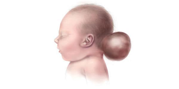
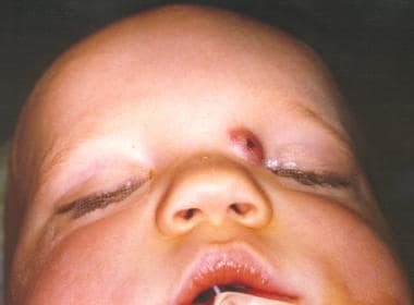
Encephalocele
Meningocele
Sphenoid Bone
Hypertelorism
Cerebrospinal Fluid Rhinorrhea
Sphenoid Sinus
Ethmoid Bone
Orbital Fractures
Neural Tube Defects
Anencephaly
Neuroendoscopy
Sella Turcica
Meningomyelocele
Abnormalities, Multiple
Ciliary Motility Disorders
Polycystic Kidney Diseases
Ectromelia
Encyclopedias as Topic
Pelvic Bones
Greek World
Caroli Disease
Thalidomide
High-resolution physical and genetic mapping of the critical region for Meckel syndrome and Mulibrey Nanism on chromosome 17q22-q23. (1/200)
Previously, we assigned the genes for two autosomal recessive disorders, Meckel syndrome (MKS; MIM 249000) and Mulibrey Nanism [MUL (muscle-liver-brain-eye Nanism); MIM 253250] that are enriched in the Finnish population, to overlapping genomic regions on chromosome 17q. Now, we report the construction of a bacterial clone contig over the critical region for both disorders. Several novel CA-repeat markers were isolated from these clones, which allowed refined mapping of the MKS and MUL loci using haplotype and linkage disequilibrium analysis. The localization of the MKS locus was narrowed to <1 cM between markers D17S1290 and 132-CA, within an approximately 800-kb region. The MUL locus was refined into an approximately 1400-kb interval between markers D17S1290 and 52-CA. The whole MKS region falls within the MUL region. In the common critical region, the conserved haplotypes were different in MKS and MUL patients. A trancript map was constructed by assigning expressed sequence tags (ESTs) and genes, derived from the human gene map, to the bacterial clone contig. Altogether, four genes and a total of 20 ESTs were precisely localized. These data provide the molecular tools for the final identification of the MKS and the MUL genes. (+info)Neural tube defects along the Texas-Mexico border, 1993-1995. (2/200)
In response to a 1991 anencephaly cluster in Cameron County, Texas, a surveillance and neural tube defect (NTD) recurrence prevention project for NTDs was implemented in the 14 Texas-Mexico border counties. For 1993-1995, NTD-affected pregnancies were identified at all gestational ages through active surveillance of multiple case-ascertainment sources. There were 87 cases of anencephaly, 96 cases of spina bifida, and 14 cases of encephalocele for respective rates of 6.4, 7.1, and 1.1 per 10,000 live births. Of the 197 NTD case-women, 93% were Hispanic. The overall, Hispanic, and Anglo NTD rates were, respectively, 14.6, 14.9, and 10.6 per 10,000 live births. The NTD rate for El Paso County (9.8 per 10,000), the most northwestern Texas county, was significantly lower (p = 0.001) than the aggregate rate for the rest of the Texas border (17.1 per 10,000). The overall Texas border rate was significantly higher (p < 0.001) than a recently estimated rate of 9.3 for California and minimally higher than a recently adjusted rate of 11.3 for the Metropolitan Atlanta Congenital Defects Program counties (p = 0.052), both of which now reflect all gestational ages. Of the 197 Texas border cases, 85% (168 cases) reached a gestational age of > or =20 weeks. Excluding cases of <20 weeks' gestation in the rate had a more marked effect on reducing the anencephaly rate (4.9 per 10,000) than the spina bifida rate (6.7 per 10,000). A country of birth was known for 153 (83%) of the 184 Hispanic case-women: 63% were born in Mexico; 24%, in Texas; and 11%, elsewhere in the United States. Rates for Mexico-born Hispanic women (15.1 per 10,000) were significantly higher than rates for United States-born Hispanic women (9.5 per 10,000) (p = 0.006). (+info)Dysgenesis of the internal carotid artery associated with transsphenoidal encephalocele: a neural crest syndrome? (3/200)
We describe two original cases of internal carotid artery dysgenesis associated with a malformative spectrum, which includes transsphenoidal encephalocele, optic nerve coloboma, hypopituitarism, and hypertelorism. Cephalic neural crest cells migrate to various regions in the head and neck where they contribute to the development of structures as diverse as the anterior skull base, the walls of the craniofacial arteries, the forebrain, and the face. Data suggest that the link between these rare malformations is abnormal neural crest development. (+info)Central brain herniation secondary to juvenile diabetic ketoacidosis. (4/200)
We present the CT, MR, and autopsy findings of central brain herniation in a 9-year-old boy undergoing treatment for diabetic ketoacidosis (DKA). Severe cerebral edema resulting in central brain herniation is an uncommon complication of the treatment of DKA but carries with it high morbidity and mortality. Radiologic imaging and autopsy findings in this case revealed striking infarctions of central brain structures. (+info)Surgical management of bacterial meningitis. (5/200)
A variety of associated lesions may require the neurosurgeon's assistance in the management of bacterial meningitis. As treatment of this infection of the central nervous system proceeds, the surgeon will have to decide about the concurrent or subsequent operative treatment of congenital dysraphic states, paraneural infections, compound fractures or penetrating wounds of thecranium or spine, or infected bypass shunts for cerebrospinal fluid (CSF). In patients with intractable meningitic infections the surgeon may have to insert a ventricular drainage-irrigation system to permit adequate perfusion of the CSF pathways with antibiotic. Hydrocephalus or subdural effusions complicating meningitis may bring the patient to the surgeon long after the infection has been cured. This paper examines these problems and outlines the current principles of management. (+info)Pregnancy in patients with Wegener's granulomatosis: report of five cases in three women. (6/200)
Five cases of pregnancy occurring in three women with previously diagnosed Wegener's granulomatosis are described. The disease was diffuse in one case and localised in the other. Initial treatment consisted of a combination of corticosteroids and intravenous cyclophosphamide in two women, and methotrexate in one. Four pregnancies ended in live births despite pre-eclampsia in two cases. One therapeutic abortion was induced because of encephalocele. Comparable reported cases were reviewed to examine the implications of immunosuppressive treatment on the fetus. A relapse occurred during pregnancy in 40% of the cases, but in 25% if only pregnancies beginning during inactive disease were taken into account. No other indicator for maternal and fetal outcome was obvious. Pregnancy should be planned after complete disappearance of disease activity. In the case of a relapse a combination of immunosuppressive drugs and corticosteroids should be chosen rather than corticosteroids alone because the outcome of pregnancy is poor in cases of undertreatment. Prematurity remains common. (+info)Transtentorial herniation after unilateral infarction of the anterior cerebral artery. (7/200)
BACKGROUND: Fatal cerebral herniation is a common complication of large ("malignant") middle cerebral artery infarcts but has not been reported in unilateral anterior cerebral artery (ACA) infarction. CASE DESCRIPTION: We report a 47-year-old woman who developed an acute left hemiparesis during an attack of migraine. Cranial CT (CCT) was normal but demonstrated narrow external cerebrospinal fluid compartments. Transcranial Doppler sonography was compatible with occlusion of the right ACA. Systemic thrombolytic therapy with tissue plasminogen activator was initiated 105 minutes after symptom onset. Follow-up CCT 24 hours after treatment revealed subtotal ACA infarction with hemorrhagic conversion. Two days later, the patient suddenly deteriorated with clinical signs of cerebral herniation, as confirmed by CCT. An extended right hemicraniectomy was immediately performed. Within 6 months, the patient regained her ability to walk but remained moderately disabled. CONCLUSIONS: This is the first reported case of unilateral ACA infarct leading to almost fatal cerebral herniation. Narrow external cerebrospinal fluid compartments in combination with early reperfusion, hemorrhagic transformation, and additional dysfunction of the blood-brain barrier promoted by tissue plasminogen activator and migraine may have contributed to this unusual course. (+info)Fixed and dilated pupils after trauma, stroke, and previous intracranial surgery: management and outcome. (8/200)
OBJECTIVES: To clarify whether different causative events (trauma, stroke, intracranial surgery), time of intervention, and treatment mode influence outcome, patients with fixed and dilated pupils (FDPs) in a prospective neurosurgical series were evaluated. METHODS: Ninety nine consecutive patients who presented with or developed one or two FDPs, were split into three groups according to the respective aetiology: 46 patients had a trauma, 41 patients a stroke (subarachnoid or intracerebral haemorrhage), and 12 patients had undergone previous elective intracranial surgery. Appropriate therapy was performed depending on the CT findings. Outcome was classified according to the Glasgow outcome scale (GOS). RESULTS: Overall mortality was 75%. In 15% outcome was unfavourable (GOS 2 and 3), and in 10% favourable (GOS 4, 5) at 24 month follow up. No differences in outcome were found between trauma, stroke, and postelective surgery groups. Unilaterally FDP was associated with a better chance of survival (46% v 13%; p<0.01). Age did not correlate with survival, but younger survivors had a significantly better outcome. Patients in whom an intracranial mass was removed surgically had a 42% survival rate, compared with 8% with conservative treatment (p<0.01). Patients with a shorter delay from FDPs to intervention had a better chance of recovery after trauma and previous intracranial surgery (p<0.05). No patient survived better than a vegetative state, if previous FDPs did not become reactive shortly after therapy. If both pupils became reactive on therapy, the chance of survival was 62%. Of these survivors 42% had a favourable outcome. CONCLUSION: Bilateral restoration of pupillary reactivity shortly after therapy is crucial for survival. Surgical evacuation of an intracranial mass, unilateral FDPs, early intervention, and younger age are related to better chances of survival or recovery. The prognosis of patients with FDPs after trauma, stroke, and previous elective intracranial surgery is similar. (+info)An Encephalocele is a type of neural tube defect that occurs when the bones of the skull do not close completely during fetal development. This results in a sac-like protrusion of the brain and the membranes that cover it through an opening in the skull. The sac may be visible on the scalp, forehead, or back of the head, and can vary in size. Encephaloceles can cause a range of symptoms, including developmental delays, intellectual disabilities, vision problems, and seizures, depending on the severity and location of the defect. Treatment typically involves surgical repair of the encephalocele soon after birth to prevent further damage to the brain and improve outcomes.
A meningocele is a type of neural tube defect that results in the herniation of the meninges (the protective membranes covering the brain and spinal cord) through a defect in the vertebral column. The meninges protrude as a sac-like structure, which may be covered by skin or a thin layer of tissue. Meningoceles usually do not contain neural tissue, but cerebrospinal fluid is present within the sac. They are typically asymptomatic unless there is compression of surrounding structures or infection. Treatment generally involves surgical repair to prevent potential complications such as meningitis or neurological damage.
The sphenoid bone is a complex, irregularly shaped bone located in the middle cranial fossa and forms part of the base of the skull. It articulates with several other bones, including the frontal, parietal, temporal, ethmoid, palatine, and zygomatic bones. The sphenoid bone has two main parts: the body and the wings.
The body of the sphenoid bone is roughly cuboid in shape and contains several important structures, such as the sella turcica, which houses the pituitary gland, and the sphenoid sinuses, which are air-filled cavities within the bone. The greater wings of the sphenoid bone extend laterally from the body and form part of the skull's lateral walls. They contain the superior orbital fissure, through which important nerves and blood vessels pass between the cranial cavity and the orbit of the eye.
The lesser wings of the sphenoid bone are thin, blade-like structures that extend anteriorly from the body and form part of the floor of the anterior cranial fossa. They contain the optic canal, which transmits the optic nerve and ophthalmic artery between the brain and the orbit of the eye.
Overall, the sphenoid bone plays a crucial role in protecting several important structures within the skull, including the pituitary gland, optic nerves, and ophthalmic arteries.
Hypertelorism is a medical term that refers to an ocular condition where the distance between two eyes (interpupillary distance) is abnormally increased. It's typically defined as an interpupillary distance that measures more than 2 standard deviations beyond the mean for a given age, gender, and race.
This condition can be associated with various genetic syndromes or conditions such as craniosynostosis (premature fusion of skull sutures), fetal alcohol syndrome, and certain chromosomal abnormalities like Down syndrome. Hypertelorism may also occur in isolation without any other associated anomalies.
It's important to note that hypertelorism can have cosmetic implications, particularly if the distance between the eyes is significantly increased, as it may affect the overall symmetry and appearance of the face. However, in most cases, this condition does not directly impact vision unless there are other related structural abnormalities of the eye or orbit.
Cerebrospinal fluid (CSF) rhinorrhea is a condition where the cerebrospinal fluid, which surrounds and protects the brain and spinal cord, leaks through the nasal cavity. This occurs due to a defect or opening in the skull base or the thin bone that separates the brain from the nasal cavity, known as the cribriform plate.
CSF rhinorrhea can result from trauma, surgery, or spontaneously due to increased pressure in the brain. It is important to diagnose and treat this condition promptly because it increases the risk of meningitis, an infection of the membranes covering the brain and spinal cord. Treatment options include bed rest, hydration, stool softeners, and sometimes surgical repair of the defect.
The sphenoid sinuses are air-filled spaces located within the sphenoid bone, which is one of the bones that make up the skull base. These sinuses are located deep inside the skull, behind the eyes and nasal cavity. They are paired and separated by a thin bony septum, and each one opens into the corresponding nasal cavity through a small opening called the sphenoethmoidal recess. The sphenoid sinuses vary greatly in size and shape between individuals. They develop during childhood and continue to grow until early adulthood. The function of the sphenoid sinuses, like other paranasal sinuses, is not entirely clear, but they may contribute to reducing the weight of the skull, resonating voice during speech, and insulating the brain from trauma.
The ethmoid bone is a paired, thin, and lightweight bone that forms part of the skull's anterior cranial fossa and contributes to the formation of the orbit and nasal cavity. It is located between the frontal bone above and the maxilla and palatine bones below. The ethmoid bone has several important features:
1. Cribriform plate: This is the horizontal, sieve-like portion that forms part of the anterior cranial fossa and serves as the roof of the nasal cavity. It contains small openings (foramina) through which olfactory nerves pass.
2. Perpendicular plate: The perpendicular plate is a vertical structure that projects downward from the cribriform plate, forming part of the nasal septum and separating the left and right nasal cavities.
3. Superior and middle nasal conchae: These are curved bony projections within the lateral walls of the nasal cavity that help to warm, humidify, and filter incoming air.
4. Lacrimal bone: The ethmoid bone articulates with the lacrimal bone, forming part of the medial wall of the orbit.
5. Frontal process: This is a thin, vertical plate that articulates with the frontal bone above the orbit.
6. Sphenoidal process: The sphenoidal process connects the ethmoid bone to the sphenoid bone posteriorly.
The ethmoid bone plays a crucial role in protecting the brain and providing structural support for the eyes, as well as facilitating respiration by warming, humidifying, and filtering incoming air.
Orbital fractures refer to breaks in the bones that make up the eye socket, also known as the orbit. These bones include the maxilla, zygoma, frontal bone, and palatine bone. Orbital fractures can occur due to trauma, such as a blunt force injury or a penetrating wound.
There are several types of orbital fractures, including:
1. Blowout fracture: This occurs when the thin bone of the orbital floor is broken, often due to a direct blow to the eye. The force of the impact can cause the eyeball to move backward, breaking the bone and sometimes trapping the muscle that moves the eye (the inferior rectus).
2. Blow-in fracture: This type of fracture involves the breakage of the orbital roof, which is the bone that forms the upper boundary of the orbit. It typically occurs due to high-impact trauma, such as a car accident or a fall from a significant height.
3. Direct fracture: A direct fracture happens when there is a break in one or more of the bones that form the walls of the orbit. This type of fracture can result from a variety of traumas, including motor vehicle accidents, sports injuries, and assaults.
4. Indirect fracture: An indirect fracture occurs when the force of an injury is transmitted to the orbit through tissues surrounding it, causing the bone to break. The most common type of indirect orbital fracture is a blowout fracture.
Orbital fractures can cause various symptoms, including pain, swelling, bruising, and double vision. In some cases, the fracture may also lead to enophthalmos (sinking of the eye into the orbit) or telecanthus (increased distance between the inner corners of the eyes). Imaging tests, such as CT scans, are often used to diagnose orbital fractures and determine the best course of treatment. Treatment may include observation, pain management, and in some cases, surgery to repair the fracture and restore normal function.
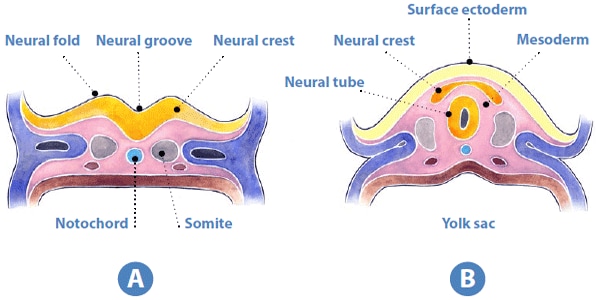
Neural Tube Defects (NTDs) are a group of birth defects that affect the brain, spine, or spinal cord. They occur when the neural tube, which forms the early brain and spinal cord of the embryo, does not close properly during fetal development. This can result in various conditions such as:
1. Anencephaly: a severe defect where most of the brain and skull are missing. Infants with anencephaly are usually stillborn or die shortly after birth.
2. Spina bifida: a condition where the spine does not close properly, leaving a portion of the spinal cord and nerves exposed. This can result in various neurological problems, including paralysis, bladder and bowel dysfunction, and hydrocephalus (fluid buildup in the brain).
3. Encephalocele: a condition where the skull does not close properly, allowing the brain to protrude through an opening in the skull. This can result in various neurological problems, including developmental delays, vision and hearing impairments, and seizures.
NTDs are thought to be caused by a combination of genetic and environmental factors, such as folic acid deficiency, obesity, diabetes, and exposure to certain medications during pregnancy. Folic acid supplementation before and during early pregnancy has been shown to reduce the risk of NTDs.
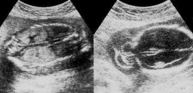
Anencephaly is a serious birth defect that affects the neural tube, which is the structure that develops into the brain and spinal cord. In anencephaly, the neural tube fails to close properly during fetal development, resulting in the absence of a major portion of the brain, skull, and scalp.
Anencephaly is typically diagnosed through prenatal ultrasound or other imaging tests. Unfortunately, it is a fatal condition, and most babies with anencephaly do not survive birth or live for more than a few hours or days after birth.
The exact cause of anencephaly is not fully understood, but it is believed to be related to genetic factors as well as environmental influences such as folic acid deficiency and exposure to certain medications or chemicals during pregnancy. Pregnant women are often advised to take folic acid supplements to reduce the risk of neural tube defects, including anencephaly.
Neuroendoscopy is a minimally invasive surgical technique that involves the use of an endoscope to access and treat various conditions within the brain and spinal column. An endoscope is a long, flexible tube with a light and camera at its tip, which allows surgeons to view and operate on internal structures through small incisions or natural openings in the body.
In neuroendoscopy, the surgeon uses the endoscope to navigate through the brain's ventricular system (fluid-filled spaces) or other narrow spaces within the skull or spine to diagnose and treat conditions such as hydrocephalus, brain tumors, arachnoid cysts, and intraventricular hemorrhage.
The benefits of neuroendoscopy include reduced trauma to surrounding tissues, shorter hospital stays, faster recovery times, and improved outcomes compared to traditional open surgical approaches. However, neuroendoscopic procedures require specialized training and expertise due to the complexity of the anatomy involved.
The Sella Turcica, also known as the Turkish saddle, is a depression or fossa in the sphenoid bone located at the base of the skull. It forms a housing for the pituitary gland, which is a small endocrine gland often referred to as the "master gland" because it controls other glands and makes several essential hormones. The Sella Turcica has a saddle-like shape, with its anterior and posterior clinoids forming the front and back of the saddle, respectively. This region is of significant interest in neuroimaging and clinical settings, as various conditions such as pituitary tumors or other abnormalities may affect the size, shape, and integrity of the Sella Turcica.
Meningomyelocele is a type of neural tube defect that affects the development of the spinal cord and the surrounding membranes known as meninges. In this condition, a portion of the spinal cord and meninges protrude through an opening in the spine, creating a sac-like structure on the back. This sac is usually covered by skin, but it may be open in some cases.
Meningomyelocele can result in various neurological deficits, including muscle weakness, paralysis, and loss of sensation below the level of the lesion. It can also cause bladder and bowel dysfunction, as well as problems with sexual function. The severity of these symptoms depends on the location and extent of the spinal cord defect.
Early diagnosis and treatment are crucial for managing meningomyelocele and preventing further complications. Treatment typically involves surgical closure of the opening in the spine to protect the spinal cord and prevent infection. Physical therapy, occupational therapy, and other supportive care measures may also be necessary to help individuals with meningomyelocele achieve their full potential for mobility and independence.
Eye abnormalities refer to any structural or functional anomalies that affect the eye or its surrounding tissues. These abnormalities can be present at birth (congenital) or acquired later in life due to various factors such as injury, disease, or aging. Some examples of eye abnormalities include:
1. Strabismus: Also known as crossed eyes, strabismus is a condition where the eyes are misaligned and point in different directions.
2. Nystagmus: This is an involuntary movement of the eyes that can be horizontal, vertical, or rotatory.
3. Cataracts: A cataract is a clouding of the lens inside the eye that can cause vision loss.
4. Glaucoma: This is a group of eye conditions that damage the optic nerve and can lead to vision loss.
5. Retinal disorders: These include conditions such as retinal detachment, macular degeneration, and diabetic retinopathy.
6. Corneal abnormalities: These include conditions such as keratoconus, corneal ulcers, and Fuchs' dystrophy.
7. Orbital abnormalities: These include conditions such as orbital tumors, thyroid eye disease, and Graves' ophthalmopathy.
8. Ptosis: This is a condition where the upper eyelid droops over the eye.
9. Color blindness: A condition where a person has difficulty distinguishing between certain colors.
10. Microphthalmia: A condition where one or both eyes are abnormally small.
These are just a few examples of eye abnormalities, and there are many others that can affect the eye and its functioning. If you suspect that you have an eye abnormality, it is important to consult with an ophthalmologist for proper diagnosis and treatment.
A syndrome, in medical terms, is a set of symptoms that collectively indicate or characterize a disease, disorder, or underlying pathological process. It's essentially a collection of signs and/or symptoms that frequently occur together and can suggest a particular cause or condition, even though the exact physiological mechanisms might not be fully understood.
For example, Down syndrome is characterized by specific physical features, cognitive delays, and other developmental issues resulting from an extra copy of chromosome 21. Similarly, metabolic syndromes like diabetes mellitus type 2 involve a group of risk factors such as obesity, high blood pressure, high blood sugar, and abnormal cholesterol or triglyceride levels that collectively increase the risk of heart disease, stroke, and diabetes.
It's important to note that a syndrome is not a specific diagnosis; rather, it's a pattern of symptoms that can help guide further diagnostic evaluation and management.
'Abnormalities, Multiple' is a broad term that refers to the presence of two or more structural or functional anomalies in an individual. These abnormalities can be present at birth (congenital) or can develop later in life (acquired). They can affect various organs and systems of the body and can vary greatly in severity and impact on a person's health and well-being.
Multiple abnormalities can occur due to genetic factors, environmental influences, or a combination of both. Chromosomal abnormalities, gene mutations, exposure to teratogens (substances that cause birth defects), and maternal infections during pregnancy are some of the common causes of multiple congenital abnormalities.
Examples of multiple congenital abnormalities include Down syndrome, Turner syndrome, and VATER/VACTERL association. Acquired multiple abnormalities can result from conditions such as trauma, infection, degenerative diseases, or cancer.
The medical evaluation and management of individuals with multiple abnormalities depend on the specific abnormalities present and their impact on the individual's health and functioning. A multidisciplinary team of healthcare professionals is often involved in the care of these individuals to address their complex needs.
Ciliary motility disorders are a group of rare genetic conditions that affect the function of cilia, which are tiny hair-like structures on the surface of cells in the body. Cilia play an important role in moving fluids and particles across the cell surface, including the movement of mucus and other substances in the respiratory system, the movement of eggs and sperm in the reproductive system, and the movement of fluid in the inner ear.
Ciliary motility disorders are caused by mutations in genes that are responsible for the proper functioning of cilia. These mutations can lead to abnormalities in the structure or function of cilia, which can result in a range of symptoms depending on the specific disorder and the parts of the body that are affected.
Some common symptoms of ciliary motility disorders include recurrent respiratory infections, chronic sinusitis, hearing loss, infertility, and situs inversus, a condition in which the major organs are reversed or mirrored from their normal positions. There are several different types of ciliary motility disorders, including primary ciliary dyskinesia, Kartagener syndrome, and immotile cilia syndrome.
Treatment for ciliary motility disorders typically involves addressing the specific symptoms and underlying causes of the disorder. This may include antibiotics to treat respiratory infections, surgery to correct structural abnormalities, or assisted reproductive technologies to help with infertility.
Neurosurgical procedures are operations that are performed on the brain, spinal cord, and peripheral nerves. These procedures are typically carried out by neurosurgeons, who are medical doctors with specialized training in the diagnosis and treatment of disorders of the nervous system. Neurosurgical procedures can be used to treat a wide range of conditions, including traumatic injuries, tumors, aneurysms, vascular malformations, infections, degenerative diseases, and congenital abnormalities.
Some common types of neurosurgical procedures include:
* Craniotomy: A procedure in which a bone flap is temporarily removed from the skull to gain access to the brain. This type of procedure may be performed to remove a tumor, repair a blood vessel, or relieve pressure on the brain.
* Spinal fusion: A procedure in which two or more vertebrae in the spine are fused together using bone grafts and metal hardware. This is often done to stabilize the spine and alleviate pain caused by degenerative conditions or spinal deformities.
* Microvascular decompression: A procedure in which a blood vessel that is causing pressure on a nerve is repositioned or removed. This type of procedure is often used to treat trigeminal neuralgia, a condition that causes severe facial pain.
* Deep brain stimulation: A procedure in which electrodes are implanted in specific areas of the brain and connected to a battery-operated device called a neurostimulator. The neurostimulator sends electrical impulses to the brain to help alleviate symptoms of movement disorders such as Parkinson's disease or dystonia.
* Stereotactic radiosurgery: A non-invasive procedure that uses focused beams of radiation to treat tumors, vascular malformations, and other abnormalities in the brain or spine. This type of procedure is often used for patients who are not good candidates for traditional surgery due to age, health status, or location of the lesion.
Neurosurgical procedures can be complex and require a high degree of skill and expertise. Patients considering neurosurgical treatment should consult with a qualified neurosurgeon to discuss their options and determine the best course of action for their individual situation.
Polycystic Kidney Disease (PKD) is a genetic disorder characterized by the growth of multiple cysts in the kidneys. These cysts are fluid-filled sacs that can vary in size and can multiply, leading to enlarged kidneys. The increased size and number of cysts can result in reduced kidney function, high blood pressure, and eventually kidney failure.
There are two main types of PKD: Autosomal Dominant Polycystic Kidney Disease (ADPKD) and Autosomal Recessive Polycystic Kidney Disease (ARPKD). ADPKD is the most common form, affecting approximately 1 in every 500 people. It typically develops in adulthood. On the other hand, ARPKD is a rarer form, affecting about 1 in every 20,000 children, and it often presents in infancy or early childhood.
In addition to kidney problems, PKD can also affect other organs, such as the liver and the heart. It's important to note that while there is no cure for PKD, various treatments can help manage symptoms and slow down the progression of the disease.
Ectromelia is a medical term that refers to the congenital absence or malformation of a limb or extremity. It is also known as "congenital amputation" or "limb reduction defect." This condition can affect any extremity, including arms, legs, hands, or feet, and can range from mild, such as a missing finger or toe, to severe, such as the absence of an entire limb.
Ectromelia can be caused by various factors, including genetic mutations, environmental factors, or a combination of both. In some cases, the cause may be unknown. Treatment options for ectromelia depend on the severity and location of the malformation and may include prosthetics, physical therapy, or surgery.
An encyclopedia is a comprehensive reference work containing articles on various topics, usually arranged in alphabetical order. In the context of medicine, a medical encyclopedia is a collection of articles that provide information about a wide range of medical topics, including diseases and conditions, treatments, tests, procedures, and anatomy and physiology. Medical encyclopedias may be published in print or electronic formats and are often used as a starting point for researching medical topics. They can provide reliable and accurate information on medical subjects, making them useful resources for healthcare professionals, students, and patients alike. Some well-known examples of medical encyclopedias include the Merck Manual and the Stedman's Medical Dictionary.
The pelvic bones, also known as the hip bones, are a set of three irregularly shaped bones that connect to form the pelvic girdle in the lower part of the human body. They play a crucial role in supporting the spine and protecting the abdominal and pelvic organs.
The pelvic bones consist of three bones:
1. The ilium: This is the largest and uppermost bone, forming the majority of the hip bone and the broad, flaring part of the pelvis known as the wing of the ilium or the iliac crest, which can be felt on the side of the body.
2. The ischium: This is the lower and back portion of the pelvic bone that forms part of the sitting surface or the "sit bones."
3. The pubis: This is the front part of the pelvic bone, which connects to the other side at the pubic symphysis in the midline of the body.
The pelvic bones are joined together at the acetabulum, a cup-shaped socket that forms the hip joint and articulates with the head of the femur (thigh bone). The pelvic bones also have several openings for the passage of blood vessels, nerves, and reproductive and excretory organs.
The shape and size of the pelvic bones differ between males and females due to their different roles in childbirth and locomotion. Females typically have a wider and shallower pelvis than males to accommodate childbirth, while males usually have a narrower and deeper pelvis that is better suited for weight-bearing and movement.
I believe there may be some confusion in your question as "Greek World" is not a medical term. If you are referring to the ancient Greek civilization, it was a significant period in human history that greatly contributed to the development of various fields including medicine. The ancient Greeks, particularly Hippocrates and his followers, are often referred to as the "Fathers of Medicine." They made substantial contributions to the field through their observations, theories, and practices which formed the foundation of much of Western medical thought. However, "Greek World" itself does not have a medical definition.
Caroli disease is a rare genetic disorder that affects the liver and bile ducts. It is characterized by abnormal dilations or sac-like structures in the intrahepatic bile ducts, which are the ducts that carry bile from the liver to the gallbladder and small intestine. These dilations can lead to recurrent cholangitis (inflammation of the bile ducts), stone formation, and liver damage.
Caroli disease is usually diagnosed in childhood or early adulthood, and it can be associated with other congenital anomalies such as polycystic kidney disease. The exact cause of Caroli disease is not fully understood, but it is believed to be inherited in an autosomal recessive manner, meaning that an individual must inherit two copies of the abnormal gene, one from each parent, to develop the condition.
Treatment for Caroli disease may include antibiotics to manage cholangitis, endoscopic procedures to remove stones or dilate strictures, and surgery to bypass or remove affected bile ducts. In severe cases, liver transplantation may be necessary. Regular monitoring of liver function and surveillance for complications are essential in the management of this condition.
Thalidomide is a pharmaceutical drug that was initially developed and marketed as a sedative and treatment for morning sickness in pregnant women. However, it was later found to cause severe birth defects when given during pregnancy, particularly damage to the limbs, ears, and eyes of the developing fetus. As a result, thalidomide was banned in many countries in the 1960s.
In recent years, thalidomide has been reintroduced as a treatment for certain medical conditions, including multiple myeloma (a type of cancer that affects plasma cells) and leprosy. It is also being studied as a potential treatment for other diseases, such as rheumatoid arthritis and Crohn's disease.
Thalidomide works by suppressing the immune system and inhibiting the formation of new blood vessels (angiogenesis). However, its use is tightly regulated due to its teratogenic effects, meaning it can cause birth defects if taken during pregnancy. Women who are pregnant or planning to become pregnant should not take thalidomide, and healthcare providers must follow strict guidelines when prescribing the drug to ensure that it is used safely and effectively.
Genetic research is a branch of biomedical science that involves the study of genes, their functions, and heredity. It aims to understand how genetic variations contribute to human health and disease by using various scientific approaches such as genetics, genomics, molecular biology, biochemistry, and bioinformatics.
Genetic research can be conducted on humans, animals, or plants, and it can focus on a variety of areas including:
1. Identifying genes associated with specific diseases or traits
2. Understanding how genes are regulated and expressed
3. Investigating the role of genetic mutations in disease development
4. Developing new diagnostic tests and treatments based on genetic information
5. Exploring evolutionary relationships between species
6. Examining ethical, legal, and social implications of genetic research.
Genetic research has led to significant advances in our understanding of many diseases, including cancer, diabetes, heart disease, and neurological disorders. It also holds great promise for personalized medicine, which tailors treatments to individual patients based on their genetic makeup.
 Encephalocele
 Facts about Encephalocele | CDC
 Encephalocele Imaging: Practice Essentials, Radiography, Computed Tomography
 Researchers interested in Encephalocele | Yale School of Medicine
 Right sided cerebellar ataxia with occipital encephalocele (arnold -chiari type iii malformation) | Pediatric Oncall Journal
 Meckel gruber syndrome: a vaginal breech delivery facilitated by encephalocele self-decompression | ADC Fetal & Neonatal Edition
 Encephalocele - Pediatrics - MSD Manual Professional Edition
 Dr Balaji Anvekar FRCR: Occipital Encephalocele MRI
 How do you fix occipital encephalocele? - Motelmexicolabali.com
 Neuro Spine Care | Children's Healthcare of Atlanta
 Optic Nerve Coloboma / Morning Glory Syndrome / Encephalocele - Retina Image Bank
 A Rare Association of Right-sided Congenital Diaphragmatic Hernia and Encephalocele: A Case Report
 Term newborn with microphthalmia, encephalocele and linear skin defect - Fingerprint
- UTMB Health Research Expert Profiles
Term newborn with microphthalmia, encephalocele and linear skin defect - Fingerprint
- UTMB Health Research Expert Profiles MEGALENCEPHALY, POLYMICROGYRIA, POLYDACTYLY AND HYDROCEPHALUS (MPPH) SYNDROME: A NEW CASE WITH OCCIPITAL ENCEPHALOCELE AND...
 Molecular and Clinical Findings in Patients With Knobloch Syndrome
 Myelomeningocele: MedlinePlus Medical Encyclopedia
 Airway management for occipital encephalocele in neonatal patients: A review of 17 cases - Journal of Neurosciences in Rural...
 Cord
Cord MyMarkets | Digital Health Tech Vision 2019
 Myles L. Pensak, MD,FACS