Eosinophilic Granuloma
Histiocytosis, Langerhans-Cell
Granuloma
Lanolin
Spinal Diseases
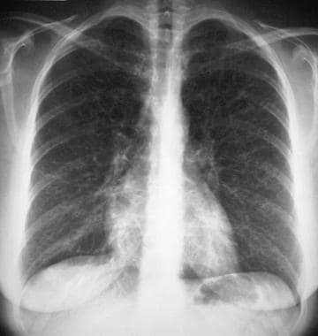
Pulmonary eosinophilic gramuloma in a child. (1/56)
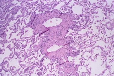
The occurrence of pulmonary eosinophilic granuloma in a 3-year-old child is described. She presented with a pneumothorax and typical radiological changes and the diagnosis was confirmed by lung biopsy. There was no objective evidence of improvement after radiotherapy when lung function was assessed by gamma scans. She died suddenly while abroad. (+info)Anaplastic gangliocytoma with eosinophilic cytoplasmic granules in a cow. (2/56)
In a 3-year-old Holstein cow, a tumor mass replaced the left olfactory bulb. The tumor was highly or moderately cellular, and consisted of tumor cells showing pleomorphism and anaplasia, sometimes with intracytoplasmic granules. The tumor showed weak reactivity for neurofilaments (NF) in most cells with distinct staining in a minority, and it was extremely rare to see neoplastic cells with positivity for glial fibrillary acidic protein (GFAP). The neoplastic cells displayed some ultrastructural features reminiscent of ganglionic cells, and the cytoplasmic granularity was due to the presence of numerous lysosomes. This tumor expressing both NF and GFAP may be histogenetically related to brain tumors of pluripotential cell origin in calves. (+info)Schistosome infection of transgenic mice defines distinct and contrasting pathogenic roles for IL-4 and IL-13: IL-13 is a profibrotic agent. (3/56)
Experimental Schistosoma mansoni infections of mice lead to a dynamic type 2 cytokine-mediated pathological process. We have used IL-4-deficient, IL-13-deficient, and IL-4/13-deficient mice to dissect the role of these cytokines in the development of immune response and pathology following S. mansoni infection. We demonstrate that while both of these cytokines are necessary to develop a robust Th2 cell-driven, eosinophil-rich granuloma response, they also perform disparate functions that identify novel sites for therapeutic intervention. IL-13-deficient mice demonstrated significantly enhanced survival following infection, which correlated with reduced hepatic fibrosis. In contrast, increased mortality was manifest in IL-4-deficient and IL-4/13-deficient mice, and this correlated with hepatocyte damage and intestinal pathology. Therefore, we demonstrate that during a dynamic type 2 cytokine disease process IL-13 is detrimental to survival following infection, whereas IL-4 is beneficial. (+info)Magnetic resonance imaging of calvarial eosinophilic granuloma with pericranial soft tissue reaction--case report. (4/56)
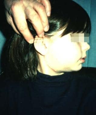
A 4-year-old girl presented with an eosinophilic granuloma in the cranial vault. Magnetic resonance (MR) imaging showed the mass as slightly low intensity on T1- and high intensity on T2-weighted images. The pericranial soft tissue was densely enhanced after gadolinium-diethylenetriaminepenta-acetic acid infusion. The mass was soft and successfully removed. Histological examination disclosed Langerhans' cell histiocytosis. MR imaging is useful for the diagnosis of calvarial eosinophilic granuloma with soft tissue involvement. (+info)Differentiation of Langerhans cells in Langerhans cell histiocytosis. (5/56)
Langerhans cell histiocytosis (LCH) consists of lesions composed of cells with a dendritic Langerhans cell (LC) phenotype. The clinical course of LCH ranges from spontaneous resolution to a chronic and sometimes lethal disease. We studied 25 patients with various clinical forms of the disease. In bone and chronic lesions, LCH cells had immature phenotype and function. They coexpressed LC antigens CD1a and Langerin together with monocyte antigens CD68 and CD14. Class II antigens were intracellular and LCH cells almost never expressed CD83 or CD86 or dendritic cell (DC)-Lamp, despite their CD40 expression. Consistently, LCH cells sorted from bone lesions (eosinophilic granuloma) poorly stimulated allogeneic T-cell proliferation in vitro. Strikingly, however, in vitro treatment with CD40L induced the expression of membrane class II and CD86 and strongly increased LCH cell allostimulatory activity to a level similar to that of mature DCs. Numerous interleukin-10-positive (IL-10(+)), Langerin(-), and CD68(+) macrophages were found within bone and lymph node lesions. In patients with self-healing and/or isolated cutaneous disease, LCH cells had a more mature phenotype. LCH cells were frequently CD14(-) and CD86(+), and macrophages were rare or absent, as were IL-10-expressing cells. We conclude that LCH cells in the bone and/or chronic forms of the disease accumulate within the tissues in an immature state and that most probably result from extrinsic signals and may be induced to differentiate toward mature DCs after CD40 triggering. Drugs that enhance the in vivo maturation of these immature DCs, or that induce their death, may be of therapeutic benefit. (+info)Eosinophilic granuloma. A different behaviour in children than in adults. (6/56)
Localised Langerhans-cell histiocytosis of bone (eosinophilic granuloma) is a benign tumour-like condition with a variable clinical course. Different forms of treatment have been reported to give satisfactory results. However, previous series all contain patients with a wide age range. Our aim was to investigate the effect of skeletal maturity on the rate of recurrence of isolated eosinophilic granuloma of bone excluding those arising in the spine. We followed up 32 patients with an isolated eosinophilic granuloma for a mean of five years; 17 were skeletally immature. No recurrences were noted in the skeletally immature group even after biopsy alone. By contrast, four of 13 skeletally mature patients had a recurrence and required further surgery. This suggests that eosinophilic granuloma has a low rate of recurrence in skeletally immature patients. (+info)Cranial fasciitis of childhood. (7/56)
Cranial fasciitis is a benign fibroblastic tumor of the skull found almost exclusively in young children. It is histologically identical to nodular fasciitis. We present the clinical, radiologic, and pathologic findings of a 7-month-old male infant with cranial fasciitis of the skull. Herein, we include the first description of this entity's diffusion-weighted imaging appearance. Although rare, cranial fasciitis can mimic more aggressive processes both clinically and radiographically. (+info)GASTRO-DUODENAL CROHN'S DISEASE. (8/56)
Gastro-duodenal Crohn's disease is rare. Thirty-one previously reported cases are briefly reviewed; histological confirmation of the diagnosis was not always possible. Details are given of a patient with pyloro-duodenal involvement accompanied by terminal ileitis and appendicitis where surgical specimens were available for study. The differential diagnosis is considered from the clinical and pathological aspects. (+info)Eosinophilic granuloma is a term used in pathology to describe a specific type of inflammatory lesion that is characterized by the accumulation of eosinophils, a type of white blood cell, and the formation of granulomas. A granuloma is a small nodular structure formed by the accumulation of immune cells, typically including macrophages, lymphocytes, and other inflammatory cells.
Eosinophilic granulomas can occur in various organs of the body, but they are most commonly found in the lungs, skin, and bones. In the lungs, eosinophilic granulomas are often associated with hypersensitivity reactions to inhaled antigens, such as dust mites or fungal spores. They can also be seen in association with certain diseases, such as Langerhans cell histiocytosis, an uncommon disorder characterized by the abnormal proliferation of a type of immune cell called Langerhans cells.
The symptoms of eosinophilic granuloma depend on the location and extent of the lesion. In the lungs, eosinophilic granulomas may cause cough, chest pain, or shortness of breath. In the skin, they may present as nodules, plaques, or ulcers. In the bones, they can cause pain, swelling, and fractures.
The diagnosis of eosinophilic granuloma is typically made based on a combination of clinical, radiological, and pathological findings. Treatment may include avoidance of known antigens, corticosteroids, or other immunosuppressive medications, depending on the severity and location of the lesion.
Langerhans cell histiocytosis (LCH) is a rare disorder characterized by the abnormal proliferation and accumulation of dendritic cells called Langerhans cells in various tissues and organs of the body. These cells are part of the immune system and normally help to fight infection. However, in LCH, an overactive immune response leads to the excessive buildup of these cells, forming granulomas that can damage organs and impair their function.
The exact cause of LCH is not fully understood, but it is thought to involve genetic mutations that lead to uncontrolled cell growth and division. The disorder can affect people of any age, although it is most commonly diagnosed in children under the age of 15.
LCH can affect a single organ or multiple organs, depending on the severity and extent of the disease. Commonly affected sites include the bones, skin, lymph nodes, lungs, liver, spleen, and pituitary gland. Symptoms vary widely depending on the location and severity of the disease, but may include bone pain, rashes, fatigue, fever, weight loss, cough, and difficulty breathing.
Treatment for LCH depends on the extent and severity of the disease. In mild cases, observation and monitoring may be sufficient. More severe cases may require chemotherapy, radiation therapy, or surgery to remove affected tissues. In some cases, immunosuppressive drugs or targeted therapies that target specific genetic mutations may be used.
Overall, LCH is a complex and poorly understood disorder that requires careful evaluation and management by a team of medical specialists. While the prognosis for patients with LCH has improved in recent years, some cases can be life-threatening or lead to long-term complications.
A granuloma is a small, nodular inflammatory lesion that occurs in various tissues in response to chronic infection, foreign body reaction, or autoimmune conditions. Histologically, it is characterized by the presence of epithelioid macrophages, which are specialized immune cells with enlarged nuclei and abundant cytoplasm, often arranged in a palisading pattern around a central area containing necrotic debris, microorganisms, or foreign material.
Granulomas can be found in various medical conditions such as tuberculosis, sarcoidosis, fungal infections, and certain autoimmune disorders like Crohn's disease. The formation of granulomas is a complex process involving both innate and adaptive immune responses, which aim to contain and eliminate the offending agent while minimizing tissue damage.
Lanolin is not strictly a medical term, but it is often used in medical contexts. Medically, lanolin is referred to as "wool fat" or "wool wax." It's a yellow, waxy substance that is secreted by the sebaceous glands of wool-bearing animals, most notably sheep. Lanolin is composed primarily of esters, alcohols, and fatty acids, and it has excellent emollient properties, making it a valuable ingredient in various medical and cosmetic products.
In medical contexts, lanolin is often used as an emollient or moisturizer in topical preparations, such as creams, ointments, and lotions. It helps to soften and soothe dry, chapped, or irritated skin by creating a protective barrier that locks in moisture. Lanolin is also used in the pharmaceutical industry as an excipient (an inactive substance that serves as a vehicle or medium for a drug) in various formulations, including tablets and capsules.
It's worth noting that some people may have allergic reactions to lanolin, so it's essential to perform a patch test before using products containing this ingredient, especially if you have sensitive skin or a history of allergies.
Orbital neoplasms refer to abnormal growths or tumors that develop in the orbit, which is the bony cavity that contains the eyeball, muscles, nerves, fat, and blood vessels. These neoplasms can be benign (non-cancerous) or malignant (cancerous), and they can arise from various types of cells within the orbit.
Orbital neoplasms can cause a variety of symptoms depending on their size, location, and rate of growth. Common symptoms include protrusion or displacement of the eyeball, double vision, limited eye movement, pain, swelling, and numbness in the face. In some cases, orbital neoplasms may not cause any noticeable symptoms, especially if they are small and slow-growing.
There are many different types of orbital neoplasms, including:
1. Optic nerve glioma: a rare tumor that arises from the optic nerve's supportive tissue.
2. Orbital meningioma: a tumor that originates from the membranes covering the brain and extends into the orbit.
3. Lacrimal gland tumors: benign or malignant growths that develop in the lacrimal gland, which produces tears.
4. Orbital lymphangioma: a non-cancerous tumor that arises from the lymphatic vessels in the orbit.
5. Rhabdomyosarcoma: a malignant tumor that develops from the skeletal muscle cells in the orbit.
6. Metastatic tumors: cancerous growths that spread to the orbit from other parts of the body, such as the breast, lung, or prostate.
The diagnosis and treatment of orbital neoplasms depend on several factors, including the type, size, location, and extent of the tumor. Imaging tests, such as CT scans and MRI, are often used to visualize the tumor and determine its extent. A biopsy may also be performed to confirm the diagnosis and determine the tumor's type and grade. Treatment options include surgery, radiation therapy, chemotherapy, or a combination of these approaches.
Spinal diseases refer to a range of medical conditions that affect the spinal column, which is made up of vertebrae (bones), intervertebral discs, facet joints, nerves, ligaments, and muscles. These diseases can cause pain, discomfort, stiffness, numbness, weakness, or even paralysis, depending on the severity and location of the condition. Here are some examples of spinal diseases:
1. Degenerative disc disease: This is a condition where the intervertebral discs lose their elasticity and height, leading to stiffness, pain, and decreased mobility.
2. Herniated disc: This occurs when the inner material of the intervertebral disc bulges or herniates out through a tear in the outer layer, causing pressure on the spinal nerves and resulting in pain, numbness, tingling, or weakness in the affected area.
3. Spinal stenosis: This is a narrowing of the spinal canal or the neural foramen (the openings where the spinal nerves exit the spinal column), which can cause pressure on the spinal cord or nerves and result in pain, numbness, tingling, or weakness.
4. Scoliosis: This is a curvature of the spine that can occur in children or adults, leading to an abnormal posture, back pain, and decreased lung function.
5. Osteoarthritis: This is a degenerative joint disease that affects the facet joints in the spine, causing pain, stiffness, and decreased mobility.
6. Ankylosing spondylitis: This is a chronic inflammatory disease that affects the spine and sacroiliac joints, leading to pain, stiffness, and fusion of the vertebrae.
7. Spinal tumors: These are abnormal growths that can occur in the spinal column, which can be benign or malignant, causing pain, neurological symptoms, or even paralysis.
8. Infections: Bacterial or viral infections can affect the spine, leading to pain, fever, and other systemic symptoms.
9. Trauma: Fractures, dislocations, or sprains of the spine can occur due to accidents, falls, or sports injuries, causing pain, neurological deficits, or even paralysis.
Lung diseases refer to a broad category of disorders that affect the lungs and other structures within the respiratory system. These diseases can impair lung function, leading to symptoms such as coughing, shortness of breath, chest pain, and wheezing. They can be categorized into several types based on the underlying cause and nature of the disease process. Some common examples include:
1. Obstructive lung diseases: These are characterized by narrowing or blockage of the airways, making it difficult to breathe out. Examples include chronic obstructive pulmonary disease (COPD), asthma, bronchiectasis, and cystic fibrosis.
2. Restrictive lung diseases: These involve stiffening or scarring of the lungs, which reduces their ability to expand and take in air. Examples include idiopathic pulmonary fibrosis, sarcoidosis, and asbestosis.
3. Infectious lung diseases: These are caused by bacteria, viruses, fungi, or parasites that infect the lungs. Examples include pneumonia, tuberculosis, and influenza.
4. Vascular lung diseases: These affect the blood vessels in the lungs, impairing oxygen exchange. Examples include pulmonary embolism, pulmonary hypertension, and chronic thromboembolic pulmonary hypertension (CTEPH).
5. Neoplastic lung diseases: These involve abnormal growth of cells within the lungs, leading to cancer. Examples include small cell lung cancer, non-small cell lung cancer, and mesothelioma.
6. Other lung diseases: These include interstitial lung diseases, pleural effusions, and rare disorders such as pulmonary alveolar proteinosis and lymphangioleiomyomatosis (LAM).
It is important to note that this list is not exhaustive, and there are many other conditions that can affect the lungs. Proper diagnosis and treatment of lung diseases require consultation with a healthcare professional, such as a pulmonologist or respiratory therapist.
 Eosinophilic granuloma
Granuloma faciale
Chronic multifocal Langerhans cell histiocytosis
Eosinophilic ulcer of the oral mucosa
Letterer-Siwe disease
Ewing sarcoma
Allergies in cats
Norwegian Forest cat
Toxocariasis
Langerhans cell histiocytosis
Megestrol acetate
Feline acne
Chondroblastoma
Flea allergy dermatitis
Sidney Farber
List of feline diseases
Ellen Cohn
List of MeSH codes (C23)
Cat skin disorders
International Classification of Diseases for Oncology
EGC
List of MeSH codes (C15)
Pathologic fracture
Cat health
Forme fruste
Ground-glass opacity
Tubulointerstitial nephritis and uveitis
List of diseases (E)
Granuloma
Eosinophilic granulomatosis with polyangiitis
Eosinophilic granuloma - Wikipedia
 Eosinophilic Granuloma (Histiocytosis X): Background, Pathophysiology, Etiology
Eosinophilic granuloma
 Eosinophilic Granuloma Complex in Dogs - Dog Owners - Merck Veterinary Manual
 Eosinophilic Granuloma Complex in Cats - Veterinary Partner - VIN
Eosinophilic Granuloma Complex in Cats - Veterinary Partner - VIN
 Eosinophilic Granuloma
- Vetacademy
Eosinophilic Granuloma
- Vetacademy
 Eosinophilic granuloma complex - Felipedia
Eosinophilic Granuloma (Histiocytosis X): Background, Pathophysiology, Epidemiology
Eosinophilic Granuloma - Two Crazy Cat Ladies
 Vet Tech Case Study: Eosinophilic Granuloma Complex
 Dr Balaji Anvekar FRCR: Eosinophilic Granuloma Skull
 Pulmonary eosinophilic granuloma (Concept Id: C1455705)
- MedGen - NCBI
Pulmonary eosinophilic granuloma (Concept Id: C1455705)
- MedGen - NCBI
 Eosinophilic granuloma complex in dogs and cats - Birnam Veterinary Clinic
Solitary Eosinophilic Granuloma of Rib: A Case Report and Literature Review
 Eosinophilic Granuloma (Rodent Ulcer) in Cats - Avnayt & Waltham's HOLISTIC TREATMENT TRADITION
 Histological and immunological studies on eosinophilic granuloma of soft tissue, so?called Kimura's disease
 Turmeric for Dogs and Cats
 Neurosurgery Clinical Studies at Children's Hospital
 38 CFR Appendix C to Part 4 - Appendix C to Part 4-Alphabetical Index of Disabilities | Electronic Code of Federal Regulations ...
 SYSTEMIC RETICULOENDOTHELIAL GRANULOMA: Comparison of Letterer-Siwe Disease, Schueller-Christian Disease and Eosinophilic...
 Skin
Skin
 USMLE | Multisystem Processes & Disorders
 Cat Skin Disorders Pictures Symptoms and Treatment
Benign Gastric Tumors: Practice Essentials, Pathophysiology, Epidemiology
Histiocytoses
 Advanced Search Results - Public Health Image Library(PHIL)
 Microcystic Variant of an Intraosseous Meningioma in the Frontal Area: A Case Report
 What Is the Best Approach for the Evaluation and Management of Endocrine Incidentalomas? | The Hospitalist
 Herbal Treatments for Dogs - FAQs and Articles: - Avnayt & Waltham's Herbal Treatments
Rodent ulcer2
- Three primary clinical lesions of EGC include indolent (also referred to as eosinophilic or rodent) ulcer, eosinophilic plaque and eosinophilic granuloma. (wikipedia.org)
- Eosinophilic Ulcer, Rodent Ulcer) - Feline indolent ulcer is a common cutaneous, mucocutaneous, and oral mucosal lesion. (felipedia.org)
Histiocytosis5
- Eosinophilic granuloma, also known as pulmonary histiocytosis X (PHX) or pulmonary Langerhans cell histiocytosis X (PLCH), is an uncommon interstitial lung disease that is epidemiologically related to tobacco smoking. (medscape.com)
- See also Imaging in Eosinophilic Granuloma of the Skeleton and Langerhans Cell Histiocytosis Imaging . (medscape.com)
- In Langerhans cell histiocytosis, excess immature Langerhans cells usually form tumors called granulomas. (medlineplus.gov)
- Older names that were sometimes used for forms of Langerhans cell histiocytosis include eosinophilic granuloma, Hand-Schüller-Christian disease, and Letterer-Siwe disease. (medlineplus.gov)
- Cystic fibrosis and Langerhans cell histiocytosis (eosinophilic granuloma) share this feature. (medscape.com)
Granulomatosis1
- Given the patient's history of recurrent sinusitis, asthma, eosinophilia, and coronary aneurysms, we diagnosed eosinophilic granulomatosis with polyangiitis ( Churg-Strauss syndrome , CSS) and cardiac involvement, and recommended high-dose corticosteroid treatment. (medscape.com)
Feline Eosinophi3
- Feline eosinophilic granuloma complex (EGC) is relatively common condition, characterized by number of patterns affecting oral cavity, skin and mucocutaneous junctions of cats. (wikipedia.org)
- Feline eosinophilic granuloma complex (EGC) is a common inflammatory skin disease of cats, which consists of a group of lesions that affect the skin, mucocutaneous junctions, and oral cavity. (felipedia.org)
- After performing a thorough physical exam, the vet suspects feline eosinophilic granuloma complex (ECG). (vettechprep.com)
Lesions7
- Eosinophilic granuloma lesions in dogs primarily consists of eosinophils with addition of various cell subtypes such macrophages, neutrophils, plasmocytes, lymphocytes, mast cells and many others. (wikipedia.org)
- Eosinophilic granuloma lesions are more like symptoms of a variety of underlying causes such as allergy or even bacterial infection. (vin.com)
- Feline eosinophilic dermatoses is the term now used to encompass EGC lesions[1]. (felipedia.org)
- Feline eosinophilic granulomas are an inflammatory skin issue that presents as a wide variation of lesions that are frequently ulcerated. (twocrazycatladies.com)
- Reports of granuloma faciale-like lesions of the oral mucosa are rare. (medscape.com)
- Roustan G, Sánchez Yus E, Salas C, Simón A. Granuloma faciale with extrafacial lesions. (medscape.com)
- Nasiri S, Rahimi H, Farnaghi A, Asadi-Kani Z. Granuloma faciale with disseminated extra facial lesions. (medscape.com)
Lesion7
- The eosinophilic granuloma, which is also called linear granuloma or collagenolytic granuloma, produces a classical swollen lower lip or chin or a classical long, narrow lesion running down the back of the thigh. (vin.com)
- Feline eosinophilic plaque is a common cutaneous lesion which may arise anywhere on the skin, but is most commonly found on the ventral abdomen and medial thighs. (felipedia.org)
- Reactive eosinophilic pleuritis: a lesion to be distinguished from pulmonary eosinophilic granuloma. (nih.gov)
- Biopsy of lesion was consistent with the features of Eosinophilic granuloma. (researchbib.com)
- Introduction: Eosinophilic ulcer of the oral mucosa (EUOM) is a lesion manifesting as ulcer with elevated margins. (bvsalud.org)
- Eosinophilic ulcer of the oral mucosa (EUOM) is considered to be a reactive lesion with a benign clinical course. (bvsalud.org)
- A mucosal variant of the skin lesion granuloma faciale. (medscape.com)
Name eosinophilic1
- Furthermore, the name eosinophilic granuloma implies a final diagnosis, but this is generally not the case, either. (vin.com)
Plaque2
- Eosinophilic granuloma complex (EGC) is a disease complex that presents in three main forms, namely an eosinophilic ulcer (also known as a rodent or indolent ulcer), an eosinophilic plaque or an eosinophilic granuloma . (birnamvet.co.za)
- Solitary, well-demarcated, brown-red plaque associated with granuloma faciale. (medscape.com)
Include eosinophilic1
- Diagnostic findings include eosinophilic pleocytosis, peripheral eosinophilia, deep white matter abnormalities on MRI, and positive Baylisascaris antibody titers on serologic testing of CSF and serum. (cdc.gov)
Biopsy4
- Biopsy findings are variable and may range from a hyperplastic, superficial and deep, perivascular dermatitis with eosinophilia to interstitial or diffuse eosinophilic dermatitis. (felipedia.org)
- Transbronchial biopsy in patients with pulmonary eosinophilic granuloma. (nih.gov)
- Immunohistochemical diagnosis of pulmonary eosinophilic granuloma on lung biopsy. (nih.gov)
- A surgical biopsy showed an infiltrate of eosinophilic cells with oval, grooved and convoluted nucleus, associated with eosinophils, lymphocytes and plasma cells ( Figure 2 ). (scirp.org)
Letterer-Siwe Di1
- SYSTEMIC reticuloendothelial granuloma is the generic term used by Wallgren 1 in 1940 to include Letterer-Siwe disease and Schüller-Christian disease. (jamanetwork.com)
19942
Called Kimura's disease1
- Increase of serum IgE with frequent localization of IgE in the germinal centres, mast cell hyperplasia in lymph nodes and changes of specific granules in the infiltrated eosinophils, such as roughness of the matrix and appearance of tubular structures together with fusing and disappearance of the core, were demonstrated in eosinophilic granuloma of the soft tissue, so-called Kimura's disease, in association with increase of anti-Candida IgE antibody. (indexindex.com)
Eosinophilia1
- EUOM has been known by different terms, including, eosinophilic ulcer, eosinophilic granuloma of tissue, traumatic granuloma, atypical histiocytic granuloma and traumatic ulcerative granuloma with stromal eosinophilia. (bvsalud.org)
Pulmonary4
- Reactive eosinophilic pulmonary vascular infiltration in patients with spontaneous pneumothorax. (nih.gov)
- Neoplasms associated with pulmonary eosinophilic granuloma. (nih.gov)
- Chronic eosinophilic pneumonia is a rare disease characterized by systemic and pulmonary manifestations. (scirp.org)
- Gaensler, E.A. and Carrington, C.B. (1977) Peripheral opacities in chronic eosinophilic pneumonia: The photographic negative of pulmonary edema. (scirp.org)
Cutaneous2
- Dermatologists, however, have apparently paid little attention to the cutaneous manifestations of systemic reticuloendothelial granuloma, judging from the paucity of articles which have appeared in dermatologic journals. (jamanetwork.com)
- Granuloma faciale (GF) is an uncommon benign chronic skin disease of unknown origin characterized by single or multiple cutaneous nodules, usually occurring over the face. (medscape.com)
Ulcer1
- We report a rare case of eosinophilic ulcer of the oral mucosa, emphasizing the clinic and histopathological aspects that are relevant for the diagnosis and treatment of this pathology. (bvsalud.org)
Infiltration1
- Histopathologic examination revealed eosinophilic infiltration and non-caseating epitheloid granulomas in the lung and mediastinal lymph nodes without vasculitis. (scirp.org)
Indolent1
- A genetic predisposition to EGC may exist because eosinophilic granulomas and indolent ulcers have been observed in a colony of specific pathogen-free cats and other cats with limited genetic diversity[4]. (felipedia.org)
Pneumonia1
- Fox, B. and Seed, W. (1980) Chronic eosinophilic pneumonia. (scirp.org)
Inflammatory skin1
- Information on common inflammatory skin condition in cats known as eosinophilic granuloma complex (EGC). (cathealth.com)
Systemic3
- In articles written within the past few years, Farber, 2 Mallory 3 and Jaffe and Lichtenstein 4 also included eosinophilic granuloma of bone in the group of systemic reticuloendothelial granulomas. (jamanetwork.com)
- No systemic involvement has been reported with granuloma faciale. (medscape.com)
- CSS is a rare form of systemic vasculitis characterized clinically by asthma, hypereosinophilia, and vasculitis, and pathologically by extravascular granulomas and necrotizing vasculitis of small vessels. (medscape.com)
Solitary2
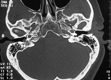
- histo pathological features are compatible with solitary Eosinophilic Granuloma. (neuroradiologycases.com)
- A review of available literature was done regarding the various modalities of treatment available for solitary eosinophilic granuloma and their results. (researchbib.com)
Eosinophils3
- Initially, it appeared that eosinophilic granuloma was just what it sounds like but as it was studied more thoroughly, it was found that there were three different classes of this condition, and not all were granulomas and not all involved eosinophils. (vin.com)
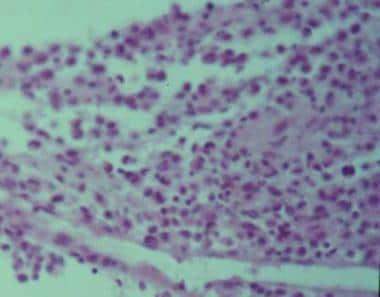
- It consists of Langerhans cells with oval, grooved and convoluted nucleus and slightly eosinophilic cytoplasm, associated with eosinophils, lymphocytes and plasma cells (H & E × 400). (scirp.org)
- Myocardial damage is caused by vasculitis, leading to coronary occlusion due to the release of toxic mediators by activated eosinophils causing direct myocardial damage, or by replacement of the myocardium with granulomas and scar tissue. (medscape.com)
Disease3
- Canine eosinophilic furunculosis is a closely related disease. (merckvetmanual.com)
- There is a form of the disease seen in dogs and cats called eosinophilic furunculosis , which is caused by an allergic reaction to insect bites and stings. (birnamvet.co.za)
- Granuloma faciale is primarily a disease of middle age (median age, 45 y). (medscape.com)
Skull1
- The granulomas, which usually occur in the skull or the long bones of the arms or legs, may cause the bone to fracture. (medlineplus.gov)
Benign1
- In humans, eosinophilic granulomas are considered as a benign tumors that occurs mainly in children and adolescents. (wikipedia.org)
Encephalitis1
- Consider Baylisascaris infection in persons with severe developmental disabilities or pica/geophagia and sudden onset of eosinophilic encephalitis. (cdc.gov)
Immunohistochemical1
- In immunohistochemical study, these eosinophilic cells expressed CD1a and S100 protein. (scirp.org)
Thighs1
- Also known as linear granuloma or collagenolytic granuloma, eosinophilic granulomas occur on the caudal thighs, face, and oral cavity (particularly the tongue and palate). (felipedia.org)
Vasculitis2
- Vassallo C, Derlino F, Croci GA, Brazzelli V, Borroni G. Chronic localized leukocytoclastic vasculitis: clinicopathological spectrum of granuloma faciale with and without extrafacial and mucosal involvement. (medscape.com)
- Autopsy revealed necrotizing vasculitis involving pericardial and intramyocardial vessels, as well as eosinophilic and giant cell inflammation of the heart and lungs, supporting the diagnosis of CSS. (medscape.com)
Nasal1
- Eosinophilic angiocentric fibrosis affecting the nasal cavity. (medscape.com)
Pathogenesis2
- In this course you will learn about pathogenesis, diagnosis and treatment of Eosinophilic granuloma. (vetacademy.org)
- [ 6 ] Innate and adaptive immunity seem to be factors in the pathogenesis of granuloma faciale. (medscape.com)
Acute1
- Neural larva migrans often presents as acute eosinophilic meningoencephalitis. (cdc.gov)
Canine1
- Canine eosinophilic granuloma (CEG) is extremely rare autoinflammatory state affecting primarily oral cavity and surrounding areas of transition between mucosa and hairy skin. (wikipedia.org)
Findings1
- Given the constellation of findings, this is most compatible with an eosinophilic granuloma. (logicalimages.com)
Larvae1
- Larvae can become encapsulated within granulomas. (cdc.gov)
Homeopathy1
- I was invited to Puerto Rico by local clients whose cat I had treated and cured through homeopathy from EGC- Eosinophilic granuloma complex- a chronic skin condition. (hpathy.com)
Occur2
- Granulomas also frequently occur in the skin, appearing as blisters, reddish bumps, or rashes which can be mild to severe. (medlineplus.gov)
- Costa Rica is considered the most endemic angiostrongiliasis en un country, and it has been shown in different reviews that most cases occur in children and males. (bvsalud.org)
Diseases1
- Ranging from simple infections to immune-related disorders, potential cat paw diseases include pillow foot, pemphigus, and eosinophilic granuloma. (cuteness.com)
Plaques1
- Multiple brown-red plaques on the face associated with granuloma faciale (same patient as in Media Files 13-16). (medscape.com)
Abdomen1
- Known as eosinophilic granuloma complex, the reaction can be present around the mouth or throat, chin or abdomen, hind legs, or footpads. (cuteness.com)
Skin1
- High local interleukin 5 production in granuloma faciale (eosinophilicum): role of clonally expanded skin-specific CD4+ cells. (medscape.com)