Ependymoma
Infratentorial Neoplasms
Spinal Cord Neoplasms
Supratentorial Neoplasms
Brain Neoplasms
Cerebral Ventricle Neoplasms
Central Nervous System Neoplasms
Cauda Equina
Siderosis
Radiotherapy, Conformal
Medulloblastoma
Fourth Ventricle
Glioma, Subependymal
Information Centers
Internet
Rare Diseases
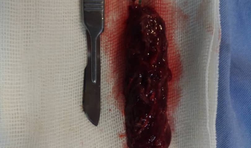
Extraneural metastasizing ependymoma of the spinal cord. (1/337)
This paper reports a case of the rare entity of an extraneural metastasizing ependymoma of the spinal cord. The tumor which arose in the conus medullaris and in the cauda equina was first diagnosed in 1956 when a thoracolumbar myeloresection was performed. At autopsy, 40 years after the primary diagnosis, a massive local tumor recurrence with extraneural metastases in the lungs, the pleura, the liver, and the thoracal and abdominal lymph nodes were found. Immunohistochemical stains of the extraneural metastases showed a strong cytoplasmatic expression of glial fibrillary acidic protein (GFAP). Neither the primary tumor nor its metastases showed any of the conventional morphological criteria of malignancy. Reviewing the literature we discuss the possible mechanism of extraneural tumor spread and the incidence of metastases with regard to the tumor type. (+info)Familial gliomas : a case report. (2/337)
Two non-twin brothers were found to have intracranial malignant neoplasms. The age of presentation was third and fourth decade but the onset was simultaneous, at the same time. Diagnosis in each of them was made by computed tomography and confirmed by histopathology. Elder among them had cellular ependymoma and the younger had oligodendroglioma. Both the brothers received radiotherapy post operatively and were surviving asymptomatically without any neurological deficit, leading active life as police constable, 12 months after surgical treatment. (+info)Molecular genetic analysis of ependymal tumors. NF2 mutations and chromosome 22q loss occur preferentially in intramedullary spinal ependymomas. (3/337)
Ependymal tumors are heterogeneous with regard to morphology, localization, age at first clinical manifestation, and prognosis. Several molecular alterations have been reported in these tumors, including allelic losses on chromosomes 10, 17, and 22 and mutations in the NF2 gene. However, in contrast to astrocytic gliomas, no consistent molecular alterations have been associated with distinct types of ependymal tumors. To evaluate whether morphological subsets of ependymomas are characterized by specific genetic lesions, we analyzed a series of 62 ependymal tumors, including myxopapillary ependymomas, subependymomas, ependymomas, and anaplastic ependymomas, for allelic losses on chromosome arms 10q and 22q and mutations in the PTEN and NF2 genes. Allelic losses on 10q and 22q were detected in 5 of 56 and 12 of 54 tumors, respectively. Six ependymomas carried somatic NF2 mutations, whereas no mutations were detected in the PTEN gene. All six of the NF2 mutations occurred in ependymomas of WHO grade II and were exclusively observed in tumors with a spinal localization (P = 0.0063). These findings suggest that a considerable fraction of spinal ependymomas are associated with molecular events involving chromosome 22 and that mutations in the NF2 gene may be of primary importance for their genesis. Furthermore, our data suggest that the more favorable clinical course of spinal ependymomas may relate to a distinct pattern of genetic alterations different from that of intracerebral ependymomas. (+info)Association of lower cranial nerve schwannoma with spinal ependymoma in ? NF2. (4/337)
A 15 year old male, who had earlier been operated for intraspinal intramedullary ependymoma, subsequently developed a right cerebello pontine (CP) angle mass. A diagnosis of right CP angle ependymoma was considered, in view of established histology of previously operated spinal lesion. Histopathological examination of the well defined extra-axial mass, which was attached with ninth cranial nerve, however revealed a schwannoma. A diagnosis of Neurofibromatosis-2 (NF2) is strongly suspected, because of well established fact, that the spinal ependymomas may have association with lower cranial nerve schwannomas in NF2. Cranial and spinal MRI screening for early diagnosis of associated, asymptomatic lesions, in suspected cases of NF2, particularly in children, is recommended. (+info)Tumour type and size are high risk factors for the syndrome of "cerebellar" mutism and subsequent dysarthria. (5/337)
OBJECTIVE: "Cerebellar mutis" and subsequent dysarthria (MSD) is a documented complication of posterior fossa surgery in children. In this prospective study the following risk factors for MSD were assessed: type, size and site of the tumour; hydrocephalus at presentation and after surgery, cerebellar incision site, postoperative infection, and cerebellar swelling. METHODS: In a consecutive series of 42 children with a cerebellar tumour, speech and neuroradiological studies (CT and MRI) were systematically analysed preoperatively and postoperatively. Speech was assessed using the Mayo Clinic lists and the severity of dysarthria using the Michigan rating scale. RESULTS: Twelve children (29%) developed MSD postoperatively. The type of tumour, midline localisation, and vermal incision were significant single independent risk factors. In addition, an interdependency of possible risk factors (tumour>5 cm, medulloblastoma) was found. CONCLUSION: MSD often occurs after paediatric cerebellar tumour removal and is most likely after removal of a medulloblastoma with a maximum lesion diameter>5 cm. (+info)Evidence for an ependymoma tumour suppressor gene in chromosome region 22pter-22q11.2. (6/337)
Ependymomas are glial tumours of the brain and spinal cord. The most frequent genetic change in sporadic ependymoma is monosomy 22, suggesting the presence of an ependymoma tumour suppressor gene on that chromosome. Clustering of ependymomas has been reported to occur in some families. From an earlier study in a family in which four cousins developed an ependymoma, we concluded that an ependymoma-susceptibility gene, which is not the NF2 gene in 22q12, might be located on chromosome 22. To localize that gene, we performed a segregation analysis with chromosome 22 markers in this family. This analysis revealed that the susceptibility gene may be located proximal to marker D22S941 in 22pter-22q11.2. Comparative genomic hybridization showed that monosomy 22 was the sole detectable genetic aberration in the tumour of one of the patients. Loss of heterozygosity studies in that tumour revealed that, in accordance to Knudson's two-hit theory of tumorigenesis, the lost chromosome 22 originated from the parent presumed to have contributed the wild-type allele of the susceptibility gene. Thus, our segregation and tumour studies collectively indicate that an ependymoma tumour suppressor gene may be present in region 22pter-22q11.2. (+info)Expression of bisecting GlcNAc in pediatric brain tumors and its association with tumor cell response to vinblastine. (7/337)
Increased expression of the bisecting GlcNAc has been correlated with tumor progression in several experimental tumor models. Its expression and function in brain tumors are, however, not yet known. In this study, we investigated expression of the bisecting GlcNAc structure in a series of pediatric brain tumors and its relationship to tumor response to vinblastine. A plant lectin (E-PHA) that recognizes the bisecting GlcNAc structure was used for detection of this molecule in a total of 90 pediatric brain tumors and normal brain tissue specimens. Our results showed that, whereas E-PHA staining was undetectable in the normal brain tissue, pediatric brain tumor specimens exhibited different levels of reactivity. Lectin staining was particularly prominent in high-grade astrocytomas (73%) and ependymomas (72%). In astrocytomas, there was a positive correlation with the tumor grade, which suggests that the bisecting GlcNAc may be of particular interest as a tumor marker for diagnosis and/or prognosis. By using a human glioma cell culture model, we have found that treatment of these cells with E-PHA lectin enhances their sensitivity to vinblastine. E-PHA interacted directly with the drug transporter P-glycoprotein and inhibited its drug efflux function. In a drug-resistant glioma cell line transfected with the mdr1 gene, drug resistance was reversed by E-PHA. Our findings indicate that: (a) expression of the bisecting GlcNAc in pediatric brain tumors may have a potential relevance as a tumor marker; and (b) glioma response to chemotherapy may be modulated through the bisecting GlcNAc. (+info)Neuropsychological consequences of cerebellar tumour resection in children: cerebellar cognitive affective syndrome in a paediatric population. (8/337)
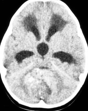
Acquired cerebellar lesions in adults have been shown to produce impairments in higher function as exemplified by the cerebellar cognitive affective syndrome. It is not yet known whether similar findings occur in children with acquired cerebellar lesions, and whether developmental factors influence their presentation. In studies to date, survivors of childhood cerebellar tumours who demonstrate long-term deficits in cognitive functions have undergone surgery as well as cranial irradiation or methotrexate treatment. Investigation of the effects of the cerebellar lesion independent of the known deleterious effects of these agents is important for understanding the role of the cerebellum in cognitive and affective development and for informing treatment and rehabilitation strategies. If the cerebellar contribution to cognition and affect is significant, then damage in childhood may influence a wide range of psychological processes, both as an immediate consequence and as these processes fail to develop normally later on. In this study we evaluated neuropsychological data in 19 children who underwent resection of cerebellar tumours but who received neither cranial irradiation nor methotrexate chemotherapy. Impairments were noted in executive function, including planning and sequencing, and in visual-spatial function, expressive language, verbal memory and modulation of affect. These deficits were common and in some cases could be dissociated from motor deficits. Lesions of the vermis in particular were associated with dysregulation of affect. Behavioural deficits were more apparent in older than younger children. These results reveal that clinically relevant neuropsychological changes may occur following cerebellar tumour resection in children. Age at the time of surgery and the site of the cerebellar lesion influence the neurobehavioural outcome. The results of the present study indicate that the cerebellar cognitive affective syndrome is evident in children as well as in adults, and they provide further clinical evidence that the cerebellum is an essential node in the distributed neural circuitry subserving higher-order behaviours. (+info)Ependymoma is a type of brain or spinal cord tumor that develops from the ependymal cells that line the ventricles (fluid-filled spaces) in the brain, or the central canal of the spinal cord. These tumors can be benign or malignant, and they can cause various symptoms depending on their location and size.
Ependymomas are relatively rare, accounting for about 2-3% of all primary brain and central nervous system tumors. They most commonly occur in children and young adults, but they can also affect older individuals. Treatment typically involves surgical removal of the tumor, followed by radiation therapy or chemotherapy, depending on the grade and location of the tumor. The prognosis for ependymomas varies widely, with some patients experiencing long-term survival and others having more aggressive tumors that are difficult to treat.
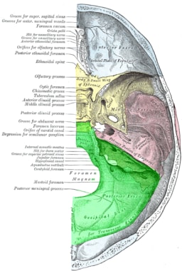
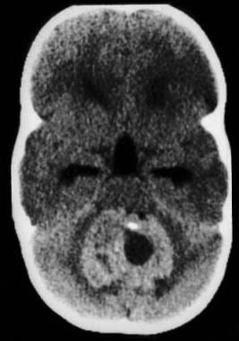
Infratentorial neoplasms refer to tumors that originate in the region of the brain called the posterior fossa, which is located below the tentorium cerebelli (a membranous structure that separates the cerebrum from the cerebellum). This area contains several important structures such as the cerebellum, pons, medulla oblongata, and fourth ventricle. Infratentorial neoplasms can be benign or malignant and can arise from various cell types including nerve cells, glial cells, or supportive tissues. They can cause a variety of symptoms depending on their location and size, such as headache, vomiting, unsteady gait, weakness, numbness, vision changes, hearing loss, and difficulty swallowing or speaking. Treatment options may include surgery, radiation therapy, and chemotherapy.
Spinal cord neoplasms refer to abnormal growths or tumors within the spinal cord. These can be benign (non-cancerous) or malignant (cancerous). They originate from the cells within the spinal cord itself (primary tumors), or they may spread to the spinal cord from other parts of the body (metastatic tumors). Spinal cord neoplasms can cause various symptoms depending on their location and size, including back pain, neurological deficits, and even paralysis. Treatment options include surgery, radiation therapy, and chemotherapy.
Supratentorial neoplasms refer to tumors that originate in the region of the brain located above the tentorium cerebelli, which is a dual layer of dura mater (the protective outer covering of the brain) that separates the cerebrum from the cerebellum. This area includes the cerebral hemispheres, basal ganglia, thalamus, hypothalamus, and pineal gland. Supratentorial neoplasms can be benign or malignant and may arise from various cell types such as neurons, glial cells, meninges, or blood vessels. They can cause a variety of neurological symptoms depending on their size, location, and rate of growth.
Brain neoplasms, also known as brain tumors, are abnormal growths of cells within the brain. These growths can be benign (non-cancerous) or malignant (cancerous). Benign brain tumors typically grow slowly and do not spread to other parts of the body. However, they can still cause serious problems if they press on sensitive areas of the brain. Malignant brain tumors, on the other hand, are cancerous and can grow quickly, invading surrounding brain tissue and spreading to other parts of the brain or spinal cord.
Brain neoplasms can arise from various types of cells within the brain, including glial cells (which provide support and insulation for nerve cells), neurons (nerve cells that transmit signals in the brain), and meninges (the membranes that cover the brain and spinal cord). They can also result from the spread of cancer cells from other parts of the body, known as metastatic brain tumors.
Symptoms of brain neoplasms may vary depending on their size, location, and growth rate. Common symptoms include headaches, seizures, weakness or paralysis in the limbs, difficulty with balance and coordination, changes in speech or vision, confusion, memory loss, and changes in behavior or personality.
Treatment for brain neoplasms depends on several factors, including the type, size, location, and grade of the tumor, as well as the patient's age and overall health. Treatment options may include surgery, radiation therapy, chemotherapy, targeted therapy, or a combination of these approaches. Regular follow-up care is essential to monitor for recurrence and manage any long-term effects of treatment.
Cerebral ventricle neoplasms refer to tumors that develop within the cerebral ventricles, which are fluid-filled spaces in the brain. These tumors can arise from various types of cells within the ventricular system, including the ependymal cells that line the ventricles, choroid plexus cells that produce cerebrospinal fluid, or other surrounding tissues.
Cerebral ventricle neoplasms can cause a variety of symptoms depending on their size and location, such as headaches, nausea, vomiting, vision changes, imbalance, weakness, or difficulty with mental tasks. The treatment options for these tumors may include surgical resection, radiation therapy, and chemotherapy, depending on the type and extent of the tumor. Regular follow-up care is essential to monitor for recurrence and manage any long-term effects of treatment.
Central nervous system (CNS) neoplasms refer to a group of abnormal growths or tumors that develop within the brain or spinal cord. These tumors can be benign or malignant, and their growth can compress or disrupt the normal functioning of surrounding brain or spinal cord tissue.
Benign CNS neoplasms are slow-growing and rarely spread to other parts of the body. However, they can still cause significant problems if they grow large enough to put pressure on vital structures within the brain or spinal cord. Malignant CNS neoplasms, on the other hand, are aggressive tumors that can invade and destroy surrounding tissue. They may also spread to other parts of the CNS or, rarely, to other organs in the body.
CNS neoplasms can arise from various types of cells within the brain or spinal cord, including nerve cells, glial cells (which provide support and insulation for nerve cells), and supportive tissues such as blood vessels. The specific type of CNS neoplasm is often used to help guide treatment decisions and determine prognosis.
Symptoms of CNS neoplasms can vary widely depending on the location and size of the tumor, but may include headaches, seizures, weakness or paralysis, vision or hearing changes, balance problems, memory loss, and changes in behavior or personality. Treatment options for CNS neoplasms may include surgery, radiation therapy, chemotherapy, or a combination of these approaches.
The Cauda Equina refers to a bundle of nerves at the lower end of the spinal cord within the vertebral column. It originates from the lumbar (L1-L5) and sacral (S1-S5) regions and looks like a horse's tail, hence the name "Cauda Equina" in Latin. These nerves are responsible for providing motor and sensory innervation to the lower extremities, bladder, bowel, and sexual organs. Any damage or compression to this region can lead to serious neurological deficits, such as bowel and bladder incontinence, sexual dysfunction, and lower limb weakness or paralysis.
Siderosis is a medical condition characterized by the abnormal accumulation of iron in various tissues and organs, most commonly in the lungs. This occurs due to the repeated inhalation of iron-containing dusts or fumes, which can result from certain industrial processes such as welding, mining, or smelting.
In the lungs, this iron deposit can lead to inflammation and fibrosis, potentially causing symptoms like coughing, shortness of breath, and decreased lung function. It is important to note that siderosis itself is not contagious or cancerous, but there may be an increased risk for lung cancer in individuals with severe and prolonged exposure to iron-containing particles.
While siderosis is generally non-reversible, the progression of symptoms can often be managed through medical interventions and environmental modifications to reduce further exposure to iron-containing dusts or fumes.
The sacrococcygeal region is the lower part of the back where the spine ends, specifically referring to the area where the sacrum (a triangular bone at the base of the spine formed by the fusion of several vertebrae) meets the coccyx (also known as the tailbone). This region is located at the very bottom of the spine and is susceptible to injury or trauma due to its position and role in supporting the body's weight. It is also a common site for birth defects, particularly in newborns.
Neurosurgical procedures are operations that are performed on the brain, spinal cord, and peripheral nerves. These procedures are typically carried out by neurosurgeons, who are medical doctors with specialized training in the diagnosis and treatment of disorders of the nervous system. Neurosurgical procedures can be used to treat a wide range of conditions, including traumatic injuries, tumors, aneurysms, vascular malformations, infections, degenerative diseases, and congenital abnormalities.
Some common types of neurosurgical procedures include:
* Craniotomy: A procedure in which a bone flap is temporarily removed from the skull to gain access to the brain. This type of procedure may be performed to remove a tumor, repair a blood vessel, or relieve pressure on the brain.
* Spinal fusion: A procedure in which two or more vertebrae in the spine are fused together using bone grafts and metal hardware. This is often done to stabilize the spine and alleviate pain caused by degenerative conditions or spinal deformities.
* Microvascular decompression: A procedure in which a blood vessel that is causing pressure on a nerve is repositioned or removed. This type of procedure is often used to treat trigeminal neuralgia, a condition that causes severe facial pain.
* Deep brain stimulation: A procedure in which electrodes are implanted in specific areas of the brain and connected to a battery-operated device called a neurostimulator. The neurostimulator sends electrical impulses to the brain to help alleviate symptoms of movement disorders such as Parkinson's disease or dystonia.
* Stereotactic radiosurgery: A non-invasive procedure that uses focused beams of radiation to treat tumors, vascular malformations, and other abnormalities in the brain or spine. This type of procedure is often used for patients who are not good candidates for traditional surgery due to age, health status, or location of the lesion.
Neurosurgical procedures can be complex and require a high degree of skill and expertise. Patients considering neurosurgical treatment should consult with a qualified neurosurgeon to discuss their options and determine the best course of action for their individual situation.
Conformal radiotherapy is a type of external beam radiation therapy that uses advanced technology to conform the radiation beam to the shape of the tumor, allowing for more precise and targeted treatment while minimizing exposure to healthy surrounding tissue. This can help reduce the risk of side effects and improve the therapeutic ratio. Conformal radiotherapy techniques include 3D conformal radiation therapy (3D-CRT), intensity-modulated radiation therapy (IMRT), and volumetric modulated arc therapy (VMAT). These techniques use sophisticated imaging and treatment planning systems to create a personalized treatment plan for each patient, based on the size, shape, and location of their tumor.
Medulloblastoma is a type of malignant brain tumor that originates in the cerebellum, which is the part of the brain located at the back of the skull and controls coordination and balance. It is one of the most common types of pediatric brain tumors, although it can also occur in adults.
Medulloblastomas are typically made up of small, round cancer cells that grow quickly and can spread to other parts of the central nervous system, such as the spinal cord. They are usually treated with a combination of surgery, radiation therapy, and chemotherapy. The exact cause of medulloblastoma is not known, but it is thought to be related to genetic mutations or abnormalities that occur during development.
The fourth ventricle is a part of the cerebrospinal fluid-filled system in the brain, located in the posterior cranial fossa and continuous with the central canal of the medulla oblongata and the cerebral aqueduct. It is shaped like a cavity with a roof, floor, and lateral walls, and it communicates rostrally with the third ventricle through the cerebral aqueduct and caudally with the subarachnoid space through the median and lateral apertures (foramina of Luschka and Magendie). The fourth ventricle contains choroid plexus tissue, which produces cerebrospinal fluid. Its roof is formed by the cerebellar vermis and the superior medullary velum, while its floor is composed of the rhomboid fossa, which includes several important structures such as the vagal trigone, hypoglossal trigone, and striae medullares.
A subependymal glioma is a rare, typically slow-growing type of brain tumor that develops in the lining of the fluid-filled spaces (ventricles) within the brain. These tumors are formed from glial cells, which are supportive cells that help nerve cells function.
Subependymal gliomas are often associated with a genetic disorder called tuberous sclerosis complex (TSC), and they account for about 10-15% of all brain tumors in TSC patients. These tumors usually appear as small, nodular growths on the walls of the ventricles, particularly near the region where the ventricles connect to the lower part of the brain (the brainstem).
Although subependymal gliomas are generally slow-growing, they can cause symptoms by obstructing the flow of cerebrospinal fluid (CSF) within the ventricles. This obstruction can lead to an increase in intracranial pressure and a condition called hydrocephalus, which may require surgical intervention to alleviate. Symptoms of subependymal gliomas can include headaches, vomiting, seizures, balance problems, and developmental delays.
Treatment options for subependymal gliomas depend on the size and location of the tumor, as well as the presence of any associated symptoms. In some cases, these tumors may not require treatment if they are small and asymptomatic. However, if the tumor is causing obstruction or symptoms, surgical removal or other treatments such as radiation therapy or chemotherapy may be necessary. Regular follow-up with imaging studies is essential to monitor the growth and development of subependymal gliomas in TSC patients.
I'm sorry for any confusion, but "Information Centers" is not a medical term per se. However, in a broad context, information centers can refer to organized locations that provide specific health-related or medical information to patients, healthcare professionals, or the general public. These can be found in hospitals, clinics, universities, government agencies, and other organizations. They offer access to various resources such as books, pamphlets, databases, and online tools, with the aim of promoting evidence-based practices, enhancing knowledge, and supporting decision-making.
I'm sorry for any confusion, but "Internet" is a term that pertains to the global network of interconnected computers and servers that enable the transmission and reception of data via the internet protocol (IP). It is not a medical term and does not have a specific medical definition. If you have any questions related to medicine or health, I'd be happy to try to help answer them for you!
A rare disease, also known as an orphan disease, is a health condition that affects fewer than 200,000 people in the United States or fewer than 1 in 2,000 people in Europe. There are over 7,000 rare diseases identified, and many of them are severe, chronic, and often life-threatening. The causes of rare diseases can be genetic, infectious, environmental, or degenerative. Due to their rarity, research on rare diseases is often underfunded, and treatments may not be available or well-studied. Additionally, the diagnosis of rare diseases can be challenging due to a lack of awareness and understanding among healthcare professionals.
 Ependymoma
 Ependymoma Pathology: Definition, Epidemiology, Etiology
 Ependymoma - Childhood: Statistics | Cancer.Net
 Ependymoma | St. Jude Research
 Ependymoma Foundation Informs and Inspires a Rare Cancer Community - NCI
 Ependymoma
 The Multifaceted Appearance of Supratentorial Ependymoma with ZFTA-MAML2 Fusion
| Free Neuropathology
The Multifaceted Appearance of Supratentorial Ependymoma with ZFTA-MAML2 Fusion
| Free Neuropathology
 An integrated in vitro and in vivo high-throughput screen identifies treatment leads for ependymoma
 A trial looking at treatment for children and young people with an ependymoma (SIOP 99) | Cancer Research UK
 Immune Checkpoint Inhibitor Nivolumab in People With Recurrent Select Rare CNS Cancers | Clinical Research Trial Listing (...
 Myxopapillary ependymoma - About the Disease - Genetic and Rare Diseases Information Center
 Ependymoma Treatment Market Size, Share, Trends 2022-2030
 Ependymoma
 Brain and Nervous System Cancers (for Parents) - East Tenneesee Children's
 Ependymoma - MeSH - NCBI
 Pediatric Ependymoma Patient Guide
 Childhood Ependymoma Treatment (PDQ®)
 Ependymoma
 Spinal Cord Ependymoma | دکتر گیو شریفی
 EPENDYMOMA AWARENESS DAY - BRAIN CANCER BABE
 What is ependymoma: Symptoms and treatment
 Ependymoma Care at Tufts Medical Center


![10 Best Clinics for Esophageal Cancer Treatment in Thailand [2023 Prices]](https://www.mymeditravel.com/cdn-cgi/image/f=auto,fit=contain,quality=75/uploads/property/gallery/5af2758efa6b7e04401f8c27/5af51dadfa6b7e4212052361/preview.jpg)