Esophagus
Barrett Esophagus
Esophagogastric Junction
Metaplasia
Gastroesophageal Reflux
Cardia
Esophagitis
Mucous Membrane
Precancerous Conditions
Esophagitis, Peptic
Esophageal Perforation
Peristalsis
Esophageal Fistula
Carcinoma, Squamous Cell
Dimethylnitrosamine
Esophageal Sphincter, Lower
Deglutition
Endoscopy, Gastrointestinal
Hypopharynx
Hernia, Hiatal
Esophageal Achalasia
Deglutition Disorders
Caustics
Stomach
Endoscopy
Fundoplication
Esophageal Sphincter, Upper
Esophageal Atresia
Hydrochloric Acid
Burns, Chemical
Biopsy
Duodenogastric Reflux
Eosinophilic Esophagitis
Upper Gastrointestinal Tract
Tracheoesophageal Fistula
Esophageal Motility Disorders
Esophageal pH Monitoring
Immunohistochemistry
Bile Reflux
Gastrointestinal Neoplasms
Numismatics
Fistula
Digestive System
Anastomosis, Surgical
Nitrosamines
Mediastinum
Retrospective Studies
Laryngopharyngeal Reflux
Larynx
Disease Progression
Risk Factors
Ulcer
Gastrectomy
Carcinoma, Basosquamous
Case-Control Studies
Duodenum
Treatment Outcome
Gastric Mucosa
Esophageal Spasm, Diffuse
Diagnostic Techniques, Digestive System
Catheter Ablation
Epithelium
Heartburn
Nodose Ganglion
Pressure
Goblet Cells
Photochemotherapy
Carcinogens
Neoplasm Staging
Esophageal Cyst
Tumor Markers, Biological
Mucin-2
Regulation and function of family 1 and family 2 UDP-glucuronosyltransferase genes (UGT1A, UGT2B) in human oesophagus. (1/2903)
Human UDP-glucuronosyltransferases (UGTs) are expressed in a tissue-specific fashion in hepatic and extrahepatic tissues [Strassburg, Manns and Tukey (1998) J. Biol. Chem. 273, 8719-8726]. Previous work suggests that these enzymes play a protective role in chemical carcinogenesis [Strassburg, Manns and Tukey (1997) Cancer Res. 57, 2979-2985]. In this study, UGT1 and UGT2 gene expression was investigated in human oesophageal epithelium and squamous-cell carcinoma in addition to the characterization of individual UGT isoforms using recombinant protein. UGT mRNA expression was characterized by duplex reverse transcriptase-PCR analysis and revealed the expression of UGT1A7, UGT1A8, UGT1A9 and UGT1A10 mRNAs. UGT1A1, UGT1A3, UGT1A4, UGT1A5 and UGT1A6 transcripts were not detected. UGT2 expression included UGT2B7, UGT2B10 and UGT2B15, but UGT2B4 mRNA was absent. UGT2 mRNA was present at significantly lower levels than UGT1 transcripts. This observation was in agreement with the analysis of catalytic activities in oesophageal microsomal protein, which was characterized by high glucuronidation rates for phenolic xenobiotics, all of which are classical UGT1 substrates. Whereas UGT1A9 was not regulated, differential regulation of UGT1A7 and UGT1A10 mRNA was observed between normal oesophageal epithelium and squamous-cell carcinoma. Expression and analysis in vitro of recombinant UGT1A7, UGT1A9, UGT1A10, UGT2B7 and UGT2B15 demonstrated that UGT1A7, UGT1A9 and UGT1A10 catalysed the glucuronidation of 7-hydroxybenzo(alpha)pyrene, as well as other environmental carcinogens, such as 2-hydroxyamino-1-methyl-6-phenylimidazo-(4, 5-beta)-pyridine. Although UGT1A9 was not regulated in the carcinoma tissue, the five-fold reduction in 7-hydroxybenzo(alpha)pyrene glucuronidation could be attributed to regulation of UGT1A7 and UGT1A10. These data elucidate an individual regulation of human UGT1A and UGT2B genes in human oesophagus and provide evidence for specific catalytic activities of individual human UGT isoforms towards environmental carcinogens that have been implicated in cellular carcinogenesis. (+info)The integrin alpha v beta 6 binds and activates latent TGF beta 1: a mechanism for regulating pulmonary inflammation and fibrosis. (2/2903)
Transforming growth factor beta (TGF beta) family members are secreted in inactive complexes with a latency-associated peptide (LAP), a protein derived from the N-terminal region of the TGF beta gene product. Extracellular activation of these complexes is a critical but incompletely understood step in regulation of TGF beta function in vivo. We show that TGF beta 1 LAP is a ligand for the integrin alpha v beta 6 and that alpha v beta 6-expressing cells induce spatially restricted activation of TGF beta 1. This finding explains why mice lacking this integrin develop exaggerated inflammation and, as we show, are protected from pulmonary fibrosis. These data identify a novel mechanism for locally regulating TGF beta 1 function in vivo by regulating expression of the alpha v beta 6 integrin. (+info)The postnatal development of the alimentary canal in the opossum. I. Oesophagus. (3/2903)
The oesophageal epithelium of the newborn opossum generally is two to three cells in depth and in some regions appears pseudostratified. By the 9th postnatal day the epithelium shows two distinct strata. Ciliated cells and occasional goblet cells also are observed within the epithelium during this stage and in subsequent stages. Cilia persist in the oesophagus of the adult opossum, but are restricted to the depths of the transverse folds found in the distal part of the organ. The epithelium covering the transverse folds of the adult likewise has an immature appearance. By 4-5 cm (ca. 20 days), the epithelium has assumed a more mature appearance and is of greater depth. This and later stages show three basic strata: a germinal layer, a spinous layer and, adjacent to the lumen, a flattened layer of cells that retain their nuclei. The epithelium throughout the postnatal period and in the adult does not undergo complete keratinization. The oesophageal glands begin as outgrowths from the epithelium just prior to 4-5 cm (ca. 20 days). The glands continue their development throughout the remainder of the postnatal period. The secretory units of the oesophageal glands of the the major portion of the secretory elements, and a light, rounded cell type which is less numerous and which occupies the terminal portions of the secretory units. Secretory material of the former appears complex, consisting of both neutral and acid glycoproteins. The secretory product of the light cell type is unknown at present. Both cell types are encompassed by myoepithelial cells. The relationship of the mitotic sequences to the observations made by microscopic examination of the developing oesophagus is discussed. (+info)Langerhans cells in the human oesophagus. (4/2903)
The dendrite cells of Langerhans, first identified in the epidermis, have now been observed in the middle and superficial layers of the normal human oesophageal mucosa. They exhibit typical Langerhans granules, but no desmosomes and tonofilaments. They often have irregular indented nuclei, with a relatively pale cytoplasm contrasting with that of the adjacent squamous cells. These cells are sometimes difficult to distinguish from intra-epithelial lymphocytes, which are also encountered in the oesophageal mucosa and which share certain ultrastructural characteristics with Langerhans cells. (+info)Oesophageal epithelial innervation in health and reflux oesophagitis. (5/2903)
BACKGROUND: The response of the oesophagus to refluxed gastric contents is likely to depend on intact neural mechanisms in the oesophageal mucosa. The epithelial innervation has not been systematically evaluated in health or reflux disease. AIMS: To study oesophageal epithelial innervation in controls, and also inflamed and non-inflamed mucosa in patients with reflux oesophagitis and healed oesophagitis. PATIENTS: Ten controls, nine patients with reflux oesophagitis, and five patients with healed oesophagitis. METHODS: Oesophageal epithelial biopsy specimens were obtained at endoscopy. The distribution of the neuronal marker protein gene product 9.5 (PGP), and the neuropeptides calcitonin gene related peptide (CGRP), neuropeptide Y (NPY), substance P (SP), and vasoactive intestinal peptide (VIP) were investigated by immunohistochemistry. Density of innervation was assessed by the proportion of papillae in each oesophageal epithelial biopsy specimen containing immunoreactive fibres (found in the subepithelium and epithelial papillae, but not penetrating the epithelium). RESULTS: The proportion of papillae positive for PGP immunoreactive nerve fibres was significantly increased in inflamed tissue when compared with controls, and non-inflamed and healed tissue. There was also a significant increase in VIP immunoreactive fibres within epithelial papillae. Other neuropeptides showed no proportional changes in inflammation. CONCLUSIONS: Epithelial biopsy specimens can be used to assess innervation in the oesophagus. The innervation of the oesophageal mucosa is not altered in non-inflamed tissue of patients with oesophagitis but alters in response to inflammation, where there is a selective increase (about three- to fourfold) in VIP containing nerves. (+info)Differential expression of Hsp27 in normal oesophagus, Barrett's metaplasia and oesophageal adenocarcinomas. (6/2903)
The protein expression patterns of normal, metaplastic and malignant oesophageal tissues were analysed by two-dimensional polyacrylamide gel electrophoresis (2D-PAGE) to identify changes associated with Barrett's metaplasia and transformation to oesophageal adenocarcinoma. Heat-shock protein 27 (Hsp27), a small heat-shock protein which is protective against cytotoxic stresses, was abundant in normal oesophagus. However, Hsp27 expression was markedly lower in Barrett's metaplasia and oesophageal adenocarcinomas. This was confirmed by immunohistochemical analysis. Hsp27 protein was most highly expressed in the upper layers of squamous epithelium and exhibited a pattern of expression that corresponded with the degree of squamous maturation. Northern and Southern analysis demonstrated Hsp27 to be regulated at the level of mRNA transcription or abundance. Normal oesophageal tissues were examined for gender differences in Hsp27 expression. Women expressed fourfold higher levels of Hsp27 mRNA, however, this difference was not appreciable in protein expression. Hsp27 protein was inducible by heat shock in Barrett's adenocarcinoma cell lines and an immortalized oesophageal epithelial cell line (HET-1A), but not by oestradiol. These results demonstrate abundant constitutive expression of the stress-response protein Hsp27 in the normal oesophagus, and suggest that low-level expression in Barrett's metaplasia may be one factor which may influence susceptibility to oesophageal adenocarcinoma development. (+info)Characterization of cytochrome P450 expression in human oesophageal mucosa. (7/2903)
The expression of cytochrome (CYP) P450 enzymes in human oesophageal mucosa was investigated in a total of 25 histologically non-neoplastic surgical tissue specimens by using specific antibodies in immunoblots and by RT-PCR mRNA analysis. The presence of CYP1A, 2E1, 3A and 4A enzymes was demonstrated by both techniques; CYP2A reactive protein was also detected by immunoblot. The presence of CYP4B1 mRNA was established but no specific antibody was available for detection of the corresponding protein by immunoblot. CYP2B6/7 mRNA was not detected in any sample. The mRNA transcripts for CYP1A1, 2E1, 4A11 and 4B1 were consistently detected in the majority of samples (>84%), whereas CYP1A2 mRNA was only detected in 11 of 19 specimens examined. An RT-PCR method to differentiate CYP3A4 and 3A5 mRNA was developed. This demonstrated CYP3A5 mRNA expression in all samples tested, whereas CYP3A4 mRNA was not detectable, suggesting that CYP3A5 is the major CYP3A protein in human oesophagus. There were significant interindividual variations in the amount of proteins, ranging from 8-fold for CYP4A to 43-fold for CYP2E1. For each patient, data on exposure to risk factors for oesophageal cancer were available, including tobacco smoke, alcohol, gastro-oesophageal reflux and hot beverage consumption. None of these risk factors or other patient characteristics (age, sex, tumour location and tumour stage) were correlated with the protein level of the individual CYP enzymes as determined by quantitation of immunoblot staining. However, the small series of samples precludes any strong conclusion concerning the lack of such correlations. There were no differences between squamous cell carcinomas and adenocarcinomas in either the qualitative or quantitative expression of the CYP enzymes. These data demonstrate that a range of CYP enzymes are expressed in human oesophageal mucosa and indicate that this tissue has the capacity to activate chemical carcinogens to reactive DNA binding metabolites. (+info)Neurocardiac and cerebral responses evoked by esophageal vago-afferent stimulation in humans: effect of varying intensities. (8/2903)
OBJECTIVE: This study was designed to determine whether esophageal vago-afferent electrostimulation, over a wide range of stimulus intensities, can sustain a cardiac vago-efferent effect by way of central nervous system processing. METHODS: Studies were performed in ten healthy male subjects (23.9 +/- 6.3 years). Esophageal electrostimulation was carried out using a stimulating electrode placed in the distal esophagus. Stimulation of esophageal vago-afferent fibres was employed using electrical impulses (200 microseconds at 0.2 Hz x 128 s) varying from 2.7 to 20 mA. Respiratory frequencies, beat-to-beat heart rate autospectra and cerebral evoked potentials were recorded at baseline and at each stimulus intensity in random order. RESULTS: With esophageal electrical stimulation, we observed a small non-significant decrease in heart rate. There was a dramatic shift of the instantaneous heart rate power spectra towards enhanced cardiac vagal modulation with intensities as low as 5 mA. This effect was sustained throughout all intensities with no further change in either the low frequency or high frequency power. Conversely, there was a linear dose response relationship between cerebral evoked potential amplitude and stimulus intensity mainly occurring above perception threshold (10 mA). Esophageal stimulation had no significant effect on heart rate or respiratory frequency at any stimulus intensity. CONCLUSIONS: These results indicate that electrical stimulation of the distal esophagus across a wide range of current intensities elicits a reproducible shift in the heart rate power spectrum towards enhanced vagal modulation. The data suggest a closed loop afferent/efferent circuitry wherein tonic visceral afferent impulses appear to elicit a phasic or modulatory vago-efferent cardiac response in healthy subjects. (+info)The esophagus is the muscular tube that connects the throat (pharynx) to the stomach. It is located in the midline of the neck and chest, passing through the diaphragm to enter the abdomen and join the stomach. The main function of the esophagus is to transport food and liquids from the mouth to the stomach for digestion.
The esophagus has a few distinct parts: the upper esophageal sphincter (a ring of muscle that separates the esophagus from the throat), the middle esophagus, and the lower esophageal sphincter (another ring of muscle that separates the esophagus from the stomach). The lower esophageal sphincter relaxes to allow food and liquids to enter the stomach and then contracts to prevent stomach contents from flowing back into the esophagus.
The walls of the esophagus are made up of several layers, including mucosa (a moist tissue that lines the inside of the tube), submucosa (a layer of connective tissue), muscle (both voluntary and involuntary types), and adventitia (an outer layer of connective tissue).
Common conditions affecting the esophagus include gastroesophageal reflux disease (GERD), Barrett's esophagus, esophageal cancer, esophageal strictures, and eosinophilic esophagitis.
Barrett esophagus is a condition in which the tissue lining of the lower esophagus changes, becoming more like the tissue that lines the intestines (intestinal metaplasia). This change can increase the risk of developing esophageal adenocarcinoma, a type of cancer. The exact cause of Barrett esophagus is not known, but it is often associated with long-term gastroesophageal reflux disease (GERD), also known as chronic acid reflux.
In Barrett esophagus, the normal squamous cells that line the lower esophagus are replaced by columnar epithelial cells. This change is usually detected during an upper endoscopy and biopsy. The diagnosis of Barrett esophagus is confirmed when the biopsy shows intestinal metaplasia in the lower esophagus.
It's important to note that not everyone with GERD will develop Barrett esophagus, and not everyone with Barrett esophagus will develop esophageal cancer. However, if you have been diagnosed with Barrett esophagus, your healthcare provider may recommend regular endoscopies and biopsies to monitor the condition and reduce the risk of cancer. Treatment options for Barrett esophagus include medications to control acid reflux, lifestyle changes, and in some cases, surgery.
Esophageal neoplasms refer to abnormal growths in the tissue of the esophagus, which is the muscular tube that connects the throat to the stomach. These growths can be benign (non-cancerous) or malignant (cancerous). Malignant esophageal neoplasms are typically classified as either squamous cell carcinomas or adenocarcinomas, depending on the type of cell from which they originate.
Esophageal cancer is a serious and often life-threatening condition that can cause symptoms such as difficulty swallowing, chest pain, weight loss, and coughing. Risk factors for esophageal neoplasms include smoking, heavy alcohol consumption, gastroesophageal reflux disease (GERD), and Barrett's esophagus. Treatment options may include surgery, radiation therapy, chemotherapy, or a combination of these approaches.
Esophagoscopy is a medical procedure that involves the visual examination of the esophagus, which is the tube that connects the throat to the stomach. This procedure is typically carried out using an esophagogastroduodenoscope (EGD), a flexible tube with a camera and light on the end.
During the procedure, the EGD is inserted through the mouth and down the throat into the esophagus, allowing the medical professional to examine its lining for any abnormalities such as inflammation, ulcers, or tumors. The procedure may also involve taking tissue samples (biopsies) for further examination and testing.
Esophagoscopy is commonly used to diagnose and monitor conditions such as gastroesophageal reflux disease (GERD), Barrett's esophagus, esophageal cancer, and other disorders affecting the esophagus. It may also be used to treat certain conditions, such as removing polyps or foreign objects from the esophagus.
Esophageal diseases refer to a range of medical conditions that affect the esophagus, which is the muscular tube that connects the throat to the stomach. Here are some common esophageal diseases with their brief definitions:
1. Gastroesophageal reflux disease (GERD): A chronic condition in which stomach acid or bile flows back into the esophagus, causing symptoms such as heartburn, chest pain, and difficulty swallowing.
2. Esophagitis: Inflammation of the esophageal lining, often caused by GERD, infection, or medication.
3. Esophageal stricture: Narrowing of the esophagus due to scarring or inflammation, which can make swallowing difficult.
4. Esophageal cancer: Cancer that forms in the tissues of the esophagus, often as a result of long-term GERD or smoking.
5. Esophageal motility disorders: Disorders that affect the normal movement and function of the esophagus, such as achalasia, diffuse spasm, and nutcracker esophagus.
6. Barrett's esophagus: A condition in which the lining of the lower esophagus changes, increasing the risk of esophageal cancer.
7. Esophageal diverticula: Small pouches that form in the esophageal wall, often causing difficulty swallowing or regurgitation.
8. Eosinophilic esophagitis (EoE): A chronic immune-mediated disorder characterized by inflammation of the esophagus due to an allergic reaction.
These are some of the common esophageal diseases, and their diagnosis and treatment may vary depending on the severity and underlying cause of the condition.
Esophageal stenosis is a medical condition characterized by the narrowing or constriction of the esophagus, which is the muscular tube that connects the throat to the stomach. This narrowing can make it difficult to swallow food and liquids, leading to symptoms such as dysphagia (difficulty swallowing), pain or discomfort while swallowing, regurgitation, and weight loss.
Esophageal stenosis can be caused by a variety of factors, including:
1. Scarring or fibrosis due to prolonged acid reflux or gastroesophageal reflux disease (GERD)
2. Radiation therapy for cancer treatment
3. Ingestion of corrosive substances
4. Eosinophilic esophagitis, an allergic condition that affects the esophagus
5. Esophageal tumors or cancers
6. Surgical complications
Depending on the underlying cause and severity of the stenosis, treatment options may include medications to manage symptoms, dilation procedures to widen the narrowed area, or surgery to remove the affected portion of the esophagus. It is important to seek medical attention if you experience any difficulty swallowing or other symptoms related to esophageal stenosis.
Esophagectomy is a surgical procedure in which part or all of the esophagus (the muscular tube that connects the throat to the stomach) is removed. This surgery is typically performed as a treatment for esophageal cancer, although it may also be used to treat other conditions such as severe damage to the esophagus from acid reflux or benign tumors.
During an esophagectomy, the surgeon will make incisions in the neck, chest, and/or abdomen to access the esophagus. The affected portion of the esophagus is then removed, and the remaining ends are reconnected, often using a section of the stomach or colon to create a new conduit for food to pass from the throat to the stomach.
Esophagectomy is a complex surgical procedure that requires significant expertise and experience on the part of the surgeon. It carries risks such as bleeding, infection, and complications related to anesthesia. Additionally, patients who undergo esophagectomy may experience difficulty swallowing, chronic pain, and other long-term complications. However, for some patients with esophageal cancer or other serious conditions affecting the esophagus, esophagectomy may be the best available treatment option.
The esophagogastric junction (EGJ) is the region of the gastrointestinal tract where the esophagus (the tube that carries food from the mouth to the stomach) meets the stomach. It serves as a physiological sphincter, which helps control the direction of flow and prevent reflux of gastric contents back into the esophagus. The EGJ is also known as the gastroesophageal junction or cardia.
Metaplasia is a term used in pathology to describe the replacement of one differentiated cell type with another differentiated cell type within a tissue or organ. It is an adaptive response of epithelial cells to chronic irritation, inflammation, or injury and can be reversible if the damaging stimulus is removed. Metaplastic changes are often associated with an increased risk of cancer development in the affected area.
For example, in the case of gastroesophageal reflux disease (GERD), chronic exposure to stomach acid can lead to metaplasia of the esophageal squamous epithelium into columnar epithelium, a condition known as Barrett's esophagus. This metaplastic change is associated with an increased risk of developing esophageal adenocarcinoma.
Gastroesophageal reflux (GER) is the retrograde movement of stomach contents into the esophagus, which can cause discomfort and symptoms. It occurs when the lower esophageal sphincter (a ring of muscle between the esophagus and stomach) relaxes inappropriately, allowing the acidic or non-acidic gastric contents to flow back into the esophagus.
Gastroesophageal reflux becomes gastroesophageal reflux disease (GERD) when it is more severe, persistent, and/or results in complications such as esophagitis, strictures, or Barrett's esophagus. Common symptoms of GERD include heartburn, regurgitation, chest pain, difficulty swallowing, and chronic cough or hoarseness.
The cardia is a term used in anatomical context to refer to the upper part of the stomach that surrounds and opens into the lower end of the esophagus. It is responsible for controlling the passage of food from the esophagus into the stomach and is also known as the cardiac orifice or cardiac sphincter. Any medical condition that affects this area, such as gastroesophageal reflux disease (GERD), can lead to symptoms like heartburn, difficulty swallowing, and chest pain.
Adenocarcinoma is a type of cancer that arises from glandular epithelial cells. These cells line the inside of many internal organs, including the breasts, prostate, colon, and lungs. Adenocarcinomas can occur in any of these organs, as well as in other locations where glands are present.
The term "adenocarcinoma" is used to describe a cancer that has features of glandular tissue, such as mucus-secreting cells or cells that produce hormones. These cancers often form glandular structures within the tumor mass and may produce mucus or other substances.
Adenocarcinomas are typically slow-growing and tend to spread (metastasize) to other parts of the body through the lymphatic system or bloodstream. They can be treated with surgery, radiation therapy, chemotherapy, targeted therapy, or a combination of these treatments. The prognosis for adenocarcinoma depends on several factors, including the location and stage of the cancer, as well as the patient's overall health and age.
Esophagoplasty is a surgical procedure that involves reconstructing or reshaping the esophagus, which is the muscular tube that connects the throat to the stomach. This procedure may be performed to treat various conditions such as esophageal atresia (a birth defect in which the esophagus does not develop properly), esophageal stricture (narrowing of the esophagus), or esophageal cancer.
During an esophagoplasty, a surgeon may use tissue from another part of the body, such as the stomach or colon, to reconstruct the esophagus. The specific technique used will depend on the individual patient's needs and the nature of their condition.
It is important to note that esophagoplasty is a complex surgical procedure that carries risks such as bleeding, infection, and complications related to anesthesia. Patients who undergo this procedure may require extensive postoperative care and rehabilitation to recover fully.
Esophagitis is a medical condition characterized by inflammation and irritation of the esophageal lining, which is the muscular tube that connects the throat to the stomach. This inflammation can cause symptoms such as difficulty swallowing, chest pain, heartburn, and acid reflux.
Esophagitis can be caused by various factors, including gastroesophageal reflux disease (GERD), infection, allergies, medications, and chronic vomiting. Prolonged exposure to stomach acid can also cause esophagitis, leading to a condition called reflux esophagitis.
If left untreated, esophagitis can lead to complications such as strictures, ulcers, and Barrett's esophagus, which is a precancerous condition that increases the risk of developing esophageal cancer. Treatment for esophagitis typically involves addressing the underlying cause, managing symptoms, and protecting the esophageal lining to promote healing.
A mucous membrane is a type of moist, protective lining that covers various body surfaces inside the body, including the respiratory, gastrointestinal, and urogenital tracts, as well as the inner surface of the eyelids and the nasal cavity. These membranes are composed of epithelial cells that produce mucus, a slippery secretion that helps trap particles, microorganisms, and other foreign substances, preventing them from entering the body or causing damage to tissues. The mucous membrane functions as a barrier against infection and irritation while also facilitating the exchange of gases, nutrients, and waste products between the body and its environment.
A precancerous condition, also known as a premalignant condition, is a state of abnormal cellular growth and development that has a higher-than-normal potential to progress into cancer. These conditions are characterized by the presence of certain anomalies in the cells, such as dysplasia (abnormal changes in cell shape or size), which can indicate an increased risk for malignant transformation.
It is important to note that not all precancerous conditions will eventually develop into cancer, and some may even regress on their own. However, individuals with precancerous conditions are often at a higher risk of developing cancer compared to the general population. Regular monitoring and appropriate medical interventions, if necessary, can help manage this risk and potentially prevent or detect cancer at an early stage when it is more treatable.
Examples of precancerous conditions include:
1. Dysplasia in the cervix (cervical intraepithelial neoplasia or CIN)
2. Atypical ductal hyperplasia or lobular hyperplasia in the breast
3. Actinic keratosis on the skin
4. Leukoplakia in the mouth
5. Barrett's esophagus in the digestive tract
Regular medical check-ups, screenings, and lifestyle modifications are crucial for individuals with precancerous conditions to monitor their health and reduce the risk of cancer development.
Peptic esophagitis is a medical condition that refers to inflammation and damage of the lining of the esophagus caused by stomach acid backing up into the esophagus. This is also known as gastroesophageal reflux disease (GERD). The term "peptic" indicates that digestive enzymes or stomach acids are involved in the cause of the condition.
Peptic esophagitis can cause symptoms such as heartburn, chest pain, difficulty swallowing, and painful swallowing. If left untreated, it can lead to complications like strictures, ulcers, and Barrett's esophagus, which is a precancerous condition. Treatment typically involves lifestyle changes, medications to reduce acid production, and sometimes surgery.
Esophageal perforation is a medical condition that refers to a hole or tear in the esophagus, which is the muscular tube that connects the throat to the stomach. This condition can occur as a result of various factors such as trauma, forceful vomiting (Boerhaave's syndrome), swallowing sharp objects, or complications from medical procedures like endoscopy.
Esophageal perforation is a serious medical emergency that requires immediate attention and treatment. If left untreated, it can lead to severe complications such as mediastinitis (inflammation of the tissue surrounding the heart), sepsis, and even death. Treatment typically involves surgical repair of the perforation, antibiotics to prevent infection, and supportive care to manage any associated symptoms or complications.
Peristalsis is an involuntary muscular movement that occurs in the digestive tract, including the esophagus, stomach, and intestines. It is characterized by alternate contraction and relaxation of the smooth muscles in the walls of these organs, which creates a wave-like motion that helps propel food, fluids, and waste through the digestive system.
The process of peristalsis begins with a narrowing or constriction of the muscle in one area of the digestive tract, followed by a relaxation of the muscle in the adjacent area. This creates a localized contraction that moves along the length of the organ, pushing its contents forward. The wave of contractions continues to move along the digestive tract until it reaches the anus, where waste is eliminated from the body.
Peristalsis plays a crucial role in maintaining proper digestion and absorption of nutrients, as well as in the elimination of waste products from the body. Disorders that affect peristalsis, such as gastrointestinal motility disorders, can lead to symptoms such as abdominal pain, bloating, constipation, or diarrhea.
Manometry is a medical test that measures pressure inside various parts of the gastrointestinal tract. It is often used to help diagnose digestive disorders such as achalasia, gastroparesis, and irritable bowel syndrome. During the test, a thin, flexible tube called a manometer is inserted through the mouth or rectum and into the area being tested. The tube is connected to a machine that measures and records pressure readings. These readings can help doctors identify any abnormalities in muscle function or nerve reflexes within the digestive tract.
An esophageal fistula is an abnormal connection or passage between the esophagus (the tube that carries food and liquids from the throat to the stomach) and another organ, such as the trachea (windpipe) or the skin. This condition can result from complications of certain medical conditions, including cancer, prolonged infection, or injury to the esophagus.
Esophageal fistulas can cause a variety of symptoms, including difficulty swallowing, coughing, chest pain, and fever. They can also lead to serious complications, such as pneumonia or sepsis, if left untreated. Treatment for an esophageal fistula typically involves surgical repair of the abnormal connection, along with management of any underlying conditions that may have contributed to its development.
Squamous cell carcinoma is a type of skin cancer that begins in the squamous cells, which are flat, thin cells that form the outer layer of the skin (epidermis). It commonly occurs on sun-exposed areas such as the face, ears, lips, and backs of the hands. Squamous cell carcinoma can also develop in other areas of the body including the mouth, lungs, and cervix.
This type of cancer usually develops slowly and may appear as a rough or scaly patch of skin, a red, firm nodule, or a sore or ulcer that doesn't heal. While squamous cell carcinoma is not as aggressive as some other types of cancer, it can metastasize (spread) to other parts of the body if left untreated, making early detection and treatment important.
Risk factors for developing squamous cell carcinoma include prolonged exposure to ultraviolet (UV) radiation from the sun or tanning beds, fair skin, a history of sunburns, a weakened immune system, and older age. Prevention measures include protecting your skin from the sun by wearing protective clothing, using a broad-spectrum sunscreen with an SPF of at least 30, avoiding tanning beds, and getting regular skin examinations.
Dimethylnitrosamine is a chemical compound with the formula (CH3)2NNO. It is a potent carcinogen, and is classified as a Class 1 carcinogen by the International Agency for Research on Cancer (IARC). It is known to cause cancer in various organs, including the liver, kidney, and lungs.
Dimethylnitrosamine is formed when nitrogen oxides react with secondary amines under conditions that are commonly encountered in industrial processes or in certain food preservation methods. It can also be found as a contaminant in some foods and cosmetics.
Exposure to dimethylnitrosamine can occur through inhalation, ingestion, or skin contact. The toxic effects of this compound are due to its ability to form DNA adducts, which can lead to mutations and cancer. It is important to minimize exposure to this compound and to take appropriate safety measures when working with it.
The lower esophageal sphincter (LES) is a specialized ring of muscle located at the junction of the esophagus and stomach. It functions as a physiological valve that regulates the direction of content flow between the esophagus and the stomach. Normally, the LES remains contracted to prevent the reflux of gastric contents into the esophagus, and it relaxes during swallowing to allow food to enter the stomach.
A dysfunctional lower esophageal sphincter may lead to gastroesophageal reflux disease (GERD), where stomach acid frequently backs up into the esophagus, causing symptoms such as heartburn, chest pain, and difficulty swallowing.
Deglutition is the medical term for swallowing. It refers to the process by which food or liquid is transferred from the mouth to the stomach through a series of coordinated muscle movements and neural responses. The deglutition process involves several stages, including oral preparatory, oral transit, pharyngeal, and esophageal phases, each of which plays a critical role in ensuring safe and efficient swallowing.
Dysphagia is the medical term for difficulty with swallowing, which can result from various underlying conditions such as neurological disorders, structural abnormalities, or muscular weakness. Proper evaluation and management of deglutition disorders are essential to prevent complications such as aspiration pneumonia, malnutrition, and dehydration.
Gastrointestinal endoscopy is a medical procedure that allows direct visualization of the inner lining of the digestive tract, which includes the esophagus, stomach, small intestine, large intestine (colon), and sometimes the upper part of the small intestine (duodenum). This procedure is performed using an endoscope, a long, thin, flexible tube with a light and camera at its tip. The endoscope is inserted through the mouth for upper endoscopy or through the rectum for lower endoscopy (colonoscopy), and the images captured by the camera are transmitted to a monitor for the physician to view.
Gastrointestinal endoscopy can help diagnose various conditions, such as inflammation, ulcers, tumors, polyps, or bleeding in the digestive tract. It can also be used for therapeutic purposes, such as removing polyps, taking tissue samples (biopsies), treating bleeding, and performing other interventions to manage certain digestive diseases.
There are different types of gastrointestinal endoscopy procedures, including:
1. Upper Endoscopy (Esophagogastroduodenoscopy or EGD): This procedure examines the esophagus, stomach, and duodenum.
2. Colonoscopy: This procedure examines the colon and rectum.
3. Sigmoidoscopy: A limited examination of the lower part of the colon (sigmoid colon) using a shorter endoscope.
4. Enteroscopy: An examination of the small intestine, which can be performed using various techniques, such as push enteroscopy, single-balloon enteroscopy, or double-balloon enteroscopy.
5. Capsule Endoscopy: A procedure that involves swallowing a small capsule containing a camera, which captures images of the digestive tract as it passes through.
Gastrointestinal endoscopy is generally considered safe when performed by experienced medical professionals. However, like any medical procedure, there are potential risks and complications, such as bleeding, infection, perforation, or adverse reactions to sedatives used during the procedure. Patients should discuss these risks with their healthcare provider before undergoing gastrointestinal endoscopy.
The hypopharynx is the lower part of the pharynx, which is the muscular tube that extends from the back of the nasal cavity and mouth to the esophagus and trachea. The hypopharynx lies posterior to the larynx and is divided into three regions: the pyriform (or piriform) sinuses, the postcricoid area, and the posterior pharyngeal wall. It serves as a passageway for both food and air, and any abnormalities or diseases in this region can lead to swallowing difficulties, aspiration, and other serious medical conditions.
A hiatal hernia is a type of hernia that occurs when a part of the stomach protrudes or squeezes through an opening (hiatus) in the diaphragm, the muscular partition between the chest and abdominal cavities. Normally, the esophagus passes through this opening to connect to the stomach, but in a hiatal hernia, a portion of the stomach also moves up into the chest cavity through the hiatus.
There are two main types of hiatal hernias: sliding and paraesophageal. In a sliding hiatal hernia, the junction between the esophagus and stomach (gastroesophageal junction) slides upward into the chest cavity, which is the most common type. Paraesophageal hiatal hernias are less common but can be more severe, as they involve the stomach herniating alongside the esophagus, potentially leading to complications like obstruction or strangulation of the blood supply to the stomach.
Many people with hiatal hernias do not experience symptoms, but some may have heartburn, acid reflux, regurgitation, difficulty swallowing, chest pain, or shortness of breath. Treatment depends on the severity and associated symptoms, ranging from lifestyle modifications and medications to surgical repair in severe cases.
Esophageal achalasia is a rare disorder of the esophagus, the tube that carries food from the mouth to the stomach. In this condition, the muscles at the lower end of the esophagus fail to relax properly during swallowing, making it difficult for food and liquids to pass into the stomach. This results in symptoms such as difficulty swallowing (dysphagia), regurgitation of food, chest pain, and weight loss. The cause of esophageal achalasia is not fully understood, but it is believed to be related to damage to the nerves that control the muscles of the esophagus. Treatment options include medications to relax the lower esophageal sphincter, botulinum toxin injections, and surgical procedures such as laparoscopic Heller myotomy or peroral endoscopic myotomy (POEM).
Deglutition disorders, also known as swallowing disorders, are conditions that affect the ability to move food or liquids from the mouth to the stomach safely and efficiently. These disorders can occur at any stage of the swallowing process, which includes oral preparation (chewing and manipulating food in the mouth), pharyngeal phase (activating muscles and structures in the throat to move food toward the esophagus), and esophageal phase (relaxing and contracting the esophagus to propel food into the stomach).
Symptoms of deglutition disorders may include coughing or choking during or after eating, difficulty initiating a swallow, food sticking in the throat or chest, regurgitation, unexplained weight loss, and aspiration (inhaling food or liquids into the lungs), which can lead to pneumonia.
Deglutition disorders can be caused by various factors, such as neurological conditions (e.g., stroke, Parkinson's disease, multiple sclerosis), structural abnormalities (e.g., narrowing or blockage of the esophagus), muscle weakness or dysfunction, and cognitive or behavioral issues. Treatment for deglutition disorders may involve dietary modifications, swallowing exercises, medications, or surgical interventions, depending on the underlying cause and severity of the condition.
Endoscopy of the digestive system, also known as gastrointestinal (GI) endoscopy, is a medical procedure that allows healthcare professionals to visually examine the inside lining of the digestive tract using a flexible tube with a light and camera attached to it, called an endoscope. This procedure can help diagnose and treat various conditions affecting the digestive system, including gastroesophageal reflux disease (GERD), ulcers, inflammatory bowel disease (IBD), and cancer.
There are several types of endoscopy procedures that focus on different parts of the digestive tract:
1. Esophagogastroduodenoscopy (EGD): This procedure examines the esophagus, stomach, and duodenum (the first part of the small intestine). It is often used to investigate symptoms such as difficulty swallowing, abdominal pain, or bleeding in the upper GI tract.
2. Colonoscopy: This procedure explores the large intestine (colon) and rectum. It is commonly performed to screen for colon cancer, as well as to diagnose and treat conditions like inflammatory bowel disease, diverticulosis, or polyps.
3. Sigmoidoscopy: Similar to a colonoscopy, this procedure examines the lower part of the colon (sigmoid colon) and rectum. It is often used as a screening tool for colon cancer and to investigate symptoms like rectal bleeding or changes in bowel habits.
4. Upper GI endoscopy: This procedure focuses on the esophagus, stomach, and duodenum, using a thin, flexible tube with a light and camera attached to it. It is used to diagnose and treat conditions such as GERD, ulcers, and difficulty swallowing.
5. Capsule endoscopy: This procedure involves swallowing a small capsule containing a camera that captures images of the digestive tract as it passes through. It can help diagnose conditions in the small intestine that may be difficult to reach with traditional endoscopes.
Endoscopy is typically performed under sedation or anesthesia to ensure patient comfort during the procedure. The images captured by the endoscope are displayed on a monitor, allowing the healthcare provider to assess the condition of the digestive tract and make informed treatment decisions.
An esophagogastroduodenoscope, often referred to as an "esophagogastroscopy" or simply "esophagoscope," is a medical device used for visual examination of the upper digestive tract, including the esophagus, stomach, and duodenum. It is a long, flexible tube with a light and camera at the end, which allows doctors to see detailed images of the inside of these organs and diagnose various conditions such as gastroesophageal reflux disease (GERD), ulcers, and cancer. The procedure of using an esophagogastroduodenoscope is called an "esophagogastroduodenoscopy" or "EGD."
In medical terms, "caustics" refer to substances that can cause burns or destroy living tissue due to their corrosive nature. They can cause chemical burns upon contact with skin, eyes, or mucous membranes, leading to inflammation, necrosis (tissue death), and potential scarring. Common caustic substances include strong acids and bases, such as sulfuric acid, hydrochloric acid, and sodium hydroxide (lye).
In dermatology, the term "caustics" may also refer to chemical peeling agents used for the treatment of various skin conditions, such as hyperpigmentation, acne scars, or fine lines. These substances, which include trichloroacetic acid (TCA) and phenol, cause a controlled injury to the skin, leading to exfoliation and the stimulation of new tissue growth. However, they must be used with caution, as improper application can result in unwanted side effects or complications.
In anatomical terms, the stomach is a muscular, J-shaped organ located in the upper left portion of the abdomen. It is part of the gastrointestinal tract and plays a crucial role in digestion. The stomach's primary functions include storing food, mixing it with digestive enzymes and hydrochloric acid to break down proteins, and slowly emptying the partially digested food into the small intestine for further absorption of nutrients.
The stomach is divided into several regions, including the cardia (the area nearest the esophagus), the fundus (the upper portion on the left side), the body (the main central part), and the pylorus (the narrowed region leading to the small intestine). The inner lining of the stomach, called the mucosa, is protected by a layer of mucus that prevents the digestive juices from damaging the stomach tissue itself.
In medical contexts, various conditions can affect the stomach, such as gastritis (inflammation of the stomach lining), peptic ulcers (sores in the stomach or duodenum), gastroesophageal reflux disease (GERD), and stomach cancer. Symptoms related to the stomach may include abdominal pain, bloating, nausea, vomiting, heartburn, and difficulty swallowing.
Endoscopy is a medical procedure that involves the use of an endoscope, which is a flexible tube with a light and camera at the end, to examine the interior of a body cavity or organ. The endoscope is inserted through a natural opening in the body, such as the mouth or anus, or through a small incision. The images captured by the camera are transmitted to a monitor, allowing the physician to visualize the internal structures and detect any abnormalities, such as inflammation, ulcers, or tumors. Endoscopy can also be used for diagnostic purposes, such as taking tissue samples for biopsy, or for therapeutic purposes, such as removing polyps or performing minimally invasive surgeries.
Fundoplication is a surgical procedure in which the upper part of the stomach (the fundus) is wrapped around the lower esophagus and then stitched into place. This procedure strengthens the lower esophageal sphincter, which helps prevent acid reflux from the stomach into the esophagus. It is commonly used to treat gastroesophageal reflux disease (GERD) and paraesophageal hernias.
The upper esophageal sphincter (UES) is a band of muscle fibers located at the upper end of the esophagus, where it meets the throat or pharynx. The UES acts as a physiological barrier between the pharynx and the esophagus, helping to prevent the reflux of gastric contents into the upper airway.
During swallowing, the UES relaxes to allow the passage of food from the mouth into the esophagus, and then contracts again to prevent the backflow of food or stomach acid into the throat. The UES also plays a role in protecting the airway during activities such as coughing, sneezing, or vomiting, by closing to prevent the entry of foreign materials or fluids into the lungs.
Abnormalities in UES function can contribute to various swallowing disorders and respiratory symptoms, such as aspiration, coughing, and choking.
Esophageal atresia is a congenital condition in which the esophagus, the tube that connects the throat to the stomach, does not develop properly. In most cases, the upper esophagus ends in a pouch instead of connecting to the lower esophagus and stomach. This condition prevents food and liquids from reaching the stomach, leading to difficulty swallowing and feeding problems in newborn infants. Esophageal atresia often occurs together with a congenital defect called tracheoesophageal fistula, in which there is an abnormal connection between the esophagus and the windpipe (trachea).
The medical definition of 'Esophageal Atresia' is:
A congenital anomaly characterized by the absence of a normal connection between the upper esophagus and the stomach, resulting in the separation of the proximal and distal esophageal segments. The proximal segment usually ends in a blind pouch, while the distal segment may communicate with the trachea through a tracheoesophageal fistula. Esophageal atresia is often associated with other congenital anomalies and can cause serious complications if not diagnosed and treated promptly after birth.
Hydrochloric acid, also known as muriatic acid, is not a substance that is typically found within the human body. It is a strong mineral acid with the chemical formula HCl. In a medical context, it might be mentioned in relation to gastric acid, which helps digest food in the stomach. Gastric acid is composed of hydrochloric acid, potassium chloride and sodium chloride dissolved in water. The pH of hydrochloric acid is very low (1-2) due to its high concentration of H+ ions, making it a strong acid. However, it's important to note that the term 'hydrochloric acid' does not directly refer to a component of human bodily fluids or tissues.
Stomach neoplasms refer to abnormal growths in the stomach that can be benign or malignant. They include a wide range of conditions such as:
1. Gastric adenomas: These are benign tumors that develop from glandular cells in the stomach lining.
2. Gastrointestinal stromal tumors (GISTs): These are rare tumors that can be found in the stomach and other parts of the digestive tract. They originate from the stem cells in the wall of the digestive tract.
3. Leiomyomas: These are benign tumors that develop from smooth muscle cells in the stomach wall.
4. Lipomas: These are benign tumors that develop from fat cells in the stomach wall.
5. Neuroendocrine tumors (NETs): These are tumors that develop from the neuroendocrine cells in the stomach lining. They can be benign or malignant.
6. Gastric carcinomas: These are malignant tumors that develop from the glandular cells in the stomach lining. They are the most common type of stomach neoplasm and include adenocarcinomas, signet ring cell carcinomas, and others.
7. Lymphomas: These are malignant tumors that develop from the immune cells in the stomach wall.
Stomach neoplasms can cause various symptoms such as abdominal pain, nausea, vomiting, weight loss, and difficulty swallowing. The diagnosis of stomach neoplasms usually involves a combination of imaging tests, endoscopy, and biopsy. Treatment options depend on the type and stage of the neoplasm and may include surgery, chemotherapy, radiation therapy, or targeted therapy.
Dilation, also known as dilatation, refers to the process of expanding or enlarging a body passage or cavity. In medical terms, it typically refers to the widening of a bodily opening or hollow organ, allowing for increased flow or access. This can occur naturally, such as during childbirth when the cervix dilates to allow for the passage of a baby, or it can be induced through medical procedures or interventions.
For example, dilation of the pupils is a natural response to darkness or certain medications, while dilation of blood vessels is a common side effect of some drugs and can also occur in response to changes in temperature or emotional state. Dilation of the stomach or intestines may be necessary for medical procedures such as endoscopies or surgeries.
It's important to note that dilation can also refer to the abnormal enlargement of a body part, such as dilated cardiomyopathy, which refers to an enlarged and weakened heart muscle.
An esophageal diverticulum is a small pouch or sac that forms as a result of a protrusion or herniation of the inner lining (mucosa) of the esophagus through the outer layer of muscle in the wall of the esophagus. Esophageal diverticula can occur in any part of the esophagus, but they are most commonly found in the lower third of the esophagus, near the junction with the stomach.
Esophageal diverticula may be congenital (present at birth) or acquired (develop later in life). Acquired esophageal diverticula are often associated with underlying conditions such as esophageal motility disorders, strictures, or tumors that increase the pressure inside the esophagus and cause the mucosa to bulge out through weakened areas of the esophageal wall.
Symptoms of esophageal diverticula may include difficulty swallowing (dysphagia), regurgitation of undigested food, chest pain, heartburn, and recurrent respiratory infections due to aspiration of food or saliva into the lungs. Treatment options for esophageal diverticula depend on the size and location of the diverticulum, as well as the presence of any underlying conditions. Small asymptomatic diverticula may not require treatment, while larger symptomatic diverticula may be treated with surgical removal or endoscopic repair.
"Foreign bodies" refer to any object or substance that is not normally present in a particular location within the body. These can range from relatively harmless items such as splinters or pieces of food in the skin or gastrointestinal tract, to more serious objects like bullets or sharp instruments that can cause significant damage and infection.
Foreign bodies can enter the body through various routes, including ingestion, inhalation, injection, or penetrating trauma. The location of the foreign body will determine the potential for harm and the necessary treatment. Some foreign bodies may pass through the body without causing harm, while others may require medical intervention such as removal or surgical extraction.
It is important to seek medical attention if a foreign body is suspected, as untreated foreign bodies can lead to complications such as infection, inflammation, and tissue damage.
Chemical burns are a type of tissue injury that results from exposure to strong acids, bases, or other corrosive chemicals. These substances can cause damage by reacting chemically with the skin or other tissues, leading to destruction of cells and potentially serious harm. The severity of a chemical burn depends on several factors, including the type and concentration of the chemical, the duration of exposure, and the amount of body surface area affected.
Chemical burns can occur through direct contact with the skin or eyes, inhalation of toxic fumes, or ingestion of harmful substances. Symptoms may include redness, pain, blistering, swelling, and irritation at the site of contact. In severe cases, chemical burns can lead to scarring, disability, or even death.
Immediate medical attention is required for chemical burns, as they can continue to cause damage until the source of the injury is removed, and appropriate first aid measures are taken. Treatment typically involves thorough cleaning and irrigation of the affected area, followed by administration of pain medication and other supportive care as needed. In some cases, skin grafting or other surgical interventions may be required to promote healing and minimize scarring.
A biopsy is a medical procedure in which a small sample of tissue is taken from the body to be examined under a microscope for the presence of disease. This can help doctors diagnose and monitor various medical conditions, such as cancer, infections, or autoimmune disorders. The type of biopsy performed will depend on the location and nature of the suspected condition. Some common types of biopsies include:
1. Incisional biopsy: In this procedure, a surgeon removes a piece of tissue from an abnormal area using a scalpel or other surgical instrument. This type of biopsy is often used when the lesion is too large to be removed entirely during the initial biopsy.
2. Excisional biopsy: An excisional biopsy involves removing the entire abnormal area, along with a margin of healthy tissue surrounding it. This technique is typically employed for smaller lesions or when cancer is suspected.
3. Needle biopsy: A needle biopsy uses a thin, hollow needle to extract cells or fluid from the body. There are two main types of needle biopsies: fine-needle aspiration (FNA) and core needle biopsy. FNA extracts loose cells, while a core needle biopsy removes a small piece of tissue.
4. Punch biopsy: In a punch biopsy, a round, sharp tool is used to remove a small cylindrical sample of skin tissue. This type of biopsy is often used for evaluating rashes or other skin abnormalities.
5. Shave biopsy: During a shave biopsy, a thin slice of tissue is removed from the surface of the skin using a sharp razor-like instrument. This technique is typically used for superficial lesions or growths on the skin.
After the biopsy sample has been collected, it is sent to a laboratory where a pathologist will examine the tissue under a microscope and provide a diagnosis based on their findings. The results of the biopsy can help guide further treatment decisions and determine the best course of action for managing the patient's condition.
Duodenogastric reflux (DGR) is a medical condition in which the contents of the duodenum, the first part of the small intestine, flow backward into the stomach. This occurs when the pyloric sphincter, a muscle that separates the stomach and duodenum, fails to function properly, allowing the reflux of duodenal juice into the stomach.
Duodenogastric refluxate typically contains bile acids, digestive enzymes, and other stomach-irritating substances. Chronic DGR can lead to gastritis (inflammation of the stomach lining), ulcers, and other gastrointestinal complications. Symptoms may include abdominal pain, bloating, nausea, vomiting, heartburn, and indigestion. Treatment usually involves medications that reduce acid production or neutralize stomach acid, as well as lifestyle modifications to minimize reflux triggers.
Eosinophilic esophagagitis (EE) is a chronic, immune-mediated disorder characterized by symptoms related to esophageal dysfunction and eosinophil-predominant inflammation. It's typically diagnosed through endoscopic biopsy that reveals more than 15 eosinophils per high power field in the esophagus, despite treatment for gastroesophageal reflux disease (GERD).
Eosinophils are a type of white blood cell that play an important role in the body's immune response. In EE, these cells accumulate in the esophagus and cause inflammation, leading to symptoms such as difficulty swallowing (dysphagia), food impaction, chest pain, heartburn, and regurgitation.
The disorder is often associated with other atopic conditions, such as asthma, allergic rhinitis, and eczema. Treatment typically involves a combination of dietary modifications, medications (such as proton pump inhibitors or corticosteroids), and esophageal dilation in cases where there is stricture formation.
The Upper Gastrointestinal (GI) Tract refers to the segment of the digestive system that includes the mouth, pharynx, esophagus, stomach, and duodenum, which is the first part of the small intestine. This region is responsible for the initial stages of digestion, such as mechanical breakdown of food by chewing and churning, and chemical breakdown through enzymes and acids. It's also where the majority of nutrient absorption occurs. Various medical conditions, including infections, inflammation, and cancers, can affect the upper GI tract.
A tracheoesophageal fistula (TEF) is an abnormal connection between the trachea (windpipe) and the esophagus (tube that carries food from the mouth to the stomach). This congenital anomaly is usually present at birth and can vary in size and location. It can cause complications such as respiratory distress, feeding difficulties, and recurrent lung infections. TEF is often treated surgically to separate the trachea and esophagus and restore their normal functions.
Esophageal motility disorders are a group of conditions that affect the normal movement (motility) of the muscles in the esophagus, which is the tube that connects the throat to the stomach. The esophageal muscles normally contract and relax in a coordinated manner to help move food from the mouth to the stomach.
In esophageal motility disorders, this muscle movement is impaired, leading to difficulty swallowing (dysphagia), chest pain, heartburn, or regurgitation of food. Some common examples of esophageal motility disorders include:
1. Achalasia: a condition in which the lower esophageal sphincter muscle fails to relax properly, preventing food from passing into the stomach.
2. Diffuse esophageal spasm: a disorder characterized by uncoordinated contractions of the esophageal muscles, leading to difficulty swallowing and chest pain.
3. Nutcracker esophagus: a condition in which the esophageal muscles contract too forcefully, causing pain and difficulty swallowing.
4. Hypertensive lower esophageal sphincter: a disorder in which the lower esophageal sphincter muscle is too tight, making it difficult to swallow and leading to symptoms such as heartburn and regurgitation.
5. Ineffective esophageal motility: a condition in which the esophageal muscles have weak or disorganized contractions, leading to difficulty swallowing and other symptoms.
Esophageal motility disorders can be diagnosed through tests such as manometry, which measures the pressure and coordination of esophageal muscle contractions, or barium swallow studies, which use X-rays to visualize the movement of food through the esophagus. Treatment may include medications, lifestyle changes, or surgery, depending on the specific disorder and its severity.
Cineradiography is a medical imaging technique that combines fluoroscopy and cinematography to record moving images of the internal structures of a patient's body. It uses a special X-ray machine with a high-speed image intensifier and a movie camera or video recorder to capture real-time, dynamic visualizations of bodily functions such as swallowing, digestion, or muscle movements.
During cineradiography, a continuous X-ray beam is passed through the patient's body while the image intensifier converts the X-rays into visible light, which is then captured by the camera or video recorder. The resulting film or digital recordings can be played back in slow motion or frame by frame to analyze the movement and function of internal organs and structures.
Cineradiography has largely been replaced by newer imaging technologies such as CT and MRI, which offer higher resolution and more detailed images without the use of radiation. However, it is still used in some specialized applications where real-time, dynamic visualization is essential for diagnosis or treatment planning.
Esophageal pH monitoring is a medical test used to measure the acidity (pH level) inside the esophagus. The test involves inserting a thin, flexible tube through the nose and down into the esophagus. The tube contains a sensor that detects changes in pH levels and transmits this information to a recording device worn by the patient.
The test typically lasts for 24 hours, during which time the patient keeps a diary of their activities and symptoms. This information is used to correlate any symptoms with changes in pH levels. The test can help diagnose gastroesophageal reflux disease (GERD) and assess the effectiveness of treatment.
It's important to note that there are some precautions to be taken before and after the test, such as avoiding certain medications that may affect the pH levels or interfere with the test results. Patients should follow their healthcare provider's instructions carefully to ensure accurate results.
Immunohistochemistry (IHC) is a technique used in pathology and laboratory medicine to identify specific proteins or antigens in tissue sections. It combines the principles of immunology and histology to detect the presence and location of these target molecules within cells and tissues. This technique utilizes antibodies that are specific to the protein or antigen of interest, which are then tagged with a detection system such as a chromogen or fluorophore. The stained tissue sections can be examined under a microscope, allowing for the visualization and analysis of the distribution and expression patterns of the target molecule in the context of the tissue architecture. Immunohistochemistry is widely used in diagnostic pathology to help identify various diseases, including cancer, infectious diseases, and immune-mediated disorders.
Bile reflux is a condition in which bile flows backward from the small intestine into the stomach and sometimes into the esophagus, causing symptoms such as heartburn, nausea, vomiting a greenish-yellow fluid (bile), and abdominal pain. Bile is a digestive fluid produced by the liver that helps to break down fats in the small intestine. Normally, a muscle called the sphincter of Oddi prevents bile from flowing backward into the stomach. However, if this muscle becomes weak or damaged, bile reflux can occur.
Bile reflux is different from gastroesophageal reflux disease (GERD), which occurs when stomach acid flows backward into the esophagus. Although both conditions can cause similar symptoms, such as heartburn and regurgitation, they require different treatments. Bile reflux can increase the risk of complications such as inflammation of the stomach lining (gastritis), ulcers, and cancer of the esophagus. If left untreated, bile reflux can lead to serious health problems, so it is important to seek medical attention if you experience symptoms.
'Digestive System Neoplasms' refer to new and abnormal growths of tissue in the digestive system that can be benign or malignant. These growths are also known as tumors, and they can occur in any part of the digestive system, including the esophagus, stomach, small intestine, large intestine (colon and rectum), liver, bile ducts, pancreas, and gallbladder. Neoplasms in the digestive system can interfere with normal digestion and absorption of nutrients, cause bleeding, obstruct the digestive tract, and spread to other parts of the body (metastasis) if they are malignant.
Benign neoplasms are not cancerous and do not usually spread to other parts of the body. They can often be removed surgically and may not require further treatment. Malignant neoplasms, on the other hand, are cancerous and can invade nearby tissues and organs and spread to other parts of the body. Treatment for malignant neoplasms in the digestive system typically involves a combination of surgery, radiation therapy, and chemotherapy.
The causes of digestive system neoplasms are varied and include genetic factors, environmental exposures, lifestyle factors (such as diet and smoking), and infectious agents. Prevention strategies may include maintaining a healthy diet, avoiding tobacco and excessive alcohol consumption, practicing safe sex, getting vaccinated against certain viral infections, and undergoing regular screenings for certain types of neoplasms (such as colonoscopies for colorectal cancer).
Gastrointestinal (GI) neoplasms refer to abnormal growths in the gastrointestinal tract, which can be benign or malignant. The gastrointestinal tract includes the mouth, esophagus, stomach, small intestine, large intestine, rectum, and anus.
Benign neoplasms are non-cancerous growths that do not invade nearby tissues or spread to other parts of the body. They can sometimes be removed completely and may not cause any further health problems.
Malignant neoplasms, on the other hand, are cancerous growths that can invade nearby tissues and organs and spread to other parts of the body through the bloodstream or lymphatic system. These types of neoplasms can be life-threatening if not diagnosed and treated promptly.
GI neoplasms can cause various symptoms, including abdominal pain, bloating, changes in bowel habits, nausea, vomiting, weight loss, and anemia. The specific symptoms may depend on the location and size of the neoplasm.
There are many types of GI neoplasms, including adenocarcinomas, gastrointestinal stromal tumors (GISTs), lymphomas, and neuroendocrine tumors. The diagnosis of GI neoplasms typically involves a combination of medical history, physical examination, imaging studies, and biopsy. Treatment options may include surgery, radiation therapy, chemotherapy, targeted therapy, or immunotherapy.
I'm sorry for any confusion, but "numismatics" is not a term typically used in medical definitions. Numismatics is the study or collection of coins, tokens, and currency, including their history, design, and cultural significance. It is not a medical concept or diagnosis. If you have any questions related to medical terminology or health concerns, I would be happy to try to help with those instead!
A fistula is an abnormal connection or passage between two organs, vessels, or body parts that usually do not connect. It can form as a result of injury, infection, surgery, or disease. A fistula can occur anywhere in the body but commonly forms in the digestive system, genital area, or urinary system. The symptoms and treatment options for a fistula depend on its location and underlying cause.
The digestive system is a complex group of organs and glands that process food. It converts the food we eat into nutrients, which the body uses for energy, growth, and cell repair. The digestive system also eliminates waste from the body. It is made up of the gastrointestinal tract (GI tract) and other organs that help the body break down and absorb food.
The GI tract includes the mouth, esophagus, stomach, small intestine, large intestine, and anus. Other organs that are part of the digestive system include the liver, pancreas, gallbladder, and salivary glands.
The process of digestion begins in the mouth, where food is chewed and mixed with saliva. The food then travels down the esophagus and into the stomach, where it is broken down further by stomach acids. The digested food then moves into the small intestine, where nutrients are absorbed into the bloodstream. The remaining waste material passes into the large intestine, where it is stored until it is eliminated through the anus.
The liver, pancreas, and gallbladder play important roles in the digestive process as well. The liver produces bile, a substance that helps break down fats in the small intestine. The pancreas produces enzymes that help digest proteins, carbohydrates, and fats. The gallbladder stores bile until it is needed in the small intestine.
Overall, the digestive system is responsible for breaking down food, absorbing nutrients, and eliminating waste. It plays a critical role in maintaining our health and well-being.
Surgical anastomosis is a medical procedure that involves the connection of two tubular structures, such as blood vessels or intestines, to create a continuous passage. This technique is commonly used in various types of surgeries, including vascular, gastrointestinal, and orthopedic procedures.
During a surgical anastomosis, the ends of the two tubular structures are carefully prepared by removing any damaged or diseased tissue. The ends are then aligned and joined together using sutures, staples, or other devices. The connection must be secure and leak-free to ensure proper function and healing.
The success of a surgical anastomosis depends on several factors, including the patient's overall health, the location and condition of the structures being joined, and the skill and experience of the surgeon. Complications such as infection, bleeding, or leakage can occur, which may require additional medical intervention or surgery.
Proper postoperative care is also essential to ensure the success of a surgical anastomosis. This may include monitoring for signs of complications, administering medications to prevent infection and promote healing, and providing adequate nutrition and hydration.
Nitrosamines are a type of chemical compound that are formed by the reaction between nitrous acid (or any nitrogen oxide) and secondary amines. They are often found in certain types of food, such as cured meats and cheeses, as well as in tobacco products and cosmetics.
Nitrosamines have been classified as probable human carcinogens by the International Agency for Research on Cancer (IARC). Exposure to high levels of nitrosamines has been linked to an increased risk of cancer, particularly in the digestive tract. They can also cause DNA damage and interfere with the normal functioning of cells.
In the medical field, nitrosamines have been a topic of concern due to their potential presence as contaminants in certain medications. For example, some drugs that contain nitrofurantoin, a medication used to treat urinary tract infections, have been found to contain low levels of nitrosamines. While the risk associated with these low levels is not well understood, efforts are underway to minimize the presence of nitrosamines in medications and other products.
The mediastinum is the medical term for the area in the middle of the chest that separates the two lungs. It contains various vital organs and structures, including:
* The heart and its blood vessels
* The trachea (windpipe) and esophagus (tube connecting the throat to the stomach)
* The thymus gland
* Lymph nodes
* Nerves, including the vagus nerve and phrenic nerves
* Connective tissue and fat
The mediastinum is enclosed by the breastbone in front, the spine in back, and the lungs on either side. Abnormalities in the structures contained within the mediastinum can lead to various medical conditions, such as tumors or infections.
Retrospective studies, also known as retrospective research or looking back studies, are a type of observational study that examines data from the past to draw conclusions about possible causal relationships between risk factors and outcomes. In these studies, researchers analyze existing records, medical charts, or previously collected data to test a hypothesis or answer a specific research question.
Retrospective studies can be useful for generating hypotheses and identifying trends, but they have limitations compared to prospective studies, which follow participants forward in time from exposure to outcome. Retrospective studies are subject to biases such as recall bias, selection bias, and information bias, which can affect the validity of the results. Therefore, retrospective studies should be interpreted with caution and used primarily to generate hypotheses for further testing in prospective studies.
Laryngopharyngeal reflux (LPR) is a condition in which the stomach contents, particularly acid, flow backward from the stomach into the larynx (voice box) and pharynx (throat). This is also known as extraesophageal reflux disease (EERD) or supraesophageal reflux disease (SERD). Unlike gastroesophageal reflux disease (GERD), where acid reflux causes symptoms such as heartburn and regurgitation, LPR may not cause classic reflux symptoms, but rather symptoms related to the upper aerodigestive tract. These can include hoarseness, throat clearing, cough, difficulty swallowing, and a sensation of a lump in the throat.
The larynx, also known as the voice box, is a complex structure in the neck that plays a crucial role in protection of the lower respiratory tract and in phonation. It is composed of cartilaginous, muscular, and soft tissue structures. The primary functions of the larynx include:
1. Airway protection: During swallowing, the larynx moves upward and forward to close the opening of the trachea (the glottis) and prevent food or liquids from entering the lungs. This action is known as the swallowing reflex.
2. Phonation: The vocal cords within the larynx vibrate when air passes through them, producing sound that forms the basis of human speech and voice production.
3. Respiration: The larynx serves as a conduit for airflow between the upper and lower respiratory tracts during breathing.
The larynx is located at the level of the C3-C6 vertebrae in the neck, just above the trachea. It consists of several important structures:
1. Cartilages: The laryngeal cartilages include the thyroid, cricoid, and arytenoid cartilages, as well as the corniculate and cuneiform cartilages. These form a framework for the larynx and provide attachment points for various muscles.
2. Vocal cords: The vocal cords are thin bands of mucous membrane that stretch across the glottis (the opening between the arytenoid cartilages). They vibrate when air passes through them, producing sound.
3. Muscles: There are several intrinsic and extrinsic muscles associated with the larynx. The intrinsic muscles control the tension and position of the vocal cords, while the extrinsic muscles adjust the position and movement of the larynx within the neck.
4. Nerves: The larynx is innervated by both sensory and motor nerves. The recurrent laryngeal nerve provides motor innervation to all intrinsic laryngeal muscles, except for one muscle called the cricothyroid, which is innervated by the external branch of the superior laryngeal nerve. Sensory innervation is provided by the internal branch of the superior laryngeal nerve and the recurrent laryngeal nerve.
The larynx plays a crucial role in several essential functions, including breathing, speaking, and protecting the airway during swallowing. Dysfunction or damage to the larynx can result in various symptoms, such as hoarseness, difficulty swallowing, shortness of breath, or stridor (a high-pitched sound heard during inspiration).
Disease progression is the worsening or advancement of a medical condition over time. It refers to the natural course of a disease, including its development, the severity of symptoms and complications, and the impact on the patient's overall health and quality of life. Understanding disease progression is important for developing appropriate treatment plans, monitoring response to therapy, and predicting outcomes.
The rate of disease progression can vary widely depending on the type of medical condition, individual patient factors, and the effectiveness of treatment. Some diseases may progress rapidly over a short period of time, while others may progress more slowly over many years. In some cases, disease progression may be slowed or even halted with appropriate medical interventions, while in other cases, the progression may be inevitable and irreversible.
In clinical practice, healthcare providers closely monitor disease progression through regular assessments, imaging studies, and laboratory tests. This information is used to guide treatment decisions and adjust care plans as needed to optimize patient outcomes and improve quality of life.
Medical Definition:
"Risk factors" are any attribute, characteristic or exposure of an individual that increases the likelihood of developing a disease or injury. They can be divided into modifiable and non-modifiable risk factors. Modifiable risk factors are those that can be changed through lifestyle choices or medical treatment, while non-modifiable risk factors are inherent traits such as age, gender, or genetic predisposition. Examples of modifiable risk factors include smoking, alcohol consumption, physical inactivity, and unhealthy diet, while non-modifiable risk factors include age, sex, and family history. It is important to note that having a risk factor does not guarantee that a person will develop the disease, but rather indicates an increased susceptibility.
Gastroscopy is a medical procedure that involves the insertion of a gastroscope, which is a thin, flexible tube with a camera and light on the end, through the mouth and into the digestive tract. The gastroscope allows the doctor to visually examine the lining of the esophagus, stomach, and duodenum (the first part of the small intestine) for any abnormalities such as inflammation, ulcers, or tumors.
The procedure is usually performed under sedation to minimize discomfort, and it typically takes only a few minutes to complete. Gastroscopy can help diagnose various conditions, including gastroesophageal reflux disease (GERD), gastritis, stomach ulcers, and Barrett's esophagus. It can also be used to take tissue samples for biopsy or to treat certain conditions, such as bleeding or the removal of polyps.
A medical definition of an ulcer is:
A lesion on the skin or mucous membrane characterized by disintegration of surface epithelium, inflammation, and is associated with the loss of substance below the normal lining. Gastric ulcers and duodenal ulcers are types of peptic ulcers that occur in the gastrointestinal tract.
Another type of ulcer is a venous ulcer, which occurs when there is reduced blood flow from vein insufficiency, usually in the lower leg. This can cause skin damage and lead to an open sore or ulcer.
There are other types of ulcers as well, including decubitus ulcers (also known as pressure sores or bedsores), which are caused by prolonged pressure on the skin.
A Gastrectomy is a surgical procedure involving the removal of all or part of the stomach. This procedure can be total (complete resection of the stomach), partial (removal of a portion of the stomach), or sleeve (removal of a portion of the stomach to create a narrow sleeve-shaped pouch).
Gastrectomies are typically performed to treat conditions such as gastric cancer, benign tumors, severe peptic ulcers, and in some cases, for weight loss in individuals with morbid obesity. The type of gastrectomy performed depends on the patient's medical condition and the extent of the disease.
Following a gastrectomy, patients may require adjustments to their diet and lifestyle, as well as potential supplementation of vitamins and minerals that would normally be absorbed in the stomach. In some cases, further reconstructive surgery might be necessary to reestablish gastrointestinal continuity.
Carcinoma, basosquamous is a rare type of skin cancer that has features of both basal cell carcinoma and squamous cell carcinoma. It is also known as metatypical carcinoma or basaloid squamous cell carcinoma. This cancer typically appears as a firm, shiny, pearly nodule or plaque on the skin, often on sun-exposed areas such as the head, neck, or hands. It can be aggressive and has a higher risk of recurrence and metastasis compared to traditional basal cell carcinomas. Treatment options include surgical excision, radiation therapy, and chemotherapy.
A case-control study is an observational research design used to identify risk factors or causes of a disease or health outcome. In this type of study, individuals with the disease or condition (cases) are compared with similar individuals who do not have the disease or condition (controls). The exposure history or other characteristics of interest are then compared between the two groups to determine if there is an association between the exposure and the disease.
Case-control studies are often used when it is not feasible or ethical to conduct a randomized controlled trial, as they can provide valuable insights into potential causes of diseases or health outcomes in a relatively short period of time and at a lower cost than other study designs. However, because case-control studies rely on retrospective data collection, they are subject to biases such as recall bias and selection bias, which can affect the validity of the results. Therefore, it is important to carefully design and conduct case-control studies to minimize these potential sources of bias.
The duodenum is the first part of the small intestine, immediately following the stomach. It is a C-shaped structure that is about 10-12 inches long and is responsible for continuing the digestion process that begins in the stomach. The duodenum receives partially digested food from the stomach through the pyloric valve and mixes it with digestive enzymes and bile produced by the pancreas and liver, respectively. These enzymes help break down proteins, fats, and carbohydrates into smaller molecules, allowing for efficient absorption in the remaining sections of the small intestine.
Treatment outcome is a term used to describe the result or effect of medical treatment on a patient's health status. It can be measured in various ways, such as through symptoms improvement, disease remission, reduced disability, improved quality of life, or survival rates. The treatment outcome helps healthcare providers evaluate the effectiveness of a particular treatment plan and make informed decisions about future care. It is also used in clinical research to compare the efficacy of different treatments and improve patient care.
Gastric mucosa refers to the innermost lining of the stomach, which is in contact with the gastric lumen. It is a specialized mucous membrane that consists of epithelial cells, lamina propria, and a thin layer of smooth muscle. The surface epithelium is primarily made up of mucus-secreting cells (goblet cells) and parietal cells, which secrete hydrochloric acid and intrinsic factor, and chief cells, which produce pepsinogen.
The gastric mucosa has several important functions, including protection against self-digestion by the stomach's own digestive enzymes and hydrochloric acid. The mucus layer secreted by the epithelial cells forms a physical barrier that prevents the acidic contents of the stomach from damaging the underlying tissues. Additionally, the bicarbonate ions secreted by the surface epithelial cells help neutralize the acidity in the immediate vicinity of the mucosa.
The gastric mucosa is also responsible for the initial digestion of food through the action of hydrochloric acid and pepsin, an enzyme that breaks down proteins into smaller peptides. The intrinsic factor secreted by parietal cells plays a crucial role in the absorption of vitamin B12 in the small intestine.
The gastric mucosa is constantly exposed to potential damage from various factors, including acid, pepsin, and other digestive enzymes, as well as mechanical stress due to muscle contractions during digestion. To maintain its integrity, the gastric mucosa has a remarkable capacity for self-repair and regeneration. However, chronic exposure to noxious stimuli or certain medical conditions can lead to inflammation, erosions, ulcers, or even cancer of the gastric mucosa.
Diffuse Esophageal Spasm (DES) is a motility disorder of the esophagus, which is the muscular tube that connects the throat to the stomach. In DES, the esophagus involuntarily and uncoordinately contracts, causing difficulty swallowing (dysphagia), chest pain, and sometimes regurgitation of food or liquids.
The term "diffuse" refers to the fact that these spasms can occur throughout the entire length of the esophagus, rather than being localized to a specific area. The exact cause of diffuse esophageal spasm is not known, but it may be associated with abnormalities in the nerve cells that control muscle contractions in the esophagus.
Diagnosis of DES typically involves a combination of medical history, physical examination, and specialized tests such as esophageal manometry or ambulatory 24-hour pH monitoring. Treatment options may include medications to relax the esophageal muscles, lifestyle modifications such as avoiding trigger foods, and in some cases, surgery.
Diagnostic techniques for the digestive system are medical tests and procedures used to diagnose and evaluate various conditions and diseases related to the gastrointestinal (GI) tract, including the esophagus, stomach, small intestine, large intestine, liver, gallbladder, pancreas, and associated organs. These techniques can be categorized into invasive and non-invasive methods.
Non-invasive diagnostic techniques:
1. Imaging tests: These include X-rays, computed tomography (CT) scans, magnetic resonance imaging (MRI), positron emission tomography (PET) scans, and ultrasounds. They help visualize the structure and function of the digestive organs without requiring any invasive procedures.
2. Laboratory tests: Blood, stool, and urine samples can be analyzed to detect signs of infection, inflammation, or other abnormalities related to digestive system disorders. Examples include complete blood count (CBC), liver function tests (LFTs), coagulation studies, and fecal occult blood test (FOBT).
3. Breath tests: These are used to diagnose conditions like lactose intolerance, small intestinal bacterial overgrowth (SIBO), or helicobacter pylori infection by analyzing the patient's exhaled air after consuming a specific substance.
Invasive diagnostic techniques:
1. Endoscopy: A thin, flexible tube with a light and camera attached to its end is inserted through the mouth or rectum to directly visualize the GI tract's inner lining. There are different types of endoscopies, such as gastroscopy (esophagus, stomach, and duodenum), colonoscopy (colon and rectum), sigmoidoscopy (lower part of the colon), and enteroscopy (small intestine).
2. Endoscopic ultrasound (EUS): This combines endoscopy with ultrasound technology to provide detailed images of the digestive organs' structure and surrounding tissues, allowing for accurate diagnosis and staging of conditions like cancer.
3. Biopsy: During an endoscopy or surgery, a small tissue sample can be taken from the affected area for further examination under a microscope to confirm a diagnosis or assess the severity of a condition.
4. Capsule endoscopy: A patient swallows a tiny camera-equipped capsule that transmits images as it passes through the GI tract, allowing doctors to diagnose conditions in the small intestine that may be difficult to reach with traditional endoscopes.
5. Imaging studies: Procedures like computed tomography (CT), magnetic resonance imaging (MRI), or positron emission tomography (PET) scans can provide detailed images of the digestive organs and help diagnose conditions like tumors, inflammation, or obstructions.
These diagnostic techniques help healthcare providers identify and manage various gastrointestinal conditions, ensuring appropriate treatment and improved patient outcomes.
Catheter ablation is a medical procedure in which specific areas of heart tissue that are causing arrhythmias (irregular heartbeats) are destroyed or ablated using heat energy (radiofrequency ablation), cold energy (cryoablation), or other methods. The procedure involves threading one or more catheters through the blood vessels to the heart, where the tip of the catheter can be used to selectively destroy the problematic tissue. Catheter ablation is often used to treat atrial fibrillation, atrial flutter, and other types of arrhythmias that originate in the heart's upper chambers (atria). It may also be used to treat certain types of arrhythmias that originate in the heart's lower chambers (ventricles), such as ventricular tachycardia.
The goal of catheter ablation is to eliminate or reduce the frequency and severity of arrhythmias, thereby improving symptoms and quality of life. In some cases, it may also help to reduce the risk of stroke and other complications associated with arrhythmias. Catheter ablation is typically performed by a specialist in heart rhythm disorders (electrophysiologist) in a hospital or outpatient setting under local anesthesia and sedation. The procedure can take several hours to complete, depending on the complexity of the arrhythmia being treated.
It's important to note that while catheter ablation is generally safe and effective, it does carry some risks, such as bleeding, infection, damage to nearby structures, and the possibility of recurrent arrhythmias. Patients should discuss the potential benefits and risks of the procedure with their healthcare provider before making a decision about treatment.
Epithelium is the tissue that covers the outer surface of the body, lines the internal cavities and organs, and forms various glands. It is composed of one or more layers of tightly packed cells that have a uniform shape and size, and rest on a basement membrane. Epithelial tissues are avascular, meaning they do not contain blood vessels, and are supplied with nutrients by diffusion from the underlying connective tissue.
Epithelial cells perform a variety of functions, including protection, secretion, absorption, excretion, and sensation. They can be classified based on their shape and the number of cell layers they contain. The main types of epithelium are:
1. Squamous epithelium: composed of flat, scalelike cells that fit together like tiles on a roof. It forms the lining of blood vessels, air sacs in the lungs, and the outermost layer of the skin.
2. Cuboidal epithelium: composed of cube-shaped cells with equal height and width. It is found in glands, tubules, and ducts.
3. Columnar epithelium: composed of tall, rectangular cells that are taller than they are wide. It lines the respiratory, digestive, and reproductive tracts.
4. Pseudostratified epithelium: appears stratified or layered but is actually made up of a single layer of cells that vary in height. The nuclei of these cells appear at different levels, giving the tissue a stratified appearance. It lines the respiratory and reproductive tracts.
5. Transitional epithelium: composed of several layers of cells that can stretch and change shape to accommodate changes in volume. It is found in the urinary bladder and ureters.
Epithelial tissue provides a barrier between the internal and external environments, protecting the body from physical, chemical, and biological damage. It also plays a crucial role in maintaining homeostasis by regulating the exchange of substances between the body and its environment.
Heartburn is not a cardiac condition, but rather a digestive disorder. The medical term for heartburn is "pyrosis." It is characterized by a burning sensation in the chest or throat, caused by the reflux of stomach acid into the esophagus. This backflow of acid can irritate the lining of the esophagus, leading to discomfort and pain. Heartburn often occurs after eating, when lying down, or during bending over, and it can be worsened by certain foods, drinks, or medications. Chronic or severe heartburn may indicate a more serious condition, such as gastroesophageal reflux disease (GERD).
An esophagostomy is a surgical opening created between the esophagus and the skin of the neck or chest. It is typically performed as an emergency procedure in cases where there is an obstruction or injury to the esophagus that cannot be managed through less invasive means. The esophagostomy provides a temporary or permanent access point for feeding, medication administration, or decompression of the esophagus.
The procedure involves creating an incision in the neck or chest and exposing the esophagus. A small opening is then made in the esophageal wall, and a tube is inserted through the opening and brought out through the skin. The tube may be secured in place with sutures or staples, and a dressing is applied to protect the site from infection.
After surgery, patients with an esophagostomy will require close monitoring and care to ensure proper healing and prevent complications such as infection, bleeding, or leakage of digestive fluids. The tube may be removed once the underlying condition has been treated and the esophagus has healed.
The nodose ganglion is a part of the human autonomic nervous system. It is a collection of nerve cell bodies that are located in the upper neck, near the junction of the skull and the first vertebra (C1). The nodose ganglion is a component of the vagus nerve (cranial nerve X), which is a mixed nerve that carries both sensory and motor fibers.
The sensory fibers in the vagus nerve provide information about the state of the internal organs to the brain, including information about the heart, lungs, and digestive system. The cell bodies of these sensory fibers are located in the nodose ganglion.
The nodose ganglion contains neurons that have cell bodies with long processes called dendrites that extend into the mucous membranes of the respiratory and digestive tracts. These dendrites detect various stimuli, such as mechanical deformation (e.g., stretch), chemical changes (e.g., pH, osmolarity), and temperature changes in the internal environment. The information detected by these dendrites is then transmitted to the brain via the sensory fibers of the vagus nerve.
In summary, the nodose ganglion is a collection of nerve cell bodies that are part of the vagus nerve and provide sensory innervation to the internal organs in the thorax and abdomen.
In medical terms, pressure is defined as the force applied per unit area on an object or body surface. It is often measured in millimeters of mercury (mmHg) in clinical settings. For example, blood pressure is the force exerted by circulating blood on the walls of the arteries and is recorded as two numbers: systolic pressure (when the heart beats and pushes blood out) and diastolic pressure (when the heart rests between beats).
Pressure can also refer to the pressure exerted on a wound or incision to help control bleeding, or the pressure inside the skull or spinal canal. High or low pressure in different body systems can indicate various medical conditions and require appropriate treatment.
Goblet cells are specialized epithelial cells that are located in various mucosal surfaces, including the respiratory and gastrointestinal tracts. They are named for their goblet-like shape, which is characterized by a narrow base and a wide, rounded top that contains secretory granules. These cells play an essential role in producing and secreting mucins, which are high molecular weight glycoproteins that form the gel-like component of mucus.
Mucus serves as a protective barrier for the underlying epithelial cells by trapping foreign particles, microorganisms, and toxins, preventing them from coming into contact with the epithelium. Goblet cells also help maintain the hydration of the mucosal surface, which is important for normal ciliary function in the respiratory tract and for the movement of food through the gastrointestinal tract.
In summary, goblet cells are secretory cells that produce and release mucins to form the mucus layer, providing a protective barrier and maintaining the homeostasis of mucosal surfaces.
Photochemotherapy is a medical treatment that combines the use of drugs and light to treat various skin conditions. The most common type of photochemotherapy is PUVA (Psoralen + UVA), where the patient takes a photosensitizing medication called psoralen, followed by exposure to ultraviolet A (UVA) light.
The psoralen makes the skin more sensitive to the UVA light, which helps to reduce inflammation and suppress the overactive immune response that contributes to many skin conditions. This therapy is often used to treat severe cases of psoriasis, eczema, and mycosis fungoides (a type of cutaneous T-cell lymphoma). It's important to note that photochemotherapy can increase the risk of skin cancer and cataracts, so it should only be administered under the close supervision of a healthcare professional.
Carcinogens are agents (substances or mixtures of substances) that can cause cancer. They may be naturally occurring or man-made. Carcinogens can increase the risk of cancer by altering cellular DNA, disrupting cellular function, or promoting cell growth. Examples of carcinogens include certain chemicals found in tobacco smoke, asbestos, UV radiation from the sun, and some viruses.
It's important to note that not all exposures to carcinogens will result in cancer, and the risk typically depends on factors such as the level and duration of exposure, individual genetic susceptibility, and lifestyle choices. The International Agency for Research on Cancer (IARC) classifies carcinogens into different groups based on the strength of evidence linking them to cancer:
Group 1: Carcinogenic to humans
Group 2A: Probably carcinogenic to humans
Group 2B: Possibly carcinogenic to humans
Group 3: Not classifiable as to its carcinogenicity to humans
Group 4: Probably not carcinogenic to humans
This information is based on medical research and may be subject to change as new studies become available. Always consult a healthcare professional for medical advice.
Neoplasm staging is a systematic process used in medicine to describe the extent of spread of a cancer, including the size and location of the original (primary) tumor and whether it has metastasized (spread) to other parts of the body. The most widely accepted system for this purpose is the TNM classification system developed by the American Joint Committee on Cancer (AJCC) and the Union for International Cancer Control (UICC).
In this system, T stands for tumor, and it describes the size and extent of the primary tumor. N stands for nodes, and it indicates whether the cancer has spread to nearby lymph nodes. M stands for metastasis, and it shows whether the cancer has spread to distant parts of the body.
Each letter is followed by a number that provides more details about the extent of the disease. For example, a T1N0M0 cancer means that the primary tumor is small and has not spread to nearby lymph nodes or distant sites. The higher the numbers, the more advanced the cancer.
Staging helps doctors determine the most appropriate treatment for each patient and estimate the patient's prognosis. It is an essential tool for communication among members of the healthcare team and for comparing outcomes of treatments in clinical trials.
An esophageal cyst is a rare, abnormal growth that forms in the wall of the esophagus, which is the muscular tube that connects the throat to the stomach. These cysts are typically filled with fluid and can vary in size. They are usually congenital, meaning they are present at birth and develop as a result of abnormal embryonic development.
Esophageal cysts are typically asymptomatic and may not cause any problems until they become large enough to compress nearby structures, such as the trachea or other parts of the digestive system. In some cases, esophageal cysts may cause difficulty swallowing, coughing, or breathing.
Diagnosis of an esophageal cyst is typically made through imaging tests, such as a CT scan or MRI, which can help to visualize the cyst and determine its size and location. Treatment usually involves surgical removal of the cyst, which is typically performed using minimally invasive techniques such as endoscopy or thoracoscopy.
It's important to note that while I strive to provide accurate information, my responses should not be used as a substitute for professional medical advice, diagnosis or treatment. If you have any concerns about your health, it is always best to consult with a healthcare provider.
Tumor markers are substances that can be found in the body and their presence can indicate the presence of certain types of cancer or other conditions. Biological tumor markers refer to those substances that are produced by cancer cells or by other cells in response to cancer or certain benign (non-cancerous) conditions. These markers can be found in various bodily fluids such as blood, urine, or tissue samples.
Examples of biological tumor markers include:
1. Proteins: Some tumor markers are proteins that are produced by cancer cells or by other cells in response to the presence of cancer. For example, prostate-specific antigen (PSA) is a protein produced by normal prostate cells and in higher amounts by prostate cancer cells.
2. Genetic material: Tumor markers can also include genetic material such as DNA, RNA, or microRNA that are shed by cancer cells into bodily fluids. For example, circulating tumor DNA (ctDNA) is genetic material from cancer cells that can be found in the bloodstream.
3. Metabolites: Tumor markers can also include metabolic products produced by cancer cells or by other cells in response to cancer. For example, lactate dehydrogenase (LDH) is an enzyme that is released into the bloodstream when cancer cells break down glucose for energy.
It's important to note that tumor markers are not specific to cancer and can be elevated in non-cancerous conditions as well. Therefore, they should not be used alone to diagnose cancer but rather as a tool in conjunction with other diagnostic tests and clinical evaluations.
Mucin-2, also known as MUC2, is a type of mucin that is primarily produced by the goblet cells in the mucous membranes lining the gastrointestinal tract. It is a large, heavily glycosylated protein that forms the gel-like structure of mucus, which provides lubrication and protection to the epithelial surfaces. Mucin-2 is the major component of intestinal mucus and plays an important role in maintaining the integrity of the gut barrier by preventing the adhesion and colonization of harmful microorganisms. Additionally, it has been shown to have anti-inflammatory properties and may play a role in regulating immune responses in the gut.
Pharyngeal diseases refer to conditions that affect the pharynx, which is the part of the throat that lies behind the nasal cavity and mouth, and above the esophagus and larynx. The pharynx plays a crucial role in swallowing, speaking, and breathing. Pharyngeal diseases can cause symptoms such as sore throat, difficulty swallowing, pain during swallowing, swollen lymph nodes, and earaches.
Some common pharyngeal diseases include:
1. Pharyngitis: Inflammation of the pharynx, often caused by a viral or bacterial infection.
2. Tonsillitis: Inflammation of the tonsils, which are two masses of lymphoid tissue located on either side of the back of the throat.
3. Epiglottitis: Inflammation of the epiglottis, a flap of cartilage that covers the windpipe during swallowing to prevent food and liquids from entering the lungs.
4. Abscesses: A collection of pus in the pharynx caused by a bacterial infection.
5. Cancer: Malignant tumors that can develop in the pharynx, often caused by smoking or heavy alcohol use.
6. Dysphagia: Difficulty swallowing due to nerve damage, muscle weakness, or structural abnormalities in the pharynx.
7. Stridor: Noisy breathing caused by a narrowed or obstructed airway in the pharynx.
Treatment for pharyngeal diseases depends on the underlying cause and may include antibiotics, pain relievers, surgery, or radiation therapy.
 Esophagus
Nutcracker esophagus
Barrett's esophagus
Horsemen of the Esophagus
Lymphocytic esophagitis
Nissen fundoplication
Throat
Gastrointestinal disease
Independent Project Records
Health effects of tobacco
Traditional English pronunciation of Latin
Esomeprazole
Prateek Sharma (gastroenterologist)
Esophageal web
Presbyphagia
Ramakant Krishnaji Deshpande
Schatzki ring
Pratylenchus zeae
Oesophagogastric junctional adenocarcinoma
Allobenedenia dischizosepta
Pseudorhabdosynochus kritskyi
Pseudorhabdosynochus americanus
Mallory-Weiss syndrome
Aube (musician)
Rotylenchulus reniformis
Hiatal hernia
Esophageal pH monitoring
Pseudorhabdosynochus hyphessometochus
Esophageal achalasia
Pseudorhabdosynochus sulamericanus
 Candida infections of the mouth, throat, and esophagus | Fungal Diseases | CDC
Esophagus - Wikipedia
 Barrett's Esophagus | Medscape
 Esophagus Disorders | MedlinePlus
Barrett Esophagus: Practice Essentials, Background, Etiology
 Esophagus | RTI
 How Curable Is Cancer of the Esophagus?
 Esophagus Function, Pictures & Anatomy | Body Maps
 Closer to a treatment for 'asthma of the esophagus' | ScienceDaily
 Esophagus News, Articles | The Scientist Magazine®
 Barrett's Esophagus Detection and Treatment | Medtronic
Radiofrequency ablation safely and effectively treats Barrett's esophagus, study suggests | ScienceDaily
 Extrachromosomal DNA in the cancerous transformation of Barrett's oesophagus | Nature
 Understanding Barrett's Esophagus - University Hospitals | University Hospitals
Esophagus Tests
 Cancer in the Esophagus | NorthShore
 Esophagus - Inflammation - Nonneoplastic Lesion Atlas
 Reproducibility of p53 and Ki-67 immunoquantitation in Barrett's esophagus
 Gastro-oesophageal reflux, including Barrett's oesophagus | Topic | NICE
 Esophagus - WellSpan Health Library
Barrett's Esophagus -- eCureMe.com
 Barrett's Oesophagus Clinical Guidelines | Cancer Council
 Statin Use May Decrease Barrett's Esophagus Risk
 Inflammation of the Esophagus in Cats | PetMD
 FDA approves Coldplay Cryoballoon for Barrett's esophagus treatment
 How long does food stay in your esophagus? - Dane101
 esophagus | The Biology Corner
 ASGE | Understanding Barrett's Esophagus
 Mesenchymal Tumors of Esophagus
Barrett's80
- This lecture, by Dr. Jason Samarasena of the University of California Irvine, dives into the clinical literature around esophageal disease progression, and discusses his approaches to managing patients with Barrett's esophagus from detection to treatment. (medtronic.com)
- Radiofrequency ablation is a safe and effective option for the treatment of dysplastic Barrett's esophagus that attains lasting response, new research suggests. (sciencedaily.com)
- Radiofrequency ablation (RFA) is a safe and effective option for the treatment of dysplastic Barrett's esophagus that attains lasting response, according to a new study in Gastroenterology , the official journal of the American Gastroenterological Association. (sciencedaily.com)
- This study reports the longest duration of follow-up of patients undergoing radiofrequency ablation for pre-cancerous Barrett's esophagus," said Nicholas J. Shaheen, MD, MPH, of the University of North Carolina at Chapel Hill and lead author of this study. (sciencedaily.com)
- Because those with dysplastic Barrett's esophagus are at highest risk for progression to cancer, such data are essential to understanding the value of ablative therapy in the setting of Barrett's esophagus. (sciencedaily.com)
- In patients with Barrett's esophagus, the normal cells lining the esophagus are replaced with tissue that is similar to the lining of the intestine. (sciencedaily.com)
- A minority of people with Barrett's esophagus develop a rare, but often deadly, type of cancer of the esophagus. (sciencedaily.com)
- If cancer is to develop, precancerous changes -- also called dysplasia -- are often found in the Barrett's esophagus. (sciencedaily.com)
- RFA is a minimally invasive treatment alternative for dysplastic Barrett's esophagus in which the inner lining of the esophagus, which contains the precancerous cells, is destroyed by applying high radiofrequency waves to it, causing a thermal injury or "burn. (sciencedaily.com)
- In March 2011, the AGA released the "American Gastroenterological Association Medical Position Statement on the Management of Barrett's Esophagus" recommending the endoscopic removal of pre-cancerous cells in patients with confirmed, high-risk Barrett's esophagus rather than surveillance. (sciencedaily.com)
- Here, to better understand the development of ecDNA, we analysed whole-genome sequencing (WGS) data from patients with oesophageal adenocarcinoma (EAC) or Barrett's oesophagus. (nature.com)
- These data included 206 biopsies in Barrett's oesophagus surveillance and EAC cohorts from Cambridge University. (nature.com)
- In the Cambridge cohorts, the frequency of ecDNA increased between Barrett's-oesophagus-associated early-stage (24%) and late-stage (43%) EAC, suggesting that ecDNA is formed during cancer progression. (nature.com)
- EAC is a highly lethal cancer that can arise from Barrett's oesophagus, a relatively common, pre-cancerous metaplastic condition that affects around 1.6% of the US population 7 . (nature.com)
- Two surveillance studies of patients with Barrett's oesophagus, including a longitudinal case-control study with multi-regional WGS sampling, and a completely independent, cross-sectional surveillance cohort, with full histological correlatives, provided us with an opportunity to study the role of ecDNA in the transition from Barrett's oesophagus to EAC. (nature.com)
- We analysed WGS data from a Cambridge University cross-sectional surveillance cohort of 206 patients with biopsy-validated Barrett's oesophagus (Supplementary Table 1 ). (nature.com)
- Poor five-year survival rates make esophageal cancer - and its precursor Barrett's esophagus - formidable foes. (uhhospitals.org)
- So far on the project, Dr. Chak and BETRNet colleagues have discovered the first gene that creates susceptibility to familial Barrett's esophagus, VSIG10L. (uhhospitals.org)
- In addition, Dr. Chak and his UH colleagues Joseph Willis, MD , and Sanford Markowitz, MD, PhD , have also identified methylation markers that are both sensitive and specific in identifying Barrett's esophagus and esophageal cancer. (uhhospitals.org)
- Publishing in the journal Cancer Epidemiology, Biomarkers and Prevention, the team found that the gene Vimentin is hypermethylated in Barrett's esophagus, and that the methylation status of Vimentin can be used to detect the condition in tissue samples. (uhhospitals.org)
- With these markers that we've identified, we've completed a pilot study with a device that allows us to detect Barrett's esophagus and esophageal cancer with high sensitivity and specificity, without endoscopy," Dr. Chak says. (uhhospitals.org)
- With this new funding, Dr. Chak is planning to screen patients at risk of Barrett's esophagus and esophageal cancer with the new, non-invasive technology. (uhhospitals.org)
- We will be reaching patients who've never been diagnosed to detect their Barrett's esophagus, which is crucial," Dr. Chak says. (uhhospitals.org)
- He and his team are also exploring improved targeted therapies for Barrett's esophagus and esophageal cancer. (uhhospitals.org)
- These same molecules become ways not only to detect Barrett's esophagus, but also methods for predicting which patients with Barrett's esophagus are going to progress to cancer. (uhhospitals.org)
- To test the reproducibility and time effectiveness of two immunoquantitation and sampling methods in Barrett's esophagus (BE) mucosa. (nih.gov)
- All NICE products on gastro-oesophageal reflux, including barrett's oesophagus. (nice.org.uk)
- Approximately 10% of patients with long-standing acid reflux develop Barrett's esophagus. (ecureme.com)
- Mild Barrett's esophagus should be followed with repeat endoscopy every six to twelve months (after above treatments). (ecureme.com)
- Severe Barrett's esophagus is often treated surgically (area surgically removed) due to the risk of invasive cancer (ablation may be considered if the patient is a poor candidate for surgery). (ecureme.com)
- This is a pre-cancerous condition and high-grade Barrett's esophagus has a 30-40% risk of turning into invasive adenocarcinoma. (ecureme.com)
- These Guidelines seek to assist Australian doctors and patients by providing up-to-date, evidence-based information about Barrett's Oesophagus and early oesophageal adenocarcinoma. (cancer.org.au)
- The Guidelines are aimed at gastroenterologists, pathologists and physicians, as well as members of teams in multi-disciplinary clinics to which patients with Barrett's Oesophagus and OAC are referred (including surgeons, radiologists, nurse practitioners etc). (cancer.org.au)
- Statin use may decrease the risk of Barrett's esophagus, according to the results of a study published online on May 2, 2014, in Gastroenterology . (pharmacytimes.com)
- The case-control study compared 303 patients diagnosed with Barrett's esophagus with a group of selective endoscopy controls and a group of primary care controls to analyze the impact of statins on the risk for the condition. (pharmacytimes.com)
- To determine statin use, researchers of the study analyzed pharmacy records during a 10-year period before patients were diagnosed with Barrett's esophagus. (pharmacytimes.com)
- Overall, 57.4% of Barrett's esophagus patients used statins, compared with 64.9% of endoscopy controls and 71.3% of primary care controls. (pharmacytimes.com)
- The results indicated that statin use was associated with a significantly lower risk for Barrett's esophagus compared with the combined control groups. (pharmacytimes.com)
- The risk was especially decreased among obese patients who used statins and for Barrett's esophagus segments of 3 cm or more. (pharmacytimes.com)
- The study did not find a significant association between Barrett's esophagus and nonstatin lipid-lowering medications. (pharmacytimes.com)
- What is Barrett's Esophagus? (asge.org)
- Barrett's esophagus is a condition in which the lining of the esophagus changes, becoming more like the lining of the small intestine rather than the esophagus. (asge.org)
- Barrett's esophagus is more common in people who have had GERD for a long period of time or who developed it at a young age. (asge.org)
- It is interesting that the frequency or the intensity of GERD symptoms, such as heartburn, does not affect the likelihood that someone will develop Barrett's esophagus. (asge.org)
- Most patients with Barrett's esophagus will not develop cancer. (asge.org)
- At the current time, a diagnosis of Barrett's esophagus can only be made using endoscopy and detecting a change in the lining of the esophagus that can be confirmed by a biopsy of the tissue. (asge.org)
- The definitive diagnosis of Barrett's esophagus requires biopsy confirmation of the change in the lining of the esophagus. (asge.org)
- When this happens, the condition is called Barrett's esophagus. (asge.org)
- Doctors believe that most cases of adenocarcinoma of the esophagus begin in Barrett's esophagus. (asge.org)
- How does my doctor test for Barrett's Esophagus? (asge.org)
- Your doctor will first perform an imaging procedure of the esophagus using endoscopy to see if there are sufficient changes for Barrett's esophagus. (asge.org)
- If your doctor suspects Barrett's esophagus, a sample of tissue (a biopsy) will be taken to make a definitive diagnosis. (asge.org)
- Barrett's tissue has a different appearance than the normal lining of the esophagus and is visible during endoscopy. (asge.org)
- Who should be screened for Barrett's Esophagus? (asge.org)
- Barrett's esophagus is twice as common in men as women. (asge.org)
- Even in patients with heartburn, Barrett's esophagus is uncommon and esophageal cancer is rare. (asge.org)
- Our Center of Excellence for Barrett's Esophagus at Fort Sanders Regional is an alliance of medical professionals, research organizations, and supporting services that provide comprehensive and well-coordinated care for patients, including pre-clinical and clinical research. (covenanthealth.com)
- Barrett's esophagus is when the normal cells that line your esophagus turn into a different type of cell due to damage in the lining of the esophagus. (covenanthealth.com)
- The new, abnormal cells are called specialized columnar cells, or Barrett's Esophagus. (covenanthealth.com)
- Having Barrett's Esophagus raises your risk of getting esophageal cancer. (covenanthealth.com)
- Barrett's Esophagus is a major complication of GERD. (covenanthealth.com)
- Our center was the first to pioneer photodynamic therapy in 1990 for the treatment of Barrett's esophagus with high grade dysplasia, and one of the first institutions to introduce radiofrequency ablation (RFA) for treatment of Barrett's esophagus. (covenanthealth.com)
- When you have Barrett's Esophagus, you want to learn as much as possible about the condition and the risk of developing into esophageal cancer. (covenanthealth.com)
- Our staff participates in ongoing research studies to discover the most effective diagnostic and treatment methods for Barrett's esophagus. (covenanthealth.com)
- We collaborate with different entities, including the College of Veterinary Medicine at the University of Tennessee, Oak Ridge National Laboratory, Gastrointestinal Associates, Innovative Pathology Services and the Anesthesia Alliance of East Tennessee to provide pre-clinical, clinical, and support services to patients with Barrett's Esophagus. (covenanthealth.com)
- We strive to provide state-of-the-art treatments for management of Barrett's esophagus, and are dedicated to achieving the best treatment outcomes and patient satisfaction through a partnership between healthcare providers, research institutions, and support services. (covenanthealth.com)
- Barrett's Esophagus After Roux-en- Y Gastric Bypass: Does Regression Occur? (sages.org)
- Barrett's esophagus (BE) is recognized as a premalignant lesion for esophageal adenocarcinoma. (sages.org)
- Barrett's esophagus is a precancerous condition that affects about 1 percent of adults in the United States and is more common in men than in women. (nyp.org)
- Barrett's esophagus occurs when the normal lining (epithelium) of the esophagus is replaced by tissue that is similar to what lines the intestine. (nyp.org)
- At the Center for Advanced Digestive Care (CADC) at NewYork-Presbyterian/Weill Cornell Medical Center, a team of doctors specializing in esophageal disorders coordinates the care of every patient with Barrett's esophagus. (nyp.org)
- All team members work together to provide seamless care to improve the health and quality of life of people with Barrett's esophagus. (nyp.org)
- Barrett's esophagus itself does not cause symptoms. (nyp.org)
- However, many individuals with Barrett's esophagus may have symptoms such as heartburn, indigestion, or a sour taste in the mouth - symptoms associated with GERD. (nyp.org)
- Barrett's esophagus is diagnosed by a gastroenterologist who performs an upper endoscopic examination of the esophagus - inspection of the inside of the esophagus using a flexible tube with a camera at its tip. (nyp.org)
- The gastroenterologist is able to see abnormal areas of tissue which indicate Barrett's esophagus. (nyp.org)
- Endoscopic ultrasound is sometimes employed to assess Barrett's esophagus. (nyp.org)
- Endoscopic ultrasound is performed in patients with "nodular" Barrett's esophagus. (nyp.org)
- Chromoendoscopy can aid in the diagnosis of Barrett's esophagus. (nyp.org)
- GERD subtypes (non-erosive reflux disease and erosive esophagitis) and related conditions (Barrett's Esophagus [BE] and Esophageal Adenocarcinoma). (cdc.gov)
Endoscopy10
- Healthcare providers usually diagnose candidiasis in the esophagus by doing an endoscopy. (cdc.gov)
- 5 yr), particularly those aged 50 years or older, have an upper endoscopy to detect or screen for Barrett esophagus. (medscape.com)
- Once Barrett esophagus has been identified, patients should undergo periodic surveillance endoscopy to identify histologic markers for increased cancer risk (dysplasia) or cancer that is at an earlier stage and is amenable to therapy. (medscape.com)
- Help find the cause of GERD symptoms if you have not been helped by medicine and your esophagus looks normal during an endoscopy test. (healthwise.net)
- Endoscopy is an internal diagnostic tool which uses an endoscope, a rigid but flexible tube equipped with a camera and methods for retrieving tissue samples, that can be inserted into a hollow cavity, such as the esophagus. (petmd.com)
- Follow up progress checkups are an important part of after care, and endoscopy examinations are usually performed to verify that the esophagus is healing properly. (petmd.com)
- In an upper endoscopy, the physician passes a thin, flexible tube called an endoscope through your mouth and into the esophagus, stomach and duodenum. (asge.org)
- Capsule Endoscopy is another test that has been used to examine the esophagus. (asge.org)
- Only an upper endoscopy procedure can allow the doctor to take a sample of the tissue from the esophagus to confirm this diagnosis, as well as to look for changes of potential dysplasia that cannot be determined on endoscopic appearance alone. (asge.org)
- These measurements are clinically important for endoscopy and endoscopic surgeries of the esophagus. (medscape.com)
Barrett Esophagus11
- In Barrett esophagus, healthy esophageal epithelium is replaced with metaplastic columnar cells-the result, it is believed, of damage from prolonged exposure of the esophagus to the refluxate of gastroesophageal reflux disease (GERD). (medscape.com)
- The inherent risk of progression from Barrett esophagus to adenocarcinoma of the esophagus has been established. (medscape.com)
- The classic picture of a patient with Barrett esophagus is a middle-aged (55 yr) white man with a chronic history of gastroesophageal reflux-for example, pyrosis, acid regurgitation, and, occasionally, dysphagia. (medscape.com)
- Pharmacologic treatment for Barrett esophagus should be the same as that for GERD, although most authorities agree that treatment should employ a proton pump inhibitor (PPI) instead of an H2-receptor antagonist, due to the relative acid insensitivity of patients with Barrett esophagus. (medscape.com)
- While PPIs have been found to be better than H2-receptor antagonists at reducing gastric acid secretion, the evidence as to whether PPIs induce regression of Barrett esophagus remains inconclusive. (medscape.com)
- The diet for patients with Barrett esophagus is the same as that recommended for patients with GERD. (medscape.com)
- Barrett esophagus (BE) is well recognized as a complication of gastroesophageal reflux disease (GERD). (medscape.com)
- This chronic damage is believed to promote the replacement of healthy esophageal epithelium with the metaplastic columnar cells of Barrett esophagus (see the image below). (medscape.com)
- Why only some people with GERD develop Barrett esophagus is not clear. (medscape.com)
- Barrett esophagus (BE). (medscape.com)
- This condition is known as Barrett esophagus. (medicinenet.com)
GERD13
- The most common problem with the esophagus is GERD (gastroesophageal reflux disease). (medlineplus.gov)
- With GERD, a muscle at the end of your esophagus does not close properly. (medlineplus.gov)
- Over time, GERD can cause damage to the esophagus. (medlineplus.gov)
- Prolonged exposure of the esophagus to the refluxate of GERD can erode the esophageal mucosa, promote inflammatory cell infiltrate, and ultimately cause epithelial necrosis. (medscape.com)
- When the stomach contents go back up into the esophagus (refluxes) repeatedly, it may cause GERD , leading to heartburn . (medicinenet.com)
- Having difficulty swallowing because an esophageal sphincter won't relax can lead to severe irritation of the esophagus, leading to GERD. (medicinenet.com)
- The most common problem is gastroesophageal reflux disease (GERD), when the sphincter at the base of the esophagus does not close properly, allowing stomach contents to leak back into the esophagus and irritate or damage it over time. (healthline.com)
- A low pH for long periods may mean that you often have abnormal backflow (reflux) of stomach acid into the esophagus ( gastroesophageal reflux disease, or GERD ). (healthwise.net)
- Check how the esophagus works before surgery for GERD. (healthwise.net)
- Another cancer, adenocarcinoma of the esophagus, occurs most commonly in people with GERD. (asge.org)
- In a few patients with GERD (about 10 to 15 percent of patients), a change in the lining of the esophagus develops near the area where the esophagus and stomach join. (asge.org)
- Gastroesophageal reflux disease (GERD) is the chronic backflow of stomach contents into the esophagus. (covenanthealth.com)
- There is no question that laparoscopic Roux -en- Y gastric bypass (LRYGB) is the best treatment option for obesity combined with GERD. (sages.org)
Gastroesophageal2
- Gastroesophageal reflux is the reflux of gastric contents other than air into or through the esophagus. (oup.com)
- At the gastroesophageal junction smooth, uniform folds in gastric fundus converge on very distal esophagus (arrow). (radiologyassistant.nl)
Endoscopic2
- Most early stage esophageal cancers can be successfully treated with advanced endoscopic techniques that leave the esophagus intact. (northshore.org)
- With this endoscopic technique, doctors use a special system to capture high-resolution images of the inner surface of the esophagus without the use of dyes. (nyp.org)
Esophageal cancer2
- Esophageal cancer is a cancer of the esophagus , which is the tube that carries food and liquid from the mouth to the stomach. (medicinenet.com)
- There are two main types of esophageal cancer: squamous cell cancer and adenocarcinoma of the esophagus. (asge.org)
Length of the esophagus1
- [ 1 ] The length of the esophagus at birth varies between 8 and 10 cm and measures about 19 cm at age 15 years. (medscape.com)
Removal of the esophagus2
- The traditional treatment for such patients is surgical removal of the esophagus. (sciencedaily.com)
- Advanced and more severe forms of the disease, though, may require the complete removal of the esophagus. (northshore.org)
Heartburn3
- Heartburn and cancer are both problems affecting the esophagus. (healthline.com)
- Esophageal symptoms may consist of heartburn, regurgitation, dysphagia (the sensation that food is not traveling through the esophagus), chest pain, or even food impaction. (iffgd.org)
- As a result, when the stomach contents back up into the esophagus it can cause heartburn. (covenanthealth.com)
Mucosa1
- The wall of the esophagus from the lumen outwards consists of mucosa, submucosa (connective tissue), layers of muscle fibers between layers of fibrous tissue, and an outer layer of connective tissue. (wikipedia.org)
Lower esophagea2
- The upper parts of the esophagus and the upper esophageal sphincter receive blood from the inferior thyroid artery, the parts of the esophagus in the thorax from the bronchial arteries and branches directly from the thoracic aorta, and the lower parts of the esophagus and the lower esophageal sphincter receive blood from the left gastric artery and the left inferior phrenic artery. (wikipedia.org)
- The esophagus is then passed through the lower esophageal sphincter, entering the stomach. (medscape.com)
Back up into the esophagus1
- This muscle also works to keep food and stomach contents from coming back up into the esophagus. (medicinenet.com)
Tissue8
- There is inflammation in the esophagus (asterisk) and in the periesophageal tissue (arrow). (nih.gov)
- Acid reflux is the result of stomach acids passing into the esophageal canal, causing irritation to the tissue lining of the esophagus. (petmd.com)
- Taking a sample of the tissue from the esophagus through an endoscope only slightly lengthens the procedure time, causes no discomfort and rarely causes complications. (asge.org)
- While the tissue lining of the stomach is able to handle digestive contents such as acid, the lining of the esophagus cannot. (covenanthealth.com)
- This abnormal tissue in the esophagus is known as "intestinal metaplasia. (nyp.org)
- During chromoendoscopy, the doctor applies a stain or dye to tissue in the esophagus to enhance its appearance and distinguish between normal and abnormal tissue. (nyp.org)
- By using light of different wavelengths, doctors can see fine features of tissue in the esophagus. (nyp.org)
- The trachea lies anterior to the esophagus and is connected to it by a loose connective tissue. (medscape.com)
Large intestine1
- We studied 50 segments of large intestine and 39 segments of esophagus from chronic chagasic patients obtained by surgery (16 colon and 4 esophagus) or autopsies performed at the Department of Surgical Pathology/Special Pathology from the Hospital of Universidade Federal do Triângulo Mineiro (UFTM), Uberaba, MG, Brazil. (hindawi.com)
Adenocarcinoma1
- Adenocarcinoma of the esophagus is increasing in frequency in the United States. (asge.org)
Chronic5
- The eosinophilic esophagitis is a chronic inflammatory disorder of the esophagus. (sciencedaily.com)
- Esophagus - Inflammation, Suppurative in a male F344/N rat from a chronic study. (nih.gov)
- At the junction between the esophagus and the stomach these cells may undergo a change (or be replaced by) a cell type called metaplastic epithelium cells, thought to be caused by chronic irritation from long-standing acid reflux. (ecureme.com)
- To compare parasitism and inflammatory process in esophagus and colon from chronic chagasic patients, immunohistochemistry was carried out to research for T. cruzi and to evaluate the inflammatory infiltrate in the muscular and myenteric plexus in 39 esophagi (20 with and 19 without megaesophagus) and 50 colons (25 with and 25 without megacolon). (hindawi.com)
- The aim of this study was to evaluate comparatively the inflammatory process in muscular and myenteric plexus of esophagus and colon from chronic chagasic patients with and without mega, with the purpose of ascertaining whether there are similarities between the process in esophagus and colon and between cases with and without evident parasitism in the organ. (hindawi.com)
Muscular tube that carries food2
- The esophagus is the muscular tube that carries food and liquids from your mouth to the stomach. (medlineplus.gov)
- Esophagitis is the term applied to inflammation of the esophagus -- the muscular tube that carries food down from the mouth cavity to the stomach. (petmd.com)
Muscles12
- Position The upper esophagus lies at the back of the mediastinum behind the trachea, adjoining along the tracheoesophageal stripe, and in front of the erector spinae muscles and the vertebral column. (wikipedia.org)
- The muscular layers that form the esophagus are closed tightly at both ends by sphincter muscles, to prevent food or liquids from leaking from the stomach back into the esophagus or mouth. (healthline.com)
- Esophagus tests can check how well the muscles in the tube work, how strong the tube is, and the pH (acid content) of the tube. (healthwise.net)
- It can show how the muscles of the esophagus are contracting when there is food or liquid in the esophagus. (healthwise.net)
- The muscles in the esophagus contract to move food and liquid from the mouth through the throat and down to the stomach. (wellspan.org)
- So basically what happens is that when you swallow, a group of muscles at the top of your esophagus relaxes and allows food to enter into it. (dane101.com)
- The goal is for students to determine if water moves down the esophagus by gravity or by the action of the muscles, peristalsis. (biologycorner.com)
- Esophageal manometry is a test used to evaluate how well the esophagus muscles work to transport liquids/food from your mouth into your stomach. (covenanthealth.com)
- The esophagus accomplishes these functions by its tubular anatomy and motility that involves the contraction and relaxation of sphincter muscles and precisely timed peristaltic waves. (oup.com)
- Your esophagus is lined with muscles that push food and liquids down. (msdmanuals.com)
- Other muscles go around the upper and lower ends of your esophagus like rings. (msdmanuals.com)
- These muscles, also called sphincters, close off the esophagus so your stomach contents can't flow back into your esophagus or throat. (msdmanuals.com)
Trachea7
- The esophagus is a fibromuscular tube, about 25 cm (10 in) long in adults, that travels behind the trachea and heart, passes through the diaphragm, and empties into the uppermost region of the stomach. (wikipedia.org)
- In humans, the esophagus generally starts around the level of the sixth cervical vertebra behind the cricoid cartilage of the trachea, enters the diaphragm at about the level of the tenth thoracic vertebra, and ends at the cardia of the stomach, at the level of the eleventh thoracic vertebra. (wikipedia.org)
- From the bifurcation of the trachea downwards, the esophagus passes behind the right pulmonary artery, left main bronchus, and left atrium. (wikipedia.org)
- The esophagus passes close to the trachea (breathing tube) and the left side of the heart. (healthline.com)
- Foregut malformations (oesophageal atresia, tracheo-oesophageal fistula, lung anomalies and congenital stenosis of the oesophagus and trachea) are relatively common anomalies occurring in 1 in 2,000-5,000 live births, although their aetiology is poorly understood. (nih.gov)
- We report here that mutant mice lacking Gli2 function exhibit foregut defects, including stenosis of the oesophagus and trachea, as well as hypoplasia and lobulation defects of the lung. (nih.gov)
- Mutant mice lacking both Gli2 and Gli3 function did not form oesophagus, trachea and lung. (nih.gov)
Esophagogastroduodenoscopy1
- This video shows a normal esophagus via esophagogastroduodenoscopy. (medscape.com)
Stomach contents to leak back1
- This allows stomach contents to leak back, or reflux, into the esophagus and irritate it. (medlineplus.gov)
Swallow4
- You may not be aware of your esophagus until you swallow something too large, too hot, or too cold. (medlineplus.gov)
- When you swallow food or drink, it goes down your throat and into your stomach through a tube called the esophagus. (dane101.com)
- Oropharyngeal dysphagia is often characterized by the complaint of difficulty initiating a swallow, transitioning the food bolus or liquid into the esophagus, meal-induced coughing or "choking," or of food "getting stuck" in the voluntary phase of swallowing. (oup.com)
- Both ends of her esophagus were separated and she was unable to swallow. (ivanhoe.com)
Biopsy1
- Using this method, your veterinarian can look directly into the esophagus in order to visually inspect it, to take pictures, and to take a sample for biopsy. (petmd.com)
Pharynx2
- PL: (o)esophagi or (o)esophaguses), colloquially known also as the food pipe or gullet, is an organ in vertebrates through which food passes, aided by peristaltic contractions, from the pharynx to the stomach. (wikipedia.org)
- Chemical burns of the mouth, pharynx and esophagus. (cdc.gov)
Posterior2
- Lymphatically, the upper third of the esophagus drains into the deep cervical lymph nodes, the middle into the superior and posterior mediastinal lymph nodes, and the lower esophagus into the gastric and celiac lymph nodes. (wikipedia.org)
- Cricopharyngeal muscle impression: Extrinsic impression on posterior esophagus by contracted muscle. (radiologyassistant.nl)
Endoscope2
- Dilating (widening) the esophagus using an endoscope. (medicinenet.com)
- For each test, a thin, flexible tube ( endoscope ) will be passed through your nose or mouth to your lower esophagus and stomach. (healthwise.net)
Inflammation2
Tube that connects3
- Candidiasis in the esophagus (the tube that connects the throat to the stomach) is called esophageal candidiasis or Candida esophagitis. (cdc.gov)
- In patients with this condition, a type of white blood cell from the immune system, the eosinophil, builds up in the lining of the tube that connects the mouth to the stomach, the esophagus. (sciencedaily.com)
- Your esophagus is the tube that connects your throat to your stomach. (healthwise.net)
Minimally invasive1
- Dr. Rothenberg performed complex esophagus surgery on Harper, using minimally invasive techniques to reconnect her esophagus to her stomach. (ivanhoe.com)
Vertebral column1
- The esophagus also has anteroposterior curvatures that correspond to the curvatures of the cervical and thoracic part of the vertebral column. (medscape.com)
Thoracic vertebra2
- The esophagus extends from the lower border of the cricoid cartilage (at the level of the sixth cervical vertebra) to the cardiac orifice of the stomach at the side of the body of the 11th thoracic vertebra. (medscape.com)
- The first curve begins a little below the commencement of the esophagus and inclines to the left as far as the root of the neck and returns to the midline at the level of fifth thoracic vertebra. (medscape.com)
Symptoms3
- Symptoms of candidiasis in the esophagus usually include pain when swallowing and difficulty swallowing. (cdc.gov)
- Contact your healthcare provider if you have symptoms that you think are related to candidiasis in the mouth, throat, or esophagus. (cdc.gov)
- If gastrointestinal reflux is the cause of the esophagitis, your veterinarian will treat the cause and the symptoms to reduce their amount and frequency, and also to prevent the esophagus from narrowing in response to the stress and trauma. (petmd.com)
Esophagitis2
- Scientists from the D'Or Institute of Research and Education (IDOR), the Federal University of Rio de Janeiro (UFRJ) and the Yale University School of Medicine have elucidated the chemical process behind a mysterious gastrointestinal disease that is becoming more frequent every day: the eosinophilic esophagitis (EoE), also known as the "asthma of the esophagus. (sciencedaily.com)
- Furthermore, the early administration of a drug that blocks the effect of MIF prevented the eosinophils accumulation in the esophagus and the development of esophagitis in treated mice. (sciencedaily.com)
Throat7
- 1 Sometimes, Candida can multiply and cause an infection if the environment inside the mouth, throat, or esophagus changes in a way that encourages fungal growth. (cdc.gov)
- Candidiasis in the mouth, throat, or esophagus is uncommon in healthy adults. (cdc.gov)
- People who get candidiasis in the esophagus often also have candidiasis in the mouth and throat. (cdc.gov)
- Candidiasis in the mouth, throat, or esophagus is usually treated with antifungal medicine. (cdc.gov)
- The exact number of cases of candidiasis in the mouth, throat, and esophagus in the United States is difficult to determine. (cdc.gov)
- This means that problems with the esophagus, such as eating something too hot, can sometimes feel like a pain close to or in the heart or throat. (healthline.com)
- In this presentation, Dr. John Pandolfino discusses the providers' approach to diagnose and treat a patient living with a disorder of the throat and esophagus. (iffgd.org)
Contractions3
- This checks the strength and pattern of muscle contractions in the esophagus. (healthwise.net)
- Your esophagus is a muscular tube about 25cm in length that moves food from your mouth to your stomach by making wave-like contractions known as peristalsis (oh I love big words). (dane101.com)
- The upper esophageal sphincter opens (2) so that food can enter the esophagus, where waves of muscular contractions, called peristalsis, propel the food downward (3). (msdmanuals.com)
Liquids2
- The esophagus is a hollow muscular tube that transports saliva, liquids, and foods from the mouth to the stomach. (healthline.com)
- Food and liquids don't just slide down your esophagus by gravity. (msdmanuals.com)
Colon6
- considering the total of esophagi (with and without mega), the frequency of T. cruzi would be 10% and 2% in the colon. (hindawi.com)
- Myositis and ganglionitis were more frequent and intense in organs with mega compared to those without mega, and in esophagus compared to colon. (hindawi.com)
- Qualitatively, inflammatory infiltration in esophagus and colon, with or without mega, was similar, consisting predominantly of T lymphocytes (CD3+), scarce macrophages (CD68+), and rare B lymphocytes (CD20+). (hindawi.com)
- Reviewing the literature, we came across few studies of direct microscopic examination of Trypanosoma cruzi in chronically infected human tissues and we did not find studies evaluating both research on T. cruzi and inflammatory infiltration simultaneously in histological sections of esophagus and colon from chagasic patients with and without mega. (hindawi.com)
- On 30 cases from autopsies, it was possible to use the esophagus and colon from the same individual. (hindawi.com)
- Histological sections were stained according to the techniques of hematoxylin-eosin (HE) and Giemsa for global histological evaluation, assessing the occurrence, and classifying the intensity of myositis in the muscularis propria and ganglionitis in the myenteric plexus of the esophagus and colon. (hindawi.com)
Disorders2
- The esophagus may be affected by gastric reflux, cancer, prominent dilated blood vessels called varices that can bleed heavily, tears, constrictions, and disorders of motility. (wikipedia.org)
- Functional GI and motility disorders which affect the upper GI tract from the esophagus to the stomach. (iffgd.org)
Cancer6
- How Curable Is Cancer of the Esophagus? (medicinenet.com)
- When the cancer has not spread outside the esophagus , surgery may improve the chances of survival. (medicinenet.com)
- When the cancer is only in the esophagus and has not spread, surgery may be the best option. (medicinenet.com)
- The cancer and a part or all the esophagus are removed. (medicinenet.com)
- Radiation therapy may also be used instead of surgery in some cases when the cancer has not spread outside the esophagus. (medicinenet.com)
- The frequency of squamous cell cancer of the esophagus in the United States has remained the same. (asge.org)
Radiofrequency ablation1
- The information from this study suggests that radiofrequency ablation, a treatment that is available in multiple centers throughout the U.S., results in the removal of precancerous cells from the esophagus, and that this removal is durable, at least out to the three-year time horizon of the study. (sciencedaily.com)
Food8
- It relaxes to let food or liquid go through it into the esophagus. (medicinenet.com)
- In severe cases, the patients may need to undergo a procedure to dilate the esophagus to let the food pass. (sciencedaily.com)
- This could slow the rate at which food or stomach acid is cleared from the esophagus. (healthwise.net)
- The esophagus is the tube carrying food from the mouth to the stomach. (ecureme.com)
- How long does food stay in your esophagus? (dane101.com)
- Have you ever wondered how long it takes for food to pass through your esophagus? (dane101.com)
- They usually can sense that the food or liquid bolus has traversed the oral cavity and has entered the esophagus. (oup.com)
- The esophagus functions as a conduit for food after it has been chewed and swallowed. (nyp.org)
Procedure1
- Called an esophagectomy or esophageal resection, this procedure involves cutting out the diseased esophagus and a small portion of the stomach. (northshore.org)
Gastric2
- The upper and middle parts of the esophagus drain into the azygos and hemiazygos veins, and blood from the lower part drains into the left gastric vein. (wikipedia.org)
- Oh, did I mention we also have a very cool thing called 'enterogastric reflex' which kinda pushes back on esophagus urging her to reduce any extra influx of goodies once she senses gastric is full! (dane101.com)
Squamous1
- In a healthy esophagus, the tube is comprised of a cell type called squamous epithelium. (ecureme.com)
Sphincter1
- A ring or valve of muscle called the upper esophageal sphincter is present at the upper end of the esophagus. (medicinenet.com)
Digestive system1
- The esophagus is one of the upper parts of the digestive system. (wikipedia.org)
Lesion1
- The severity grade depends on the extent of the lesion in the esophagus and the density of the cellular infiltrate. (nih.gov)
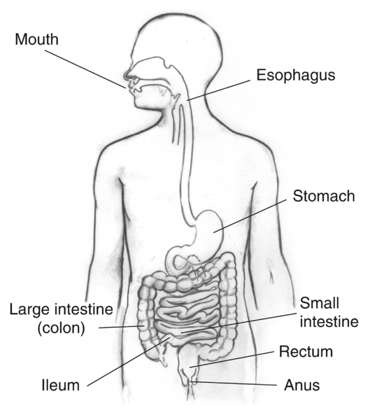


![Recombinant Anti-p53 antibody [E26] KO Tested (ab32389) | Abcam](https://www.abcam.com/ps/products/32/ab32389/Images/ab32389-389819-antip53antibodyimmunocytochemistryimmunofluorescencehap1human.jpg)