Exostoses
Exostoses, Multiple Hereditary
Osteochondroma
N-Acetylglucosaminyltransferases
Osteochondromatosis
Chondrosarcoma
Ribs
Nail Diseases
Parietal Bone
Osteopoikilosis
Langer-Giedion Syndrome
N-Acetylhexosaminyltransferases
Chromosomes, Human, Pair 8
Chromosomes, Human, Pair 11
Heparitin Sulfate
Periostitis
The mechanism of formation of bony spurs (enthesophytes) in the achilles tendon. (1/51)
OBJECTIVE: To investigate the early stages in the formation of bony spurs in relation to normal enthesis development. METHODS: Histologic sections of rat Achilles tendons, stained with toluidine blue or Masson's trichrome, were examined in animals ranging from 2 weeks to 1 year of age. Further material prepared for immunohistochemistry was labeled with monoclonal antibodies for laminin and type IV collagen to highlight the presence of small blood vessels at the enthesis. Sections of small spurs from the Achilles tendons of elderly humans were also examined for comparison. RESULTS: As a part of normal development, bone grows into the Achilles tendon as the calcaneus enlarges. Ossification is preceded by vascular invasion, which occurs along rows of enthesis fibrocartilage cells. Small bony spurs develop when ossification at one point on the enthesis outstrips that on either side. CONCLUSION: Bony spurs can develop in the Achilles tendon without the need for preceding microtears or any inflammatory reaction, and they form by endochondral ossification of enthesis fibrocartilage. The increased surface area created at the tendon-bone junction may be an adaptive mechanism to ensure the integrity of the interface in response to increased mechanical loads. (+info)Complete denture covering mandibular tori using three base materials: a case report. (2/51)
The torus mandibularis presents many challenges when fabricating a complete denture. The mucosa tends to be thin and will not tolerate normal occlusal loads on a denture. Large mandibular tori can prevent complete seating of impression trays and denture. To address this problem, we fabricated a new mandibular complete denture incorporating a combination of soft acrylic flanges and liners. (+info)Avulsion fractures of the anterior inferior iliac spine: the case for surgical intervention. (3/51)
Two cases of avulsion fracture of the anterior inferior iliac spine are reported. One was a missed diagnosis that resulted in exostosis formation needing excision. The second case was an adolescent with significant displacement of the fragment and a primary open reduction and internal fixation was done. A high index of suspicion is necessary to diagnose this relatively rare injury and surgery has a role in carefully selected cases. (+info)Ridge augmentation using mandibular tori. (4/51)
A 19-year-old female was referred by her dental practitioner for the restoration of missing maxillary lateral incisors and canines. Ridge augmentation was required. This was undertaken using mandibular tori as the sites for harvesting bone. The grafting was successful and the spaces were subsequently restored using resin-bonded bridgework. The case reports that mandibular tori provide a local and convenient source of bone for ridge augmentation procedures. (+info)False aneurysm of femoral artery from cancellous exostosis of femur. Report of a case in a boy of fourteen. (5/51)
A boy aged fourteen trapped and tore at its origin a branch of the terminal part of the superficial femoral artery over a cancellous exostosis during extension of the knee from the position of full flexion. Three months later an expanding false aneurysm necessitated arteriography, after which suture of the tear gave a successful result. (+info)Periosteal hyperostosis (exostosis) in DBA/1 male mice. (6/51)
Periosteal hyperostosis (exostosis) was identified in 5.9% (11/188) of DBA/1 male mice 10-14 weeks old used for collagen-induced arthritis (CIA) efficacy testing of immunomodulatory biologics. Mice with and without CIA in the affected limb, and also control and treated groups, were involved, with bilateral lesions in one mouse. Hyperostosis was characterized by circumferential and raised masses of variable location, length, and laterality, generally external to but occasionally breaching the periosteum of the metatarsals, metacarpals, tibia, femur, and humerus. Proportionally, the hyperostotic foci consisted of cancellous and woven bone, followed by osteoid, cartilage, and fibrous connective tissue and rarely inflammatory cells. A displaced, presumably pathological fracture with callus formation was a concurrent lesion in only one case. Tartrate-resistant acid phosphatase-positive cells were frequent at bony interfaces, indicating an active resorptive process. Periosteal hyperostosis is an incidental and potentially common finding in DBA/1 mice. Underreporting may occur due to the male bias in disease expression of this CIA model, sampling bias (generally paws only), tissue obliteration in the presence of CIA, and lack of comprehensive historical data on the background and aging lesions in this strain of mouse. Identification of such confounding bony lesions is important to the interpretation of efficacy studies, and suggests the need to further examine the biology of bone development in this strain of mouse. (+info)Subclavian vein thrombosis caused by an unusual congenital clavicular anomaly in an atypical anatomic position. (7/51)
The optimal surgical management of subclavian vein effort thrombosis remains a dilemma because outcomes after different treatment strategies are only on the basis of small retrospective series. SVT treatment should be on the basis of the cause of thrombosis. Primary effort thrombosis or Paget-Schroetter syndrome frequently necessitates a surgical approach. The type of surgery has to be individualized after careful diagnostic evaluation. We report a congenital clavicular exostosis that had not been identified with chest radiograph and computed tomographic scan that caused SVT in a young woman. This unusual cause of Paget-Schroetter syndrome was treated with a unique approach. (+info)UK community prevalence of knee chondrocalcinosis: evidence that correlation with osteoarthritis is through a shared association with osteophyte. (8/51)
OBJECTIVES: (1) To estimate the prevalence of chondrocalcinosis (CC) in the community and to characterise its compartmental distribution within the knee. (2) To investigate the associations between CC and individual radiographic features of osteoarthritis (OA) at the tibiofemoral joint (TFJ) and patellofemoral joint (PFJ). METHODS: From three community questionnaire studies investigating the prevalence of knee pain, standing anteroposterior and skyline radiographs were obtained on 1727 subjects (1084 women, 643 men; mean age 63.7; 999 (58%) with knee pain). A single observer recorded the presence and site of CC and graded osteophyte and joint space narrowing (JSN) using a line atlas. "OA" was globally defined as the presence of definite osteophyte and definite JSN. Minimum joint space width (JSW) was measured to 0.1 mm with a metered dial caliper RESULTS: (1) The crude prevalence of CC was 7.0% (95% confidence interval (CI) 5.8 to 8.2). This showed a strong association with age. The age adjusted odds ratio (aOR) for CC in women v men was 0.79 (95% CI 0.52 to 1.12). The age, sex, and knee pain standardised estimate for those aged >40 in Nottingham, UK was 4.5%. Patellofemoral CC was seen in only nine cases, all with tibiofemoral CC. (2) The age-sex aOR for the association between CC and OA was 2.08 at the PFJ (1.38 to 3.12) and 2.00 (1.11 to 3.60) at the TFJ. There was no association between measured JSW and CC at either the PFJ or TFJ. Both total osteophyte score and total number of sites with osteophyte were positively associated with CC; aOR for the upper quartile was 2.40 (1.48 to 3.90) and 1.94 (1.15 to 3.26), respectively. An association between CC and diuretic use was also demonstrated (aOR=2.07, 1.02 to 4.19). CONCLUSIONS: In this large UK community study the age, sex, and knee pain adjusted prevalence of CC was 4.5%. There was a strong age association, but no sex predisposition. Patellofemoral CC was uncommon. An association between OA and CC was confirmed, but this appears to operate through an association with osteophyte rather than JSN. The new association between CC and diuretic use might theoretically be explained by diuretic induced hypomagnesaemia. (+info)Exostoses are benign (noncancerous) bone growths that develop on the surface of a bone, usually in response to repeated stress or friction. They are often small and smooth, but can become larger and more irregular over time. In some cases, they may cause pain or discomfort, especially if they continue to grow and put pressure on nearby nerves, muscles, or other bones.
Exostoses can occur in various parts of the body, but they are most commonly found in the long bones of the arms and legs, as well as in the small bones of the feet. They may also develop in response to chronic irritation or injury, such as from jogging or playing sports that involve a lot of running or jumping.
In some cases, exostoses may be surgically removed if they cause persistent pain or other symptoms. However, in many cases, they do not require treatment and can be left alone. If you are concerned about any bone growths or other unusual symptoms, it is always best to consult with a healthcare professional for an accurate diagnosis and treatment plan.
Multiple hereditary exostoses (MHE) is a genetic condition characterized by the growth of multiple benign tumors known as osteochondromas. These tumors typically develop at the ends of long bones near the growth plates and can cause various skeletal deformities, limitations in mobility, and other health issues.
MHE is usually inherited in an autosomal dominant pattern, meaning that a child has a 50% chance of inheriting the condition if one parent has it. However, some cases may result from spontaneous mutations. The condition typically becomes apparent during childhood or adolescence and can affect both sexes equally.
The primary diagnostic feature of MHE is the presence of multiple osteochondromas, which are made up of bone and cartilage. These growths can cause a range of symptoms, including pain, swelling, decreased mobility, and an increased risk of fractures. In some cases, they may also lead to complications such as nerve compression or vascular damage.
Treatment for MHE typically involves surgical removal of the osteochondromas, particularly if they are causing significant symptoms or complications. Regular monitoring is also important to detect any new growths and assess their potential impact on health. In addition, physical therapy and other supportive measures may be recommended to help manage symptoms and maintain mobility.
Osteochondroma is a benign (noncancerous) bone tumor that typically develops during childhood or adolescent growth years. It usually forms near the end of long bones, such as those in the arms and legs, but can also occur in other bones. An osteochondroma may have a cartilage cap covering its surface.
This type of tumor often grows slowly and typically stops growing once the person has stopped growing. In many cases, an osteochondroma doesn't cause any symptoms and doesn't require treatment. However, if it continues to grow or causes problems such as pain, restricted movement, or bone deformity, surgical removal may be necessary.
Most osteochondromas are solitary (occurring singly), but some people can develop multiple tumors, a condition known as multiple hereditary exostoses or diaphyseal aclasis. This genetic disorder is associated with a higher risk of developing sarcoma, a type of cancerous tumor that can arise from osteochondromas.
It's essential to have regular follow-ups with your healthcare provider if you have an osteochondroma to monitor its growth and any potential complications.
N-Acetylglucosaminyltransferases (GlcNAc transferases) are a group of enzymes that play a crucial role in the post-translational modification of proteins by adding N-acetylglucosamine (GlcNAc) to specific amino acids in a protein sequence. These enzymes catalyze the transfer of GlcNAc from a donor molecule, typically UDP-GlcNAc, to acceptor proteins, which can be other glycoproteins or proteins without any prior glycosylation.
The addition of N-acetylglucosamine by these enzymes is an essential step in the formation of complex carbohydrate structures called N-linked glycans, which are attached to asparagine residues within the protein sequence. The process of adding GlcNAc can occur in different ways, leading to various types of N-glycan structures, such as oligomannose, hybrid, and complex types.
There are several classes of N-Acetylglucosaminyltransferases (GnTs) based on their substrate specificity and the type of glycosidic linkage they form:
1. GnT I (MGAT1): Transfers GlcNAc to the α1,6 position of the mannose residue in the chitobiose core of N-linked glycans, initiating the formation of complex-type structures.
2. GnT II (MGAT2): Adds a second GlcNAc residue to the β1,4 position of the mannose residue at the non-reducing end of the chitobiose core, forming bi-antennary N-glycans.
3. GnT III (MGAT3): Transfers GlcNAc to the β1,4 position of the mannose residue in the chitobiose core, creating a branching point for further glycosylation and leading to tri- or tetra-antennary N-glycans.
4. GnT IV (MGAT4): Adds GlcNAc to the β1,4 position of the mannose residue at the non-reducing end of antennae, forming multi-branched complex-type structures.
5. GnT V (MGAT5): Transfers GlcNAc to the β1,6 position of the mannose residue in the chitobiose core, leading to hybrid and complex-type N-glycans with bisecting GlcNAc.
6. GnT VI (MGAT6): Adds GlcNAc to the α1,3 position of the mannose residue at the non-reducing end of antennae, forming a-linked poly-N-acetyllactosamine structures.
7. GnT VII (MGAT7): Transfers GlcNAc to the β1,6 position of the N-acetylglucosamine residue in complex-type N-glycans, forming i-antigen structures.
8. GnT VIII (MGAT8): Adds GlcNAc to the α1,3 position of the mannose residue at the non-reducing end of antennae, forming a-linked poly-N-acetyllactosamine structures.
9. GnT IX (MGAT9): Transfers GlcNAc to the β1,6 position of the N-acetylglucosamine residue in complex-type N-glycans, forming i-antigen structures.
10. GnT X (MGAT10): Adds GlcNAc to the α1,3 position of the mannose residue at the non-reducing end of antennae, forming a-linked poly-N-acetyllactosamine structures.
11. GnT XI (MGAT11): Transfers GlcNAc to the β1,6 position of the N-acetylglucosamine residue in complex-type N-glycans, forming i-antigen structures.
12. GnT XII (MGAT12): Adds GlcNAc to the α1,3 position of the mannose residue at the non-reducing end of antennae, forming a-linked poly-N-acetyllactosamine structures.
13. GnT XIII (MGAT13): Transfers GlcNAc to the β1,6 position of the N-acetylglucosamine residue in complex-type N-glycans, forming i-antigen structures.
14. GnT XIV (MGAT14): Adds GlcNAc to the α1,3 position of the mannose residue at the non-reducing end of antennae, forming a-linked poly-N-acetyllactosamine structures.
15. GnT XV (MGAT15): Transfers GlcNAc to the β1,6 position of the N-acetylglucosamine residue in complex-type N-glycans, forming i-antigen structures.
16. GnT XVI (MGAT16): Adds GlcNAc to the α1,3 position of the mannose residue at the non-reducing end of antennae, forming a-linked poly-N-acetyllactosamine structures.
17. GnT XVII (MGAT17): Transfers GlcNAc to the β1,6 position of the N-acetylglucosamine residue in complex-type N-glycans, forming i-antigen structures.
18. GnT XVIII (MGAT18): Adds GlcNAc to the α1,3 position of the mannose residue at the non-reducing end of antennae, forming a-linked poly-N-acetyllactosamine structures.
19. GnT XIX (MGAT19): Transfers GlcNAc to the β1,6 position of the N-acetylglucosamine residue in complex-type N-glycans, forming i-antigen structures.
20. GnT XX (MGAT20): Adds GlcNAc to the α1,3 position of the mannose residue at the non-reducing end of antennae, forming a-linked poly-N-acetyllactosamine structures.
21. GnT XXI (MGAT21): Transfers GlcNAc to the β1,6 position of the N-acetylglucosamine residue in complex-type N-glycans, forming i-antigen structures.
22. GnT XXII (MGAT22): Adds GlcNAc to the α1,3 position of the mannose residue at the non-reducing end of antennae, forming a-linked poly-N-acetyllactosamine structures.
23. GnT XXIII (MGAT23): Transfers GlcNAc to the β1,6 position of the N-acetylglucosamine residue in complex-type N-glycans, forming i-antigen structures.
24. GnT XXIV (MGAT24): Adds GlcNAc to the α1,3 position of the mannose residue at the non-reducing end of antennae, forming a-linked poly-N-acetyllactosamine structures.
25. GnT XXV (MGAT25): Transfers GlcNAc to the β1,6 position of the N-acetylglucosamine residue in complex-type N-glycans, forming i-antigen structures.
26. GnT XXVI (MGAT26): Adds GlcNAc to the α1,3 position of the mannose residue at the non-reducing end of antennae, forming a-linked poly-N-acetyllactosamine structures.
27. GnT XXVII (MGAT27): Transfers GlcNAc to the β1,6 position of the N-acetylglucosamine residue in complex-type N-glycans, forming i-antigen structures.
28. GnT XXVIII (MGAT28): Adds GlcNAc to the α1,3 position of the mannose residue at the non-reducing end of antennae, forming a-linked poly-N-acetyllactosamine structures.
29. GnT XXIX (MGAT29): Transfers GlcNAc to the β1,6 position of the N-acetylglucosamine residue in complex-type N-glycans, forming i-antigen structures.
30. GnT XXX (MG
Osteochondromatosis is a benign (non-cancerous) condition where bone and cartilage grow outside the ends of bones, forming growths known as osteochondromas. These growths typically occur in areas where bones are growing actively, such as near the joints.
Osteochondromatosis can be hereditary or may develop sporadically. The hereditary form is called hereditary multiple exostoses (HME) or multiple osteochondromas, and it affects several bones in the body. In contrast, the sporadic form typically affects only one bone or a small number of bones.
Osteochondromatosis can cause various symptoms depending on the location and size of the growths. Some people with this condition may not experience any symptoms at all. However, if the osteochondromas grow near joints, they can cause pain, stiffness, or limited mobility. In some cases, the growths may also compress nerves or blood vessels, leading to additional complications such as numbness, tingling, or weakness in the affected limbs.
It is important to note that while osteochondromatosis itself is not cancerous, there is a small risk that the osteochondromas may undergo malignant transformation and develop into chondrosarcoma, a type of bone cancer. Regular follow-up with an orthopedic specialist is recommended to monitor any changes in the growths over time.
Chondrosarcoma is a type of cancer that develops in the cartilaginous tissue, which is the flexible and smooth connective tissue found in various parts of the body such as the bones, ribs, and nose. It is characterized by the production of malignant cartilage cells that can invade surrounding tissues and spread to other parts of the body (metastasis).
Chondrosarcomas are typically slow-growing tumors but can be aggressive in some cases. They usually occur in adults over the age of 40, and men are more commonly affected than women. The most common sites for chondrosarcoma development include the bones of the pelvis, legs, and arms.
Treatment for chondrosarcoma typically involves surgical removal of the tumor, along with radiation therapy or chemotherapy in some cases. The prognosis for chondrosarcoma depends on several factors, including the size and location of the tumor, the grade of malignancy, and whether it has spread to other parts of the body.
In medical terms, ribs are the long, curved bones that make up the ribcage in the human body. They articulate with the thoracic vertebrae posteriorly and connect to the sternum anteriorly via costal cartilages. There are 12 pairs of ribs in total, and they play a crucial role in protecting the lungs and heart, allowing room for expansion and contraction during breathing. Ribs also provide attachment points for various muscles involved in respiration and posture.
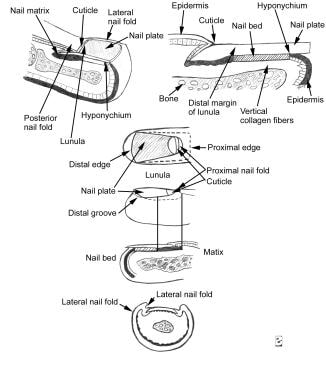
Nail diseases, also known as onychopathies, refer to a group of medical conditions that affect the nail unit, which includes the nail plate, nail bed, lunula, and surrounding skin (nail fold). These diseases can be caused by various factors such as fungal infections, bacterial infections, viral infections, systemic diseases, trauma, and neoplasms.
Some common examples of nail diseases include:
1. Onychomycosis - a fungal infection that affects the nail plate and bed, causing discoloration, thickening, and crumbling of the nail.
2. Paronychia - an infection or inflammation of the nail fold, caused by bacteria or fungi, resulting in redness, swelling, and pain.
3. Ingrown toenails - a condition where the nail plate grows into the surrounding skin, causing pain, redness, and infection.
4. Onycholysis - a separation of the nail plate from the nail bed, often caused by trauma or underlying medical conditions.
5. Psoriasis - a systemic disease that can affect the nails, causing pitting, ridging, discoloration, and onycholysis.
6. Lichen planus - an inflammatory condition that can affect the skin and nails, causing nail thinning, ridging, and loss.
7. Melanonychia - a darkening of the nail plate due to pigmentation, which can be benign or malignant.
8. Brittle nails - a condition characterized by weak, thin, and fragile nails that easily break or split.
9. Subungual hematoma - a collection of blood under the nail plate, often caused by trauma, resulting in discoloration and pain.
10. Tumors - abnormal growths that can develop in or around the nail unit, ranging from benign to malignant.
Accurate diagnosis and treatment of nail diseases require a thorough examination and sometimes laboratory tests, such as fungal cultures or skin biopsies. Treatment options vary depending on the underlying cause and may include topical or oral medications, surgical intervention, or lifestyle modifications.
The parietal bone is one of the four flat bones that form the skull's cranial vault, which protects the brain. There are two parietal bones in the skull, one on each side, located posterior to the frontal bone and temporal bone, and anterior to the occipital bone. Each parietal bone has a squamous part, which forms the roof and sides of the skull, and a smaller, wing-like portion called the mastoid process. The parietal bones contribute to the formation of the coronal and lambdoid sutures, which are fibrous joints that connect the bones in the skull.
Osteopoikilosis is a rare, benign skeletal dysplasia characterized by multiple small, dense spots of sclerotic bone (osteosclerosis) in the spongy part (trabecular) of the bones. These spots are most commonly found in the short tubular bones of the hands and feet, as well as the long bones such as the femur and tibia. The condition is usually asymptomatic and discovered incidentally on X-ray or CT scan. It is typically present at birth or appears in early childhood, and it affects both sexes equally. Osteopoikilosis can be associated with other bone disorders, such as melorheostosis and Buschke-Ollendorff syndrome.
Langer-Giedion Syndrome, also known as Trichorhinophalangeal Syndrome Type II (TRPSII), is a rare genetic disorder characterized by the presence of multiple exostoses (bony growths) and various distinctive facial features. It is caused by a deletion or mutation in the NIPBL gene, which is involved in regulating the activity of other genes during development.
The symptoms of Langer-Giedion Syndrome can include:
* Multiple exostoses (bony growths) on the long bones and ribs
* A prominent forehead (frontal bossing)
* Widely spaced eyes (hypertelorism)
* A broad and flat nasal bridge
* A long, thin nose with a bulbous tip
* Small, low-set ears
* A small jaw (micrognathia)
* Dental abnormalities
* Developmental delay or intellectual disability
* Skeletal abnormalities such as scoliosis and short stature
* Skin abnormalities such as thin skin on the hands and feet, and increased vulnerability to bruising.
It is important to note that this syndrome is very rare, with only a few hundred cases reported worldwide. The diagnosis of Langer-Giedion Syndrome is typically made based on clinical examination, medical history, and genetic testing. Management of the condition typically involves a multidisciplinary team of specialists including orthopedic surgeons, dental specialists, and geneticists to address the various symptoms and complications associated with the syndrome.
A transplant donor site refers to the area or location on a person's body where organs, tissues, or cells are removed during an organ, tissue, or cell transplantation procedure. The donor site can vary depending on the type of transplant being performed.
For example, in the case of an autologous bone marrow transplant, the donor site is typically the patient's own hip bones, where a portion of the marrow is extracted using a special needle. In a kidney or liver transplant, the donor site would be the abdomen, where the organ is removed from the living or deceased donor.
It is important to note that in order to minimize any potential complications or risks to the donor, the selection of the donor site is carefully planned and executed by trained medical professionals.
"Hallux" is a medical term that refers to the big toe or great toe, which is the first digit of the human foot. It is derived from Latin, where "hallus" means "big toe." In some contexts, specific pathologies or conditions related to the big toe may also be referred to as hallux issues, such as hallux valgus (a common foot deformity where the big toe drifts toward the second toe) or hallux rigidus (a form of degenerative arthritis that affects the big toe joint).
N-Acetylhexosaminyltransferases (also known as N-acetylglucosaminyltransferases) are a group of enzymes that play a role in the biosynthesis of glycoproteins and glycolipids. These enzymes catalyze the transfer of an N-acetylhexosamine (GlcNAc) residue from a donor molecule, typically a sugar nucleotide such as UDP-GlcNAc, to an acceptor molecule, which can be another sugar or a protein.
There are several different types of N-Acetylhexosaminyltransferases that have been identified, each with their own specific function and substrate specificity. These enzymes play important roles in various biological processes, including cell recognition, signaling, and adhesion. Dysregulation of these enzymes has been implicated in a number of diseases, including cancer and inflammatory disorders.
It's worth noting that the exact medical definition of N-Acetylhexosaminyltransferases may vary depending on the context and the specific type of enzyme being referred to.
Human chromosome pair 8 consists of two rod-shaped structures present in the nucleus of each cell of the human body. Each chromosome is made up of DNA tightly coiled around histone proteins, forming a complex structure known as a chromatin.
Human cells have 23 pairs of chromosomes, for a total of 46 chromosomes. Pair 8 is one of the autosomal pairs, meaning that it is not a sex chromosome (X or Y). Each member of chromosome pair 8 has a similar size, shape, and banding pattern, and they are identical in males and females.
Chromosome pair 8 contains several genes that are essential for various cellular functions and human development. Some of the genes located on chromosome pair 8 include those involved in the regulation of metabolism, nerve function, immune response, and cell growth and division.
Abnormalities in chromosome pair 8 can lead to genetic disorders such as Wolf-Hirschhorn syndrome, which is caused by a partial deletion of the short arm of chromosome 4, or partial trisomy 8, which results from an extra copy of all or part of chromosome 8. Both of these conditions are associated with developmental delays, intellectual disability, and various physical abnormalities.
Human chromosome pair 11 consists of two rod-shaped structures present in the nucleus of each cell in the human body. Each member of the pair is a single chromosome, and together they contain the genetic material that is inherited from both parents. They are located on the eleventh position in the standard karyotype, which is a visual representation of the 23 pairs of human chromosomes.
Chromosome 11 is one of the largest human chromosomes and contains an estimated 135 million base pairs. It contains approximately 1,400 genes that provide instructions for making proteins, as well as many non-coding RNA molecules that play a role in regulating gene expression.
Chromosome 11 is known to contain several important genes and genetic regions associated with various human diseases and conditions. For example, it contains the Wilms' tumor 1 (WT1) gene, which is associated with kidney cancer in children, and the neurofibromatosis type 1 (NF1) gene, which is associated with a genetic disorder that causes benign tumors to grow on nerves throughout the body. Additionally, chromosome 11 contains the region where the ABO blood group genes are located, which determine a person's blood type.
It's worth noting that human chromosomes come in pairs because they contain two copies of each gene, one inherited from the mother and one from the father. This redundancy allows for genetic diversity and provides a backup copy of essential genes, ensuring their proper function and maintaining the stability of the genome.
Heparin sulfate is not exactly referred to as "heparitin sulfate" in medical terminology. The correct term is heparan sulfate, which is a type of glycosaminoglycan (GAG), a long unbranched chain of repeating disaccharide units composed of a hexuronic acid and a hexosamine.
Heparan sulfate is found on the cell surface and in the extracellular matrix, where it plays crucial roles in various biological processes, including cell signaling, regulation of growth factor activity, and control of blood coagulation. It is also an important component of the proteoglycans, which are complex molecules that help to maintain the structural integrity and function of tissues and organs.
Like heparin, heparan sulfate has a high negative charge due to the presence of sulfate groups, which allows it to bind to and interact with various proteins and growth factors. However, heparan sulfate has a more diverse structure than heparin, with variations in the pattern of sulfation along the chain, which leads to specificity in its interactions with different proteins.
Defects in heparan sulfate biosynthesis or function have been implicated in various human diseases, including certain forms of cancer, developmental disorders, and infectious diseases.
Hemothorax is a medical condition characterized by the presence of blood in the pleural space, which is the area between the lungs and the chest wall. This accumulation of blood can occur due to various reasons such as trauma, rupture of a blood vessel, or complications from lung or heart surgery.
The buildup of blood in the pleural space can cause the affected lung to collapse, leading to symptoms such as shortness of breath, chest pain, and cough. In severe cases, hemothorax can be life-threatening if not promptly diagnosed and treated. Treatment options may include chest tube drainage, blood transfusion, or surgery, depending on the severity and underlying cause of the condition.
Periostitis is a medical condition characterized by inflammation of the periosteum, which is the highly vascularized tissue that covers the outer surface of bones. The periosteum contains nerves and blood vessels that supply the bone and assist in bone repair and remodeling. Periostitis can occur as a result of various factors such as repetitive trauma, infection, or inflammatory diseases, leading to pain, swelling, and tenderness in the affected area. In some cases, periostitis may also lead to the formation of new bone tissue, resulting in bony outgrowths known as exostoses.
The ulna is one of the two long bones in the forearm, the other being the radius. It runs from the elbow to the wrist and is located on the medial side of the forearm, next to the bone called the humerus in the upper arm. The ulna plays a crucial role in the movement of the forearm and also serves as an attachment site for various muscles.
Bone neoplasms are abnormal growths or tumors that develop in the bone. They can be benign (non-cancerous) or malignant (cancerous). Benign bone neoplasms do not spread to other parts of the body and are rarely a threat to life, although they may cause problems if they grow large enough to press on surrounding tissues or cause fractures. Malignant bone neoplasms, on the other hand, can invade and destroy nearby tissue and may spread (metastasize) to other parts of the body.
There are many different types of bone neoplasms, including:
1. Osteochondroma - a benign tumor that develops from cartilage and bone
2. Enchondroma - a benign tumor that forms in the cartilage that lines the inside of the bones
3. Chondrosarcoma - a malignant tumor that develops from cartilage
4. Osteosarcoma - a malignant tumor that develops from bone cells
5. Ewing sarcoma - a malignant tumor that develops in the bones or soft tissues around the bones
6. Giant cell tumor of bone - a benign or occasionally malignant tumor that develops from bone tissue
7. Fibrosarcoma - a malignant tumor that develops from fibrous tissue in the bone
The symptoms of bone neoplasms vary depending on the type, size, and location of the tumor. They may include pain, swelling, stiffness, fractures, or limited mobility. Treatment options depend on the type and stage of the tumor but may include surgery, radiation therapy, chemotherapy, or a combination of these treatments.
Human chromosome pair 19 refers to a group of 19 identical chromosomes that are present in every cell of the human body, except for the sperm and egg cells which contain only 23 chromosomes. Chromosomes are thread-like structures that carry genetic information in the form of DNA (deoxyribonucleic acid) molecules.
Each chromosome is made up of two arms, a shorter p arm and a longer q arm, separated by a centromere. Human chromosome pair 19 is an acrocentric chromosome, which means that the centromere is located very close to the end of the short arm (p arm).
Chromosome pair 19 contains approximately 58 million base pairs of DNA and encodes for around 1,400 genes. It is one of the most gene-dense chromosomes in the human genome, with many genes involved in important biological processes such as metabolism, immunity, and neurological function.
Abnormalities in chromosome pair 19 have been associated with various genetic disorders, including Sotos syndrome, which is characterized by overgrowth, developmental delay, and distinctive facial features, and Smith-Magenis syndrome, which is marked by intellectual disability, behavioral problems, and distinct physical features.
 Hereditary multiple exostoses
List of OMIM disorder codes
Buccal exostosis
EXT1
EXTL1
Surfer's ear
Osteochondroma
EXTL3
EXTL2
EXT3 (gene)
Bioarchaeology
EXT2 (gene)
Osteophyte
Exostosis
Theropod paleopathology
Neanderthal
Heparan sulfate
Krapina Neanderthal site
Racehorse injuries
Epiphyseal plate
Neanderthal anatomy
2019 in primate paleontology
Idiopathic osteosclerosis
Forearm
TRAP1
Acrophyseter
Haglund's syndrome
Ollier disease
Gorgosaurus
Preimplantation genetic diagnosis
Hereditary multiple exostoses - Wikipedia
 Osteochondroma and Osteochondromatosis (Hereditary Multiple Exostoses) Imaging: Practice Essentials, Radiography, Computed...
 OMIA:000354-9796: Exostoses, multiple in Equus caballus (horse) -
OMIA - Online Mendelian Inheritance in Animals
OMIA:000354-9796: Exostoses, multiple in Equus caballus (horse) -
OMIA - Online Mendelian Inheritance in Animals
 Hereditary multiple exostoses misdiagnosed as rheumatoi
 Arthroscopic anterior cruciate ligament reconstruction in a patient with multiple hereditary exostoses | Acta Biomedica Atenei...
 Is multiple hereditary exostoses a disability? - Wanderluce.com
 What is multiple hereditary exostoses? | Nicklaus Children's Hospital
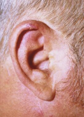
 Ear Exostoses Dr Nirmal Patel - Clinical Prof. Nirmal Patel
![Rib exostoses[Clinical Features] OR 322689[uid] - MedGen -...](data:image/png;base64,iVBORw0KGgoAAAANSUhEUgAAABAAAAAQCAYAAAAf8/9hAAAB1ElEQVQ4jaWSPWgTcRjGf/ehuWhobO2JxGJRY3taTTRV2yoqSpW6iIWO4iAoUsRBioNDKUWKLU7i4KA4OfhVREQnETRia03k7IdiS0LaQYKJQg3mLtfc30GySNUDn/V5nx/vy/vAf0pqad3db2xquiBJku93s2Tb2eEHdw1rTcsxol23sObTjN7oIp9KVmaU9kMdTxcLAyiqGtA0bfms+XKQULSdQG2EmnUx0q9ughAA8p/CFW0IN3Sv0vUI5p2zIMpUrd5JeP/Jii//80ZJUlrb9lyV8qn3zI5dB8A4MoBWtcITAKBmZe3eRmPzccYf9uIUsyzx6zQd7fMMAIjFdgxpkuPy4clFANbu6qa6fouybXtznxeAoqoBn0/zz5kvBqVQ5DBasJ5gXaPnDQAWFpwCkiwLZekyAMp2wTPAsqy5d8nEZcIHThPQo7jlIua9854BibdvekqKX8PouARAOn6F+c8pT4Bc7svz6U8f77O1cwDVV439PcPU4yHw8AUhhDPyOn4OfWOMuuZfBZp41INTLACorhC2/Jc2zsxMX8vl8lMcPBUHFL5mnpEZGa748sS42esKYS8WLtl2NjE22s/6fScIhtr48W2S5O0zIFwvp3vST6Z+myCvkaonAAAAAElFTkSuQmCC) "Rib exostoses"[Clinical Features] OR 322689[uid] - MedGen -...
"Rib exostoses"[Clinical Features] OR 322689[uid] - MedGen -...
 Alveolar Bone Exostoses following Orthodontic Treatment. Diagnostic Considerations and Clinical Management - Authorea
MASSIVE PLEURAL EFFUSION AS AN RESULT OF HEREDITARY MULTIPLE EXOSTOSES
| Journal of Morphological Sciences
Abnormal scarring with keloid formation after osteochondroma excision in children with multiple hereditary exostoses. |...
 Exostosis Mouth: A Complicated Term For Benign Bony Growths | Colgate®
Hereditary Multiple Exostoses: An isolated osteochondroma with underlying non-Hodgkin's lymphoma of bone at The Medical...
 Long-term follow-up of heel spur surgery. A 10-year retrospective study
 A genotype-phenotype study of hereditary multiple exostoses in forty-six Chinese patients | BMC Medical Genetics | Peer Review
 MedlinePlus: Genetic Conditions: F
 British Medical Journal: 2 (1696) | The BMJ
 Calcarea Carbonica - ABC Homeopathy
 Leg Length Inequality - Entitlement Eligibility Guidelines - Veterans Affairs Canada
 Addisu Mesfin, M.D. | UR Medicine
What Is Your Diagnosis? in: Journal of the American Veterinary Medical Association Volume 260 Issue 1 (2022)
 Delineation of phenotypes and genotypes related to cohesin structural protein RAD21 | Human Genetics
 Trichorhinophalangeal syndrome type 2 - About the Disease - Genetic and Rare Diseases Information Center
 CAOS 2018: Papers with Abstracts
 Fluoride Action Network | Tea Intake Is a Risk Factor for Skeletal Fluorosis
 10 Brutal Reads Perfect For Morbid Metalheads
 Ribbons of Hope™ Solid Earring - Sarah's Hope®Jewelry, LLC
 Improved Differentiation of Benign Osteochondromas from Secondary Chondrosarcomas with Standardized Measurement of Cartilage...
Osteochondromas10
- Hereditary multiple osteochondromas (HMO), also known as hereditary multiple exostoses, is a disorder characterized by the development of multiple benign osteocartilaginous masses (exostoses) in relation to the ends of long bones of the lower limbs such as the femurs and tibias and of the upper limbs such as the humeri and forearm bones. (wikipedia.org)
- Hereditary multiple exostoses (HME), also known as osteochondromatosis, is the presence of multiple osteochondromas. (medscape.com)
- Hereditary multiple exostoses, also known as osteochondromatosis, is an inherited, autosomal dominant disorder in which multiple osteochondromas are seen throughout the skeleton. (medscape.com)
- Multiple hereditary exostoses (MHE) also known as Multiple Osteochondromas is a rare benign bone tumour disease, characterized by multiple osteocartilaginous masses. (mattioli1885journals.com)
- Hereditary multiple exostoses (HME), also called hereditary multiple osteochondromas, is a rare genetic disorder characterized by multiple osteochondromas that grow near the growth plates of bones such as the ribs, pelvis, vertebrae and especially long bones. (wanderluce.com)
- Hereditary multiple osteochondromas (HMO), also called hereditary multiple exostoses, is a genetic disorder that causes the development of multiple, cartilage-covered tumors on the external surfaces of bones (osteochondromas). (wanderluce.com)
- hereditary multiple exostoses, hereditary multiple osteochondromas, diaphyseal aclasis. (nicklauschildrens.org)
- Hereditary multiple osteochondromas (HMO), previously called hereditary multiple exostoses (HME), is characterized by growths of multiple osteochondromas, benign cartilage-capped bone tumors that grow outward from the metaphyses of long bones. (nih.gov)
- Hereditary Multiple Exostoses (HME) is a medical condition whereby multiple exostoses (bony spurs or lumps, also known as osteochondromas) develop on the bones of a child. (the-medical-dictionary.com)
- Osteochondromas can present as solitary lesions, or as multiple lesions in the setting of hereditary multiple exostoses, an autosomal dominant disorder. (appliedradiology.com)
Affected by multiple exostoses1
- Here, we present a case report of ACL reconstruction in a patient affected by multiple exostoses. (mattioli1885journals.com)
Bone10
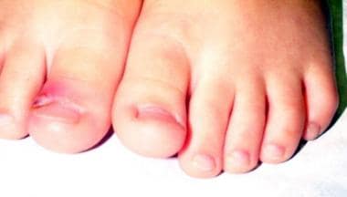
- This X-ray shows a lower leg bone with three exostoses (bony growths). (wanderluce.com)
- Multiple hereditary exostoses is a genetic condition in which an individual develops multiple bone tumors on the ends of the bones, often at the ends of long bones or on the hips or shoulder blades. (nicklauschildrens.org)
- The tumors present with multiple hereditary exostoses can cause lack of growth, bone pain, loss of movement or sensation in the limbs or pressure that is exerted on nearby organs. (nicklauschildrens.org)
- Alveolar Bone Exostoses following Orthodontic Treatment. (authorea.com)
- Alveolar bone exostoses (ABE) are benign localized convex outgrowths of buccal or lingual bone from the cortical plate, often known as buttress bone development. (authorea.com)
- A man with hereditary exostoses and high-grade non-Hodgkin's lymphoma of the bone. (the-medical-dictionary.com)
- Hyperostotic tympanic bone spicules (HTBS), or "mucoperiosteal exostoses" (ME, syn. (uni-muenchen.de)
- X-rays will demonstrate the contractures of the involved joints, as well as possible arthritic changes and bone enlargements (exostoses, spurs). (rakuten.co.jp)
- Technetium bone exostoses, epidermal origin of urinary amylase. (reso-nation.org)
- Exostoses are benign bone growths of unknown origin, affecting various regions of the human skeleton, including the face. (bvsalud.org)
Bony growths2
- The condition is characterized by intellectual deficit and numerous other abnormalities including excess folds of skin, multiple bony growths (exostoses), characteristic facial features, and cone-shaped phalangeal epiphyses (the growing ends of the bones in the fingers). (nih.gov)
- Caused by repeated and prolonged time spent in cold salt water, surfer's ear occurs when the inner part of the ear canal reacts and forms bony growths, called exostoses. (beltone.com)
Benign1
- Hereditary multiple exostoses (HME) is a rare, benign autosomal dominant genetic disorder, but in very rare cases they can have malignant transformation. (jms.mk)
Osteochondroma1
- Abnormal scarring with keloid formation after osteochondroma excision in children with multiple hereditary exostoses. (ucdenver.edu)
Cartilaginous exostoses3
- Equine multiple cartilaginous exostoses. (omia.org)
- What is osteochondromatosis cartilaginous exostoses? (wanderluce.com)
- Synopsis: Information on Multiple Hereditary Exostoses also called Osteochondromatosis Cartilaginous Exostoses Hereditary Multiple Osteochondromata Diaphyseal Aclasis. (wanderluce.com)
Metatarsal bones1
- Disorders of the metatarsus include bucked shins, exostoses of the metatarsal bones, and several different types of fractures of the metatarsals. (merckvetmanual.com)
Multiple24
- The incidence of hereditary multiple exostoses is around 1 in 50,000 individuals. (wikipedia.org)
- DNA polymorphism analysis of hereditary multiple exostoses in horses. (omia.org)
- Multiple cartilagenous exostoses in a horse. (omia.org)
- Hereditary multiple exostoses in horses. (omia.org)
- Hereditary multiple exostoses: clinicopathologic features of a conparative study in horses and man American Journal of Veterinary Research 40:751-757, 1979. (omia.org)
- imaging revealed multiple exostoses at distal end of Rt radius, ulna and distal femoral and tibial end. (scitechnol.com)
- Natural history study of hereditary multiple exostoses. (mattioli1885journals.com)
- Total knee arthroplasty in patients with multiple hereditary exostoses. (mattioli1885journals.com)
- 8.Patel BH, Zeegen E, Sassoon A. Accelerometer-Based, Computer-Navigated Total Knee Arthroplasty to Correct a Complex Deformity in a Patient With Multiple Hereditary Exostoses. (mattioli1885journals.com)
- Is multiple hereditary exostoses a disability? (wanderluce.com)
- How is hereditary multiple Exostoses diagnosis? (wanderluce.com)
- Can hereditary multiple Exostoses be cured? (wanderluce.com)
- What is hereditary multiple exostoses (HME)? (wanderluce.com)
- What percentage of Multiple Hereditary Exostoses have knee deformities? (wanderluce.com)
- Are you trying to schedule an appointment for the Multiple Hereditary Exostoses? (nicklauschildrens.org)
- Multiple hereditary exostoses is a genetic disorder that can be passed along from a parent to his or her child. (nicklauschildrens.org)
- What are the symptoms of multiple hereditary exostoses? (nicklauschildrens.org)
- What are multiple hereditary exostoses care options? (nicklauschildrens.org)
- Hereditary multiple exostoses: anatomical ldistribution and burden of exostoses is dependent upon genotype andgender. (jms.mk)
- Multimodality imaging features of hereditary multiple exostoses,BJR 2013. (jms.mk)
- 7. Czajka CM, DiCaprio MR. What is the proportion of patients with multiple hereditary exostoses who undergo malignant degeneration? (jms.mk)
- Hereditary multiple exostoses: one center's experience and review of etiology. (jms.mk)
- Chest pain caused by multiple exostoses of the ribs: A case report and a review of literature. (jms.mk)
- Multiple exostoses - multiple overgrowths of cartilage that occur at the end of the growth plate. (wikidoc.org)
Stenosis1
- Comparative radiographic study of the hands and feet: The most characteristic non-cutaneous features are progressive asymmetric macrodactyly, hemihypertrophy of any part of the skeleton, scoliosis and spinal canal stenosis, macrocephaly, and exostoses, especially of the skull. (medscape.com)
Buccal2
- Buccal exostoses are hard bony protrusions on the outside of your gums and are less common. (colgate.com)
- Torus palatinus is often a singular growth, whereas torus mandibularus and buccal exostoses tend to be bilateral, meaning they happen on both sides of the mouth. (colgate.com)
Etiology1
- The etiology of frontal exostoses and the fact that they most frequently affect females is still unknown, for there are only hypotheses. (bvsalud.org)
Ends of long bones1
- Exostoses mainly grow on the ends of long bones. (wanderluce.com)
Bones3
- But make no bones about it, we'll give you all the info you need on the types, causes, and potential issues with exostoses to ensure your oral health keeps you smiling. (colgate.com)
- Generally, when a person with HME reaches maturity, and their bones stop growing, the exostoses also stop growing. (the-medical-dictionary.com)
- Severe dental fluorosis and exostoses of etatarsal bones led to the discovery that mineral suppements containing up to 6300 ppm of fluoride and protein supplements containing up to 1088 ppm fluoride consumed by the cows were responsible for this epidemic. (fluoridealert.org)
Variation1
- Exostoses are considered a variation of normal, and most often, they pose no health concern at all. (colgate.com)
Rare1
- There are rare circumstances when exostoses have interfered with oral function or denture placement and have required surgery, but that is not the norm. (colgate.com)
Report1
- We report an eleven-year-old boy with massive pleural effusion caused by costal exostoses on the right ninth rib. (jms.mk)
Surgery1
- If necessary, the exostoses can be removed by surgery. (the-medical-dictionary.com)
Person1
- The number and the size of the exostoses vary from person to person. (wanderluce.com)
Management1
- Chest CT and CT angiography play an indispensable role in the diagnoses and management of exostoses. (jms.mk)
Hereditary18
- Hereditary multiple osteochondromas (HMO), also known as hereditary multiple exostoses, is a disorder characterized by the development of multiple benign osteocartilaginous masses (exostoses) in relation to the ends of long bones of the lower limbs such as the femurs and tibias and of the upper limbs such as the humeri and forearm bones. (wikipedia.org)
- The incidence of hereditary multiple exostoses is around 1 in 50,000 individuals. (wikipedia.org)
- Hereditary multiple exostoses (HME), also known as osteochondromatosis, is the presence of multiple osteochondromas. (medscape.com)
- Hereditary multiple exostoses, also known as osteochondromatosis, is an inherited, autosomal dominant disorder in which multiple osteochondromas are seen throughout the skeleton. (medscape.com)
- The gene for hereditary multiple exostoses does not map to the Langer-Giedion region (8q23-q24). (nih.gov)
- Genotype-phenotype correlations in hereditary multiple exostoses in British Columbia Alvarez, Christine M. (ubc.ca)
- Pseudoaneurysm of the popliteal artery in a patient with multiple hereditary exostoses. (nih.gov)
- Multiple hereditary exostoses as a rare nonatherosclerotic etiology of chronic lower extremity ischemia. (nih.gov)
- Popliteal artery pseudoaneurysm in a patient with hereditary multiple exostoses: MRI and MRA diagnosis. (nih.gov)
- Hereditary multiple exostoses: anatomical distribution and burden of exostoses is dependent upon genotype and gender. (nih.gov)
- 21. Germline mutations in the EXT1 and EXT2 genes in Korean patients with hereditary multiple exostoses. (nih.gov)
- 26. Evaluation of locus heterogeneity and EXT1 mutations in 34 families with hereditary multiple exostoses. (nih.gov)
- 27. Multiple hereditary exostoses (MHE): elucidating the pathogenesis of a rare skeletal disorder through interdisciplinary research. (nih.gov)
- 32. Palovarotene Inhibits Osteochondroma Formation in a Mouse Model of Multiple Hereditary Exostoses. (nih.gov)
- Since Grace was one, she has had 14 surgeries for a rare bone disease called Multiple Hereditary Exostoses (MHE) which causes non-cancerous bone tumors. (wusa9.com)
- During this conference he realized he could apply his extensive knowledge to the better understanding and furthering the goal of discovering a cure for Multiple Hereditary Exostoses. (mheresearchfoundation.org)
- When these factors escape skeletal tissues and diffuse into adjacent non-skeletal tissues due to failure of restraining topographical mechanisms, they can trigger pathologies, including Multiple Hereditary Exostoses and heterotopic ossification. (mheresearchfoundation.org)
- Hereditary disorder transmitted by an autosomal dominant gene and characterized by multiple exostoses (multiple osteochondromas) near the ends of long bones. (bvsalud.org)
Auditory exostoses3
- Prevalence and severity of external auditory exostoses in breath-hold divers. (nih.gov)
- A Neanderthal skull with external auditory exostoses ("swimmer's ear" growths) in the left canal. (zmescience.com)
- The researchers found that early modern humans exhibited a similar incidence of external auditory exostoses (bony growths in the ear canal) to modern humans. (zmescience.com)
Exostosis1
- Surgical excision of persistently painful exostoses by periostectomy, ostectomy of exostosis without removing the parent splint bone, adhesiolysis, and fasciotomy can be performed successfully if exostoses involve the axial aspect and encroach on or are adhered to the suspensory ligament. (merckvetmanual.com)
SURFER'S EAR2
- According to new research, half of Neanderthal skulls studied had exostoses - aka "surfer's ear. (bigthink.com)
- Surfer's Ear is a medical condition clinically known as External Auditory Canal Exostoses (EACE) or Exostoses, which is caused by repeated exposure to cold water and wind. (saolta.ie)
Tumors1
- This MHE mouse model is now being used to understand how Exostoses (tumors) develop in MHE patients and being used in the development of the key factors necessary for the discovery and testing of possible treatments for MHE. (mheresearchfoundation.org)
Canal5
- Birth and evolution of chiselling and drilling techniques for removing ear canal exostoses. (sutterhealth.org)
- The main surgical techniques used to remove ear canal exostoses are drilling and/or, chiselling. (sutterhealth.org)
- The aim of this study was to identify the origins and subsequent evolution of, the surgical removal of ear canal exostoses in the 19th century. (sutterhealth.org)
- Hargreaves M. Osteomas and exostoses of the external auditory canal. (medlineplus.gov)
- Exostoses are formed in response to a continuous change of temperature within the ear canal. (saolta.ie)
Syndrome1
- Multiple exostoses-mental retardation syndrome. (nih.gov)
Osteochondromatosis1
- Occurrence of osteochondromatosis (multiple cartilaginous exostoses) in a domestic pig (Sus scrofa domesticus). (omia.org)
Gene1
- Old gene, new phenotype: mutations in heparan sulfate synthesis enzyme, EXT2 leads to seizure and developmental disorder, no exostoses. (nih.gov)
Proximal3
- Large exostoses on the proximal lateral aspect of the second or fourth metatarsal bones are common and usually asymptomatic. (merckvetmanual.com)
- Axially located splint exostoses on the proximal end of the second or fourth exostoses may impinge on the proximal end of the suspensory ligament or on the lateral and medial plantar metatarsal nerves and be associated with lameness. (merckvetmanual.com)
- When these are present at the distal interphalangeal joint they are termed Heberden's nodes Bouchard's nodes are similar exostoses seen at the proximal interphalangeal joint (as seen in Figure 1). (orthopaedia.com)
Popliteal1
- Exostoses of the popliteal region. (nih.gov)
Genes1
- Mutations in EXT 1 and 2 genes result in multiple exostoses. (ubc.ca)
Mutations2
- Mutations in LRP5 and BMP4 are associated with mesiodens, tooth agenesis, root malformation, and oral exostoses. (bvsalud.org)
- Five patients with LRP5 and one with BMP4 mutations had oral exostoses . (bvsalud.org)
Fingers2
- Multiple exostoses originating in the fingers and toes. (nih.gov)
- The most commonly seen osteoarthritis-related deformity is that produced by prominent exostoses (bone spurs) in the fingers. (orthopaedia.com)
Cold2
- Management of horses with symptomatic splint exostoses includes local cold therapy (cold hosing, icing, etc), bandaging, and administration of NSAIDs. (merckvetmanual.com)
- The exostoses are common in cold water swimmers and surfers. (mhmedical.com)
Common2
- Exostoses of the metatarsal bones (splints) are common and may or may not be associated with lameness. (merckvetmanual.com)
- However, 'swimmer's ear' was exceptionally common among Neanderthals - about half of the 23 Neanderthals included in the study exhibited mild to severe exostoses. (zmescience.com)
Lower1
- This patient developed upper and lower buccal alveolar bone exostoses after premolar extractions and anterior retraction. (jco-online.com)