Facial Nerve Injuries
Facial Nerve
Facial Paralysis
Facial Nerve Diseases
Facial Muscles
Sciatic Nerve
Cranial Nerve Neoplasms
Cranial Nerve Injuries
Optic Nerve Injuries
Neuroma, Acoustic
Wounds and Injuries
Peripheral Nerves
Parotid neoplasms: a report of 250 cases and review of the literature. (1/71)
A 25-year experience with parotid tumors was reviewed. From a total of 250 neoplasms, 173 were histologically benign and 77 were malignant. Benign mixed tumors accounted for 59% of all lesions. Clinical parameters used to diagnose parotid neoplasms were found to be unreliable in determining whether a given tumor was benign or malignant. The mean age for malignant lesions was 10 years greater than for benign lesions. The phenomenon of malignant transformation of a benign tumor was considered in four patients. Complete surgical excision is the safest and preferred method for diagnosis. Preoperative needle or incisional biopsy are associated with a high degree of local recurrence. The appropriate management of any parotid tumor is predicated on special histological type. Local excision or enucleation no longer have a place in the surgical management of benign parotid tumors. Postoperative tumor recurrence and morbidity are directly related to awareness of surgical anatomy and pursuit of correct surgical techniques for adequate resection. The five-year recurrence rate for 102 benign mixed tumors was 6%. Recurrence in malignant tumors varied with specific histological types but was generally high. Five-year survival for all malignant parotid tumors was 48%. (+info)Exacerbation of facial motoneuron loss after facial nerve transection in severe combined immunodeficient (scid) mice. (2/71)
The immune system functions to protect an organism against microbial infections and may be involved in the reparative response to nerve injury. The goal of this study was to determine whether the immune system plays a role in regulating motoneuron survival after a peripheral nerve injury. After a right facial nerve axotomy, facial motoneuron (FMN) survival in C.B-17 (+/+) wild-type mice was found to be 87 +/- 3.0% of the unaxotomized left side control. In contrast, facial nerve axotomy in C.B-17 (-/-) severe combined immunodeficient (scid) mice, lacking functional T and B lymphocytes, resulted in an average FMN survival of 55 +/- 3.5% relative to the unaxotomized left side control. This represented an approximately 40% decrease in FMN survival compared with wild-type controls. The reconstitution of scid mice with wild-type splenocytes containing T and B lymphocytes restored FMN survival in these mice to the level of the wild-type controls. These results suggest that immune cells associated with acquired immunity play a role in regulating motoneuron survival after a peripheral nerve injury. (+info)Impaired axonal regeneration in alpha7 integrin-deficient mice. (3/71)
The interplay between growing axons and the extracellular substrate is pivotal for directing axonal outgrowth during development and regeneration. Here we show an important role for the neuronal cell adhesion molecule alpha7beta1 integrin during peripheral nerve regeneration. Axotomy led to a strong increase of this integrin on regenerating motor and sensory neurons, but not on the normally nonregenerating CNS neurons. alpha7 and beta1 subunits were present on the axons and their growth cones in the regenerating facial nerve. Transgenic deletion of the alpha7 subunit caused a significant reduction of axonal elongation. The associated delay in the reinnervation of the whiskerpad, a peripheral target of the facial motor neurons, points to an important role for this integrin in the successful execution of axonal regeneration. (+info)Spatial relationship between vestibular schwannoma and facial nerve on three-dimensional T2-weighted fast spin-echo MR images. (4/71)
BACKGROUND AND PURPOSE: During surgical removal of a vestibular schwannoma, correct identification of the facial nerve is necessary for its preservation and continuing function. We prospectively analyzed the spatial relationship between vestibular schwannomas and the facial nerve using 3D T2-weighted and postcontrast T1-weighted spin-echo (SE) MR imaging. METHODS: Twenty-two patients with a unilateral vestibular schwannoma were examined with MR imaging. The position and spatial relationship of the facial nerve to adjacent tumor within the internal auditory canal (IAC) and cerebellopontine angle cistern (CPA) were assessed on multiplanar reformatted 3D T2-weighted fast spin-echo (FSE) images and on postcontrast transverse and coronal T1-weighted SE images. The entrance of the nerve into the bony canal at the meatal foramen and the nerve root exit zone along the brain stem were used as landmarks to follow the nerve course proximally and distally on all images. RESULTS: The spatial relationship between vestibular schwannoma and facial nerve could not be detected on postcontrast T1-weighted SE images. In 86% of the patients, the position of the nerve in relation to the tumor was discernible on multiplanar reformatted 3D T2-weighted FSE images. In tumors with a maximal diameter up to 10 mm, the entire nerve course was visible; in tumors with a diameter of 11 to 24 mm, only segments of the facial nerve were visible; and in tumors larger than 25 mm, the facial nerve could not be seen, owing to focal nerve thinning and obliteration of landmarks within the IAC and CPA. CONCLUSION: Identification of the facial nerve and its position relative to an adjacent vestibular schwannoma is possible on multiplanar reformatted 3D T2-weighted FSE images but not on postcontrast T1-weighted SE images. Detection of this spatial relationship depends on the tumor's size and location. (+info)Complete and long-term rescue of lesioned adult motoneurons by lentiviral-mediated expression of glial cell line-derived neurotrophic factor in the facial nucleus. (5/71)
To date, delivery of neurotrophic factors has only allowed to transiently protect axotomized facial motoneurons against cell death. In the present report, long-term protection of these neurons was evaluated by continuously expressing the neurotrophic factor glial cell line-derived neurotrophic factor (GDNF) within the facial nucleus using a lentiviral vector system. The viral vector was injected unilaterally into the facial nucleus of 4-month-old Balb/C mice. In contrast to axotomy in other adult rodents, facial nerve lesion in these animals leads to a progressive and sustained loss and/or atrophy of >50% of the motoneurons. This model thus represents an attractive model to evaluate potential protective effects of neurotrophic factors for adult-onset motoneuron diseases, such as amyotrophic lateral sclerosis. One month after unilateral lentiviral vector injection, the facial nerve was sectioned, and the animals were killed 3 months later. Viral delivery of the GDNF gene led to long-term expression and extensive diffusion of GDNF within the brainstem. In addition, axotomized motoneurons were completely protected against cell death, because 95% of the motoneurons were present as demonstrated by both Nissl staining and choline acetyltransferase immunoreactivity. Furthermore, GDNF prevented lesion-induced neuronal atrophy and maintained proximal motoneuron axons, despite the absence of target cell reinnervation. This is the first evidence that viral-mediated delivery of GDNF close to the motoneuron cell bodies of the facial nucleus of adult mice can lead to complete and long-term protection against lesion-induced cell death. (+info)Conditional gene ablation of Stat3 reveals differential signaling requirements for survival of motoneurons during development and after nerve injury in the adult. (6/71)
Members of the ciliary neurotrophic factor (CNTF)/leukemia inhibitory factor (LIF)/cardiotrophin gene family are potent survival factors for embryonic and lesioned motoneurons. These factors act via receptor complexes involving gp130 and LIFR-beta and ligand binding leads to activation of various signaling pathways, including phosphorylation of Stat3. The role of Stat3 in neuronal survival was investigated in mice by Cre-mediated gene ablation in motoneurons. Cre is expressed under the neurofilament light chain (NF-L) promoter, starting around E12 when these neurons become dependent on neurotrophic support. Loss of motoneurons during the embryonic period of naturally occurring cell death is not enhanced in NF-L-Cre; Stat3(flox/KO) mice although motoneurons isolated from these mice need higher concentrations of CNTF for maximal survival in culture. In contrast, motoneuron survival is significantly reduced after facial nerve lesion in the adult. These neurons, however, can be rescued by the addition of neurotrophic factors, including CNTF. Stat3 is essential for upregulation of Reg-2 and Bcl-xl expression in lesioned motoneurons. Our data show that Stat3 activation plays an essential role for motoneuron survival after nerve lesion in postnatal life but not during embryonic development, indicating that signaling requirements for motoneuron survival change during maturation. (+info)Local injection of botulinum toxin type A for hemifacial spasm. (7/71)
The preliminary experience of botulinum toxin treatment for hemifacial spasm is reported in this study. Five patients were treated with 10 injections of botulinum toxin in total. Botulinum toxin had a good to excellent effect in all cases. Improvement was observed 2 weeks to 1 month after the injection. The duration of improvement was 0-9 months (mean 4.2 months). The peak rank tended to decrease and the duration of improvement increased after several treatments. Hemifacial spasm caused by the anterior inferior cerebellar artery tended to subside easily. In contrast, compression by the vertebral artery was more refractory. Continuous facial spasm caused by operative trauma subsided after the injection, but paroxysmal spasm still occurred when eating or laughing. Spasm caused by trauma disappeared 4.5 months after the injection. The complications, which were facial nerve paresis in two cases (3 injections, 30%) and diplopia in one case (1 injection, 10%), were transient and subsided in 2 weeks. (+info)Transplantation of olfactory mucosa minimizes axonal branching and promotes the recovery of vibrissae motor performance after facial nerve repair in rats. (8/71)
The occurrence of abnormally associated movements is inevitable after facial nerve transection. The reason for this post-paralytic syndrome is poor guidance of regrowing axons, whereby a given muscle group is reinnervated by misrouted axonal branches. Olfactory ensheathing glia have been shown to reduce axonal sprouting and stimulate axonal regeneration after transplantation into the spinal cord. In the present study, we asked whether transplantation of olfactory mucosa (OM) would also reduce sprouting of a damaged peripheral pure motor nerve. The adult facial nerve was transected, and the effect of the OM placed at the lesion site was analyzed with regard to the accuracy of target reinnervation, axonal sprouting of motoneurons, and vibrissal motor performance. Accuracy of target reinnervation and axonal sprouting were studied using preoperative/postoperative labeling and triple retrograde labeling of facial motoneurons, respectively. The vibrissal motor performance was monitored using a video-based motion analysis. We show here that implantation of OM, compared with simple facial-facial anastomosis, (1) improved the protraction, amplitude, angular velocity, and acceleration of vibrissal movements up to 80% of the control values, (2) reduced the percentage of branching motoneurons from 76 to 39%, and (3) improved the accuracy of reinnervation from 22 to 49%. Moreover, we present evidence, that transplanted OM but not buccal mucous membrane induced a sustained upregulation of trophic factors at the lesion site. It is concluded that transplantation of OM to the transected facial nerve significantly improves nerve regeneration. (+info)Facial nerve injuries refer to damages or trauma inflicted on the facial nerve, also known as the seventh cranial nerve (CN VII). This nerve is responsible for controlling the muscles involved in facial expressions, eyelid movement, and taste sensation in the front two-thirds of the tongue.
There are two main types of facial nerve injuries:
1. Peripheral facial nerve injury: This type of injury occurs when damage affects the facial nerve outside the skull base, usually due to trauma from cuts, blunt force, or surgical procedures in the parotid gland or neck region. The injury may result in weakness or paralysis on one side of the face, known as Bell's palsy, and may also impact taste sensation and salivary function.
2. Central facial nerve injury: This type of injury occurs when damage affects the facial nerve within the skull base, often due to stroke, brain tumors, or traumatic brain injuries. Central facial nerve injuries typically result in weakness or paralysis only on the lower half of the face, as the upper motor neurons responsible for controlling the upper face receive innervation from both sides of the brain.
Treatment for facial nerve injuries depends on the severity and location of the damage. For mild to moderate injuries, physical therapy, protective eyewear, and medications like corticosteroids and antivirals may be prescribed. Severe cases might require surgical intervention, such as nerve grafts or muscle transfers, to restore function. In some instances, facial nerve injuries may heal on their own over time, particularly when the injury is mild and there is no ongoing compression or tension on the nerve.
The facial nerve, also known as the seventh cranial nerve (CN VII), is a mixed nerve that carries both sensory and motor fibers. Its functions include controlling the muscles involved in facial expressions, taste sensation from the anterior two-thirds of the tongue, and secretomotor function to the lacrimal and salivary glands.
The facial nerve originates from the brainstem and exits the skull through the internal acoustic meatus. It then passes through the facial canal in the temporal bone before branching out to innervate various structures of the face. The main branches of the facial nerve include:
1. Temporal branch: Innervates the frontalis, corrugator supercilii, and orbicularis oculi muscles responsible for eyebrow movements and eyelid closure.
2. Zygomatic branch: Supplies the muscles that elevate the upper lip and wrinkle the nose.
3. Buccal branch: Innervates the muscles of the cheek and lips, allowing for facial expressions such as smiling and puckering.
4. Mandibular branch: Controls the muscles responsible for lower lip movement and depressing the angle of the mouth.
5. Cervical branch: Innervates the platysma muscle in the neck, which helps to depress the lower jaw and wrinkle the skin of the neck.
Damage to the facial nerve can result in various symptoms, such as facial weakness or paralysis, loss of taste sensation, and dry eyes or mouth due to impaired secretion.
Facial paralysis is a loss of facial movement due to damage or dysfunction of the facial nerve (cranial nerve VII). This nerve controls the muscles involved in facial expressions, such as smiling, frowning, and closing the eyes. Damage to one side of the facial nerve can cause weakness or paralysis on that side of the face.
Facial paralysis can result from various conditions, including:
1. Bell's palsy - an idiopathic (unknown cause) inflammation of the facial nerve
2. Trauma - skull fractures, facial injuries, or surgical trauma to the facial nerve
3. Infections - Lyme disease, herpes zoster (shingles), HIV/AIDS, or bacterial infections like meningitis
4. Tumors - benign or malignant growths that compress or invade the facial nerve
5. Stroke - damage to the brainstem where the facial nerve originates
6. Congenital conditions - some people are born with facial paralysis due to genetic factors or birth trauma
Symptoms of facial paralysis may include:
* Inability to move one or more parts of the face, such as the eyebrows, eyelids, mouth, or cheeks
* Drooping of the affected side of the face
* Difficulty closing the eye on the affected side
* Changes in saliva and tear production
* Altered sense of taste
* Pain around the ear or jaw
* Speech difficulties due to weakened facial muscles
Treatment for facial paralysis depends on the underlying cause. In some cases, such as Bell's palsy, spontaneous recovery may occur within a few weeks to months. However, physical therapy, medications, and surgical interventions might be necessary in other situations to improve function and minimize complications.
Facial nerve diseases refer to a group of medical conditions that affect the function of the facial nerve, also known as the seventh cranial nerve. This nerve is responsible for controlling the muscles of facial expression, and it also carries sensory information from the taste buds in the front two-thirds of the tongue, and regulates saliva flow and tear production.
Facial nerve diseases can cause a variety of symptoms, depending on the specific location and extent of the nerve damage. Common symptoms include:
* Facial weakness or paralysis on one or both sides of the face
* Drooping of the eyelid and corner of the mouth
* Difficulty closing the eye or keeping it closed
* Changes in taste sensation or dryness of the mouth and eyes
* Abnormal sensitivity to sound (hyperacusis)
* Twitching or spasms of the facial muscles
Facial nerve diseases can be caused by a variety of factors, including:
* Infections such as Bell's palsy, Ramsay Hunt syndrome, and Lyme disease
* Trauma or injury to the face or skull
* Tumors that compress or invade the facial nerve
* Neurological conditions such as multiple sclerosis or Guillain-Barre syndrome
* Genetic disorders such as Moebius syndrome or hemifacial microsomia
Treatment for facial nerve diseases depends on the underlying cause and severity of the symptoms. In some cases, medication, physical therapy, or surgery may be necessary to restore function and relieve symptoms.
Peripheral nerve injuries refer to damage or trauma to the peripheral nerves, which are the nerves outside the brain and spinal cord. These nerves transmit information between the central nervous system (CNS) and the rest of the body, including sensory, motor, and autonomic functions. Peripheral nerve injuries can result in various symptoms, depending on the type and severity of the injury, such as numbness, tingling, weakness, or paralysis in the affected area.
Peripheral nerve injuries are classified into three main categories based on the degree of damage:
1. Neuropraxia: This is the mildest form of nerve injury, where the nerve remains intact but its function is disrupted due to a local conduction block. The nerve fiber is damaged, but the supporting structures remain intact. Recovery usually occurs within 6-12 weeks without any residual deficits.
2. Axonotmesis: In this type of injury, there is damage to both the axons and the supporting structures (endoneurium, perineurium). The nerve fibers are disrupted, but the connective tissue sheaths remain intact. Recovery can take several months or even up to a year, and it may be incomplete, with some residual deficits possible.
3. Neurotmesis: This is the most severe form of nerve injury, where there is complete disruption of the nerve fibers and supporting structures (endoneurium, perineurium, epineurium). Recovery is unlikely without surgical intervention, which may involve nerve grafting or repair.
Peripheral nerve injuries can be caused by various factors, including trauma, compression, stretching, lacerations, or chemical exposure. Treatment options depend on the type and severity of the injury and may include conservative management, such as physical therapy and pain management, or surgical intervention for more severe cases.
Facial muscles, also known as facial nerves or cranial nerve VII, are a group of muscles responsible for various expressions and movements of the face. These muscles include:
1. Orbicularis oculi: muscle that closes the eyelid and raises the upper eyelid
2. Corrugator supercilii: muscle that pulls the eyebrows down and inward, forming wrinkles on the forehead
3. Frontalis: muscle that raises the eyebrows and forms horizontal wrinkles on the forehead
4. Procerus: muscle that pulls the medial ends of the eyebrows downward, forming vertical wrinkles between the eyebrows
5. Nasalis: muscle that compresses or dilates the nostrils
6. Depressor septi: muscle that pulls down the tip of the nose
7. Levator labii superioris alaeque nasi: muscle that raises the upper lip and flares the nostrils
8. Levator labii superioris: muscle that raises the upper lip
9. Zygomaticus major: muscle that raises the corner of the mouth, producing a smile
10. Zygomaticus minor: muscle that raises the nasolabial fold and corner of the mouth
11. Risorius: muscle that pulls the angle of the mouth laterally, producing a smile
12. Depressor anguli oris: muscle that pulls down the angle of the mouth
13. Mentalis: muscle that raises the lower lip and forms wrinkles on the chin
14. Buccinator: muscle that retracts the cheek and helps with chewing
15. Platysma: muscle that depresses the corner of the mouth and wrinkles the skin of the neck.
These muscles are innervated by the facial nerve, which arises from the brainstem and exits the skull through the stylomastoid foramen. Damage to the facial nerve can result in facial paralysis or weakness on one or both sides of the face.
The sciatic nerve is the largest and longest nerve in the human body, running from the lower back through the buttocks and down the legs to the feet. It is formed by the union of the ventral rami (branches) of the L4 to S3 spinal nerves. The sciatic nerve provides motor and sensory innervation to various muscles and skin areas in the lower limbs, including the hamstrings, calf muscles, and the sole of the foot. Sciatic nerve disorders or injuries can result in symptoms such as pain, numbness, tingling, or weakness in the lower back, hips, legs, and feet, known as sciatica.
Cranial nerve neoplasms refer to abnormal growths or tumors that develop within or near the cranial nerves. These nerves are responsible for transmitting sensory and motor information between the brain and various parts of the head, neck, and trunk. There are 12 pairs of cranial nerves, each with a specific function and location in the skull.
Cranial nerve neoplasms can be benign or malignant and may arise from the nerve itself (schwannoma, neurofibroma) or from surrounding tissues that invade the nerve (meningioma, epidermoid cyst). The growth of these tumors can cause various symptoms depending on their size, location, and rate of growth. Common symptoms include:
* Facial weakness or numbness
* Double vision or other visual disturbances
* Hearing loss or tinnitus (ringing in the ears)
* Difficulty swallowing or speaking
* Loss of smell or taste
* Uncontrollable eye movements or drooping eyelids
Treatment for cranial nerve neoplasms depends on several factors, including the type, size, location, and extent of the tumor, as well as the patient's overall health. Treatment options may include surgery, radiation therapy, chemotherapy, or a combination of these approaches. Regular follow-up care is essential to monitor for recurrence or complications.
Cranial nerve injuries refer to damages or trauma to one or more of the twelve cranial nerves (CN I through CN XII). These nerves originate from the brainstem and are responsible for transmitting sensory information (such as vision, hearing, smell, taste, and balance) and controlling various motor functions (like eye movement, facial expressions, swallowing, and speaking).
Cranial nerve injuries can result from various causes, including head trauma, tumors, infections, or neurological conditions. The severity of the injury may range from mild dysfunction to complete loss of function, depending on the extent of damage to the nerve. Treatment options vary based on the type and location of the injury but often involve a combination of medical management, physical therapy, surgical intervention, or rehabilitation.
Hypoglossal nerve injuries refer to damages or impairments to the twelfth cranial nerve, also known as the hypoglossal nerve. This nerve is primarily responsible for controlling the movements of the tongue.
An injury to this nerve can result in various symptoms, depending on the severity and location of the damage. These may include:
1. Deviation of the tongue to one side when protruded (usually away from the side of the lesion)
2. Weakness or paralysis of the tongue muscles
3. Difficulty with speaking, swallowing, and articulation
4. Changes in taste and sensation on the back of the tongue (in some cases)
Hypoglossal nerve injuries can occur due to various reasons, such as trauma, surgical complications, tumors, or neurological disorders like stroke or multiple sclerosis. Treatment for hypoglossal nerve injuries typically focuses on managing symptoms and may involve speech and language therapy, exercises to strengthen the tongue muscles, and, in some cases, surgical intervention.
Nerve regeneration is the process of regrowth and restoration of functional nerve connections following damage or injury to the nervous system. This complex process involves various cellular and molecular events, such as the activation of support cells called glia, the sprouting of surviving nerve fibers (axons), and the reformation of neural circuits. The goal of nerve regeneration is to enable the restoration of normal sensory, motor, and autonomic functions impaired due to nerve damage or injury.
Optic nerve injuries refer to damages or trauma inflicted on the optic nerve, which is a crucial component of the visual system. The optic nerve transmits visual information from the retina to the brain, enabling us to see. Injuries to the optic nerve can result in various visual impairments, including partial or complete vision loss, decreased visual acuity, changes in color perception, and reduced field of view.
These injuries may occur due to several reasons, such as:
1. Direct trauma to the eye or head
2. Increased pressure inside the eye (glaucoma)
3. Optic neuritis, an inflammation of the optic nerve
4. Ischemia, or insufficient blood supply to the optic nerve
5. Compression from tumors or other space-occupying lesions
6. Intrinsic degenerative conditions affecting the optic nerve
7. Toxic exposure to certain chemicals or medications
Optic nerve injuries are diagnosed through a comprehensive eye examination, including visual acuity testing, slit-lamp examination, dilated fundus exam, and additional diagnostic tests like optical coherence tomography (OCT) and visual field testing. Treatment options vary depending on the cause and severity of the injury but may include medications, surgery, or vision rehabilitation.
An acoustic neuroma, also known as vestibular schwannoma, is not actually a neuroma but rather a benign (noncancerous) tumor that develops on the vestibular nerve. This nerve is one of the two nerves that transmit sound and balance information from the inner ear to the brain. The tumor arises from an overproduction of Schwann cells, which normally provide a protective covering for the nerve fibers. As the tumor grows, it can press against the hearing and balance nerves, causing symptoms such as hearing loss, ringing in the ear (tinnitus), unsteadiness, and disequilibrium. In some cases, acoustic neuromas can become quite large and cause additional symptoms by pressing on nearby cranial nerves. Treatment options include observation, radiation therapy, or surgical removal of the tumor.
A wound is a type of injury that occurs when the skin or other tissues are cut, pierced, torn, or otherwise broken. Wounds can be caused by a variety of factors, including accidents, violence, surgery, or certain medical conditions. There are several different types of wounds, including:
* Incisions: These are cuts that are made deliberately, often during surgery. They are usually straight and clean.
* Lacerations: These are tears in the skin or other tissues. They can be irregular and jagged.
* Abrasions: These occur when the top layer of skin is scraped off. They may look like a bruise or a scab.
* Punctures: These are wounds that are caused by sharp objects, such as needles or knives. They are usually small and deep.
* Avulsions: These occur when tissue is forcibly torn away from the body. They can be very serious and require immediate medical attention.
Injuries refer to any harm or damage to the body, including wounds. Injuries can range from minor scrapes and bruises to more severe injuries such as fractures, dislocations, and head trauma. It is important to seek medical attention for any injury that is causing significant pain, swelling, or bleeding, or if there is a suspected bone fracture or head injury.
In general, wounds and injuries should be cleaned and covered with a sterile bandage to prevent infection. Depending on the severity of the wound or injury, additional medical treatment may be necessary. This may include stitches for deep cuts, immobilization for broken bones, or surgery for more serious injuries. It is important to follow your healthcare provider's instructions carefully to ensure proper healing and to prevent complications.
Peripheral nerves are nerve fibers that transmit signals between the central nervous system (CNS, consisting of the brain and spinal cord) and the rest of the body. These nerves convey motor, sensory, and autonomic information, enabling us to move, feel, and respond to changes in our environment. They form a complex network that extends from the CNS to muscles, glands, skin, and internal organs, allowing for coordinated responses and functions throughout the body. Damage or injury to peripheral nerves can result in various neurological symptoms, such as numbness, weakness, or pain, depending on the type and severity of the damage.
A facial expression is a result of the contraction or relaxation of muscles in the face that change the physical appearance of an individual's face to convey various emotions, intentions, or physical sensations. Facial expressions can be voluntary or involuntary and are a form of non-verbal communication that plays a crucial role in social interaction and conveying a person's state of mind.
The seven basic facial expressions of emotion, as proposed by Paul Ekman, include happiness, sadness, fear, disgust, surprise, anger, and contempt. These facial expressions are universally recognized across cultures and can be detected through the interpretation of specific muscle movements in the face, known as action units, which are measured and analyzed in fields such as psychology, neurology, and computer vision.
 Rhytidectomy
 Facial Nerve Disorders and Injuries - UAB Medicine
 Chicago Facial Nerve Injury or Paralysis Attorney
 Dynamic Reconstruction for Facial Nerve Paralysis: Overview, Anatomy of the Facial Nerve, Etiology in Prognosis and Treatment
 Topic: Surgical Management of Traumatic Facial Nerve Injuries
 Changes In Various Hormone Levels In The Rabbit Traumatic Facial Nerve Injury Model | AVESİS
 Oral Facial Nerve Injuries | Oral and Facial Surgery Centers of Massachusetts | Dental Office Burlington, MA
 Plastic Surgery Techniques 2016 Involve Regenerative Medicine: What Patients Can Expect
 Craniotomy.pptx
 Paul O. Dutcher, M.D. | UR Medicine
 Salivary gland biopsy: MedlinePlus Medical Encyclopedia
 Face Lift | Aspirus Health Care
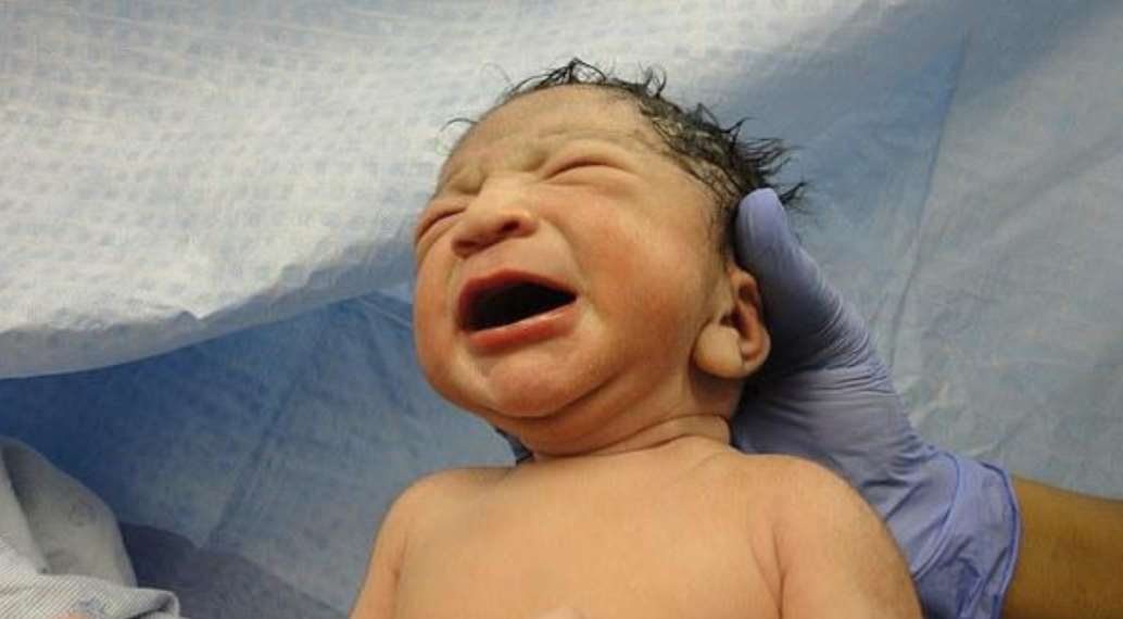
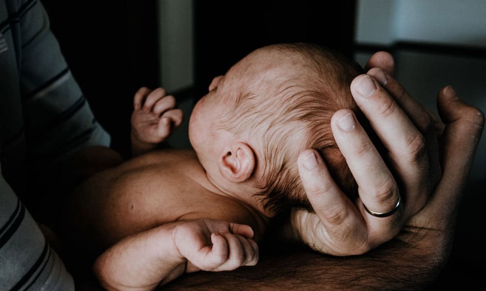
 Birth Injuries
 Homepage - SPOHNC: Support for People with Oral and Head and Neck Cancer
 Kaneohe Birth Injury Lawyer | Davis Levin Livingston
 What Is the Most Common Birth Injury? | Houston Birth Injury Attorney
 Private Consultants' Special Interests word selected: PERSONAL in town: MIDDLESBROUGH
 Kirurgisk korreksjon av ansiktslammelse - NEL - Norsk Elektronisk Legehåndbok
 Hemifacial Spasm (12.10.2012)
 Fanconi Anemia Research Fund
 Parotidectomy, Facial Nerve Dissection and Sialoceles