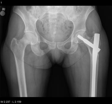
Femoral Neck Fractures
Fracture Fixation, Internal
Hemiarthroplasty
Fracture Healing
Hip Fractures
Femur Head
Femur Head Necrosis
Fractures, Spontaneous
Fractures, Ununited
Fracture Fixation
Hip Joint
Reoperation
Bone Density
Osteoporotic Fractures
Bone Nails
Osteoporosis
Bone Malalignment
Cementation
Radius Fractures
Fractures, Malunited
Fracture Fixation, Intramedullary
Prosthesis Failure
Fractures, Stress
Bone Plates
Postoperative Complications
Osteoarthritis, Hip
Bone Cements
Head and Neck Neoplasms
Treatment Outcome
Fibula
Dislocations
Leg Length Inequality
Follow-Up Studies
Osteomalacia
Retrospective Studies
Delayed Diagnosis
Absorptiometry, Photon
Orthopedic Procedures
Recovery of Function
Range of Motion, Articular
Technetium Tc 99m Medronate
Lithuania
Osteoporosis, Postmenopausal
Durapatite
Joint Prosthesis
Rib Fractures
Skull Fractures
Acetabulum
Prospective Studies
Lumbar Vertebrae
Bone Wires
Neck Pain
Metacarpal Bones
Hip
Risk Factors
Bone Diseases, Metabolic
Fractures, Compression
Bone Density Conservation Agents
Talus
Joint Capsule
Regional survey of femoral neck fractures. (1/520)
In the South-west Thames Region 2619 patients (2105 women and 514 men) were discharged with a diagnosis of femoral neck fracture in 1974. The equivalent of a 250-bedded hospital was occupied throughout the year. The incidence, average length of stay, and mortality rate rose with increasing age and there were differences in these indices in the five health areas. These results confirm the enormous burden placed on the hospital service by patients with fracture of the femoral neck but suggest that differences in practice in the five areas may contribute to the size of the problem. (+info)Carotid sinus hypersensitivity--a modifiable risk factor for fractured neck of femur. (2/520)
BACKGROUND: the potential impact on morbidity, mortality and health care economics makes it important to identify patients at risk of fracture, in particular fractured neck of femur (FNOF). Older patients with carotid sinus hypersensitivity (CSH) are more likely to have unexplained falls and to experience fractures, particularly FNOF. Our objective was to determine the prevalence of CSH in patients with FNOF. DESIGN: case-controlled prospective series. METHODS: consecutive cases were admissions over 65 years with FNOF. Controls were consecutive patients admitted for elective hip surgery, frail elderly people admitted to hospital medical wards and day-hospital patients. All patients had a clinical assessment of cognitive function, physical abilities and history of previous syncope, falls and dizziness, in addition to repeated carotid sinus massage with continuous heart rate and phasic blood pressure measurement. RESULTS: heart rate slowing and fall in systolic blood pressure was greater for patients with FNOF than those admitted for elective hip surgery (P < 0.05 and P < 0.001). CSH was present in 36% of the FNOF group, none of the elective surgery group, 13% of the acutely ill controls and 17% of the outpatients. It was more likely to be present in FNOF patients with a previous history of unexplained falls or an unexplained fall causing the index fracture. The heart rate and systolic blood pressure responses to carotid sinus stimulation were reproducible. CONCLUSION: older patients with an acute neck of femur fracture who do not give a clear history of an accidental fall or who have had previously unexplained falls are likely to have CSH. CSH may be a modifiable risk factor for older patients at risk of hip fracture. (+info)Acute fracture of the neck of the femur. An assessment of perfusion of the head by dynamic MRI. (3/520)
We performed dynamic MRI of the femoral head within 48 hours of injury on 22 patients with subcapital fracture of the neck of the femur and on a control group of 20 of whom ten were healthy subjects and ten were patients with an intertrochanteric fracture. Three MRI patterns emerged when the results between the fractured side and the contralateral femoral head were compared. In all of the control group and in those patients who had undisplaced fractures (Garden stages I and II), perfusion of the femoral head was considered to be at the same level as on the unaffected side. In patients with displaced fractures (Garden stages III and IV) almost all the femoral heads on the fractured side were impaired or totally avascular, although some had the same level of perfusion as the unaffected side. We conclude that dynamic MRI, a new non-invasive imaging technique, is useful for evaluating the perfusion of the femoral head. (+info)Hip fracture and bone histomorphometry in a young adult with cystic fibrosis. (4/520)
A 25-yr-old male with cystic fibrosis sustained a fragility fracture of the left femoral neck, which required surgical correction. He had several risk factors for the development of low bone density and despite treatment with an oral bisphosphonate, his bone mineral density reduced further. The patient died 2 yrs after sustaining the fracture. Bone specimens obtained at post mortem demonstrated severe cortical and trabecular osteopenia, but the histological features were not typical of osteoporosis or osteomalacia. Osteoporosis is thought to be a common complication of cystic fibrosis. The novel histomorphometric appearances reported here suggest that the bone disease of cystic fibrosis may be more complex and possibly unique. Labelled bone biopsies are required to clarify the bone defect leading to low bone density in cystic fibrosis patients so that appropriate therapeutic strategies can be developed. (+info)Fractures due to hypocalcemic convulsion. (5/520)
We report on two cases of patients in whom hypocalcemic seizures during hemodialysis led to right scapular body fracture in one and bilateral femoral neck fractures in the other. (+info)Is McMurray's osteotomy obsolete? (6/520)
A review of the method of performing, advantages, disadvantages of McMurray's displacement osteotomy with regard to treatment of nonunion of transcervical fracture neck femur with viable femoral head was carried out in this study of ten cases, in view of the abandonment of the procedure in favour of angulation osteotomy. Good results obtained in the series attest to the usefulness of McMurray's osteotomy in the difficult problem of nonunion of transcervical fracture neck femur in well selected cases with certain advantages over the angulation osteotomy due to the 'Armchair effect'. (+info)Hip dislocations associated with ipsilateral femoral neck fracture. (7/520)
Two cases of subcapital fracture associated with hip dislocation, treated with primary uncemented Total Hip Replacement are presented. (+info)Reflex sympathetic dystrophy in hypophosphataemic osteomalacia with femoral neck fracture: a case report. (8/520)
We report a male patient who presented with suspicion of skeletal metastases based upon an abnormal 99-mTc bone scan, which showed increased uptake at both femoral heads, left femoral neck, and several ribs. The images also suggested reflex sympathetic dystrophy, subcapital fracture of the left femur, and rib fractures. A diagnosis of hypophosphataemic osteomalacia was finally made. (+info)A femoral neck fracture is a type of hip fracture that occurs in the narrow, vertical section of bone just below the ball of the femur (thigh bone) that connects to the hip socket. This area is called the femoral neck. Femoral neck fractures can be categorized into different types based on their location and the direction of the fractured bone.
These fractures are typically caused by high-energy trauma, such as car accidents or falls from significant heights, in younger individuals. However, in older adults, particularly those with osteoporosis, femoral neck fractures can also result from low-energy trauma, like a simple fall from standing height.
Femoral neck fractures are often serious and require prompt medical attention. Treatment usually involves surgery to realign and stabilize the broken bone fragments, followed by rehabilitation to help regain mobility and strength. Potential complications of femoral neck fractures include avascular necrosis (loss of blood flow to the femoral head), nonunion or malunion (improper healing), and osteoarthritis in the hip joint.
Fracture fixation, internal, is a surgical procedure where a fractured bone is fixed using metal devices such as plates, screws, or rods that are implanted inside the body. This technique helps to maintain the alignment and stability of the broken bone while it heals. The implants may be temporarily or permanently left inside the body, depending on the nature and severity of the fracture. Internal fixation allows for early mobilization and rehabilitation, which can result in a faster recovery and improved functional outcome.
Hemiarthroplasty is a surgical procedure where only one half (hemi-) of a joint is replaced with an artificial component, usually a metal ball attached to a stem that fits into the bone. This procedure is most commonly performed on the shoulder or hip joints. In a hip hemiarthroplasty, it involves replacing the femoral head (the ball part of the thighbone) which has been damaged due to fracture or arthritis. The acetabulum (socket part of the pelvis) is not replaced and remains as it is. This procedure aims to relieve pain, restore mobility, and improve joint function.
Fracture healing is the natural process by which a broken bone repairs itself. When a fracture occurs, the body responds by initiating a series of biological and cellular events aimed at restoring the structural integrity of the bone. This process involves the formation of a hematoma (a collection of blood) around the fracture site, followed by the activation of inflammatory cells that help to clean up debris and prepare the area for repair.
Over time, specialized cells called osteoblasts begin to lay down new bone matrix, or osteoid, along the edges of the broken bone ends. This osteoid eventually hardens into new bone tissue, forming a bridge between the fracture fragments. As this process continues, the callus (a mass of newly formed bone and connective tissue) gradually becomes stronger and more compact, eventually remodeling itself into a solid, unbroken bone.
The entire process of fracture healing can take several weeks to several months, depending on factors such as the severity of the injury, the patient's age and overall health, and the location of the fracture. In some cases, medical intervention may be necessary to help promote healing or ensure proper alignment of the bone fragments. This may include the use of casts, braces, or surgical implants such as plates, screws, or rods.
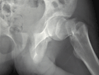
The "femur neck" is the narrow, upper part of the femur (thigh bone) where it connects to the pelvis. It is the region through which the femoral head articulates with the acetabulum to form the hip joint. The femur neck is a common site for fractures, especially in older adults with osteoporosis.
A hip fracture is a medical condition referring to a break in the upper part of the femur (thigh) bone, which forms the hip joint. The majority of hip fractures occur due to falls or direct trauma to the area. They are more common in older adults, particularly those with osteoporosis, a condition that weakens bones and makes them more prone to breaking. Hip fractures can significantly impact mobility and quality of life, often requiring surgical intervention and rehabilitation.
Hip arthroplasty, also known as hip replacement surgery, is a medical procedure where the damaged or diseased joint surfaces of the hip are removed and replaced with artificial components. These components typically include a metal or ceramic ball that replaces the head of the femur (thigh bone), and a polyethylene or ceramic socket that replaces the acetabulum (hip socket) in the pelvis.
The goal of hip arthroplasty is to relieve pain, improve joint mobility, and restore function to the hip joint. This procedure is commonly performed in patients with advanced osteoarthritis, rheumatoid arthritis, hip fractures, or other conditions that cause significant damage to the hip joint.
There are several types of hip replacement surgeries, including traditional total hip arthroplasty, partial (hemi) hip arthroplasty, and resurfacing hip arthroplasty. The choice of procedure depends on various factors, such as the patient's age, activity level, overall health, and the extent of joint damage.
After surgery, patients typically require rehabilitation to regain strength, mobility, and function in the affected hip. With proper care and follow-up, most patients can expect significant pain relief and improved quality of life following hip arthroplasty.
A bone fracture is a medical condition in which there is a partial or complete break in the continuity of a bone due to external or internal forces. Fractures can occur in any bone in the body and can vary in severity from a small crack to a shattered bone. The symptoms of a bone fracture typically include pain, swelling, bruising, deformity, and difficulty moving the affected limb. Treatment for a bone fracture may involve immobilization with a cast or splint, surgery to realign and stabilize the bone, or medication to manage pain and prevent infection. The specific treatment approach will depend on the location, type, and severity of the fracture.
The femoral head is the rounded, ball-like top portion of the femur (thigh bone) that fits into the hip socket (acetabulum) to form the hip joint. It has a smooth, articular cartilage surface that allows for smooth and stable articulation with the pelvis. The femoral head is connected to the femoral neck, which is a narrower section of bone that angles downward and leads into the shaft of the femur. Together, the femoral head and neck provide stability and range of motion to the hip joint.
A femoral fracture is a medical term that refers to a break in the thigh bone, which is the longest and strongest bone in the human body. The femur extends from the hip joint to the knee joint and is responsible for supporting the weight of the upper body and allowing movement of the lower extremity. Femoral fractures can occur due to various reasons such as high-energy trauma, low-energy trauma in individuals with weak bones (osteoporosis), or as a result of a direct blow to the thigh.
Femoral fractures can be classified into different types based on their location, pattern, and severity. Some common types of femoral fractures include:
1. Transverse fracture: A break that occurs straight across the bone.
2. Oblique fracture: A break that occurs at an angle across the bone.
3. Spiral fracture: A break that occurs in a helical pattern around the bone.
4. Comminuted fracture: A break that results in multiple fragments of the bone.
5. Open or compound fracture: A break in which the bone pierces through the skin.
6. Closed or simple fracture: A break in which the bone does not pierce through the skin.
Femoral fractures can cause severe pain, swelling, bruising, and difficulty walking or bearing weight on the affected leg. Diagnosis typically involves a physical examination, medical history, and imaging tests such as X-rays or CT scans. Treatment may involve surgical intervention, including the use of metal rods, plates, or screws to stabilize the bone, followed by rehabilitation and physical therapy to restore mobility and strength.
Bone screws are medical devices used in orthopedic and trauma surgery to affix bone fracture fragments or to attach bones to other bones or to metal implants such as plates, rods, or artificial joints. They are typically made of stainless steel or titanium alloys and have a threaded shaft that allows for purchase in the bone when tightened. The head of the screw may have a hexagonal or star-shaped design to allow for precise tightening with a screwdriver. Bone screws come in various shapes, sizes, and designs, including fully threaded, partially threaded, cannulated (hollow), and headless types, depending on their intended use and location in the body.
A comminuted fracture is a type of bone break where the bone is shattered into three or more pieces. This type of fracture typically occurs after high-energy trauma, such as a car accident or a fall from a great height. Commminuted fractures can also occur in bones that are weakened by conditions like osteoporosis or cancer. Because of the severity and complexity of comminuted fractures, they often require extensive treatment, which may include surgery to realign and stabilize the bone fragments using metal screws, plates, or rods.
A hip prosthesis, also known as a total hip replacement, is a surgical implant designed to replace the damaged or diseased components of the human hip joint. The procedure involves replacing the femoral head (the ball at the top of the thigh bone) and the acetabulum (the socket in the pelvis) with artificial parts, typically made from materials such as metal, ceramic, or plastic.
The goal of a hip prosthesis is to relieve pain, improve joint mobility, and restore function, allowing patients to return to their normal activities and enjoy an improved quality of life. The procedure is most commonly performed in individuals with advanced osteoarthritis, rheumatoid arthritis, or other degenerative conditions that have caused significant damage to the hip joint.
There are several different types of hip prostheses available, each with its own unique design and set of benefits and risks. The choice of prosthesis will depend on a variety of factors, including the patient's age, activity level, overall health, and specific medical needs. In general, however, all hip prostheses are designed to provide a durable, long-lasting solution for patients suffering from debilitating joint pain and stiffness.
Femoral head necrosis, also known as avascular necrosis of the femoral head, is a medical condition that results from the interruption of blood flow to the femoral head, which is the rounded end of the thigh bone that fits into the hip joint. This lack of blood supply can cause the bone tissue to die, leading to the collapse of the femoral head and eventually resulting in hip joint damage or arthritis.
The condition can be caused by a variety of factors, including trauma, alcohol abuse, corticosteroid use, radiation therapy, and certain medical conditions such as sickle cell disease and lupus. Symptoms may include pain in the hip or groin, limited range of motion, and difficulty walking. Treatment options depend on the severity and progression of the necrosis and may include medication, physical therapy, or surgical intervention.
Spontaneous fractures are bone breaks that occur without any identifiable trauma or injury. They are typically caused by underlying medical conditions that weaken the bones, making them more susceptible to breaking under normal stress or weight. The most common cause of spontaneous fractures is osteoporosis, a condition characterized by weak and brittle bones. Other potential causes include various bone diseases, certain cancers, long-term use of corticosteroids, and genetic disorders affecting bone strength.
It's important to note that while the term "spontaneous" implies that the fracture occurred without any apparent cause, it is usually the result of an underlying medical condition. Therefore, if you experience a spontaneous fracture, seeking medical attention is crucial to diagnose and manage the underlying cause to prevent future fractures and related complications.
Ununited fracture is a medical term used to describe a fractured bone that has failed to heal properly. This condition is also known as a nonunion fracture. In a normal healing process, the broken ends of the bone will grow together, or "unite," over time as new bone tissue forms. However, in some cases, the bones may not reconnect due to various reasons such as infection, poor blood supply, excessive motion at the fracture site, or inadequate stabilization of the fracture.
Ununited fractures can cause significant pain, swelling, and deformity in the affected area. They may also lead to a decreased range of motion, weakness, and instability in the joint near the fracture. Treatment for ununited fractures typically involves surgical intervention to promote bone healing, such as bone grafting or internal fixation with screws or plates. In some cases, electrical stimulation or ultrasound therapy may also be used to help promote bone growth and healing.
Fracture fixation is a surgical procedure in orthopedic trauma surgery where a fractured bone is stabilized using various devices and techniques to promote proper healing and alignment. The goal of fracture fixation is to maintain the broken bone ends in correct anatomical position and length, allowing for adequate stability during the healing process.
There are two main types of fracture fixation:
1. Internal fixation: In this method, metal implants like plates, screws, or intramedullary rods are inserted directly into the bone to hold the fragments in place. These implants can be either removed or left in the body once healing is complete, depending on the type and location of the fracture.
2. External fixation: This technique involves placing pins or screws through the skin and into the bone above and below the fracture site. These pins are then connected to an external frame that maintains alignment and stability. External fixators are typically used when there is significant soft tissue damage, infection, or when internal fixation is not possible due to the complexity of the fracture.
The choice between internal and external fixation depends on various factors such as the type and location of the fracture, patient's age and overall health, surgeon's preference, and potential complications. Both methods aim to provide a stable environment for bone healing while minimizing the risk of malunion, nonunion, or deformity.
A hip dislocation is a medical emergency that occurs when the head of the femur (thighbone) slips out of its socket in the pelvis. This can happen due to high-energy trauma, such as a car accident or a severe fall. Hip dislocations can also occur in people with certain health conditions that make their hips more prone to displacement, such as developmental dysplasia of the hip.
There are two main types of hip dislocations: posterior and anterior. In a posterior dislocation, the femur head moves out of the back of the socket, which is the most common type. In an anterior dislocation, the femur head moves out of the front of the socket. Both types of hip dislocations can cause severe pain, swelling, and difficulty moving the affected leg.
Immediate medical attention is necessary for a hip dislocation to realign the bones and prevent further damage. Treatment typically involves sedation or anesthesia to relax the muscles around the joint, followed by a closed reduction procedure to gently guide the femur head back into the socket. In some cases, surgery may be required to repair any associated injuries, such as fractures or damaged ligaments. After treatment, physical therapy and rehabilitation are usually necessary to restore strength, mobility, and function to the affected hip joint.
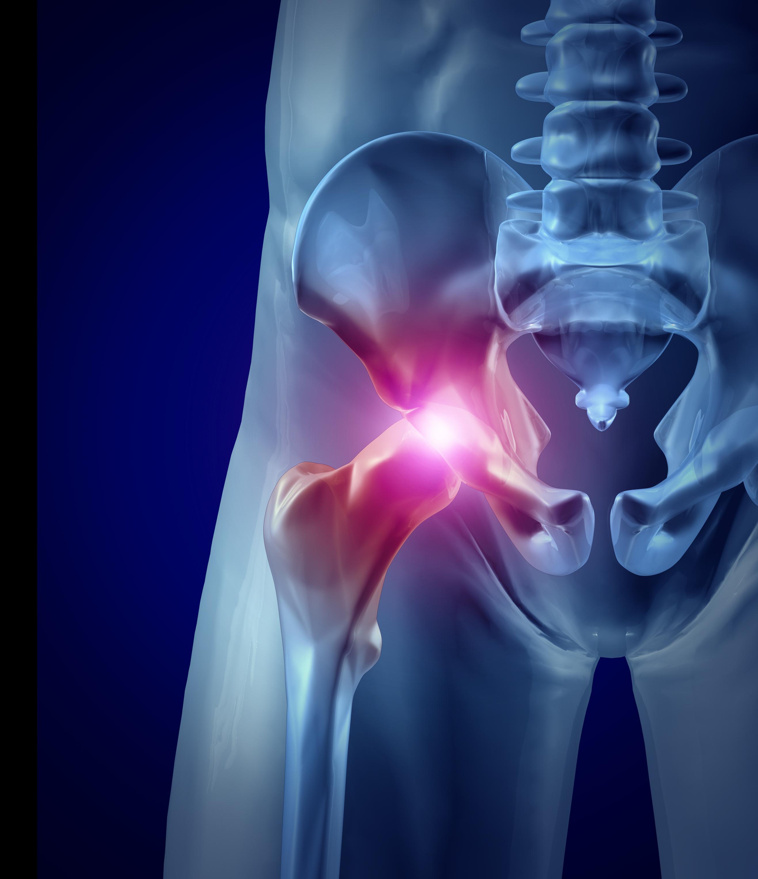
The hip joint, also known as the coxal joint, is a ball-and-socket type synovial joint that connects the femur (thigh bone) to the pelvis. The "ball" is the head of the femur, while the "socket" is the acetabulum, a concave surface on the pelvic bone.
The hip joint is surrounded by a strong fibrous capsule and is reinforced by several ligaments, including the iliofemoral, ischiofemoral, and pubofemoral ligaments. The joint allows for flexion, extension, abduction, adduction, medial and lateral rotation, and circumduction movements, making it one of the most mobile joints in the body.
The hip joint is also supported by various muscles, including the gluteus maximus, gluteus medius, gluteus minimus, iliopsoas, and other hip flexors and extensors. These muscles provide stability and strength to the joint, allowing for weight-bearing activities such as walking, running, and jumping.
A reoperation is a surgical procedure that is performed again on a patient who has already undergone a previous operation for the same or related condition. Reoperations may be required due to various reasons, such as inadequate initial treatment, disease recurrence, infection, or complications from the first surgery. The nature and complexity of a reoperation can vary widely depending on the specific circumstances, but it often carries higher risks and potential complications compared to the original operation.
Bone density refers to the amount of bone mineral content (usually measured in grams) in a given volume of bone (usually measured in cubic centimeters). It is often used as an indicator of bone strength and fracture risk. Bone density is typically measured using dual-energy X-ray absorptiometry (DXA) scans, which provide a T-score that compares the patient's bone density to that of a young adult reference population. A T-score of -1 or above is considered normal, while a T-score between -1 and -2.5 indicates osteopenia (low bone mass), and a T-score below -2.5 indicates osteoporosis (porous bones). Regular exercise, adequate calcium and vitamin D intake, and medication (if necessary) can help maintain or improve bone density and prevent fractures.
Osteoporotic fractures are breaks or cracks in bones that occur as a result of osteoporosis, a condition characterized by weak and brittle bones. Osteoporosis causes bones to lose density and strength, making them more susceptible to fractures, even from minor injuries or falls.
The most common types of osteoporotic fractures are:
1. Hip fractures: These occur when the upper part of the thigh bone (femur) breaks, often due to a fall. Hip fractures can be serious and may require surgery and hospitalization.
2. Vertebral compression fractures: These occur when the bones in the spine (vertebrae) collapse, causing height loss, back pain, and deformity. They are often caused by everyday activities, such as bending or lifting.
3. Wrist fractures: These occur when the bones in the wrist break, often due to a fall. Wrist fractures are common in older adults with osteoporosis.
4. Other fractures: Osteoporotic fractures can also occur in other bones, such as the pelvis, ribs, and humerus (upper arm bone).
Prevention is key in managing osteoporosis and reducing the risk of osteoporotic fractures. This includes getting enough calcium and vitamin D, engaging in regular weight-bearing exercise, avoiding smoking and excessive alcohol consumption, and taking medications as prescribed by a healthcare provider.
I believe you are referring to "bone pins" or "bone nails" rather than "bone nails." These terms are used in the medical field to describe surgical implants made of metal or biocompatible materials that are used to stabilize and hold together fractured bones during the healing process. They can also be used in spinal fusion surgery to provide stability and promote bone growth between vertebrae.
Bone pins or nails typically have a threaded or smooth shaft, with a small diameter that allows them to be inserted into the medullary canal of long bones such as the femur or tibia. They may also have a head or eyelet on one end that allows for attachment to external fixation devices or other surgical instruments.
The use of bone pins and nails has revolutionized orthopedic surgery, allowing for faster healing times, improved stability, and better functional outcomes for patients with fractures or spinal deformities.
Arthroplasty is a surgical procedure to restore the integrity and function of a joint. The term is derived from two Greek words: "arthro" meaning joint, and "plasty" meaning to mold or form. There are several types of arthroplasty, but most involve resurfacing the damaged joint cartilage with artificial materials such as metal, plastic, or ceramic.
The goal of arthroplasty is to relieve pain, improve mobility, and restore function in a joint that has been damaged by arthritis, injury, or other conditions. The most common types of arthroplasty are total joint replacement (TJR) and partial joint replacement (PJR).
In TJR, the surgeon removes the damaged ends of the bones in the joint and replaces them with artificial components called prostheses. These prostheses can be made of metal, plastic, or ceramic materials, and are designed to mimic the natural movement and function of the joint.
In PJR, only one side of the joint is resurfaced, typically because the damage is less extensive. This procedure is less invasive than TJR and may be recommended for younger patients who are still active or have a higher risk of complications from a full joint replacement.
Other types of arthroplasty include osteotomy, in which the surgeon cuts and reshapes the bone to realign the joint; arthrodesis, in which the surgeon fuses two bones together to create a stable joint; and resurfacing, in which the damaged cartilage is removed and replaced with a smooth, artificial surface.
Arthroplasty is typically recommended for patients who have tried other treatments, such as physical therapy, medication, or injections, but have not found relief from their symptoms. While arthroplasty can be highly effective in relieving pain and improving mobility, it is not without risks, including infection, blood clots, and implant failure. Patients should discuss the benefits and risks of arthroplasty with their healthcare provider to determine if it is the right treatment option for them.
A spinal fracture, also known as a vertebral compression fracture, is a break in one or more bones (vertebrae) of the spine. This type of fracture often occurs due to weakened bones caused by osteoporosis, but it can also result from trauma such as a car accident or a fall.
In a spinal fracture, the front part of the vertebra collapses, causing the height of the vertebra to decrease, while the back part of the vertebra remains intact. This results in a wedge-shaped deformity of the vertebra. Multiple fractures can lead to a hunched forward posture known as kyphosis or dowager's hump.
Spinal fractures can cause pain, numbness, tingling, or weakness in the back, legs, or arms, depending on the location and severity of the fracture. In some cases, spinal cord compression may occur, leading to more severe symptoms such as paralysis or loss of bladder and bowel control.
Osteoporosis is a systemic skeletal disease characterized by low bone mass, deterioration of bone tissue, and disruption of bone architecture, leading to increased risk of fractures, particularly in the spine, wrist, and hip. It mainly affects older people, especially postmenopausal women, due to hormonal changes that reduce bone density. Osteoporosis can also be caused by certain medications, medical conditions, or lifestyle factors such as smoking, alcohol abuse, and a lack of calcium and vitamin D in the diet. The diagnosis is often made using bone mineral density testing, and treatment may include medication to slow bone loss, promote bone formation, and prevent fractures.
Bone malalignment is a term used to describe the abnormal alignment or positioning of bones in relation to each other. This condition can occur as a result of injury, deformity, surgery, or disease processes that affect the bones and joints. Bone malalignment can cause pain, stiffness, limited mobility, and an increased risk of further injury. In some cases, bone malalignment may require treatment such as bracing, physical therapy, or surgery to correct the alignment and improve function.
In the medical field, cementation refers to the process of using a type of dental cement or bonding agent to attach a dental restoration (such as a crown, bridge, or false tooth) to a natural tooth or implant. The cement helps to create a strong and secure attachment, while also helping to seal the restoration and prevent the entry of bacteria and saliva.
Dental cement can be made from various materials, including glass ionomers, resin-modified glass ionomers, zinc phosphate, and polycarboxylate cements. The choice of cement depends on several factors, such as the type of restoration being attached, the location in the mouth, and the patient's individual needs and preferences.
Cementation is an important step in many dental procedures, as it helps to ensure the longevity and success of the restoration. Proper technique and material selection are crucial for achieving a successful cementation that will last for years to come.
A radius fracture is a break in the bone that runs from the wrist to the elbow, located on the thumb side of the forearm. Radius fractures can occur as a result of a fall, direct blow to the forearm, or a high-energy collision such as a car accident. There are various types of radius fractures, including:
1. Distal radius fracture: A break at the end of the radius bone, near the wrist joint, which is the most common type of radius fracture.
2. Radial shaft fracture: A break in the middle portion of the radius bone.
3. Radial head and neck fractures: Breaks in the upper part of the radius bone, near the elbow joint.
4. Comminuted fracture: A complex radius fracture where the bone is broken into multiple pieces.
5. Open (compound) fracture: A radius fracture with a wound or laceration in the skin, allowing for communication between the outside environment and the fractured bone.
6. Intra-articular fracture: A radius fracture that extends into the wrist joint or elbow joint.
7. Torus (buckle) fracture: A stable fracture where one side of the bone is compressed, causing it to buckle or bend, but not break completely through.
Symptoms of a radius fracture may include pain, swelling, tenderness, bruising, deformity, limited mobility, and in some cases, numbness or tingling in the fingers. Treatment options depend on the type and severity of the fracture but can range from casting to surgical intervention with implant fixation.
Malunited fractures refer to a type of fracture where the bones do not heal in their proper alignment or position. This can occur due to various reasons such as inadequate reduction of the fracture fragments during initial treatment, improper casting or immobilization, or failure of the patient to follow proper immobilization instructions. Malunited fractures can result in deformity, limited range of motion, and decreased functionality of the affected limb. Additional treatments such as surgery may be required to correct the malunion and restore normal function.
Intramedullary fracture fixation is a surgical technique used to stabilize and align bone fractures. In this procedure, a metal rod or nail is inserted into the marrow cavity (intramedullary canal) of the affected bone, spanning the length of the fracture. The rod is then secured to the bone using screws or other fixation devices on either side of the fracture. This provides stability and helps maintain proper alignment during the healing process.
The benefits of intramedullary fixation include:
1. Load sharing: The intramedullary rod shares some of the load bearing capacity with the bone, which can help reduce stress on the healing bone.
2. Minimal soft tissue dissection: Since the implant is inserted through the medullary canal, there is less disruption to the surrounding muscles, tendons, and ligaments compared to other fixation methods.
3. Biomechanical stability: Intramedullary fixation provides rotational and bending stiffness, which helps maintain proper alignment of the fracture fragments during healing.
4. Early mobilization: Patients with intramedullary fixation can often begin weight bearing and rehabilitation exercises earlier than those with other types of fixation, leading to faster recovery times.
Common indications for intramedullary fracture fixation include long bone fractures in the femur, tibia, humerus, and fibula, as well as certain pelvic and spinal fractures. However, the choice of fixation method depends on various factors such as patient age, fracture pattern, location, and associated injuries.
Prosthesis failure is a term used to describe a situation where a prosthetic device, such as an artificial joint or limb, has stopped functioning or failed to meet its intended purpose. This can be due to various reasons, including mechanical failure, infection, loosening of the device, or a reaction to the materials used in the prosthesis.
Mechanical failure can occur due to wear and tear, manufacturing defects, or improper use of the prosthetic device. Infection can also lead to prosthesis failure, particularly in cases where the prosthesis is implanted inside the body. The immune system may react to the presence of the foreign material, leading to inflammation and infection.
Loosening of the prosthesis can also cause it to fail over time, as the device becomes less stable and eventually stops working properly. Additionally, some people may have a reaction to the materials used in the prosthesis, leading to tissue damage or other complications that can result in prosthesis failure.
In general, prosthesis failure can lead to decreased mobility, pain, and the need for additional surgeries or treatments to correct the problem. It is important for individuals with prosthetic devices to follow their healthcare provider's instructions carefully to minimize the risk of prosthesis failure and ensure that the device continues to function properly over time.
Stress fractures are defined as small cracks or severe bruising in bones that occur from repetitive stress or overuse. They most commonly occur in weight-bearing bones, such as the legs and feet, but can also occur in the arms, hips, and back. Stress fractures differ from regular fractures because they typically do not result from a single, traumatic event. Instead, they are caused by repeated stress on the bone that results in microscopic damage over time. Athletes, military personnel, and individuals who engage in high-impact activities or have weak bones (osteoporosis) are at increased risk of developing stress fractures. Symptoms may include pain, swelling, tenderness, and difficulty walking or bearing weight on the affected bone.
Bone plates are medical devices used in orthopedic surgery to stabilize and hold together fractured or broken bones during the healing process. They are typically made of surgical-grade stainless steel, titanium, or other biocompatible materials. The plate is shaped to fit the contour of the bone and is held in place with screws that are inserted through the plate and into the bone on either side of the fracture. This provides stability and alignment to the broken bones, allowing them to heal properly. Bone plates can be used to treat a variety of fractures, including those that are complex or unstable. After healing is complete, the bone plate may be left in place or removed, depending on the individual's needs and the surgeon's recommendation.
In medical terms, the "neck" is defined as the portion of the body that extends from the skull/head to the thorax or chest region. It contains 7 cervical vertebrae, muscles, nerves, blood vessels, lymphatic vessels, and glands (such as the thyroid gland). The neck is responsible for supporting the head, allowing its movement in various directions, and housing vital structures that enable functions like respiration and circulation.
Postoperative complications refer to any unfavorable condition or event that occurs during the recovery period after a surgical procedure. These complications can vary in severity and may include, but are not limited to:
1. Infection: This can occur at the site of the incision or inside the body, such as pneumonia or urinary tract infection.
2. Bleeding: Excessive bleeding (hemorrhage) can lead to a drop in blood pressure and may require further surgical intervention.
3. Blood clots: These can form in the deep veins of the legs (deep vein thrombosis) and can potentially travel to the lungs (pulmonary embolism).
4. Wound dehiscence: This is when the surgical wound opens up, which can lead to infection and further complications.
5. Pulmonary issues: These include atelectasis (collapsed lung), pneumonia, or respiratory failure.
6. Cardiovascular problems: These include abnormal heart rhythms (arrhythmias), heart attack, or stroke.
7. Renal failure: This can occur due to various reasons such as dehydration, blood loss, or the use of certain medications.
8. Pain management issues: Inadequate pain control can lead to increased stress, anxiety, and decreased mobility.
9. Nausea and vomiting: These can be caused by anesthesia, opioid pain medication, or other factors.
10. Delirium: This is a state of confusion and disorientation that can occur in the elderly or those with certain medical conditions.
Prompt identification and management of these complications are crucial to ensure the best possible outcome for the patient.
Osteoarthritis (OA) of the hip is a degenerative joint disease that affects the articular cartilage and subchondral bone of the hip joint. It is characterized by the progressive loss of cartilage, remodeling of bone, osteophyte formation (bone spurs), cysts, and mild to moderate inflammation. The degenerative process can lead to pain, stiffness, limited range of motion, and crepitus (grating or crackling sound) during movement.
In the hip joint, OA typically affects the femoral head and acetabulum. As the articular cartilage wears away, the underlying bone becomes exposed and can lead to bone-on-bone contact, which is painful. The body responds by attempting to repair the damage through remodeling of the subchondral bone and formation of osteophytes. However, these changes can further limit joint mobility and exacerbate symptoms.
Risk factors for OA of the hip include age, obesity, genetics, previous joint injury or surgery, and repetitive stress on the joint. Treatment options may include pain management (such as NSAIDs, physical therapy, and injections), lifestyle modifications (such as weight loss and exercise), and, in severe cases, surgical intervention (such as hip replacement).
The femur is the medical term for the thigh bone, which is the longest and strongest bone in the human body. It connects the hip bone to the knee joint and plays a crucial role in supporting the weight of the body and allowing movement during activities such as walking, running, and jumping. The femur is composed of a rounded head, a long shaft, and two condyles at the lower end that articulate with the tibia and patella to form the knee joint.
Osteonecrosis is a medical condition characterized by the death of bone tissue due to the disruption of blood supply. Also known as avascular necrosis, this process can lead to the collapse of the bone and adjacent joint surfaces, resulting in pain, limited mobility, and potential deformity if left untreated. Osteonecrosis most commonly affects the hips, shoulders, and knees, but it can occur in any bone. The condition may be caused by trauma, corticosteroid use, alcohol abuse, certain medical conditions (like sickle cell disease or lupus), or for no apparent reason (idiopathic).
Bone cements are medical-grade materials used in orthopedic and trauma surgery to fill gaps between bone surfaces and implants, such as artificial joints or screws. They serve to mechanically stabilize the implant and provide a smooth, load-bearing surface. The two most common types of bone cement are:
1. Polymethylmethacrylate (PMMA) cement: This is a two-component system consisting of powdered PMMA and liquid methyl methacrylate monomer. When mixed together, they form a dough-like consistency that hardens upon exposure to air. PMMA cement has been widely used for decades in joint replacement surgeries, such as hip or knee replacements.
2. Calcium phosphate (CP) cement: This is a two-component system consisting of a powdered CP compound and an aqueous solution. When mixed together, they form a paste that hardens through a chemical reaction at body temperature. CP cement has lower mechanical strength compared to PMMA but demonstrates better biocompatibility, bioactivity, and the ability to resorb over time.
Both types of bone cements have advantages and disadvantages, and their use depends on the specific surgical indication and patient factors.
Head and neck neoplasms refer to abnormal growths or tumors in the head and neck region, which can be benign (non-cancerous) or malignant (cancerous). These tumors can develop in various sites, including the oral cavity, nasopharynx, oropharynx, larynx, hypopharynx, paranasal sinuses, salivary glands, and thyroid gland.
Benign neoplasms are slow-growing and generally do not spread to other parts of the body. However, they can still cause problems if they grow large enough to press on surrounding tissues or structures. Malignant neoplasms, on the other hand, can invade nearby tissues and organs and may also metastasize (spread) to other parts of the body.
Head and neck neoplasms can have various symptoms depending on their location and size. Common symptoms include difficulty swallowing, speaking, or breathing; pain in the mouth, throat, or ears; persistent coughing or hoarseness; and swelling or lumps in the neck or face. Early detection and treatment of head and neck neoplasms are crucial for improving outcomes and reducing the risk of complications.
Treatment outcome is a term used to describe the result or effect of medical treatment on a patient's health status. It can be measured in various ways, such as through symptoms improvement, disease remission, reduced disability, improved quality of life, or survival rates. The treatment outcome helps healthcare providers evaluate the effectiveness of a particular treatment plan and make informed decisions about future care. It is also used in clinical research to compare the efficacy of different treatments and improve patient care.
The fibula is a slender bone located in the lower leg of humans and other vertebrates. It runs parallel to the larger and more robust tibia, and together they are known as the bones of the leg or the anterior tibial segment. The fibula is the lateral bone in the leg, positioned on the outside of the tibia.
In humans, the fibula extends from the knee joint proximally to the ankle joint distally. Its proximal end, called the head of the fibula, articulates with the lateral condyle of the tibia and forms part of the inferior aspect of the knee joint. The narrowed portion below the head is known as the neck of the fibula.
The shaft of the fibula, also called the body of the fibula, is a long, thin structure that descends from the neck and serves primarily for muscle attachment rather than weight-bearing functions. The distal end of the fibula widens to form the lateral malleolus, which is an important bony landmark in the ankle region. The lateral malleolus articulates with the talus bone of the foot and forms part of the ankle joint.
The primary functions of the fibula include providing attachment sites for muscles that act on the lower leg, ankle, and foot, as well as contributing to the stability of the ankle joint through its articulation with the talus bone. Fractures of the fibula can occur due to various injuries, such as twisting or rotational forces applied to the ankle or direct trauma to the lateral aspect of the lower leg.
A dislocation is a condition in which a bone slips out of its normal position in a joint. This can happen as a result of trauma or injury, such as a fall or direct blow to the body. Dislocations can cause pain, swelling, and limited mobility in the affected area. In some cases, a dislocation may also damage surrounding tissues, such as ligaments, tendons, and nerves.
Dislocations are typically treated by reducing the dislocation, which means putting the bone back into its normal position. This is usually done with the help of medication to relieve pain and relaxation techniques to help the person stay still during the reduction. In some cases, surgery may be necessary to repair damaged tissues or if the dislocation cannot be reduced through other methods. After the dislocation has been reduced, the joint may be immobilized with a splint or sling to allow it to heal properly.
It is important to seek medical attention promptly if you suspect that you have a dislocation. If left untreated, a dislocation can lead to further complications, such as joint instability and chronic pain.
'Leg length inequality' (LLIS) is a condition where there is a discrepancy in the lengths of an individual's lower extremities, specifically the bones of the thigh (femur) and/or the leg (tibia/fibula). This discrepancy can be congenital or acquired due to various causes such as fractures, infections, or surgical procedures. The inequality can lead to functional scoliosis, lower back pain, and other musculoskeletal issues. It is typically diagnosed through physical examination and imaging studies like X-rays, and may be treated with various methods including orthotics, shoe lifts, or in some cases, surgical intervention.
Osteotomy is a surgical procedure in which a bone is cut to shorten, lengthen, or change its alignment. It is often performed to correct deformities or to realign bones that have been damaged by trauma or disease. The bone may be cut straight across (transverse osteotomy) or at an angle (oblique osteotomy). After the bone is cut, it can be realigned and held in place with pins, plates, or screws until it heals. This procedure is commonly performed on bones in the leg, such as the femur or tibia, but can also be done on other bones in the body.
Follow-up studies are a type of longitudinal research that involve repeated observations or measurements of the same variables over a period of time, in order to understand their long-term effects or outcomes. In medical context, follow-up studies are often used to evaluate the safety and efficacy of medical treatments, interventions, or procedures.
In a typical follow-up study, a group of individuals (called a cohort) who have received a particular treatment or intervention are identified and then followed over time through periodic assessments or data collection. The data collected may include information on clinical outcomes, adverse events, changes in symptoms or functional status, and other relevant measures.
The results of follow-up studies can provide important insights into the long-term benefits and risks of medical interventions, as well as help to identify factors that may influence treatment effectiveness or patient outcomes. However, it is important to note that follow-up studies can be subject to various biases and limitations, such as loss to follow-up, recall bias, and changes in clinical practice over time, which must be carefully considered when interpreting the results.
Osteomalacia is a medical condition characterized by the softening of bones due to defective bone mineralization, resulting from inadequate vitamin D, phosphate, or calcium. It mainly affects adults and is different from rickets, which occurs in children. The primary symptom is bone pain, but muscle weakness can also occur. Prolonged osteomalacia may lead to skeletal deformities and an increased risk of fractures. Treatment typically involves supplementation with vitamin D, calcium, and sometimes phosphate.
Retrospective studies, also known as retrospective research or looking back studies, are a type of observational study that examines data from the past to draw conclusions about possible causal relationships between risk factors and outcomes. In these studies, researchers analyze existing records, medical charts, or previously collected data to test a hypothesis or answer a specific research question.
Retrospective studies can be useful for generating hypotheses and identifying trends, but they have limitations compared to prospective studies, which follow participants forward in time from exposure to outcome. Retrospective studies are subject to biases such as recall bias, selection bias, and information bias, which can affect the validity of the results. Therefore, retrospective studies should be interpreted with caution and used primarily to generate hypotheses for further testing in prospective studies.
Delayed diagnosis is a term used in the medical field to describe a situation where a medical condition or disease was not diagnosed in a timely manner, despite the patient having sought medical attention and presented with symptoms that should have led to an earlier diagnosis. This can occur due to various reasons such as failure to recognize symptoms, misinterpretation of test results, lack of appropriate follow-up care, or communication breakdowns between healthcare providers and patients.
A delayed diagnosis can result in worsening of the medical condition, increased severity of symptoms, decreased treatment options, and potentially poorer outcomes for the patient. It may also lead to additional medical expenses, longer recovery times, and emotional distress for the patient and their family members. In some cases, a delayed diagnosis may be considered medical malpractice if it can be shown that the healthcare provider failed to meet the standard of care required in diagnosing the condition.
Prosthesis design is a specialized field in medical device technology that involves creating and developing artificial substitutes to replace a missing body part, such as a limb, tooth, eye, or internal organ. The design process typically includes several stages: assessment of the patient's needs, selection of appropriate materials, creation of a prototype, testing and refinement, and final fabrication and fitting of the prosthesis.
The goal of prosthesis design is to create a device that functions as closely as possible to the natural body part it replaces, while also being comfortable, durable, and aesthetically pleasing for the patient. The design process may involve collaboration between medical professionals, engineers, and designers, and may take into account factors such as the patient's age, lifestyle, occupation, and overall health.
Prosthesis design can be highly complex, particularly for advanced devices such as robotic limbs or implantable organs. These devices often require sophisticated sensors, actuators, and control systems to mimic the natural functions of the body part they replace. As a result, prosthesis design is an active area of research and development in the medical field, with ongoing efforts to improve the functionality, comfort, and affordability of these devices for patients.
Photon Absorptiometry is a medical technique used to measure the absorption of photons (light particles) by tissues or materials. In clinical practice, it is often used as a non-invasive method for measuring bone mineral density (BMD). This technique uses a low-energy X-ray beam or gamma ray to penetrate the tissue and then measures the amount of radiation absorbed by the bone. The amount of absorption is related to the density and thickness of the bone, allowing for an assessment of BMD. It can be used to diagnose osteoporosis and monitor treatment response in patients with bone diseases. There are two types of photon absorptiometry: single-photon absorptiometry (SPA) and dual-photon absorptiometry (DPA). SPA uses one energy level, while DPA uses two different energy levels to measure BMD, providing more precise measurements.
Orthopedic procedures are surgical or nonsurgical methods used to treat musculoskeletal conditions, including injuries, deformities, or diseases of the bones, joints, muscles, ligaments, and tendons. These procedures can range from simple splinting or casting to complex surgeries such as joint replacements, spinal fusions, or osteotomies (cutting and repositioning bones). The primary goal of orthopedic procedures is to restore function, reduce pain, and improve the quality of life for patients.
"Recovery of function" is a term used in medical rehabilitation to describe the process in which an individual regains the ability to perform activities or tasks that were previously difficult or impossible due to injury, illness, or disability. This can involve both physical and cognitive functions. The goal of recovery of function is to help the person return to their prior level of independence and participation in daily activities, work, and social roles as much as possible.
Recovery of function may be achieved through various interventions such as physical therapy, occupational therapy, speech-language therapy, and other rehabilitation strategies. The specific approach used will depend on the individual's needs and the nature of their impairment. Recovery of function can occur spontaneously as the body heals, or it may require targeted interventions to help facilitate the process.
It is important to note that recovery of function does not always mean a full return to pre-injury or pre-illness levels of ability. Instead, it often refers to the person's ability to adapt and compensate for any remaining impairments, allowing them to achieve their maximum level of functional independence and quality of life.
An ulna fracture is a break in the ulna bone, which is one of the two long bones in the forearm. The ulna is located on the pinky finger side of the forearm and functions to support the elbow joint and assist in rotation and movement of the forearm. Ulna fractures can occur at various points along the bone, including the shaft, near the wrist, or at the elbow end of the bone. Symptoms may include pain, swelling, bruising, tenderness, deformity, limited mobility, and in some cases, numbness or tingling in the fingers. Treatment typically involves immobilization with a cast or splint, followed by rehabilitation exercises to restore strength and range of motion. In severe cases, surgery may be required to realign and stabilize the fractured bone.
Articular Range of Motion (AROM) is a term used in physiotherapy and orthopedics to describe the amount of movement available in a joint, measured in degrees of a circle. It refers to the range through which synovial joints can actively move without causing pain or injury. AROM is assessed by measuring the degree of motion achieved by active muscle contraction, as opposed to passive range of motion (PROM), where the movement is generated by an external force.
Assessment of AROM is important in evaluating a patient's functional ability and progress, planning treatment interventions, and determining return to normal activities or sports participation. It is also used to identify any restrictions in joint mobility that may be due to injury, disease, or surgery, and to monitor the effectiveness of rehabilitation programs.
Technetium Tc 99m Medronate is a radiopharmaceutical agent used in nuclear medicine for bone scintigraphy. It is a technetium-labeled bisphosphonate compound, which accumulates in areas of increased bone turnover and metabolism. This makes it useful for detecting and evaluating various bone diseases and conditions, such as fractures, tumors, infections, and arthritis.
The "Tc 99m" refers to the radioisotope technetium-99m, which has a half-life of approximately 6 hours and emits gamma rays that can be detected by a gamma camera. The medronate component is a bisphosphonate molecule that binds to hydroxyapatite crystals in bone tissue, allowing the radiolabeled compound to accumulate in areas of active bone remodeling.
Overall, Technetium Tc 99m Medronate is an important tool in nuclear medicine for diagnosing and managing various musculoskeletal disorders.
"Time-to-Treatment" is a medical term that refers to the duration of time taken from the identification or diagnosis of a medical condition to the initiation of appropriate treatment. This interval is crucial in determining the prognosis and outcome of various medical conditions, particularly those that require prompt intervention such as stroke, myocardial infarction (heart attack), and sepsis.
Reducing time-to-treatment can significantly improve patient outcomes by minimizing the risk of complications, reducing morbidity and mortality, and enhancing the effectiveness of therapeutic interventions. Therefore, healthcare providers strive to optimize and streamline their processes to ensure timely and effective treatment for their patients.
I'm sorry for any confusion, but "Lithuania" is not a medical term. It is a country in Northern Europe, the southernmost of the three Baltic states. If you have any questions about medical terms or concepts, I would be happy to try and help answer those for you!
Postmenopausal osteoporosis is a specific type of osteoporosis that occurs in women after they have gone through menopause. It is defined as a skeletal disorder characterized by compromised bone strength, leading to an increased risk of fractures. In this condition, the decline in estrogen levels that occurs during menopause accelerates bone loss, resulting in a decrease in bone density and quality, which can lead to fragility fractures, particularly in the hips, wrists, and spine.
It's important to note that while postmenopausal osteoporosis is more common in women, men can also develop osteoporosis due to other factors such as aging, lifestyle choices, and medical conditions.
Dura Mater: The tough, outer membrane that covers the brain and spinal cord.
Hydroxyapatite: A naturally occurring mineral form of calcium apatite, also known as dahllite, with the formula Ca5(PO4)3(OH), is the primary mineral component of biological apatites found in bones and teeth.
Therefore, "Durapatite" isn't a recognized medical term, but it seems like it might be a combination of "dura mater" and "hydroxyapatite." If you meant to ask about a material used in medical or dental applications that combines properties of both dura mater and hydroxyapatite, please provide more context.
In the field of medicine, "time factors" refer to the duration of symptoms or time elapsed since the onset of a medical condition, which can have significant implications for diagnosis and treatment. Understanding time factors is crucial in determining the progression of a disease, evaluating the effectiveness of treatments, and making critical decisions regarding patient care.
For example, in stroke management, "time is brain," meaning that rapid intervention within a specific time frame (usually within 4.5 hours) is essential to administering tissue plasminogen activator (tPA), a clot-busting drug that can minimize brain damage and improve patient outcomes. Similarly, in trauma care, the "golden hour" concept emphasizes the importance of providing definitive care within the first 60 minutes after injury to increase survival rates and reduce morbidity.
Time factors also play a role in monitoring the progression of chronic conditions like diabetes or heart disease, where regular follow-ups and assessments help determine appropriate treatment adjustments and prevent complications. In infectious diseases, time factors are crucial for initiating antibiotic therapy and identifying potential outbreaks to control their spread.
Overall, "time factors" encompass the significance of recognizing and acting promptly in various medical scenarios to optimize patient outcomes and provide effective care.
A joint prosthesis, also known as an artificial joint or a replacement joint, is a surgical implant used to replace all or part of a damaged or diseased joint. The most common types of joint prostheses are total hip replacements and total knee replacements. These prostheses typically consist of a combination of metal, plastic, and ceramic components that are designed to replicate the movement and function of a natural joint.
Joint prostheses are usually recommended for patients who have severe joint pain or mobility issues that cannot be adequately managed with other treatments such as physical therapy, medication, or lifestyle changes. The goal of joint replacement surgery is to relieve pain, improve joint function, and enhance the patient's quality of life.
Joint prostheses are typically made from materials such as titanium, cobalt-chrome alloys, stainless steel, polyethylene plastic, and ceramics. The choice of material depends on a variety of factors, including the patient's age, activity level, weight, and overall health.
While joint replacement surgery is generally safe and effective, there are risks associated with any surgical procedure, including infection, blood clots, implant loosening or failure, and nerve damage. Patients who undergo joint replacement surgery typically require several weeks of rehabilitation and physical therapy to regain strength and mobility in the affected joint.
Rib fractures are breaks or cracks in the bones that make up the rib cage, which is the protective structure around the lungs and heart. Rib fractures can result from direct trauma to the chest, such as from a fall, motor vehicle accident, or physical assault. They can also occur from indirect forces, such as during coughing fits in people with weakened bones (osteoporosis).
Rib fractures are painful and can make breathing difficult, particularly when taking deep breaths or coughing. In some cases, rib fractures may lead to complications like punctured lungs (pneumothorax) or collapsed lungs (atelectasis), especially if multiple ribs are broken in several places.
It is essential to seek medical attention for suspected rib fractures, as proper diagnosis and management can help prevent further complications and promote healing. Treatment typically involves pain management, breathing exercises, and, in some cases, immobilization or surgery.
A skull fracture is a break in one or more of the bones that form the skull. It can occur from a direct blow to the head, penetrating injuries like gunshot wounds, or from strong rotational forces during an accident. There are several types of skull fractures, including:
1. Linear Skull Fracture: This is the most common type, where there's a simple break in the bone without any splintering, depression, or displacement. It often doesn't require treatment unless it's near a sensitive area like an eye or ear.
2. Depressed Skull Fracture: In this type, a piece of the skull is pushed inward toward the brain. Surgery may be needed to relieve pressure on the brain and repair the fracture.
3. Diastatic Skull Fracture: This occurs along the suture lines (the fibrous joints between the skull bones) that haven't fused yet, often seen in infants and young children.
4. Basilar Skull Fracture: This involves fractures at the base of the skull. It can be serious due to potential injury to the cranial nerves and blood vessels located in this area.
5. Comminuted Skull Fracture: In this severe type, the bone is shattered into many pieces. These fractures usually require extensive surgical repair.
Symptoms of a skull fracture can include pain, swelling, bruising, bleeding (if there's an open wound), and in some cases, clear fluid draining from the ears or nose (cerebrospinal fluid leak). Severe fractures may cause brain injury, leading to symptoms like confusion, loss of consciousness, seizures, or neurological deficits. Immediate medical attention is necessary for any suspected skull fracture.
The acetabulum is the cup-shaped cavity in the pelvic bone (specifically, the os coxa) where the head of the femur bone articulates to form the hip joint. It provides a stable and flexible connection between the lower limb and the trunk, allowing for a wide range of movements such as flexion, extension, abduction, adduction, rotation, and circumduction. The acetabulum is lined with articular cartilage, which facilitates smooth and frictionless movement of the hip joint. Its stability is further enhanced by various ligaments, muscles, and the labrum, a fibrocartilaginous rim that deepens the socket and increases its contact area with the femoral head.
Prospective studies, also known as longitudinal studies, are a type of cohort study in which data is collected forward in time, following a group of individuals who share a common characteristic or exposure over a period of time. The researchers clearly define the study population and exposure of interest at the beginning of the study and follow up with the participants to determine the outcomes that develop over time. This type of study design allows for the investigation of causal relationships between exposures and outcomes, as well as the identification of risk factors and the estimation of disease incidence rates. Prospective studies are particularly useful in epidemiology and medical research when studying diseases with long latency periods or rare outcomes.
The lumbar vertebrae are the five largest and strongest vertebrae in the human spine, located in the lower back region. They are responsible for bearing most of the body's weight and providing stability during movement. The lumbar vertebrae have a characteristic shape, with a large body in the front, which serves as the main weight-bearing structure, and a bony ring in the back, formed by the pedicles, laminae, and processes. This ring encloses and protects the spinal cord and nerves. The lumbar vertebrae are numbered L1 to L5, starting from the uppermost one. They allow for flexion, extension, lateral bending, and rotation movements of the trunk.
A mandibular fracture is a break or crack in the lower jaw (mandible) bone. It can occur at any point along the mandible, but common sites include the condyle (the rounded end near the ear), the angle (the curved part of the jaw), and the symphysis (the area where the two halves of the jaw meet in the front). Mandibular fractures are typically caused by trauma, such as a direct blow to the face or a fall. Symptoms may include pain, swelling, bruising, difficulty chewing or speaking, and malocclusion (misalignment) of the teeth. Treatment usually involves immobilization with wires or screws to allow the bone to heal properly.
Bone transplantation, also known as bone grafting, is a surgical procedure in which bone or bone-like material is transferred from one part of the body to another or from one person to another. The graft may be composed of cortical (hard outer portion) bone, cancellous (spongy inner portion) bone, or a combination of both. It can be taken from different sites in the same individual (autograft), from another individual of the same species (allograft), or from an animal source (xenograft). The purpose of bone transplantation is to replace missing bone, provide structural support, and stimulate new bone growth. This procedure is commonly used in orthopedic, dental, and maxillofacial surgeries to repair bone defects caused by trauma, tumors, or congenital conditions.
I'm not aware of a medical term called "bone wires." The term "wiring" is used in orthopedic surgery to describe the use of metal wire to hold bones or fractures in place during healing. However, I couldn't find any specific medical definition or term related to "bone wires." It may be a colloquialism, a term used in a specific context, or a term from science fiction. If you could provide more context about where you encountered this term, I might be able to give a more accurate answer.
Neck pain is discomfort or soreness in the neck region, which can extend from the base of the skull to the upper part of the shoulder blades, caused by injury, irritation, or inflammation of the muscles, ligaments, or nerves in the cervical spine. The pain may worsen with movement and can be accompanied by stiffness, numbness, tingling, or weakness in the neck, arms, or hands. In some cases, headaches can also occur as a result of neck pain.
The metacarpal bones are the long slender bones that make up the middle part of the hand, located between the carpals (wrist bones) and the phalanges (finger bones). There are five metacarpal bones in total, with one for each finger and thumb. Each bone has a base attached to the carpals, a shaft, and a head that connects to the phalanges. The metacarpal bones play a crucial role in hand function, providing stability and support during gripping and manipulation movements.
In medical terms, the hip is a ball-and-socket joint where the rounded head of the femur (thigh bone) fits into the cup-shaped socket, also known as the acetabulum, of the pelvis. This joint allows for a wide range of movement in the lower extremities and supports the weight of the upper body during activities such as walking, running, and jumping. The hip joint is surrounded by strong ligaments, muscles, and tendons that provide stability and enable proper functioning.
Medical Definition:
"Risk factors" are any attribute, characteristic or exposure of an individual that increases the likelihood of developing a disease or injury. They can be divided into modifiable and non-modifiable risk factors. Modifiable risk factors are those that can be changed through lifestyle choices or medical treatment, while non-modifiable risk factors are inherent traits such as age, gender, or genetic predisposition. Examples of modifiable risk factors include smoking, alcohol consumption, physical inactivity, and unhealthy diet, while non-modifiable risk factors include age, sex, and family history. It is important to note that having a risk factor does not guarantee that a person will develop the disease, but rather indicates an increased susceptibility.
A tooth fracture is a dental health condition characterized by a break or crack in the tooth structure. It can occur in different parts of the tooth, including the crown (the visible part), root, or filling. Tooth fractures can result from various factors such as trauma, biting or chewing on hard objects, grinding or clenching teeth, and having large, old amalgam fillings that weaken the tooth structure over time. Depending on the severity and location of the fracture, it may cause pain, sensitivity, or affect the tooth's functionality and appearance. Treatment options for tooth fractures vary from simple bonding to root canal treatment or even extraction in severe cases. Regular dental check-ups are essential for early detection and management of tooth fractures.
Metabolic bone diseases are a group of conditions that affect the bones and are caused by disorders in the body's metabolism. These disorders can result in changes to the bone structure, density, and strength, leading to an increased risk of fractures and other complications. Some common examples of metabolic bone diseases include:
1. Osteoporosis: a condition characterized by weak and brittle bones that are more likely to break, often as a result of age-related bone loss or hormonal changes.
2. Paget's disease of bone: a chronic disorder that causes abnormal bone growth and deformities, leading to fragile and enlarged bones.
3. Osteomalacia: a condition caused by a lack of vitamin D or problems with the body's ability to absorb it, resulting in weak and soft bones.
4. Hyperparathyroidism: a hormonal disorder that causes too much parathyroid hormone to be produced, leading to bone loss and other complications.
5. Hypoparathyroidism: a hormonal disorder that results in low levels of parathyroid hormone, causing weak and brittle bones.
6. Renal osteodystrophy: a group of bone disorders that occur as a result of chronic kidney disease, including osteomalacia, osteoporosis, and high turnover bone disease.
Treatment for metabolic bone diseases may include medications to improve bone density and strength, dietary changes, exercise, and lifestyle modifications. In some cases, surgery may be necessary to correct bone deformities or fractures.
A compression fracture is a type of bone fracture that occurs when there is a collapse of a vertebra in the spine. This type of fracture is most commonly seen in the thoracic and lumbar regions of the spine. Compression fractures are often caused by weakened bones due to osteoporosis, but they can also result from trauma or tumors that weaken the bone.
In a compression fracture, the front part (anterior) of the vertebra collapses, while the back part (posterior) remains intact, causing the height of the vertebra to decrease. This can lead to pain, deformity, and decreased mobility. In severe cases, multiple compression fractures can result in a condition called kyphosis, which is an abnormal curvature of the spine that leads to a hunchback appearance.
Compression fractures are typically diagnosed through imaging tests such as X-rays, CT scans, or MRI scans. Treatment may include pain medication, bracing, physical therapy, or in some cases, surgery. Preventive measures such as maintaining a healthy diet, getting regular exercise, and taking medications to prevent or treat osteoporosis can help reduce the risk of compression fractures.
Multiple trauma, also known as polytrauma, is a medical term used to describe severe injuries to the body that are sustained in more than one place or region. It often involves damage to multiple organ systems and can be caused by various incidents such as traffic accidents, falls from significant heights, high-energy collisions, or violent acts.
The injuries sustained in multiple trauma may include fractures, head injuries, internal bleeding, chest and abdominal injuries, and soft tissue injuries. These injuries can lead to a complex medical situation requiring immediate and ongoing care from a multidisciplinary team of healthcare professionals, including emergency physicians, trauma surgeons, critical care specialists, nurses, rehabilitation therapists, and mental health providers.
Multiple trauma is a serious condition that can result in long-term disability or even death if not treated promptly and effectively.
An intra-articular fracture is a type of fracture that involves the joint surface or articular cartilage of a bone. These types of fractures can occur in any joint, but they are most commonly seen in the weight-bearing joints such as the knee, ankle, and wrist.
Intra-articular fractures can be caused by high-energy trauma, such as motor vehicle accidents or falls from significant heights, or by low-energy trauma, such as a simple fall in older adults with osteoporosis.
These types of fractures are often complex and may involve displacement or depression of the joint surface, which can increase the risk of developing post-traumatic arthritis. Therefore, prompt diagnosis and appropriate treatment are essential to ensure optimal outcomes and minimize long-term complications. Treatment options for intra-articular fractures may include surgical fixation with plates, screws, or pins, as well as joint replacement in some cases.
Bone density conservation agents, also known as anti-resorptive agents or bone-sparing drugs, are a class of medications that help to prevent the loss of bone mass and reduce the risk of fractures. They work by inhibiting the activity of osteoclasts, the cells responsible for breaking down and reabsorbing bone tissue during the natural remodeling process.
Examples of bone density conservation agents include:
1. Bisphosphonates (e.g., alendronate, risedronate, ibandronate, zoledronic acid) - These are the most commonly prescribed class of bone density conservation agents. They bind to hydroxyapatite crystals in bone tissue and inhibit osteoclast activity, thereby reducing bone resorption.
2. Denosumab (Prolia) - This is a monoclonal antibody that targets RANKL (Receptor Activator of Nuclear Factor-κB Ligand), a key signaling molecule involved in osteoclast differentiation and activation. By inhibiting RANKL, denosumab reduces osteoclast activity and bone resorption.
3. Selective estrogen receptor modulators (SERMs) (e.g., raloxifene) - These medications act as estrogen agonists or antagonists in different tissues. In bone tissue, SERMs mimic the bone-preserving effects of estrogen by inhibiting osteoclast activity and reducing bone resorption.
4. Hormone replacement therapy (HRT) - Estrogen hormone replacement therapy has been shown to preserve bone density in postmenopausal women; however, its use is limited due to increased risks of breast cancer, cardiovascular disease, and thromboembolic events.
5. Calcitonin - This hormone, secreted by the thyroid gland, inhibits osteoclast activity and reduces bone resorption. However, it has largely been replaced by other more effective bone density conservation agents.
These medications are often prescribed for individuals at high risk of fractures due to conditions such as osteoporosis or metabolic disorders that affect bone health. It is essential to follow the recommended dosage and administration guidelines to maximize their benefits while minimizing potential side effects. Regular monitoring of bone density, blood calcium levels, and other relevant parameters is also necessary during treatment with these medications.
The talus is a bone in the foot that articulates with the tibia and fibula to form the ankle joint, also known as the talocrural joint. It is unique because it doesn't have any muscle attachments and gets its blood supply from surrounding vessels. Its main function is to transfer weight and force during movement from the lower leg to the foot.
A joint capsule is the fibrous sac that encloses a synovial joint, which is a type of joint characterized by the presence of a cavity filled with synovial fluid. The joint capsule provides stability and strength to the joint, while also allowing for a range of motion. It consists of two layers: an outer fibrous layer and an inner synovial membrane. The fibrous layer is made up of dense connective tissue that helps to stabilize the joint, while the synovial membrane produces synovial fluid, which lubricates the joint and reduces friction during movement.
An accidental fall is an unplanned, unexpected event in which a person suddenly and involuntarily comes to rest on the ground or other lower level, excluding intentional changes in position (e.g., jumping to catch a ball) and landings that are part of a planned activity (e.g., diving into a pool). Accidental falls can occur for various reasons, such as environmental hazards, muscle weakness, balance problems, visual impairment, or certain medical conditions. They are a significant health concern, particularly among older adults, as they can lead to serious injuries, loss of independence, reduced quality of life, and increased mortality.
 Femoral neck
Pauwel's angle
Hip fracture
G-arm medical imaging
Trendelenburg gait
Natalia Pavlovna Paley
Stress fracture
Occult fracture
Femoral fracture
John Chalmers (surgeon)
List of orthopedic implants
Garden classification
Intracapsular fracture
Single photon absorptiometry
Pulsed electromagnetic field therapy
Slipped capital femoral epiphysis
Trabecular bone score
Johanna Budwig
San Baw
Medial circumflex femoral artery
Queen Paola of Belgium
Femoral head fracture
Shannon Rowbury
Seinsheimer classification
Townes Van Zandt
Phemister graft
Sclerostin
Lesser trochanter
Health issues in athletics
Tony Greenfield
X-ray of hip dysplasia
Varus deformity
 Femoral Neck Fracture: Background, Etiology, Epidemiology
![Geriatric femoral neck fractures: hemiarthroplasty implant cost and selection trends from 2006-2018 [presentation]](data:image/png;base64,iVBORw0KGgoAAAANSUhEUgAAABAAAAAQCAMAAAAoLQ9TAAAAQlBMVEVHcExZF5dTHZhDaZE9ftsAm0kAm0k9fts/fOAGmz8AnUc8gN09fts9ftsAm0kAm0kAm0kAnUc9ftsAm0lSI5hSIZhBqe7oAAAAEnRSTlMAI+AQqalWVx8f4drsqqryWtDTgttsAAAAXklEQVQYlZXMyxKAIAhAUcQeavYC+f9fLQxtWyyYOXcYAL5N1BVeb3yXQGvzyMwxENHwOjm3tFLtRbwV9emkFLGbifUDloL6hWZ4ikf0ZisxdNeSAHI3wLHrzt0/5wJAeQX65ICUdQAAAABJRU5ErkJggg==) Geriatric femoral neck fractures: hemiarthroplasty implant cost and selection trends from 2006-2018 [presentation]
Geriatric femoral neck fractures: hemiarthroplasty implant cost and selection trends from 2006-2018 [presentation]
 Optimum Configuration of Cannulated Compression Screws for the Fixation of Unstable Femoral Neck Fractures: Finite Element...
 Femoral Neck Fractures, Garden Classification - Physiopedia
 RePub, Erasmus University Repository:
Factors Associated With Mortality After Surgical Management of Femoral Neck Fractures
RePub, Erasmus University Repository:
Factors Associated With Mortality After Surgical Management of Femoral Neck Fractures
 Dual mobility cups do not reduce the revision risk for patients with acute femoral neck fracture : A matched cohort study from...
 Femoral Neck Stress Fractures - Knee & Sports - Orthobullets
 The Role of The Medial Buttress Plate In The Treatment of Pauwels Type Ⅱ And Ⅲ Femoral Neck Fracture: A Retrospective Study And...
Femoral Neck Shortening After Fracture Fixation With Multiple Cancellous Screws: Incidence and Effect on Function - McMaster...
 Trauma | Femoral Neck Fractures - Orthohub
ConceptNet 5: femoral neck fracture
 Femoral neck system for Basicervical femoral neck fractures
 Stress fracture in the femoral neck - Sportnetdoc
Femoral neck system for Transcervical shear fractures of the femoral neck
 Displaced Femoral Neck Fracture in | Clinical Trials | medthority.com
 Femoral Neck Fracture Surgery Elmhurst | Hip Hemiarthroplasty Lombard | Hinsdale
Femoral neck - Wikipedia
Simultaneous bilateral femoral neck fracture and end-stage renal disease in a 76-year-old woman: A case report<...
 Femoral Neck Fracture Quiz: Check Possibility & Treatment with Ubie AI Symptom Checker
 Anatomy Model Femoral Fracture
 ScholarWorks @ UTRGV - Research Colloquium: Femoral Neck Fracture Management in the Young Patient: A Case Review
Early contrast-enhanced MR imaging assessment of femoral head viability after femoral neck fracture] - SWOO
 Slipped Capital Femoral Epiphysis following Internal Fixation for Fracture Neck of Femur. - Dr. Deepak Sharan
 A New Case of Fracture of a Modular Femoral Neck Device After a Total Hip Arthroplasty
 Age related changes in the bone microstructure in patients with femoral neck fractures. | Injury;51 Suppl 1: S12-S18, 2020 Apr...
 Hip Fracture: The Choice of Surgery | SpringerLink
Biomechanical Analysis of Novel "Pauwels" Screw Fixation for Pauwels Type-III Vertical Sheer Femoral Neck Fractures -...
 The experience of the early results of minimally invasive treatment of fractures of the femoral neck in patients with...
Comparative finite element analysis of four different internal fixation implants for Pauwels type III femoral neck fractures in...
Fixation27
- In the present study, we evaluated the mechanical outcome of different configurations of cannulated compression screws for the fixation of Pauwels type III femoral neck fracture and the stress distribution around the holes corresponding to fixation protocol after screws removal. (hindawi.com)
- Five fixation systems were assembled to the fracture models. (hindawi.com)
- Our study indicated that different cannulated compression screws fixation configurations for the unstable femoral neck fractures showed the different mechanical efficiency. (hindawi.com)
- Even if fractures have healed uneventfully, internal fixation can sometimes cause functional impairment and local irritation [ 9 , 10 ]. (hindawi.com)
- Low-angle fixation in fractures of the femoral neck. (physio-pedia.com)
- Anatomic reduction and stable fixation contribute to satisfactory outcomes in the treatment of nonelderly displaced femoral neck fractures. (researchsquare.com)
- The orthopaedic surgeon's goals of obtaining and maintaining anatomic reduction until bony union have been addressed by a number of surgical approaches and fixation constructs, including a sliding hip screw device, proximal femoral locking plates, cephalomedullary nails, and multiple cannulated parallel lag screws 7 - 9 . (researchsquare.com)
- 3 proposed that anatomic reduction and fixation, coupled with a medial buttress plate that resists shearing forces, may reduce the historically high complication rate of vertical femoral neck fractures. (researchsquare.com)
- The inclusion criteria were: (1) age between 18 to 65 years, (2) femoral neck fracture, Garden type III-IV, (3) fixation should be conducted less than 10 days from injury, (4) American Academy of Anesthesiology class 1 or 2, (5) no other fracture in the lower limb, (6) a minimum of 2 years follow-up. (researchsquare.com)
- For low-grade fractures (Garden types 1 and 2), standard treatment is fixation of the fracture in situ with screws or a sliding screw/plate device. (wikipedia.org)
- Augat P, Bliven E, Hackl S. Biomechanics of Femoral Neck Fractures and Implications for Fixation. (ubiehealth.com)
- First, an anterior approach (Smith-Peterson) to obtain an anatomic reduction, and a direct lateral approach for the definitive fixation using the Synthes Femoral Neck System (FNS). (utrgv.edu)
- This technique for open reduction internal fixation (ORIF) is part of a larger case series that reports in more detail on specific outcomes and potential complications that once can expect when treating femoral neck fractures in young individuals. (utrgv.edu)
- Treatment options for displaced proximal femoral fractures include closed reduction percutaneous pinning (CRPP), open reduction and internal fixation (ORIF), Hemiarthroplasty (HA), or total hip arthroplasty (THA). (utrgv.edu)
- Newer constructs such as the Synthes Femoral Neck System (FNS) build on the strengths of traditional methods of fixation, such as cannulated screws and the sliding hip screw, to allow for controlled compression of the fracture while achieving greater rotational stability to decrease the rate of failure and increase union rates. (utrgv.edu)
- Evaluation of femoral head viability is important because the outcome of internal fixation is adversely affected by the development of capital osteonecrosis. (swoo.nl)
- Slipped Capital Femoral Epiphysis following Internal Fixation for Fracture Neck of Femur. (deepaksharan.com)
- Hip fractures are operated with either prosthesis or various kinds of fracture fixation devices, with the aim of immediate mobilisation with full weight-bearing. (springer.com)
- Baumgaertner MR, Solberg BD (1997) Awareness of tip-apex distance reduces failure of fixation of trochanteric fractures of the hip. (springer.com)
- Chirodian N, Arch B, Parker MJ (2005) Sliding hip screw fixation of trochanteric hip fractures: outcome of 1024 procedures. (springer.com)
- Dolatowski FC, Adampour M, Frihagen F, Stavem K, Erik Utvåg S, Hoelsbrekken SE (2016) Preoperative posterior tilt of at least 20° increased the risk of fixation failure in Garden-I and -II femoral neck fractures. (springer.com)
- A study by Song et al indicated that in patients aged 50 years or older with valgus-angulated femoral neck fractures, the risk of AVN of the femoral head and fixation failure after screw osteosynthesis is predicted by the severity of the initial deformity. (medscape.com)
- Finite Element Evaluation of the Femoral Neck System as Prophylactic Fixation to Prevent Contralateral Hip Fractures LaMonica J, Rhee B, Milligan K, Leslie M , Tommasini S, Wiznia D. Finite Element Evaluation of the Femoral Neck System as Prophylactic Fixation to Prevent Contralateral Hip Fractures. (yale.edu)
- Simulating Prophylactic Fixation Methods for Osteoporotic Femoral Neck Fracture Prevention Roytman G, Ramji A, Beitler B, Yoo B, Leslie M , Baumgaertner M, Tommasini S, Wiznia D. Simulating Prophylactic Fixation Methods for Osteoporotic Femoral Neck Fracture Prevention. (yale.edu)
- There is a comminuted femoral neck fracture with femoral nail and screw fixation. (fda.gov)
- Ideal length of thread forms for screws used in screw fixation of nondisplaced femoral neck fractures. (massgeneral.org)
- [ 4-6 ] A series of decisions must be made before proceeding with definitive diaphyseal femur fracture fixation, including patient positioning, surgical approach, and implant type. (medscape.com)
Total hip arthropl5
- Materials and Methods: This observational cohort study based on the Swedish Arthroplasty Register (SAR) compared 2242 patients with dual mobility cups (DMC) and 6726 with conventional total hip arthroplasty (cTHA), all due to acute fracture 2005-2019. (lu.se)
- Conclusions: As treatment of acute femoral neck fractures, total hip arthroplasty with a dual mobility cup have similar outcome in terms of revisions in general, and due to dislocation or infections specifically, as one with conventional bearing. (lu.se)
- This report addresses a new case of a modular femoral neck fracture after total hip arthroplasty. (openorthopaedicsjournal.com)
- Burgers PT, Van Geene AR, Van den Bekerom MP, Van Lieshout EM, Blom B, Aleem IS, Bhandari M, Poolman RW (2012) Total hip arthroplasty versus hemiarthroplasty for displaced femoral neck fractures in the healthy elderly: a meta-analysis and systematic review of randomized trials. (springer.com)
- Total hip arthroplasty with femoral osteotomy for proximal femoral deformity. (medscape.com)
Bone36
- Predisposing factors, such as anatomic variations, relative osteopenia, poor physical conditioning, systemic medical conditions that demineralize bone, or temporary inactivity, can make bone more susceptible to stress fractures. (medscape.com)
- Stress fractures, especially in trabecular bone, have shown a decrease in bone mineral content. (medscape.com)
- Irreversible bone loss places the patient at a higher risk for fractures. (medscape.com)
- Femoral neck fractures may be either displaced, where the bone is moved out of its original position, or non-displaced, where there is no instability of the bone. (drtomsmith.com)
- These fractures may disrupt the blood supply to the fractured portion of the bone. (drtomsmith.com)
- Treatment of a femoral neck fracture will depend on your age and the extent of displacement of the fractured bone. (drtomsmith.com)
- The femoral neck (femur neck or neck of the femur) is a flattened pyramidal process of bone, connecting the femoral head with the femoral shaft, and forming with the latter a wide angle opening medialward. (wikipedia.org)
- in the vast majority of cases, a hip fracture is a fragility fracture due to a fall or minor trauma in someone with weakened osteoporotic bone. (wikipedia.org)
- Most hip fractures in people with normal bone are the result of high-energy trauma such as car accidents, falling from heights, or sports injuries. (wikipedia.org)
- In elderly patients with displaced or intracapsular fractures many surgeons prefer to undertake a hemiarthroplasty, replacing the broken part of the bone with a metal implant. (wikipedia.org)
- Age related changes in the bone microstructure in patients with femoral neck fractures. (bvsalud.org)
- However, the reasons behind this consistent increase in the fracture risk can't be completely justified by the decrease in the bone mineral density . (bvsalud.org)
- A 2 cm × 1 cm Ø cylindrical trabecular bone sample was collected from the femoral heads and preserved in formaldehyde . (bvsalud.org)
- The present results suggest that the progressive increase in the hip fracture risk in elderly patients could be partially explained by the lower bone protein content in this age group . (bvsalud.org)
- Challenges are osteoporotic bone, bone vascularity, muscle-attachments, maintaining fracture reduction and slow fracture healing in the often-elderly population and, although reduced in recent years, still 5-20 % of patients need a reoperation, mainly depending on fracture type and choice of surgery. (springer.com)
- The reasons for the cut-out are: a suboptimal position of the hip-screw in the femoral head, the type of fracture and poor bone quality. (uni-luebeck.de)
- Concerning the density and compression force of osteoporotic femoral bone polyurethane foam according to the terms of the Association for Standard Testing Material (ASTMF 1839-97) was used as test material. (uni-luebeck.de)
- The polyurethane foam Lumoltan 200 with a compression force of 3.3 Mpa and a density of 0.192 g/cm 3 was used to reproduce the osteoporotic bone of the femoral fragment (density 12 lbm/ft 3 ). (uni-luebeck.de)
- Avascular necrosis (AVN) of the femoral head is a pathologic process that results from interruption of blood supply to the bone. (medscape.com)
- Ischemic insult to the femoral head results in infarcted subchondral bone. (medscape.com)
- This experimental study utilized deep learning techniques in the detection of bone fractures in radiography. (jointdrs.org)
- Missing this diagnosis could lead to a completely displaced femoral neck fracture, AVN, nonunion of the bone, and eventual varus deformity. (medscape.com)
- Most hip fractures result from low-energy falls in elderly patients who have weakened or osteoporotic bone. (orthoinfo.org)
- In some cases, the bone may be so weak that the fracture occurs spontaneously while someone is walking or standing. (orthoinfo.org)
- Possible contributing factors to the overload could be a not properly healed bone fracture and / or not adherence to the postoperative protocol (patient behavior). (fda.gov)
- We also performed electromagnetic (EM) computer simulations of virtual prototypes of bone cancer, femur fracture, and diabetic foot ulcers utilizing different frequencies and power applications to build an accurate differential diagnosis. (intechopen.com)
- Overview of Fractures A fracture is a break in a bone. (msdmanuals.com)
- Most fractures result from a single, significant force applied to normal bone. (msdmanuals.com)
- Subtle evidence of fracture (eg, as when fractures are minimally displaced or impacted) can include irregularities in femoral neck trabecular density or bone cortex. (msdmanuals.com)
- Fractures of the skull, as with fractures of any bone, occur when biomechanical stresses exceed the bone's tolerance. (radiopaedia.org)
- It is essential that a bone algorithm is used if undisplaced fractures are to be visualized. (radiopaedia.org)
- Fractures will appear as discontinuities in the bone and may or may not be displaced. (radiopaedia.org)
- Femoral neck bone mineral density was obtained from DXA femur file. (cdc.gov)
- ABSTRACT Vertebral fracture, the hallmark of osteoporosis, usually occurs in postmenopausal women with low bone mineral density (BMD). (who.int)
- Bone mass measurement, fracture risk, and screening for osteoporosis. (cdc.gov)
- Synthesis and Characterization of a Biocomposite Bone Bandage for Controlled Delivery of Bone-Active Drugs in Fracture Nonunions, ACS Biomater. (lu.se)
Hemiarthroplasty5
- Hip hemiarthroplasty or partial hip replacement is suggested if you have a displaced fracture. (drtomsmith.com)
- Azegami S, Gurusamy KS, Parker MJ (2011) Cemented versus uncemented hemiarthroplasty for hip fractures: a systematic review of randomised controlled trials. (springer.com)
- Enocson A, Tidermark J, Tornkvist H, Lapidus LJ (2008) Dislocation of hemiarthroplasty after femoral neck fracture: better outcome after the anterolateral approach in a prospective cohort study on 739 consecutive hips. (springer.com)
- One dosage of topical tranexamic acid may be given to an older patient undergoing bipolar hemiarthroplasty for a fractured femoral neck in order to minimize post-operative blood loss. (ijoro.org)
- Whether bipolar hemiarthroplasty (BH) for displaced femoral neck fractures has benefit over unipolar hemiarthroplasty (UH) remains controversial. (biomedcentral.com)
Allowing the femoral head1
- Type IV is a complete fracture with total displacement of fracture fragments, allowing the femoral head to rotate back to its anatomical position within the acetabulum. (physio-pedia.com)
Trauma7
- Femoral neck fractures in young patients are usually caused by high-energy trauma. (medscape.com)
- Femoral neck fractures in healthy nonelderly individuals are relatively uncommon and are usually caused by high energy trauma 1 . (researchsquare.com)
- In this episode, we review the high-yield topic of Femoral Neck Fractures from the Trauma section. (orthohub.xyz)
- The x-rays done at the trauma department have shown no periprosthetic fracture but a fracture of the modular neck. (openorthopaedicsjournal.com)
- The x-rays done at the trauma department have shown a correct positioning of the acetabular and femoral components and no periprosthetic fracture but a fracture of the modular neck. (openorthopaedicsjournal.com)
- We are happy to invite you to embark on a learning journey with our forthcoming Trauma webinar, centered around the key to mastering the intricate art of managing segmental fractures of the lower extremities. (sicot.org)
- Diaphyseal femur fractures, which commonly result from a high-energy mechanism of injury, may be isolated or associated with multisystem trauma. (medscape.com)
Complications6
- Recognition of the disabling complications of femoral neck fractures requires meticulous attention to detail in their management. (medscape.com)
- But subtrochanteric fractures and other complications related to hardware removal have been well documented [ 11 - 13 ]. (hindawi.com)
- BACKGROUND: Hip fractures are recognized as one of the most devastating injuries impacting older adults because of the complications that follow. (eur.nl)
- The cut-out of the sliding screw is one of the most common complications in the treatment of intertrochanteric fractures. (uni-luebeck.de)
- Treating the fracture and getting the patient out of bed as soon as possible will help prevent medical complications such as bed sores, blood clots, and pneumonia. (orthoinfo.org)
- Complications are more common among older patients with a displaced femoral neck fracture. (msdmanuals.com)
Geriatric2
Lateral7
- Our aim was to compare revision rate in general, as well as due to dislocation and infection, after DMC and conventional THA due to femoral neck fracture, for each of the two groups of lateral and posterior approach. (lu.se)
- The sliding allows for a controlled collapse of the fracture without lateral implant protrusion. (aofoundation.org)
- The lateral femoral cutaneous nerve (LFCN) is identified and carefully retracted medially with sartorius. (utrgv.edu)
- After having used an antero-lateral approach to the hip, the head and the proximal aspect of the neck were removed. (openorthopaedicsjournal.com)
- Diagnosis of a suspected hip fracture begins with an anteroposterior pelvis x-ray and a cross-table lateral view. (msdmanuals.com)
- Users can choose to assign FRAX scores that reflect previous fracture defined only by self-reported fracture data, or they can assign FRAX scores that reflect both self-reported fracture data and morphometric vertebral fracture obtained from a lateral spine scan. (cdc.gov)
- Information on vertebral fracture from a lateral dual-energy x-ray absorptiometry (DXA) scan is available in the vertebral fracture assessment examination file (DXXVFA_H) and can also be defined using vertebral dimensions provided in the vertebral morphometry files. (cdc.gov)
Dislocation4
- Introduction: Dual mobility cups (DMC) are gaining in popularity, as a method to reduce the risk of dislocation after fracture-related hip arthroplasty. (lu.se)
- Further studies with high external validity are needed regarding total dislocation rate, patient-reported outcome, cost-effectiveness and learning curve, to make a definitive recommendation on the use of DMC for fracture cases. (lu.se)
- Bensen AS, Jakobsen T, Krarup N (2014) Dual mobility cup reduces dislocation and re-operation when used to treat displaced femoral neck fractures. (springer.com)
- Fractures of the femoral head are rare and are usually the result of a high-impact injury or are part of a fracture dislocation of the hip. (orthoinfo.org)
Elderly11
- Displaced Femoral Neck Fracture in Elderly Patient. (medthority.com)
- Hip fracture is a common occurrence in the elderly. (mssm.edu)
- The extensive literature has created partial treatment consensus: Undisplaced femoral neck fractures seem adequately treated with parallel screws/pins or a sliding hip screw, while the displaced femoral neck fractures should be given a prosthesis in elderly patients. (springer.com)
- Adam P, Philippe R, Ehlinger M, Roche O, Bonnomet F, Molé D, Fessy MH, French Society of Orthopaedic Surgery and Traumatology (SoFCOT) (2012) Dual mobility cups hip arthroplasty as a treatment for displaced fracture of the femoral neck in the elderly. (springer.com)
- Hip fractures in the elderly: a worldwide projection. (ijoro.org)
- Femoral fractures are a major health issue faced by the elderly population. (jointdrs.org)
- Hip fracture surgery is also considered as a high-risk operation because patients requiring such surgery are usually elderly with multiple comorbidities. (pain.org.tw)
- Although the classic presentation of a hip fracture is an elderly patient who is in extreme pain, a young, healthy athlete usually has the same presentation. (medscape.com)
- Management of Hip Fractures in the Elderly: Evidence- Based Clinical Practice Guideline. (medscape.com)
- Most hip fractures occur in elderly patients whose bones have become weakened by osteoporosis . (orthoinfo.org)
- Early Surgery in Femoral Neck Fractures in Elderly: Does Preoperative ASA Score Matter? (scirp.org)
Implant8
- Therefore, surgeons are asked to remove implant after fracture healing by some patients with persistent complaints. (hindawi.com)
- In the MCS group, there were one avascular necrosis, five postoperative nonunion, five implant failure, and two femoral neck shortening. (researchsquare.com)
- For this surgery, your surgeon will make an incision over the outside of the hip, remove the femoral head and replace it with a metal implant. (drtomsmith.com)
- Selection of the appropriate implant for a particular patient requires individualized assessment of patient-related (eg, growth potential, activity level, life expectancy, and medical comorbidities) and fracture-related (eg, location, orientation, comminution) factors. (utrgv.edu)
- Implant options for the treatment of intertrochanteric fractures of the hip: rationale, evidence, and recommendations. (yale.edu)
- Socci AR, Casemyr NE, Leslie MP , Baumgaertner MR. Implant options for the treatment of intertrochanteric fractures of the hip: rationale, evidence, and recommendations. (yale.edu)
- Event description: it was reported that the patient was implanted on an unknown date and underwent a revision surgery on (b)(6) 2021 due to implant fracture. (fda.gov)
- It was reported that patient underwent revision surgery due to implant fracture. (fda.gov)
Stress fracture3
- The patient with a stress fracture may present more subtly, reporting pain in the anterior groin or thigh. (medscape.com)
- Increased pain on the extremes of hip rotation, an abduction lurch, and an inability to stand on the involved leg may indicate a femoral neck stress fracture. (medscape.com)
- If a femoral neck stress fracture is suggested, it must be excluded. (medscape.com)
Underwent3
- The retrospective study included 342 patients who underwent femoral neck fractures treated at our institution from April 2012 to February 2018. (researchsquare.com)
- A total of 29 consecutive patients who suffered an intracapsular hip fracture and underwent joint replacement surgery between May 2012 and March 2013 were included in this study. (bvsalud.org)
- Herein, we present a 66-year-old woman who suffered from right femoral neck fracture and underwent bipolar arthroplasty. (pain.org.tw)
Ipsilateral Femoral Neck2
- Diagnosis and Management of Ipsilateral Femoral Neck and Shaft Fractures. (nih.gov)
- and the displacement of unrecognized fractures, including ipsilateral femoral neck fractures. (medscape.com)
Patients23
- We aimed to identify factors associated with increased risk of mortality within 24 months of a femoral neck fracture in patients aged ≥50 years enrolled in the FAITH and HEALTH trials. (eur.nl)
- 69 nonelderly patients with Garden type III-IV femoral neck fracture were retrospectively analyzed. (researchsquare.com)
- A buttress plate in the medial region of the femur neck with cannulated screws offers patients the best biomechanical microenvironment needed for fracture healing without obviously blood-supply disruption, especially in Pauwels type Ⅱ and III. (researchsquare.com)
- Reporting the rare case of a frail 76-year-old woman admitted with bilateral hip fracture and end-stage renal disease, we explain the important role played by the OGU and its flexible multidisciplinary approach for providing comprehensive care to patients with multimorbidity and clinical complexity. (mssm.edu)
- A systematic review of undisplaced femoral neck fracture treatments for patients over 65 years of age, with a focus on union rates and avascular necrosis. (ubiehealth.com)
- Inclusion Criteria: Our criteria includes all patients 50 years old and younger who are admitted and treated for displaced femoral neck fractures, diagnosed by x-ray and CT. (utrgv.edu)
- METHODS: We performed MRI of the femoral head in 48 hours of injury, on 10 patients with femoral neck fracture. (swoo.nl)
- RESULTS: Radiographic follow-up showed femoral head osteonecrosis in two patients. (swoo.nl)
- On contrast-enhanced MR images of these patients, the femoral head did not enhance and was lower in signal intensity than were the enhancing femoral shaft and neck distal to the fracture and the enhancing femoral head on the unaffected side. (swoo.nl)
- Ban I, Birkelund L, Palm H, Brix M, Troelsen A (2012) Circumferential wires as a supplement to intramedullary nailing in unstable trochanteric hip fractures: 4 reoperations in 60 patients followed for 1 year. (springer.com)
- Bretherton CP, Parker MJ (2015) Early surgery for patients with a fracture of the hip decreases 30-day mortality. (springer.com)
- Patients with an initial valgus and posterior tilt of greater than 15° (B1.1.2 fracture) were found to have a 17-fold higher risk of treatment failure than did patients with a tilt in both planes of less than 15° (B1.2.1 fracture). (medscape.com)
- The case suggests that fascia iliaca block might be considered for perioperative analgesia for the management of patients with pulmonary hypertensionand femoral neck fracture. (pain.org.tw)
- Patients with hip fractures may present in a variety of ways, ranging from an 80-year-old woman reporting hip pain after a trivial fall to a 30-year-old man in hemorrhagic shock after a high-speed motor vehicle accident. (medscape.com)
- Most of these fractures occur in patients 65 years of age or older who are injured in household or community falls. (orthoinfo.org)
- In these patients, even a simple twisting or tripping injury may lead to a fracture. (orthoinfo.org)
- These fractures are most common among older patients, particularly those with osteoporosis, and usually result from ground level falls. (msdmanuals.com)
- In patients with femoral neck fractures, risk of osteonecrosis is increased because the fracture often disrupts the blood supply to the femoral head. (msdmanuals.com)
- In contrast, patients with impacted fractures may be able to walk and have only mild pain and no visible deformity. (msdmanuals.com)
- However, x-rays are occasionally normal, particularly in patients with subcapital fractures or severe osteoporosis. (msdmanuals.com)
- van der Vliet QMJ, Lucas RC, Velmahos G, Houwert RM, Leenen LPH, Hietbrink F, Heng M. Foot fractures in polytrauma patients: Injury characteristics and timing of diagnosis. (massgeneral.org)
- Intertrochanteric osteotomy combined with acetabular shelfplasty in young patients with severe deformity of the femoral head and secondary osteoarthritis. (medscape.com)
- Other reference modalities and clinical examinations are required in patients with bilateral diaphyseal femur fractures. (medscape.com)
Slipped Capital F1
- Slipped capital femoral epiphysis (See also the Medscape Reference articles Slipped Capital Femoral Epiphysis Surgery [in the Orthopedic Surgery section] and Slipped Capital Femoral Epiphysis [in the Sports Medicine section]. (medscape.com)
Surgery7
- Surgery may be indicated depending on the type of fracture. (physio-pedia.com)
- The exclusion criteria were (1) pathological fractures, (2) severe blood and immune system diseases, (3) severe multiple traumas or a previous history of ipsilateral hip or femur surgery, and (4) conditions such as osteoarthritis and post-dysplastic deformities. (researchsquare.com)
- Surgery for a femoral neck fracture is performed under general or spinal anesthesia. (drtomsmith.com)
- Surgery is required to treat the break in the majority of fractures. (ubiehealth.com)
- Clifton R, Haleem S, Mckee A, Parker MJ (2008) Closed suction surgical wound drainage after hip fracture surgery: a systematic review and meta-analysis of randomised controlled trials. (springer.com)
- Hand and wrist conditions including arthritis, carpal tunnel syndrome, Dupuytren's contracture, and fractures or sprains, with treatment options such as surgery or physical therapy. (healthgrades.com)
- Bhashyam AR, Basilico M, Weaver MJ, Harris MB, Heng M. Using historical variation in opioid prescribing immediately after fracture surgery to guide maximum initial prescriptions. (massgeneral.org)
Include femoral head1
- Early radiographic findings in femoral head AVN include femoral head lucency and subchondral sclerosis. (medscape.com)
Diagnosis3
- 6 ] Pelvic X ray (PXR) is the simplest, cheapest, and fastest method for the diagnosis of femoral fractures. (jointdrs.org)
- The benefits of using deep learning techniques are early diagnosis of fractures, early initiation of treatment, shortening of postoperative recovery time, and prevention of increased costs due to misdiagnosis. (jointdrs.org)
- The osteoporosis questionnaire includes items on self-reported physician's diagnosis of hip fracture (OSQ010a), wrist fracture (OSQ010b), spine fracture (OSQ010c) and any other fracture (OSQ080). (cdc.gov)
Nonunion5
- These fractures are often associated with multiple injuries and high rates of avascular necrosis and nonunion. (medscape.com)
- However, the optimal definitive management of such fractures remains controversial and a challenging orthopaedic problem due to the complexity of anatomical relationship and a vulnerable blood supply to the femoral head, which could result in malunion, nonunion, or avascular necrosis 4 - 6 . (researchsquare.com)
- Bartonicek J, Skala-Rosenbaum J, Dousa P. Valgus intertrochanteric osteotomy for nonunion of trochanteric fractures. (medscape.com)
- Nonunion of subcapital femoral neck fractures. (medscape.com)
- Achieving and maintaining adequate fracture reduction is critical to prevent nonunion and malunion, which result in substantial disability-adjusted life-year burden. (medscape.com)
Subtrochanteric fractures1
- cite web}}: CS1 maint: multiple names: authors list (link) Area of subtrochanteric fractures: Mark A Lee. (wikipedia.org)
Osteoporosis3
- When stress fractures occur in the subtrochanteric region of the hip, they are usually associated with prolonged use of certain osteoporosis medications. (orthoinfo.org)
- All participants were assigned "'no"' for secondary osteoporosis risk factor because this risk factor does not affect FRAX scores when femur neck BMD has been included in the calculation (Kanis, J 2007). (cdc.gov)
- Information on previous fracture in NHANES 2013-2014 is available from two sources: the osteoporosis questionnaire (OSQ_H) and the vertebral fracture assessment examination files. (cdc.gov)
Intertrochanteric fractures of the hip1
- Bhandari M, Schemitsch E, Jönsson A, Zlowodzki M, Haidukewych GJ (2009) Gamma nails revisited: gamma nails versus compression hip screws in the management of intertrochanteric fractures of the hip: a meta-analysis. (springer.com)
Surgical6
- However, femoral neck fractures are rarely stable enough to manage with only conservative therapy and usually require surgical repair. (drtomsmith.com)
- Surgical procedures in femoral neck fractures in Finland: a nationwide study between 1998 and 2011. (ijoro.org)
- Surgical treatment of AVN can be broadly categorized as either prophylactic measures (to retard progression) or reconstruction procedures (after femoral head collapse). (medscape.com)
- Not only is CT sensitive to the detection fractures but it is also able to exquisitely characterize their extent and allow for surgical planning. (radiopaedia.org)
- In contrast, depressed fractures will often require surgical intervention for cosmesis and reduction in the incidence of post-traumatic epilepsy 1 . (radiopaedia.org)
- To fully prepare for successful surgical management of diaphyseal femur fractures, surgeons must consider appropriate patient positioning and necessary tools, including surgical tables, traction devices, and instruments. (medscape.com)
Sacral Fracture2
Postoperative2
- Aim of current study was fragmented necks of the femur can be treated with Tranexamic acid to reduce postoperative blood loss. (ijoro.org)
- Intraoperative assessment for associated femoral neck fractures and postoperative clinical examination of the hip and knee are imperative to the successful management of diaphyseal femur fractures. (medscape.com)
Osteoarthritis2
- The hip joint model shows the femoral fractures that occur most commonly in practice as well as typical wear and tear symptoms of the hip joint (coxarthrosis or hip osteoarthritis). (anatomywarehouse.com)
- Femoral fracture and hip osteoarthritis mounted on base. (anatomywarehouse.com)
Subcapital3
- Subcapital: femoral head/neck junction (intracapsular fracture). (physio-pedia.com)
- Severity of a subcapital fracture is graded by the Garden classification of hip fractures. (physio-pedia.com)
- Subcapital and intertrochanteric fractures are the most common types. (msdmanuals.com)
Below the trochanters1
- Hip fractures may occur in the head, neck, or area between or below the trochanters (prominences) of the femur. (msdmanuals.com)
Avascular necrosis2
- It is simple and predicts the development of Avascular necrosis of the femoral head . (physio-pedia.com)
- 1] (See also the Medscape Reference article Imaging in Avascular Necrosis of the Femoral Head. (medscape.com)
Osteonecrosis1
- Osteonecrosis of the femoral head was first described in 1738 by Munro. (medscape.com)
Intracapsular1
- The Garden classification is the most commonly used to classify intracapsular femoral neck fractures [1] . (physio-pedia.com)
Unstable3
- Garden stage III and IV are unstable fractures and hence treated with hemi or total hip replacement . (physio-pedia.com)
- The purpose of this study was to compare the effectiveness of multiple cannulated screws combined with medial buttress plate or not for the treatment of unstable femoral neck fracture. (researchsquare.com)
- The stable trochanteric fractures are well treated with a sliding hip screw, while intramedullary nails seem superior for the unstable trochanteric and the sub-trochanteric fractures. (springer.com)
Occur5
- Most hip fractures occur in the femoral neck or intertrochanteric area. (orthoinfo.org)
- Spontaneous fractures usually occur in the femoral neck. (orthoinfo.org)
- Stress fractures or fractures from repeated impact may also occur in the femoral neck. (orthoinfo.org)
- It is possible that the nail was fractured due to fatigue which can occur due to a cyclic overloading. (fda.gov)
- Given that 1.1 to 2.9 million diaphyseal femur fractures occur each year, an emphasis has been placed on the efficient and effective management of diaphyseal femur fractures to maximize good patient outcomes. (medscape.com)
Reduction3
- Fracture reduction should start with a closed attempt. (aofoundation.org)
- After the capsulotomy is performed, apply traction on the limb and manipulate the head and neck with hooks or K-wires, which can be inserted to act as joysticks until an anatomical reduction is achieved. (aofoundation.org)
- Reduction of diaphyseal femur fractures should be attained in the least invasive manner, via percutaneous reduction techniques, if possible, to preserve fracture biology and promote successful fracture healing. (medscape.com)
Injuries2
- however, hip fractures are much more common and are often devastating injuries. (medscape.com)
- In addition to fractures, musculoskeletal injuries include Joint dislocations. (msdmanuals.com)
Distal1
- We could not remove the distal aspect of the neck, which remained in the bore of the femoral stem (Fig. 2a , b ). (openorthopaedicsjournal.com)
Head20
- Fractures to the femoral neck can completely or partially disconnect the femoral head from the rest of the femur. (drtomsmith.com)
- Radiographic image of a broken shenton's curve, which shows the relationship of the femoral head to the acetabulum, indicates a femoral neck fracture. (drtomsmith.com)
- Your surgeon will replace the femoral head as well as the socket of the hip joint with artificial metal implants. (drtomsmith.com)
- AIMS: To evaluate the use of MR imaging, before and after i.v. administration of Magnevist, for assessing the femoral head perfusion after femoral neck fracture. (swoo.nl)
- the femoral head on the fractured side showed contrast enhancement similar to that on the healthy side. (swoo.nl)
- CONCLUSIONS: These preliminary results indicate that contrast-enhanced MR imaging may be useful for noninvasive evaluation of femoral head perfusion after femoral neck fracture. (swoo.nl)
- the neck head junction, which has thus far not been described in the relevant literature (Fig. 1a , b ). (openorthopaedicsjournal.com)
- The femoral head relative protein content progressively decreases with age. (bvsalud.org)
- AVN of the hip is poorly understood, but this process is the final common pathway of traumatic or nontraumatic factors that compromise the already precarious circulation of the femoral head. (medscape.com)
- Femoral head ischemia results in the death of marrow and osteocytes and usually results in the collapse of the necrotic segment. (medscape.com)
- In approximately 1835, Cruveilhier depicted femoral head morphologic changes secondary to interruption of blood flow. (medscape.com)
- With disease progression, subchondral collapse (ie, crescent sign) and femoral head flattening become evident radiographically. (medscape.com)
- Joint space narrowing is the end result of untreated femoral head AVN. (medscape.com)
- Information regarding the deleterious effects of alcohol and corticosteroids on femoral head circulation should be disseminated to those who are at risk for AVN. (medscape.com)
- Traumatic AVN is simply a result of mechanical disruption of blood flow to the femoral head. (medscape.com)
- Similarly, a displaced femoral neck fracture can damage the fragile retinacular vessels, which supply the femoral head and result in femoral head necrosis. (medscape.com)
- It has a rounded shape that fits around the femoral head. (orthoinfo.org)
- The area of the femur below the ball (femoral head). (orthoinfo.org)
- Femoral head fractures are extremely rare and are usually the result of a high-velocity event. (orthoinfo.org)
- How much varus is optimal with proximal femoral osteotomy to preserve the femoral head in legg-calve-perthes disease? (medscape.com)
Pauwels3
- The Pauwels type III of femoral neck fracture was created in 3-matic software and the models of cannulated compression screws were constructed using UG-NX software. (hindawi.com)
- Femoral neck fracture type of Pauwels III [ 14 ] was created in 3-matic software (Materialize, Belgian). (hindawi.com)
- The Pauwels classification is based on the angle that is formed by the fracture line and the horizontal line. (hindawi.com)
Acetabulum1
- Fractures of the acetabulum and pelvis are addressed in separate articles. (orthoinfo.org)
Shaft3
- Plancher and Donshik reported a prevalence rate of at least 10% for ipsilateral femoral shaft fractures, of which 30% are missed on the initial presentation. (medscape.com)
- The central guide wire through the neck axis needs to be adjusted according to the patient's anatomy and to reflect the neck-shaft angle (130°±5° possible). (aofoundation.org)
- The area below the neck of the femur and above the long part or shaft of the femur. (orthoinfo.org)
Femur neck2
- Femur, neck - Fracture 31B. (aofoundation.org)
- FRAX score was calculated for participants 40 years and older who had valid femur neck, height, and weight data. (cdc.gov)
Varus2
- The femoral nail is fractured at the screw level with varus angulation at the nail fracture site. (fda.gov)
- Intrinsic passive stiffness of 2 constructs of varus proximal femoral osteotomy: external fixator or blade plate. (medscape.com)
Classification1
- Classification of femoral neck stress fractures. (medscape.com)
Treatment4
- Treatment recommendations depend on the patient's age and fracture type [ 2 ]. (hindawi.com)
- The purpose of this study is to present our preferred technique for treatment of displaced femoral neck fractures in the young patient. (utrgv.edu)
- Skull fractures, if closed and undisplaced, rarely need any direct management, with treatment being aimed at any associated injury (e.g. extradural hematoma). (radiopaedia.org)
- The use of trochanteric slide osteotomy in the treatment of displaced acetabular fractures. (medscape.com)
Basicervical1
- Basicervical: base of femoral neck ( extracapsular fracture). (physio-pedia.com)
Acetabular Fractures1
- Acetabular Fractures and Pelvic Fractures . (orthoinfo.org)
Injury3
- Femoral neck fracture (FNF) is a major public health problem and a common injury encountered by orthopaedic surgeons, which accounts for about 50% of hip fractures [ 1 ]. (hindawi.com)
- Skull fractures are common in the setting of both closed traumatic brain injury and penetrating brain injury . (radiopaedia.org)
- When a fracture is identified, a careful search for adjacent soft tissue injury should be undertaken. (radiopaedia.org)
Vertebral fracture1
- BMD values at the spine and femoral neck were compared in 34 women with vertebral fracture and 34 controls. (who.int)
Patient with a hip fracture1
- Most of the time, a patient with a hip fracture will be taken by ambulance to a hospital emergency room. (orthoinfo.org)
Management5
- Femoral neck fractures: current management. (physio-pedia.com)
- Prevention and Management of Hip Fracture on Older People. (wikipedia.org)
- Management of Pediatric Femoral Neck Fracture. (ubiehealth.com)
- The optimal management of femoral neck fractures in the young patient remains controversial. (utrgv.edu)
- Seize the opportunity to connect with the vanguard of fracture management techniques. (sicot.org)
Methods2
- METHODS: Two multivariable Cox proportional hazards regressions were used to investigate potential prognostic factors that may be associated with mortality within 90 days and 24 months of hip fracture. (eur.nl)
- Although femoral neck fracture detection rates using magnetic resonance imaging (MRI), computed tomography (CT), and radionuclide methods are higher, their routine use is not cost- effective. (jointdrs.org)


.jpg)