Fibroma, Ossifying
Fibroma, Desmoplastic
Fibrous Dysplasia of Bone
Jaw Neoplasms
Poxviridae
Gingival Neoplasms
Cementoma
Chondroma
Thecoma
Leporipoxvirus
Chondroblastoma
Radiography, Panoramic
Osteoma
Palatal Neoplasms
Rhabdomyoma
Maxillary Sinus Neoplasms
Heart Neoplasms
Dental Cementum
Myxoma virus
Neoplasms, Connective Tissue
Collagen Type XII
Femoral Neoplasms
Gingival Diseases
Chromium Isotopes
Ethmoid Sinus
Jaw Cysts
Odontoma
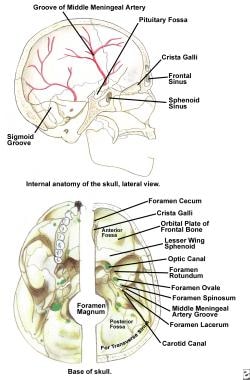
Frontal Bone
Tumor Virus Infections
Granuloma, Giant Cell
Curettage
Togo
Tooth, Unerupted
Cemento-ossifying fibroma presenting as a mass of the parapharyngeal and masticator space. (1/70)
We report a case of cemento-ossifying fibroma that presented as a large extraosseous mass in the masticator and parapharyngeal space. CT scanning and MR imaging showed a large extraosseous mass with central conglomerated, well-matured ossified nodules and fatty marrow. The central matured ossified nodules were of low density on CT scans and high signal intensity on T1- and T2-weighted MR images. Multiplanar reformatted CT scans revealed the origin of the mass to be at the extraction site of the right lower second molar tooth. (+info)Ossifying fibroma in a llama. (2/70)
A 4.5-year-old llama was admitted for evaluation of a firm mass rostral and ventral to the medial canthus of the left eye. Mucopurulent nasal discharge and absence of airflow through the left nostril were noted. Radiographs of the skull revealed a sharply demarcated soft tissue mass with faint mineralization. Endoscopy of the nasal passages revealed a mucosa-covered mass originating in the area of the second premolar, extending to the edge of the soft palate, and obstructing the airway. Examination of the oral cavity revealed a missing second molar and a mass protruding 2-cm from the empty alveolus. An ossifying fibroma, a previously unreported tumor in llamas, was diagnosed at postmortem examination. (+info)The hyperparathyroidism-jaw tumour syndrome in a Portuguese kindred. (3/70)
The hyperparathyroidism-jaw tumour (HPT-JT) syndrome is an autosomal dominant disease characterized by the occurrence of parathyroid tumours and fibro-osseous tumours of the jaw bones. Some HPT-JT patients may also develop renal abnormalities, which include Wilms' tumours, hamartomas and polycystic disease. The HPT-JT gene has been mapped to chromosome 1q25-q31, and we report the clinical and genetic findings in a kindred from central Portugal. HPT-JT was observed in six members from three generations; all had primary hyperparathyroidism (five had parathyroid adenomas, one a parathyroid carcinoma). Ossifying jaw fibromas affecting the maxilla and/or mandible were observed in 5/6. Renal cysts (<2.5 cm) were observed in four. Genetic studies using 18 polymorphic loci from chromosome 1q25-q31, together with leukocyte DNA from 11 family members and tumour DNA from three parathyroids (two adenomas and one carcinoma), revealed loss of tumour heterozygosity in the parathyroid carcinoma only, and the retained haplotype was found to cosegregate with the disease in the six affected members. A new Portuguese kindred with the HPT-JT syndrome that maps to chromosome 1q25-q31 has been identified, and these findings will help in the further characterization of this inherited disorder. (+info)Odontogenic cysts, odontogenic tumors, fibroosseous, and giant cell lesions of the jaws. (4/70)
Odontogenic cysts that can be problematic because of recurrence and/or aggressive growth include odontogenic keratocyst (OKC), calcifying odontogenic cyst, and the recently described glandular odontogenic cyst. The OKC has significant growth capacity and recurrence potential and is occasionally indicative of the nevoid basal cell carcinoma syndrome. There is also an orthokeratinized variant, the orthokeratinized odontogenic cyst, which is less aggressive and is not syndrome associated. Ghost cell keratinization, which typifies the calcifying odontogenic cyst, can be seen in solid lesions that have now been designated odontogenic ghost cell tumor. The glandular odontogenic cyst contains mucous cells and ductlike structures that may mimic central mucoepidermoid carcinoma. Several odontogenic tumors may provide diagnostic challenges, particularly the cystic ameloblastoma. Identification of this frequently underdiagnosed cystic tumor often comes after one or more recurrences and a destructive course. Other difficult lesions include malignant ameloblastomas, calcifying epithelial odontogenic tumor, squamous odontogenic tumor, and clear-cell odontogenic tumor. Histologic identification of myxofibrous lesions of the jaws (odontogenic myxoma, odontogenic fibroma, desmoplastic fibroma) is necessary to avoid the diagnostic pitfall of overdiagnosis of similar-appearing follicular sacs and dental pulps. Fibroosseous lesions of the jaws show considerable microscopic overlap and include fibrous dysplasia, ossifying fibroma, periapical cementoosseous dysplasia, and low-grade chronic osteomyelitis. The term fibrous dysplasia is probably overused in general practice and should be reserved for the rare lesion that presents as a large, expansile, diffuse opacity of children and young adults. The need to use clinicopathologic correlation in assessing these lesions is of particular importance. Central giant cell granuloma is a relatively common jaw lesion of young adults that has an unpredictable behavior. Microscopic diagnosis is relatively straightforward; however, this lesion continues to be somewhat controversial because of its disputed classification (reactive versus neoplastic) and because of its management (surgical versus. medical). Its relationship to giant cell tumor of long bone remains undetermined. (+info)Surgery and religiosity. (5/70)
Variances in religious practices and beliefs require health care providers to find alternative ways of meeting medical standards of care in cases where religious beliefs preclude the use of blood transfusions or blood products. We report a case of head and neck tumor with low hemoglobin in which we utilized temporary arterial ligation to aid in minimal blood loss. (+info)Sinonasal ossifying fibroma with fluid-fluid levels on MR images. (6/70)
Ossifying fibroma is a rare benign neoplasm that usually affects mandibular and maxillary bones. In this report, we present a case of sinonasal ossifying fibroma with fluid-fluid levels and posterior extension toward the torus tubarius on MR images. (+info)Cemento-ossifying mandibular fibroma: a presentation of two cases and review of the literature. (7/70)
We present two clinical cases of radiolucent mandibular lesions in young women that simulated chronic periapical infectious pathology. The detection of both cases was fortuitous since they were totally asymptomatic. Diagnosis was reached in one case (upon periapical surgery and anatomo-pathologic study) after endodontic treatment and after verifying non-resolution of affected periapical area. The other case was an extensive lesion, which involved the periapices of the four inferior incisors in which surgery was directly performed upon verifying pulp vitality of these teeth. After surgery endodontic treatment was performed on the teeth that had lost their vitality. In both cases the histopathologic tests revealed the presence of a cemento-ossifying fibroma, the initial clinical and radiographic diagnosis of which could easily be overlooked. (+info)Cemento-ossifying fibroma of mandibular gingiva: single case report. (8/70)
We report a case of a woman presenting a giant cementoossifying fibroma depending of the mandibular gingivae. The evolution of the process was 20 years. Cemento-ossifying fibroma is a relatively rare tumour classified between fibroosseous lesions. This lesion appears within the bone although in some occasions it involves the gingivae soft tissues, as the case we present. It is a slow-growing and well-defined tumorous lesion, because of this, it is considered as a benign lesion. The histologic findings alone may be similar to other pathologies such as osteoblastoma, low-grade osteosarcoma and particularly to fibrous dysplasia. An accurate diagnosis requires careful clinical, radiological and histological correlation in order to make an optimal treatment and an excellent outcome. (+info)A fibroma is a benign (non-cancerous) tumor that consists primarily of fibrous or connective tissue. It can occur in various parts of the body, including the skin, mouth, and internal organs. The term "fibroma" is often used to describe any benign fibrous growth, but there are specific types of fibromas such as dermatofibroma (found in the skin), oral fibroma (found in the mouth), and benign fibrous histiocytoma (found in soft tissues).
It's important to note that while fibromas are generally harmless, they can cause discomfort or problems depending on their size and location. If a fibroma is causing issues or there's concern about its growth or malignancy, it should be evaluated by a healthcare professional for potential removal or further assessment.
A fibroma, ossifying is a benign (non-cancerous) tumor that typically develops in the periodontal ligament, which is the tissue that connects the tooth to the jawbone. This type of fibroma is characterized by the formation of bone-like tissue within the tumor. It usually appears as a firm, slow-growing nodule or mass that can cause pain or discomfort, particularly when biting down on the affected tooth.
The exact cause of ossifying fibromas is not well understood, but they are thought to arise from an overgrowth of cells in the periodontal ligament. They are more common in women than men and typically occur in people between the ages of 20 and 40. Treatment usually involves surgical removal of the tumor, along with any affected tissue or teeth. In some cases, recurrence may occur, so regular follow-up appointments with a dental professional are recommended.
Desmoplastic fibroma is a very rare benign (non-cancerous) tumor of the connective tissue. It typically develops in the bones, but can also occur in soft tissues. The tumor is characterized by the overgrowth of collagen-producing cells (fibroblasts), leading to the formation of a firm, fibrous mass. Desmoplastic fibromas are slow-growing and typically do not spread to other parts of the body (metastasize). However, they can cause significant damage to the affected bone or tissue as they grow, potentially leading to fractures or deformities. Treatment usually involves surgical removal of the tumor.
Fibrous Dysplasia of Bone is a rare, benign bone disorder that is characterized by the replacement of normal bone tissue with fibrous (scar-like) and immature bone tissue. This results in weakened bones that are prone to fractures, deformities, and pain. The condition can affect any bone in the body but most commonly involves the long bones of the legs, arms, and skull. It can occur as an isolated finding or as part of a genetic disorder called McCune-Albright syndrome. The exact cause of fibrous dysplasia is not fully understood, but it is believed to result from a genetic mutation that occurs during early bone development. There is no cure for fibrous dysplasia, and treatment typically focuses on managing symptoms and preventing complications.
Odontogenic tumors are a group of neoplasms that originate from the dental tissues or their remnants, including the odontogenic epithelium, ectomesenchyme, and/or their derivatives. These tumors can be benign or malignant and may affect the jaw bones and surrounding structures. They can cause various symptoms, such as swelling, pain, loosening of teeth, and altered bite. The classification of odontogenic tumors includes a wide range of entities with different biological behaviors, clinical features, and treatment approaches. Accurate diagnosis is essential for proper management and prognosis.
Mandibular neoplasms refer to abnormal growths or tumors that develop in the mandible, which is the lower jawbone. These growths can be benign (non-cancerous) or malignant (cancerous). Benign neoplasms are typically slow-growing and rarely spread to other parts of the body, while malignant neoplasms can invade surrounding tissues and may metastasize (spread) to distant sites.
Mandibular neoplasms can have various causes, including genetic mutations, exposure to certain chemicals or radiation, and infection with certain viruses. The symptoms of mandibular neoplasms may include swelling or pain in the jaw, difficulty chewing or speaking, numbness in the lower lip or chin, loose teeth, and/or a lump or mass in the mouth or neck.
The diagnosis of mandibular neoplasms typically involves a thorough clinical examination, imaging studies such as X-rays, CT scans, or MRI scans, and sometimes a biopsy to confirm the type and extent of the tumor. Treatment options depend on the type, stage, and location of the neoplasm, and may include surgery, radiation therapy, chemotherapy, or a combination of these approaches. Regular follow-up care is essential to monitor for recurrence or metastasis.
Jaw neoplasms refer to abnormal growths or tumors in the jawbone (mandible) or maxilla (upper jaw). These growths can be benign (non-cancerous) or malignant (cancerous). Benign neoplasms are not considered life-threatening, but they can still cause problems by invading nearby tissues and causing damage. Malignant neoplasms, on the other hand, can spread to other parts of the body and can be life-threatening if not treated promptly and effectively.
Jaw neoplasms can present with various symptoms such as swelling, pain, loose teeth, numbness or tingling in the lips or tongue, difficulty chewing or swallowing, and jaw stiffness or limited movement. The diagnosis of jaw neoplasms typically involves a thorough clinical examination, imaging studies such as X-rays, CT scans, or MRI, and sometimes a biopsy to determine the type and extent of the tumor.
Treatment options for jaw neoplasms depend on several factors, including the type, size, location, and stage of the tumor, as well as the patient's overall health and medical history. Treatment may involve surgery, radiation therapy, chemotherapy, or a combination of these modalities. Regular follow-up care is essential to monitor for recurrence or metastasis (spread) of the neoplasm.
Maxillary neoplasms refer to abnormal growths or tumors in the maxilla, which is the upper jaw bone. These growths can be benign (non-cancerous) or malignant (cancerous). Benign neoplasms are slow-growing and do not spread to other parts of the body, while malignant neoplasms can invade surrounding tissues and spread to distant sites.
Maxillary neoplasms can cause various symptoms such as swelling, pain, numbness, loose teeth, or difficulty in chewing or swallowing. They may also cause nasal congestion, nosebleeds, or visual changes if they affect the eye or orbit. The diagnosis of maxillary neoplasms usually involves a combination of clinical examination, imaging studies such as CT or MRI scans, and biopsy to determine the type and extent of the tumor.
Treatment options for maxillary neoplasms depend on several factors, including the type, size, location, and stage of the tumor, as well as the patient's overall health and preferences. Treatment may include surgery, radiation therapy, chemotherapy, or a combination of these modalities. Regular follow-up care is essential to monitor for recurrence or metastasis and ensure optimal outcomes.
Poxviridae is a family of large, complex, double-stranded DNA viruses that includes many significant pathogens affecting humans and animals. The most well-known member of this family is the Variola virus, which causes smallpox in humans, a highly contagious and deadly disease that has been eradicated through global vaccination efforts. Other important human pathogens in this family include the Monkeypox virus, which can cause a smallpox-like illness, and the Molluscum contagiosum virus, which causes benign skin tumors.
Poxviruses have a unique ability to replicate in the cytoplasm of host cells, rather than in the nucleus like many other DNA viruses. They also have a complex structure, with a large, brick-shaped virion that contains a lateral body, a core, and an outer envelope. The genome of poxviruses is relatively large, ranging from 130 to 375 kilobases in length, and encodes many genes involved in viral replication, host immune evasion, and modulation of host cell processes.
Poxviridae is further divided into two subfamilies: Chordopoxvirinae, which includes viruses that infect vertebrates, and Entomopoxvirinae, which includes viruses that infect insects. The Chordopoxvirinae subfamily is divided into several genera, including Orthopoxvirus (which includes Variola, Monkeypox, and Vaccinia viruses), Parapoxvirus (which includes Orf virus and Bovine papular stomatitis virus), and Yatapoxvirus (which includes Yaba monkey tumor virus and Tanapox virus).
Overall, Poxviridae is a diverse family of viruses that pose significant public health and agricultural threats, and continue to be the subject of ongoing research and development efforts aimed at understanding their biology and developing new vaccines and therapies.
Gingival neoplasms refer to abnormal growths or tumors that occur in the gingiva, which are the part of the gums that surround the teeth. These growths can be benign (non-cancerous) or malignant (cancerous). Benign neoplasms include conditions such as fibromas, papillomas, and hemangiomas, while malignant neoplasms are typically squamous cell carcinomas.
Gingival neoplasms can present with a variety of symptoms, including swelling, bleeding, pain, and loose teeth. They may also cause difficulty with chewing, speaking, or swallowing. The exact cause of these neoplasms is not always known, but risk factors include tobacco use, alcohol consumption, poor oral hygiene, and certain viral infections.
Diagnosis of gingival neoplasms typically involves a thorough clinical examination, including a dental exam and biopsy. Treatment options depend on the type and stage of the neoplasm, but may include surgery, radiation therapy, chemotherapy, or a combination of these approaches. Regular dental check-ups and good oral hygiene practices can help to detect gingival neoplasms at an early stage and improve treatment outcomes.
Cementoma is a benign (non-cancerous) tumor that primarily affects the jaw bones, particularly the lower jaw (mandible). It is characterized by the growth of abnormal cementum-like tissue within the bone. Cementum is a hard tissue that covers the roots of teeth and helps anchor them to the jawbone.
There are different types of cementomas, including:
1. Periapical cemental dysplasia (PCD): This type of cementoma usually affects the anterior region of the lower jaw and is often associated with non-vital teeth. It typically presents as a small, radiopaque (dark) area on an X-ray.
2. Florid cemento-osseous dysplasia (FCOD): FCOD is a more widespread form of cementoma that affects multiple areas of the jawbones. It primarily affects middle-aged women and can cause significant bone remodeling, leading to radiopaque lesions on X-rays.
3. Gigantiform cementoma: This rare, aggressive type of cementoma typically affects children and adolescents. It can cause rapid bone growth and expansion, resulting in facial deformities and functional impairments.
4. Ossifying fibroma: Although not strictly a cementoma, ossifying fibroma shares some similarities with these tumors. It is characterized by the formation of both bone and cementum-like tissue within the lesion.
Treatment for cementomas depends on their size, location, and growth rate. Small, asymptomatic lesions may not require treatment, while larger or symptomatic ones might need surgical removal to prevent complications such as tooth displacement, infection, or pathological fractures. Regular follow-ups with dental X-rays are essential to monitor the progression of these lesions.
A chondroma is a benign, slow-growing tumor that develops in the cartilage. Cartilage is a type of connective tissue found in various parts of the body, including the joints, ribcage, and nose. Chondromas are most commonly found in the hands and feet.
Chondromas are typically small, measuring less than 2 centimeters in diameter, and they usually do not cause any symptoms. However, if a chondroma grows large enough to press on nearby nerves or blood vessels, it may cause pain, numbness, or weakness in the affected area.
Chondromas are usually diagnosed through imaging tests such as X-rays, CT scans, or MRI scans. If a chondroma is suspected based on these tests, a biopsy may be performed to confirm the diagnosis and rule out other types of tumors.
Treatment for chondromas typically involves surgical removal of the tumor. In most cases, this can be done using minimally invasive techniques that allow for quicker recovery times. After surgery, patients will need to follow up with their healthcare provider to ensure that the tumor has been completely removed and to monitor for any signs of recurrence.
A thecoma is a type of ovarian sex cord-stromal tumor, which are rare tumors that develop from the supporting cells (stromal cells) or the cells that produce hormones (sex cord cells) in the ovary. These tumors account for about 2% of all ovarian tumors.
Thecomas specifically arise from stromal cells that produce estrogen and other sex hormones. They are typically slow-growing and may not cause any symptoms, or they may cause symptoms related to hormonal imbalances such as irregular menstrual periods, vaginal bleeding, or postmenopausal bleeding. In some cases, thecomas can also grow large enough to cause abdominal discomfort or bloating.
Most thecomas are benign (non-cancerous), but a small percentage of them can be malignant (cancerous) and may spread to other parts of the body. Treatment for thecomas typically involves surgical removal of the tumor, and in some cases, hormonal therapy or chemotherapy may also be recommended.
Leporipoxvirus is a genus of viruses in the Poxviridae family, which includes double-stranded DNA viruses. This genus primarily consists of pathogens that infect rabbits and hares. Two well-known examples of Leporipoxviruses are myxoma virus and rabbit (hare) fibroma virus.
1. Myxoma Virus: It is the causative agent of myxomatosis, a often fatal disease in European rabbits (Oryctolagus cuniculus). The virus is transmitted through insect vectors, primarily mosquitoes and fleas. Infected rabbits develop skin lesions, swelling around the eyes and genitals, and eventually die due to internal organ failure.
2. Rabbit (Hare) Fibroma Virus: This Leporipoxvirus causes benign tumors called fibromas in rabbits and hares. The tumors typically develop on the skin or mucous membranes but can also occur internally. While these growths are not fatal, they can cause significant stress and discomfort for affected animals.
It is important to note that Leporipoxviruses do not pose a direct threat to humans as they primarily infect rabbits and hares. However, researchers study these viruses due to their potential applications in cancer therapy and vaccine development.
Oncogenic viruses are a type of viruses that have the ability to cause cancer in host cells. They do this by integrating their genetic material into the DNA of the infected host cell, which can lead to the disruption of normal cellular functions and the activation of oncogenes (genes that have the potential to cause cancer). This can result in uncontrolled cell growth and division, ultimately leading to the formation of tumors. Examples of oncogenic viruses include human papillomavirus (HPV), hepatitis B virus (HBV), and human T-cell leukemia virus type 1 (HTLV-1). It is important to note that only a small proportion of viral infections lead to cancer, and the majority of cancers are not caused by viruses.
Chondroblastoma is a rare, benign (non-cancerous) bone tumor that typically develops in the epiphysis, which is the rounded end of a long bone near a joint. It primarily affects children and adolescents, with around 90% of cases occurring before the age of 20.
The tumor arises from chondroblasts, cells responsible for producing cartilage during bone growth. Chondroblastoma is usually slow-growing and typically causes localized pain, swelling, or tenderness in the affected area. In some cases, it may weaken the bone and lead to fractures.
Treatment generally involves surgical removal of the tumor, followed by curettage (scraping) of the surrounding bone tissue and replacement with bone grafts or substitutes. Recurrence is possible but rare, and long-term prognosis is usually favorable.
Panoramic radiography is a specialized type of dental X-ray imaging that captures a panoramic view of the entire mouth, including the teeth, upper and lower jaws, and surrounding structures. It uses a special machine that rotates around the head, capturing images as it moves. This technique provides a two-dimensional image that is helpful in diagnosing and planning treatment for various dental conditions such as impacted teeth, bone abnormalities, and jaw disorders.
The panoramic radiograph can also be used to assess the development and positioning of wisdom teeth, detect cysts or tumors in the jaws, and evaluate the effects of trauma or injury to the mouth. It is a valuable tool for dental professionals as it allows them to see a comprehensive view of the oral structures, which may not be visible with traditional X-ray techniques.
It's important to note that while panoramic radiography provides valuable information, it should be used in conjunction with other diagnostic tools and clinical examinations to ensure accurate diagnosis and treatment planning.
Osteoma is a benign (noncancerous) tumor that is made up of mature bone tissue. It usually grows slowly over a period of years and is most commonly found in the skull or jaw, although it can occur in other bones of the body as well. Osteomas are typically small, but they can grow to be several centimeters in size. They may cause symptoms if they press on nearby tissues or structures, such as nerves or blood vessels. In some cases, osteomas may not cause any symptoms and may only be discovered during routine imaging studies. Treatment for osteoma is typically not necessary unless it is causing problems or growing rapidly. If treatment is needed, it may involve surgical removal of the tumor.
Palatal neoplasms refer to abnormal growths or tumors that occur on the palate, which is the roof of the mouth. These growths can be benign (non-cancerous) or malignant (cancerous). Benign neoplasms are typically slower growing and less likely to spread, while malignant neoplasms are more aggressive and can invade nearby tissues and organs.
Palatal neoplasms can have various causes, including genetic factors, environmental exposures, and viral infections. They may present with symptoms such as mouth pain, difficulty swallowing, swelling or lumps in the mouth, bleeding, or numbness in the mouth or face.
The diagnosis of palatal neoplasms typically involves a thorough clinical examination, imaging studies, and sometimes biopsy to determine the type and extent of the growth. Treatment options depend on the type, size, location, and stage of the neoplasm but may include surgery, radiation therapy, chemotherapy, or a combination of these approaches. Regular follow-up care is essential to monitor for recurrence or spread of the neoplasm.
Rhabdomyoma is a rare, benign tumor that arises from the striated muscle tissue, which is the type of muscle that enables movement and action in the body. These tumors most commonly occur in the heart (cardiac rhabdomyomas) or in the head and neck region (extracardiac rhabdomyomas). Cardiac rhabdomyomas are often associated with genetic disorders such as tuberous sclerosis complex, while extracardiac rhabdomyomas can be found in various locations like the skin, tongue, or skeletal muscles.
Cardiac rhabdomyomas typically appear in infancy or early childhood and may not cause any symptoms. However, they can potentially lead to complications such as heart rhythm abnormalities, obstruction of blood flow, or heart failure. Extracardiac rhabdomyomas are usually slow-growing and asymptomatic but can cause issues depending on their size and location. Surgical removal may be necessary if the tumor interferes with vital functions or causes discomfort.
It is essential to note that while rhabdomyomas are generally benign, they can undergo malignant transformation in rare cases, leading to a more aggressive form called rhabdomyosarcoma. Regular follow-ups and monitoring are crucial for early detection and management of any changes in the tumor's behavior.
Maxillary sinus neoplasms refer to abnormal growths or tumors that develop in the maxillary sinuses, which are located in the upper part of your cheekbones, below your eyes. These growths can be benign (non-cancerous) or malignant (cancerous).
Benign neoplasms may include conditions such as an osteoma (a benign bone tumor), a papilloma (a benign growth of the lining of the sinus), or a fibrous dysplasia (a condition where bone is replaced by fibrous tissue).
Malignant neoplasms, on the other hand, can be primary (originating in the maxillary sinuses) or secondary (spreading to the maxillary sinuses from another site in the body). Common types of malignant tumors that arise in the maxillary sinus include squamous cell carcinoma, adenocarcinoma, and mucoepidermoid carcinoma.
Symptoms of maxillary sinus neoplasms may include nasal congestion, nosebleeds, facial pain or numbness, vision changes, and difficulty swallowing or speaking. Treatment options depend on the type, size, and location of the tumor but may include surgery, radiation therapy, chemotherapy, or a combination of these approaches.
Heart neoplasms are abnormal growths or tumors that develop within the heart tissue. They can be benign (noncancerous) or malignant (cancerous). Benign tumors, such as myxomas and rhabdomyomas, are typically slower growing and less likely to spread, but they can still cause serious complications if they obstruct blood flow or damage heart valves. Malignant tumors, such as angiosarcomas and rhabdomyosarcomas, are fast-growing and have a higher risk of spreading to other parts of the body. Symptoms of heart neoplasms can include shortness of breath, chest pain, fatigue, and irregular heart rhythms. Treatment options depend on the type, size, and location of the tumor, and may include surgery, radiation therapy, or chemotherapy.
Dental cementum is a type of hard connective tissue that covers the root of a tooth. It is primarily composed of calcium salts and collagen fibers, and it serves to attach the periodontal ligaments (the fibers that help secure the tooth in its socket) to the tooth's root. Cementum also helps protect the root of the tooth and contributes to the maintenance of tooth stability. It continues to grow and deposit new layers throughout an individual's life, which can be seen as incremental lines called "cementum annulations."
Myxoma virus (MYXV) is a member of the Poxviridae family, specifically in the Leporipoxvirus genus. It is a double-stranded DNA virus that naturally infects European rabbits (Oryctolagus cuniculus) and causes a fatal disease called myxomatosis. The virus is transmitted through insect vectors such as mosquitoes and fleas, and it replicates in the cytoplasm of infected cells.
Myxoma virus has been studied extensively as a model organism for viral pathogenesis and host-pathogen interactions. It has also been explored as a potential oncolytic virus for cancer therapy due to its ability to selectively infect and kill certain types of cancer cells while leaving normal cells unharmed. However, it is important to note that the use of Myxoma virus in humans is still experimental and requires further research and development before it can be considered safe and effective for therapeutic purposes.
Neoplasms of connective tissue are abnormal growths or tumors that develop from the cells that form the body's supportive framework, including bones, cartilage, tendons, ligaments, and other connective tissues. These neoplasms can be benign (non-cancerous) or malignant (cancerous), and they can cause various symptoms depending on their location and size.
There are several types of connective tissue neoplasms, including:
1. Fibroma: A benign tumor that arises from fibrous connective tissue.
2. Fibrosarcoma: A malignant tumor that develops from fibrous connective tissue.
3. Lipoma: A benign tumor that arises from fat cells.
4. Liposarcoma: A malignant tumor that develops from fat cells.
5. Chondroma: A benign tumor that arises from cartilage.
6. Chondrosarcoma: A malignant tumor that develops from cartilage.
7. Osteoma: A benign tumor that arises from bone.
8. Osteosarcoma: A malignant tumor that develops from bone.
9. Giant cell tumors: Benign or malignant tumors that contain many giant cells, which are large, multinucleated cells.
10. Synovial sarcoma: A malignant tumor that arises from the synovial tissue that lines joints and tendons.
Connective tissue neoplasms can cause various symptoms depending on their location and size. For example, a benign lipoma may cause a painless lump under the skin, while a malignant osteosarcoma may cause bone pain, swelling, and fractures. Treatment options for connective tissue neoplasms include surgery, radiation therapy, chemotherapy, or a combination of these approaches.
Bone neoplasms are abnormal growths or tumors that develop in the bone. They can be benign (non-cancerous) or malignant (cancerous). Benign bone neoplasms do not spread to other parts of the body and are rarely a threat to life, although they may cause problems if they grow large enough to press on surrounding tissues or cause fractures. Malignant bone neoplasms, on the other hand, can invade and destroy nearby tissue and may spread (metastasize) to other parts of the body.
There are many different types of bone neoplasms, including:
1. Osteochondroma - a benign tumor that develops from cartilage and bone
2. Enchondroma - a benign tumor that forms in the cartilage that lines the inside of the bones
3. Chondrosarcoma - a malignant tumor that develops from cartilage
4. Osteosarcoma - a malignant tumor that develops from bone cells
5. Ewing sarcoma - a malignant tumor that develops in the bones or soft tissues around the bones
6. Giant cell tumor of bone - a benign or occasionally malignant tumor that develops from bone tissue
7. Fibrosarcoma - a malignant tumor that develops from fibrous tissue in the bone
The symptoms of bone neoplasms vary depending on the type, size, and location of the tumor. They may include pain, swelling, stiffness, fractures, or limited mobility. Treatment options depend on the type and stage of the tumor but may include surgery, radiation therapy, chemotherapy, or a combination of these treatments.
Collagen type XII is a type of collagen that is found in the extracellular matrix of various tissues, including tendons, ligaments, and skin. It is a fibril-associated collagen that is closely associated with collagens type I and III. Collagen type XII has been shown to play a role in regulating the organization and diameter of collagen fibrils. Mutations in the gene for collagen type XII have been associated with certain types of muscular dystrophy and Bethlem myopathy, which are genetic disorders that affect muscle strength and tone. Additionally, it has been suggested to play a role in the development of osteoarthritis.
Femoral neoplasms refer to abnormal growths or tumors that develop in the femur, which is the long thigh bone in the human body. These neoplasms can be benign (non-cancerous) or malignant (cancerous). Benign femoral neoplasms are slow-growing and rarely spread to other parts of the body, while malignant neoplasms are aggressive and can invade nearby tissues and organs, as well as metastasize (spread) to distant sites.
There are various types of femoral neoplasms, including osteochondromas, enchondromas, chondrosarcomas, osteosarcomas, and Ewing sarcomas, among others. The specific type of neoplasm is determined by the cell type from which it arises and its behavior.
Symptoms of femoral neoplasms may include pain, swelling, stiffness, or weakness in the thigh, as well as a palpable mass or limited mobility. Diagnosis typically involves imaging studies such as X-rays, CT scans, or MRI, as well as biopsy to determine the type and grade of the tumor. Treatment options may include surgery, radiation therapy, chemotherapy, or a combination of these approaches, depending on the type, size, location, and stage of the neoplasm.
Gingival diseases are infections or inflammations that affect the gingiva, which is the part of the gum around the base of the teeth. These diseases can be caused by bacteria found in dental plaque and can lead to symptoms such as redness, swelling, bleeding, and receding gums. If left untreated, gingival diseases can progress to periodontal disease, a more serious condition that can result in tooth loss. Common types of gingival diseases include gingivitis and periodontitis.
Skull neoplasms refer to abnormal growths or tumors that develop within the skull. These growths can be benign (non-cancerous) or malignant (cancerous). They can originate from various types of cells, such as bone cells, nerve cells, or soft tissues. Skull neoplasms can cause various symptoms depending on their size and location, including headaches, seizures, vision problems, hearing loss, and neurological deficits. Treatment options include surgery, radiation therapy, and chemotherapy. It is important to note that a neoplasm in the skull can also refer to metastatic cancer, which has spread from another part of the body to the skull.
Chromium isotopes are different forms of the chemical element Chromium (Cr), which have different numbers of neutrons in their atomic nuclei. This results in each isotope having a different atomic mass, although they all have the same number of protons (24) and therefore share the same chemical properties.
The most common and stable chromium isotopes are Chromium-52 (Cr-52), Chromium-53 (Cr-53), Chromium-54 (Cr-54), and Chromium-56 (Cr-56). The other less abundant isotopes of Chromium, such as Chromium-50 (Cr-50) and Chromium-51 (Cr-51), are radioactive and undergo decay to become stable isotopes.
Chromium is an essential trace element for human health, playing a role in the metabolism of carbohydrates, lipids, and proteins. It is also used in various industrial applications, such as in the production of stainless steel and other alloys.
The ethmoid sinuses are a pair of air-filled spaces located in the ethmoid bone, which is a part of the skull that forms the upper portion of the nasal cavity and the inner eye socket. These sinuses are divided into anterior and posterior groups and are present in adults, but not at birth. They continue to grow and develop until early adulthood.
The ethmoid sinuses are lined with mucous membrane, which helps to warm, humidify, and filter the air we breathe. They are surrounded by a network of blood vessels and nerves, making them susceptible to inflammation and infection. Inflammation of the ethmoid sinuses can lead to conditions such as sinusitis, which can cause symptoms such as nasal congestion, headache, and facial pain.
Paranasal sinus neoplasms refer to abnormal growths or tumors that develop within the paranasal sinuses, which are air-filled cavities located inside the skull near the nasal cavity. These tumors can be benign (noncancerous) or malignant (cancerous), and they can arise from various types of tissue within the sinuses, such as the lining of the sinuses (mucosa), bone, or other soft tissues.
Paranasal sinus neoplasms can cause a variety of symptoms, including nasal congestion, nosebleeds, facial pain or numbness, and visual disturbances. The diagnosis of these tumors typically involves a combination of imaging studies (such as CT or MRI scans) and biopsy to determine the type and extent of the tumor. Treatment options may include surgery, radiation therapy, chemotherapy, or a combination of these approaches, depending on the specific type and stage of the neoplasm.
A jaw cyst is a pathological cavity filled with fluid or semi-fluid material, which forms within the jaw bones. They are typically classified as odontogenic (developing from tooth-forming tissues) or non-odontogenic (developing from other tissues). The most common types of odontogenic jaw cysts include dentigerous cysts (formed around the crown of an unerupted tooth) and follicular cysts (formed from the inflammation of a developing tooth's tissue). Non-odontogenic cysts, such as nasopalatine duct cysts and keratocystic odontogenic tumors, can also occur in the jaw bones. Jaw cysts may cause symptoms like swelling, pain, or displacement of teeth, but some may not present any symptoms until they grow large enough to be detected on a radiographic examination. Treatment typically involves surgical removal of the cyst and, if necessary, reconstruction of the affected bone.
Chondromatosis is a medical condition characterized by the abnormal formation of multiple cartilaginous nodules or masses within a joint or soft tissue. It is often seen in synovial joints, where the synovial membrane that lines the joint cavity produces these cartilage nodules.
There are two types of chondromatosis: primary and secondary. Primary chondromatosis, also known as synovial chondromatosis, is a rare condition where the cartilaginous nodules develop spontaneously within the synovium. Over time, these nodules may calcify or ossify, turning into bone-like structures. Secondary chondromatosis occurs as a result of degenerative joint disease, trauma, or other underlying conditions that cause cartilage to break off and float freely in the synovial fluid, eventually forming nodules.
Symptoms of chondromatosis may include joint pain, swelling, stiffness, and limited range of motion. In some cases, the condition may lead to osteoarthritis or other joint complications if left untreated. Treatment typically involves surgical removal of the cartilaginous nodules, followed by management of any underlying conditions that may have contributed to the development of chondromatosis.
Odontoma is a type of odontogenic tumor, which means it arises from the tissues that form teeth. It is considered a benign or non-cancerous tumor and is typically slow-growing. Odontomas are usually asymptomatic and are often discovered on routine dental X-rays or during procedures such as wisdom tooth removal.
Odontomas can be classified into two types: complex and compound. Complex odontomas are composed of a haphazard mixture of dental tissue, including enamel, dentin, and cementum, while compound odontomas contain small tooth-like structures called denticles.
These tumors typically occur in the posterior region of the jaw, and while they are usually asymptomatic, some patients may experience symptoms such as swelling, pain, or displacement of teeth. Treatment for odontomas typically involves surgical removal of the tumor.
The frontal bone is the bone that forms the forehead and the upper part of the eye sockets (orbits) in the skull. It is a single, flat bone that has a prominent ridge in the middle called the superior sagittal sinus, which contains venous blood. The frontal bone articulates with several other bones, including the parietal bones at the sides and back, the nasal bones in the center of the face, and the zygomatic (cheek) bones at the lower sides of the orbits.
A myoma, also known as a leiomyoma or fibroid, is a benign (noncancerous) tumor that originates from the smooth muscle cells in the wall of a visceral organ. The term "myoma" is often used to describe these growths when they occur in the uterus, where they are typically referred to as uterine fibroids. Uterine fibroids can vary in size, shape, and location within the uterine wall. They are quite common, especially among women of reproductive age, and may not always cause symptoms. However, in some cases, they can lead to issues such as heavy menstrual bleeding, pelvic pain, or infertility. Myomas can also occur in other organs, like the gastrointestinal tract, but they are most frequently found in the uterus.
A tumor virus infection is a condition in which a person's cells become cancerous or transformed due to the integration and disruption of normal cellular functions by a viral pathogen. These viruses are also known as oncoviruses, and they can cause tumors or cancer by altering the host cell's genetic material, promoting uncontrolled cell growth and division, evading immune surveillance, and inhibiting apoptosis (programmed cell death).
Examples of tumor viruses include:
1. DNA tumor viruses: These are double-stranded DNA viruses that can cause cancer in humans. Examples include human papillomavirus (HPV), hepatitis B virus (HBV), and Merkel cell polyomavirus (MCV).
2. RNA tumor viruses: Also known as retroviruses, these single-stranded RNA viruses can cause cancer in humans. Examples include human T-cell leukemia virus type 1 (HTLV-1) and human immunodeficiency virus (HIV).
Tumor virus infections are responsible for approximately 15-20% of all cancer cases worldwide, making them a significant public health concern. Prevention strategies, such as vaccination against HPV and HBV, have been shown to reduce the incidence of associated cancers.
A giant cell granuloma is a type of non-cancerous (benign) lesion characterized by the presence of large collections of immune cells called macrophages, which have fused together to form multinucleated giant cells. These lesions can occur in various tissues throughout the body but are most commonly found in the oral cavity and jawbone.
Giant cell granulomas can be further classified into two types: central (or bone) giant cell granuloma and peripheral giant cell granuloma. Central giant cell granulomas arise from the bone, while peripheral giant cell granulomas occur in the soft tissues of the gingiva or mouth lining.
Central giant cell granulomas are more aggressive than peripheral ones and can cause significant bone destruction if left untreated. They typically affect younger individuals, with a higher prevalence in females than males. The exact cause of central giant cell granulomas is not well understood but may be associated with local trauma, hormonal imbalances, or genetic factors.
Peripheral giant cell granulomas are less aggressive and usually present as painless, slow-growing nodules on the gums. They typically affect adults, with a higher prevalence in females than males. Peripheral giant cell granulomas may be associated with local irritants such as plaque, calculus, or dental restorations.
Treatment for giant cell granulomas depends on their size, location, and aggressiveness. Surgical excision is the most common treatment approach, but other options such as curettage, corticosteroid injections, or medication therapy may also be considered. Regular follow-up appointments with a healthcare provider are essential to monitor for recurrence.
Curettage is a medical procedure that involves scraping or removing tissue from the lining of an organ or body cavity, typically performed using a curette, which is a long, thin surgical instrument with a looped or sharp end. In gynecology, curettage is often used to remove tissue from the uterus during a procedure called dilation and curettage (D&C) to diagnose or treat abnormal uterine bleeding, or to remove residual placental or fetal tissue following a miscarriage or abortion. Curettage may also be used in other medical specialties to remove damaged or diseased tissue from areas such as the nose, throat, or skin.
I'm sorry for any confusion, but "Togo" is not a medical term. It is the name of a country in West Africa, known officially as the Togolese Republic. If you have any questions about medical terminology or health-related topics, please don't hesitate to ask!
A tooth is classified as "unerupted" when it has not yet penetrated through the gums and entered the oral cavity. This can apply to both primary (baby) teeth and permanent (adult) teeth. The reasons for a tooth's failure to erupt can vary, including crowding of teeth, lack of sufficient space, or anatomical barriers such as bone or soft tissue. In some cases, unerupted teeth may need to be monitored or treated, depending on the specific situation and any symptoms experienced by the individual.
A lipoma is a common, benign (non-cancerous) soft tissue growth. It is composed of adipose or fatty tissue and typically found just beneath the skin, but they can also occur deeper within the body. Lipomas are usually round, moveable, and painless, although they may cause discomfort if they grow large enough to put pressure on nearby nerves or if they're located in a sensitive area. They generally grow slowly over time. Surgical removal is an option if the lipoma becomes bothersome or grows significantly in size. It's important to note that while lipomas are typically harmless, any new lumps or bumps should be evaluated by a healthcare professional to confirm the diagnosis and rule out other more serious conditions.
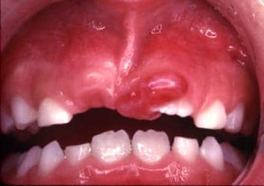
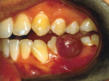
 Peripheral ossifying fibroma
Central ossifying fibroma
Non-ossifying fibroma
Juvenile active ossifying fibroma
Index of oral health and dental articles
Epulis
Peripheral odontogenic fibroma
Jaffe-Campanacci syndrome
Bone tumor
Xanthogranulomatous inflammation
Parish Sedghizadeh
Pyogenic granuloma
Ameloblastic fibroma
Peripheral giant-cell granuloma
Metaphysis
Fibroma
List of MeSH codes (C04)
NOF
Epulis fissuratum
Skhul and Qafzeh hominins
List of diseases (F)
International Classification of Diseases for Oncology
Nasal septum
Edmontosaurus
Peripheral ossifying fibroma - Wikipedia
 Peripheral Ossifying Fibroma: Case Report
 Oral Fibromas and Fibromatoses: Background, Fibroma, Giant Cell Fibroma
 Natural Remedies for Ossifying Fibroma - The Hidden Cures
 Peripheral Ossifying Fibroma in Infant: A Case Report | JDPS
 Non-ossifying fibroma (Radiopaedia 23651-23780 bone window) - NC Commons
Ossifying fibroma of maxilla- A case report | International Journal of Current Research
 Peripheral Cemanto-Ossifying Fibroma - A Case Report, IJSR, Call for Papers, Online Journal
Indian Journal of Multidisciplinary Dentistry - Peripheral ossifying fibroma - Report of a case : Download PDF
Peripheral ossifying fibroma and gingival pigmentation: a case report | International Journal of Current Research
Vascularized fibula flap reconstruction of the maxilla after a juvenile ossifying fibroma removal - hnj.science
 Oral and maxillofacial pathology case of the month: Trabecular juvenile ossifying fibroma. - Fingerprint
- Scholars @ UT...
Oral and maxillofacial pathology case of the month: Trabecular juvenile ossifying fibroma. - Fingerprint
- Scholars @ UT...
![Saini G[au] - Search Results - PubMed](data:image/png;base64,iVBORw0KGgoAAAANSUhEUgAAABAAAAAQCAMAAAAoLQ9TAAAARVBMVEVHcEwoU45gYmYAUpQAUpRPYGVgYmZLXnJgYmYAUZUAUpRJXnIAUpQAUpRgYmYAUpRgYmZgYmZhYmYAUpQAUpQAUpRgYmaDiPJuAAAAFXRSTlMADOJ+6QewGO8/uTRqtH7GdFJ11p1bCL3TAAAAZUlEQVQYlV2PVw7AIAxDTeney7n/UcsoldX3E+VJOAboEi7MBpHWMs1ADlG8u7UYWauwyZFeRQVPOhG2o+aiwhByJxUx91Jxhje3iJSqGfHuLKI0+0TpXvY1twCOPlFh5pa/++MB0vIOBm+1zaoAAAAASUVORK5CYII=) Saini G[au] - Search Results - PubMed
Saini G[au] - Search Results - PubMed
 Radiology Quiz 9348 | Radiopaedia.org
 General Dentistry Details
Juvenile psammomatoid ossifying fibroma with fluid-fluid levels: an unusual presentation | Egyptian Journal of Radiology and...
Oral Fibromas and Fibromatoses: Background, Fibroma, Giant Cell Fibroma
 Ghent University Academic Bibliography
 Hyperparathyroidism-jaw tumor syndrome: MedlinePlus Genetics
 Select Diagnosis | A Nonulcerated, Slowly Growing Mass of the Mandible Selectdiagonosis | DentalCare.com
 Optic Nerve Decompression Surgery - Medical Clinical Policy Bulletins | Aetna
 2020-2021 BCSC Basic and Clinical Science Course™
 horse
horse
 Et tilfelle av sentralt ossifiserende fibrom | Den norske tannlegeforenings Tidende
M.D. MUTLU DURAN
 Worrisome and Incidental Signs on Knee Radiographs in Clinical Practice: Malignant Primary Bone Tumors and Benign Bone Lesions ...
James A. Orsini<...
prof. dr. Koenraad Verstraete
 Oral and Craniofacial Reconstruction Using Mesenchymal Stem Cells - CIRM
Cemento-ossifyi3
- Peripheral cemento-ossifying fibroma is a relatively rare tumour classified between fibroosseous lesions. (ijsr.net)
- The cemento-ossifying fibroma is a central neoplasm of bone as well as periodontium which has caused considerable controversy because of confusion regarding terminology and the criteria for its diagnosis. (ijsr.net)
- The cemento-ossifying fibroma is odontogenic in origin, whereas ossifying fibroma is of bony origin. (ijsr.net)
Fibrous dysplasia and ossifyi3
- The concept of fibroosseous lesions of bone has evolved over the last several decades and now includes two major entities: fibrous dysplasia and ossifying fibroma. (ijsr.net)
- The differential includes fibrous dysplasia and ossifying fibroma / fibroxanthoma of bone . (radiopaedia.org)
- Histomorphological evaluation of osteocalcin and cytokeratin in fibrous dysplasia and ossifying fibroma of the jaw. (pakmedinet.com)
Peripheral giant cel2
- The clinical differential diagnosis of a fibroma depends on its clinical presentation and location and includes giant cell fibroma, neurofibroma , peripheral giant cell granuloma , schwannoma, granular cell tumor, mucocele , and benign and malignant salivary gland tumors (eg, see Salivary Gland Neoplasms ). (medscape.com)
- The clinical differential diagnosis includes squamous papilloma , irritation fibroma, pyogenic granuloma , and peripheral giant cell granuloma . (medscape.com)
Juvenile8
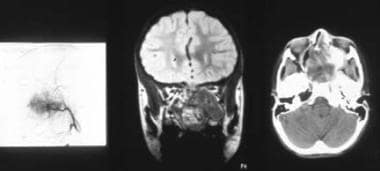
- For citation: Grachev N.S., Gorozhanina A.I., Petrovsky Y.V., Zyabkin I.V., Yunusov A.S., Vorozhtsov I.N., Babaskina N.V. Vascularized fibula flap reconstruction of the maxilla after juvenile ossifying fibroma removal. (hnj.science)
- The aim of this article was to present a clinical case of juvenile ossifying fibroma in a child involving the maxilla, spreading into the maxillary sinus, nasal cavity, ethmoid sinus, and the orbit. (hnj.science)
- Oral and maxillofacial pathology case of the month: Trabecular juvenile ossifying fibroma. (uthscsa.edu)
- Dive into the research topics of 'Oral and maxillofacial pathology case of the month: Trabecular juvenile ossifying fibroma. (uthscsa.edu)
- Fibro-osseous dysplasia (juvenile ossifying fibroma), a variant of fibrous dysplasia, is characterized histologically by spicules of bone rimmed by osteoblasts (Fig 14-19). (aao.org)
- Juvenile ossifying fibroma of the mandible: a case report. (acibadem.com.tr)
- Juvenile ossifying fibroma is an uncommon clinical entity, its aggressive local behaviour and high recurrence rate mean that it is important to make an early diagnosis, apply the appropriate treatment and, especially, follow the patient up over the long term. (annexpublishers.com)
- In the current article we report a case of juvenile ossifying fibroma-WHO type in 12yr old patient which was clinical and histopathologically challenging as it was asymptomatic and at an unusual location. (annexpublishers.com)
Pyogenic1
- Some researchers believe peripheral ossifying fibromas to be related to pyogenic fibromas and, in some instances, are the result of a pyogenic granuloma which has undergone fibrosis and calcification. (wikipedia.org)
Reactive4
- The Peripheral ossifying fibroma is a reactive proliferative lesion, non neoplastic, slow growth, which can produce recurrence after removal. (bvsalud.org)
- While the terminology implies a benign neoplasm, most if not all fibromas represent reactive focal fibrous hyperplasia due to trauma or local irritation. (medscape.com)
- Peripheral ossifying fibroma is a relatively common gingival growth of reactive rather than neoplastic in nature. (peertechzpublications.org)
- Peripheral ossifying fibroma (POF) is a reactive lesion of the gingival tissues, usually originates from the periodontal ligament cells. (journalcra.com)
Lesions1
- Lesions histologically similar to peripheral ossifying fibroma have been given various names in existing literature. (ijsr.net)
Lesion4
- The lesion is considered part of an ossifying fibroma, but that is usually considered to be a jaw tumor. (wikipedia.org)
- The term peripheral ossifying fibroma has been criticized as this lesion is not related to the ossifying fibroma of bone and is not a fibroma. (wikipedia.org)
- The aim of this paper is to describe a case of peripheral ossifying fibroma in a patient 40, female, exophytic lesion in the jaw, unusual for its large dimensions and with a history of three recurrences, leading to facial asymmetry. (bvsalud.org)
- She underwent surgery to remove the lesion along with the likely irritants, and the pathological diagnosis of peripheral ossifying fibroma. (bvsalud.org)
Fibro-osseous1
- Ossifying fibroma is considered to be a rare benign fibro-osseous neoplasm of the jaw. (journalcra.com)
Bone4
- A peripheral ossifying fibroma, also known as ossifying fibrous epulis, is "a gingival nodule which is composed of a cellular fibroblastic connective tissue stroma which is associated with the formation of randomly dispersed foci of mineralised products, which consists of bone, cementum-like tissue, or a dystrophic calcification. (wikipedia.org)
- Additionally, highly developed bone or cementum is more likely to be present when the peripheral ossifying fibroma has existed for a longer period of time. (wikipedia.org)
- The most common benign bone tumours that cause pathological fractures in children are unicameral bone cysts, aneurysmal bone cysts, non-ossifying fibromas and fibrous dysplasia. (lu.se)
- Peripheral odontogenic fibroma (formerly known as fibromatous and ossifying epulis) is a benign, often slow-growing tumor that arises from periodontal structures (gums, ligaments, and bone). (pleasefireme.com)
Histologically1
- Histologically, a fibroma is an unencapsulated, solid, nodular mass of dense and sometimes hyalinized fibrous connective tissue that is often arranged in haphazard fascicles. (medscape.com)
Odontogenic3
- What is an odontogenic fibroma? (pleasefireme.com)
- Central odontogenic fibroma (COF) is an extremely rare benign tumor that accounts for 0.1% of all odontogenic tumors. (pleasefireme.com)
- What causes odontogenic fibroma? (pleasefireme.com)
Resection2
- 2 ] In malignant and borderline tumors, en-bloc resection is in the foreground. (jointdrs.org)
- For the proximal fibula, Malawer described two types of en-bloc resection: Type I, marginal resection, includes 2 to 3 cm of the diaphysis of the proximal fibula and resection of the muscles around the fibula, while in Type II, wide extra-compartment resection, proximal fibula, 5 to 6 cm diaphysis, anterior and lateral muscle compartments, anterior tibial artery and peroneal nerve should be resected. (jointdrs.org)
Case2
- We report here a clinical case of peripheral ossifying fibroma in a 3 month old infant .clinical and histopathological features along with etiopathogenesis and differential diagnosis are also discussed. (peertechzpublications.org)
- A Rare Case of Ossifying Fibroma with Cystic Degeneration: Diagnostic Challenge with Literature Review. (bvsalud.org)
Recurrent1
- Treatment of a recurrent ameloblastic fibroma. (utsouthwestern.edu)
Benign tumor1
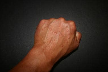
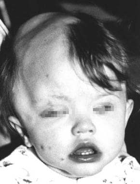
- People with hyperparathyroidism-jaw tumor syndrome may also have a type of benign tumor called a fibroma in the jaw. (medlineplus.gov)
Prevalence1
- The prevalence of peripheral ossifying fibromas is highest around 10 - 19 years of age. (wikipedia.org)
Maxilla1
- An incisional biopsy was carried out and the report was suggestive of ossifying fibroma of the maxilla. (journalcra.com)
Giant1
- Giant cell fibroma is the oral counterpart of fibrous papule of the nose . (medscape.com)
Oral4
- The fibroma, also referred to as irritation fibroma, is by far the most common of the oral fibrous tumorlike growths. (medscape.com)
- A fibroma may occur at any oral site, but it is seen most often on the buccal mucosa along the plane of occlusion of the maxillary and mandibular teeth as depicted below. (medscape.com)
- Both sporadic sclerotic fibromas and those associated with the syndrome have also been described in the oral cavity, mainly in the buccal and labial mucosa. (medscape.com)
- En 51 år gammel kvinne ble henvist til spesialist i oral kirurgi for fjernelse av en oppklaring på ca. 2 cm i diameter rundt rotspissen av 36. (tannlegetidende.no)
Occur2
- Fibromas are most often observed in adults, but they may occur in individuals of any age and either sex. (medscape.com)
- Ossifying fibroma are usually 1.5cm or less and occur on patients from 25-25 years old. (bauersmiles.com)
Syndrome1
- The sclerotic fibroma was first described as a component of Cowden syndrome . (medscape.com)
Color1
- The color of peripheral ossifying fibromas ranges from red to pink, and is frequently ulcerated. (wikipedia.org)
Mandible5
- The tumor seemed to be between spongiosa and inflammatory bone, and after histology the diagnosis came back of a central ossifying fibroma of the mandible, COF. (newlinemedical.com)
- A case of cemento-ossifying fibroma involving the right side of mandible is described in a 38 year-old male patient, with description of clinical, radiographic, and histologic features. (journalcra.com)
- Giant central ossifying fibroma of the mandible: Report of case. (thejcdp.com)
- Ossifying fibroma involving maxilla and mandible. (thejcdp.com)
- Osteofibrous dysplasia of the mandible, which occurs exclusively in adults, commonly is referred to as ossifying fibroma. (medscape.com)
Tumors9
- A non-ossifying fibroma (NOF) is one of the most common bone tumors found in children between 8-20 years of age. (luskinoic.org)
- The clinical differential diagnosis of a fibroma depends on its clinical presentation and location and includes giant cell fibroma, neurofibroma , peripheral giant cell granuloma , schwannoma, granular cell tumor, mucocele , and benign and malignant salivary gland tumors (eg, see Salivary Gland Neoplasms ). (medscape.com)
- Heterozygous pathogenic germline variants in CDC73 predispose to the development of primary hyperparathyroidism (pHPT) and, less frequently, ossifying fibroma of the jaw and renal and uterine tumors. (medscape.com)
- Giant ossifying fibroma: A clinicopathologic study of 8 tumors. (thejcdp.com)
- The exact causes of non-ossifying fibroids are still unknown, but certain factors are thought to play a role in the development of these benign bone tumors. (tunisie-esthetic.com)
- Non-ossifying fibroids are usually benign tumors that do not require surgery or active treatment. (tunisie-esthetic.com)
- Like other bone tumors, a large non-ossifying fibroma can weaken the bone and increase the risk of fractures. (uchicagomedicine.org)
- Follow-up, excision/curettage + adjuvant therapies or en-bloc resection can be performed in benign tumors. (jointdrs.org)
- 2 , 4 , 6 , 15 , 16 , 19 ] In malignant tumors and metastases, wide resection (en-bloc resection of the involved ramus section) and/or combinations of non-surgical medical treatments such as chemotherapy and radiotherapy are performed according to the type and stage of the tumor. (jointdrs.org)
Cortical2
- 23. Non-ossifying fibromas and fibrous cortical defects around the knee - an epidemiologic survey in a Japanese pediatric population. (nih.gov)
- A non-ossifying fibroma , also known as a cortical fibrous lesion, is a benign tumor that affects the bones. (tunisie-esthetic.com)
Peripheral ossifyin5
- Some researchers believe peripheral ossifying fibromas to be related to pyogenic fibromas and, in some instances, are the result of a pyogenic granuloma which has undergone fibrosis and calcification. (wikipedia.org)
- The color of peripheral ossifying fibromas ranges from red to pink, and is frequently ulcerated. (wikipedia.org)
- The prevalence of peripheral ossifying fibromas is highest around 10 - 19 years of age. (wikipedia.org)
- citation needed] Peripheral ossifying fibromas appear microscopically as a combination of a mineralized product and fibrous proliferation. (wikipedia.org)
- The sample comprised a total 50 cases of ossifying fibromas [25 central ossifying fibromas (COF) and 25 peripheral ossifying fibromas (POF)] inclusive of cemento-ossifying fibromas. (thejcdp.com)
Cemento-Ossifying Fibroma2
- Occurrence of cemento-ossifying fibroma in patients over 60 years of age is unusual and had not been reported. (ajol.info)
- Surgical diode laser excision for peripheral cemento-ossifying fibroma: A case report and literature review. (thejcdp.com)
Pyogenic granuloma2
- The clinical differential diagnosis includes squamous papilloma , irritation fibroma, pyogenic granuloma , and peripheral giant cell granuloma . (medscape.com)
- Results: Pyogenic granuloma (PG), gingival squamous cell carcinoma (GSCC), and peripheral ossifying fibroma (POF) were the three most common LGOs in this study. (ui.ac.id)
Chondromyxoid fibroma1
- 26. Epiphyseal chondromyxoid fibroma with prominent adipose tissue: an unusual radiologic and histologic presentation. (nih.gov)
Abstract1
- Abstract: Central ossifying fibroma is a rare slow growing destructive benign fibro-osseous tumor. (saudijournals.com)
Granuloma1
- Commonly used synonyms for POF include peripheral fibroma with calcification, calcifying fibroblastic granuloma, peripheral cementifying fibroma, and calcifying or ossifying fibrous epulis. (oraljournal.com)
Fibrous epulis1
- A peripheral ossifying fibroma, also known as ossifying fibrous epulis, is "a gingival nodule which is composed of a cellular fibroblastic connective tissue stroma which is associated with the formation of randomly dispersed foci of mineralised products, which consists of bone, cementum-like tissue, or a dystrophic calcification. (wikipedia.org)
Osteofibrous1
- 21. Congenital ossifying fibroma (osteofibrous dysplasia) of the tibia--a case report. (nih.gov)
Tumor8
- The lesion is considered part of an ossifying fibroma, but that is usually considered to be a jaw tumor. (wikipedia.org)
- Histologically, the differential diagnosis included non-ossifying fibroma, giant cell tumor of bone, and ossifying fibroma. (utrgv.edu)
- People with hyperparathyroidism-jaw tumor syndrome may also have a type of benign tumor called a fibroma in the jaw. (medlineplus.gov)
- Some people with HPT-JT also develop a benign tumor in the jaw called an ossifying fibroma. (nih.gov)
- Nausea, vomiting, or loss of appetite: these symptoms are not exclusive to non-ossifying fibroids but can occur if the tumor becomes very large or spreads into delicate areas of bone. (tunisie-esthetic.com)
- Diagnosis of non-ossifying fibroids usually involves a series of examinations and tests to confirm the presence of the tumor and assess its size and location. (tunisie-esthetic.com)
- Surgery may be a treatment option for patients with non-ossifying fibroids, particularly if the tumor is very symptomatic or has progressed. (tunisie-esthetic.com)
- Similar to osteochrondroma, a non-ossifying fibroma is a benign (noncancerous), slow-growing tumor that usually appears in leg bones, particularly the femur (thigh bone) and the humerus (shin bone). (uchicagomedicine.org)
Pathological diagnosis2
- She underwent surgery to remove the lesion along with the likely irritants, and the pathological diagnosis of peripheral ossifying fibroma. (bvsalud.org)
- Radiographic imaging combined with histology gave a final pathological diagnosis most consistent with non-ossifying fibroma. (utrgv.edu)
Clinical3
- This article presents a case of peripheral ossifying fibroma in a 25-year-old female along with the clinical, histopathologic, and treatment details. (oraljournal.com)
- To study the clinical, histological features of peripheral and central ossifying fibromas and also to compare between them. (thejcdp.com)
- Peripheral ossifying fibroma: A clinical and immunohistochemical study of four cases. (thejcdp.com)
Congenital1
- [ 4 ] They proposed this term as a replacement for ossifying fibroma because of the supposed congenital origin of the condition, the histologic resemblance to fibrous dysplasia , and the apparent exclusive involvement of the tibia and fibula. (medscape.com)
Multifocal2
Recurrence1
- At the 1-year follow-up visit, she continued to have minor discomfort and slight swelling on the posteromedial aspect of the right ankle but had no recurrence of the ossified loose bodies. (hindawi.com)
Neoplasms1
- Neoplasms composed of bony tissue, whether normal or of a soft tissue which has become ossified. (bvsalud.org)
Tibia1
- [ 2 ] Subsequently, Kempson reported two cases affecting the tibia of young children and named the lesion ossifying fibroma. (medscape.com)
Irritation1
- The fibroma, also referred to as irritation fibroma, is by far the most common of the oral fibrous tumorlike growths. (medscape.com)
Asymptomatic1
- Monitoring: Regular monitoring is recommended for asymptomatic non-ossifying fibroids to monitor changes in size and thickness. (tunisie-esthetic.com)
Ossification1
- Pathogenesis of osteochondroma is still controversial, but some osteochondromas can be replaced by osseous tissue through the process of enchondral ossification, and these chondromas are ossified over a period of time finally [ 2 ]. (hindawi.com)
Adults1
- Fibromas are most often observed in adults, but they may occur in individuals of any age and either sex. (medscape.com)
Solitary1
- Peripheral ossifying fibroma is a relatively uncommon, solitary, non-neoplastic lesion, predominantly seen in gingiva. (oraljournal.com)
Central1
- central odontogenic fibroma and others. (mayoclinic.org)
Occurs1
- Ossifying fibroma occurs mostly in craniofacial bones, the peripheral types how sa contiguous relationship with the PDL, occurring solely on the soft tissues overlying the alveolar process. (oraljournal.com)
Posterior1
- Imaging revealed several ossified loose bodies in the posterior ankle extra-articular space. (hindawi.com)
Jaws1
- Cemento-ossifying fibromas in the jaws of Hong Kong Chinese. (thejcdp.com)
Researchers1
- Researchers believe that non-ossifying fibroids may be caused by a disruption in the normal regulation of bone growth and function. (tunisie-esthetic.com)
Rare1
- Ossifying Fibroma is a rare bone disorder in which scar tissue (fibrous tissue) grows where normal bone should be. (thehiddencures.com)
Bones1
- Some theories also suggest that trauma and injury to the bones may contribute to the formation of non-ossifying fibroids, although this has not been conclusively proven. (tunisie-esthetic.com)
Children2
- 38. [Non-ossifying fibroma of the bone in children]. (nih.gov)
- Studies have shown that non-ossifying fibroids have a slightly more frequent predisposition to develop in growing children and adolescents, suggesting that growth hormone and calcium levels may play a role in their appearance. (tunisie-esthetic.com)
Multiple1
- 39. Multiple non-ossifying fibromas. (nih.gov)
Term1
- The term peripheral ossifying fibroma has been criticized as this lesion is not related to the ossifying fibroma of bone and is not a fibroma. (wikipedia.org)
Cases4
- . CDC73 mutations have been identified in a subset of sporadic cases but aberrant expression of the encoded protein, parafibromin, has not been demonstrated in ossifying fibroma. (nih.gov)
- Ossifying fibromas of the jaw bone: 20 cases. (thejcdp.com)
- Ossifying fibroma: A clinicopathologic study of sixty-four cases. (thejcdp.com)
- Aim: The present study analyses the mineralized components, cellularity, stromal density and stromal composition (nature of collagen, presence of elastic and oxytalan fibres) in cases of ossifying fibroma (OF), fibrous dysplasia (FD) and peripheral ossifying fibroma (POF). (manipal.edu)